524 The Journal of family PracTice | SePTemBer 2015 | Vol 64, no 9
Feras Ghazal, DDS; Mohammed Ahmad, MD; Hussein Elrawy, DDS; Tamer Said, MDDepartment of Oral Health (Drs. Ghazal and Elrawy) and Department of Family Medicine/Geriatrics (Drs. Ahmad and Said), MetroHealth Medical Center, Cleveland, Ohio
The authors reported no potential conflict of interest relevant to this article.
Zeroing in on the cause of your patient's facial painThe overlapping characteristics of facial pain can make it difficult to pinpoint the cause. This article, with a handy at-a-glance table, can help.
Facial pain is a common complaint: Up to 22% of adults in the United States experience orofacial pain during any 6-month period.1 Yet this type of pain can be dif-
ficult to diagnose due to the many structures of the face and mouth, pain referral patterns, and insufficient diagnostic tools.
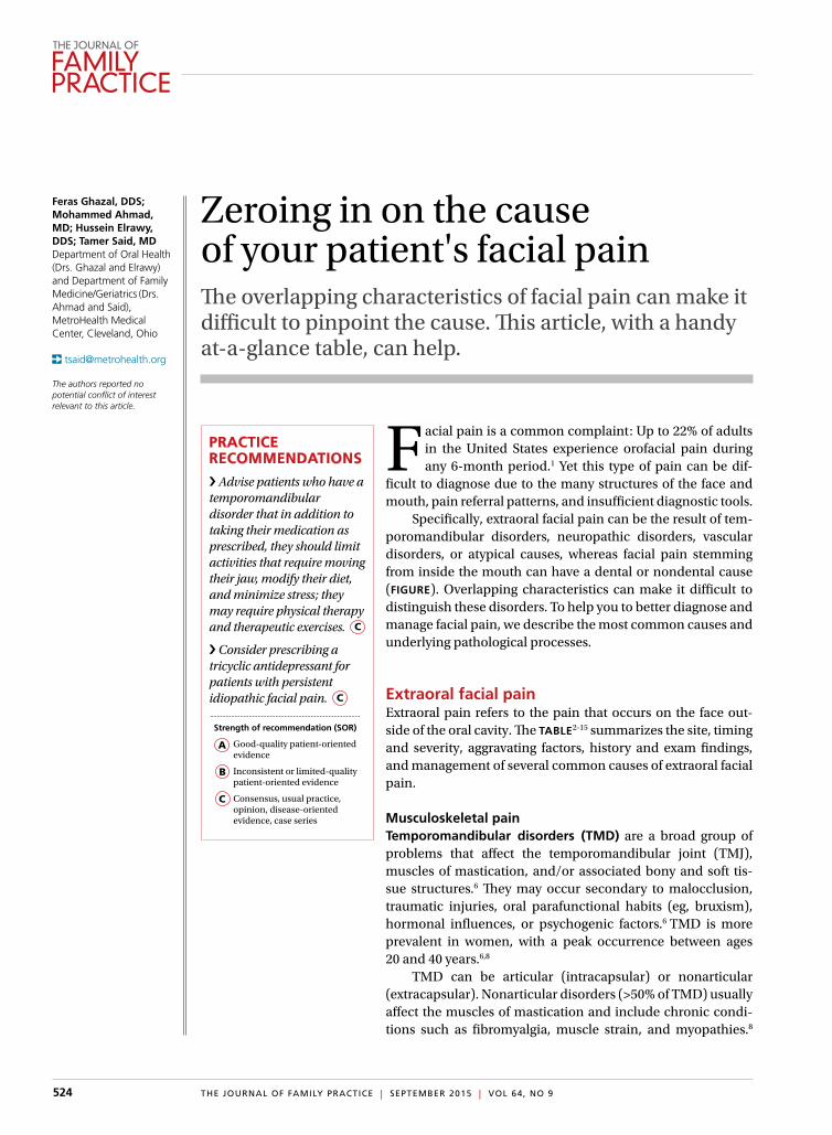
Specifically, extraoral facial pain can be the result of tem-poromandibular disorders, neuropathic disorders, vascular disorders, or atypical causes, whereas facial pain stemming from inside the mouth can have a dental or nondental cause (FIGURE). Overlapping characteristics can make it difficult to distinguish these disorders. To help you to better diagnose and manage facial pain, we describe the most common causes and underlying pathological processes.
Extraoral facial painExtraoral pain refers to the pain that occurs on the face out-side of the oral cavity. The TABLE2-15 summarizes the site, timing and severity, aggravating factors, history and exam findings, and management of several common causes of extraoral facial pain.
Musculoskeletal painTemporomandibular disorders (TMD) are a broad group of problems that affect the temporomandibular joint (TMJ), muscles of mastication, and/or associated bony and soft tis-sue structures.6 They may occur secondary to malocclusion, traumatic injuries, oral parafunctional habits (eg, bruxism), hormonal influences, or psychogenic factors.6 TMD is more prevalent in women, with a peak occurrence between ages 20 and 40 years.6,8
TMD can be articular (intracapsular) or nonarticular (extracapsular). Nonarticular disorders (>50% of TMD) usually affect the muscles of mastication and include chronic condi-tions such as fibromyalgia, muscle strain, and myopathies.8
PRAcTIcE REcoMMEnDATIonS
› Advise patients who have a temporomandibular disorder that in addition to taking their medication as prescribed, they should limit activities that require moving their jaw, modify their diet, and minimize stress; they may require physical therapy and therapeutic exercises. C
› Consider prescribing a tricyclic antidepressant for patients with persistent idiopathic facial pain. C
Strength of recommendation (SoR)
Good-quality patient-oriented evidence
Inconsistent or limited-quality patient-oriented evidence
Consensus, usual practice, opinion, disease-oriented evidence, case series
A
B
C
525JfPonline.com Vol 64, no 9 | SePTemBer 2015 | The Journal of family PracTice
Muscle-related pain and dysfunction are believed to arise from parafunctional hab-its such as bruxism or clenching. Articular disorders include synovitis/capsulitis, joint effusion, trauma/fracture, internal derange-ment (disturbance in the normal anatomic relationship between the disc and condyle), arthritis, and neoplasm.16
z What you’ll see. Orofacial pain (usu-ally dull and located in the preauricular region), joint noise, and restricted jaw func-tion are key signs and symptoms of TMD. Exacerbation of pain with mandibular func-tions (eg, chewing, yawning, or swallowing) is a pathognomonic sign. Joint sounds such as clicking or crepitus are common. In most cases, crepitus correlates with osteoarthritis.6 Nonspecific TMD symptoms include head-ache, earache, insomnia, tinnitus, and neck and shoulder pain.6
The gold standard of diagnosis of TMD consists of taking a detailed history, evaluating the patient’s head and neck, and conducting a general physical examination and behavioral/psychological assessment.17 Imaging of the TMJ and associated structures is essential.17
z Treatment. Nonsteroidal anti-inflam-matory drugs, opioids, muscle relaxants, antidepressants, anticonvulsants, anxiolytics, and corticosteroids are options for treating TMD.6,8 Isometric jaw exercises, maxilloman-dibular appliances, and physical therapy are valuable adjuncts for pain relief. Advise pa-tients to establish a self-care routine to reduce TMJ pain that might include changing their head posture or sleeping position, and limit-ing activities that require using their jaw, such as clenching, bruxism, and excessive gum chewing. Some patients may need to adopt a non-chewing diet that consists of liquid or pu-reed food. Massage and moist heat can help relax muscles of mastication and improve range of motion.
Approximately 5% of patients with TMD undergo surgery, typically simple arthrocen-tesis, arthroscopy, arthrotomy, or modified condylotomy.6 Total joint replacement is indi-cated only for patients with severely damaged joints with end-stage disease when all other conservative treatments have failed. Joint re-placement primarily restores form and func-tion; pain relief is a secondary benefit.8
FIGURE
Causes of facial pain
Musculoskeletal
Temporo- mandibular disorders
neuropathic
Trigeminal neuralgia
Post-traumatic trigeminal pain
Glossopharyngeal neuralgia
Postherpetic neuralgia
numb chin syndrome
Vascular
Giant cell arteritis
malignancy
Atypical
Persistent idiopathic facial pain
Dental
caries
Pulpitis
Periapical disease
cracked tooth
Sensitivity
alveolar osteitis
Periodontal disease
Pericoronitis
nondental
Salivary gland disorders
Sinusitis
cancer
mucosal disorders
Burning mouth syndrome
atypical odontalgia
Extraoral oral
Facial pain
conTinued

526 The Journal of family PracTice | SePTemBer 2015 | Vol 64, no 9
neuropathic painTrigeminal neuralgia (Tn) is sudden, usually unilateral, severe, brief, stabbing, recurrent
episodes of pain in the distribution of one or more branches of the trigeminal nerve.9 It most commonly presents in the lower
TABLE
Extraoral facial pain: Differential diagnosis2-15
Site Timing; severity aggravating factors history and physical exam management
Temporomandibular disorders
TmJ, muscles of mastication, ear. may radiate to the neck
abrupt but often constant; moderate to severe
chewing that is prolonged, opening mouth
clicking or locking of TmJ, headaches, bruxism. attrition of teeth, tenderness along the TmJ, clicking, reduced opening of mouth
nSaids, muscle relaxants, surgery
Trigeminal neuralgia
along the distribution of the second and third division of the trigeminal nerve
Sudden onset, lasts seconds to minutes, up to 30 attacks daily; moderate to severe
eating, light touch, cold, some attacks could be spontaneous
depression, fear of pain returning. attack can be triggered with light touch; sensory changes very rare
anticonvulsants
Post-traumatic trigeminal pain
Trigeminal area 3-6 months after traumatic event; moderate to severe
Touch, cold or heat history of trauma or dental procedure. Sensory changes, including allodynia
Tcas, pregabalin, gabapentin
Glossopharyngeal neuralgia
deep in the ear, throat, and posterior tongue
Sudden attacks, lasts seconds to minutes, often multiple attacks daily; moderate to severe
chewing, talking, drinking, swallowing
Syncope (rare). Provoked by light touch
anticonvulsants, Tcas, neuropath-ic medications, local anesthetics, surgery
Postherpetic neuralgia
Site of zoster rash constant or intermittent; moderate to severe
light touch history of zoster. Skin changes. hyperesthesia, hypoesthesia, allodynia
anticonvulsants, Tcas
numb chin syndrome
over the chin in the region supplied by the mental nerve
abrupt or gradual odontogenic causes (eg, dental abscess, dental trauma, osteomyelitis)
Systemic causes (sarcoidosis, hiV, malignancy)
hypoesthesia, paresthesia, thermalgesic anesthesia or pain over the chin. if related to dental causes: percussion- induced pain, loosening of teeth. if malignancy is present, constitutional symptoms may be seen
Varies based on etiology
Persistent idiopathic facial pain
not well localized constant; moderate to severe
Stress, fatigue other chronic pain, significant life events. exam is usually normal
Tcas, cBT
Giant cell arteritis
Temporal region Sudden onset, continuous; moderate to severe
mastication Vision changes, often associated with polymyalgia rheumatica. Temporal area tenderness
Prednisone
cBT, cognitive behavioral therapy; hiV, human immunodeficiency virus; nSaids, nonsteroidal anti-inflammatory drugs; Tcas, tricyclic antidepressants; TmJ, temporomandibular joint.
528 The Journal of family PracTice | SePTemBer 2015 | Vol 64, no 9
2 branches of the trigeminal nerve and usu-ally is caused by compression of the trigemi-nal nerve root by vascular or nonvascular causes.4 The pain is severe and can profound-ly impact a patient’s quality of life.
TN attacks typically last from a few sec-onds to up to 2 minutes. As many as 30 at-tacks can occur daily, with refractory periods between attacks. After the initial attack, indi-viduals are left with a residual dull or burning pain. TN can be triggered by face washing, teeth brushing, speaking, eating, shaving, or cold wind.4
z Diagnosis can be tricky because more than half of patients with TN experience less severe pain after the main sharp attack; this presentation is called TN type II.7 A detailed patient history and careful evaluation can help identify patients with TN type II. TN can be misdiagnosed as TMD, especially if it pres-ents unilaterally.15
z Treatment. Anticonvulsants are the pri-mary medications used to treat TN.
z Post-traumatic trigeminal pain is usu-ally the result of an injury or dental procedure, such as facial trauma, tooth extraction, root canal, or dental implants.12,18,19 Nerve injury is assumed to be the cause. This type of pain can start within 3 to 6 months of a trauma. It is located in the trigeminal area and patients describe it as burning, tingling and, at times, sharp.15 Patients who have sustained injury to the lingual or inferior alveolar nerves have re-ported feeling “pins and needles.”12
Common triggers include temperature changes or simple touch. Not all injuries re-sult in pain; some patients may have only sensory impairment15 or sensory deficits such as allodynia or hypoesthesia.
z Treatment. The first line of treatment for post-traumatic trigeminal pain is tricyclic antidepressants (TCAs) followed by pregaba-lin or gabapentin.14
z Glossopharyngeal neuralgia (Gn) is similar in presentation to TN but is much rarer.15 GN pain occurs deep in the throat, ear, or posterior tongue.15 When the pain occurs in the inner ear, GN can be misdiagnosed as TMD. In most cases, no cause of GN can be determined.
Patients describe GN pain as shooting, sharp, and electrical shock-like, lasting from
seconds to minutes, with recurrent attacks throughout the day. Like TN, GN can pres-ent as episodes of attacks that last weeks to months. Triggers include chewing, drink-ing, swallowing, and talking, as well as light touch.13,15 Some patients with GN experience syncope due to the anatomical proximity of the vagus nerve.14
z Treatment. Anticonvulsants are the first-line treatment for GN. Local anesthetics or surgery can be considered for patients who don’t improve after medical therapy.15
z Postherpetic neuralgia (PHn) can cause facial pain when the characteristic ve-sicular rash of the varicella zoster virus (shin-gles) occurs on the face. PHN usually affects the first division of trigeminal nerve, but the second and third divisions can be affected as well.13
z What you’ll see. The acute phase of PHN begins a few days before the initial rash has resolved and can last up to a month af-ter. A new pain may begin one to 6 months after the initial rash has healed.20 This pain, which patients often describe as sharp, stab-bing, or burning, can be constant or inter-mittent. Dysesthesia, hypoesthesia, and allodynia may also occur within the affected dermatome.
PHN is usually diagnosed based on the patient’s history and clinical presentation. However, direct fluorescent antibody stain, viral culture, or polymerase chain reaction performed on vesicular fluid from a herpetic lesion during the initial rash are the labora-tory tests of choice if confirmation is needed.
z Treatment. PHN is managed with anti-convulsants and TCAs.
z numb chin syndrome (ncS) is charac-terized by hypoesthesia, paresthesia, therm-algesic anesthesia, or pain over the chin in the region supplied by the mental nerve, a terminal branch of the mandibular division of the trigeminal nerve.5,21,22
NCS can be caused by odontogenic conditions, such as dental abscess, dental anesthesia, dental trauma, or osteomyeli-tis; systemic conditions such as amyloido-sis, sickle cell disease, sarcoidosis, multiple sclerosis, human immunodeficiency virus, or diabetes; or malignancies such as lym-phoma, leukemia, breast cancer, lung can-
Exacerbation of orofacial pain with mandibular functions such as chewing, yawning, or swallowing is a patho g nomonic sign of temporo- mandibular disorder.

diAgnosing fACiAl pAin
529JfPonline.com Vol 64, no 9 | SePTemBer 2015 | The Journal of family PracTice
Diagnosis of trigeminal neuralgia can be tricky; more than half of patients experience less severe pain after the main sharp attack.
cer, prostate cancer, or head and neck cancers.21 In one study of patients with NCS, cancer was the cause of the condition in 89% of patients.22
z What you’ll see. NCS is characterized by numbness of the skin in the lower lip, chin and mucous membrane inside the lip that extends to the midline.5 Depending upon the etiology, patients may present with percus-sion-induced pain, loosening of teeth, se-questra, and mobility of fractured segments. Patients with metastatic malignancy may de-velop constitutional symptoms.
z Making the diagnosis. Panoramic radi-ography is a useful starting point. If possible, a computerized tomography scan of the head and neck should also be done. Nuclear bone scintigraphy (bone scanning) may help iden-tify bone disease such as osteomyelitis. A bi-opsy may be needed if a mass lesion is present.
z Treatment. In NCS that is the result of a dental etiology, the prognosis usually is good. For example, NCS that is the result of an ab-scess usually resolves after the abscess is drained. However, if NCS is caused by metas-tasis, the prognosis is grim; the average length of survival after diagnosis is approximately 5 months if NCS is caused by mandibular metastasis and 12 months if leptomeningeal metastasis is present. Treatment does little to affect the outcome in these cases.21,22
Atypical painPersistent idiopathic facial pain (PIFP), pre-viously known as atypical facial pain, is a persistent facial pain that does not have the classical characteristics of cranial neuralgias and for which there is no obvious cause.2,10,23
PIFP is not triggered by any of the factors that typically precipitate neuralgias.2 The onset may be spontaneous or associated with den-tal intervention or facial injury, but it usually does not have a demonstrable local cause.24,25
Neuropathic mechanisms that might be at work in PIFP include nociceptor sensitiza-tion, phenotypic changes and ectopic activity from the nociceptors, central sensitization possibly maintained by ongoing activity from initially damaged peripheral tissues, sympa-thetic abnormal activity, alteration of seg-mental inhibitory control, or hyperactivity or hypoactivity of descending controls.2
PIFP is most frequently reported in wom-en in their 40s and 50s.25 The history of a pa-tient with PIFP often include mood disorders, chronic pain, or poor coping skills.14 Patients complain of a steady, unilateral, poorly local-ized pain that is deep, constant, aching, pull-ing, or crushing. It is usually present all day, every day. The constancy of the pain is its dis-tinguishing feature. In the beginning, this pain may be in a limited area on one side of the face, usually the nasolabial folds or the angle of the mandible. Later, it may affect both sides of the face and extend to the neck and upper limbs.23,24 Most patients with PIFP report oth-er symptoms, including headache, neck and backache, dermatitis, pruritus, irritable bowel, and dysfunctional uterine bleeding.26
z Making the diagnosis. A targeted his-tory and accurate clinical examination are essential.2,10 Although there are no formal di-agnostic criteria, a patient can be assumed to have PIFP if:2,10
• There is pain in the face for most of the day or all day, every day.
• Initially, the pain may be confined to a portion of the face, but it is poorly localized and deep.
• The pain is not associated with other physical signs or loss of sensation.
• Imaging does not reveal an obvious anatomic or structural cause.
z Treatment. Treatment of PIFP can be difficult and unsatisfactory.23 Counseling to educate patients about the chronic and non-malignant nature of the illness is the mainstay of treatment, followed by pharmacotherapy.23 TCAs have shown a moderate effect in several trials. Gabapentin, topiramate, carbamaze-pine, and pregabalin also have shown limited to modest benefit in some patients. Surgical therapies appear to be of little or no use.23 Ex-perimental treatments such as pulsed radio-frequency, low-energy level diode laser have shown success in small studies.10,23
Vascular painGiant cell arteritis (GcA) is a systemic, chronic vasculitis involving the large and me-dium-sized vessels, mainly the extracranial branches of the carotid artery.6,11 It predomi-nantly affects people older than age 50 and is
530 The Journal of family PracTice | SePTemBer 2015 | Vol 64, no 9
A distinguishing feature of persistent idiopathic facial pain is that the pain is present all day, every day.
more common among women and those of Scandinavian ethnicity.27
The cause of GCA is unclear. Genetic predisposition linked to humoral and cell-mediated immunity is believed to play a role.28
Familial aggregation and predominance of the HLA-DR4 allele has been reported in patients with GCA.6
z What you’ll see. The most common signs and symptoms of GCA are temporal headache (seen in two-thirds of patients), jaw claudication and tenderness, and swell-ing of the temporal artery.6,11 The headache of GCA usually is unilateral, severe, boring or lancinating, and localized to the temporal or occipital regions of the scalp.6 Other oro-facial manifestations include trismus, throat pain that develops while chewing, changes in tongue sensation and tongue claudication, tooth pain, dysphagia, dysarthria, subman-dibular mass, lip and chin numbness, macro-glossia, glossitis, lip and tongue necrosis, and facial swelling.11
Visual symptoms include diplopia, pto-sis, and possibly blindness if treatment is not instituted at first suspicion. Ocular symptoms result from anterior ischemic optic neuropa-thy, posterior ischemic optic neuropathy, or central retinal or cilioretinal artery occlu-sion.6,28 Patients have also reported low-grade fever, asthenia, anorexia, weight loss, and generalized aches.11,28
z Making the diagnosis. Arterial biopsy is the gold standard for diagnosis of GCA. It is usually performed on the temporal artery and is positive in 80% to 95% of people with the condition.28 Other useful lab tests include erythrocyte sedimentation rate (ESR; elevat-ed), white blood cell count (mildly elevated), and C-reactive protein (elevated).
z Treatment. Prednisone is used to treat GCA, in initial doses ranging from 30 to 80 mg. A maintenance dose may be required for up to 2 years, with close follow-up and pe-riodic ESR measurements.28
z Malignancy is a rare cause of facial pain. The pain may be due to metastasis of ex-tracranial bony or soft tissue as it compresses cervical and cranial nerves.3 Lung cancer can cause referred pain in the periauricular region by compressing the vagus nerve, and this pain can be misdiagnosed as dental pain,
atypical facial pain, TMD, or TN.3,29 The facial pain of lung cancer is unilateral and on the same side as the lung neoplasm, and com-monly is referred to the jaw, ear, or temporal region. While many patients have continu-ous pain, some report intermittent pain or pain that lasts for hours.3 Facial pain caused by a malignancy is differentiated from other sources of facial pain by the presence of asso-ciated symptoms such as weight loss, cough, and hemoptysis.
z Treatment. Treatment can include ra-diation or chemotherapy.29
The mouth is often the source of lower facial pain Pain in the oral cavity is the most common cause of pain in the lower face.15 Intraoral pain usually is caused by disease in the fol-lowing structures:
1. Dentition (eg, caries, dentin sensitiv-ity, pulpal disease)
2. Periodontium (eg, gingivitis, acute or chronic periodontal disease, sensitiv-ity related to gum recession, alveolar bone pathology)
3. Other soft and hard tissues, such as the palate, floor of mouth, buccal mu-cosa, non-tooth supporting bone, and tongue (eg, mucosal diseases, neo-plasms, pain related to parafunction or trauma).
Rarely, intraoral pain may be referred. For example, myofascial pain might cause diffuse tooth pain.30
See TABLE W131-35 at the end of this article at jfponline.com for a summary of the etiology, signs/symptoms, diagnosis, and management of these and other dental causes of oral facial pain.
Nondental causes of oral facial pain can be associated with oral mucosal disorders, malignant disease and its therapy, salivary gland disorders, maxillary sinusitis, burning mouth syndrome, or atypical odontalgia. See TABLE W236-41 at jfponline.com for a more de-tailed description of these conditions. JFP
coRRESPonDEncETamer h. Said, md, metrohealth medical center, 2500 metrohealth drive, cleveland, ohio 44109; [email protected]

diAgnosing fACiAl pAin
531JfPonline.com Vol 64, no 9 | SePTemBer 2015 | The Journal of family PracTice
1. Lipton JA, Ship JA, Larach-Robinson D. Estimated prevalence and distribution of reported orofacial pain in the United States. J Am Dent Assoc. 1993;124:115-1121.
2. Agostoni E, Frigerio R, Santoro P. Atypical facial pain: clinical con-siderations and differential diagnosis. Neurol Sci. 2005;26:S71-S74.
3. Bajwa Z, Ho C, Khan S, et al. Overview of craniofacial pain. UpTo-Date Web site. Available at: http://www.uptodate.com/contents/overview-of-craniofacial-pain. Accessed January 28, 2015.
4. Bendtsen L, Birk S, Kasch H, et al. Reference programme: Di-agnosis and treatment of headache disorders and facial pain. Danish Headache Society, 2nd Edition, 2012. J Headache Pain. 2012;13:S1-S29.
5. Divya KS, Moran NA, Atkin PA. Numb chin syndrome: a case se-ries and discussion. Br Dent J. 2010;208:157-160.
6. Kapur N, Kamel IR, Herlich A. Oral and craniofacial pain: di-agnosis, pathophysiology, and treatment. Int Anesthesiol Clin. 2003;41:115-150.
7. Limonadi FM, McCartney S, Burchiel KJ. Design of an artificial neural network for diagnosis of facial pain syndromes. Stereotact Funct Neurosurg. 2006;84:212-220.
8. Liu F, Steinkeler A. Epidemiology, diagnosis, and treatment of temporomandibular disorders. Dent Clin North Am. 2013;57:465-479.
9. Merskey H, Bogduk N (eds). Classification of Chronic Pain. De-scriptors of Chronic Pain Syndromes and Definition of Pain Terms, 2nd ed. Seattle, WA: International Association for the Study of Pain Press; 1994.
10. Nguyen CT, Wang MB. Complementary and integrative treat-ments: atypical facial pain. Otolaryngol Clin North Am. 2013;46:367-382.
11. Reiter S, Winocur E, Goldsmith C, et al. Giant cell arteritis mis-diagnosed as temporomandibular disorder: a case report and review of the literature. J Orofac Pain. 2009;23:360-365.
12. Renton T, Adey-Viscuso D, Meechan JG, et al. Trigeminal nerve injuries in relation to local anaesthesia in mandibular injections. Br Dent J. 2010;209:E15.
13. Shephard MK, Macgregor EA, Zakrzewska JM. Orofacial pain: a guide for the headache physician. Headache. 2014;54:22-39.
14. Zakrzewska JM. Differential diagnosis of facial pain and guide-lines for management. Br J Anaesth. 2013;111:95-104.
15. Zakrzewska JM. Multi-dimensionality of chronic pain of the oral cavity and face. J Headache Pain. 2013;14:37.
16. Herb K, Cho S, Stiles MA. Temporomandibular joint pain and dysfunction. Curr Pain Headache Rep. 2006;10:408-414.
17. American Society of Temporomandibular Joint Surgeons. Guide-lines for diagnosis and management of disorders involving the temporomandibular joint and related musculoskeletal struc-tures. Cranio. 2003;21:68-76.
18. Benoliel R, Zadik Y, Eliav E, et al. Peripheral painful traumatic tri-geminal neuropathy: clinical features in 91 cases and proposal of novel diagnostic criteria. J Orofac Pain. 2012;26:49-58.
19. Brooke RI. Atypical odontalgia. A report of twenty-two cases. Oral Surg Oral Med Oral Pathol. 1980;49:196-199.
20. Bouhassira D, Chassany O, Gaillat J, et al. Patient perspective on herpes zoster and its complications: an observational prospec-tive study in patients aged over 50 years in general practice. Pain. 2012;153:342-349.
21. Baskaran RK, Krishnamoorthy, Smith M. Numb chin syn-
drome—a reflection of systemic malignancy. World J Surg Oncol. 2006;4:52.
22. Lata J, Kumar P. Numb chin syndrome: a case report and review of the literature. Indian J Dent Res. 2010;21:135-137.
23. Cornelissen P, van Kleef M, Mekhail N, et al. Evidence-based interventional pain medicine according to clinical diagnoses. 3. Persistent idiopathic facial pain. Pain Pract. 2009;9:443-448.
24. Didier H, Marchetti C, Borromeo G, et al. Persistent idiopathic fa-cial pain: multidisciplinary approach and assumption of comor-bidity. Neurol Sci. 2010;31:S189-S195.
25. Klasser G. Management of persistent idiopathic facial pain. J Can Dent Assoc. 2013;79:d71.
26. Abiko Y, Matsuoka H, Chiba I, et al. Current evidence on atypi-cal odontalgia: diagnosis and clinical management. Int J Dent. 2012;2012:518548.
27. Sheldon CA, White VA, Holland SP. Giant cell arteritis presenting with bilateral loss of vision and jaw pain: reminder of a potentially devastating condition. J Can Dent Assoc. 2011;77:b55.
28. Rockey JG, Anand R. Tongue necrosis secondary to temporal arte-ritis: a case report and literature review. Oral Surg Oral Med Oral Pathol Oral Radiol Endod. 2002;94:471-473.
29. Sarlani E, Schwartz AH, Greenspan JD, et al. Facial pain as first manifestation of lung cancer: a case of lung cancer-related cluster headache and a review of the literature. J Orofac Pain. 2003;17:262-267.
30. Kumar A, Brennan MT. Differential diagnosis of orofacial pain and temporomandibular disorder. Dent Clin North Am. 2013;57:419-428.
31. Laudenbach JM, Simon Z. Common dental and periodontal diseases: evaluation and management. Med Clin North Am. 2014;98:1239-1260.
32. Napeñas JJ. Intraoral pain disorders. Dent Clin North Am. 2013;57:429-447.
33. Vickers ER, Zakrzewska JM. Dental causes of orofacial pain. In: Orofacial Pain. Zakrzewska JM, ed. Oxford, UK: Oxford University Press; 2009:69-81.
34. Pierse JE, Dym H, Clarkson E. Diagnosis and management of common postextraction complications. Dent Clin North Am. 2012;56:75-93.
35. Renton T. Dental (odontogenic) pain. Br J Pain. 2011;5:2-7.
36. Yatani H, Komiyama O, Matsuka Y, et al. Systematic review and recommendations for nonodontogenic toothache. J Oral Rehabil. 2014;41:843-852.
37. Klasser GD, Fischer DJ, Epstein JB. Burning mouth syndrome: recognition, understanding, and management. Oral Maxillofac Surg Clin North Am. 2008;20:255-271.
38. Balasubramaniam R, Turner LN, Fischer D, et al. Non-odon-togenic toothache revisited. Open Journal of Stomatology. 2011;1:92-102.
39. Patton LL, Siegel MA, Benoliel R, et al. Management of burning mouth syndrome: systematic review and management recom-mendations. Oral Surg Oral Med Oral Pathol Oral Radiol Endod. 2007;103:S39.e1-e13.
40. Cascarini L, McGurk M. Epidemiology of salivary gland in-fections. Oral Maxillofac Surg Clin North Am. 2009;21:353-357.
41. Hegarty AM, Zakrzewska JM. Differential diagnosis for orofacial pain, including sinusitis, TMD, trigeminal neuralgia. Dent Up-date. 2011;38:396-400,402-403,405-406.
The most common signs and symptoms of giant cell arteritis are temporal headache, jaw claudication and tenderness, and swelling of the temporal artery.
references
from the authors who bring you clinical inquiries and Purls comes a relevant, concise, and clinically useful journal to assist you in delivering the best care to your patients – all without industry support.
Evidence-Based Practice is published monthly by the family Physicians inquiries network.
JfP readers pay our new discount price of $119 for 12 issues and 48 AMA PRA Category 1 Credits™.
To subscribe or request a sample issue, call (573) 256-2066 or e-mail [email protected]
ADVERTISEMENT

The Journal of family PracTice | SePTemBer 2015 | Vol 64, no 9531A
TABLE W1
Oral pain of dental origin31-35
disorder Site etiology Signs/symptoms making the diagnosis management
caries enamel, dentin, and cementum of the teeth
Bacterial invasion of tooth structure
Thermal sensitivity and/or pain when exposed to sweet or acidic foods
risk assessment, clinical examination and interpretation of radiographs
incipient caries: monitoring, remineralization, and control of risk factors. deep caries: excavation and restoration
Pulpitis Pulp of the teeth
caries extend into pulp. Trauma, exposed dentin or cementum, and iatrogenic insults
reversible pulpitis: discomfort that goes away within seconds of removal of stimulus
irreversible: sharp, spon-taneous, lingering pain
Thorough history. clinical exam and pulp test
interpretation of radiographs
reversible: excava-tion and restoration
irreversible: root canal treatment
Periapical diseases
apical periodontal tissues of the teeth
infection from diseased pulp spreads into apical tissues
acute periapical disease: sharp, throbbing, spon-taneous pain, pain on biting, and/or swelling
chronic periapical disease: mild discomfort, sinus tract
clinical exam and tests such as percussion, palpation
interpretation of radiographs
root canal treat-ment or extraction with or without antibiotics. abscess with fluctuant swelling: incision and drainage
cracked tooth
dentin and/or pulp of the teeth
Trauma, extensive restorations
incomplete fracture of dentin may or may not extend into the pulp
Sharp momentary pain on exposure to cold. Pain associated with release of biting pressure
clinical history/exam. Periodontal probing, bite test. Trans-illumination with magnification
cone beam cT
Stabilization with a band, overlay, or crown
root canal treat-ment or extraction
Generalized sensitivity
exposed dentin and/or cementum of the teeth
Gingival recession and/or abrasion of enamel. most commonly due to incorrect brushing of teeth. also due to recent scaling or tooth wear
extreme sensitivity to cold fluids and air. Sharp localized pain that disappears immediately after removal of stimulus
Thorough history of symptoms and clinical examination shows exposed cementum and/or dentin
improved oral hygiene practices, reduction of dietary acid, desensitizing agents and restorations
alveolar osteitis
extraction socket, mainly after mandibu-lar tooth extraction
lysis of a fully formed clot before it is replaced with granulation tissue
moderate to severe deep, continuous, aching, radiating pain
Thorough patient history. clinical exam shows the tissue around the socket is tender and white necrotic bone is exposed
irrigation using saline or chlorhexi-dine. Placement of an obtundent dressing. analgesics
Periodontal disease
Supporting tissues of the teeth: gingiva, periodontal ligament, and alveolar bone
Bacteria-induced inflammation of soft tissues and alveolar bone surrounding the teeth. Traumatic injury to the soft tissue
may be mild, persistent, or episodic dull pain
clinical history of pain and associated symptoms. clinical exam reveals gingival/periodontal pockets and/or abscess associated with vital teeth. interpre-tation of radiographs
Scaling, root planing, and curettage. Periodontal abscess: drainage by incision or through pocket orifice
Pericoronitis Gingival and mucosal tissues surrounding the crown of an erupting tooth
inflammation and infection of the tissues around an erupting tooth, especially molars
Pain may be continuous or intermittent, rang-ing from mild to severe. Pain may radiate to ear, throat, and floor of mouth. difficulty in opening/closing the jaw
history of trismus and discomfort during mouth opening/closing. clinical exam shows erythema-tous and edematous tissue along with indentation of the opposing tooth
removal of overly-ing operculum, irrigation with 2% chlorhexidine, debridement.
analgesics
cT, computed tomography.

TK
diAgnosing fACiAl pAin
JfPonline.com Vol 64, no 9 | SePTemBer 2015 | The Journal of family PracTice 531B
TABLE W2
Oral pain of nondental origin36-41
condition Site etiology Signs/symptoms making the diagnosis
management
oral mucosal disorders (oral candidiasis, herpes virus, recurrent aphthous stomati-tis, lichen planus, pemphigus)
oral mucosal epithelium
infection, reactive process, systemic disorders or dysplasia
Vesicles, erosions, ulcer-ations, erythema, pseudo- membrane formation, hyperkeratosis and hyper-algesia. herpes zoster of the face may be associated with toothache
risk assessment, clinical exam, and testing
Treatment of underlying mucosal condition
Pain associated with malignant disease and its therapy
multiple sites oral mucositis second-ary to chemotherapy and radiation therapy
longstanding ulcerations, secondary infection, infiltration into adjacent peripheral nerves
injuries to peripheral nerves: pulpitis-like pain
oral mucositis: erythema, ulceration, pseudomem-brane formation and shedding
angioleiomyoma and methemoglobinemia cause toothache
Thorough clinical exam of hard and soft oral tissues and appropriate judgment
early diagnosis, referral, and management of underlying malignant disease
Salivary gland disorders
Typically localized to the gland itself
Bacteria, localized and systemic viruses, autoimmune diseases, secondary to sialoliths and strictures and congenital disorders
Swollen painful salivary glands, induration and erythema of overlying skin. increase in pain with chewing and dry mouth
Pain can be local-ized by palpa-tion. radiograph of the gland and ducts
Screening tests for dry mouth
acute phase: supportive treatment with analge-sics, hydration, antibiot-ics, and parasympatho-mimetics
Sialoliths: surgical re-moval of salivary stones
maxillary sinusitis Posterior maxilla and maxillary teeth
intranasal and sinus abnormalities, inflammation or anatomic variations
infection of maxillary sinus is perceived as dental pain in maxillary posterior teeth and vice versa
facial pain and fullness in maxillary posterior region. continuous dull or diffuse lingering pain. Teeth are sensitive to percus-sion, mastication, and/or temperature. cough, headache, ear pain, nasal congestion, and discharge
intraoral exam of the teeth and extraoral exam of the sinus. fiber optic illumination of the sinus may reveal changes in the affected region
Symptomatic: deconges-tants, antihistamines, mucolytic agents, alpha-adrenergic agents, corti-costeroids and analge-sics. if sinusitis persists >7 days and features purulent discharge: antibiotics
Burning mouth syndrome
multiple sites; mostly affects anterior two-thirds of the tongue, followed by dorsum and lateral borders of the tongue, lips, palate, and cheeks
Primary/essential BmS: no local or systemic causes, involves periph-eral or central neuro-pathological pathways
Secondary BmS: local, systemic, and psycho-logical factors
Burning pain, dysgeusia, dysesthesia, loss of taste, and paresthesia
Thorough, comprehensive history of pre-senting symp-toms. identify un-derlying factors, such as mucosal disease, stress, nutritional defi-ciencies, diabetes, and medication adverse effects
management of under-lying condition
Systemic medications (eg, benzodiazepines, anticonvulsants, Tcas, SSris), topical medica-tions (eg, capsaicin, clonazepam), cBT
atypical odontalgia Teeth or in a tooth socket after extraction
not clearly defined. Possibly neuropathic, vascular, or psychogenic mechanisms. Pain usually, but not necessarily, follows a dental procedure or trauma
Pain can be aching, burn-ing, throbbing, or shoot-ing. moderate to severe in intensity
diagnosis of exclusion
Pain management
Topical analgesics: lidocaine/prilocaine or capsaicin cream
Tcas, SSris, anticon-vulsants, tramadol, oxycodone. adjunct therapies: acupuncture, self-hypnosis, cBT
BmS, burning mouth syndrome; cBT, cognitive behavioral therapy; SSris, selective serotonin reuptake inhibitors; Tcas, tricyclic antidepressants.
Recommended