THE ULTRASTUCTURE OF THE ARTICULAR TISSUE OF THE MANDIBULAR CONDYLE IN THE RAT
J. APPLETON
Electron Microscope Unit. Department of Dental Surgery. University of Liverpool. England
Summary-~-The ultrastructure of the fibrous articular tissue of the rat mandibular condyle at 20 and 80 days of age was investigated. The articular tissue was composed of collagen fihres. immature elastic fibres and fibroblasts. At 20 days of age the articular surface was formed by a smooth electron-dense layer about 0.15 pm thick. Some fibroblast cell pro- cesses extended through the surface layer into the lower joint space. At 80 days of age the articular surface was formed by a distinct, even layer of fibrillar material up to 1.3 pm thick. There were no fibroblast cells close to the articular surface. Numerous membrane- bound vesicles of varying electron density were present in the fibrous tissue. The collagen fibres. elastic fibres and articular surface layers probably fulfil functional requirements which are specifically related to their physical properties.
INTRODLCTION
In the mandibular .joint (Collins (2~ t/l.. 1946). unlike most other synovial joints except the Sterno-clavicular and acromio-clavicular. the articular tissues are not formed of hyaline cartilage but of fibrous tissue. This fibrous tissue covers the membrane bone of the cranial component of the mandibular joint and the remains of the secondary cartilage of the condylar process of the mandible. Furthermore, the joint space is divided by a fibrous intra-articular disc of similar composition to the articular tissues (Barnett, Davies and MacConaill, 1961).
There have been studies on the anatomy and his- tology of the mandibular joint in many species and in particular in the rat (Cabrini and Erausquin. 1941: Collinsrtul., 1946;CunaL Bhaskar and Weinman, 1956; Furstman, 1966; Greenspan and Blackwood, 1966; Hiiemae. 1966; Appleton, 1969) but in these studies little attention has been given to the articular tissues. Some investigations have been made, however, on the distribution of elastic fibres in the articular tissue of several species (Miles and Dawson, 1962; Frommer and Monroe, 1966). To date, there have only been two published accounts on the ultrastructure of the articular tissues in the mandibular joint, both on the guinea pig (Silva and Hart, 1967; Silva, 1969) but numerous ultrastructural investigations have been made of typical hyaline cartilage from synovial joints in many species (Wright, Dowson and Kerr, 1973).
It is evident that the fibrous articular tissues in the mandibular joint fulfil some specific functional require- ments related to their structure and composition. Therefore this study examined the ultrastructure of the articular tissues of the mandibular condylar com- ponent of the mandibular joint in the 20 and 80 day old rat. The intention was to investigate the ultrastuc- tural changes that occur in the articular tissues from just before weaning to the establishment of proper masticatory function in the mature animal.
MATERIALS AND METHODS
Black and white rats of both sexes at 20 and 80 days of age were used. Under Nembutal anaesthesia
an incision was made between the car and the eye and the overlying muscles reflected to expose the mandibular joint. In the rat, this joint is formed between the inferior surface of the zygomatic process of the squamosal and the condylar process of the mandible. The condylar process was divided beneath the condylar head and the condyle was removed with the articular disc intact in order to avoid damage to the articular surface of the mandibular condyle. The articular disc was carefully removed and the ar- titular surface was then washed vigorously in a stream of normal saline for several minutes before fixation for 2 hr in 6.25 per cent cacodylate-buffered gluteraldehyde pH 7.4 (Sabatini, Bensch and Barnett, 1963) followed by washing for several hours in caco- dylate buffer and post-fixation in Verona1 acetate-buf- fered 1 per cent osmium tetroxide pH 7.2. The tissues were then washed in Verona1 acetate buffer, dehyd- rated, routinely embedded in Araldite and sectioned on a Reichert 0MU2 ultramicrotome using a dia- mond knife. Sections were mounted on copper grids, stained with a 25 per cent solution of uranyl acetate in methanol and examined in a Philips E.M. 300 elec- tron microscope.
RESL LTS
The articular tissue was composed of collagen and elastic fibres and numerous cells resembling hbro- blasts (Fig. 1). The articular surface. however. was formed by a smooth electron-dense amorphous layer up to about 0.15 pm thick continuous with the interfibrillar matrix of the articular tissue. Immcdi- ately beneath the surface there were small bundles of collagen fibres orientated parallel and at right angles to the plane of section and some individual fibres separated by matrix ground substance (Fig. I and Fig. 2a). Deep to the articular surface the col- lagen fibres were arranged in larger bundles orientated parallel and at right angles to the plane of section. For a depth of 25 pm from the articular surface the bundles contained fewer fibres and were separated by
824 J. Appleton
more matrix ground substance (Fig. 1 and Fig. 2a). Individual elastic fibres of indeterminate length
were present in large numbers throughout the articu- lar tissue. The fibres were randomly orientated and branching was not evident (Fig. I). Some fibres were close to the articular surface but were never seen forming part of the articular surface (Fig. 3a). Each elastic fibre was about 0.35 Ltrn in thickness and con- sisted of a parallel arrangement of fibrils about 8 nm in diameter (Fig. 3a.b). The fibres were associated with an electron-dense amorphous material which occupied the central region within each elastic fibre (Fig. 3c.d). Occasionally this electron-dense amor- phous material was discontinuous and electron-trans- lucent areas were also evident (Fig. 3a.c).
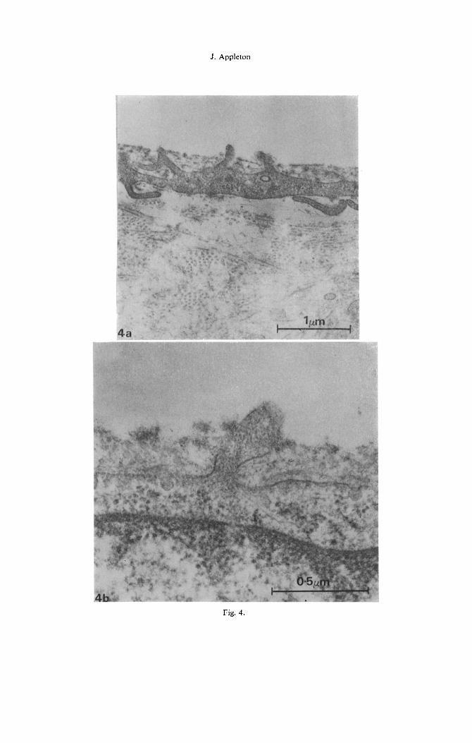
The fibroblast-like cells were numerous and dis- persed throughout the articular tissue (Fig. 1). Many of these cells were close to the articular surface and frequently several cell processes from each ccl] extended through the electron-dense amorphous layer at the articular surface (Fig. 4a.b).
The articular tissue was composed of collagen and elastic fibres, and cells resembling fibroblasts which were more widely separated than at 20 days of age (Fig. 5). The articular surface was formed by a distinct even layer of fibrillar material up to I.3 pm in depth (Fig. 5 and Fig. 2a,b). This layer was discontinuous over small areas of the surface. The distribution, organisation and dimensions of collagen and elastic fibres were similar to that at 20 days of age except that the collagen fibre bundles were larger deep within the articular tissue (Fig. 5).
There were no cells in close proximity to the articu- lar surface and no cells processes extended through the surface. Throughout the articular tissues. however, there were electron-translucent areas which contained cell debris and membrane-bound vesicles of varying electron density up to about 250 nm in diameter. These vesicles were also scattered throughout the fibrous tissues between and within the individual col- lagen fibre bundles (Fig. 5).
DISCUSSION
The large bundles of collagen fibrcs. elastic fibres and fibroblasts forming the articular tissue of the mandibular condyle of the rat present a different his- tological and ultrastructural picture from that seen in typical hyaline articular cartilage. For a depth of 3 pm-5 pm the mandibular condylar articular tissue is very similar to hyaline articular cartilage in con- sisting of small bundles of collagen fibres 2@25 nm in diameter lying parallel to the articular surface (Gardener, 1972). One important difference is the pre- sence of large numbers of elastic fibres (Miles and Dawson, 1962; Appleton. 1969) which arc invariably closely associated with the collagen fibres and in the immature animal may approach the articular surface. These fibres differ from mature elastic fibres and closely resemble the immature elastic fibres described in the articular tissue of the guinea-pig mandibular joint (Silva and Hart, 1967; Silva, 1969) in calf liga- mentum nuchae (Fahrenbach. Sandberg and Cleary.
1966) and foetal rat tendon (Greenlee, Ross and Hart- man, 1966). The collagen and elastic fibres probably fulfil some functional requirements specifically related to their physical properties. The elastic fibres prob- ably provide the necessary resilience to the periarticu- lar tissues and articular tissues in the absence of ground substance containing chondroitin sulphate (Griffen and Sharpe, 1962: Miles and Dawson, 1962).
The origin and prccisc nature of the fine but dis- tinct layer seen coating the collagen fibrcs of the ar- titular surface of the condylar cartilage. in thin sec- tions in unknown (Appleton. 1969). It is not analo- gous in any way to the thicker membrane observed coating hyaline articular cartilage surfaces when viewed under polarized light and termed the “lamina splendens” by McConaill (1951). The layer seen in the electron microscope may result from precipitation of whole or part of the synovial fluid following fixa- tion and dehydration or it may be a me-existing sur- face layer structurally distinct from the articular carti- lage. The fact that it is not removed by vigorous washing in 0.1 N saline prior to fixation would sug- gest that it is an integral part of the surface cartilage (Balazs, Bloom and Swann, 1966). Material aspirated from the surface of articular cartilage in the carpome- tacarpal and tarsometatarsal joints of cattle contained chiefly a glycosaminoglycan which had equimolar amounts of hexuronic acid and glucosamine (Balazs et cd., 1966)Whether this material represents the thin layer seen at the surface in the electron microscope is uncertain. It has been reported, however, that this surface layer is more readily visualized by incorporat- ing cetyl pyridinium chloride or Ruthenium red into the fixation process (Gardener, 1972).
Cell proccsscs extending close to and through the articular surface arc a noticeable and constant feature in the young animal. It may be speculated that either the cells are producing material which is secreted into the synovial cavity or acquiring nutrients via the synovial fluid. In the adult. cells are not seen close to the surface but accumulations of cell debris and vesicles are common. It is evident from this study that the surface layer appears thinner and structurally different prior to weaning at 20 days than in the adult at 80 days whet-c the joint is fully developed and func- tional. Its ultrastructural appcarancc. however. is un- doubtably &ected by fixation and dehydration. Nevertheless, this marked change in appearance may suggest that this layer plays an important role in the mechanism or mechanisms of joint lubrication.
~~ckrlol~lc~d~lc~l?lclir The technical assistance of Mrs. V. R. Bradley is gratefully acknowledged.
Appleton .I. 1969. The fine structure of the condylar carti- lage of the rat mandible. PhD. Thesis, London.
Bala% E. A.. Bloom G. D. and Swarm D. A. 1966. Fine structure and glycosaminoglycan content of the surface layer of articular cartilage. F&I PKK. 2.5. I8 IS IX 16.
Barnett C. H.. Davies D. V. and MacConaill M. A. 1961. Stw0t+1/ .Ioinl.s; T/wir Sffxc.l~rrc twd ,2/I~w/xntics Long- mans. Green. London.
Cabrini R. and Erausquin J. 1941. La articulation tcmpro- maxilat- de la rata. Rcrto. otlmt. B. rl~rcs 29. 2X5 470.
Articular tissue of the mandibular condyle 825
Collins D. A.. Becks H.. Simpson M. E. and Eavans H. Griffin C. J. and Sharpe C. J. 1962. Distribution of elastic M. 1946. Growth and transformation of the mandibular tissues in the human temporomandibular meniscus espe- joint in the rat. I Normal female rats. Am. J. Orthod. cially in respect to compression areas. hst. /hut. J. 7. Oral Sttrg. 32, 431 ~~442. 72278.
Cunat J. J.. Bhaskar S. N. and Weinman J. P. 1956. Devel- opment of the squamosal mandibular articulation in the rat. J. dent. Res. 35, 533-546.
Fahrenbach W. H., Sandberg L. B. and Cleary E. G. 1966. Ultrastructural studies on early elastogenesis. Anat. Rec. 155. 563~ 516.
Hiimae K. M. 1966. The development. structure and func- tion of the mandibular joint in the rat. PhD. Thesis, London.
Frommer J. and Monroe C. W. 1966. Development and distribution of elastic fibres in the mandibular joint of the mouse. A comparison of foetal suckling. juvenile and adult stages. Anat. Rec. 156, 333-345.
Furstman L. L. 1966. Normal age changes in the rat man- dibular joint. J. drnt. Rex 45. 291-296.
Gardner D. L. 1972. The influence of microscopic tech- nology on knowledge of cartilage surface structure. Ann. rheunr. Dis. 31. 235-258.
MacConaill M. A. 1951. The movement of bones and joints, IV. The mechanical structure of articular carti- lage. J. Bone Jt. Surg. 33B. 251.-259.
Miles A. E. W. and Dawson J. A. 1962. Elastic tibres in the elastic fibrous tissue of some joints. Archs oral Biol. 7. 249m 252.
Greenlee T. K., Ross R. and Hartman J. L. 1966. The
Sabatini D. D.. Bensch K. and Barnett R. J. 1963. Cytoche- mistry and electron microscopy. The preservation of cellular ultrastructure and enzymatic activity by alde- hyde fixation. J. Cell Biol. 17. 19-58.
Silva D. G. and Hart J. A. L. 1967. Ultrastructural obscr- vations on the mandibular condyle of the guinea-pig. J. Ultrastruct. Res. 20, 227-243.
fine structure of elastic fibres. J. Cell Biol. 30. 59-71. Silva D. G. 1969. Further ultrastructural studies on the Grcenspan J. S., Blackwood H. J. J. 1966. Histochemical temporo-mandibular joint of the guinea-pig. J. Clrru-
studies of chondrocyte function in the cartilage of the struct. Res. 26, 14X-162. mandibular condyle of the rat. J. Anut. (Land) 100. 615.- Wright W., Dowson D. and Kerr J. 1973. The structure 626. of joints. Int. Rec. Connect. Tissw Rcs. 6. It& 125.
Figs. l-5 overleaf.
826 J. Appleton
Fig. 1. The articular tissue of the rat mandibular condyle at 20 days of age. There is a fine distinct electron-dense layer forming the surface. Randomly orientated bundles of collagen and individual elastic fibres (arrowed) are present. Some of the cells resembling fibroblasts are close to the articular surface.
X 8000
Fig. 2. (a) Articular surface of 20-day rat mandibular condyle showing the electron-dense surface layer about 0,15/(m in thickness. x 30.000
(b) Articular surface of X0-day rat mandibular condyle shohing the distmct tilamentous surface layer up to 1.3 jtm in thickness. x 30.000
Fig. 3. Elastic tibres are seen in longitudinal (a.b). oblique (c) and transverse section (d). Each fibre is composed of hbrils which enclose an electron-opaque amorphous material (cd). Occasionally elec- tron-translucent areas are present (arrowed) (a.d). (a) x 20.000, (b) x 62,000. (c) x 62.000, (d) x 62,000
Fig. 4. (a) A cell close to the articular surface uith several cell processes extending through the surface. x 30.000. (b) Cell process extending through the articular surface. The process does not have a distinct
electron dense coat. x 80.000
Fig. 5. The articular tissue of the rat mandibular condyle at 80 days of age. There is a distinct filamen- tous layer forming the surface. Large bundles of randomly orientated collagen fibres and individual elastic fibres (arrowed) are present. There are few cells with none close to the surface but numerous
accumulations of vesicles (\) of Larying electron density are evident. x 8000

Articular tissue of the mandibular condyle
Fig. 1.
A.O.B. f.p. 826

J. Appleton
Fig. 2.

Articular tissue of the mandibular condyle
Fig. 3.
J. Appleton
Fig. 4.

Articular tissue of the mandibular condyle
Fig. 5.
Recommended


![Current Advances in Mandibular Condyle Reconstruction · The LIPUS is considered the preferred method of mechanical stimulation, also known as “preferred bioreactor” [25]. 5](https://img.dokumen.tips/doc/110x75/5e96a9d67ba2de640562addd/current-advances-in-mandibular-condyle-reconstruction-the-lipus-is-considered-the.jpg)