THE ROLE OF ADIPOSE TRIGLYCERIDE LIPASE IN HEPATIC LIPID
METABOLISM, NON-ALCOHOLIC FATTY LIVER DISEASE AND INSULIN
RESISTANCE
A DISSERTATION SUBMITTED TO THE FACULTY OF THE GRADUATE SCHOOL
OF THE UNIVERSITY OF MINNESOTA BY
KUOK TEONG ONG
IN PARTIAL FULFILLMENT OF THE REQUIREMENTS FOR DEGREE OF
DOCTOR OF PHILOSOPHY
DR. DOUGLAS G MASHEK, ADVISOR
JUNE 2013

© KUOK TEONG ONG 2013
i
Acknowledgements
First, I would like to thank my graduate advisor Dr. Mashek for his guidance and
support. His jovial and positive attitude always helps me in times of difficulties
with lab work. Without him, I would not have been able to learn so much about
the numerous aspects of being a successful researcher.
I, also, would like to thank the members of my committee, Drs. Xiaoli Chen,
Howard Towle, David Brown, and Chi Chen for their valuable inputs throughout
these 5 years. Seeking advice from experts of various fields has provided me
with a more interdisciplinary education.
Not to forget the lab members of Mashek lab including Mara Mashek who has
incessantly provided me with technical guidance and friendship in the lab. I would
also like to thank other current graduate students of the lab including Aishwarya
Sathyanarayan and Mallory Franklin who have always been very helpful and
reliable peers. Both postdoctoral fellows in the lab, Drs. Salmaan Khan and Edith
Hayden have also been instrumental in providing technical and scientific
assistance. Of course, the undergraduates including Katie Ress, Ellen Fisher and
Joe O’Brien are such a big helping hand especially with the preparation of lab
equipment and animal experiments. Last but not least, I would like to
acknowledge past lab members: Dr. So Young Bu, Teng Zhao and Jillian Tholen.

ii
Last but not least, I would like to thank the funding sources including the National
Institutes of Health (RO1-DK0822574 and 1RC2ES018781 to Andrew S.
Greenberg; and DK090364 and R56-DK085008 to Douglas G. Mashek), the
American Diabetes Association (7-08-RA-57 to Andrew G. Greenberg and 7-07-
JF-43 to Douglas G. Mashek) the USDA, Agricultural Research Service (under
Contract no. 58-1950-7-707 to Andrew S. Greenberg) and the Minnesota Obesity
Center (NIH DK050456).

iii
Dedication
This thesis will not have materialized without the love and support I have
received throughout my entire life from my family and great friends back in
Malaysia.
A big token of appreciation to my dad and mom who are always there for me no
matter what happens to guide and provide me with their utmost love to keep me
going despite our distance. Their cheerful and positive outlook at life never stops
to impress and pick me up when I need them the most.
To my younger brother who is in medical school and never cease to be the
reason why I always persevere to be a better person so that I can set myself as a
great role model.
Also to my closest friends in Minnesota and throughout the country, especially
my college friends from Madison who have provided me a good reason to stay in
the US and undergo a great American experience, both educationally and
personally.

iv
Abstract
Hepatic triglyceride (TAG) accumulation leads to the development of non-
alcoholic fatty liver disease (NAFLD), which is strongly correlated with other
metabolic diseases including obesity, insulin resistance and type II diabetes.
While the TAG synthetic pathway has been well-researched, our knowledge of
the TAG hydrolysis pathway, especially in the liver, is scant. The research
project is aimed at understanding the role and mechanisms of hepatic adipose
triglyceride lipase (ATGL) and its downstream lipid metabolites in mediating the
development of NAFLD and insulin resistance. To elucidate the metabolic
functions of hepatic ATGL, we employed adenovirus-mediated knockdown and
overexpression in primary hepatocyte cultures and mouse models. We have
shown that ATGL is a key TAG hydrolase in the liver that preferentially channels
fatty acids (FAs) to mitochondrial β-oxidation, but does not affect VLDL synthesis
and secretion. Additionally, ATGL positively regulates PPAR-α and its target
gene expression to influence β-oxidation transcriptionally. Liver FA binding
protein (LFABP), a major intracellular FA carrier, is not necessary for ATGL-
regulated changes in the expression of PPAR-α and its target genes or for the
shuttling of hydrolyzed FA to the mitochondria. Moreover, the PPAR-α agonist
fenofibrate is unable to normalize the expression of PPAR-α target genes in
ATGL knockdown mice, suggesting that ATGL regulates PPAR-α target gene
expression in a LFABP- and ligand-independent mechanism. Interestingly,
despite enhanced TAG content, mice lacking hepatic ATGL are actually more
v
glucose tolerant without exhibiting impaired insulin signaling. ATGL knockdown
also normalizes glucose intolerance in HF diet-induced obese mice. Hepatocytes
isolated from mice receiving ATGL knockdown adenovirus display higher glucose
oxidation and lower glucose production compared to control cells. Thus, hepatic
ATGL knockdown enhances glucose tolerance by increasing hepatic glucose
utilization, and uncouples impairments in insulin action from hepatic TAG
accumulation. Taken together, hepatic ATGL is a major player in TAG
catabolism and FA oxidation. Further investigation is warranted to understand the
mechanisms through which ATGL mediates FA oxidation, PPAR-α activity and
the uncoupling of hepatic TAG accumulation from impaired insulin signaling and
insulin resistance.

vi
Table of Contents
Acknowledgements ............................................................................................... i
Dedication ............................................................................................................ iii
Abstract ................................................................................................................iv
List of Tables ...................................................................................................... viii
List of Figures .......................................................................................................ix
Chapter 1:
The Role of Hepatic Lipolysis in the Development of Fatty Liver Disease and
Insulin Resistance ................................................................................................ 1
Non-alcoholic Fatty Liver Disease ..................................................................... 2
Lipid Metabolism and Insulin Resistance .......................................................... 4
Lipid Droplet Proteins ........................................................................................ 6
Adipose Triglyceride Lipase ............................................................................ 11
Regulation of ATGL ......................................................................................... 14
Other TAG Hydrolases .................................................................................... 19
Current Objectives........................................................................................... 21
Chapter 2:
Adipose Triglyceride Lipase is a Major Hepatic Lipase that Regulates
Triacylglycerol Turnover and Fatty Acid Signaling and Partitioning .................... 35
Introduction ..................................................................................................... 37
Materials and Methods .................................................................................... 39
Results ............................................................................................................ 44
Discussion ....................................................................................................... 60
References ...................................................................................................... 65
vii
Chapter 3:
Hepatic ATGL Knockdown Uncouples Glucose Intolerance from Liver TAG
Accumulation ...................................................................................................... 70
Introduction ..................................................................................................... 72
Materials and Methods .................................................................................... 74
Results ............................................................................................................ 79
Discussion ....................................................................................................... 94
References ...................................................................................................... 99
Chapter 4:
Hepatic ATGL Mediates Fatty Acid Oxidation and PPAR- Signaling Through an
LFABP-Independent Mechanism ...................................................................... 104
Introduction ................................................................................................... 106
Materials and Methods .................................................................................. 108
Results .......................................................................................................... 112
Discussion ..................................................................................................... 123
References .................................................................................................... 128
Chapter 5:
Conclusions and Perspectives.......................................................................... 132
References .................................................................................................... 137
Bibliography ...................................................................................................... 139

viii
List of Tables
Chapter 2:
Adipose Triglyceride Lipase Is a Major Hepatic Lipase That Regulates
Triacylglycerol Turnover and Fatty Acid Signaling and Partitioning
Table 1. Effects of hepatic lipases and lipid droplet proteins on fatty acid
metabolism…………………………………………………………………………..58

ix
List of Figures
Chapter 1:
The Role of Hepatic Lipolysis in the Development of Fatty Liver Disease and
Insulin Resistance
Figure 1. Lipolysis in adipose and oxidative tissues under fasting
conditions……………………………………………………………………………10
Chapter 2:
Adipose Triglyceride Lipase Is a Major Hepatic Lipase That Regulates
Triacylglycerol Turnover and Fatty Acid Signaling and Partitioning
Figure 1. Adenovirus-mediated shRNA treatment suppresses hepatic ATGL
expression and TAG hydrolase…………………………………………………...45
Figure 2. Hepatic ATGL knockdown induces steatosis…………………….….47
Figure 3. Hepatic ATGL regulates TAG turnover……………………………….50
Figure 4. Hepatic ATGL does not regulate TAG secretion…………………....54
Figure 5. Hepatic ATGL promotes fatty acid oxidation…………………………56
Figure 6. Hepatic ATGL regulates oxidative gene expression………………..57
Figure 7. PPAR-α agonism does not rescue the effects of ATGL
knockdown………………………………………………………………...………...59
Chapter 3:
Hepatic ATGL Knockdown Uncouples Glucose Intolerance from Liver TAG
Accumulation
Figure 1. Adenoviral delivery of Atgl shRNA increases whole-body glucose
tolerance without altering hepatic insulin signaling……………………………..81
Supplemental Figure S1. Adenoviral delivery of Atgl shRNA does not alter
insulin signaling in adipose or muscle tissues…………………………………...82
Figure 2. Adenovirus-mediated Atgl shRNA alters tissue weights and liver
TAG in DIO mice…………………………………………………………………....84
x
Figure 3. Hepatic ATGL deficiency normalizes glucose intolerance without
affecting hepatic insulin signaling in DIO mice…………………………………..87
Figure 4. Hepatic ATGL knockdown diet-dependently alters DAG…………...89
Figure 5. Hepatic ATGL knockdown alters substrate utilization in DIO
mice………………………………………………………………………………….91
Figure 6. Adenoviral delivery of Atgl shRNA reduces glucose production in
DIO mice……………………………………………………………………………..92
Chapter 4:
Hepatic ATGL Mediates PPAR- Signaling Through an LFABP Independent
Mechanism
Figure 1. ATGL knockdown increases liver weight of LFABP knockout
mice…………………………………………………………………………………113
Figure 2. ATGL overexpression reduces liver weight of LFABP knockout
mice………………………………………………………………………………...115
Figure 3. ATGL manipulation does not modulate serum lipid in LFABP KO
mice…………………………………………………………………………………116
Figure 4. LFABP deletion does not block ATGL-mediated β-oxidation……..118
Figure 5. LFABP is not required for transcriptional regulation of oxidative
genes downstream of ATGL……………………………………………………..120
Figure 6. LFABP deletion does not enhance suppression of PPAR-α target
genes induced by ATGL knockdown…………………………………………....122

1
CHAPTER 1
The Role of Hepatic Lipolysis in the
Development of Fatty Liver Disease and
Insulin Resistance
Kuok Teong Ong wrote this chapter in its entirety.
2
Non-alcoholic Fatty Liver Disease
Non-alcoholic fatty liver disease (NAFLD) is the most common liver disease and
is comprised of several stages including its most mild form, hepatic steatosis,
which is frequently characterized by increased hepatic triglyceride (TAG)
accumulation. In clinical terms, NAFLD is defined by hepatic fat content
exceeding 5% to 10% by weight (1). NAFLD can advance to more severe stages
of liver diseases such as nonalcoholic steatohepatitis (NASH), cirrhosis and
fibrosis that are characterized by inflammation, hepatocyte injury and organ
failure (2). NAFLD has been associated with numerous metabolic diseases
including insulin resistance, diabetes and obesity, which along with other
abnormalities such as increased blood pressure, elevated levels of blood TAG
and decreased high-density lipoprotein cholesterol are classified as the metabolic
syndrome (3).
It is estimated that approximately 20-30% of the general population suffers
from NAFLD (4). However, the prevalence of NAFLD in obese patients (BMI ≥ 30)
is about 75% while almost every morbidly obese individual (BMI ≥ 40) exhibits
NAFLD with 25-70% of them having NASH (5–7). Up to 75% of diabetic patients
are found to display hepatic triglyceride accumulation, suggesting that metabolic
syndrome and NAFLD are tightly linked (8). In fact, scientific evidence also
suggests that NAFLD is one of the pathological outcomes of metabolic diseases
(9,10).
3
Although the mechanism that leads to fatty liver disease remains unclear,
the etiology of NAFLD has been linked to insulin resistance (1). Nevertheless, it
is still unknown whether insulin resistance or enhanced TAG accumulation
develops first. Peripheral insulin resistance may contribute to the development of
fatty liver disease by increasing plasma FA, glucose and insulin. In particular,
adipocytes resistant to the actions of insulin contribute to the increased rate of
triglyceride breakdown and fatty acid (FA) delivery to the liver. Simultaneously,
hyperinsulinemic conditions stimulate hepatic TAG synthesis and decrease
hepatic FA oxidation by mitochondria. Together, these effects result in FA
accumulation in the liver which is counteracted by formation of TAG, leading to
hepatic steatosis. In short, hepatic lipid accumulation can be attributed to four
metabolic dysfunctions: increased hepatic FA content from adipose TAG, dietary
lipids or de novo lipogenesis; enhanced TAG synthesis; decreased FA oxidation;
and lack of TAG secretion in the form of VLDL (11).
4
Lipid Metabolism and Insulin Resistance
While hepatic TAG accumulation is frequently linked to hepatic insulin resistance,
some animal models of NAFLD do not exhibit insulin resistance (12–15). These
discrepancies may be attributed to the type of lipid intermediates accumulating,
the cellular location and FA composition of the metabolites and the pathways
leading to hepatic fat accumulation (11). Hepatic insulin resistance is linked to
overexpression of hepatic lipoprotein lipase which upregulates FA flux into the
liver and enhances hepatic TAG and acyl-CoA accumulation (16). The
lipogenesis pathway has been implicated in the pathology of insulin resistance as
depicted by protection from hepatic steatosis and insulin resistance in ob/ob mice
lacking transcriptional regulators of de novo lipogenesis including sterol
regulatory element binding protein 1c (SREBP-1c) or carbohydrate responsive
element binding protein (ChREBP) (17,18). Furthermore, it has been shown that
enhanced SREBP-1c activity in ob/ob mice stimulates upregulation of glycerol-3-
phosphate acyltransferase 1 (GPAT1) mRNA expression and hepatic glycerolipid
content (19). Findings from GPAT1 knockout mice and rats with hepatic
overexpression of GPAT1 indicate that lipid accumulation from de novo
glycerolipid synthesis plays a role in the etiology of hepatic insulin resistance
without obesity (20,21). Interestingly, rat hepatocytes overexpressing
mitochondrial GPAT1 also exhibit reduced β-oxidation rate suggesting that
attenuated FA oxidation may contribute to the development of insulin resistance.
Indeed, stimulating β-oxidation by activating peroxisome proliferator-activated
receptor-alpha (PPAR-α) with its agonist, Wy-14,643 decreases hepatic TAG

5
content and enhances insulin sensitivity in ob/ob mice and lipoatrophic A-ZIP/F-1
mice (22,23). However, the relationship between FA oxidation and insulin
resistance remains elusive because prolonged fasting or inhibition of a β-
oxidation mediator, carnitine palmitoyl transferase I (CPT1) induces only fatty
liver phenotype but not insulin resistance (13,24). Several studies reveal that
PPAR-α knockout mice fed a HF-coconut oil diet, but not HF-palm oil or HF-lard
diet, are protected from hepatic insulin resistance, suggesting that diet type
influences the risk of developing insulin resistance (15,25). To that end, rats on
HF-salfflower oil diet for only three days exhibit increased hepatic DAG and
protein kinase C activity, which is postulated to cause hepatic insulin resistance
leading to whole-body insulin resistance (26). Nevertheless, several animal
models with elevated hepatic TAG, DAG and ceramide do not appear to be
insulin resistant (12,14). A potential explanation for this discrepancy is the
existence of divergent pools of lipid metabolites in various cellular locations that
regulate differential pathways with insulin signaling being one of them.
Specifically, liver specific overexpression of diacylglycerol acyltransferase 2
(DGAT2) induces hepatic steatosis characterized by enhanced hepatic TAG,
DAG, and ceramides without affecting markers of glucose tolerance or insulin
sensitivity (12). In addition, blocking VLDL secretion from the liver via deletion of
microsomal triglyceride transfer protein increases hepatic TAG, DAG and
ceramides levels but insulin and glucose action remains normal compared to
control mice (14). More research on other pathways associated with lipid
6
metabolism will be beneficial in elucidating the mechanisms that regulate the
development of insulin resistance.
Lipid Droplet Proteins
Lipid droplets (LDs) are intracellular and cytoplasmic organelles that function as
a depot for neutral lipids in most types of cells (27). The major constituents of the
core of most LDs are TAG and sterol ester, although other lipid forms including
free cholesterol, retinol ester, DAG and FA can be found in the LD core (28,29).
The core of LD is surrounded by a phospholipid monolayer. Once considered an
inert storage space for neutral lipids, LDs have recently been intensely studied.
New findings demonstrate that LDs are dynamic intracellular organelles involved
in regulating lipid breakdown and numerous metabolic functions, including
protein degradation and temporal protein sequestration (27). Our understanding
of LD formation and breakdown was greatly advanced with the discovery of LD
proteins, notably members of the PAT-domain family proteins. PAT-domain
family proteins initially were comprised of perilipin, adipose differentiation-related
protein or adipophilin/adipocyte differentiation-related protein (ADRP) and tail-
interacting protein of 47 kDA (TIP47), which share a common conserved
sequence of amino acids (30). Two additional LD proteins, S3-12 and OXPAT
were later added to the PAT group (31–33). A new systematic nomenclature was
recently introduced for the PAT proteins as follows: perilipin 1 (PLIN1) for
perilipin; perilipin 2 (PLIN2) for adipophilin/ADRP; perilipin 3 (PLIN3) for
PP17/TIP47; perilipin 4 (PLIN4) for S3-12, and perilipin 5 (PLIN5) for OXPAT
(34).These LD proteins partake in many different activities not limited to lipid
7
metabolism including signaling, cytoskeletal organization and RNA metabolism,
consistent with the notion of LD being a dynamic structure (35–37). Dynamic
alteration in morphology of LDs depending on metabolic state may be partially
accounted for by the observation of hetereogeneity in PAT proteins between LDs.
The most well-studied family member of PAT is PLIN1 which is in expression
limited to adipocytes and steroidogenic cells (38). Another important LD protein,
PLIN5 is highly detectable in tissues with high rates of FA oxidation such as
fasted liver and skeletal muscle (30). The other two founding members of the
PAT LD protein group, PLIN2 and PLIN3 are expressed in most cell types
although ADRP can only be found minimally in mature adipocytes (39). Taken
together, differential localization of LD proteins suggests that each of them may
have specialized functions in the regulation of LD metabolism.
It is now known that changes in the regulation of LD physiology and
metabolism affect the pathogenesis of metabolic diseases such as diabetes (40).
For example, PLIN1 null mice are lean but develop insulin resistance with aging
(40). To that end, expressing mutated PLIN1 cells leads to unsuppressed TAG
hydrolysis when compared to wild type PLIN1 (41). In agreement with increased
lipolytic rate, patients with PLIN1 polymorphisms exhibit reduced body fat mass
(40). Thus, further understanding of the mechanisms through which other LD
proteins mediate the metabolism of LD may elucidate the pathophysiology of
metabolic disorders.
8
TAG Hydrolysis
While most studies have focused on how TAG synthesis contributes to NAFLD,
less is known regarding the role of TAG catabolism in the etiology of NAFLD.
Lipolysis is a critical physiological process that provides FA as a substrate for
energy via oxidation especially during fasting or famine. High lipolytic activity
occurs in the white adipose tissue (WAT) where TAG synthesized to store
excess calories is hydrolyzed by a cascade of lipases to yield three FA molecules
and one glycerol molecule. Dysregulation of lipid hydrolysis is believed to
contribute to the escalating rate of metabolic diseases including obesity, diabetes,
insulin resistance, and NAFLD. For example, obese rodents and humans have
been shown to exhibit increased basal lipolytic rate but decreased
catecholamine-induced lipolytic rate (42). While most studies have focused on
the anabolic pathway of lipid metabolism and its contribution to metabolic
diseases including NAFLD, very little is known about the role of lipolysis in non-
adipose tissue in metabolic syndrome (12,43,44). For that reason, determining
the major lipases that facilitate the breakdown of TAG is of paramount
importance to shed light into the contribution of lipolysis to the development of
metabolic diseases.
The first lipase catalyzing hormone-induced lipolysis to be discovered in
white adipose tissue was hormone sensitive lipase (HSL) (45). HSL has a broad
substrate specificity towards TAG, DAG, monoacylglycerol (MG), cholesteryl
esters and retinyl esters (46) although this enzyme exhibits the highest substrate

9
preference for DAG and the lowest for TAG. The three major domains of HSL are
the C-terminal domain which contains the catalytic triad (serine 423, aspartate
703, and histidine 733), a regulatory domain encoding phosphorylation sites and
the N-terminal domain which mediates protein-protein and protein-lipid
interactions (47). Regulation of HSL enzyme activity is controlled by PKA-
mediated phosphorylation of HSL (47). β-adrenergic stimulation which induces
PKA-mediated phosphorylation of HSL enzyme only upregulates HSL enzyme
activity by two-fold although it has been shown that β-adrenergic stimulation and
PKA activation induces up to a 100-fold induction of FA and glycerol secretion
(38). These findings suggest that there must be another regulatory mechanism
which determines the enzymatic activity of HSL. Indeed, researchers discovered
a protein called perilipin 1 (PLIN1), the first identified LD protein, which is highly
expressed in WAT and steroidogenic tissues and interacts directly with HSL (48–
50). Early research shows that translocation of HSL to LD and HSL enzyme
activation requires dissociation of PLIN1 (51,52). However, researchers have
shown that unphosphorylated PLIN1 mutants still enable translocation of HSL to
lipid droplet while PLIN1 phosphorylation is needed for normal lipolytic activity of
HSL (53). Many other tissues such as liver, cardiac and skeletal muscles do not
express PLIN1, indicating that a different LD protein or mechanism is involved in
the regulation of HSL activity. Until recently, HSL has been postulated to be the
major lipase that catalyzes breakdown of TAG and DAG in most tissues including
adipose tissue, skeletal muscles and liver (54). Generation of HSL-knockout mice
changed our understanding of the role of HSL. HSL-knockout mice exhibit normal

10
lipid and energy metabolism and are not overweight or obese (55,56). These
transgenic mice, instead, have lower WAT mass and are resistant to diet-induced
obesity (57). Interestingly, HSL deficiency only causes DAG but not TAG
accumulation which agrees with its high substrate specificity towards DAG (58).
This finding implies that HSL is the rate-limiting enzyme for DAG catalysis and
there is a separate enzyme that mainly promotes hydrolysis of TAG.
Figure 1. Lipolysis in adipose and oxidative tissues under fasting
conditions. β-adrenergic stimulation of lipolysis leads to the hydrolysis of TAG
where ATGL cleaves the first ester bond in TAGs, HSL hydrolyzes DAGs, and
MG lipase (MGL) breaks down MGs to form both FAs and glycerol. For full
hydrolytic activity, ATGL interacts with its coactivator protein CGI-58, whereas
HSL translocates to the LD upon phosphorylation, and interacts with
phosphorylated PLIN1. In oxidative tissues PLIN1 is not present on LDs. Instead,
PLIN5 is expressed and interacts with both ATGL and CGI-58, facilitating LD
localization of these proteins.
Figure courtesy of Cell Metabolism © 2012 (59)
11
Adipose Triglyceride Lipase
Three different groups in 2004 discovered adipose triglyceride lipase (ATGL),
which catalyzes the breakdown of TAG (60–62). The nomenclature of this
enzyme includes ATGL, desnutrin and calcium-independent phospholipase A2δ
(iPLA2δ) (60–62). Mammalian ATGL belongs to a group of five phospholipases
named patatin-like phospholipase domain-containing 1 to 5 (PNPLA1-5) (63,64).
This family of enzymes contains a patatin-domain which encodes the major
glycoprotein in potato with known DAG, MG and phospholipase but not TAG
hydrolase activity (65,66). Characterization of human ATGL protein shows that
the active site of the patatin-containing domain is a catalytic dyad comprising of
serine 47 within a canonical GXSXG sequence, and aspartate 166 (67,68).
Structural study of the enzyme also demonstrates that the catalytic dyad is
embedded within a α/β hydrolase or esterase region (69). It has been postulated
that a hydrophobic region mediates the binding of ATGL to LD (61). Similar to
HSL enzyme, ATGL also possesses phosphorylation sites, namely serine 404
and serine 428 in the C-terminal region, suggesting that ATGL may possibly be
regulated by post-translational modification via phosphorylation (35). Intriguingly,
human and murine ATGL only share 84% sequence homology. These
differences may partially be explained by the extra 19 amino acids in human
ATGL compared to mouse ATGL and the high presence of a proline-rich
sequence in the C-terminal region of human ATGL. Early study shows that ATGL
has higher substrate specificity for TAG (approximately 10-fold over DAG)
compared to HSL in adipose tissue (61). Additionally, ATGL has been shown to
12
exhibit a lower rate of phospholipase and transacylase activity compared to TAG
hydrolase activity (60,70). Unlike HSL, ATGL does not catalyze hydrolysis of MG,
CE or RE (61). The physiological and metabolic role of ATGL in lipid metabolism
was further elucidated with the generation of ATGL-knockout mice (71). ATGL-
knockout mice exhibit a marked decrease in TAG hydrolase activity in WAT and
brown adipose tissue (BAT) (82% and 85%, respectively) (71). Furthermore,
while basal release of FFAs and glycerol from gonadal white fat explants of
ATGL-knockout mice remains similar to that of wild type, isoproterenol-stimulated
lipolysis is decreased substantially in ATGL-knockout mice (71). Consistent with
decreased TAG hydrolase activity, ATGL-knockout adipose explants incubated
with isoproterenol for two hours releases 74% fewer FFAs and 78% less glycerol
in comparison with wild type explants. In contrast to HSL-knockout mice, ATGL-
knockout mice accumulate TAG in multiple tissues including WAT, BAT, cardiac
muscle, kidney and liver, suggesting that ATGL also plays a major role in
nonadipose tissues (71). Taken together with findings from previous studies,
whole body ATGL deletion in mice pinpoints that ATGL is the rate-limiting
enzyme that hydrolyzes TAG while HSL predominantly catalyzes degradation of
DAG.
MG lipase (MGL) is known to be the rate-limiting enzyme for the
catabolism of MGs derived from extracellular TAG hydrolysis, intracellular TAG
hydrolysis and intracellular phospholipid hydrolysis (59). MGL has been shown
to localize to cell membranes, cytoplasm and LDs (59). This enzyme is
ubiquitously expressed but is found abundantly in adipose tissue. MGL contains
13
a consensus GXSXG motif within a catalytic triad and also shares homology with
esterases, lysophospholipases and haloperoxides (59). In fact, its crystal
structure was recently defined and now it is known that MGL exhibits the classic
fold of the α/β hydrolases whereby an apolar helix-domain lid mediates the
interaction of MGL with its substrate (72,73). Several mutant mouse models
have shown that MGL suppression leads to impaired lipolysis and increased MG
accumulation in both adipose and non-adipose tissues (74–76). Apart from lipid
catabolism, MGL has also been implicated in the hydrolysis of 2-
arachidonylglycerol, an abundant endocannabinoid (74,75). Taken together, the
lipolytic cascade primarily consists of ATGL, HSL and MGL which sequentially
hydrolyze TAG, DAG and MAG, respectively (Fig. 1).

14
Regulation of ATGL
Whereas regulation of HSL enzyme is well characterized, less is known about
the regulatory mechanism of ATGL activity. Studies in WAT from HSL-knockout
mice demonstrate that lipolytic rate can still be upregulated by β-adrenergic
stimulation despite deletion of HSL in WAT, suggesting that ATGL enzymatic
activity may be regulated hormonally (58,77). Nevertheless, unlike HSL, ATGL is
not phosphorylated by PKA, although phosphorylation of ATGL is possible on
two conserved serine residues (35,61).
Interestingly, the ATGL orthologue in C. elegans, ATGL-1 is
phosphorylated at multiple sites by AMPK leading to the inactivation of enzyme
activity and lifespan extension of C. elegans larvae (78). However, AMPK has
recently been found to phosphorylate ATGL at serine 406 to activate lipolysis in
adipocytes and in vivo (79). Nutritional status also influences ATGL mRNA
expression whereby mRNA levels are upregulated during fasting but are
decreased with feeding (62). Parallel with this observation, administration of
insulin reduces ATGL mRNA expression in murine 3T3-L1 adipocytes (80,81).
Interestingly, it has been discovered that the ATGL is regulated at the
transcriptional level by several nuclear receptors including forkhead box protein
O1 (FoxO1) and peroxisome proliferator-activated receptor-γ (PPAR-γ) (80,82).
FoxO1 is a transcription factor which plays a role in energy metabolism including
gluconeogenesis and its activity is inhibited by insulin and growth factors such as
insulin-like growth factor 1 and epidermal growth factor (83). Chakrabarti and
Kandror show that FoxO1 directly induces expression of ATGL while inhibition of

15
FoxO1 is the mechanism by which insulin suppresses ATGL mRNA levels (82).
PPAR-γ is a known regulator of adipocyte differentiation and administration of a
PPAR-γ agonist, rosiglitazone, upregulates ATGL mRNA expression and the rate
of lipolysis in several adipose models (84,85).
A recent study has identified a protein, G0/G1 switch gene 2 (G0S2), that
negatively regulates ATGL (86). Its function in cell-cycle regulation remains
unknown, although it was first discovered to be associated with re-entry of cells
from G0 into G1 phase (87). G0S2 is highly expressed in WAT, BAT, liver and
heart, and plays a role in adipogenic differentiation of preadipocytes as
suggested by its upregulation during this process (86,88). G0S2 is hormonally
and metabolically regulated whereby its expression increases with insulin,
glucose and ligands for the PPAR family and decreases in response to tumor
necrosis factor alpha (TNF-α) and β-adrenergic agonist isoproterenol (88–91).
Yang et al. demonstrated that overexpressing G0S2 in HeLa cells inhibits LD
degradation by ATGL (86). Mutagenesis test and co-immunoprecipitation
revealed that ATGL and G0S2 interact directly via the patatin domain of ATGL
and hydrophobic domain of G0S2 (86). Interestingly, knockdown of ATGL leads
to reduced G0S2 protein levels overall and absence of G0S2 in the LD fraction,
implying that interaction with ATGL is required for G0S2 to bind to LD (86). G0S2
also has an inhibitory effect on lipolysis in adipocytes as demonstrated by
enhanced basal and isoproterenol-stimulated FFA and glycerol release in G0S2
siRNA-treated 3T3-L1 adipocytes (86). A study has reported that ATGL is a
receptor of pigment epithelium-derived factor (PEDF), a multifunctional

16
glycoprotein involved in neuronal survival and differentiation and possesses anti-
angiogenesis and antitumor properties (70). Chung et al. discovered that PEDF
not only binds to ATGL but also modulates its TAG hydrolase activity (92).
PEDF-null mice and hepatocytes exhibit increased TAG accumulation compared
to their controls, but restoration of PEDF expression in hepatocytes reverses
steatosis, suggesting that PEDF is an important regulator of ATGL activity (92).
Comparative gene identification-58 (CGI-58), also known as α/β-hydrolase
domain containing 5 (ABHD5), has been shown to enhance TAG hydrolase
activity of mouse ATGL by 20-fold in COS-7 cells (93). Intriguingly, human ATGL
is also activated by CGI-58, but only by approximately 5-fold (93). CGI-58 also
contains a canonical esterase/lipase motif but the catalytic serine within GXSXG
is replaced by asparagine, thus rendering it without lipase activity (94) . During
basal (non-stimulated) state, CGI-58 is tightly bound to LD via interaction with
PLIN1 in 3T3-L1 adipocytes (95). Upon β-adrenergic stimulation and
phosphorylation of PLIN1 during starvation or fasting, CGI-58 dissociates from
LD and disperses to the cytosol (95). Specifically, Miyoshi et al. have
demonstrated that phosphorylation of serine-517 on PLIN1 is a required activity
prior to hydrolytic activity of ATGL (96). In addition, fluorescence resonance
energy transfer experiment demonstrates that CGI-58 colocalizes with ATGL
once dissociated from PLIN1 (97). Current findings on CGI-58 suggest that
binding of CGI-58 to PLIN1 prevents activation of ATGL while dissociation of
CGI-58 allows interaction of CGI-58 to ATGL, thus activating the lipase. In vitro
studies show that CGI-58 knockdown decreases PKA-activated FA and glycerol

17
release substantially (98,99). To that end, overexpression of CGI-58 and ATGL
synergistically decreases TAG content in COS-7 cells (93). However, the exact
mechanism of ATGL activation by CGI-58 remains unclear. While ATGL lacking
its hydrophobic region fails to degrade LD, coexpression of CGI-58 and the
mutant form of ATGL results in significant LD breakdown, suggesting that CGI-58
is able to activate the hydrolase activity of ATGL without having ATGL bound to
LD surface (100). CGI-58 has also been shown to attenuate the inhibitory effects
of G0S2 on ATGL (86). However, the presence of CGI-58 is not sufficient to
prevent dose-dependent inhibition of ATGL by G0S2 (86). Moreover, co-
immunoprecipitation experiment showed that the presence of CGI-58 does not
affect interaction between G0S2 and ATGL (86,100). This observation suggests
that CGI-58 and G0S2 do not directly compete with each other in the regulation
of ATGL hydrolase activity despite having the N-terminal of ATGL as a common
binding site.
Another LD protein, PLIN5 has been shown to facilitate the targeting of
CGI-58 to the surface of LD and promotes interaction of CGI-58 with ATGL in
cardiomyocytes (101). However, increased interaction between PLIN5 and ATGL
decreases lipolysis (102). In agreement with this observation, overexpression of
PLIN5 in hepatocytes increases cellular TAG content by decreasing lipolysis and
FA oxidation (103). Cardiac-specific PLIN5 overexpression also enhances TAG
amount by providing a lipolytic barrier to prevent from excessive release of free
FAs (104). Both microscopy and immunoblotting results demonstrate that ADRP
inhibits TAG hydrolysis by decreasing the presence of ATGL on the LD surface
18
(105). Notably, knockdown of both ADRP and TIP47 result in larger but fewer
LDs which is attributed to relentless LD fusion and increased localization of
ATGL on LDs (106)
Regulation of the localization of ATGL on LD is also an important factor in
determining its metabolic role. Studies performed in Drosophila (D.)
melanogaster L2 cells implicate the function of vesicular transport in ATGL
delivery to the LD (107). Specifically, deletion of ADP-ribosylation factor 1, small
GTP-binding protein 1, the guanine-nucleotide exchange factor Golgi-Brefeldin A
resistance factor, or deficiency of the coatamer protein coat-complex I and II,
translocation of ATGL from the ER to LDs is abolished (108,109).
Recently, ubiquitin-like domain 8, a membrane-embedded recruitment factor
associated with TAG synthesis in the ER has been shown to inhibit the activity of
ATGL by recruiting p97/VCP (a hexameric, ring-shaped ATPase segregase)
(110). Ubiquitin-like domain 8also directly binds to ATGL and dissociates its
coactivator, CGI-58 (110).
19
Other TAG Hydrolases
Given that ATGL and HSL account for more than 90% of the TAG hydrolytic
activity in white adipose tissue, other TAG hydrolases must exist especially in
non-adipose tissues, which have low expression of these two lipases (77). In
fact, several members of the carboxylesterase and PNPLA families have been
shown to possess TAG hydrolase activity. Carboxyl esterase-3 (Ces3) or
triglyceride hydrolase-1 (TGH) is highly expressed in the liver and adipose tissue
while to a less extent in kidney heart and small intestine (111). Apart from its
function in the assembly and secretion of hepatic VLDL (112), Ces3 also plays a
role in LD maturation and formation (113). Two members of the PNPLA family,
PNPLA4 and PNPLA5 possess TAG hydrolase, DAG transacylase and retinyl
ester hydrolase activity in vitro (114). PNPLA3 (also called adiponutrin) shares
the highest homology to ATGL (over 50% amino acid homology) and was first
discovered as a nutritionally regulated adipose-specific protein (115). PNPLA3 is
highly expressed in the adipose tissue (116). PNPLA3 mutation (I148M) in
humans is strongly correlated with NAFLD, hepatic fibrosis and liver cirrhosis,
indicating that PNPLA3 may be an important TAG lipase (117–119). In addition,
PNPLA3 also contains the α−β−α sandwich structure and the GXSXG motif
within a catalytic dyad. Consistent with its structure, several studies have shown
that PNPLA3 exhibits TAG hydrolase and DAG transacylase activity
(60,120,121). However, mice lacking PNPLA3 do not exhibit any overt phenotype
pertaining to insulin resistance, NAFLD or obesity (122,123). In accordance to
these studies, overexpressing the I148M mutated PNPLA3 enhances TAG
20
content, but overexpressing wild type PNPLA3 in the liver does not affect hepatic
TAG levels (120), suggesting a more complex mechanism underlying the role of
PNPLA3 in hepatic TAG metabolism.
Another known TAG hydrolase is arylacetamide deacetylase (AADA),
which is located in the lumen of the ER (124) and shares protein sequence
homology with the active site of HSL (125). AADA is expressed in the liver and
intestine (125). Expression of AADA is upregulated during fasting and is
attenuated in in PPARα deficient mice (126). When AADA is overexpressed in
McA-RH7777 cells, TAG content is attenuated and FA oxidation is increased
while apoB-containing lipoprotein is reduced (124). Based on these studies, there
are other hepatic TAG hydrolases that may play a role in the pathogenesis of
metabolic abnormalities including insulin resistance and NAFLD.

21
Current Objectives
Dysfunctional lipid metabolism plays a key role in the development of numerous
metabolic diseases. Hepatic TAG accumulation leads to the development of
NAFLD, which increases hepatic insulin resistance, glucose synthesis, and VLDL
production. These changes increase the risk of cardiovascular diseases and
Type 2 Diabetes and play a key role in the development of obesity-related
comorbidities given that nearly all obese patients have NAFLD. Therefore, it is of
interest to advance our understanding of the mechanisms that are involved in
hepatic TAG accumulation in order to prevent or alleviate NAFLD and its
associated metabolic complications.
Given that the research examining the pathogenesis of NAFLD has
focused on the role of TAG synthesis, the importance of TAG catabolism is
relatively unknown. Since the discovery of ATGL in 2004, most of the studies on
this lipase have been performed in adipose tissue. Although ATGL is expressed
at a lower level in liver compared to adipose tissue, ATGL serves an important
role in non-adipose tissue as evidenced by ectopic lipid accumulation in other
tissues such as liver and cardiac muscle in ATGL knockout mice even when
serum free fatty acid (FA) levels are decreased (71). Furthermore, these
knockout mice have lower hepatic TAG hydrolase activity supporting a role for
ATGL in hepatic TAG hydrolysis.
In order to investigate the role of hepatic ATGL in energy metabolism and
whether its dysfunction may lead to NAFLD and insulin resistance, we utilized
adenovirus-mediated approaches to manipulate hepatic ATGL expression in both
22
mouse and tissue culture models. The studies presented herein aim to answer
the following questions: What is the major role of hepatic ATGL in lipid and
systemic energy metabolism? Does ATGL contribute to the development of
NAFLD and insulin resistance? What are the mechanisms through which ATGL
regulates hepatic energy metabolism?
23
References
1. Salt WB 2nd. Nonalcoholic fatty liver disease (NAFLD): a comprehensive review. J Insur Med. 2004;36:27–41.
2. Lee RG. Nonalcoholic steatohepatitis: a study of 49 patients. Hum. Pathol. 1989;20:594–598.
3. Fabbrini E, Sullivan S, Klein S. Obesity and nonalcoholic fatty liver disease: biochemical, metabolic, and clinical implications. Hepatology. 2010;51:679–689.
4. Utzschneider KM, Kahn SE. Review: The role of insulin resistance in nonalcoholic fatty liver disease. J. Clin. Endocrinol. Metab. 2006;91:4753–4761.
5. Bellentani S, Saccoccio G, Masutti F, Crocè LS, Brandi G, Sasso F, et al. Prevalence of and risk factors for hepatic steatosis in Northern Italy. Ann. Intern. Med. 2000;132:112–117.
6. Dixon JB, Bhathal PS, O’Brien PE. Nonalcoholic fatty liver disease: predictors of nonalcoholic steatohepatitis and liver fibrosis in the severely obese. Gastroenterology. 2001;121:91–100.
7. García-Monzón C, Martín-Pérez E, Iacono OL, Fernández-Bermejo M, Majano PL, Apolinario A, et al. Characterization of pathogenic and prognostic factors of nonalcoholic steatohepatitis associated with obesity. J. Hepatol. 2000;33:716–724.
8. Gupte P, Amarapurkar D, Agal S, Baijal R, Kulshrestha P, Pramanik S, et al. Non-alcoholic steatohepatitis in type 2 diabetes mellitus. J. Gastroenterol. Hepatol. 2004;19:854–858.
9. Pagano G, Pacini G, Musso G, Gambino R, Mecca F, Depetris N, et al. Nonalcoholic steatohepatitis, insulin resistance, and metabolic syndrome: further evidence for an etiologic association. Hepatology. 2002;35:367–372.
10. Abdelmalek MF, Diehl AM. Nonalcoholic fatty liver disease as a complication of insulin resistance. Med. Clin. North Am. 2007;91:1125–1149, ix.
11. Nagle CA, Klett EL, Coleman RA. Hepatic triacylglycerol accumulation and insulin resistance. J. Lipid Res. 2009;50 Suppl:S74–79.
12. Monetti M, Levin MC, Watt MJ, Sajan MP, Marmor S, Hubbard BK, et al. Dissociation of hepatic steatosis and insulin resistance in mice overexpressing DGAT in the liver. Cell Metab. 2007;6:69–78.
24
13. Grefhorst A, Hoekstra J, Derks TGJ, Ouwens DM, Baller JFW, Havinga R, et al. Acute hepatic steatosis in mice by blocking beta-oxidation does not reduce insulin sensitivity of very-low-density lipoprotein production. Am. J. Physiol. Gastrointest. Liver Physiol. 2005;289:G592–598.
14. Minehira K, Young SG, Villanueva CJ, Yetukuri L, Oresic M, Hellerstein MK, et al. Blocking VLDL secretion causes hepatic steatosis but does not affect peripheral lipid stores or insulin sensitivity in mice. J. Lipid Res. 2008;49:2038–2044.
15. Guerre-Millo M, Rouault C, Poulain P, André J, Poitout V, Peters JM, et al. PPAR-alpha-null mice are protected from high-fat diet-induced insulin resistance. Diabetes. 2001;50:2809–2814.
16. Kim JK, Fillmore JJ, Chen Y, Yu C, Moore IK, Pypaert M, et al. Tissue-specific overexpression of lipoprotein lipase causes tissue-specific insulin resistance. Proc. Natl. Acad. Sci. U.S.A. 2001;98:7522–7527.
17. Dentin R, Benhamed F, Hainault I, Fauveau V, Foufelle F, Dyck JRB, et al. Liver-specific inhibition of ChREBP improves hepatic steatosis and insulin resistance in ob/ob mice. Diabetes. 2006;55:2159–2170.
18. Yahagi N, Shimano H, Hasty AH, Matsuzaka T, Ide T, Yoshikawa T, et al. Absence of sterol regulatory element-binding protein-1 (SREBP-1) ameliorates fatty livers but not obesity or insulin resistance in Lep(ob)/Lep(ob) mice. J. Biol. Chem. 2002;277:19353–19357.
19. Lindén D, William-Olsson L, Rhedin M, Asztély A-K, Clapham JC, Schreyer S. Overexpression of mitochondrial GPAT in rat hepatocytes leads to decreased fatty acid oxidation and increased glycerolipid biosynthesis. J. Lipid Res. 2004;45:1279–1288.
20. Neschen S, Morino K, Hammond LE, Zhang D, Liu Z-X, Romanelli AJ, et al. Prevention of hepatic steatosis and hepatic insulin resistance in mitochondrial acyl-CoA:glycerol-sn-3-phosphate acyltransferase 1 knockout mice. Cell Metab. 2005;2:55–65.
21. Nagle CA, An J, Shiota M, Torres TP, Cline GW, Liu Z-X, et al. Hepatic overexpression of glycerol-sn-3-phosphate acyltransferase 1 in rats causes insulin resistance. J. Biol. Chem. 2007;282:14807–14815.
22. Ide T, Tsunoda M, Mochizuki T, Murakami K. Enhancement of insulin signaling through inhibition of tissue lipid accumulation by activation of peroxisome proliferator-activated receptor (PPAR) alpha in obese mice. Med. Sci. Monit. 2004;10:BR388–395.
23. Chou CJ, Haluzik M, Gregory C, Dietz KR, Vinson C, Gavrilova O, et al. WY14,643, a peroxisome proliferator-activated receptor alpha (PPARalpha )
25
agonist, improves hepatic and muscle steatosis and reverses insulin resistance in lipoatrophic A-ZIP/F-1 mice. J. Biol. Chem. 2002;277:24484–24489.
24. Heijboer AC, Donga E, Voshol PJ, Dang Z-C, Havekes LM, Romijn JA, et al. Sixteen hours of fasting differentially affects hepatic and muscle insulin sensitivity in mice. J. Lipid Res. 2005;46:582–588.
25. Patsouris D, Reddy JK, Müller M, Kersten S. Peroxisome proliferator-activated receptor alpha mediates the effects of high-fat diet on hepatic gene expression. Endocrinology. 2006;147:1508–1516.
26. Samuel VT, Liu Z-X, Qu X, Elder BD, Bilz S, Befroy D, et al. Mechanism of hepatic insulin resistance in non-alcoholic fatty liver disease. J. Biol. Chem. 2004;279:32345–32353.
27. Murphy DJ. The biogenesis and functions of lipid bodies in animals, plants and microorganisms. Prog. Lipid Res. 2001;40:325–438.
28. Thiele C, Spandl J. Cell biology of lipid droplets. Curr. Opin. Cell Biol. 2008;20:378–385.
29. Goodman JM. The gregarious lipid droplet. J. Biol. Chem. 2008;283:28005–28009.
30. Bickel PE, Tansey JT, Welte MA. PAT proteins, an ancient family of lipid droplet proteins that regulate cellular lipid stores. Biochim. Biophys. Acta. 2009;1791:419–440.
31. Wolins NE, Skinner JR, Schoenfish MJ, Tzekov A, Bensch KG, Bickel PE. Adipocyte protein S3-12 coats nascent lipid droplets. J. Biol. Chem. 2003;278:37713–37721.
32. Yamaguchi T, Matsushita S, Motojima K, Hirose F, Osumi T. MLDP, a novel PAT family protein localized to lipid droplets and enriched in the heart, is regulated by peroxisome proliferator-activated receptor alpha. J. Biol. Chem. 2006;281:14232–14240.
33. Wolins NE, Quaynor BK, Skinner JR, Tzekov A, Croce MA, Gropler MC, et al. OXPAT/PAT-1 is a PPAR-induced lipid droplet protein that promotes fatty acid utilization. Diabetes. 2006;55:3418–3428.
34. Kimmel AR, Brasaemle DL, McAndrews-Hill M, Sztalryd C, Londos C. Adoption of PERILIPIN as a unifying nomenclature for the mammalian PAT-family of intracellular lipid storage droplet proteins. J. Lipid Res. 2010;51:468–471.

26
35. Bartz R, Zehmer JK, Zhu M, Chen Y, Serrero G, Zhao Y, et al. Dynamic activity of lipid droplets: protein phosphorylation and GTP-mediated protein translocation. J. Proteome Res. 2007;6:3256–3265.
36. Wan H-C, Melo RCN, Jin Z, Dvorak AM, Weller PF. Roles and origins of leukocyte lipid bodies: proteomic and ultrastructural studies. FASEB J. 2007;21:167–178.
37. Brasaemle DL, Dolios G, Shapiro L, Wang R. Proteomic analysis of proteins associated with lipid droplets of basal and lipolytically stimulated 3T3-L1 adipocytes. J. Biol. Chem. 2004;279:46835–46842.
38. Zechner R, Kienesberger PC, Haemmerle G, Zimmermann R, Lass A. Adipose triglyceride lipase and the lipolytic catabolism of cellular fat stores. J. Lipid Res. 2009;50:3–21.
39. Ducharme NA, Bickel PE. Lipid droplets in lipogenesis and lipolysis. Endocrinology. 2008;149:942–949.
40. Greenberg AS, Coleman RA, Kraemer FB, McManaman JL, Obin MS, Puri V, et al. The role of lipid droplets in metabolic disease in rodents and humans. J. Clin. Invest. 2011;121:2102–2110.
41. Gandotra S, Le Dour C, Bottomley W, Cervera P, Giral P, Reznik Y, et al. Perilipin deficiency and autosomal dominant partial lipodystrophy. N. Engl. J. Med. 2011;364:740–748.
42. Jocken JW, Blaak EE. Catecholamine-induced lipolysis in adipose tissue and skeletal muscle in obesity. Physiol Behav. 2008;94:219–30.
43. Kantartzis K, Machicao F, Machann J, Schick F, Fritsche A, Häring H-U, et al. The DGAT2 gene is a candidate for the dissociation between fatty liver and insulin resistance in humans. Clin. Sci. 2009;116:531–537.
44. Choi SS, Diehl AM. Hepatic triglyceride synthesis and nonalcoholic fatty liver disease. Curr. Opin. Lipidol. 2008;19:295–300.
45. Vaughan M, Berger JE, Steinberg D. Hormone-Sensitive Lipase and Monoglyceride Lipase Activities in Adipose Tissue. J. Biol. Chem. 1964;239:401–409.
46. Yeaman SJ. Hormone-sensitive lipase--a multipurpose enzyme in lipid metabolism. Biochim. Biophys. Acta. 1990;1052:128–132.
47. Yeaman SJ. Hormone-sensitive lipase--new roles for an old enzyme. Biochem. J. 2004;379:11–22.
27
48. Brasaemle DL. Thematic review series: adipocyte biology. The perilipin family of structural lipid droplet proteins: stabilization of lipid droplets and control of lipolysis. J. Lipid Res. 2007;48:2547–2559.
49. Tansey JT, Sztalryd C, Hlavin EM, Kimmel AR, Londos C. The central role of perilipin a in lipid metabolism and adipocyte lipolysis. IUBMB Life. 2004;56:379–385.
50. Greenberg AS, Egan JJ, Wek SA, Garty NB, Blanchette-Mackie EJ, Londos C. Perilipin, a major hormonally regulated adipocyte-specific phosphoprotein associated with the periphery of lipid storage droplets. J. Biol. Chem. 1991;266:11341–11346.
51. Clifford GM, Londos C, Kraemer FB, Vernon RG, Yeaman SJ. Translocation of hormone-sensitive lipase and perilipin upon lipolytic stimulation of rat adipocytes. J. Biol. Chem. 2000;275:5011–5015.
52. Sztalryd C, Xu G, Dorward H, Tansey JT, Contreras JA, Kimmel AR, et al. Perilipin A is essential for the translocation of hormone-sensitive lipase during lipolytic activation. J. Cell Biol. 2003;161:1093–1103.
53. Miyoshi H, Souza SC, Zhang H-H, Strissel KJ, Christoffolete MA, Kovsan J, et al. Perilipin promotes hormone-sensitive lipase-mediated adipocyte lipolysis via phosphorylation-dependent and -independent mechanisms. J. Biol. Chem. 2006;281:15837–15844.
54. Wang S, Soni KG, Semache M, Casavant S, Fortier M, Pan L, et al. Lipolysis and the integrated physiology of lipid energy metabolism. Mol. Genet. Metab. 2008;95:117–126.
55. Wang SP, Laurin N, Himms-Hagen J, Rudnicki MA, Levy E, Robert MF, et al. The adipose tissue phenotype of hormone-sensitive lipase deficiency in mice. Obes. Res. 2001;9:119–128.
56. Haemmerle G, Zimmermann R, Strauss JG, Kratky D, Riederer M, Knipping G, et al. Hormone-sensitive lipase deficiency in mice changes the plasma lipid profile by affecting the tissue-specific expression pattern of lipoprotein lipase in adipose tissue and muscle. J. Biol. Chem. 2002;277:12946–12952.
57. Harada K, Shen W-J, Patel S, Natu V, Wang J, Osuga J, et al. Resistance to high-fat diet-induced obesity and altered expression of adipose-specific genes in HSL-deficient mice. Am. J. Physiol. Endocrinol. Metab. 2003;285:E1182–1195.
58. Haemmerle G, Zimmermann R, Hayn M, Theussl C, Waeg G, Wagner E, et al. Hormone-sensitive lipase deficiency in mice causes diglyceride
28
accumulation in adipose tissue, muscle, and testis. J. Biol. Chem. 2002;277:4806–4815.
59. Zechner R, Zimmermann R, Eichmann TO, Kohlwein SD, Haemmerle G, Lass A, et al. FAT SIGNALS--lipases and lipolysis in lipid metabolism and signaling. Cell Metab. 2012;15:279–291.
60. Jenkins CM, Mancuso DJ, Yan W, Sims HF, Gibson B, Gross RW. Identification, cloning, expression, and purification of three novel human calcium-independent phospholipase A2 family members possessing triacylglycerol lipase and acylglycerol transacylase activities. J. Biol. Chem. 2004;279:48968–48975.
61. Zimmermann R, Strauss JG, Haemmerle G, Schoiswohl G, Birner-Gruenberger R, Riederer M, et al. Fat mobilization in adipose tissue is promoted by adipose triglyceride lipase. Science. 2004;306:1383–1386.
62. Villena JA, Roy S, Sarkadi-Nagy E, Kim K-H, Sul HS. Desnutrin, an adipocyte gene encoding a novel patatin domain-containing protein, is induced by fasting and glucocorticoids: ectopic expression of desnutrin increases triglyceride hydrolysis. J. Biol. Chem. 2004;279:47066–47075.
63. Wilson PA, Gardner SD, Lambie NM, Commans SA, Crowther DJ. Characterization of the human patatin-like phospholipase family. J. Lipid Res. 2006;47:1940–1949.
64. Zechner R, Strauss JG, Haemmerle G, Lass A, Zimmermann R. Lipolysis: pathway under construction. Curr. Opin. Lipidol. 2005;16:333–340.
65. Andrews DL, Beames B, Summers MD, Park WD. Characterization of the lipid acyl hydrolase activity of the major potato (Solanum tuberosum) tuber protein, patatin, by cloning and abundant expression in a baculovirus vector. Biochem. J. 1988;252:199–206.
66. Senda K, Yoshioka H, Doke N, Kawakita K. A cytosolic phospholipase A2 from potato tissues appears to be patatin. Plant Cell Physiol. 1996;37:347–353.
67. Smirnova E, Goldberg EB, Makarova KS, Lin L, Brown WJ, Jackson CL. ATGL has a key role in lipid droplet/adiposome degradation in mammalian cells. EMBO Rep. 2006;7:106–113.
68. Lake AC, Sun Y, Li J-L, Kim JE, Johnson JW, Li D, et al. Expression, regulation, and triglyceride hydrolase activity of Adiponutrin family members. J. Lipid Res. 2005;46:2477–2487.
69. Schneider G, Neuberger G, Wildpaner M, Tian S, Berezovsky I, Eisenhaber F. Application of a sensitive collection heuristic for very large

29
protein families: evolutionary relationship between adipose triglyceride lipase (ATGL) and classic mammalian lipases. BMC Bioinformatics. 2006;7:164.
70. Notari L, Baladron V, Aroca-Aguilar JD, Balko N, Heredia R, Meyer C, et al. Identification of a lipase-linked cell membrane receptor for pigment epithelium-derived factor. J. Biol. Chem. 2006;281:38022–38037.
71. Haemmerle G, Lass A, Zimmermann R, Gorkiewicz G, Meyer C, Rozman J, et al. Defective lipolysis and altered energy metabolism in mice lacking adipose triglyceride lipase. Science. 2006;312:734–737.
72. Bertrand T, Augé F, Houtmann J, Rak A, Vallée F, Mikol V, et al. Structural basis for human monoglyceride lipase inhibition. J. Mol. Biol. 2010;396:663–673.
73. Labar G, Bauvois C, Borel F, Ferrer J-L, Wouters J, Lambert DM. Crystal structure of the human monoacylglycerol lipase, a key actor in endocannabinoid signaling. Chembiochem. 2010;11:218–227.
74. Schlosburg JE, Blankman JL, Long JZ, Nomura DK, Pan B, Kinsey SG, et al. Chronic monoacylglycerol lipase blockade causes functional antagonism of the endocannabinoid system. Nat. Neurosci. 2010;13:1113–1119.
75. Chanda PK, Gao Y, Mark L, Btesh J, Strassle BW, Lu P, et al. Monoacylglycerol lipase activity is a critical modulator of the tone and integrity of the endocannabinoid system. Mol. Pharmacol. 2010;78:996–1003.
76. Taschler U, Radner FPW, Heier C, Schreiber R, Schweiger M, Schoiswohl G, et al. Monoglyceride lipase deficiency in mice impairs lipolysis and attenuates diet-induced insulin resistance. J. Biol. Chem. 2011;286:17467–17477.
77. Schweiger M, Schreiber R, Haemmerle G, Lass A, Fledelius C, Jacobsen P, et al. Adipose triglyceride lipase and hormone-sensitive lipase are the major enzymes in adipose tissue triacylglycerol catabolism. J. Biol. Chem. 2006;281:40236–40241.
78. Narbonne P, Roy R. Caenorhabditis elegans dauers need LKB1/AMPK to ration lipid reserves and ensure long-term survival. Nature. 2009;457:210–214.
79. Ahmadian M, Abbott MJ, Tang T, Hudak CSS, Kim Y, Bruss M, et al. Desnutrin/ATGL is regulated by AMPK and is required for a brown adipose phenotype. Cell Metab. 2011;13:739–748.
30
80. Kim JY, Tillison K, Lee J-H, Rearick DA, Smas CM. The adipose tissue triglyceride lipase ATGL/PNPLA2 is downregulated by insulin and TNF-alpha in 3T3-L1 adipocytes and is a target for transactivation by PPARgamma. Am. J. Physiol. Endocrinol. Metab. 2006;291:E115–127.
81. Kralisch S, Klein J, Lossner U, Bluher M, Paschke R, Stumvoll M, et al. Isoproterenol, TNFalpha, and insulin downregulate adipose triglyceride lipase in 3T3-L1 adipocytes. Mol. Cell. Endocrinol. 2005;240:43–49.
82. Chakrabarti P, Kandror KV. FoxO1 controls insulin-dependent adipose triglyceride lipase (ATGL) expression and lipolysis in adipocytes. J. Biol. Chem. 2009;284:13296–13300.
83. Haeusler RA, Kaestner KH, Accili D. FoxOs function synergistically to promote glucose production. J. Biol. Chem. 2010;285:35245–35248.
84. Kershaw EE, Schupp M, Guan H-P, Gardner NP, Lazar MA, Flier JS. PPARgamma regulates adipose triglyceride lipase in adipocytes in vitro and in vivo. Am. J. Physiol. Endocrinol. Metab. 2007;293:E1736–1745.
85. Shen W-J, Patel S, Yu Z, Jue D, Kraemer FB. Effects of rosiglitazone and high fat diet on lipase/esterase expression in adipose tissue. Biochim. Biophys. Acta. 2007;1771:177–184.
86. Yang X, Lu X, Lombès M, Rha GB, Chi Y-I, Guerin TM, et al. The G(0)/G(1) switch gene 2 regulates adipose lipolysis through association with adipose triglyceride lipase. Cell Metab. 2010;11:194–205.
87. Russell L, Forsdyke DR. A human putative lymphocyte G0/G1 switch gene containing a CpG-rich island encodes a small basic protein with the potential to be phosphorylated. DNA Cell Biol. 1991;10:581–591.
88. Zandbergen F, Mandard S, Escher P, Tan NS, Patsouris D, Jatkoe T, et al. The G0/G1 switch gene 2 is a novel PPAR target gene. Biochem. J. 2005;392:313–324.
89. Teunissen BEJ, Smeets PJH, Willemsen PHM, De Windt LJ, Van der Vusse GJ, Van Bilsen M. Activation of PPARdelta inhibits cardiac fibroblast proliferation and the transdifferentiation into myofibroblasts. Cardiovasc. Res. 2007;75:519–529.
90. Ma L, Robinson LN, Towle HC. ChREBP*Mlx is the principal mediator of glucose-induced gene expression in the liver. J. Biol. Chem. 2006;281:28721–28730.
91. Parikh H, Carlsson E, Chutkow WA, Johansson LE, Storgaard H, Poulsen P, et al. TXNIP regulates peripheral glucose metabolism in humans. PLoS Med. 2007;4:e158.
31
92. Chung C, Doll JA, Gattu AK, Shugrue C, Cornwell M, Fitchev P, et al. Anti-angiogenic pigment epithelium-derived factor regulates hepatocyte triglyceride content through adipose triglyceride lipase (ATGL). J. Hepatol. 2008;48:471–478.
93. Lass A, Zimmermann R, Haemmerle G, Riederer M, Schoiswohl G, Schweiger M, et al. Adipose triglyceride lipase-mediated lipolysis of cellular fat stores is activated by CGI-58 and defective in Chanarin-Dorfman Syndrome. Cell Metab. 2006;3:309–319.
94. Lefèvre C, Jobard F, Caux F, Bouadjar B, Karaduman A, Heilig R, et al. Mutations in CGI-58, the gene encoding a new protein of the esterase/lipase/thioesterase subfamily, in Chanarin-Dorfman syndrome. Am. J. Hum. Genet. 2001;69:1002–1012.
95. Subramanian V, Rothenberg A, Gomez C, Cohen AW, Garcia A, Bhattacharyya S, et al. Perilipin A mediates the reversible binding of CGI-58 to lipid droplets in 3T3-L1 adipocytes. J. Biol. Chem. 2004;279:42062–42071.
96. Miyoshi H, Perfield JW 2nd, Souza SC, Shen W-J, Zhang H-H, Stancheva ZS, et al. Control of adipose triglyceride lipase action by serine 517 of perilipin A globally regulates protein kinase A-stimulated lipolysis in adipocytes. J. Biol. Chem. 2007;282:996–1002.
97. Granneman JG, Moore H-PH, Granneman RL, Greenberg AS, Obin MS, Zhu Z. Analysis of lipolytic protein trafficking and interactions in adipocytes. J. Biol. Chem. 2007;282:5726–5735.
98. Bezaire V, Mairal A, Ribet C, Lefort C, Girousse A, Jocken J, et al. Contribution of adipose triglyceride lipase and hormone-sensitive lipase to lipolysis in hMADS adipocytes. J. Biol. Chem. 2009;284:18282–18291.
99. Granneman JG, Moore H-PH, Krishnamoorthy R, Rathod M. Perilipin controls lipolysis by regulating the interactions of AB-hydrolase containing 5 (Abhd5) and adipose triglyceride lipase (Atgl). J. Biol. Chem. 2009;284:34538–34544.
100. Lu X, Yang X, Liu J. Differential control of ATGL-mediated lipid droplet degradation by CGI-58 and G0S2. Cell Cycle. 2010;9:2719–2725.
101. Granneman JG, Moore H-PH, Mottillo EP, Zhu Z. Functional interactions between Mldp (LSDP5) and Abhd5 in the control of intracellular lipid accumulation. J. Biol. Chem. 2009;284:3049–3057.
102. Wang H, Bell M, Sreenevasan U, Hu H, Liu J, Dalen K, et al. Unique regulation of adipose triglyceride lipase (ATGL) by perilipin 5, a lipid droplet-associated protein. J. Biol. Chem. 2011;286:15707–15715.
32
103. Li H, Song Y, Zhang L-J, Gu Y, Li F-F, Pan S-Y, et al. LSDP5 enhances triglyceride storage in hepatocytes by influencing lipolysis and fatty acid β-oxidation of lipid droplets. PLoS ONE. 2012;7:e36712.
104. Pollak NM, Schweiger M, Jaeger D, Kolb D, Kumari M, Schreiber R, et al. Cardiac-specific overexpression of perilipin 5 provokes severe cardiac steatosis via the formation of a lipolytic barrier. J. Lipid Res. 2013;54:1092–1102.
105. Listenberger LL, Ostermeyer-Fay AG, Goldberg EB, Brown WJ, Brown DA. Adipocyte differentiation-related protein reduces the lipid droplet association of adipose triglyceride lipase and slows triacylglycerol turnover. J. Lipid Res. 2007;48:2751–2761.
106. Bell M, Wang H, Chen H, McLenithan JC, Gong D-W, Yang R-Z, et al. Consequences of lipid droplet coat protein downregulation in liver cells: abnormal lipid droplet metabolism and induction of insulin resistance. Diabetes. 2008;57:2037–2045.
107. Beller M, Sztalryd C, Southall N, Bell M, Jäckle H, Auld DS, et al. COPI complex is a regulator of lipid homeostasis. PLoS Biol. 2008;6:e292.
108. Soni KG, Mardones GA, Sougrat R, Smirnova E, Jackson CL, Bonifacino JS. Coatomer-dependent protein delivery to lipid droplets. J. Cell. Sci. 2009;122:1834–1841.
109. Ellong EN, Soni KG, Bui Q-T, Sougrat R, Golinelli-Cohen M-P, Jackson CL. Interaction between the triglyceride lipase ATGL and the Arf1 activator GBF1. PLoS ONE. 2011;6:e21889.
110. Olzmann JA, Richter CM, Kopito RR. Spatial regulation of UBXD8 and p97/VCP controls ATGL-mediated lipid droplet turnover. Proc. Natl. Acad. Sci. U.S.A. 2013;110:1345–1350.
111. Dolinsky VW, Sipione S, Lehner R, Vance DE. The cloning and expression of a murine triacylglycerol hydrolase cDNA and the structure of its corresponding gene. Biochim. Biophys. Acta. 2001;1532:162–172.
112. Wei E, Ben Ali Y, Lyon J, Wang H, Nelson R, Dolinsky VW, et al. Loss of TGH/Ces3 in mice decreases blood lipids, improves glucose tolerance, and increases energy expenditure. Cell Metab. 2010;11:183–193.
113. Wang H, Wei E, Quiroga AD, Sun X, Touret N, Lehner R. Altered lipid droplet dynamics in hepatocytes lacking triacylglycerol hydrolase expression. Mol. Biol. Cell. 2010;21:1991–2000.

33
114. Kienesberger PC, Oberer M, Lass A, Zechner R. Mammalian patatin domain containing proteins: a family with diverse lipolytic activities involved in multiple biological functions. J. Lipid Res. 2009;50 Suppl:S63–68.
115. Baulande S, Lasnier F, Lucas M, Pairault J. Adiponutrin, a transmembrane protein corresponding to a novel dietary- and obesity-linked mRNA specifically expressed in the adipose lineage. J. Biol. Chem. 2001;276:33336–33344.
116. Coleman RA, Mashek DG. Mammalian triacylglycerol metabolism: synthesis, lipolysis, and signaling. Chem. Rev. 2011;111:6359–6386.
117. Romeo S, Kozlitina J, Xing C, Pertsemlidis A, Cox D, Pennacchio LA, et al. Genetic variation in PNPLA3 confers susceptibility to nonalcoholic fatty liver disease. Nat. Genet. 2008;40:1461–1465.
118. Krawczyk M, Grünhage F, Zimmer V, Lammert F. Variant adiponutrin (PNPLA3) represents a common fibrosis risk gene: non-invasive elastography-based study in chronic liver disease. J. Hepatol. 2011;55:299–306.
119. Yuan X, Waterworth D, Perry JRB, Lim N, Song K, Chambers JC, et al. Population-based genome-wide association studies reveal six loci influencing plasma levels of liver enzymes. Am. J. Hum. Genet. 2008;83:520–528.
120. He S, McPhaul C, Li JZ, Garuti R, Kinch L, Grishin NV, et al. A sequence variation (I148M) in PNPLA3 associated with nonalcoholic fatty liver disease disrupts triglyceride hydrolysis. J. Biol. Chem. 2010;285:6706–6715.
121. Huang Y, Cohen JC, Hobbs HH. Expression and characterization of a PNPLA3 protein isoform (I148M) associated with nonalcoholic fatty liver disease. J. Biol. Chem. 2011;286:37085–37093.
122. Basantani MK, Sitnick MT, Cai L, Brenner DS, Gardner NP, Li JZ, et al. Pnpla3/Adiponutrin deficiency in mice does not contribute to fatty liver disease or metabolic syndrome. J. Lipid Res. 2011;52:318–329.
123. Chen W, Chang B, Li L, Chan L. Patatin-like phospholipase domain-containing 3/adiponutrin deficiency in mice is not associated with fatty liver disease. Hepatology. 2010;52:1134–1142.
124. Lo V, Erickson B, Thomason-Hughes M, Ko KWS, Dolinsky VW, Nelson R, et al. Arylacetamide deacetylase attenuates fatty-acid-induced triacylglycerol accumulation in rat hepatoma cells. J. Lipid Res. 2010;51:368–377.

34
125. Probst MR, Beer M, Beer D, Jenö P, Meyer UA, Gasser R. Human liver arylacetamide deacetylase. Molecular cloning of a novel esterase involved in the metabolic activation of arylamine carcinogens with high sequence similarity to hormone-sensitive lipase. J. Biol. Chem. 1994;269:21650–21656.
126. Trickett JI, Patel DD, Knight BL, Saggerson ED, Gibbons GF, Pease RJ. Characterization of the rodent genes for arylacetamide deacetylase, a putative microsomal lipase, and evidence for transcriptional regulation. J. Biol. Chem. 2001;276:39522–39532.
35
CHAPTER 2
Adipose Triglyceride Lipase is a Major
Hepatic Lipase that Regulates Triacylglycerol
Turnover and Fatty Acid Signaling and
Partitioning
Kuok Teong Ong, Mara T. Mashek, So Young Bu, Andrew S.
Greenberg and Douglas G. Mashek. Hepatology. 2011 Jan;53(1):116-26.
This chapter contains an original research article previously published.

36
Despite advances in our understanding of the ways in which nutrient oversupply
and triacylglycerol (TAG) anabolism contribute to hepatic steatosis, little is known
about the lipases responsible for regulating hepatic TAG turnover. Recent
studies have identified adipose triglyceride lipase (ATGL) as a major lipase in
adipose tissue, although its role in the liver is largely unknown. Thus, we tested
the contribution of ATGL to hepatic lipid metabolism and signaling. Adenovirus-
mediated knockdown of hepatic ATGL resulted in steatosis in mice and
decreased hydrolysis of TAG in primary hepatocyte cultures and in vitro assays.
In addition to altering TAG hydrolysis, ATGL was shown to play a significant role
in partitioning hydrolyzed fatty acids between metabolic pathways. Although
ATGL gain and loss of function did not alter hepatic TAG secretion, fatty acid
oxidation was increased by ATGL overexpression and decreased by ATGL
knockdown. The effects on fatty acid oxidation coincided with decreased
expression of peroxisome proliferator-activated receptor alpha (PPAR-α) and its
target genes in mice with suppressed hepatic ATGL expression. However, Ppar-
α agonism was unable to normalize the effects of ATGL knockdown on Ppar-α
target gene expression, and this suggests that ATGL influences PPAR-α activity
independently of ligand-induced activation. Conclusion: Taken together, these
data show that ATGL is a major hepatic TAG lipase that plays an integral role in
fatty acid partitioning and signaling to control energy metabolism.
37
Introduction
Hepatic steatosis represents the most common form of liver disease in both
adults and children in the US (1–3). In addition to being a precursor to fibrosis,
cirrhosis and cancer, hepatic steatosis is also tightly linked to Type 2 Diabetes,
obesity and cardiovascular disease (4). Because TAG content defines steatosis,
the regulation of hepatic lipid metabolism is an integral part of disease etiology.
To date, most studies on hepatic steatosis have focused upon enzymes involved
in TAG synthesis or how energy oversupply leads to TAG accumulation.
However, little is known about lipases responsible for controlling hepatic TAG
hydrolysis and how this process contributes to the development of steatosis.
Hormone-sensitive lipase has received the most research attention for its role
in regulating lipolysis especially in adipose tissue. However, in 2004, several
groups indentified a novel TAG lipase that is highly expressed in adipose tissue
(5–7). This lipase, which is commonly known as ATGL (aliases include desnutrin,
phospholipase A2- and pigment epithelium-derived factor receptor), is a
member of the patatin domain-containing family. Characterization of this lipase
revealed that it has high substrate specificity for TAG [10 fold over diacylglycerol
(DAG)] especially compared to hormone-sensitive lipase, which preferentially
hydrolyzes DAG (6). ATGL null mice have impaired rates of lipolysis and,
consequently, have increased adipose tissue mass (6,8). Because of its high
expression and prominent role in white adipose tissue, most research has
focused upon ATGL in the context of this tissue. However, ATGL is expressed at
lower levels in non-adipose tissues such as heart, muscle and liver (6,9). Its
38
importance outside of adipose tissue is evidenced by the ectopic lipid
accumulation in most tissues of ATGL null mice including increased TAG in
cardiac muscle (21-fold), skeletal muscle (3-fold) and liver (2.3-fold) (6,8).
Despite these changes it is difficult to determine if the effects of global ATGL
ablation on hepatic metabolism are direct or due to the broad effects of ATGL on
other tissues. In this report, we show that ATGL is an important hepatic lipase
that governs TAG turnover and lipid partitioning and signaling to influence the
development of steatosis.

39
Materials and Methods
Animals, diets and adenoviral administration. All animal protocols were
approved by the University of Minnesota Institutional Animal Care and Use
Committee. Eight week old C57/Bl6 male mice were purchased from Jackson
Laboratory and housed under controlled temperature and lighting (20-22°C;
12:12-h light-dark cycle). Mice were allowed to acclimate for 1 wk prior to
adenoviral injections. Adenoviruses that encode mouse ATGL shRNA and
control shRNA that targets non-specific mRNA sequence were generated as
described previously (10). Mice were injected with 1 x 10⁹ plaque forming units
of adenovirus containing ATGL shRNA or non-targeting shRNA control via the
tail vein. Mice had free access to water and were fed with either chow
(TD.94045) or a 45% fat diet (TD.09404) from Harlan Teklad Premier Laboratory
Diets following adenovirus administration. The chow diet contained 19% protein,
64% carbohydrate and 17% fat as a % of total calories and the fat source was
soybean oil (70g/kg). The high fat diet contained 19% protein, 35%
carbohydrates and 45% fat with lard (195 g/kg) and soybean oil (30 g/kg)
comprising the fat sources. Exactly one week following adenovirus injection,
mice were sacrificed for tissue and serum collection after an overnight fast. For
studies involving administration of fenofibrate, starting one day after adenovirus
injection, mice were gavaged daily with fenofibrate (125 mg/kg body weight)
suspended in 0.5% carboxymethylcellulose. This dosage of fenofibrate is within
the range of doses used to affect PPAR-α and hepatic energy metabolism in
mice (11–13).
40
Primary hepatocyte isolation and culture. Mouse primary hepatocytes were
isolated from 10-12 wk old C57/Bl6 male mice with free access to water and
chow diet by the collagenase perfusion method. Hepatocytes were isolated and
cultured exactly as we have described previously (14).
Cell adenoviral transduction, radiolabeling, and lipid analysis. Adenovirus
expressing ATGL or green fluorescent protein (GFP) that serves as a control
virus was generated as described previously (10). After 4 h of plating, cells were
exposed to either adenovirus expressing GFP or ATGL at 10 MOI for 24 h. For
knockdown studies, cells were treated with adenovirus containing ATGL shRNA
and control shRNA and were cultured in maintenance media for 66 h unless
otherwise noted. After 24 h for overexpression studies and 66 h for knockdown
studies, cells were pulsed with 500 µM [1-14C]oleate bound to fatty acid-free BSA
in a 3:1 molar ratio for 1.5 h. Some cells were harvested to measure radiolabel
incorporation into cellular lipid fractions. Parallel incubations were washed with
PBS and the wells were replaced with new media lacking labeled fatty acid for an
additional 6 h of chase period followed by collection of cells for lipid extraction.
Lipid extracts were further fractionated and radioisotopes quantified (14). Fatty
acid oxidation to acid-soluble metabolites (ASM) and CO2 was measured as
described previously (15).

41
RNA isolation, qRT-PCR and immunoblotting. RNA was processed and gene
expression was measured with qRT-PCR (14). For all analysis, gene expression
was normalized to ribosomal protein L32 and melting curve analysis was
performed on all samples to verify primer specificity. Primers are listed in
Supporting Table 1. Electrophoreses and Western blotting of liver samples were
performed as described previously (14) using antibodies against ATGL (Cell
Signaling Technology) and β-actin (Sigma).
TAG hydrolase activity assay. Liver tissues were homogenized in Buffer 1 (100
mM potassium phosphate, pH 7.0) and were subsequenlty centrifuged at
100,000 x g at 4°C for 1 h. The infranatant layer was isolated and its protein
concentration was determined. For preparation of substrate, 445 µg of triolein
[oleoyl-9, 10-3H(N)] (60 Ci/mmol) and 33 µg of phosphatidylcholine were
reconstituted in Buffer 2 (0.25 mM sucrose, 1 mM EDTA, 1 mM DTT, pH 7.0) and
25% BSA. Subsequently, the substrate was emulsified by sonication. To initiate
hydrolase activity assay, 50 µg of total protein lysate in 100 µL of Buffer 1 and
100 µL of substrate were incubated at 37°C for 1 h with shaking. The reaction
was terminated by adding 3.25 ml of methanol:chloroform:heptane (10:9:7) and 1
ml of 0.1 M potassium carbonate, 0.1 M boric acid (pH 10.5). Following
centrifugation at 800 x g for 15 min, 750 µL of the top heptane phase containing
radiolabeled fatty acids were counted for radioactivity.

42
Liver TAG analysis. TAG was extracted from liver tissues or hepatocytes
according to a method from Folch (16). Snap-frozen liver tissues were
homogenized in sterile water and extracted twice with chloroform:methanol. The
chloroform layer was isolated and dried under nitrogen gas and lipids were
dissolved in Triton X-100. Liver TAG was quantified with a TAG colorimetric
enzymatic kit (Stanbio). For analysis of TAG composition, lipid extracts were
separated with TLC as described (17) and bands corresponding to TAG were
scraped and methylated with HCl-Methanol. Fatty acid methyl esters were
extracted with multiple hexane washes followed by GC analyses with a fused
silica capillary column (Supelco Omegawax, model 122-7032), 30 m x 0.25 mm
inner diameter (ID) x 0.25 µm film thickness, and Hewlett-Packard Agilent 5890
GC system with FID. The temperature program was as follows: 50°C with a 2
min hold; ramp: 10°C/min to 250°C with a 15 min hold. Constant pressure of 20
psi was applied throughout the run of 37 min per sample. Analyses were initiated
by injection of 1 µL of sample at a split ratio of 20:1 and injector temperature of
250°C. The FID temperature was set at 300°C with air and hydrogen flow rates of
433 and 37 ml/min. Specific fatty acid methyl esters were identified based on
retention time using a reference standard purchased from Nu-Chek Prep, Inc.
Serum measurements. Colorimetric enzymatic assay kits were used to
measure serum fatty acid levels (Wako Chemicals) and serum TAG and β-
hydroxybutyrate (Stanbio). Serum glucose and insulin concentrations were
43
quantified using a glucose autokit (Wako Chemicals) and Ultra Sensitive mouse
insulin ELISA kit (Crystal Chem), respectively.
Determination of hepatic TAG secretion. One week following adenovirus
treatment, overnight fasted mice were injected via the tail vein with 500 mg/kg of
Tyloxapol (Sigma-Aldrich), an inhibitor of triglyceride clearance. Blood samples
were collected 0, 1, 2 and 3 h after Tyloxapol administration and serum TAG was
determined as described above.
Histological analysis. Liver tissues were fixed in 10% formalin, embedded in
paraffin, sectioned and stained for hematoxylin and eosin. For Oil Red O
staining in hepatocytes, plates containing cells were rinsed with PBS followed by
fixation of the cells with 3.7% formaldehyde for 2 min. Cells were then washed
again with PBS and 0.3% Oil Red O solution (in isopropanol) was added to the
plate for 30 min at room temperature to stain the cells. Cells were repeatedly
washed with PBS and were subsequently imaged with Carl Zeiss Axiovert 200
inverted fluorescent microscope.
Statistical analysis. Results are expressed as mean ± standard error of the
mean. Statistical analysis was performed using unpaired Student t-test. Values
of P<0.05 were considered statistically significant.

44
Results
Adenovirus delivery of ATGL shRNA efficiently suppresses ATGL
expression and TAG hydrolase activity. To determine the effects of hepatic
ATGL, we injected mice via the tail vein with adenoviruses expressing scrambled
shRNA (cont shRNA) or shRNA targeted against ATGL (ATGL shRNA). After 7
d, hepatic ATGL mRNA in ATGL shRNA treated mice was reduced ~70% (Fig.
1A) and ATGL protein was suppressed ~80% (Fig. 1B) compared to mice treated
with cont shRNA. We next measured hepatic TAG hydrolase activity from the
different treatment groups using [3H]trioleate as the substrate. Consistent with a
robust knockdown of ATGL, the ATGL shRNA decreased TAG hydrolase activity
~40% relative to controls (Fig. 1C). ATGL knockdown did not influence body
weight in mice fed chow or high fat diets for 7 d following transduction (Fig. 1D);
a negative control group injected with saline showed similar ATGL expression
and body weight as those receiving cont shRNA (data not shown). High fat
feeding for 7 d following administration of adenoviruses resulted in a significant
increase in gonadal fat pad weight in control mice, however, this increase was
completely abrogated in mice treated with ATGL shRNA (Fig. 1E) suggesting that
manipulating hepatic ATGL impacted adipose metabolism.
Hepatic ATGL knockdown causes steatosis. Despite its relatively low
expression in the liver, ATGL null mice have hepatic steatosis although it is
unclear if this is a direct effect of loss of hepatic ATGL or due to changes in

45
Figure 1. Adenovirus-mediated shRNA treatment suppresses hepatic ATGL
expression and TAG hydrolase activity. C57/Bl6 mice at 8-10 weeks of age
were infected with control or ATGL shRNA adenovirus (n = 8-10 per group) and
were fed with either chow or high fat (HF) diets. After an overnight fast, animals
were sacrificed 7 d post-infection followed by determination of mRNA (A) and
protein (B) expression of hepatic ATGL liver cytosolic extracts from chow fed
mice. (C) Cytosolic extracts were also used to measure hepatic TAG hydrolase
activity from chow-fed mice (n = 3-4). Body (D) and gonadal fat (E) weights of
mice fed chow or HF diets. Data are presented as means ± SEM. *P<0.05
versus control shRNA group.

46
extrahepatic metabolism (6, 9). Thus, we evaluated the liver-specific effects of
ATGL knockdown on the development of steatosis. After 7 d following adenoviral
treatments, ATGL knockdown resulted in a ~30% increase in liver weight (Fig. 2A)
regardless of diet. Histological analysis of liver specimens revealed abundant
lipid droplet accumulation following ATGL knockdown in chow and high fat diets
(Fig. 2B). Further analysis revealed that hepatic TAG content was more than
doubled in mice treated with ATGL shRNA confirming that suppression of ATGL
leads to steatosis (Fig. 2C). Additionally, the role of ATGL on influencing TAG
composition is not known. Therefore, we quantified the composition of fatty acids
in TAG from mice treated with cont or ATGL shRNA. Knockdown of ATGL
caused a significant reduction in C16:0, C18:0 and C18:3, but increased C18:1
content in TAG by ~40% (Fig. 2D). Thus, ATGL is an important hepatic lipase
that regulates both TAG content and composition.
Hepatic ATGL alters TAG turnover. Given the increase in hepatic TAG content
and reduced TAG hydrolase activity in mice with suppressed hepatic ATGL
expression, we next sought to characterize the effects of ATGL on TAG turnover.
To do so, we performed pulse-chase experiments with [1-14C]oleate in primary
mouse hepatocytes treated with ATGL knockdown or overexpression
adenoviruses. ATGL knockdown did not influence the amount of oleate
incorporated into TAG during the 1.5 h pulse period, but blunted the loss of
[14C]TAG during the chase period by ~80% compared to cells transduced with
control shRNA (Fig. 3A).

47
Figure 2. Hepatic ATGL knockdown induces steatosis. (A) Liver weights in
mice treated with control or ATGL shRNA in both chow and HF diet groups (n =
8-10). (B) Liver sections from these mice were stained with hematoxylin and
eosin (H&E) and imaged at 20X magnification. (C) Liver TAG was quantified
using a colorimetric enzymatic assay kit in mice infected with control or ATGL
shRNA (n = 8-10). (D) In order to determine composition of TAG, lipids extracted
from livers of chow-fed mice were separated by TLC and TAG was methylated
with 5% HCl in methanol to produce fatty acid methyl esters (FAMEs), which
were analyzed by GC (n = 6). Data are presented as means ± SEM. *P<0.05
versus control shRNA group.

48
Similar effects of ATGL on hepatic TAG turnover were also observed with a
second siRNA (data not shown). ATGL knockdown did not influence cholesterol
ester metabolism, but did increase oleate incorporation into PL and DAG during
the pulse and enhanced radiolabled PL and DAG loss during the chase period
(Fig. 3B-D). Consistent with the above data, pulse-chase experiments in
hepatocytes overexpressing ATGL showed that ATGL decreased incorporation
of [14C]oleate during the pulse period and increased the rate of [14C]TAG loss
during the chase period (Fig. 3E). Longer term (8 h) labeling, which more closely
reflects the effects of ATGL on lipid turnover revealed that ATGL knockdown
increased [14C]TAG by approximately 30% without affecting PL and CE (Fig. 3F).
To further characterize the role of ATGL in mediating fatty acid-induced lipid
accumulation, hepatocytes were treated with overexpression or knockdown
adenoviruses and then exposed to 500 µM oleate for 30 h. As shown in Fig. 3G,
overexpression of ATGL almost completely prevented lipid droplet accumulation
as evidenced by Oil Red O staining, whereas, ATGL knockdown resulted in
increased lipid droplet formation as expected. Smaller lipid droplets were also
observed in cells with suppressed ATGL expression has been previously
reported (18).
49
Figure 3. Hepatic ATGL regulates TAG turnover. Primary mouse hepatocytes
were isolated from 8-10 wk old C57/Bl6 chow-fed mice and cells were
transduced with control or ATGL shRNA adenovirus for 66 h, at which time pulse
(1.5 h) and chase (6 h) experiments were performed with 500 µM [1-14C]oleate.
(A) TAG, (B) PL, (C) CE and (D) DAG were isolated from cells by lipid extraction
and separated by TLC to measure incorporation of radiolabeled oleate into
different lipid species (n = 3-5). (E) Cells were transduced with Ad-GFP and Ad-
ATGL virus for 24 h, at which time pulse (1.5 h) and chase (6 h) experiments
were performed with 500 µM [1-14C]oleate (n = 3). (F) Cells were transduced with
control and ATGL shRNA for 66 h, at which time cells were pulsed with 500 µM
[1-14C]oleate for 8 h. TAG, PL & CE were isolated from harvested cells by TLC to
measure incorporation of radiolabeled oleate into different lipid species (n = 3).
(G) Primary hepatocytes were transduced with ATGL knockdown or
overexpression adenovirus as described above and then exposed to 500 µM of
oleate for 30 h, at which time cells were washed, fixed and stained with Oil Red
O followed by imaging with light microscopy at 20X magnification (representative
of 3 experiments). Data are presented as means ± SEM. CE, cholesteryl ester;
DAG, diacylglycerol; PL, phospholipid. *P< 0.05 versus control shRNA group.
#P<0.05 versus pulse period.
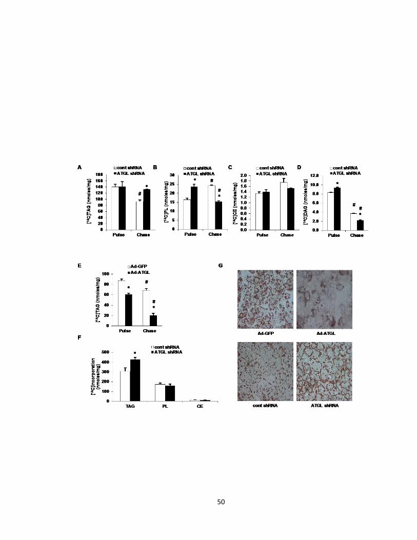
50
51
Hepatic ATGL does not influence TAG secretion. To gain insight into the
effects of hepatic ATGL knockdown on whole-body fatty acid metabolism, we
quantified serum FFA and TAG. Regardless of diet, the concentrations of these
serum metabolites were unaltered following hepatic ATGL knockdown (Fig. 4A,B).
Previous studies have shown that cytosolic TAG undergoes hydrolysis to DAG or
monoacylglycerol prior to resterification and incorporation into VLDL (19). Thus,
it was surprising that mice treated with ATGL shRNA had unaltered fasted serum
TAG levels despite having attenuated hepatic TAG hydrolysis. Based upon
these findings, we next questioned if ATGL regulates the rate of hepatic TAG
secretion. To quantify rates of hepatic TAG secretion, fasted mice were injected
with Tyloxapol, an inhibitor of lipoprotein lipase. In agreement with the similar
fasting TAG values between treatment groups, rates of hepatic TAG secretion
were unchanged in response to ATGL knockdown (Fig. 4C).
We next extended these studies to primary hepatocyte cultures. Cells
were pulsed with 500 µM [1-14C]oleate for 1.5 h, at which time media was
changed and appearance of [14C]TAG in the media was measured after 6 h to
quantify the contribution of ATGL and TAG hydrolysis to hepatic TAG secretion.
In support of the in vivo data, ATGL knockdown did not alter TAG secretion in
primary hepatocytes (Fig. 4D). Similar results were obtained when cells were
labeled with [1-14C]oleate for 8 h (Fig. 4E). We also overexpressed ATGL and
found that despite enhanced TAG hydrolysis (Fig. 3E) ATGL overexpression did
not alter TAG secretion (Fig. 4F). Thus, despite potent effects on intracellular
52
TAG hydrolysis, ATGL does not appear to be involved in channeling hydrolyzed
fatty acids to VLDL synthesis.
Hepatic ATGL promotes fatty acid oxidation. Intracellular fatty acids have two
predominant routes of disposal in the liver, export via VLDL and β-oxidation.
Given the pronounced effects of ATGL on hepatic TAG turnover, we next
explored if ATGL influenced fatty acid oxidation. Serum β-hydroxybuytrate
concentrations were similar in mice treated with control or ATGL shRNA
suggesting that perhaps hepatic β-oxidation was not influenced by hepatic ATGL
(Fig 5A). However, ATGL knockdown in primary hepatocytes resulted in a 30%
decrease in ASM production when cells were pulsed with 500 µM [1-14C]oleate
(Fig. 5B). However, these data reflect primarily oxidation of fatty acid derived
from exogenous uptake.
Thus, we also quantified fatty acid oxidation during the chase period, which
reflects oxidation of fatty acids derived primarily from intracellular TAG hydrolysis.
Under these conditions, ATGL shRNA resulted in a ~70% decrease in fatty acid
oxidation to both ASM and CO2 compared with cells treated with control shRNA
(Fig. 5B,C). Similarly, we also measured fatty acid oxidation during pulse and
chase periods following ATGL overexpression. In support of the above
experiments, ATGL overexpression increased fatty acid oxidation to ASM 40%
during the pulse period and caused an even more robust increase in fatty acid
oxidation to both ASM and CO2 during the chase period (Fig. 5D,E). Previously,
we have shown that overexpression of ATGL in rat hepatocytes increases PPAR-
53
Figure 4. Hepatic ATGL does not regulate TAG secretion. Mice fed the chow
diet were treated with control or ATGL shRNA adenovirus and were fasted
overnight followed by harvesting of serum for analysis of (A) free fatty acid (FFA)
and (B) TAG (n = 8-10). (C) For measurement of hepatic TAG production,
overnight fasted mice fed the chow diet were treated with Tyloxapol and blood
collections were performed at 0, 1, 2 and 3 h intervals (n = 4-6) and serum TAG
was subsequently analyzed. (D) Primary hepatocytes were transduced with
control or ATGL shRNA for 66 h, at which time pulse (1.5 h) and chase (6 h)
experiments were performed with 500 µM [1-14C]oleate. [14C]TAG was quantified
during the chase period. (E) Primary hepatocytes were transduced with control or
ATGL shRNA for 66 h followed by 8 h of pulse with 500 µM [1-14C]oleate. Cells
were harvested and radiolabeled oleate incorporation into different lipid fractions
was determined (n = 3). (F) Primary hepatocytes were also transduced with Ad-
GFP or Ad-ATGL adenoviruses and after 24 h pulse (1.5 h) and chase (6 h)
experiments were performed with 500 µM [1-14C]oleate and [14C]TAG secretion
during the chase period was measured. Data are presented as means ± SEM.

54
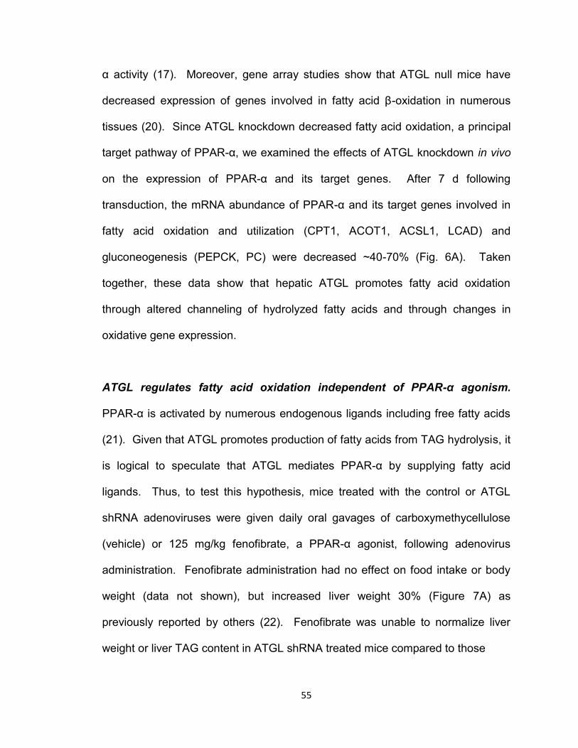
55
α activity (17). Moreover, gene array studies show that ATGL null mice have
decreased expression of genes involved in fatty acid β-oxidation in numerous
tissues (20). Since ATGL knockdown decreased fatty acid oxidation, a principal
target pathway of PPAR-α, we examined the effects of ATGL knockdown in vivo
on the expression of PPAR-α and its target genes. After 7 d following
transduction, the mRNA abundance of PPAR-α and its target genes involved in
fatty acid oxidation and utilization (CPT1, ACOT1, ACSL1, LCAD) and
gluconeogenesis (PEPCK, PC) were decreased ~40-70% (Fig. 6A). Taken
together, these data show that hepatic ATGL promotes fatty acid oxidation
through altered channeling of hydrolyzed fatty acids and through changes in
oxidative gene expression.
ATGL regulates fatty acid oxidation independent of PPAR-α agonism.
PPAR-α is activated by numerous endogenous ligands including free fatty acids
(21). Given that ATGL promotes production of fatty acids from TAG hydrolysis, it
is logical to speculate that ATGL mediates PPAR-α by supplying fatty acid
ligands. Thus, to test this hypothesis, mice treated with the control or ATGL
shRNA adenoviruses were given daily oral gavages of carboxymethycellulose
(vehicle) or 125 mg/kg fenofibrate, a PPAR-α agonist, following adenovirus
administration. Fenofibrate administration had no effect on food intake or body
weight (data not shown), but increased liver weight 30% (Figure 7A) as
previously reported by others (22). Fenofibrate was unable to normalize liver
weight or liver TAG content in ATGL shRNA treated mice compared to those

56
Figure 5. Hepatic ATGL promotes fatty acid oxidation. (A) Serum β-
hydroxybutyrate was measured with a colorimetric enzymatic kit in serum
samples collected from chow-fed mice 7 d after infection with control or ATGL
shRNA adenovirus and following an overnight fast. Primary hepatocytes isolated
from chow-fed mice were transduced with control or ATGL shRNA for 66 h and
Ad-GFP or Ad-ATGL for 24 h, at which time pulse (1.5 h) and chase (6 h)
experiments were performed with 500 µM [1-14C]oleate. (B-E) Media from
hepatocytes were harvested after pulse and chase, and CO2 and ASM were
quantified as outlined in the experimental procedures to measure fatty acid
oxidation. Data are presented as means ± SEM. *P<0.05 versus control shRNA
group. #P<0.05 versus pulse period.
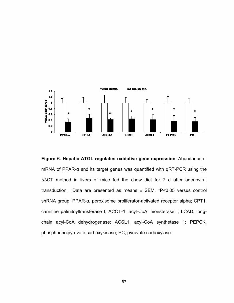
57
Figure 6. Hepatic ATGL regulates oxidative gene expression. Abundance of
mRNA of PPAR-α and its target genes was quantified with qRT-PCR using the
∆∆CT method in livers of mice fed the chow diet for 7 d after adenoviral
transduction. Data are presented as means ± SEM. *P<0.05 versus control
shRNA group. PPAR-α, peroxisome proliferator-activated receptor alpha; CPT1,
carnitine palmitoyltransferase I; ACOT-1, acyl-CoA thioesterase I; LCAD, long-
chain acyl-CoA dehydrogenase; ACSL1, acyl-CoA synthetase 1; PEPCK,
phosphoenolpyruvate carboxykinase; PC, pyruvate carboxylase.
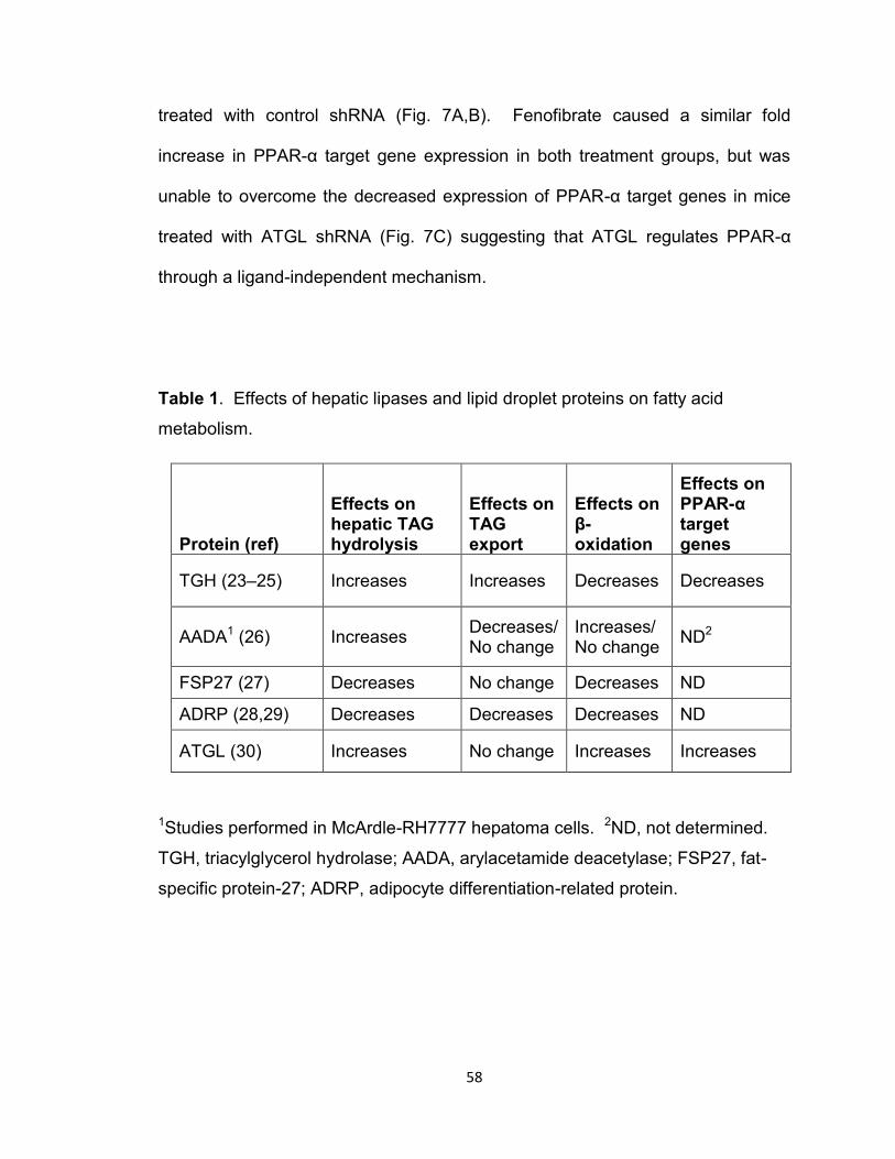
58
treated with control shRNA (Fig. 7A,B). Fenofibrate caused a similar fold
increase in PPAR-α target gene expression in both treatment groups, but was
unable to overcome the decreased expression of PPAR-α target genes in mice
treated with ATGL shRNA (Fig. 7C) suggesting that ATGL regulates PPAR-α
through a ligand-independent mechanism.
Table 1. Effects of hepatic lipases and lipid droplet proteins on fatty acid
metabolism.
Protein (ref)
Effects on hepatic TAG hydrolysis
Effects on TAG export
Effects on β-oxidation
Effects on PPAR-α target genes
TGH (23–25) Increases Increases Decreases Decreases
AADA1 (26) Increases Decreases/ No change
Increases/ No change
ND2
FSP27 (27) Decreases No change Decreases ND
ADRP (28,29) Decreases Decreases Decreases ND
ATGL (30) Increases No change Increases Increases
1Studies performed in McArdle-RH7777 hepatoma cells. 2ND, not determined.
TGH, triacylglycerol hydrolase; AADA, arylacetamide deacetylase; FSP27, fat-
specific protein-27; ADRP, adipocyte differentiation-related protein.
59
Figure 7. PPAR-α agonism does not rescue the effects of ATGL knockdown.
One day after adenoviral injections, mice were treated with 125 mg/kg of
fenofibrate suspended in 0.5% carboxylmethylcellulose for 6 d via oral gavage.
Mice were fed with the HF diet and sacrificed 7 d after infection following an
overnight fast. Liver weight (A) and triglyceride (B) were measured in mice
treated with fenofibrate (FF) and compared to vehicle treated mice (n = 7-8). (C)
mRNA abundance of oxidative genes was determined in liver tissues of mice
treated with FF (n=6). Data are presented as means ± SEM. *P<0.05 versus
control shRNA group. #P<0.05 versus pulse period.

60
Discussion
Despite the importance of hepatic TAG in local and systemic energy metabolism
and disease etiology, the major lipases responsible for TAG mobilization in the
liver are largely unknown. Herein, we show ATGL to be a principal TAG lipase in
the liver consistent with its crucial role in TAG hydrolysis in other tissues such as
adipose, heart and muscle. Interestingly, pulse-chase experiments in
hepatocytes revealed that ATGL knockdown almost completely blocked TAG
hydrolysis. However, livers from mice treated with ATGL shRNA still possessed
~60% of TAG hydrolase activity suggesting that other lipases also contribute to
hepatic TAG hydrolysis. Although these studies clearly show that ATGL impacts
TAG hydrolysis, the discrepancies between TAG hydrolase activity assays and
TAG turnover in cells also suggest that there are many additional factors that
contribute to TAG hydrolysis that are not reflected by in vitro assays. For
example, several proteins such as comparative gene identification-58, pigment
epithelium-derived factor, G0/G1 switch gene 2 and numerous lipid droplet
proteins have potent effects on ATGL-mediated TAG hydrolysis and are not
present on synthetic lipid droplets used for in vitro assays (31–33). Thus, it is
likely that the contribution of ATGL to hepatic TAG turnover likely varies
depending upon the presence of the various inhibitors and activators and may in
fact represent more than in vitro assays indicate.
The current studies provide evidence that ATGL acts as a branch point in
partitioning hydrolyzed fatty acids between oxidative and VLDL synthetic
61
pathways. As the complexity of the lipid droplet and control of TAG turnover is
unraveled, it is becoming apparent that specific lipid droplet proteins and lipases
differentially partition fatty acids to distinct metabolic fates. Recent studies have
shown that ablation of triacylglycerol hydrolase, which is located exclusively on
ER, suppresses hepatic TAG turnover and TAG export, but increases oxidation
(23). Coinciding with the increased fatty acid oxidation in mice lacking
triacylglycerol hydrolase is an increase in oxidative gene expression.
Overexpression of arylacetamide deacetylase, which possesses TAG hydrolase
activity, increases hepatic TAG turnover and fatty acid oxidation, but decreases
TAG secretion (26). Taken together with the current findings, these data suggest
that distinct lipases differentially channel fatty acids between β-oxidation and
VLDL synthesis. Additionally, it is unclear how reducing TAG hydrolysis by both
ATGL and triacylglycerol hydrolase result in opposing effects on oxidative gene
expression. In addition to lipases, several lipid droplet proteins may also
contribute to differential partitioning of hydrolyzed fatty acids. Overexpression of
fat specific protein-27, a recently identified lipid droplet protein, decreases TAG
turnover and fatty acid oxidation without altering TAG export from hepatocytes
(27). Despite promoting TAG accumulation, perilipin 5 (also known as OXPAT)
also promotes fatty acid oxidation consistent with its high expression levels in
oxidative tissues (34). Thus, there appears to be a coordinated regulation of
TAG metabolism by lipid droplet proteins and lipases that determines the
metabolic fate of hydrolyzed fatty acids (Table 1). Defining the mechanism
through which specific pools of TAG are hydrolyzed and channeled between
62
oxidative and anabolic pathways will provide valuable insight into the regulation
of hepatic energy metabolism.
The effects of ATGL on fatty acid channeling highlight the importance of
TAG hydrolysis in supplying substrates for β-oxidation. Although intracellular
fatty acids can be derived from numerous sources, TAG hydrolysis and
exogenous uptake included, the partitioning of these fatty acids between
metabolic pathways appears to be dissimilar. Recently, using stable isotope
infusions in humans, Kanaley et al. showed that resting muscle preferentially
oxidizes fatty acids derived from TAG hydrolysis compared to those derived from
exogenous uptake (35). In support, previous work in rat hepatocytes has
suggested that fatty acids derived from TAG hydrolysis are more readily oxidized
compared to those supplied as fatty acids in the media (36). Given the
importance of intracellular TAG in supplying fatty acids for oxidation, our data
would suggest that ATGL plays a critical role in controlling substrate oxidation.
Additionally, although this study only focused upon male mice, effects of
estrogen on ATGL have been reported (37). Thus, it remains to be determined if
the effects of ATGL observed in the current study would be similar in female mice.
In addition to its direct effects on TAG hydrolysis, ATGL also appears to
influence hepatic energy metabolism through changes in gene expression. The
current study shows that ATGL knockdown reduces the expression of genes
involved in fatty acid oxidation and gluconeogenesis. Microarray analysis of
tissues from global ATGL knockout mice revealed decreased expression of
oxidative genes in numerous tissues (20). Additionally, we have previously
63
shown that ATGL overexpression increases PPAR-α activity in rat hepatocytes
(17). Thus, these data suggest that ATGL may modulate fatty acid channeling at
least in part through changes in oxidative gene expression. In an attempt to
recover oxidative gene expression, mice were gavaged with fenofibrate following
administration of adenoviruses. Fenofibrate treatment resulted in a robust
induction of oxidative gene expression between both treatment groups, but was
unable to normalize gene expression in ATGL shRNA treated groups to those of
control animals. Thus, these data suggest that the effects of ATGL on gene
expression are independent of PPAR-α ligand binding. Although the mechanism
explaining the effects of ATGL on gene expression remain to be elucidated, it is
likely that the alterations in gene expression contribute to the metabolic effects of
ATGL knockdown. Additionally, it could be postulated that changes in PPAR-α
activity could influence fatty acid partitioning through alterations in hepatic TAG
export. However, the role of PPAR-α in hepatic TAG secretion is unclear.
Studies in mice and hepatocytes show that PPAR-α decreases hepatic TAG
secretion (38–40) although not all data are consistent (41). Fibrate
administration in humans lowers serum TAG through increased VLDL clearance,
but rates of hepatic TAG secretion are unaltered (42,43). Although the direct
effects of PPAR-α on hepatic TAG secretion are not resolved, perhaps changes
in metabolic enzymes, such as ATGL, may mediate the effects of PPAR-α.
Previous studies have shown that hepatic ATGL is activated in response to
fasting and that ATGL contains a PPAR-γ responsive peroxisome proliferator

64
response element (44,45). Ongoing studies will further characterize the
transcriptional and post-transcriptional regulation of hepatic ATGL.
The present studies shows that ATGL knockdown results in increased
C18:1 and lower C16:0, C18:0 and C18:3 in hepatic TAG. Although we cannot
rule out a role for ATGL in selective hepatic fatty acid uptake and esterification or
in altering de novo fatty acid synthesis, the effects of ATGL knockdown on
hepatic TAG fatty acid composition suggest that ATGL may show substrate
specificity towards different fatty acids. Given the potent effects of ATGL on fatty
acid partitioning and signaling, it is plausible that some of these effects could be
due to changes in hydrolysis and metabolism of specific fatty acids rather than
ATGL itself.
In summary, this study identifies ATGL as a major hepatic TAG lipase that
has important roles in fatty acid trafficking and signaling. Given these important
characteristics, alterations in hepatic ATGL expression or activity is likely to
contribute to a host of metabolic diseases including non-alcoholic fatty liver
disease.

65
References
1. Browning JD, Szczepaniak LS, Dobbins R, Nuremberg P, Horton JD, Cohen JC, et al. Prevalence of hepatic steatosis in an urban population in the United States: impact of ethnicity. Hepatology. 2004;40:1387–1395.
2. Watanabe S, Yaginuma R, Ikejima K, Miyazaki A. Liver diseases and metabolic syndrome. J. Gastroenterol. 2008;43:509–518.
3. Lavine JE, Schwimmer JB. Nonalcoholic fatty liver disease in the pediatric population. Clin Liver Dis. 2004;8:549–558, viii–ix.
4. Fabbrini E, Sullivan S, Klein S. Obesity and nonalcoholic fatty liver disease: biochemical, metabolic, and clinical implications. Hepatology. 2010;51:679–689.
5. Jenkins CM, Mancuso DJ, Yan W, Sims HF, Gibson B, Gross RW. Identification, cloning, expression, and purification of three novel human calcium-independent phospholipase A2 family members possessing triacylglycerol lipase and acylglycerol transacylase activities. J. Biol. Chem. 2004;279:48968–48975.
6. Zimmermann R, Strauss JG, Haemmerle G, Schoiswohl G, Birner-Gruenberger R, Riederer M, et al. Fat mobilization in adipose tissue is promoted by adipose triglyceride lipase. Science. 2004;306:1383–1386.
7. Villena JA, Roy S, Sarkadi-Nagy E, Kim K-H, Sul HS. Desnutrin, an adipocyte gene encoding a novel patatin domain-containing protein, is induced by fasting and glucocorticoids: ectopic expression of desnutrin increases triglyceride hydrolysis. J. Biol. Chem. 2004;279:47066–47075.
8. Haemmerle G, Lass A, Zimmermann R, Gorkiewicz G, Meyer C, Rozman J, et al. Defective lipolysis and altered energy metabolism in mice lacking adipose triglyceride lipase. Science. 2006;312:734–737.
9. Kershaw EE, Hamm JK, Verhagen LAW, Peroni O, Katic M, Flier JS. Adipose triglyceride lipase: function, regulation by insulin, and comparison with adiponutrin. Diabetes. 2006;55:148–157.
10. Miyoshi H, Perfield JW 2nd, Souza SC, Shen W-J, Zhang H-H, Stancheva ZS, et al. Control of adipose triglyceride lipase action by serine 517 of perilipin A globally regulates protein kinase A-stimulated lipolysis in adipocytes. J. Biol. Chem. 2007;282:996–1002.
66
11. Dana SL, Hoener PA, Bilakovics JM, Crombie DL, Ogilvie KM, Kauffman RF, et al. Peroxisome proliferator-activated receptor subtype-specific regulation of hepatic and peripheral gene expression in the Zucker diabetic fatty rat. Metab. Clin. Exp. 2001;50:963–971.
12. Seo YS, Kim JH, Jo NY, Choi KM, Baik SH, Park J-J, et al. PPAR agonists treatment is effective in a nonalcoholic fatty liver disease animal model by modulating fatty-acid metabolic enzymes. J. Gastroenterol. Hepatol. 2008;23:102–109.
13. Kwanyuen P, Witherspoon SM, Creech DR, Colton HM, Falls JG, Cariello NF. Flow cytometric assessment of peroxisome proliferation from frozen liver of fibrate-treated monkeys. Int. J. Toxicol. 2006;25:41–47.
14. Bu SY, Mashek MT, Mashek DG. Suppression of long chain acyl-CoA synthetase 3 decreases hepatic de novo fatty acid synthesis through decreased transcriptional activity. J. Biol. Chem. 2009;284:30474–30483.
15. Lewin TM, Wang S, Nagle CA, Van Horn CG, Coleman RA. Mitochondrial glycerol-3-phosphate acyltransferase-1 directs the metabolic fate of exogenous fatty acids in hepatocytes. Am. J. Physiol. Endocrinol. Metab. 2005;288:E835–844.
16. Folch J, Lees M, Stanley GHS. A simple method for the isolation and purification of total lipides from animal tissues. J. Biol. Chem. 1957;226:497–509.
17. Sapiro JM, Mashek MT, Greenberg AS, Mashek DG. Hepatic triacylglycerol hydrolysis regulates peroxisome proliferator-activated receptor alpha activity. J. Lipid Res. 2009;50:1621–1629.
18. Smirnova E, Goldberg EB, Makarova KS, Lin L, Brown WJ, Jackson CL. ATGL has a key role in lipid droplet/adiposome degradation in mammalian cells. EMBO Rep. 2006;7:106–113.
19. Wiggins D, Gibbons GF. The lipolysis/esterification cycle of hepatic triacylglycerol. Its role in the secretion of very-low-density lipoprotein and its response to hormones and sulphonylureas. Biochem. J. 1992;284 ( Pt 2):457–462.
20. Pinent M, Hackl H, Burkard TR, Prokesch A, Papak C, Scheideler M, et al. Differential transcriptional modulation of biological processes in adipocyte triglyceride lipase and hormone-sensitive lipase-deficient mice. Genomics. 2008;92:26–32.
21. Kliewer SA, Forman BM, Blumberg B, Ong ES, Borgmeyer U, Mangelsdorf DJ, et al. Differential expression and activation of a family of murine
67
peroxisome proliferator-activated receptors. Proc. Natl. Acad. Sci. U.S.A. 1994;91:7355–7359.
22. Lalwani ND, Reddy MK, Qureshi SA, Sirtori CR, Abiko Y, Reddy JK. Evaluation of selected hypolipidemic agents for the induction of peroxisomal enzymes and peroxisome proliferation in the rat liver. Hum Toxicol. 1983;2:27–48.
23. Wei E, Alam M, Sun F, Agellon LB, Vance DE, Lehner R. Apolipoprotein B and triacylglycerol secretion in human triacylglycerol hydrolase transgenic mice. J Lipid Res. 2007;48:2597–606.
24. Wei E, Ben Ali Y, Lyon J, Wang H, Nelson R, Dolinsky VW, et al. Loss of TGH/Ces3 in mice decreases blood lipids, improves glucose tolerance, and increases energy expenditure. Cell Metab. 2010;11:183–193.
25. Wang H, Wei E, Quiroga AD, Sun X, Touret N, Lehner R. Altered lipid droplet dynamics in hepatocytes lacking triacylglycerol hydrolase expression. Mol. Biol. Cell. 2010;21:1991–2000.
26. Lo V, Erickson B, Thomason-Hughes M, Ko KWS, Dolinsky VW, Nelson R, et al. Arylacetamide deacetylase attenuates fatty-acid-induced triacylglycerol accumulation in rat hepatoma cells. J. Lipid Res. 2010;51:368–377.
27. Matsusue K, Kusakabe T, Noguchi T, Takiguchi S, Suzuki T, Yamano S, et al. Hepatic steatosis in leptin-deficient mice is promoted by the PPARgamma target gene Fsp27. Cell Metab. 2008;7:302–311.
28. Magnusson B, Asp L, Boström P, Ruiz M, Stillemark-Billton P, Lindén D, et al. Adipocyte differentiation-related protein promotes fatty acid storage in cytosolic triglycerides and inhibits secretion of very low-density lipoproteins. Arterioscler. Thromb. Vasc. Biol. 2006;26:1566–1571.
29. Chang BH-J, Li L, Paul A, Taniguchi S, Nannegari V, Heird WC, et al. Protection against fatty liver but normal adipogenesis in mice lacking adipose differentiation-related protein. Mol. Cell. Biol. 2006;26:1063–1076.
30. Reid BN, Ables GP, Otlivanchik OA, Schoiswohl G, Zechner R, Blaner WS, et al. Hepatic overexpression of hormone-sensitive lipase and adipose triglyceride lipase promotes fatty acid oxidation, stimulates direct release of free fatty acids, and ameliorates steatosis. J. Biol. Chem. 2008;283:13087–13099.
31. Chung C, Doll JA, Gattu AK, Shugrue C, Cornwell M, Fitchev P, et al. Anti-angiogenic pigment epithelium-derived factor regulates hepatocyte triglyceride content through adipose triglyceride lipase (ATGL). J. Hepatol. 2008;48:471–478.
68
32. Lass A, Zimmermann R, Haemmerle G, Riederer M, Schoiswohl G, Schweiger M, et al. Adipose triglyceride lipase-mediated lipolysis of cellular fat stores is activated by CGI-58 and defective in Chanarin-Dorfman Syndrome. Cell Metab. 2006;3:309–319.
33. Yang X, Lu X, Lombès M, Rha GB, Chi Y-I, Guerin TM, et al. The G(0)/G(1) switch gene 2 regulates adipose lipolysis through association with adipose triglyceride lipase. Cell Metab. 2010;11:194–205.
34. Wolins NE, Quaynor BK, Skinner JR, Tzekov A, Croce MA, Gropler MC, et al. OXPAT/PAT-1 is a PPAR-induced lipid droplet protein that promotes fatty acid utilization. Diabetes. 2006;55:3418–3428.
35. Kanaley JA, Shadid S, Sheehan MT, Guo Z, Jensen MD. Relationship between plasma free fatty acid, intramyocellular triglycerides and long-chain acylcarnitines in resting humans. J. Physiol. (Lond.). 2009;587:5939–5950.
36. Lankester DL, Brown AM, Zammit VA. Use of cytosolic triacylglycerol hydrolysis products and of exogenous fatty acid for the synthesis of triacylglycerol secreted by cultured rat hepatocytes. J. Lipid Res. 1998;39:1889–1895.
37. Wohlers LM, Spangenburg EE. 17beta-estradiol supplementation attenuates ovariectomy-induced increases in ATGL signaling and reduced perilipin expression in visceral adipose tissue. J. Cell. Biochem. 2010;110:420–427.
38. Tordjman K, Bernal-Mizrachi C, Zemany L, Weng S, Feng C, Zhang F, et al. PPARalpha deficiency reduces insulin resistance and atherosclerosis in apoE-null mice. J. Clin. Invest. 2001;107:1025–1034.
39. Hahn SE, Goldberg DM. Modulation of lipoprotein production in Hep G2 cells by fenofibrate and clofibrate. Biochem. Pharmacol. 1992;43:625–633.
40. Staels B, Dallongeville J, Auwerx J, Schoonjans K, Leitersdorf E, Fruchart JC. Mechanism of action of fibrates on lipid and lipoprotein metabolism. Circulation. 1998;98:2088–2093.
41. Bijland S, Pieterman EJ, Maas ACE, van der Hoorn JWA, van Erk MJ, van Klinken JB, et al. Fenofibrate increases very low density lipoprotein triglyceride production despite reducing plasma triglyceride levels in APOE*3-Leiden.CETP mice. J. Biol. Chem. 2010;285:25168–25175.
42. Watts GF, Ji J, Chan DC, Ooi EMM, Johnson AG, Rye K-A, et al. Relationships between changes in plasma lipid transfer proteins and apolipoprotein B-100 kinetics during fenofibrate treatment in the metabolic syndrome. Clin. Sci. 2006;111:193–199.

69
43. Fabbrini E, Mohammed BS, Korenblat KM, Magkos F, McCrea J, Patterson BW, et al. Effect of fenofibrate and niacin on intrahepatic triglyceride content, very low-density lipoprotein kinetics, and insulin action in obese subjects with nonalcoholic fatty liver disease. J. Clin. Endocrinol. Metab. 2010;95:2727–2735.
44. Kershaw EE, Schupp M, Guan H-P, Gardner NP, Lazar MA, Flier JS. PPARgamma regulates adipose triglyceride lipase in adipocytes in vitro and in vivo. Am. J. Physiol. Endocrinol. Metab. 2007;293:E1736–1745.
45. Kim JY, Tillison K, Lee J-H, Rearick DA, Smas CM. The adipose tissue triglyceride lipase ATGL/PNPLA2 is downregulated by insulin and TNF-alpha in 3T3-L1 adipocytes and is a target for transactivation by PPARgamma. Am. J. Physiol. Endocrinol. Metab. 2006;291:E115–127.

70
CHAPTER 3
Hepatic ATGL Knockdown Uncouples
Glucose Intolerance from Liver TAG
Accumulation
Kuok Teong Ong, Mara T. Mashek, So Young Bu and Douglas G. Mashek. FASEB J. 2013 Jan;27(1):313-21
This chapter contains an original research article previously published.
71
Adipose triglyceride lipase (ATGL) is the predominant triacylglycerol (TAG)
hydrolase in mammals, however, the tissue specific effects of ATGL outside of
adipose tissue have not been well characterized. Hence, we tested the
contribution of hepatic ATGL on mediating glucose tolerance and insulin action.
Glucose or insulin tolerance tests and insulin signaling were performed in
C57BL/6 mice administered with control (non-gene specific shRNA) or Atgl
shRNA adenoviruses. Glucose and lipid metabolism assays were conducted in
primary hepatocytes isolated from mice transduced with control or Atgl shRNA
adenoviruses. Knocking down hepatic ATGL completely abrogated the increase
in serum insulin following either 1 or 12 weeks of feeding a high fat (HF) diet
despite higher hepatic TAG content. Glucose tolerance tests demonstrated that
ATGL knockdown normalized glucose tolerance in HF diet-fed mice. The
observed improvements in glucose tolerance were present despite unaltered
hepatic insulin signaling and increased liver TAG. Mice with suppressed hepatic
ATGL had reduced hepatic glucose production in vivo and hepatocytes isolated
from Atgl shRNA-treated mice displayed a 26% decrease in glucose production
and a 38% increase in glucose oxidation compared to control cells. Taken
together, these data suggest that hepatic ATGL knockdown enhances glucose
tolerance by increasing hepatic glucose utilization and uncouples impairments in
insulin action from hepatic TAG accumulation.

72
Introduction
Insulin resistance is a metabolic state that is predictive of a host of metabolic
diseases including Type 2 Diabetes and cardiovascular diseases. Hepatic
steatosis has been shown to positively correlate with insulin resistance and may
be an important component in the development of metabolic diseases resulting
from insulin resistance (1). In fact, excessive liver fat is a stronger predictor of
insulin resistance than total or visceral fat in humans (2,3). Until recently, most
studies have examined the importance of TAG synthetic enzymes in linking
hepatic steatosis to insulin resistance. However, the contribution of dysregulated
lipolysis in non-adipose tissue to the pathophysiology of insulin resistance is
unclear and has only recently gained attention among researchers.
In 2004, three separate groups discovered a novel lipase, adipose
triglyceride lipase (ATGL), with higher substrate specificity for TAG than hormone
sensitive lipase (HSL), which was previously thought to be the predominant TAG
hydrolase (4–6). Although the expression of ATGL is lower in the liver compared
to adipose tissues, we and others have shown that ATGL is a major hepatic TAG
hydrolase (7,8). Since ATGL is a key TAG lipase, it would be interesting to
determine whether it is involved in the pathogenesis of insulin resistance
especially in the event of hepatic steatosis. The role of ATGL in insulin action
was first characterized in ATGL knockout (Atgl KO) mice, which exhibit increased
glucose tolerance and insulin sensitivity (9). This finding was unexpected given
that Atgl KO mice have increased adipose tissue mass and ectopic TAG
73
deposition in insulin-sensitive tissues including skeletal muscles, heart and liver
(9). Further investigation in Atgl KO mice indicated that insulin signaling is
enhanced in skeletal muscle and white adipose tissue, but attenuated in liver and
brown adipose tissue (10). Increased glucose utilization and uptake in the liver,
cardiac muscle and skeletal muscle as a result of an inability to release non-
esterified fatty acids (NEFA) in Atgl KO mice were proposed to mediate the
observed improvement in glucose tolerance and insulin sensitivity.
In addition, Turpin et al. has reported that isolated hepatocytes from Atgl
KO mice display no change in insulin action (11). However, when hepatic ATGL
is overexpressed in vivo, whole-body glucose tolerance and Akt phosphorylation
are mildly enhanced (11). This observed phenotype may be partially attributed to
attenuated gluconeogenesis as depicted by decreased glucose levels during
pyruvate tolerance tests and decreased gluconeogenic gene expression.
Interestingly, Wu et al. has recently shown that glucose and insulin sensitivity
remain unaltered in liver-specific ATGL knockout mice that were fed control or
high-fat (HF) diet (8). They also noted that glucose production does not change
with liver-specific ATGL deletion (8).
Given the paradoxical phenotypes in the two models and the improvement
in glucose tolerance of Atgl KO mice, it is important to further examine liver-
specific effects of ATGL on whole-body glucose tolerance and insulin action.
Herein, we test the contribution of hepatic ATGL to liver energy metabolism and
insulin resistance via adenovirus-mediated approach and reveal that ATGL
uncouples glucose intolerance and impaired insulin signaling from steatosis.

74
Materials and Methods
Animals, diets and adenovirus administration. All animal protocols were
approved by the University of Minnesota Institutional Animal Care and Use
Committee. Six to eight week old C57/Bl6 male mice were purchased from
Harlan Laboratories and housed under controlled temperature and lighting (20-
22°C; 12:12-h light-dark cycle). All mice were acclimatized for 1 week prior to
adenovirus injections. Adenoviruses that encoded mouse Atgl short hairpin RNA
(shRNA) and control shRNA targeting a nonspecific mRNA sequence were
generated as described previously (12). The mice were injected with 1 x 10⁹ pfu
of an adenovirus containing Atgl shRNA or a non-targeting shRNA control (cont
shRNA) via the tail vein. The mice had free access to water and were fed either a
purified control diet (TD.94045) or a 45% fat diet (TD.09404) from Harlan Teklad
Premier Laboratory (Madison, WI, USA) after adenovirus administration. The
control diet contained protein (19% of total calories), carbohydrates (64%), and
fat (17%) in the form of soybean oil (70 g/kg). The high-fat (HF) diet contained
protein (19%), carbohydrates (35%) and fat (45%) with lard (195 g/kg) and
soybean oil (30 g/kg) as the fat sources. Exactly 1 week following adenovirus
injection, the mice were sacrificed for tissue and serum collection after an
overnight fast. Another group of 6-8 week old C57/Bl6 male mice were fed either
a control or HF diet (diet-induced obese or DIO mice) for 12 weeks. Immediately
following the 12 week feeding period, mice were transduced with the control or
Atgl shRNA adenovirus. Mice were sacrificed 1 week after adenovirus injection
75
to harvest tissue and serum samples. Mice fed either control or HF diets for 12
weeks were fasted for 4 h prior to experiments or euthanasia and harvesting of
tissues and serum.
Measurement of mitochondrial FA oxidation. Liver sections were quickly
harvested from anesthetized mice and mitochondria were isolated as described
previously (13). Isolated mitochondria were added to reaction medium containing
[1-14C]palmitate (2.13 GBq/mmol; Perkin Elmer, Waltham, MA, USA) in a 25 mL
flask. The flask was quickly sealed with a double seal stopper and perchloric acid
was added. The flask was shaken for 1 h at room temperature. Bovine serum
albumin (BSA) and water were added to the acid-treated medium followed by
centrifugation twice to separate particulate from supernatant. Supernatant
containing [14C]-labeled acid-soluble metabolites (ASM) was quantified with
scintillation counting.
RNA isolation, RT-PCR and real-time quantitative PCR analysis. RNA was
extracted with Trizol from liver tissues followed by reverse-transcription with
SuperScript® VILO™ cDNA Synthesis Kit (Invitrogen, Carlsbad, CA, USA) to
generate cDNA. Gene expression was quantified as described previously (7).

76
Liver TAG and glycogen analysis and serum measurements. TAG was
extracted from liver tissues according to the method described by Folch (14).
Liver and serum TAG levels were quantified with a TAG colorimetric enzymatic
kit (Stanbio, Boerne, TX, USA). Liver glycogen content was determined as
described previously (15). Serum FA and glucose levels were determined with
colorimetric enzymatic assay kits from Wako Chemicals (Richmond, VA, USA)
while serum insulin concentrations were assessed with Ultra Sensitive mouse
insulin ELISA kit from Crystal Chem (Downers Grove, IL, USA).
Oral glucose tolerance and insulin tolerance tests. For glucose tolerance
tests, a 20% dextrose solution (2 g of glucose/kg body weight) was administered
via oral gavage to the mice. To perform insulin tolerance tests, mice were
injected intraperitoneally with 1 U insulin/kg body weight (Humulin N from Eli Lilly,
Indianapolis, IN, USA). For pyruvate tolerance tests, mice received 2 g/kg body
weight of sterile-filtered sodium pyruvate. Blood glucose concentrations prior to
administration and at 15, 30, 60 and 90 min post-injection were measured with
AlphaTRAK glucose meter and test strips (Abbott, Chicago, IL, USA).
Insulin signaling. After a 4 h fast, mice were injected intraperitoneally with 1U
insulin/kg body weight or saline (vehicle). After 10 min, liver, adipose and muscle
tissues were snap frozen from euthanized mice. Liver preparation,
electrophoresis and immunoblotting were done as described (7). The following

77
antibodies were used: anti-β-actin monoclonal (Sigma-Aldrich, Saint Louis, MO,
USA); anti-pIRS1 (Tyr989) polyclonal (Santa Cruz Biotechnology, Santa Cruz,
CA, USA); anti-Akt polyclonal, anti-pAkt (Ser473) polyclonal, anti-IRS1 polyclonal,
anti-pIRS1 (Ser636/639) polyclonal, anti-GSK-3α and anti-pGSK-3α (Ser21) (Cell
Signaling Technology, Danvers, MA, USA). Bands were visualized by ECL
chemiluminescense (GE Healthcare, Little Chalfont, Buckinghamshire, UK).
Glucose production and oxidation assays. Glucose production was measured
as performed previously (16). Briefly, primary hepatocytes were isolated and
cultured in M199 plating media (Invitrogen) free of insulin as described previously
(7). The medium was then replaced with 1 ml of glucose production buffer
consisting of glucose-free DMEM (pH 7.4), without phenol red, supplemented
with 20 mM sodium lactate and 2 mM sodium pyruvate (with or without 100 nm
insulin). After 3 h, 0.5 ml of medium was collected and the glucose concentration
measured with a colorimetric glucose assay (Wako Chemicals) and normalized
to the total protein content determined by BCA protein assay (Thermo Scientific,
Waltham, MA, USA). To determine glucose oxidation in primary hepatocytes,
approximately 5 x 10⁵ isolated primary hepatocytes were suspended in Krebs-
Ringer-phosphate buffer (KRP) containing 5.5 mM glucose, 1 μCi of
[14C6]glucose, 135 mM NaCl, 5.4 mM KCl, 1.4 mM CaCl2, 1.4 mM MgSO4, and
10 mM sodium pyrophosphate (pH 7.4) in a 25 mL flask. The flask was promptly
sealed with a cap containing a center well and filter paper and was shaken in a
37°C water bath. After 1 h, 200 µL of 1M sodium hydroxide was added to the
78
filter paper-containing well and 200 µL of 20% sulfuric acid transferred to the cell
suspension to release any unbound CO2. The flasks were shaken for an
additional 1 h after which the center wells were transferred to vials for scintillation
counting.
Statistical analysis. Data are expressed as mean SE. Statistical analyses
were performed using Student’s t test or ANOVA where appropriate.
79
Results
Hepatic ATGL regulates serum glucose and insulin. To examine the effects
of hepatic ATGL deficiency on serum markers of glucose tolerance and insulin
sensitivity, we measured serum glucose and insulin concentrations in mice
administered with adenovirus encoding scrambled shRNA (control shRNA) or
shRNA targeting against Atgl (Atgl shRNA). Previously, we characterized the
efficacy of the Atgl shRNA adenovirus and reported that hepatic ATGL
knockdown in mice results in a ~45% decrease in hepatic TAG hydrolase activity
and more than doubles hepatic TAG content using the same treatment regiment
as described herein (7). ATGL knockdown decreased serum glucose levels in
mice fed either a control or HF diet 7 days after adenovirus administration (Fig.
1A). Additionally, ATGL knockdown completely blocked the increase in serum
insulin levels in mice fed the HF diet (Fig. 1B).
Hepatic ATGL knockdown enhances whole-body glucose tolerance but not
hepatic insulin signaling. To further characterize the role of hepatic ATGL in
modulating glucose tolerance and insulin sensitivity, we performed both oral
glucose tolerance tests (OGTT) and insulin tolerance tests (ITT). Mice receiving
the Atgl shRNA adenovirus had lower glucose area under the curve (AUC)
compared to control mice suggesting that hepatic ATGL deficiency confers
enhanced glucose tolerance (Fig. 1C). Interestingly, there was no difference
80
Figure 1. Adenoviral delivery of Atgl shRNA increases whole-body glucose
tolerance without altering hepatic insulin signaling. Serum glucose (A) and
insulin (B) levels were measured 7 days after control or Atgl shRNA
administration in mice fed a control or HF diet (n = 6-10). Following adenovirus
administration, mice were fed a HF diet for 6 days followed by an overnight fast
prior to oral glucose tolerance test (C) and insulin tolerance test (D) and
respective area under the curve (AUC) graphs (n = 5). E) Hepatic insulin
signaling was assessed by immunoblotting to determine insulin-stimulated
phosphorylation of Akt and IRS1in mice fed a HF diet for 7 days (n = 6-10). *P <
0.05 versus control shRNA group and #P < 0.05 versus control diet group or
saline-injected group.
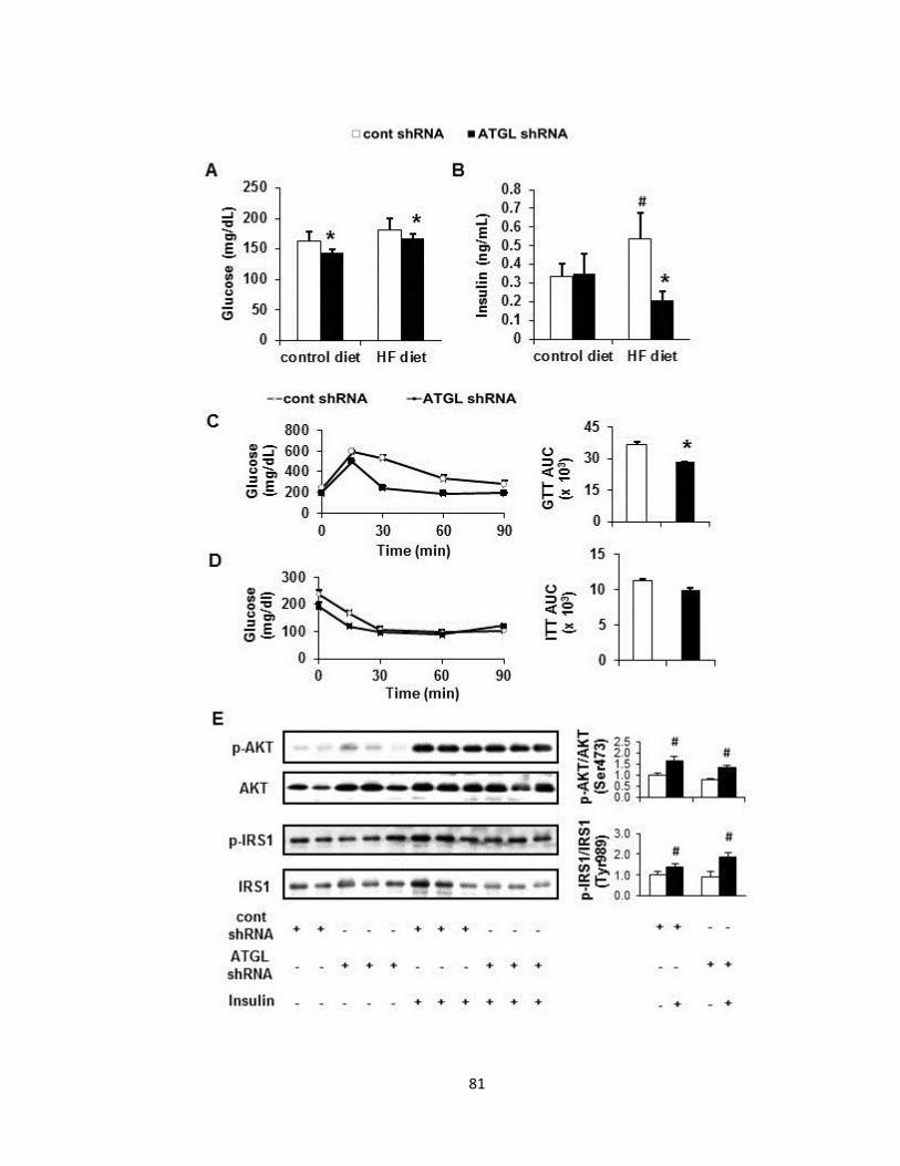
81
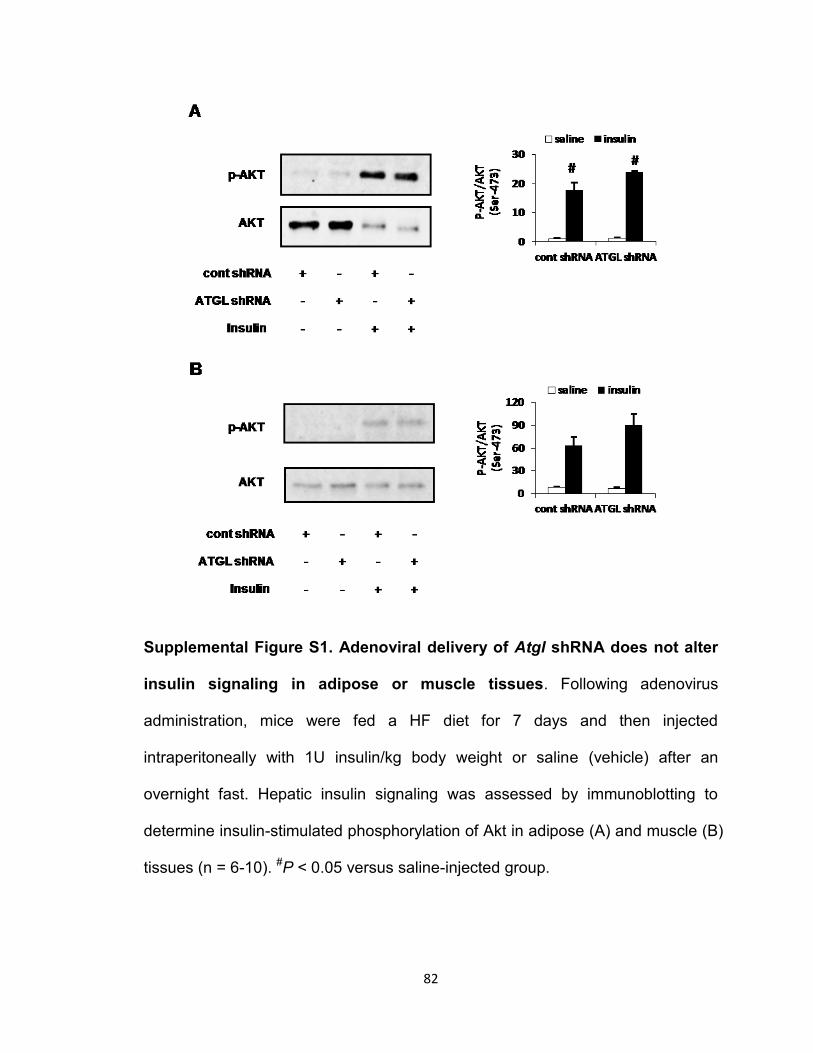
82
Supplemental Figure S1. Adenoviral delivery of Atgl shRNA does not alter
insulin signaling in adipose or muscle tissues. Following adenovirus
administration, mice were fed a HF diet for 7 days and then injected
intraperitoneally with 1U insulin/kg body weight or saline (vehicle) after an
overnight fast. Hepatic insulin signaling was assessed by immunoblotting to
determine insulin-stimulated phosphorylation of Akt in adipose (A) and muscle (B)
tissues (n = 6-10). #P < 0.05 versus saline-injected group.
83
between the two adenovirus-treated groups during the ITT (Fig. 1D). Defective
hepatic insulin signaling is frequently observed with increased lipid accumulation
in the liver (17,18). ATGL knockdown did not affect insulin-stimulated
phosphorylation of protein kinase B (AktSer473) or insulin receptor substrate 1
(IRS-1Tyr989) (Fig. 1E). This finding was unexpected given that Atgl shRNA-
treated mice fed the HF diet exhibited more than a 100% increase in TAG levels
when compared to control mice as previously shown (0.24 versus 0.10 mg/mg
protein) (7). Insulin-stimulated phosphorylation of protein kinase B (AktSer473) in
adipose or muscle tissue also remained unchanged when hepatic ATGL was
suppressed (Fig. S1). Hence, decreasing hepatic ATGL dissociates hepatic
steatosis from insulin resistance.
Hepatic ATGL deficiency exacerbates hepatic steatosis in DIO mice. Given
that ATGL knockdown confers glucose tolerance without changing insulin
sensitivity following short-term feeding of control or HF diets, we further
investigated the effects of adenoviral administration of Atgl shRNA on mice fed a
HF diet for 12 weeks. To perform this study, we initiated an ad libitum feeding
regimen of either a control or HF diet for 12 weeks. Subsequently, mice were
given control or Atgl shRNA adenovirus injections and sacrificed 1 week later.
Administration of the Atgl shRNA adenovirus resulted in a robust decrease in Atgl
mRNA and protein levels regardless of diet (Fig. 2A and 2B). While body weights
were not different in mice fed the control diet, hepatic ATGL knockdown
decreased body weight by approximately 10% in the DIO mice when compared

84
Figure 2. Adenovirus-mediated Atgl shRNA alters tissue weights and liver
TAG in DIO mice. mRNA (A) and protein levels (B) of liver ATGL in mice
administered with control or Atgl shRNA adenovirus (n= 2-5). Body (C) and liver
(D) weights of mice fed control or HF diets for 12 weeks and subsequently
transduced with adenoviruses (n = 8-10). E) Liver TAG concentrations were
determined with a colorimetric kit (n = 8-10). *P < 0.05 versus control shRNA
group and #P < 0.05 versus control diet group.

85
to control mice 7 days later (Fig. 2C). Mice treated with Atgl shRNA exhibited
approximately a 30% increase in liver weight regardless of diet type (Fig. 2D).
The changes in liver weight can be explained by increased hepatic TAG content
in the control and HF diet-fed mice (Fig. 2E). These findings show that ATGL
exacerbates hepatic steatosis under a 12-week HF diet regimen.
Hepatic ATGL mediates serum glucose and insulin in DIO mice. We
evaluated serum glucose and insulin levels to examine whether Atgl shRNA
treatment could normalize serum markers of glucose action and insulin sensitivity
in DIO mice. Hepatic ATGL knockdown resulted in decreased serum glucose
levels within the DIO group comparable to those of mice fed the control diet (Fig.
3A). Twelve weeks of HF feeding more than doubled serum insulin levels, which
were recovered to basal levels by hepatic ATGL knockdown (Fig. 3B). This
observation was in agreement with our previous result in mice fed a HF diet for 1
week (Fig. 1B). Overall, the effects of ATGL knockdown were more pronounced
in DIO mice.
Hepatic ATGL knockdown normalizes glucose intolerance without altering
hepatic insulin signaling in DIO mice. Given that ATGL knockdown decreased
serum glucose and insulin levels in DIO mice, we hypothesized that hepatic
ATGL knockdown might reverse glucose intolerance commonly observed in DIO
mice (19). Thus, we performed OGTT 7 days after DIO or control diet-fed mice

86
Figure 3. Hepatic ATGL deficiency normalizes glucose intolerance without
affecting hepatic insulin signaling in DIO mice. Serum glucose (A) and insulin
(B) levels were measured 7 day after control or Atgl shRNA administration in
mice fed control or HF diets for 12 weeks (n = 5). C) Oral glucose tolerance test
and corresponding AUC graph (n = 5). D) Seven days after adenovirus delivery
of control or Atgl shRNA, mice underwent a 4 h fast and were injected
intraperitoneally with saline or insulin at 1 unit /kg of body weight and sacrificed
10 min later. Hepatic insulin signaling was assessed by immunoblotting to
determine insulin-stimulated phosphorylation of Akt, GSK-3α and IRS1 (n = 3-5).
*P < 0.05 versus control shRNA group and #P < 0.05 versus control diet group or
saline-injected group.
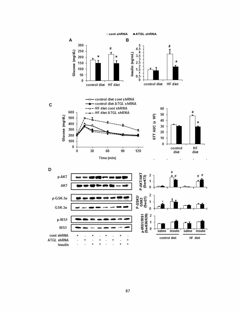
87

88
were administered adenoviruses. As expected, serum glucose levels were
significantly higher in DIO mice compared to control diet-fed mice receiving
control shRNA treatment after an oral glucose load (Fig. 3C). However, hepatic
ATGL knockdown completely normalized the glucose response in DIO mice to
basal levels. Consistent with previous results in mice fed a short-term HF diet,
hepatic steatosis induced by Atgl shRNA did not manifest in attenuated activation
of insulin cascade mediators including AKTSer473 or IRS1Ser636/639 (Fig. 3D).
Although activation of glycogen synthase kinase 3 alpha (GSK-3αSer21) was
enhanced with ATGL knockdown, this effect was abolished under insulin-
stimulated condition. Collectively, these findings show that hepatic ATGL
knockdown promotes steatosis, but improves glucose tolerance without
influencing hepatic insulin signaling.
Hepatic ATGL knockdown alters DAG content and composition depending
upon dietary treatments. DAG is widely accepted as a key lipid metabolite
involved in insulin resistance. Given that ATGL activity is a source of cellular
DAG, we measured DAG content and composition. ATGL knockdown did not
influence hepatic DAG content in DIO mice but resulted in a robust increase in
DAG content in mice fed the control diet for 12 weeks (Fig. 4A). Interestingly,
gene expression of Dgat1 remained unchanged while Dgat2 expression was only
decreased in mice fed the HF diet but not the control diet (Fig. 4B and C). In the
control diet-fed group, the percentage of palmitate, stearate and arachidonate
decreased while oleate increased in response to hepatic ATGL knockdown (Fig.

89
Figure 4. Hepatic ATGL knockdown diet-dependently alters DAG. A)
Hepatic DAG content expressed per unit of tissue weight in mice fed control or
high fat diets for 12 weeks (n = 7-9). mRNA abundance of liver Dgat1 (B) and
Dgat2 (C) was quantified in adenovirus-infected mice. D) Fatty acid composition
of DAG in mice fed the control diet or high fat diet for 12 weeks (n = 7). *P < 0.05
versus control shRNA group and #P < 0.05 versus control diet group.

90
4D). These changes in DAG composition mirror those of TAG composition in
control fed mice treated with the Atgl shRNA adenovirus (7). Feeding a HF diet
for 3 months negated any differences in DAG composition between treatment
groups (Fig. 4D).
Hepatic ATGL alters substrate utilization in DIO mice. We have previously
shown that hepatic ATGL knockdown results in attenuated hepatic FA oxidation
in hepatocyte cultures (7). In support of the aforementioned result, we also
observed that Atgl shRNA treatment resulted in lower mitochondrial oxidation in
DIO mice (Fig. 5A). Several studies have shown that inhibition of FA oxidation
leads to shift in substrate oxidation resulting in enhanced glucose catabolism
(20,21). Given that DIO mice displayed enhanced glucose tolerance when
deficient in hepatic ATGL, we hypothesized that hepatic glucose metabolism may
be altered. Hence, we determined liver glycogen levels and observed that Atgl
shRNA treatment decreased liver glycogen content in DIO mice without
impacting control diet-fed mice (Fig. 5B). In addition, glucose oxidation was
enhanced in hepatocyte suspensions isolated from mice receiving Atgl shRNA
treatment, supporting our previous observation and hypothesis (Fig. 5C).
Interestingly, however, while gene expression of hepatic glucokinase (Gk) was
upregulated in control diet-fed mice, ATGL knockdown had no effect in DIO mice
(Fig. 5D). Gene expression of pyruvate dehydrogenase kinase 2 (Pdk2) also
showed no difference between DIO mice receiving Atgl and control shRNA
treatment (Fig. 5D).
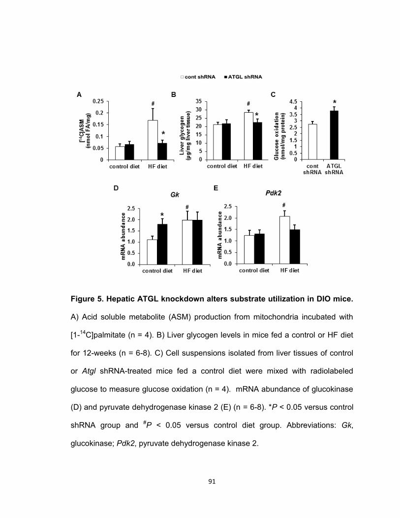
91
Figure 5. Hepatic ATGL knockdown alters substrate utilization in DIO mice.
A) Acid soluble metabolite (ASM) production from mitochondria incubated with
[1-14C]palmitate (n = 4). B) Liver glycogen levels in mice fed a control or HF diet
for 12-weeks (n = 6-8). C) Cell suspensions isolated from liver tissues of control
or Atgl shRNA-treated mice fed a control diet were mixed with radiolabeled
glucose to measure glucose oxidation (n = 4). mRNA abundance of glucokinase
(D) and pyruvate dehydrogenase kinase 2 (E) (n = 6-8). *P < 0.05 versus control
shRNA group and #P < 0.05 versus control diet group. Abbreviations: Gk,
glucokinase; Pdk2, pyruvate dehydrogenase kinase 2.

92
Figure 6. Adenoviral delivery of Atgl shRNA reduces glucose production in
DIO mice. A) Gluconeogenesis assay in hepatocytes isolated from mice fed the
HF diet for 7 days after adenoviral transduction (n = 4). mRNA abundance of
phosphoenolpyruvate carboxykinase (B) and pyruvate carboxylase (C) was
quantified in the livers of mice fed a control or HF diet for 12-weeks after
adenovirus treatment (n = 6-8). D) Following adenovirus administration, mice
were fed a HF diet for 6 days followed by an overnight fast prior to pyruvate
tolerance test. Incremental area under the curve (IAUC) is depicted (n = 4). *P <

93
0.05 versus control shRNA group. Abbreviations: Pepck, phosphoenolpyruvate
carboxykinase; Pc, pyruvate carboxylase.
Hepatic ATGL regulates gluconeogenesis. Increased hepatic glucose
production is a hallmark of insulin resistance and glucose intolerance (22,23).
Thus, we tested if hepatic ATGL influenced glucose production. ATGL
knockdown led to an approximately 25% reduction in production of glucose in
primary mouse hepatocytes isolated 7 days after adenoviral transduction (Fig.
6A). Quantitative real time PCR analysis revealed that mice receiving the Atgl
shRNA adenovirus had approximately 40% less expression of
phosphoenolpyruvate carboxykinase (Pepck) or pyruvate carboxylase (Pc)
whether fed control or HF diet for 12 weeks (Fig. 6B and C). To further
corroborate these findings, we performed pyruvate tolerance test (PTT) in mice
fed the HF diet for 1 week and found that mice infected by Atgl shRNA
adenovirus had reduced glucose production compared to control mice (Fig. 6D).
Taken together, these data suggest the improvements in glucose tolerance
following hepatic ATGL knockdown are due to increased glucose utilization and
reduced hepatic glucose output.
94
Discussion
While the role of the hepatic TAG synthetic pathway in elucidating the
pathological association between hepatic steatosis and insulin resistance has
been the focus of much research, the contribution of the TAG catabolic pathway
has been less characterized (24–26). To delineate the function of hepatic ATGL
in modulating whole-body glucose tolerance and hepatic insulin sensitivity, we
utilized adenoviral delivered shRNA to decrease hepatic ATGL expression. In
this report, we reveal that 7 days after receiving Atgl shRNA treatment, mice had
lower serum glucose levels regardless of diet despite enhanced hepatic TAG
content. In addition, the increase in serum insulin levels resulting from consuming
a HF diet was abolished by hepatic ATGL knockdown. Intriguingly, ATGL
knockdown also conferred enhanced glucose tolerance but had no impact on
insulin action. A separate study which overexpressed hepatic Atgl mRNA
approximately 4-fold observed minimal changes to glucose tolerance but found
that hepatic insulin signaling was enhanced (11). However, in our study, we
showed that hepatic insulin signaling was not altered in hepatic ATGL deficient
mice, suggestive that presence of ATGL is not necessary for normal functioning
of hepatic insulin signaling.
Because ATGL deficiency in the liver led to improved glucose tolerance in
mice fed HF diet for only 7 days, we next investigated whether Atgl shRNA could
normalize glucose intolerance and insulin sensitivity in DIO mice fed a HF diet for
12 weeks. Both serum glucose and insulin levels were recovered to basal levels
95
7 days after administration of Atgl shRNA. Furthermore, glucose intolerance
observed in DIO mice was normalized with hepatic ATGL knockdown. Hepatic
insulin signaling remained unaltered in hepatic ATGL-deficient DIO mice
compared to control DIO mice. A recent study performed in Atgl KO mice fed a
HF diet for 4 weeks also revealed that glucose tolerance and insulin sensitivity
were enhanced, suggesting that hepatic ATGL may contribute to the observed
phenotype based on the findings of the current study (9). In contrast, insulin and
glucose tolerance were unaltered in mice with a liver specific deletion of ATGL
(8). These disparities may be accounted for by the two different methods that
were used to decrease hepatic ATGL expression. It is very possible that the liver-
specific Atgl KO mice adapted to earlier metabolic changes since birth while the
effects we observed in adenovirus-mediated ATGL knockdown mice were more
representative of short term ATGL deficiency. Additionally, we observed the
most pronounced effects of ATGL knockdown under high fat feeding conditions,
which were not tested in the previous study (8).
One of the key findings in the current study is the uncoupling of glucose
intolerance and insulin resistance from hepatic steatosis. Moreover, defective
insulin signaling was not observed in ATGL-deficient mice despite TAG
accumulation in the liver. Several other models have also shown increased
steatosis without alterations in insulin signaling or impaired glucose tolerance
(25–29). Although steatosis is linked to insulin resistance, lipid intermediates
such as diacylglycerol (DAG) are thought to impair insulin signaling rather than
TAG itself (30). Surprisingly, hepatic ATGL knockdown in mice fed the control

96
diet resulted in a 4-fold increase in DAG without alterations in insulin signaling.
This observation is in agreement with a separate study which showed that ATGL
overexpression in the liver decreased hepatic DAG levels without affecting insulin
signal transduction (11). We showed that mRNA expression of Dgat2 was
markedly downregulated in the liver of mice lacking ATGL under a HF diet
consistent with a separate study performed in liver-specific Atgl KO mice (8).
Nevertheless, the effects of altering hepatic DAG content via Dgat2 expression
manipulation on insulin action appeared to be paradoxical as demonstrated in
two separate animal models (24,25). Thus, the detailed mechanisms regulating
hepatic DAG levels remain poorly understood. In the current study it should be
noted that although mice fed the control diet with suppressed hepatic ATGL
expression had elevated DAG, the percentage of saturated acyl chains in the
DAG were reduced. This is important given recent data showing that saturated
DAG impairs insulin signaling in hepatocytes (31) and may explain why no
alterations in insulin signaling were observed despite higher total DAG content. It
should be noted that the changes in FA profile of DAG only occurred in control
diet-fed mice and are consistent with the FA profile of TAG in our previous study
(7).
We have previously shown that hepatic ATGL deficiency leads to lowering
of β-oxidation without affecting VLDL secretion (7). Consistent with these data,
hepatic mitochondrial oxidation was downregulated in DIO mice administered
with Atgl shRNA in comparison with control DIO mice. Given that FA oxidation is
inhibited, we speculate that substrate utilization will be switched to glucose
97
oxidation as elaborated in the Randall cycle (32,33). In fact, Atgl KO mice had
higher respiratory quotient than control mice during fasting, indicating that the
main source of energy was glucose oxidation (9). In agreement with this
hypothesis, this study showed that glucose oxidation was higher in hepatocytes
isolated from mice treated with Atgl shRNA compared to those of control mice. In
support of the current study, inhibiting CPT1 has been shown to increase glucose
oxidation in humans and rodents (34–36). In addition, transgenic mice
overexpressing glucokinase (Gk) in the liver exhibited decreased blood glucose
levels and increased glucose tolerance suggesting that increased glucose
oxidation may contribute to enhanced glucose tolerance (37). Very similar
phenotypes have also been observed in mice overexpressing extra copies of the
Gk gene locus and transgenic mice expressing the human Gk gene in the
liver (38,39). Chronic expression of Gk in HF diet-fed mice not only improved
glucose tolerance but also slightly increased hepatic TAG accumulation (40).
These results further corroborate the notion that the switch of substrate utilization
from fat to glucose is a compensatory feedback to cope with the inability to
hydrolyze TAG due to deficiency in hepatic ATGL.
In accordance with decreased hepatic mitochondrial oxidation in DIO mice
lacking hepatic ATGL, we have previously demonstrated that mice deficient in
liver ATGL also exhibited approximately 40-70% less in gene expression of
peroxisome proliferator-activated receptor alpha (PPAR-α) and its target genes
involved in fatty acid utilization and glucose production (7). Several studies have
been suggestive of a functional association between PPAR-α and gluconeogenic

98
genes (41–43). A group has recently shown that PPAR-α can bind to putative
PPAR-response elements in glucose-6-phosphatase and PEPCK promoters in
human and mouse hepatocytes (44). Hence, these findings indicate that the
observed downregulation of hepatic gluconeogenic genes in mice lacking liver
ATGL may occur by modulating activity of PPAR-α. The detailed mechanism
through which ATGL controls PPAR- activity has not been elucidated, but
studies are currently underway to further define this regulation.
In summary, this study demonstrates that hepatic ATGL deficiency
manifests in hepatic steatosis without impairing hepatic insulin sensitivity. Instead,
glucose tolerance is improved especially in DIO mice. Decreased hepatic
glucose production and enhanced glucose oxidation are two mechanisms
contributing to the improvement in glucose tolerance. These important findings
provide new insights into the role of hepatic ATGL and TAG hydrolysis in hepatic
energy metabolism and in the etiology of steatosis and glucose intolerance.

99
References
1. Fabbrini E, Sullivan S, Klein S. Obesity and nonalcoholic fatty liver disease: biochemical, metabolic, and clinical implications. Hepatology. 2010;51:679–689.
2. Fabbrini E, Magkos F, Mohammed BS, Pietka T, Abumrad NA, Patterson BW, et al. Intrahepatic fat, not visceral fat, is linked with metabolic complications of obesity. Proc. Natl. Acad. Sci. U.S.A. 2009;106:15430–15435.
3. Stefan N, Kantartzis K, Machann J, Schick F, Thamer C, Rittig K, et al. Identification and characterization of metabolically benign obesity in humans. Arch. Intern. Med. 2008;168:1609–1616.
4. Zimmermann R, Strauss JG, Haemmerle G, Schoiswohl G, Birner-Gruenberger R, Riederer M, et al. Fat mobilization in adipose tissue is promoted by adipose triglyceride lipase. Science. 2004;306:1383–1386.
5. Villena JA, Roy S, Sarkadi-Nagy E, Kim K-H, Sul HS. Desnutrin, an adipocyte gene encoding a novel patatin domain-containing protein, is induced by fasting and glucocorticoids: ectopic expression of desnutrin increases triglyceride hydrolysis. J. Biol. Chem. 2004;279:47066–47075.
6. Jenkins CM, Mancuso DJ, Yan W, Sims HF, Gibson B, Gross RW. Identification, cloning, expression, and purification of three novel human calcium-independent phospholipase A2 family members possessing triacylglycerol lipase and acylglycerol transacylase activities. J. Biol. Chem. 2004;279:48968–48975.
7. Ong KT, Mashek MT, Bu SY, Greenberg AS, Mashek DG. Adipose triglyceride lipase is a major hepatic lipase that regulates triacylglycerol turnover and fatty acid signaling and partitioning. Hepatology. 2011;53:116–126.
8. Wu JW, Wang SP, Alvarez F, Casavant S, Gauthier N, Abed L, et al. Deficiency of liver adipose triglyceride lipase in mice causes progressive hepatic steatosis. Hepatology. 2011;54:122–132.
9. Haemmerle G, Lass A, Zimmermann R, Gorkiewicz G, Meyer C, Rozman J, et al. Defective lipolysis and altered energy metabolism in mice lacking adipose triglyceride lipase. Science. 2006;312:734–737.

100
10. Kienesberger PC, Lee D, Pulinilkunnil T, Brenner DS, Cai L, Magnes C, et al. Adipose triglyceride lipase deficiency causes tissue-specific changes in insulin signaling. J. Biol. Chem. 2009;284:30218–30229.
11. Turpin SM, Hoy AJ, Brown RD, Rudaz CG, Honeyman J, Matzaris M, et al. Adipose triacylglycerol lipase is a major regulator of hepatic lipid metabolism but not insulin sensitivity in mice. Diabetologia. 2011;54:146–156.
12. Miyoshi H, Perfield JW 2nd, Souza SC, Shen W-J, Zhang H-H, Stancheva ZS, et al. Control of adipose triglyceride lipase action by serine 517 of perilipin A globally regulates protein kinase A-stimulated lipolysis in adipocytes. J. Biol. Chem. 2007;282:996–1002.
13. Frezza C, Cipolat S, Scorrano L. Organelle isolation: functional mitochondria from mouse liver, muscle and cultured fibroblasts. Nat Protoc. 2007;2:287–295.
14. Folch J, Lees M, Stanley GHS. A simple method for the isolation and purification of total lipides from animal tissues. J. Biol. Chem. 1957;226:497–509.
15. Lo S, Russell JC, Taylor AW. Determination of glycogen in small tissue samples. J Appl Physiol. 1970;28:234–236.
16. Lee J-M, Seo W-Y, Song K-H, Chanda D, Kim YD, Kim D-K, et al. AMPK-dependent repression of hepatic gluconeogenesis via disruption of CREB.CRTC2 complex by orphan nuclear receptor small heterodimer partner. J. Biol. Chem. 2010;285:32182–32191.
17. Michael MD, Kulkarni RN, Postic C, Previs SF, Shulman GI, Magnuson MA, et al. Loss of insulin signaling in hepatocytes leads to severe insulin resistance and progressive hepatic dysfunction. Mol. Cell. 2000;6:87–97.
18. Samuel VT, Liu Z-X, Qu X, Elder BD, Bilz S, Befroy D, et al. Mechanism of hepatic insulin resistance in non-alcoholic fatty liver disease. J. Biol. Chem. 2004;279:32345–32353.
19. Ahrén B, Scheurink AJ. Marked hyperleptinemia after high-fat diet associated with severe glucose intolerance in mice. Eur. J. Endocrinol. 1998;139:461–467.
20. Lopaschuk GD, McNeil GF, McVeigh JJ. Glucose oxidation is stimulated in reperfused ischemic hearts with the carnitine palmitoyltransferase 1 inhibitor, Etomoxir. Mol. Cell. Biochem. 1989;88:175–179.

101
21. Martin C, Odeon M, Cohen R, Beylot M. Mechanisms of the glucose lowering effect of a carnitine palmitoyl transferase inhibitor in normal and diabetic rats. Metab. Clin. Exp. 1991;40:420–427.
22. Mitrakou A, Kelley D, Mokan M, Veneman T, Pangburn T, Reilly J, et al. Role of reduced suppression of glucose production and diminished early insulin release in impaired glucose tolerance. N. Engl. J. Med. 1992;326:22–29.
23. He L, Sabet A, Djedjos S, Miller R, Sun X, Hussain MA, et al. Metformin and insulin suppress hepatic gluconeogenesis through phosphorylation of CREB binding protein. Cell. 2009;137:635–646.
24. Choi CS, Savage DB, Kulkarni A, Yu XX, Liu Z-X, Morino K, et al. Suppression of diacylglycerol acyltransferase-2 (DGAT2), but not DGAT1, with antisense oligonucleotides reverses diet-induced hepatic steatosis and insulin resistance. J. Biol. Chem. 2007;282:22678–22688.
25. Monetti M, Levin MC, Watt MJ, Sajan MP, Marmor S, Hubbard BK, et al. Dissociation of hepatic steatosis and insulin resistance in mice overexpressing DGAT in the liver. Cell Metab. 2007;6:69–78.
26. Kantartzis K, Machicao F, Machann J, Schick F, Fritsche A, Häring H-U, et al. The DGAT2 gene is a candidate for the dissociation between fatty liver and insulin resistance in humans. Clin. Sci. 2009;116:531–537.
27. Chakravarthy MV, Pan Z, Zhu Y, Tordjman K, Schneider JG, Coleman T, et al. “New” hepatic fat activates PPARalpha to maintain glucose, lipid, and cholesterol homeostasis. Cell Metab. 2005;1:309–322.
28. Brown JM, Betters JL, Lord C, Ma Y, Han X, Yang K, et al. CGI-58 knockdown in mice causes hepatic steatosis but prevents diet-induced obesity and glucose intolerance. J. Lipid Res. 2010;51:3306–3315.
29. Minehira K, Young SG, Villanueva CJ, Yetukuri L, Oresic M, Hellerstein MK, et al. Blocking VLDL secretion causes hepatic steatosis but does not affect peripheral lipid stores or insulin sensitivity in mice. J. Lipid Res. 2008;49:2038–2044.
30. Erion DM, Shulman GI. Diacylglycerol-mediated insulin resistance. Nat. Med. 2010;16:400–402.
31. Zhang C, Wendel AA, Keogh MR, Harris TE, Chen J, Coleman RA. Glycerolipid signals alter mTOR complex 2 (mTORC2) to diminish insulin signaling. Proc. Natl. Acad. Sci. U.S.A. 2012;109:1667–1672.

102
32. Randle PJ, Garland PB, Hales CN, Newsholme EA. The glucose fatty-acid cycle. Its role in insulin sensitivity and the metabolic disturbances of diabetes mellitus. Lancet. 1963;1:785–789.
33. Hue L, Taegtmeyer H. The Randle cycle revisited: a new head for an old hat. Am. J. Physiol. Endocrinol. Metab. 2009;297:E578–591.
34. Ratheiser K, Schneeweiss B, Waldhäusl W, Fasching P, Korn A, Nowotny P, et al. Inhibition by etomoxir of carnitine palmitoyltransferase I reduces hepatic glucose production and plasma lipids in non-insulin-dependent diabetes mellitus. Metab. Clin. Exp. 1991;40:1185–1190.
35. Conti R, Mannucci E, Pessotto P, Tassoni E, Carminati P, Giannessi F, et al. Selective reversible inhibition of liver carnitine palmitoyl-transferase 1 by teglicar reduces gluconeogenesis and improves glucose homeostasis. Diabetes. 2011;60:644–651.
36. Hübinger A, Knode O, Susanto F, Reinauer H, Gries FA. Effects of the carnitine-acyltransferase inhibitor etomoxir on insulin sensitivity, energy expenditure and substrate oxidation in NIDDM. Horm. Metab. Res. 1997;29:436–439.
37. Ferre T, Riu E, Bosch F, Valera A. Evidence from transgenic mice that glucokinase is rate limiting for glucose utilization in the liver. FASEB J. 1996;10:1213–1218.
38. Niswender KD, Shiota M, Postic C, Cherrington AD, Magnuson MA. Effects of increased glucokinase gene copy number on glucose homeostasis and hepatic glucose metabolism. J. Biol. Chem. 1997;272:22570–22575.
39. Hariharan N, Farrelly D, Hagan D, Hillyer D, Arbeeny C, Sabrah T, et al. Expression of human hepatic glucokinase in transgenic mice liver results in decreased glucose levels and reduced body weight. Diabetes. 1997;46:11–16.
40. Winzell MS, Coghlan M, Leighton B, Frangioudakis G, Smith DM, Storlien LH, et al. Chronic glucokinase activation reduces glycaemia and improves glucose tolerance in high-fat diet fed mice. Eur. J. Pharmacol. 2011;663:80–86.
41. Im S-S, Kim M-Y, Kwon S-K, Kim T-H, Bae J-S, Kim H, et al. Peroxisome proliferator-activated receptor {alpha} is responsible for the up-regulation of hepatic glucose-6-phosphatase gene expression in fasting and db/db Mice. J. Biol. Chem. 2011;286:1157–1164.
42. Bernal-Mizrachi C, Weng S, Feng C, Finck BN, Knutsen RH, Leone TC, et al. Dexamethasone induction of hypertension and diabetes is PPAR-alpha dependent in LDL receptor-null mice. Nat. Med. 2003;9:1069–1075.

103
43. Koo S-H, Satoh H, Herzig S, Lee C-H, Hedrick S, Kulkarni R, et al. PGC-1 promotes insulin resistance in liver through PPAR-alpha-dependent induction of TRB-3. Nat. Med. 2004;10:530–534.
44. Fan Y, Guo Y, Hamblin M, Chang L, Zhang J, Chen YE. Inhibition of gluconeogenic genes by calcium-regulated heat-stable protein 1 via repression of peroxisome proliferator-activated receptor α. J. Biol. Chem. 2011;286:40584–40594.

104
CHAPTER 4
Hepatic ATGL Mediates Fatty Acid Oxidation
and PPAR- Signaling Through an LFABP-
Independent Mechanism
Kuok Teong Ong, Mara T. Mashek, Nicholas O. Davidson and Douglas G. Mashek.
105
Adipose triglyceride lipase (ATGL) catalyzes the rate-limiting step in
triacylglycerol (TAG) hydrolysis in most tissues. We have shown that hepatic
ATGL preferentially channels hydrolyzed fatty acids (FAs) to β-oxidation and
induces peroxisome proliferator-activated receptor-α (PPAR-α) target gene
expression. Nevertheless, the mechanisms that mediate this pathway are
unknown. We hypothesized that the FA carrier liver fatty acid binding protein
(LFABP) may be involved in transporting FAs from lipid droplets to the nucleus
for ligand delivery or mitochondria for β-oxidation. Thus, we suppressed or
overexpressed hepatic ATGL expression in wild type (WT) or LFABP knockout
(LFABP KO) mice and primary hepatocytes to determine the contribution of
LFABP in mediating the channeling of hydrolyzed FA. ATGL knockdown
increased liver weight and TAG content and ATGL overexpression decreased
liver weight to a similar extent in both strains. Surprisingly, manipulating ATGL
expression in LFABP KO mice resulted in similar changes in PPAR-α and its
target genes compared to WT mice suggesting that LFABP is not required to
carry FA from ATGL to PPAR-α. Furthermore, LFABP did not impair the effects
of ATGL on channeling of hydrolyzed FAs to oxidative pathways in primary
hepatocyte cultures. Taken together, we conclude that LFABP is not required to
channel ATGL-hydrolyzed FAs to mitochondria for β-oxidation or mediate its
effects on PPAR-α.
106
Introduction
Adipose triglyceride lipase (ATGL) is a major TAG hydrolase in the liver
(130,131). Intracellular free FAs can be exported from the liver via VLDL
secretion or metabolized by β-oxidation. ATGL knockout mice exhibit reduced
levels of ketone bodies, suggesting that ATGL may promote β-oxidation. In
support of this notion, we have previously shown that hepatic suppression of
ATGL leads to the inhibition of FA β-oxidation without affecting VLDL secretion
(130). Consistent with these results, several other studies have also observed
that ATGL overexpression or deletion affects FA oxidation but not VLDL
secretion in vitro and in vivo (131,132). In addition, FA oxidative genes including
peroxisome proliferator-activated receptor alpha (PPAR-α) and its downstream
target are downregulated in the mice receiving ATGL shRNA compared to control
mice (130). Others have also reported that ATGL regulates PPAR-α in heart,
small intestine and brown adipose tissues (79,133,134). Nevertheless, the
mechanisms through which ATGL channels hydrolyzed FAs to the mitochondria
for oxidation or the nucleus for transcriptional activity are currently unknown.
Liver fatty acid binding protein (LFABP) is a FA carrier involved in the long
chain fatty acid (LCFA) transport throughout the cytoplasm and channeling of
LCFA and LCFA-CoA to the mitochondria for oxidation (135). LFABP has been
shown to interact with CPT1, the rate-limiting step in mitochondrial LCFA
oxidation (135–137). In addition, small amount of LFABP has been found to
associate with mitochondria (138). These observations imply that LFABP may
mediate the transport of FA to the mitochondria. LFABP gene ablation in cultured

107
primary hepatocytes and isolated hepatocytes from LFABP null mice has been
shown to suppress mitochondrial fatty acid oxidation (139). Additionally, two
separate studies have determined that LFABP binds PPAR-α with a high affinity
and manipulating LFABP expression alters PPAR-α transcription of genes coding
for LCFA oxidative enzymes, suggesting that LFABP also regulates FA oxidation
via transcriptional mechanisms (140,141).
Given the functional and physiological significance of LFABP in FA
oxidation, we hypothesize that LFABP is necessary for the transport of ATGL-
hydrolyzed FAs to the mitochondria for oxidation or to the nucleus to regulate
gene expression. To investigate the role of LFABP in ATGL-mediated β-oxidation,
we overexpressed (Ad-ATGL) or knocked down ATGL (ATGL shRNA) in wild
type (WT) and whole body LFABP knockout (LFABP KO) mice. Despite the
importance of LFABP as a FA carrier, our findings strongly suggest that LFABP
is not required for the channeling of FAs produced by ATGL activity to the
mitochondria for oxidation or to the nucleus for transcriptional regulation of
PPAR-α.
108
Materials and Methods
Animals, diets and adenoviral administration. All animal protocols were
approved by the University of Minnesota Institutional Animal Care and Use
Committee. Male LFABP KO mice on a C57BL/6J background were generated
as previously described (142). All mice were housed under controlled
temperature and lighting (20-22°C; 12:12-h light-dark cycle). Eight to ten week
old wild-type or LFABP KO mice were injected via the tail vein with Ad-GFP or
Ad-ATGL for overexpression studies or adenoviruses encoding ATGL short
hairpin RNA (ATGL shRNA) or control shRNA targeting a nonspecific messenger
RNA (mRNA). Adenoviruses were generated as described previously (23, 24).
All mice had free access to water and were fed a purified diet (TD.94045) from
Harlan Teklad Premier Laboratory Diets (Madison, WI) for a week following
adenoviral administration. The control diet contained protein (19% of total
calories), carbohydrates (64%), and fat (17%), and the fat source was soybean
oil (70 g/kg). Exactly one week after adenovirus injection, mice were sacrificed
for liver tissues and serum collection after an overnight or 4 h fast.
Primary hepatocyte isolation. Mouse primary hepatocytes were isolated from
8-10 weeks old WT or LFABP KO male mice with free access to water and
control diet by the collagenase perfusion method described previously (143).
Hepatocytes were plated on collagen-coated multiwell tissue culture plates (Nunc)

109
for 4 h with M199 plating media (Invitrogen) that contained 23 mM HEPES, 26
mM sodium bicarbonate, 10% FBS, 1% penicillin/streptomycin, 100 nM
dexamethasone, 100 nM insulin, and 11 mM glucose. M199 maintenance media
contained 23 mM HEPES, 26 mM sodium bicarbonate, 1% penicillin/streptomycin,
5.5 mM glucose, 100 µM carnitine, 10 nM dexamethasone, and 10 nM insulin.
Cells were maintained in a humidified incubator at 37°C, 5% CO2.
Cell adenoviral infection and radiolabeling. Adenovirus expressing ATGL or
green fluorescent protein (GFP) that serves as a control virus was generated as
described previously (3). After 4 hours of plating, primary hepatocytes were
exposed to either adenovirus expressing GFP or ATGL at 100 multiplicities of
infection for 24 hr. Cells were pulsed with 500 µM [1-14C]oleate bound to fatty
acid-free BSA in a 3:1 molar ratio for 1.5 hr. Some cells were harvested for
measure radiolabel incorporation into cellular lipid fractions. Subsequently, cells
were washed with phosphate-buffered saline solution and the wells were
replaced with new media lacking labeled fatty acid for an additional 6 hr of chase
period followed by collection of cells and lipid extraction. Lipid samples were
separated into different fractions by thin layer chromatography (TLC) on 0.25-mm
Silica Gel G plates in a hexane:ethyl ether:acetic acid (80:20:2, v/v)solvent
system). TAG fractions were identified and measured by AR-2000 radio-TLC
Imaging Scanner. Total radiolabeled lipids were quantified by scintillation counter
(LS6000IC, Beckman) after addition of Bio-Safe II cocktail.

110
Measurement of FA oxidation. Following chase periods as described above,
radiolabeled medium were transferred to Eppendorf tubes containing 20% bovine
serum albumin (BSA). Subsequently, perchloric acid was added to the tubes and
vortexed. Following an overnight incubation at room temperature, the tubes were
centrifuged and the supernatant fraction was transferred to another set of tubes
containing 20% BSA. The supernatant that contained [14C]-labeled acid-soluble
metabolites (ASM) were quantified with scintillation counter.
RNA isolation, RT-PCR and real –time quantitative PCR analysis. RNA was
extracted with Trizol from liver tissues followed by reverse-transcription with
SuperSript III First-Strand Synthesis SuperMix (Invitrogen). Gene expression was
quantified using a SYBR GreenER Two-Step qRT-PCR Kit (Invitrogen) and an
Applied Biosystems StepOne Plus Real-Time PCR system. Data were analyzed
using the delta-delta CT method. For all analysis, gene expression was
normalized to ribosomal protein L32. Melting curve analysis was performed on all
samples to verify primer specificity.
Liver TAG and FA analysis. TAG was extracted from liver tissues or
hepatocytes according to a method from Folch (4). Snap-frozen liver tissues
were homogenized in sterile water and extracted twice with chloroform:methanol.
Chloroform layer was isolated and dried under nitrogen gas, and redissolved in
Triton X-100. Liver TAG was quantified with a TAG colorimetric enzymatic kit

111
(Stanbio). Liver free FAs were measured by a free fatty acid quantification kit
(Sigma) as outlined by the manufacturer’s guideline.
Serum measurements. Serum FA and β-hydroxybutyrate levels were
determined with colorimetric enzymatic assay kits from Wako Chemicals while
serum TAG was measured with a colorimetric assay kit from Stanbio.
112
Results
ATGL suppression affects liver phenotype of LFABP knockout mice. Given
that the deletion of LFABP has been shown to decrease hepatic TAG
accumulation (142) and ATGL is a major hepatic TAG hydrolase (130), we set
out to investigate the effects of ATGL knockdown on the liver phenotype of
LFABP KO mice. ATGL knockdown had no effect on body weight in WT or
LFABP KO mice when compared to control shRNA treatment following 7 d of
treatment(Fig. 1A). ATGL knockdown led to increased liver weight in WT mice
and this effect was replicated in LFABP KO mice (Fig. 1B). These observations
could be explained by enhanced hepatic TAG accumulation in both WT and
LFABP KO mice treated with ATGL shRNA adenovirus (Fig. 1D). Although
ATGL knockdown increased liver weights and TAG content in both WT and
LFABP KO mice, the absence of LFABP attenuated this increase. Interestingly,
LFABP KO mice exhibited an increase in epididymal fat weight compared to WT
mice and this change was abrogated by ATGL shRNA administration (Fig. 1C).
Surprisingly, however, hepatic ATGL knockdown did not alter the content of liver
free FAs (Fig. 1E).
ATGL overexpression reduces liver weight of LFABP knockout mice. To
further examine the importance of LFABP in mediating the effects of ATGL on
hepatic lipid metabolism, we also overexpressed ATGL in both WT and LFABP
KO mice. Consistent with the above data, overexpressing ATGL did not alter
body weight of WT or LFABP KO mice (Fig. 2A). Increasing the expression of

113
Figure 1. ATGL knockdown increases liver weight of LFABP knockout
mice. WT or LFABP KO mice at 8 – 10 weeks of age were infected with control
or ATGL shRNA adenovirus (n = 6 – 10 per group). Mice were fed with purified
control diet and sacrificed 7 d post-infection. Body (A), liver (B) and adipose (C)
weight of WT or LFABP KO mice were measured following an overnight fasting.
Liver TAG (D) and FFA (E) were quantified using a colorimetric enzymatic assay
kit in mice infected with control or ATGL shRNA. Data are presented as means ±
SE. Comparisons between groups were made using Student’s t test. *, p < 0.05
versus control shRNA group and #, p < 0.05 versus WT mice.
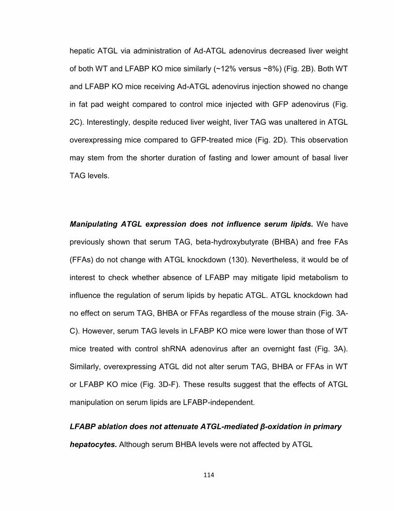
114
hepatic ATGL via administration of Ad-ATGL adenovirus decreased liver weight
of both WT and LFABP KO mice similarly (~12% versus ~8%) (Fig. 2B). Both WT
and LFABP KO mice receiving Ad-ATGL adenovirus injection showed no change
in fat pad weight compared to control mice injected with GFP adenovirus (Fig.
2C). Interestingly, despite reduced liver weight, liver TAG was unaltered in ATGL
overexpressing mice compared to GFP-treated mice (Fig. 2D). This observation
may stem from the shorter duration of fasting and lower amount of basal liver
TAG levels.
Manipulating ATGL expression does not influence serum lipids. We have
previously shown that serum TAG, beta-hydroxybutyrate (BHBA) and free FAs
(FFAs) do not change with ATGL knockdown (130). Nevertheless, it would be of
interest to check whether absence of LFABP may mitigate lipid metabolism to
influence the regulation of serum lipids by hepatic ATGL. ATGL knockdown had
no effect on serum TAG, BHBA or FFAs regardless of the mouse strain (Fig. 3A-
C). However, serum TAG levels in LFABP KO mice were lower than those of WT
mice treated with control shRNA adenovirus after an overnight fast (Fig. 3A).
Similarly, overexpressing ATGL did not alter serum TAG, BHBA or FFAs in WT
or LFABP KO mice (Fig. 3D-F). These results suggest that the effects of ATGL
manipulation on serum lipids are LFABP-independent.
LFABP ablation does not attenuate ATGL-mediated β-oxidation in primary
hepatocytes. Although serum BHBA levels were not affected by ATGL

115
Figure 2. ATGL overexpression reduces liver weight of LFABP knockout
mice. WT or LFABP KO mice at 8 – 10 weeks of age were injected with Ad-GFP
or Ad-ATGL adenovirus (n = 6 – 8 per group). Mice were fed with purified control
diet and sacrificed 7 d post-infection. Body (A), liver (B) and adipose (C) weight
of WT or LFABP KO mice were measured following a 4 h fasting. Liver FFA (D)
was quantified using a colorimetric enzymatic assay kit in mice infected with Ad-
GFP or Ad-ATGL shRNA Data are presented as means ± SE. Comparisons
between groups were made using Student’s t test. *, p < 0.05 versus control
shRNA group and #, p < 0.05 versus WT mice.
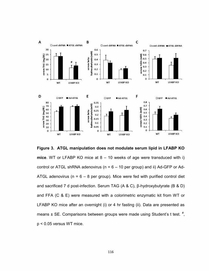
116
Figure 3. ATGL manipulation does not modulate serum lipid in LFABP KO
mice. WT or LFABP KO mice at 8 – 10 weeks of age were transduced with i)
control or ATGL shRNA adenovirus (n = 6 – 10 per group) and ii) Ad-GFP or Ad-
ATGL adenovirus (n = 6 – 8 per group). Mice were fed with purified control diet
and sacrificed 7 d post-infection. Serum TAG (A & C), β-hydroxybutyrate (B & D)
and FFA (C & E) were measured with a colorimetric enzymatic kit from WT or
LFABP KO mice after an overnight (i) or 4 hr fasting (ii). Data are presented as
means ± SE. Comparisons between groups were made using Student’s t test. #,
p < 0.05 versus WT mice.

117
knockdown or overexpression in vivo, our previous study has shown that hepatic
ATGL regulates β-oxidation in primary hepatocyte cultures (130). Thus, we
extended our in vivo studies to primary hepatocyte cultures to gain insights into
the role of LFABP in ATGL-mediated β-oxidation. We performed pulse-chase
experiments with [1-14C]oleate in primary mouse hepatocytes isolated from WT
or LFABP KO mice. During the pulse period, overexpressing ATGL decreased
incorporation of [14C]oleate into the TAG fraction of primary hepatocytes derived
from both WT and LFABP KO mice to a similar extent (Fig. 4A & D). Intriguingly,
LFABP deletion abolished the ATGL overexpression-induced increase in pulse-
labeled ASM, indicating that LFABP regulates oxidation of exogenous FAs (Fig.
4B). To determine oxidation of FAs derived primarily from intracellular TAG
hydrolysis (primarily ATGL-hydrolyzed FAs), we quantified ASM as a measure of
FA oxidation during the chase period. ATGL overexpression increased ASM by
almost two fold in the WT mice-derived hepatocytes and this increase was
duplicated in primary hepatocytes isolated from LFABP KO mice, suggesting that
LFABP is not needed for ATGL-mediated increase in β–oxidation (Fig. 4C). To
ensure that this observation was not due to a compensatory effect by other FABP
isoforms, we also utilized a pan-specific FABP inhibitor, HTS01037 (100 µM),
during the chase period in primary hepatocytes isolated from WT mice.
Administration of Ad-ATGL doubled ASM while addition of HTS01037 during the
chase period failed to abolish the Ad- ATGL induced increase in ASM (Fig. 4E).
Taken together, these data indicate that LFABP is not a major carrier of ATGL-
hydrolyzed FAs and may be more important for channeling exogenous FAs for

118
Figure 4. LFABP deletion does not block ATGL-mediated β-oxidation.
Primary mouse hepatocyes were isolated from 8 – 10 weeks old WT or LFABP
KO mice using collagenase perfusion method as outlined in the experimental
procedures section. Cells were transduced with Ad-GFP or Ad-ATGL for 24 hr, at
which time pulse (1.5 hr) and chase (6 hr) experiments were performed with 500
µM [1-14C] oleate. A: TAG fractions were isolated from harvested cells by thin
layer chromatography to measure incorporation of radiolabeled oleate into TAG.
Media from hepatocytes were harvested at the end of pulse (B) and chase (C)
periods followed by quantification of acid soluble metabolites (ASM) as outlined
in the experimental procedures to measure fatty acid oxidation. During chase,
a pan-specific FABP inhibitor, HTS01037 was added followed by measurement
of TAG incorporation (D) and ASM E) as outlined above. Data are presented as
means ± SE. Comparisons between groups were made using Student’s t test. *,
p < 0.05 versus control shRNA group and #, p < 0.05 versus WT mice.
119
the purpose of mitochondrial oxidation.
LFABP is not necessary to transcriptionally regulate oxidative genes
downstream of ATGL. To further examine whether LFABP serves as a carrier of
ATGL-hydrolyzed FAs to the nucleus to influence oxidative genes, we also
quantified PPAR-α target gene expression in WT or LFABP KO mice
administered with Ad-ATGL adenovirus. In WT mice, hepatic ATGL
overexpression led to approximately 0.4 to 2-fold increase in PPAR-α and its
target genes (Fig. 5A-F). Despite the absence of LFABP, overexpressing ATGL
in the liver still induced gene expression of PPAR-α, ACOT1, LCAD and ACSL1
(0.3 to 2.5-fold) in LFABP KO mice (Fig. 5A, C, D & E). These results show that
LFABP does not mediate the effects of ATGL on PPAR- activity and suggest
that an alternate mechanism links changes in ATGL activity to transcriptional
regulation.
LFABP deletion does not enhance the effects of ATGL knockdown on
PPAR-α and its target genes. We have previously shown that hepatic ATGL
promotes fatty acid oxidation via changes in the expression of PPAR-α and its
target genes (130). Interestingly, LFABP has been shown to bind directly to
PPAR-α and may play a role in PPAR-α activation (140). Thus, we investigated
the effect of ATGL knockdown on PPAR-α-regulated genes in LFABP KO mice.
Interestingly, only some of the genes such as PPAR-α, ACOT1 and ACSL1 were
120
Figure 5. LFABP is not required for transcriptional regulation of oxidative
genes downstream of ATGL. WT or LFABP KO mice at 8 – 10 weeks of age
were transduced with Ad-GFP or Ad-ATGL adenovirus (n = 6 – 8 per group).
Liver tissues were collected 7 d post-transduction after a 4 h fasting. Liver tissues
were used to synthesize cDNA via reverse transcription PCR from isolated
mRNA. mRNA expression of oxidative genes including PPAR-α (A), CPT1 (B),
ACOT1 (C), LCAD (D), ACSL1 (E) and PEPCK (F) were quantified with real-time
quantitative PCR using the ∆∆CT method. Data are presented as means ± S.D.
Comparisons between groups were made using Student’s t test. *, p < 0.05
versus control shRNA group and #, p < 0.05 versus WT mice. PPAR-α,
peroxisome proliferator-activated receptor alpha; CPT1, carnitine
palmitoyltransferase I; ACOT1, acyl-CoA thioesterase I; LCAD, long-chain acyl-
CoA dehydrogenase; ACSL1, acyl-CoA synthetase 1; PEPCK,
phosphoenolpyruvate carboxykinase.
121
downregulated in LFABP KO compared to WT mice injected with control shRNA
adenovirus (Fig. 6A, C & E). These discrepancies have been observed in several
studies due to differences in experimental model (in vitro versus in vivo), fasting
duration, diet type, age and sex (144–146). The fold change between the two
genotypes within the same virus treatment group is very similar across all the
genes, which further supports the data in the ATGL overexpression model
showing that ATGL mediates PPAR- through a LFABP-independent mechanism
(Fig. 6A-F).

122
Figure 6. LFABP deletion does not enhance suppression of PPAR-α target
genes induced by ATGL knockdown. WT or LFABP KO mice at 8 – 10 weeks
of age were infected with control or ATGL shRNA adenovirus (n = 6 – 10 per
group) followed by harvesting of liver tissues 7 d later after an overnight fasting.
Liver tissues were used to produce cDNA via reverse transcription PCR from
isolated mRNA. mRNA expression of oxidative genes including PPAR-α (A),
CPT1 (B), ACOT1 (C), LCAD (D), ACSL1 (E) and PEPCK (F) were quantified
with real-time quantitative PCR using the ∆∆CT method. Data are presented as
means ± S.D. Comparisons between groups were made using Student’s t test. *,
p < 0.05 versus control shRNA group and #, p < 0.05 versus WT mice. PPAR-α,
peroxisome proliferator-activated receptor alpha; CPT1, carnitine
palmitoyltransferase I; ACOT1, acyl-CoA thioesterase I; LCAD, long-chain acyl-
CoA dehydrogenase; ACSL1, acyl-CoA synthetase 1; PEPCK,
phosphoenolpyruvate carboxykinase.

123
Discussion
The first evidence suggesting that ATGL regulates FA oxidation was shown by
ATGL knockout mice exhibiting lower plasma ketone bodies compared to control
mice (71). Interestingly, hepatic overexpression of ATGL in McA-RH7777 cells
also promotes FA oxidation (132). Our group was the first to reveal that ATGL
preferentially exports TAG-hydrolyzed FAs to β-oxidation instead of VLDL
production and secretion in the liver (130). Moreover, the effect of ATGL on β-
oxidation appears to be mediated by transcriptional changes in PPAR-α and its
target genes (130). Wu et al. also showed that hepatic palmitate oxidation and
PPAR-α gene expression are downregulated in liver-specific ATGL knockout
mice (131). However, the ATGL-mediated channeling of FA to the β-oxidation
pathway is currently not well understood. Given that ATGL activity yields FA, it is
reasonable to speculate that the FA may be transported to the mitochondria for
oxidation or to the nucleus for modulating transcriptional activity of PPAR-α target
genes.
Hence, we hypothesized that a hepatic FA carrier such as LFABP may
mediate this function. While LFABP has been shown to play an essential function
in regulating both FA oxidation and transcriptional activity of PPAR-α, its role in
channeling specifically ATGL-hydrolyzed FAs to the mitochondria or nucleus is
unclear. To examine this role, we manipulated ATGL expression in WT or LFABP
KO mice. From this study, we found that either knocking down or overexpressing
ATGL in the liver of LFABP KO mice is still sufficient to influence liver weight or
124
TAG, suggesting that the TAG hydrolase activity of ATGL is not dependent on
LFABP. Consistent with our previous study in WT mice, ATGL knockdown or
overexpression does not influence serum lipid markers in LFABP KO mice.
Despite not observing a change in ketone bodies (serum beta-hydroxybutyrate)
when hepatic ATGL is suppressed in vivo, ASM (FA oxidation) is reduced in
primary hepatocyte cultures transduced with ATGL shRNA adenovirus (130).
Therefore, we also explored whether LFABP is needed to regulate FA
oxidation mediated by ATGL. The fold induction of FA oxidation as measured by
ASM in primary hepatocytes isolated from LFABP KO mice is similar to those
derived from WT mice. Furthermore, using a pan-inhibitor to suppress activity of
all FABP isoforms has no effect on attenuating the induction of ASM by
overexpressing ATGL. This observation is consistent with a previous finding that
shows other isoforms of FABP are not upregulated in LFABP KO mice (142).
While LFABP has been shown to be critical for acute FA uptake (147), hepatic
free FA levels do not differ between WT and LFABP KO mice under fasting
condition (142,147). In addition, acute hepatic deposition of radiolabeled oleate
is substantially lower in LFABP KO mice compared to WT mice under fed state in
relation to fasted state (147). In agreement with these in vivo data, we show that
LFABP deletion abrogates any increase in ASM induced by ATGL
overexpression. These findings strongly suggest that LFAPB may be more
important for channeling of exogenous FAs but not hydrolyzed FAs. Taken
together, these findings strongly suggest that ATGL-mediated β-oxidation does
not require LFABP or any other FABP family members as a FA transporter.
125
The next logical step is to speculate that other FA transporters which can
potentially mediate the shuttling of FAs to the mitochondria would be upregulated
as a compensatory mechanism in LFABP KO mice. Interestingly, Newberry et al.
have shown that other putative FA transporters including sterol carrier protein
(SCP-2 & SCP-x) and members of the fatty acid transporter family are not
enhanced in response to the ablation of LFABP in the liver during fasting despite
reduction in hepatic FA oxidation (142). However, another study has indicated
that LFABP deletion results in upregulation of SCP-2 and acyl-CoA binding
protein (ACBP), by 45 and 80%, respectively (148). ACBP has previously been
observed to partition fatty acids between esterification reactions and beta-
oxidation (149). Despite this compensation effect, LFABP KO mice still exhibit a
reduction in ketone bodies or FA oxidation when fasted for 12 hr. Hence, the
functions of other putative FA transporters especially SCP-2 and ACBP are
unlikely to involve the shuttling of FAs to the mitochondria for β-oxidation.
Now that we have largely discounted the role of FA transporters, another
potential mechanism may involve a direct association or interaction between lipid
droplets (LDs) and mitochondria where ATGL-hydrolyzed FAs are handed off
directly from the LD to the mitochondrion for oxidation. In fact, mitochondria are
often observed to be closely located to LDs in adipocytes, heart and liver (150–
155). Given that oxidative and metabolically active tissues often have higher
rates of lipolysis and FA oxidation, conditions that increase energy demand may
enhance these interactions. In fact, in exercise-trained dogs, goats and humans,
every LD in muscle tissue is observed to be in contact with at least one
126
mitochondrion (156,157). In addition, mitochondria and LDs also form chains of
alternating organelles in type I fibers of human skeletal muscle (158). When
lipolysis is activated with resveratrol in hepatocytes, more mitochondria are
observed to directly attach to LDs than those treated with vehicle (159). Notably,
perilipin 5, a LD protein commonly found in oxidative tissues, plays an important
role in linking LDs to mitochondria in cardiomyocytes and muscle tissues
(160,161). However, a separate study has presented that knocking down PLIN5
in hepatocytes stimulates lipolysis and slightly increases the mitochondrial
content and level of fatty-acid β-oxidation in the mitochondria, suggesting that
PLIN5 is antagonistic towards β-oxidation.
Although several studies have shown that LFABP may mediate the effects
of FAs on activating PPAR-α gene expression and activity (140,162,163),
whether LFABP is critical in regulating activation of PPAR-α by ATGL-hydrolyzed
FAs is unknown. Herein, we reveal that LFABP ablation does not abolish the
effect of manipulating ATGL on the expression of PPAR-α or its target genes in
comparison with control mice, suggesting that LFABP is not a major transporter
of ATGL-hydrolyzed FAs to the nucleus. These observations are in agreement
with a study which has shown that LFABP is not required for the action of PPAR-
α during fasting (139). Given that free FAs are derived primarily from adipose
tissue during fasting, LFABP is possibly a major carrier of exogenous-derived
FAs but not TAG-hydrolyzed FAs. In fact, a recent study has indicated that
LFABP delivers polyunsaturated FAs (PUFA) including omega-3 FAs to the
nuclei to augment induction of PPAR-α target genes involved in β-oxidation (163).
127
LFABP has been shown to bind and deliver fluorescent-labeled omega-3 PUFA
into the nucleus or nucleoplasm (164). In addition, saturated FAs but not PUFA
could normalize diet-induced obesity and hepatic steatosis in female LFABP KO
mice, suggesting that LFABP’s role in FA trafficking may be dependent on FA
species (165). Given that PUFA is an essential FA that can only be obtained from
the diet, it is very likely that LFABP is a key player in the delivery of exogenous
FAs but not ATGL-hydrolyzed FAs.
In short, this study demonstrates that LFABP, despite being a major FA
carrier and an abundant cytosolic protein, is not necessary for the process of
shuttling FAs broken down by ATGL to the mitochondria. Furthermore, LFABP
plays no essential role in delivering ATGL-hydrolyzed FAs to the nucleus to
modulate oxidative genes. These finding allude to a more complex mechanism
linking ATGL to downstream changes in hepatic energy metabolism.
128
References
1. Ong KT, Mashek MT, Bu SY, Greenberg AS, Mashek DG. Adipose triglyceride lipase is a major hepatic lipase that regulates triacylglycerol turnover and fatty acid signaling and partitioning. Hepatology. 2011;53:116–126.
2. Wu JW, Wang SP, Alvarez F, Casavant S, Gauthier N, Abed L, et al. Deficiency of liver adipose triglyceride lipase in mice causes progressive hepatic steatosis. Hepatology. 2011;54:122–132.
3. Reid BN, Ables GP, Otlivanchik OA, Schoiswohl G, Zechner R, Blaner WS, et al. Hepatic overexpression of hormone-sensitive lipase and adipose triglyceride lipase promotes fatty acid oxidation, stimulates direct release of free fatty acids, and ameliorates steatosis. J. Biol. Chem. 2008;283:13087–13099.
4. Haemmerle G, Moustafa T, Woelkart G, Büttner S, Schmidt A, van de Weijer T, et al. ATGL-mediated fat catabolism regulates cardiac mitochondrial function via PPAR-α and PGC-1. Nat. Med. 2011;17:1076–1085.
5. Obrowsky S, Chandak PG, Patankar JV, Povoden S, Schlager S, Kershaw EE, et al. Adipose triglyceride lipase is a TG hydrolase of the small intestine and regulates intestinal PPARα signaling. J. Lipid Res. 2013;54:425–435.
6. Ahmadian M, Abbott MJ, Tang T, Hudak CSS, Kim Y, Bruss M, et al. Desnutrin/ATGL is regulated by AMPK and is required for a brown adipose phenotype. Cell Metab. 2011;13:739–748.
7. Atshaves BP, Martin GG, Hostetler HA, McIntosh AL, Kier AB, Schroeder F. Liver fatty acid-binding protein and obesity. J. Nutr. Biochem. 2010;21:1015–1032.
8. Bhuiyan AK, Pande SV. Carnitine palmitoyltransferase activities: effects of serum albumin, acyl-CoA binding protein and fatty acid binding protein. Mol. Cell. Biochem. 1994;139:109–116.
9. Woldegiorgis G, Bremer J, Shrago E. Substrate inhibition of carnitine palmitoyltransferase by palmitoyl-CoA and activation by phospholipids and proteins. Biochim. Biophys. Acta. 1985;837:135–140.
10. Bordewick U, Heese M, Börchers T, Robenek H, Spener F. Compartmentation of hepatic fatty-acid-binding protein in liver cells and its
129
effect on microsomal phosphatidic acid biosynthesis. Biol. Chem. Hoppe-Seyler. 1989;370:229–238.
11. Erol E, Kumar LS, Cline GW, Shulman GI, Kelly DP, Binas B. Liver fatty acid binding protein is required for high rates of hepatic fatty acid oxidation but not for the action of PPARalpha in fasting mice. FASEB J. 2004;18:347–349.
12. Wolfrum C, Borrmann CM, Borchers T, Spener F. Fatty acids and hypolipidemic drugs regulate peroxisome proliferator-activated receptors alpha - and gamma-mediated gene expression via liver fatty acid binding protein: a signaling path to the nucleus. Proc. Natl. Acad. Sci. U.S.A. 2001;98:2323–2328.
13. Hostetler HA, McIntosh AL, Atshaves BP, Storey SM, Payne HR, Kier AB, et al. L-FABP directly interacts with PPARalpha in cultured primary hepatocytes. J. Lipid Res. 2009;50:1663–1675.
14. Newberry EP, Xie Y, Kennedy S, Han X, Buhman KK, Luo J, et al. Decreased hepatic triglyceride accumulation and altered fatty acid uptake in mice with deletion of the liver fatty acid-binding protein gene. J. Biol. Chem. 2003;278:51664–51672.
15. Miyoshi H, Perfield JW 2nd, Souza SC, Shen W-J, Zhang H-H, Stancheva ZS, et al. Control of adipose triglyceride lipase action by serine 517 of perilipin A globally regulates protein kinase A-stimulated lipolysis in adipocytes. J. Biol. Chem. 2007;282:996–1002.
16. Folch J, Lees M, Stanley GHS. A simple method for the isolation and purification of total lipides from animal tissues. J. Biol. Chem. 1957;226:497–509.
17. McIntosh AL, Atshaves BP, Hostetler HA, Huang H, Davis J, Lyuksyutova OI, et al. Liver type fatty acid binding protein (L-FABP) gene ablation reduces nuclear ligand distribution and peroxisome proliferator-activated receptor-alpha activity in cultured primary hepatocytes. Arch. Biochem. Biophys. 2009;485:160–173.
18. Newberry EP, Xie Y, Kennedy SM, Luo J, Davidson NO. Protection against Western diet-induced obesity and hepatic steatosis in liver fatty acid-binding protein knockout mice. Hepatology. 2006;44:1191–1205.
19. Martin GG, Atshaves BP, McIntosh AL, Payne HR, Mackie JT, Kier AB, et al. Liver fatty acid binding protein gene ablation enhances age-dependent weight gain in male mice. Mol. Cell. Biochem. 2009;324:101–115.
130
20. Haemmerle G, Lass A, Zimmermann R, Gorkiewicz G, Meyer C, Rozman J, et al. Defective lipolysis and altered energy metabolism in mice lacking adipose triglyceride lipase. Science. 2006;312:734–737.
21. Martin GG, Danneberg H, Kumar LS, Atshaves BP, Erol E, Bader M, et al. Decreased liver fatty acid binding capacity and altered liver lipid distribution in mice lacking the liver fatty acid-binding protein gene. J. Biol. Chem. 2003;278:21429–21438.
22. Martin GG, Huang H, Atshaves BP, Binas B, Schroeder F. Ablation of the liver fatty acid binding protein gene decreases fatty acyl CoA binding capacity and alters fatty acyl CoA pool distribution in mouse liver. Biochemistry. 2003;42:11520–11532.
23. Yang Y, Pritchard PH, Bhuiyan J, Seccombe DW, Moghadasian MH. Overexpression of acyl-coA binding protein and its effects on the flux of free fatty acids in McA-RH 7777 cells. Lipids. 2001;36:595–600.
24. Cohen AW, Razani B, Schubert W, Williams TM, Wang XB, Iyengar P, et al. Role of caveolin-1 in the modulation of lipolysis and lipid droplet formation. Diabetes. 2004;53:1261–1270.
25. Novikoff AB, Novikoff PM, Rosen OM, Rubin CS. Organelle relationships in cultured 3T3-L1 preadipocytes. J. Cell Biol. 1980;87:180–196.
26. Blanchette-Mackie EJ, Dwyer NK, Barber T, Coxey RA, Takeda T, Rondinone CM, et al. Perilipin is located on the surface layer of intracellular lipid droplets in adipocytes. J. Lipid Res. 1995;36:1211–1226.
27. Blanchette-Mackie EJ, Scow RO. Movement of lipolytic products to mitochondria in brown adipose tissue of young rats: an electron microscope study. J. Lipid Res. 1983;24:229–244.
28. Jacob S. Lipid droplet accumulation in the heart during fasting. Acta Histochem. 1987;82:149–152.
29. Vilaró S, Llobera M. Uptake and metabolism of Intralipid by rat liver: an electron-microscopic study. J. Nutr. 1988;118:932–940.
30. Vock R, Hoppeler H, Claassen H, Wu DX, Billeter R, Weber JM, et al. Design of the oxygen and substrate pathways. VI. structural basis of intracellular substrate supply to mitochondria in muscle cells. J. Exp. Biol. 1996;199:1689–1697.
31. Tarnopolsky MA, Rennie CD, Robertshaw HA, Fedak-Tarnopolsky SN, Devries MC, Hamadeh MJ. Influence of endurance exercise training and sex on intramyocellular lipid and mitochondrial ultrastructure, substrate use,

131
and mitochondrial enzyme activity. Am. J. Physiol. Regul. Integr. Comp. Physiol. 2007;292:R1271–1278.
32. Shaw CS, Jones DA, Wagenmakers AJM. Network distribution of mitochondria and lipid droplets in human muscle fibres. Histochem. Cell Biol. 2008;129:65–72.
33. Shiozaki M, Hayakawa N, Shibata M, Koike M, Uchiyama Y, Gotow T. Closer association of mitochondria with lipid droplets in hepatocytes and activation of Kupffer cells in resveratrol-treated senescence-accelerated mice. Histochem. Cell Biol. 2011;136:475–489.
34. Wang H, Sreenevasan U, Hu H, Saladino A, Polster BM, Lund LM, et al. Perilipin 5, a lipid droplet-associated protein, provides physical and metabolic linkage to mitochondria. J. Lipid Res. 2011;52:2159–2168.
35. Bosma M, Minnaard R, Sparks LM, Schaart G, Losen M, de Baets MH, et al. The lipid droplet coat protein perilipin 5 also localizes to muscle mitochondria. Histochem. Cell Biol. 2012;137:205–216.
36. Wolfrum C, Börchers T, Sacchettini JC, Spener F. Binding of fatty acids and peroxisome proliferators to orthologous fatty acid binding proteins from human, murine, and bovine liver. Biochemistry. 2000;39:1469–1474.
37. Petrescu AD, Huang H, Martin GG, McIntosh AL, Storey SM, Landrock D, et al. Impact of L-FABP and glucose on polyunsaturated fatty acid induction of PPARα-regulated β-oxidative enzymes. Am. J. Physiol. Gastrointest. Liver Physiol. 2013;304:G241–256.
38. McIntosh AL, Huang H, Atshaves BP, Wellberg E, Kuklev DV, Smith WL, et al. Fluorescent n-3 and n-6 very long chain polyunsaturated fatty acids: three-photon imaging in living cells expressing liver fatty acid-binding protein. J. Biol. Chem. 2010;285:18693–18708.
39. Newberry EP, Kennedy SM, Xie Y, Sternard BT, Luo J, Davidson NO. Diet-induced obesity and hepatic steatosis in L-Fabp / mice is abrogated with SF, but not PUFA, feeding and attenuated after cholesterol supplementation. Am. J. Physiol. Gastrointest. Liver Physiol. 2008;294:G307–314.

132
CHAPTER 5
Conclusions and Perspectives
Kuok Teong Ong wrote this chapter in its entirety.
133
Over the last several decades, an increase in the supply of high calorie foods has
led to a substantial weight gain among Americans. In America, the average daily
caloric intake has increased by 570 kcal between 1977 and 2006 (166) while
daily occupation-related energy expenditure has decreased by more than 100
calories per day in the last 50 years (167). Thus, it is no surprise that the
incidence of individuals who are overweight and obesity have increased by two-
and three-fold, respectively, over the last five decades (168). The health risks of
obesity are significant as this condition is highly correlated with diabetes, NAFLD
and cardiovascular diseases, collectively referred to as metabolic syndrome. The
healthcare costs of metabolic syndrome are estimated to be $600 billion annually
and increase about 25% for each additional risk factor associated with metabolic
syndrome (169). Even though weight loss is an effective option for metabolic
syndrome treatment, difficulties in achieving sustained weight loss are common.
Consequently, many have sought for pharmacological solutions. While there are
drugs on the market for treating obesity or diabetes, there is no known
medication that specifically targets NAFLD. Thus, it is critical to understand the
pathophysiology of NAFLD and its association with the other components of the
metabolic syndrome.
The discovery of ATGL has piqued the interest of many researchers to
characterize this lipase and its importance in the development of NAFLD, obesity
and diabetes. Our group is the first to show that ATGL is a predominant TAG
hydrolase in the liver that preferentially channels hydrolyzed FAs to the
mitochondria for β-oxidation (130). Nevertheless, hepatic ATGL knockdown does

134
not alter serum ketone bodies under fasting condition. This discrepancy indicates
that a systemic regulation of β-oxidation may play a role to maintain homeostasis
when hepatic ATGL is suppressed. Thus, it will be interesting to investigate
whether extra-hepatic tissues secrete protein hormones or factors as signaling
molecules to regulate hepatic ATGL and lipid metabolism under different
physiological conditions.
Given that NAFLD may result from impaired β-oxidation, it is imperative to
understand the role of ATGL in the pathogenesis of NAFLD. We have shown that
a specific FA carrier, LFABP, is not necessary for ATGL-mediated β-oxidation or
PPAR-α expression. Other potential FA carriers including SCP-2 and ACBP are
unlikely to play this role given that their upregulation could not normalize the
reduction of ketones bodies or FA oxidation in LFABP KO mice (148). If no FA
transporter is needed to carry ATGL-hydrolyzed FAs to the mitochondria, LD may
interact directly with the mitochondria to facilitate the transfer of FAs. While
perilipin 5, a LD protein linking LDs to mitochondria, has been shown to
negatively regulate lipolysis and β-oxidation, it is possible that other LD proteins
may positively regulate both lipolysis and β-oxidation via LD-mitochondria
interaction (103). For example, synaptosomal-associated protein 23 is a
mediator of mitochondria-LD interaction and its suppression leads to attenuated
β-oxidation (170). Thus, future proteomics and functional analyses of LDs may
reveal new LD proteins that are required to facilitate transfer of ATGL-hydrolyzed
FAs via LD-mitochondria interaction.
135
Apart from FAs, ATGL activity also yields DAG. Given that DAG is a
secondary messenger molecule and our study has shown that ATGL influences
DAG composition, it will be of interest to investigate whether distinct DAG
species (product of ATGL activity) may regulate downstream pathways including
FA oxidation and gluconeogenesis, and the molecular and transcriptional
mechanisms underlying these processes. Moreover, ATGL exhibits DAG
transacylase activity (116) but it has yet to be determined if different DAG
species influences their fate (being acylated or hydrolyzed by HSL) and if a
switch or regulatory mechanism exists to control the two opposing activities.
Aside from ATGL, there are other known TAG hydrolases including TGH
and AADA. Both TGH and AADA are localized to the ER and have different
effects on TAG export or β-oxidation (116,130). Thus, separate pools of FAs may
exist and their fate may depend on the specific lipase and intracellular
localization. It is currently not known whether these ER lipases are upregulated
with ATGL deficiency or knockdown. A recent study has revealed that ATGL
knockout, but not HSL knockout, induces the expression of other TAG lipases
including esterase 1, esterase 10 and esterase 21 in white and brown adipose
tissues (171). It will be very interesting to investigate whether these lipases
interact with ATGL or other LD proteins to form a complex that regulates the
metabolism of LD. Recent studies have also revealed autophagy as a regulator
of TAG hydrolysis and LD metabolism (172,173). Whether ATGL is involved in
autophagy-mediated lipid metabolism is not known and warrants further
investigation into its role and potential mechanisms.
136
Although NAFLD is frequently correlated with insulin resistance and
type II diabetes (40,174), several animal models have shown a dissociation of
these processes (175). In agreement with these models, mice lacking hepatic
ATGL are not insulin resistant in comparison with control mice despite enhanced
TAG accumulation (176). The improvement in the glucose tolerance of ATGL
knockdown mice is due to attenuated hepatic glucose production and increased
glucose utilization, potentially a mechanism to compensate for impaired FA
oxidation. It has been reported that free FAs can induce hepatic glucose
production (177,178). To date, it remains unknown whether hepatic ATGL-
hydrolyzed FAs can serve as a ligand or signal to induce hepatic
gluconeogenesis. Hence, it will be of interest to further investigate the
mechanisms underlying the crosstalk between gluconeogenesis and ATGL
action. Our preliminary data (not shown) show that proteins involved in glucose
metabolism may be present on LDs based on proteomics analysis. This
observation suggests that ATGL may interact with other glycolytic or
gluconeogenic proteins to regulate both lipid and glucose metabolism.
In short, ATGL is a major player in lipid catabolism in the liver and may
contribute to the etiology of NAFLD. Future research will be needed to
understand the crosstalk between ATGL and other proteins involved in glucose
and LD metabolism and how it relates to the pathogenesis of NAFLD and its
associated comorbidities. Further understanding may pave way to the first
therapeutics specifically targeting NAFLD.
137
References
1. Duffey KJ, Popkin BM. Energy density, portion size, and eating occasions: contributions to increased energy intake in the United States, 1977-2006. PLoS Med. 2011;8:e1001050.
2. Church TS, Thomas DM, Tudor-Locke C, Katzmarzyk PT, Earnest CP, Rodarte RQ, et al. Trends over 5 decades in U.S. occupation-related physical activity and their associations with obesity. PLoS ONE. 2011;6:e19657.
3. Parikh NI, Pencina MJ, Wang TJ, Lanier KJ, Fox CS, D’Agostino RB, et al. Increasing trends in incidence of overweight and obesity over 5 decades. Am. J. Med. 2007;120:242–250.
4. Rice BH, Cifelli CJ, Pikosky MA, Miller GD. Dairy components and risk factors for cardiometabolic syndrome: recent evidence and opportunities for future research. Adv Nutr. 2011;2:396–407.
5. Ong KT, Mashek MT, Bu SY, Greenberg AS, Mashek DG. Adipose triglyceride lipase is a major hepatic lipase that regulates triacylglycerol turnover and fatty acid signaling and partitioning. Hepatology. 2011;53:116–126.
6. Martin GG, Huang H, Atshaves BP, Binas B, Schroeder F. Ablation of the liver fatty acid binding protein gene decreases fatty acyl CoA binding capacity and alters fatty acyl CoA pool distribution in mouse liver. Biochemistry. 2003;42:11520–11532.
7. Li H, Song Y, Zhang L-J, Gu Y, Li F-F, Pan S-Y, et al. LSDP5 enhances triglyceride storage in hepatocytes by influencing lipolysis and fatty acid β-oxidation of lipid droplets. PLoS ONE. 2012;7:e36712.
8. Jägerström S, Polesie S, Wickström Y, Johansson BR, Schröder HD, Højlund K, et al. Lipid droplets interact with mitochondria using SNAP23. Cell Biol. Int. 2009;33:934–940.
9. Coleman RA, Mashek DG. Mammalian triacylglycerol metabolism: synthesis, lipolysis, and signaling. Chem. Rev. 2011;111:6359–6386.
10. Morak M, Schmidinger H, Riesenhuber G, Rechberger GN, Kollroser M, Haemmerle G, et al. Adipose triglyceride lipase (ATGL) and hormone-sensitive lipase (HSL) deficiencies affect expression of lipolytic activities in mouse adipose tissues. Mol. Cell Proteomics. 2012;11:1777–1789.

138
11. Dong H, Czaja MJ. Regulation of lipid droplets by autophagy. Trends Endocrinol. Metab. 2011;22:234–240.
12. Singh R, Kaushik S, Wang Y, Xiang Y, Novak I, Komatsu M, et al. Autophagy regulates lipid metabolism. Nature. 2009;458:1131–1135.
13. Tziomalos K, Athyros VG, Karagiannis A. Non-alcoholic fatty liver disease in type 2 diabetes: pathogenesis and treatment options. Curr Vasc Pharmacol. 2012;10:162–172.
14. Greenberg AS, Coleman RA, Kraemer FB, McManaman JL, Obin MS, Puri V, et al. The role of lipid droplets in metabolic disease in rodents and humans. J. Clin. Invest. 2011;121:2102–2110.
15. Sun Z, Lazar MA. Dissociating fatty liver and diabetes. Trends Endocrinol. Metab. 2013;24:4–12.
16. Ong KT, Mashek MT, Bu S, Mashek DG. Hepatic ATGL knockdown uncouples glucose intolerance from liver TAG accumulation. FASEB J. 2012;
17. Lam TKT, Carpentier A, Lewis GF, van de Werve G, Fantus IG, Giacca A. Mechanisms of the free fatty acid-induced increase in hepatic glucose production. Am. J. Physiol. Endocrinol. Metab. 2003;284:E863–873.
18. Lam TKT, van de Werve G, Giacca A. Free fatty acids increase basal hepatic glucose production and induce hepatic insulin resistance at different sites. Am. J. Physiol. Endocrinol. Metab. 2003;284:E281–290.

139
Bibliography
Abdelmalek, M.F., and Diehl, A.M. (2007). Nonalcoholic fatty liver disease as a
complication of insulin resistance. Med. Clin. North Am. 91, 1125–1149, ix.
Ahmadian, M., Abbott, M.J., Tang, T., Hudak, C.S.S., Kim, Y., Bruss, M.,
Hellerstein, M.K., Lee, H.-Y., Samuel, V.T., Shulman, G.I., et al. (2011).
Desnutrin/ATGL is regulated by AMPK and is required for a brown adipose
phenotype. Cell Metab. 13, 739–748.
Ahrén, B., and Scheurink, A.J. (1998). Marked hyperleptinemia after high-fat diet
associated with severe glucose intolerance in mice. Eur. J. Endocrinol. 139, 461–
467.
Andrews, D.L., Beames, B., Summers, M.D., and Park, W.D. (1988).
Characterization of the lipid acyl hydrolase activity of the major potato (Solanum
tuberosum) tuber protein, patatin, by cloning and abundant expression in a
baculovirus vector. Biochem. J. 252, 199–206.
Atshaves, B.P., Martin, G.G., Hostetler, H.A., McIntosh, A.L., Kier, A.B., and
Schroeder, F. (2010). Liver fatty acid-binding protein and obesity. J. Nutr.
Biochem. 21, 1015–1032.
Bartz, R., Zehmer, J.K., Zhu, M., Chen, Y., Serrero, G., Zhao, Y., and Liu, P.
(2007). Dynamic activity of lipid droplets: protein phosphorylation and GTP-
mediated protein translocation. J. Proteome Res. 6, 3256–3265.
Basantani, M.K., Sitnick, M.T., Cai, L., Brenner, D.S., Gardner, N.P., Li, J.Z.,
Schoiswohl, G., Yang, K., Kumari, M., Gross, R.W., et al. (2011).
Pnpla3/Adiponutrin deficiency in mice does not contribute to fatty liver disease or
metabolic syndrome. J. Lipid Res. 52, 318–329.
Baulande, S., Lasnier, F., Lucas, M., and Pairault, J. (2001). Adiponutrin, a
transmembrane protein corresponding to a novel dietary- and obesity-linked
140
mRNA specifically expressed in the adipose lineage. J. Biol. Chem. 276, 33336–
33344.
Bell, M., Wang, H., Chen, H., McLenithan, J.C., Gong, D.-W., Yang, R.-Z., Yu, D.,
Fried, S.K., Quon, M.J., Londos, C., et al. (2008). Consequences of lipid droplet
coat protein downregulation in liver cells: abnormal lipid droplet metabolism and
induction of insulin resistance. Diabetes 57, 2037–2045.
Bellentani, S., Saccoccio, G., Masutti, F., Crocè, L.S., Brandi, G., Sasso, F.,
Cristanini, G., and Tiribelli, C. (2000). Prevalence of and risk factors for hepatic
steatosis in Northern Italy. Ann. Intern. Med. 132, 112–117.
Beller, M., Sztalryd, C., Southall, N., Bell, M., Jäckle, H., Auld, D.S., and Oliver, B.
(2008). COPI complex is a regulator of lipid homeostasis. PLoS Biol. 6, e292.
Bernal-Mizrachi, C., Weng, S., Feng, C., Finck, B.N., Knutsen, R.H., Leone, T.C.,
Coleman, T., Mecham, R.P., Kelly, D.P., and Semenkovich, C.F. (2003).
Dexamethasone induction of hypertension and diabetes is PPAR-alpha
dependent in LDL receptor-null mice. Nat. Med. 9, 1069–1075.
Bertrand, T., Augé, F., Houtmann, J., Rak, A., Vallée, F., Mikol, V., Berne, P.F.,
Michot, N., Cheuret, D., Hoornaert, C., et al. (2010). Structural basis for human
monoglyceride lipase inhibition. J. Mol. Biol. 396, 663–673.
Bezaire, V., Mairal, A., Ribet, C., Lefort, C., Girousse, A., Jocken, J.,
Laurencikiene, J., Anesia, R., Rodriguez, A.-M., Ryden, M., et al. (2009).
Contribution of adipose triglyceride lipase and hormone-sensitive lipase to
lipolysis in hMADS adipocytes. J. Biol. Chem. 284, 18282–18291.
Bhuiyan, A.K., and Pande, S.V. (1994). Carnitine palmitoyltransferase activities:
effects of serum albumin, acyl-CoA binding protein and fatty acid binding protein.
Mol. Cell. Biochem. 139, 109–116.
141
Bickel, P.E., Tansey, J.T., and Welte, M.A. (2009). PAT proteins, an ancient
family of lipid droplet proteins that regulate cellular lipid stores. Biochim. Biophys.
Acta 1791, 419–440.
Bijland, S., Pieterman, E.J., Maas, A.C.E., van der Hoorn, J.W.A., van Erk, M.J.,
van Klinken, J.B., Havekes, L.M., van Dijk, K.W., Princen, H.M.G., and Rensen,
P.C.N. (2010). Fenofibrate increases very low density lipoprotein triglyceride
production despite reducing plasma triglyceride levels in APOE*3-Leiden.CETP
mice. J. Biol. Chem. 285, 25168–25175.
Blanchette-Mackie, E.J., and Scow, R.O. (1983). Movement of lipolytic products
to mitochondria in brown adipose tissue of young rats: an electron microscope
study. J. Lipid Res. 24, 229–244.
Blanchette-Mackie, E.J., Dwyer, N.K., Barber, T., Coxey, R.A., Takeda, T.,
Rondinone, C.M., Theodorakis, J.L., Greenberg, A.S., and Londos, C. (1995).
Perilipin is located on the surface layer of intracellular lipid droplets in adipocytes.
J. Lipid Res. 36, 1211–1226.
Bordewick, U., Heese, M., Börchers, T., Robenek, H., and Spener, F. (1989).
Compartmentation of hepatic fatty-acid-binding protein in liver cells and its effect
on microsomal phosphatidic acid biosynthesis. Biol. Chem. Hoppe-Seyler 370,
229–238.
Bosma, M., Minnaard, R., Sparks, L.M., Schaart, G., Losen, M., de Baets, M.H.,
Duimel, H., Kersten, S., Bickel, P.E., Schrauwen, P., et al. (2012). The lipid
droplet coat protein perilipin 5 also localizes to muscle mitochondria. Histochem.
Cell Biol. 137, 205–216.
Brasaemle, D.L. (2007). Thematic review series: adipocyte biology. The perilipin
family of structural lipid droplet proteins: stabilization of lipid droplets and control
of lipolysis. J. Lipid Res. 48, 2547–2559.
142
Brasaemle, D.L., Dolios, G., Shapiro, L., and Wang, R. (2004). Proteomic
analysis of proteins associated with lipid droplets of basal and lipolytically
stimulated 3T3-L1 adipocytes. J. Biol. Chem. 279, 46835–46842.
Brown, J.M., Betters, J.L., Lord, C., Ma, Y., Han, X., Yang, K., Alger, H.M.,
Melchior, J., Sawyer, J., Shah, R., et al. (2010). CGI-58 knockdown in mice
causes hepatic steatosis but prevents diet-induced obesity and glucose
intolerance. J. Lipid Res. 51, 3306–3315.
Browning, J.D., Szczepaniak, L.S., Dobbins, R., Nuremberg, P., Horton, J.D.,
Cohen, J.C., Grundy, S.M., and Hobbs, H.H. (2004). Prevalence of hepatic
steatosis in an urban population in the United States: impact of ethnicity.
Hepatology 40, 1387–1395.
Bu, S.Y., Mashek, M.T., and Mashek, D.G. (2009). Suppression of long chain
acyl-CoA synthetase 3 decreases hepatic de novo fatty acid synthesis through
decreased transcriptional activity. J. Biol. Chem. 284, 30474–30483.
Chakrabarti, P., and Kandror, K.V. (2009). FoxO1 controls insulin-dependent
adipose triglyceride lipase (ATGL) expression and lipolysis in adipocytes. J. Biol.
Chem. 284, 13296–13300.
Chakravarthy, M.V., Pan, Z., Zhu, Y., Tordjman, K., Schneider, J.G., Coleman, T.,
Turk, J., and Semenkovich, C.F. (2005). “New” hepatic fat activates PPARalpha
to maintain glucose, lipid, and cholesterol homeostasis. Cell Metab. 1, 309–322.
Chanda, P.K., Gao, Y., Mark, L., Btesh, J., Strassle, B.W., Lu, P., Piesla, M.J.,
Zhang, M.-Y., Bingham, B., Uveges, A., et al. (2010). Monoacylglycerol lipase
activity is a critical modulator of the tone and integrity of the endocannabinoid
system. Mol. Pharmacol. 78, 996–1003.
Chang, B.H.-J., Li, L., Paul, A., Taniguchi, S., Nannegari, V., Heird, W.C., and
Chan, L. (2006). Protection against fatty liver but normal adipogenesis in mice
lacking adipose differentiation-related protein. Mol. Cell. Biol. 26, 1063–1076.

143
Chen, W., Chang, B., Li, L., and Chan, L. (2010). Patatin-like phospholipase
domain-containing 3/adiponutrin deficiency in mice is not associated with fatty
liver disease. Hepatology 52, 1134–1142.
Choi, S.S., and Diehl, A.M. (2008). Hepatic triglyceride synthesis and
nonalcoholic fatty liver disease. Curr. Opin. Lipidol. 19, 295–300.
Choi, C.S., Savage, D.B., Kulkarni, A., Yu, X.X., Liu, Z.-X., Morino, K., Kim, S.,
Distefano, A., Samuel, V.T., Neschen, S., et al. (2007). Suppression of
diacylglycerol acyltransferase-2 (DGAT2), but not DGAT1, with antisense
oligonucleotides reverses diet-induced hepatic steatosis and insulin resistance. J.
Biol. Chem. 282, 22678–22688.
Chou, C.J., Haluzik, M., Gregory, C., Dietz, K.R., Vinson, C., Gavrilova, O., and
Reitman, M.L. (2002). WY14,643, a peroxisome proliferator-activated receptor
alpha (PPARalpha ) agonist, improves hepatic and muscle steatosis and
reverses insulin resistance in lipoatrophic A-ZIP/F-1 mice. J. Biol. Chem. 277,
24484–24489.
Chung, C., Doll, J.A., Gattu, A.K., Shugrue, C., Cornwell, M., Fitchev, P., and
Crawford, S.E. (2008). Anti-angiogenic pigment epithelium-derived factor
regulates hepatocyte triglyceride content through adipose triglyceride lipase
(ATGL). J. Hepatol. 48, 471–478.
Church, T.S., Thomas, D.M., Tudor-Locke, C., Katzmarzyk, P.T., Earnest, C.P.,
Rodarte, R.Q., Martin, C.K., Blair, S.N., and Bouchard, C. (2011). Trends over 5
decades in U.S. occupation-related physical activity and their associations with
obesity. PLoS ONE 6, e19657.
Clifford, G.M., Londos, C., Kraemer, F.B., Vernon, R.G., and Yeaman, S.J.
(2000). Translocation of hormone-sensitive lipase and perilipin upon lipolytic
stimulation of rat adipocytes. J. Biol. Chem. 275, 5011–5015.
144
Cohen, A.W., Razani, B., Schubert, W., Williams, T.M., Wang, X.B., Iyengar, P.,
Brasaemle, D.L., Scherer, P.E., and Lisanti, M.P. (2004). Role of caveolin-1 in
the modulation of lipolysis and lipid droplet formation. Diabetes 53, 1261–1270.
Coleman, R.A., and Mashek, D.G. (2011). Mammalian triacylglycerol metabolism:
synthesis, lipolysis, and signaling. Chem. Rev. 111, 6359–6386.
Conti, R., Mannucci, E., Pessotto, P., Tassoni, E., Carminati, P., Giannessi, F.,
and Arduini, A. (2011). Selective reversible inhibition of liver carnitine palmitoyl-
transferase 1 by teglicar reduces gluconeogenesis and improves glucose
homeostasis. Diabetes 60, 644–651.
Dana, S.L., Hoener, P.A., Bilakovics, J.M., Crombie, D.L., Ogilvie, K.M.,
Kauffman, R.F., Mukherjee, R., and Paterniti, J.R., Jr (2001). Peroxisome
proliferator-activated receptor subtype-specific regulation of hepatic and
peripheral gene expression in the Zucker diabetic fatty rat. Metab. Clin. Exp. 50,
963–971.
Dentin, R., Benhamed, F., Hainault, I., Fauveau, V., Foufelle, F., Dyck, J.R.B.,
Girard, J., and Postic, C. (2006). Liver-specific inhibition of ChREBP improves
hepatic steatosis and insulin resistance in ob/ob mice. Diabetes 55, 2159–2170.
Dixon, J.B., Bhathal, P.S., and O’Brien, P.E. (2001). Nonalcoholic fatty liver
disease: predictors of nonalcoholic steatohepatitis and liver fibrosis in the
severely obese. Gastroenterology 121, 91–100.
Dolinsky, V.W., Sipione, S., Lehner, R., and Vance, D.E. (2001). The cloning and
expression of a murine triacylglycerol hydrolase cDNA and the structure of its
corresponding gene. Biochim. Biophys. Acta 1532, 162–172.
Dong, H., and Czaja, M.J. (2011). Regulation of lipid droplets by autophagy.
Trends Endocrinol. Metab. 22, 234–240.
Ducharme, N.A., and Bickel, P.E. (2008). Lipid droplets in lipogenesis and
lipolysis. Endocrinology 149, 942–949.
145
Duffey, K.J., and Popkin, B.M. (2011). Energy density, portion size, and eating
occasions: contributions to increased energy intake in the United States, 1977-
2006. PLoS Med. 8, e1001050.
Ellong, E.N., Soni, K.G., Bui, Q.-T., Sougrat, R., Golinelli-Cohen, M.-P., and
Jackson, C.L. (2011). Interaction between the triglyceride lipase ATGL and the
Arf1 activator GBF1. PLoS ONE 6, e21889.
Erion, D.M., and Shulman, G.I. (2010). Diacylglycerol-mediated insulin resistance.
Nat. Med. 16, 400–402.
Erol, E., Kumar, L.S., Cline, G.W., Shulman, G.I., Kelly, D.P., and Binas, B.
(2004). Liver fatty acid binding protein is required for high rates of hepatic fatty
acid oxidation but not for the action of PPARalpha in fasting mice. FASEB J. 18,
347–349.
Fabbrini, E., Magkos, F., Mohammed, B.S., Pietka, T., Abumrad, N.A., Patterson,
B.W., Okunade, A., and Klein, S. (2009). Intrahepatic fat, not visceral fat, is
linked with metabolic complications of obesity. Proc. Natl. Acad. Sci. U.S.A. 106,
15430–15435.
Fabbrini, E., Sullivan, S., and Klein, S. (2010a). Obesity and nonalcoholic fatty
liver disease: biochemical, metabolic, and clinical implications. Hepatology 51,
679–689.
Fabbrini, E., Mohammed, B.S., Korenblat, K.M., Magkos, F., McCrea, J.,
Patterson, B.W., and Klein, S. (2010b). Effect of fenofibrate and niacin on
intrahepatic triglyceride content, very low-density lipoprotein kinetics, and insulin
action in obese subjects with nonalcoholic fatty liver disease. J. Clin. Endocrinol.
Metab. 95, 2727–2735.
Fan, Y., Guo, Y., Hamblin, M., Chang, L., Zhang, J., and Chen, Y.E. (2011).
Inhibition of gluconeogenic genes by calcium-regulated heat-stable protein 1 via
repression of peroxisome proliferator-activated receptor α. J. Biol. Chem. 286,
40584–40594.
146
Ferre, T., Riu, E., Bosch, F., and Valera, A. (1996). Evidence from transgenic
mice that glucokinase is rate limiting for glucose utilization in the liver. FASEB J.
10, 1213–1218.
Folch, J., Lees, M., and Stanley, G.H.S. (1957). A simple method for the isolation
and purification of total lipides from animal tissues. J. Biol. Chem. 226, 497–509.
Frezza, C., Cipolat, S., and Scorrano, L. (2007). Organelle isolation: functional
mitochondria from mouse liver, muscle and cultured fibroblasts. Nat Protoc 2,
287–295.
Gandotra, S., Le Dour, C., Bottomley, W., Cervera, P., Giral, P., Reznik, Y.,
Charpentier, G., Auclair, M., Delépine, M., Barroso, I., et al. (2011). Perilipin
deficiency and autosomal dominant partial lipodystrophy. N. Engl. J. Med. 364,
740–748.
García-Monzón, C., Martín-Pérez, E., Iacono, O.L., Fernández-Bermejo, M.,
Majano, P.L., Apolinario, A., Larrañaga, E., and Moreno-Otero, R. (2000).
Characterization of pathogenic and prognostic factors of nonalcoholic
steatohepatitis associated with obesity. J. Hepatol. 33, 716–724.
Goodman, J.M. (2008). The gregarious lipid droplet. J. Biol. Chem. 283, 28005–
28009.
Granneman, J.G., Moore, H.-P.H., Granneman, R.L., Greenberg, A.S., Obin,
M.S., and Zhu, Z. (2007). Analysis of lipolytic protein trafficking and interactions
in adipocytes. J. Biol. Chem. 282, 5726–5735.
Granneman, J.G., Moore, H.-P.H., Krishnamoorthy, R., and Rathod, M. (2009a).
Perilipin controls lipolysis by regulating the interactions of AB-hydrolase
containing 5 (Abhd5) and adipose triglyceride lipase (Atgl). J. Biol. Chem. 284,
34538–34544.
147
Granneman, J.G., Moore, H.-P.H., Mottillo, E.P., and Zhu, Z. (2009b). Functional
interactions between Mldp (LSDP5) and Abhd5 in the control of intracellular lipid
accumulation. J. Biol. Chem. 284, 3049–3057.
Greenberg, A.S., Egan, J.J., Wek, S.A., Garty, N.B., Blanchette-Mackie, E.J.,
and Londos, C. (1991). Perilipin, a major hormonally regulated adipocyte-specific
phosphoprotein associated with the periphery of lipid storage droplets. J. Biol.
Chem. 266, 11341–11346.
Greenberg, A.S., Coleman, R.A., Kraemer, F.B., McManaman, J.L., Obin, M.S.,
Puri, V., Yan, Q.-W., Miyoshi, H., and Mashek, D.G. (2011). The role of lipid
droplets in metabolic disease in rodents and humans. J. Clin. Invest. 121, 2102–
2110.
Grefhorst, A., Hoekstra, J., Derks, T.G.J., Ouwens, D.M., Baller, J.F.W., Havinga,
R., Havekes, L.M., Romijn, J.A., and Kuipers, F. (2005). Acute hepatic steatosis
in mice by blocking beta-oxidation does not reduce insulin sensitivity of very-low-
density lipoprotein production. Am. J. Physiol. Gastrointest. Liver Physiol. 289,
G592–598.
Guerre-Millo, M., Rouault, C., Poulain, P., André, J., Poitout, V., Peters, J.M.,
Gonzalez, F.J., Fruchart, J.C., Reach, G., and Staels, B. (2001). PPAR-alpha-
null mice are protected from high-fat diet-induced insulin resistance. Diabetes 50,
2809–2814.
Gupte, P., Amarapurkar, D., Agal, S., Baijal, R., Kulshrestha, P., Pramanik, S.,
Patel, N., Madan, A., Amarapurkar, A., and Hafeezunnisa (2004). Non-alcoholic
steatohepatitis in type 2 diabetes mellitus. J. Gastroenterol. Hepatol. 19, 854–
858.
Haemmerle, G., Zimmermann, R., Strauss, J.G., Kratky, D., Riederer, M.,
Knipping, G., and Zechner, R. (2002a). Hormone-sensitive lipase deficiency in
mice changes the plasma lipid profile by affecting the tissue-specific expression

148
pattern of lipoprotein lipase in adipose tissue and muscle. J. Biol. Chem. 277,
12946–12952.
Haemmerle, G., Zimmermann, R., Hayn, M., Theussl, C., Waeg, G., Wagner, E.,
Sattler, W., Magin, T.M., Wagner, E.F., and Zechner, R. (2002b). Hormone-
sensitive lipase deficiency in mice causes diglyceride accumulation in adipose
tissue, muscle, and testis. J. Biol. Chem. 277, 4806–4815.
Haemmerle, G., Lass, A., Zimmermann, R., Gorkiewicz, G., Meyer, C., Rozman,
J., Heldmaier, G., Maier, R., Theussl, C., Eder, S., et al. (2006). Defective
lipolysis and altered energy metabolism in mice lacking adipose triglyceride
lipase. Science 312, 734–737.
Haemmerle, G., Moustafa, T., Woelkart, G., Büttner, S., Schmidt, A., van de
Weijer, T., Hesselink, M., Jaeger, D., Kienesberger, P.C., Zierler, K., et al. (2011).
ATGL-mediated fat catabolism regulates cardiac mitochondrial function via
PPAR-α and PGC-1. Nat. Med. 17, 1076–1085.
Haeusler, R.A., Kaestner, K.H., and Accili, D. (2010). FoxOs function
synergistically to promote glucose production. J. Biol. Chem. 285, 35245–35248.
Hahn, S.E., and Goldberg, D.M. (1992). Modulation of lipoprotein production in
Hep G2 cells by fenofibrate and clofibrate. Biochem. Pharmacol. 43, 625–633.
Harada, K., Shen, W.-J., Patel, S., Natu, V., Wang, J., Osuga, J., Ishibashi, S.,
and Kraemer, F.B. (2003). Resistance to high-fat diet-induced obesity and
altered expression of adipose-specific genes in HSL-deficient mice. Am. J.
Physiol. Endocrinol. Metab. 285, E1182–1195.
Hariharan, N., Farrelly, D., Hagan, D., Hillyer, D., Arbeeny, C., Sabrah, T.,
Treloar, A., Brown, K., Kalinowski, S., and Mookhtiar, K. (1997). Expression of
human hepatic glucokinase in transgenic mice liver results in decreased glucose
levels and reduced body weight. Diabetes 46, 11–16.

149
He, L., Sabet, A., Djedjos, S., Miller, R., Sun, X., Hussain, M.A., Radovick, S.,
and Wondisford, F.E. (2009). Metformin and insulin suppress hepatic
gluconeogenesis through phosphorylation of CREB binding protein. Cell 137,
635–646.
He, S., McPhaul, C., Li, J.Z., Garuti, R., Kinch, L., Grishin, N.V., Cohen, J.C., and
Hobbs, H.H. (2010). A sequence variation (I148M) in PNPLA3 associated with
nonalcoholic fatty liver disease disrupts triglyceride hydrolysis. J. Biol. Chem. 285,
6706–6715.
Heijboer, A.C., Donga, E., Voshol, P.J., Dang, Z.-C., Havekes, L.M., Romijn, J.A.,
and Corssmit, E.P.M. (2005). Sixteen hours of fasting differentially affects hepatic
and muscle insulin sensitivity in mice. J. Lipid Res. 46, 582–588.
Hostetler, H.A., McIntosh, A.L., Atshaves, B.P., Storey, S.M., Payne, H.R., Kier,
A.B., and Schroeder, F. (2009). L-FABP directly interacts with PPARalpha in
cultured primary hepatocytes. J. Lipid Res. 50, 1663–1675.
Huang, Y., Cohen, J.C., and Hobbs, H.H. (2011). Expression and
characterization of a PNPLA3 protein isoform (I148M) associated with
nonalcoholic fatty liver disease. J. Biol. Chem. 286, 37085–37093.
Hübinger, A., Knode, O., Susanto, F., Reinauer, H., and Gries, F.A. (1997).
Effects of the carnitine-acyltransferase inhibitor etomoxir on insulin sensitivity,
energy expenditure and substrate oxidation in NIDDM. Horm. Metab. Res. 29,
436–439.
Hue, L., and Taegtmeyer, H. (2009). The Randle cycle revisited: a new head for
an old hat. Am. J. Physiol. Endocrinol. Metab. 297, E578–591.
Ide, T., Tsunoda, M., Mochizuki, T., and Murakami, K. (2004). Enhancement of
insulin signaling through inhibition of tissue lipid accumulation by activation of
peroxisome proliferator-activated receptor (PPAR) alpha in obese mice. Med. Sci.
Monit. 10, BR388–395.

150
Im, S.-S., Kim, M.-Y., Kwon, S.-K., Kim, T.-H., Bae, J.-S., Kim, H., Kim, K.-S., Oh,
G.-T., and Ahn, Y.-H. (2011). Peroxisome proliferator-activated receptor {alpha}
is responsible for the up-regulation of hepatic glucose-6-phosphatase gene
expression in fasting and db/db Mice. J. Biol. Chem. 286, 1157–1164.
Jacob, S. (1987). Lipid droplet accumulation in the heart during fasting. Acta
Histochem. 82, 149–152.
Jägerström, S., Polesie, S., Wickström, Y., Johansson, B.R., Schröder, H.D.,
Højlund, K., and Boström, P. (2009). Lipid droplets interact with mitochondria
using SNAP23. Cell Biol. Int. 33, 934–940.
Jenkins, C.M., Mancuso, D.J., Yan, W., Sims, H.F., Gibson, B., and Gross, R.W.
(2004). Identification, cloning, expression, and purification of three novel human
calcium-independent phospholipase A2 family members possessing
triacylglycerol lipase and acylglycerol transacylase activities. J. Biol. Chem. 279,
48968–48975.
Jocken, J.W., and Blaak, E.E. (2008). Catecholamine-induced lipolysis in
adipose tissue and skeletal muscle in obesity. Physiol Behav 94, 219–230.
Kanaley, J.A., Shadid, S., Sheehan, M.T., Guo, Z., and Jensen, M.D. (2009).
Relationship between plasma free fatty acid, intramyocellular triglycerides and
long-chain acylcarnitines in resting humans. J. Physiol. (Lond.) 587, 5939–5950.
Kantartzis, K., Machicao, F., Machann, J., Schick, F., Fritsche, A., Häring, H.-U.,
and Stefan, N. (2009). The DGAT2 gene is a candidate for the dissociation
between fatty liver and insulin resistance in humans. Clin. Sci. 116, 531–537.
Kershaw, E.E., Hamm, J.K., Verhagen, L.A.W., Peroni, O., Katic, M., and Flier,
J.S. (2006). Adipose triglyceride lipase: function, regulation by insulin, and
comparison with adiponutrin. Diabetes 55, 148–157.

151
Kershaw, E.E., Schupp, M., Guan, H.-P., Gardner, N.P., Lazar, M.A., and Flier,
J.S. (2007). PPARgamma regulates adipose triglyceride lipase in adipocytes in
vitro and in vivo. Am. J. Physiol. Endocrinol. Metab. 293, E1736–1745.
Kienesberger, P.C., Oberer, M., Lass, A., and Zechner, R. (2009a). Mammalian
patatin domain containing proteins: a family with diverse lipolytic activities
involved in multiple biological functions. J. Lipid Res. 50 Suppl, S63–68.
Kienesberger, P.C., Lee, D., Pulinilkunnil, T., Brenner, D.S., Cai, L., Magnes, C.,
Koefeler, H.C., Streith, I.E., Rechberger, G.N., Haemmerle, G., et al. (2009b).
Adipose triglyceride lipase deficiency causes tissue-specific changes in insulin
signaling. J. Biol. Chem. 284, 30218–30229.
Kim, J.K., Fillmore, J.J., Chen, Y., Yu, C., Moore, I.K., Pypaert, M., Lutz, E.P.,
Kako, Y., Velez-Carrasco, W., Goldberg, I.J., et al. (2001). Tissue-specific
overexpression of lipoprotein lipase causes tissue-specific insulin resistance.
Proc. Natl. Acad. Sci. U.S.A. 98, 7522–7527.
Kim, J.Y., Tillison, K., Lee, J.-H., Rearick, D.A., and Smas, C.M. (2006). The
adipose tissue triglyceride lipase ATGL/PNPLA2 is downregulated by insulin and
TNF-alpha in 3T3-L1 adipocytes and is a target for transactivation by
PPARgamma. Am. J. Physiol. Endocrinol. Metab. 291, E115–127.
Kimmel, A.R., Brasaemle, D.L., McAndrews-Hill, M., Sztalryd, C., and Londos, C.
(2010). Adoption of PERILIPIN as a unifying nomenclature for the mammalian
PAT-family of intracellular lipid storage droplet proteins. J. Lipid Res. 51, 468–
471.
Kliewer, S.A., Forman, B.M., Blumberg, B., Ong, E.S., Borgmeyer, U.,
Mangelsdorf, D.J., Umesono, K., and Evans, R.M. (1994). Differential expression
and activation of a family of murine peroxisome proliferator-activated receptors.
Proc. Natl. Acad. Sci. U.S.A. 91, 7355–7359.
Koo, S.-H., Satoh, H., Herzig, S., Lee, C.-H., Hedrick, S., Kulkarni, R., Evans,
R.M., Olefsky, J., and Montminy, M. (2004). PGC-1 promotes insulin resistance

152
in liver through PPAR-alpha-dependent induction of TRB-3. Nat. Med. 10, 530–
534.
Kralisch, S., Klein, J., Lossner, U., Bluher, M., Paschke, R., Stumvoll, M., and
Fasshauer, M. (2005). Isoproterenol, TNFalpha, and insulin downregulate
adipose triglyceride lipase in 3T3-L1 adipocytes. Mol. Cell. Endocrinol. 240, 43–
49.
Krawczyk, M., Grünhage, F., Zimmer, V., and Lammert, F. (2011). Variant
adiponutrin (PNPLA3) represents a common fibrosis risk gene: non-invasive
elastography-based study in chronic liver disease. J. Hepatol. 55, 299–306.
Kwanyuen, P., Witherspoon, S.M., Creech, D.R., Colton, H.M., Falls, J.G., and
Cariello, N.F. (2006). Flow cytometric assessment of peroxisome proliferation
from frozen liver of fibrate-treated monkeys. Int. J. Toxicol. 25, 41–47.
Labar, G., Bauvois, C., Borel, F., Ferrer, J.-L., Wouters, J., and Lambert, D.M.
(2010). Crystal structure of the human monoacylglycerol lipase, a key actor in
endocannabinoid signaling. Chembiochem 11, 218–227.
Lake, A.C., Sun, Y., Li, J.-L., Kim, J.E., Johnson, J.W., Li, D., Revett, T., Shih,
H.H., Liu, W., Paulsen, J.E., et al. (2005). Expression, regulation, and triglyceride
hydrolase activity of Adiponutrin family members. J. Lipid Res. 46, 2477–2487.
Lalwani, N.D., Reddy, M.K., Qureshi, S.A., Sirtori, C.R., Abiko, Y., and Reddy,
J.K. (1983). Evaluation of selected hypolipidemic agents for the induction of
peroxisomal enzymes and peroxisome proliferation in the rat liver. Hum Toxicol 2,
27–48.
Lam, T.K.T., Carpentier, A., Lewis, G.F., van de Werve, G., Fantus, I.G., and
Giacca, A. (2003a). Mechanisms of the free fatty acid-induced increase in
hepatic glucose production. Am. J. Physiol. Endocrinol. Metab. 284, E863–873.
153
Lam, T.K.T., van de Werve, G., and Giacca, A. (2003b). Free fatty acids increase
basal hepatic glucose production and induce hepatic insulin resistance at
different sites. Am. J. Physiol. Endocrinol. Metab. 284, E281–290.
Lankester, D.L., Brown, A.M., and Zammit, V.A. (1998). Use of cytosolic
triacylglycerol hydrolysis products and of exogenous fatty acid for the synthesis
of triacylglycerol secreted by cultured rat hepatocytes. J. Lipid Res. 39, 1889–
1895.
Lass, A., Zimmermann, R., Haemmerle, G., Riederer, M., Schoiswohl, G.,
Schweiger, M., Kienesberger, P., Strauss, J.G., Gorkiewicz, G., and Zechner, R.
(2006). Adipose triglyceride lipase-mediated lipolysis of cellular fat stores is
activated by CGI-58 and defective in Chanarin-Dorfman Syndrome. Cell Metab. 3,
309–319.
Lavine, J.E., and Schwimmer, J.B. (2004). Nonalcoholic fatty liver disease in the
pediatric population. Clin Liver Dis 8, 549–558, viii–ix.
Lee, R.G. (1989). Nonalcoholic steatohepatitis: a study of 49 patients. Hum.
Pathol. 20, 594–598.
Lee, J.-M., Seo, W.-Y., Song, K.-H., Chanda, D., Kim, Y.D., Kim, D.-K., Lee, M.-
W., Ryu, D., Kim, Y.-H., Noh, J.-R., et al. (2010). AMPK-dependent repression of
hepatic gluconeogenesis via disruption of CREB.CRTC2 complex by orphan
nuclear receptor small heterodimer partner. J. Biol. Chem. 285, 32182–32191.
Lefèvre, C., Jobard, F., Caux, F., Bouadjar, B., Karaduman, A., Heilig, R.,
Lakhdar, H., Wollenberg, A., Verret, J.L., Weissenbach, J., et al. (2001).
Mutations in CGI-58, the gene encoding a new protein of the
esterase/lipase/thioesterase subfamily, in Chanarin-Dorfman syndrome. Am. J.
Hum. Genet. 69, 1002–1012.
Lewin, T.M., Wang, S., Nagle, C.A., Van Horn, C.G., and Coleman, R.A. (2005).
Mitochondrial glycerol-3-phosphate acyltransferase-1 directs the metabolic fate

154
of exogenous fatty acids in hepatocytes. Am. J. Physiol. Endocrinol. Metab. 288,
E835–844.
Li, H., Song, Y., Zhang, L.-J., Gu, Y., Li, F.-F., Pan, S.-Y., Jiang, L.-N., Liu, F., Ye,
J., and Li, Q. (2012). LSDP5 enhances triglyceride storage in hepatocytes by
influencing lipolysis and fatty acid β-oxidation of lipid droplets. PLoS ONE 7,
e36712.
Lindén, D., William-Olsson, L., Rhedin, M., Asztély, A.-K., Clapham, J.C., and
Schreyer, S. (2004). Overexpression of mitochondrial GPAT in rat hepatocytes
leads to decreased fatty acid oxidation and increased glycerolipid biosynthesis. J.
Lipid Res. 45, 1279–1288.
Listenberger, L.L., Ostermeyer-Fay, A.G., Goldberg, E.B., Brown, W.J., and
Brown, D.A. (2007). Adipocyte differentiation-related protein reduces the lipid
droplet association of adipose triglyceride lipase and slows triacylglycerol
turnover. J. Lipid Res. 48, 2751–2761.
Lo, S., Russell, J.C., and Taylor, A.W. (1970). Determination of glycogen in small
tissue samples. J Appl Physiol 28, 234–236.
Lo, V., Erickson, B., Thomason-Hughes, M., Ko, K.W.S., Dolinsky, V.W., Nelson,
R., and Lehner, R. (2010). Arylacetamide deacetylase attenuates fatty-acid-
induced triacylglycerol accumulation in rat hepatoma cells. J. Lipid Res. 51, 368–
377.
Lopaschuk, G.D., McNeil, G.F., and McVeigh, J.J. (1989). Glucose oxidation is
stimulated in reperfused ischemic hearts with the carnitine palmitoyltransferase 1
inhibitor, Etomoxir. Mol. Cell. Biochem. 88, 175–179.
Lu, X., Yang, X., and Liu, J. (2010). Differential control of ATGL-mediated lipid
droplet degradation by CGI-58 and G0S2. Cell Cycle 9, 2719–2725.
155
Ma, L., Robinson, L.N., and Towle, H.C. (2006). ChREBP*Mlx is the principal
mediator of glucose-induced gene expression in the liver. J. Biol. Chem. 281,
28721–28730.
Magnusson, B., Asp, L., Boström, P., Ruiz, M., Stillemark-Billton, P., Lindén, D.,
Borén, J., and Olofsson, S.-O. (2006). Adipocyte differentiation-related protein
promotes fatty acid storage in cytosolic triglycerides and inhibits secretion of very
low-density lipoproteins. Arterioscler. Thromb. Vasc. Biol. 26, 1566–1571.
Martin, C., Odeon, M., Cohen, R., and Beylot, M. (1991). Mechanisms of the
glucose lowering effect of a carnitine palmitoyl transferase inhibitor in normal and
diabetic rats. Metab. Clin. Exp. 40, 420–427.
Martin, G.G., Danneberg, H., Kumar, L.S., Atshaves, B.P., Erol, E., Bader, M.,
Schroeder, F., and Binas, B. (2003a). Decreased liver fatty acid binding capacity
and altered liver lipid distribution in mice lacking the liver fatty acid-binding
protein gene. J. Biol. Chem. 278, 21429–21438.
Martin, G.G., Huang, H., Atshaves, B.P., Binas, B., and Schroeder, F. (2003b).
Ablation of the liver fatty acid binding protein gene decreases fatty acyl CoA
binding capacity and alters fatty acyl CoA pool distribution in mouse liver.
Biochemistry 42, 11520–11532.
Martin, G.G., Atshaves, B.P., McIntosh, A.L., Payne, H.R., Mackie, J.T., Kier,
A.B., and Schroeder, F. (2009). Liver fatty acid binding protein gene ablation
enhances age-dependent weight gain in male mice. Mol. Cell. Biochem. 324,
101–115.
Matsusue, K., Kusakabe, T., Noguchi, T., Takiguchi, S., Suzuki, T., Yamano, S.,
and Gonzalez, F.J. (2008). Hepatic steatosis in leptin-deficient mice is promoted
by the PPARgamma target gene Fsp27. Cell Metab. 7, 302–311.
McIntosh, A.L., Atshaves, B.P., Hostetler, H.A., Huang, H., Davis, J.,
Lyuksyutova, O.I., Landrock, D., Kier, A.B., and Schroeder, F. (2009). Liver type
fatty acid binding protein (L-FABP) gene ablation reduces nuclear ligand

156
distribution and peroxisome proliferator-activated receptor-alpha activity in
cultured primary hepatocytes. Arch. Biochem. Biophys. 485, 160–173.
McIntosh, A.L., Huang, H., Atshaves, B.P., Wellberg, E., Kuklev, D.V., Smith,
W.L., Kier, A.B., and Schroeder, F. (2010). Fluorescent n-3 and n-6 very long
chain polyunsaturated fatty acids: three-photon imaging in living cells expressing
liver fatty acid-binding protein. J. Biol. Chem. 285, 18693–18708.
Michael, M.D., Kulkarni, R.N., Postic, C., Previs, S.F., Shulman, G.I., Magnuson,
M.A., and Kahn, C.R. (2000). Loss of insulin signaling in hepatocytes leads to
severe insulin resistance and progressive hepatic dysfunction. Mol. Cell 6, 87–97.
Minehira, K., Young, S.G., Villanueva, C.J., Yetukuri, L., Oresic, M., Hellerstein,
M.K., Farese, R.V., Jr, Horton, J.D., Preitner, F., Thorens, B., et al. (2008).
Blocking VLDL secretion causes hepatic steatosis but does not affect peripheral
lipid stores or insulin sensitivity in mice. J. Lipid Res. 49, 2038–2044.
Mitrakou, A., Kelley, D., Mokan, M., Veneman, T., Pangburn, T., Reilly, J., and
Gerich, J. (1992). Role of reduced suppression of glucose production and
diminished early insulin release in impaired glucose tolerance. N. Engl. J. Med.
326, 22–29.
Miyoshi, H., Souza, S.C., Zhang, H.-H., Strissel, K.J., Christoffolete, M.A.,
Kovsan, J., Rudich, A., Kraemer, F.B., Bianco, A.C., Obin, M.S., et al. (2006).
Perilipin promotes hormone-sensitive lipase-mediated adipocyte lipolysis via
phosphorylation-dependent and -independent mechanisms. J. Biol. Chem. 281,
15837–15844.
Miyoshi, H., Perfield, J.W., 2nd, Souza, S.C., Shen, W.-J., Zhang, H.-H.,
Stancheva, Z.S., Kraemer, F.B., Obin, M.S., and Greenberg, A.S. (2007). Control
of adipose triglyceride lipase action by serine 517 of perilipin A globally regulates
protein kinase A-stimulated lipolysis in adipocytes. J. Biol. Chem. 282, 996–1002.
Monetti, M., Levin, M.C., Watt, M.J., Sajan, M.P., Marmor, S., Hubbard, B.K.,
Stevens, R.D., Bain, J.R., Newgard, C.B., Farese, R.V., Sr, et al. (2007).
157
Dissociation of hepatic steatosis and insulin resistance in mice overexpressing
DGAT in the liver. Cell Metab. 6, 69–78.
Morak, M., Schmidinger, H., Riesenhuber, G., Rechberger, G.N., Kollroser, M.,
Haemmerle, G., Zechner, R., Kronenberg, F., and Hermetter, A. (2012). Adipose
triglyceride lipase (ATGL) and hormone-sensitive lipase (HSL) deficiencies affect
expression of lipolytic activities in mouse adipose tissues. Mol. Cell Proteomics
11, 1777–1789.
Murphy, D.J. (2001). The biogenesis and functions of lipid bodies in animals,
plants and microorganisms. Prog. Lipid Res. 40, 325–438.
Nagle, C.A., An, J., Shiota, M., Torres, T.P., Cline, G.W., Liu, Z.-X., Wang, S.,
Catlin, R.L., Shulman, G.I., Newgard, C.B., et al. (2007). Hepatic overexpression
of glycerol-sn-3-phosphate acyltransferase 1 in rats causes insulin resistance. J.
Biol. Chem. 282, 14807–14815.
Nagle, C.A., Klett, E.L., and Coleman, R.A. (2009). Hepatic triacylglycerol
accumulation and insulin resistance. J. Lipid Res. 50 Suppl, S74–79.
Narbonne, P., and Roy, R. (2009). Caenorhabditis elegans dauers need
LKB1/AMPK to ration lipid reserves and ensure long-term survival. Nature 457,
210–214.
Neschen, S., Morino, K., Hammond, L.E., Zhang, D., Liu, Z.-X., Romanelli, A.J.,
Cline, G.W., Pongratz, R.L., Zhang, X.-M., Choi, C.S., et al. (2005). Prevention of
hepatic steatosis and hepatic insulin resistance in mitochondrial acyl-
CoA:glycerol-sn-3-phosphate acyltransferase 1 knockout mice. Cell Metab. 2,
55–65.
Newberry, E.P., Xie, Y., Kennedy, S., Han, X., Buhman, K.K., Luo, J., Gross,
R.W., and Davidson, N.O. (2003). Decreased hepatic triglyceride accumulation
and altered fatty acid uptake in mice with deletion of the liver fatty acid-binding
protein gene. J. Biol. Chem. 278, 51664–51672.
158
Newberry, E.P., Xie, Y., Kennedy, S.M., Luo, J., and Davidson, N.O. (2006).
Protection against Western diet-induced obesity and hepatic steatosis in liver
fatty acid-binding protein knockout mice. Hepatology 44, 1191–1205.
Newberry, E.P., Kennedy, S.M., Xie, Y., Sternard, B.T., Luo, J., and Davidson,
N.O. (2008). Diet-induced obesity and hepatic steatosis in L-Fabp / mice is
abrogated with SF, but not PUFA, feeding and attenuated after cholesterol
supplementation. Am. J. Physiol. Gastrointest. Liver Physiol. 294, G307–314.
Niswender, K.D., Shiota, M., Postic, C., Cherrington, A.D., and Magnuson, M.A.
(1997). Effects of increased glucokinase gene copy number on glucose
homeostasis and hepatic glucose metabolism. J. Biol. Chem. 272, 22570–22575.
Notari, L., Baladron, V., Aroca-Aguilar, J.D., Balko, N., Heredia, R., Meyer, C.,
Notario, P.M., Saravanamuthu, S., Nueda, M.-L., Sanchez-Sanchez, F., et al.
(2006). Identification of a lipase-linked cell membrane receptor for pigment
epithelium-derived factor. J. Biol. Chem. 281, 38022–38037.
Novikoff, A.B., Novikoff, P.M., Rosen, O.M., and Rubin, C.S. (1980). Organelle
relationships in cultured 3T3-L1 preadipocytes. J. Cell Biol. 87, 180–196.
Obrowsky, S., Chandak, P.G., Patankar, J.V., Povoden, S., Schlager, S.,
Kershaw, E.E., Bogner-Strauss, J.G., Hoefler, G., Levak-Frank, S., and Kratky, D.
(2013). Adipose triglyceride lipase is a TG hydrolase of the small intestine and
regulates intestinal PPARα signaling. J. Lipid Res. 54, 425–435.
Olzmann, J.A., Richter, C.M., and Kopito, R.R. (2013). Spatial regulation of
UBXD8 and p97/VCP controls ATGL-mediated lipid droplet turnover. Proc. Natl.
Acad. Sci. U.S.A. 110, 1345–1350.
Ong, K.T., Mashek, M.T., Bu, S.Y., Greenberg, A.S., and Mashek, D.G. (2011).
Adipose triglyceride lipase is a major hepatic lipase that regulates triacylglycerol
turnover and fatty acid signaling and partitioning. Hepatology 53, 116–126.

159
Ong, K.T., Mashek, M.T., Bu, S., and Mashek, D.G. (2012). Hepatic ATGL
knockdown uncouples glucose intolerance from liver TAG accumulation. FASEB
J.
Pagano, G., Pacini, G., Musso, G., Gambino, R., Mecca, F., Depetris, N.,
Cassader, M., David, E., Cavallo-Perin, P., and Rizzetto, M. (2002). Nonalcoholic
steatohepatitis, insulin resistance, and metabolic syndrome: further evidence for
an etiologic association. Hepatology 35, 367–372.
Parikh, H., Carlsson, E., Chutkow, W.A., Johansson, L.E., Storgaard, H., Poulsen,
P., Saxena, R., Ladd, C., Schulze, P.C., Mazzini, M.J., et al. (2007a). TXNIP
regulates peripheral glucose metabolism in humans. PLoS Med. 4, e158.
Parikh, N.I., Pencina, M.J., Wang, T.J., Lanier, K.J., Fox, C.S., D’Agostino, R.B.,
and Vasan, R.S. (2007b). Increasing trends in incidence of overweight and
obesity over 5 decades. Am. J. Med. 120, 242–250.
Patsouris, D., Reddy, J.K., Müller, M., and Kersten, S. (2006). Peroxisome
proliferator-activated receptor alpha mediates the effects of high-fat diet on
hepatic gene expression. Endocrinology 147, 1508–1516.
Petrescu, A.D., Huang, H., Martin, G.G., McIntosh, A.L., Storey, S.M., Landrock,
D., Kier, A.B., and Schroeder, F. (2013). Impact of L-FABP and glucose on
polyunsaturated fatty acid induction of PPARα-regulated β-oxidative enzymes.
Am. J. Physiol. Gastrointest. Liver Physiol. 304, G241–256.
Pinent, M., Hackl, H., Burkard, T.R., Prokesch, A., Papak, C., Scheideler, M.,
Hämmerle, G., Zechner, R., Trajanoski, Z., and Strauss, J.G. (2008). Differential
transcriptional modulation of biological processes in adipocyte triglyceride lipase
and hormone-sensitive lipase-deficient mice. Genomics 92, 26–32.
Pollak, N.M., Schweiger, M., Jaeger, D., Kolb, D., Kumari, M., Schreiber, R.,
Kolleritsch, S., Markolin, P., Grabner, G.F., Heier, C., et al. (2013). Cardiac-
specific overexpression of perilipin 5 provokes severe cardiac steatosis via the
formation of a lipolytic barrier. J. Lipid Res. 54, 1092–1102.
160
Probst, M.R., Beer, M., Beer, D., Jenö, P., Meyer, U.A., and Gasser, R. (1994).
Human liver arylacetamide deacetylase. Molecular cloning of a novel esterase
involved in the metabolic activation of arylamine carcinogens with high sequence
similarity to hormone-sensitive lipase. J. Biol. Chem. 269, 21650–21656.
Randle, P.J., Garland, P.B., Hales, C.N., and Newsholme, E.A. (1963). The
glucose fatty-acid cycle. Its role in insulin sensitivity and the metabolic
disturbances of diabetes mellitus. Lancet 1, 785–789.
Ratheiser, K., Schneeweiss, B., Waldhäusl, W., Fasching, P., Korn, A., Nowotny,
P., Rohac, M., and Wolf, H.P. (1991). Inhibition by etomoxir of carnitine
palmitoyltransferase I reduces hepatic glucose production and plasma lipids in
non-insulin-dependent diabetes mellitus. Metab. Clin. Exp. 40, 1185–1190.
Reid, B.N., Ables, G.P., Otlivanchik, O.A., Schoiswohl, G., Zechner, R., Blaner,
W.S., Goldberg, I.J., Schwabe, R.F., Chua, S.C., Jr, and Huang, L.-S. (2008).
Hepatic overexpression of hormone-sensitive lipase and adipose triglyceride
lipase promotes fatty acid oxidation, stimulates direct release of free fatty acids,
and ameliorates steatosis. J. Biol. Chem. 283, 13087–13099.
Rice, B.H., Cifelli, C.J., Pikosky, M.A., and Miller, G.D. (2011). Dairy components
and risk factors for cardiometabolic syndrome: recent evidence and opportunities
for future research. Adv Nutr 2, 396–407.
Romeo, S., Kozlitina, J., Xing, C., Pertsemlidis, A., Cox, D., Pennacchio, L.A.,
Boerwinkle, E., Cohen, J.C., and Hobbs, H.H. (2008). Genetic variation in
PNPLA3 confers susceptibility to nonalcoholic fatty liver disease. Nat. Genet. 40,
1461–1465.
Russell, L., and Forsdyke, D.R. (1991). A human putative lymphocyte G0/G1
switch gene containing a CpG-rich island encodes a small basic protein with the
potential to be phosphorylated. DNA Cell Biol. 10, 581–591.
Salt, W.B., 2nd (2004). Nonalcoholic fatty liver disease (NAFLD): a
comprehensive review. J Insur Med 36, 27–41.
161
Samuel, V.T., Liu, Z.-X., Qu, X., Elder, B.D., Bilz, S., Befroy, D., Romanelli, A.J.,
and Shulman, G.I. (2004). Mechanism of hepatic insulin resistance in non-
alcoholic fatty liver disease. J. Biol. Chem. 279, 32345–32353.
Sapiro, J.M., Mashek, M.T., Greenberg, A.S., and Mashek, D.G. (2009). Hepatic
triacylglycerol hydrolysis regulates peroxisome proliferator-activated receptor
alpha activity. J. Lipid Res. 50, 1621–1629.
Schlosburg, J.E., Blankman, J.L., Long, J.Z., Nomura, D.K., Pan, B., Kinsey,
S.G., Nguyen, P.T., Ramesh, D., Booker, L., Burston, J.J., et al. (2010). Chronic
monoacylglycerol lipase blockade causes functional antagonism of the
endocannabinoid system. Nat. Neurosci. 13, 1113–1119.
Schneider, G., Neuberger, G., Wildpaner, M., Tian, S., Berezovsky, I., and
Eisenhaber, F. (2006). Application of a sensitive collection heuristic for very large
protein families: evolutionary relationship between adipose triglyceride lipase
(ATGL) and classic mammalian lipases. BMC Bioinformatics 7, 164.
Schweiger, M., Schreiber, R., Haemmerle, G., Lass, A., Fledelius, C., Jacobsen,
P., Tornqvist, H., Zechner, R., and Zimmermann, R. (2006). Adipose triglyceride
lipase and hormone-sensitive lipase are the major enzymes in adipose tissue
triacylglycerol catabolism. J. Biol. Chem. 281, 40236–40241.
Senda, K., Yoshioka, H., Doke, N., and Kawakita, K. (1996). A cytosolic
phospholipase A2 from potato tissues appears to be patatin. Plant Cell Physiol.
37, 347–353.
Seo, Y.S., Kim, J.H., Jo, N.Y., Choi, K.M., Baik, S.H., Park, J.-J., Kim, J.S., Byun,
K.S., Bak, Y.-T., Lee, C.H., et al. (2008). PPAR agonists treatment is effective in
a nonalcoholic fatty liver disease animal model by modulating fatty-acid
metabolic enzymes. J. Gastroenterol. Hepatol. 23, 102–109.
Shaw, C.S., Jones, D.A., and Wagenmakers, A.J.M. (2008). Network distribution
of mitochondria and lipid droplets in human muscle fibres. Histochem. Cell Biol.
129, 65–72.

162
Shen, W.-J., Patel, S., Yu, Z., Jue, D., and Kraemer, F.B. (2007). Effects of
rosiglitazone and high fat diet on lipase/esterase expression in adipose tissue.
Biochim. Biophys. Acta 1771, 177–184.
Shiozaki, M., Hayakawa, N., Shibata, M., Koike, M., Uchiyama, Y., and Gotow, T.
(2011). Closer association of mitochondria with lipid droplets in hepatocytes and
activation of Kupffer cells in resveratrol-treated senescence-accelerated mice.
Histochem. Cell Biol. 136, 475–489.
Singh, R., Kaushik, S., Wang, Y., Xiang, Y., Novak, I., Komatsu, M., Tanaka, K.,
Cuervo, A.M., and Czaja, M.J. (2009). Autophagy regulates lipid metabolism.
Nature 458, 1131–1135.
Smirnova, E., Goldberg, E.B., Makarova, K.S., Lin, L., Brown, W.J., and Jackson,
C.L. (2006). ATGL has a key role in lipid droplet/adiposome degradation in
mammalian cells. EMBO Rep. 7, 106–113.
Soni, K.G., Mardones, G.A., Sougrat, R., Smirnova, E., Jackson, C.L., and
Bonifacino, J.S. (2009). Coatomer-dependent protein delivery to lipid droplets. J.
Cell. Sci. 122, 1834–1841.
Staels, B., Dallongeville, J., Auwerx, J., Schoonjans, K., Leitersdorf, E., and
Fruchart, J.C. (1998). Mechanism of action of fibrates on lipid and lipoprotein
metabolism. Circulation 98, 2088–2093.
Stefan, N., Kantartzis, K., Machann, J., Schick, F., Thamer, C., Rittig, K.,
Balletshofer, B., Machicao, F., Fritsche, A., and Häring, H.-U. (2008).
Identification and characterization of metabolically benign obesity in humans.
Arch. Intern. Med. 168, 1609–1616.
Subramanian, V., Rothenberg, A., Gomez, C., Cohen, A.W., Garcia, A.,
Bhattacharyya, S., Shapiro, L., Dolios, G., Wang, R., Lisanti, M.P., et al. (2004).
Perilipin A mediates the reversible binding of CGI-58 to lipid droplets in 3T3-L1
adipocytes. J. Biol. Chem. 279, 42062–42071.

163
Sun, Z., and Lazar, M.A. (2013). Dissociating fatty liver and diabetes. Trends
Endocrinol. Metab. 24, 4–12.
Sztalryd, C., Xu, G., Dorward, H., Tansey, J.T., Contreras, J.A., Kimmel, A.R.,
and Londos, C. (2003). Perilipin A is essential for the translocation of hormone-
sensitive lipase during lipolytic activation. J. Cell Biol. 161, 1093–1103.
Tansey, J.T., Sztalryd, C., Hlavin, E.M., Kimmel, A.R., and Londos, C. (2004).
The central role of perilipin a in lipid metabolism and adipocyte lipolysis. IUBMB
Life 56, 379–385.
Tarnopolsky, M.A., Rennie, C.D., Robertshaw, H.A., Fedak-Tarnopolsky, S.N.,
Devries, M.C., and Hamadeh, M.J. (2007). Influence of endurance exercise
training and sex on intramyocellular lipid and mitochondrial ultrastructure,
substrate use, and mitochondrial enzyme activity. Am. J. Physiol. Regul. Integr.
Comp. Physiol. 292, R1271–1278.
Taschler, U., Radner, F.P.W., Heier, C., Schreiber, R., Schweiger, M.,
Schoiswohl, G., Preiss-Landl, K., Jaeger, D., Reiter, B., Koefeler, H.C., et al.
(2011). Monoglyceride lipase deficiency in mice impairs lipolysis and attenuates
diet-induced insulin resistance. J. Biol. Chem. 286, 17467–17477.
Teunissen, B.E.J., Smeets, P.J.H., Willemsen, P.H.M., De Windt, L.J., Van der
Vusse, G.J., and Van Bilsen, M. (2007). Activation of PPARdelta inhibits cardiac
fibroblast proliferation and the transdifferentiation into myofibroblasts. Cardiovasc.
Res. 75, 519–529.
Thiele, C., and Spandl, J. (2008). Cell biology of lipid droplets. Curr. Opin. Cell
Biol. 20, 378–385.
Tordjman, K., Bernal-Mizrachi, C., Zemany, L., Weng, S., Feng, C., Zhang, F.,
Leone, T.C., Coleman, T., Kelly, D.P., and Semenkovich, C.F. (2001).
PPARalpha deficiency reduces insulin resistance and atherosclerosis in apoE-
null mice. J. Clin. Invest. 107, 1025–1034.
164
Trickett, J.I., Patel, D.D., Knight, B.L., Saggerson, E.D., Gibbons, G.F., and
Pease, R.J. (2001). Characterization of the rodent genes for arylacetamide
deacetylase, a putative microsomal lipase, and evidence for transcriptional
regulation. J. Biol. Chem. 276, 39522–39532.
Turpin, S.M., Hoy, A.J., Brown, R.D., Rudaz, C.G., Honeyman, J., Matzaris, M.,
and Watt, M.J. (2011). Adipose triacylglycerol lipase is a major regulator of
hepatic lipid metabolism but not insulin sensitivity in mice. Diabetologia 54, 146–
156.
Tziomalos, K., Athyros, V.G., and Karagiannis, A. (2012). Non-alcoholic fatty liver
disease in type 2 diabetes: pathogenesis and treatment options. Curr Vasc
Pharmacol 10, 162–172.
Utzschneider, K.M., and Kahn, S.E. (2006). Review: The role of insulin
resistance in nonalcoholic fatty liver disease. J. Clin. Endocrinol. Metab. 91,
4753–4761.
Vaughan, M., Berger, J.E., and Steinberg, D. (1964). Hormone-Sensitive Lipase
and Monoglyceride Lipase Activities in Adipose Tissue. J. Biol. Chem. 239, 401–
409.
Vilaró, S., and Llobera, M. (1988). Uptake and metabolism of Intralipid by rat liver:
an electron-microscopic study. J. Nutr. 118, 932–940.
Villena, J.A., Roy, S., Sarkadi-Nagy, E., Kim, K.-H., and Sul, H.S. (2004).
Desnutrin, an adipocyte gene encoding a novel patatin domain-containing protein,
is induced by fasting and glucocorticoids: ectopic expression of desnutrin
increases triglyceride hydrolysis. J. Biol. Chem. 279, 47066–47075.
Vock, R., Hoppeler, H., Claassen, H., Wu, D.X., Billeter, R., Weber, J.M., Taylor,
C.R., and Weibel, E.R. (1996). Design of the oxygen and substrate pathways. VI.
structural basis of intracellular substrate supply to mitochondria in muscle cells. J.
Exp. Biol. 199, 1689–1697.
165
Wan, H.-C., Melo, R.C.N., Jin, Z., Dvorak, A.M., and Weller, P.F. (2007). Roles
and origins of leukocyte lipid bodies: proteomic and ultrastructural studies.
FASEB J. 21, 167–178.
Wang, H., Wei, E., Quiroga, A.D., Sun, X., Touret, N., and Lehner, R. (2010).
Altered lipid droplet dynamics in hepatocytes lacking triacylglycerol hydrolase
expression. Mol. Biol. Cell 21, 1991–2000.
Wang, H., Bell, M., Sreenevasan, U., Hu, H., Liu, J., Dalen, K., Londos, C.,
Yamaguchi, T., Rizzo, M.A., Coleman, R., et al. (2011a). Unique regulation of
adipose triglyceride lipase (ATGL) by perilipin 5, a lipid droplet-associated protein.
J. Biol. Chem. 286, 15707–15715.
Wang, H., Sreenevasan, U., Hu, H., Saladino, A., Polster, B.M., Lund, L.M.,
Gong, D., Stanley, W.C., and Sztalryd, C. (2011b). Perilipin 5, a lipid droplet-
associated protein, provides physical and metabolic linkage to mitochondria. J.
Lipid Res. 52, 2159–2168.
Wang, S., Soni, K.G., Semache, M., Casavant, S., Fortier, M., Pan, L., and
Mitchell, G.A. (2008). Lipolysis and the integrated physiology of lipid energy
metabolism. Mol. Genet. Metab. 95, 117–126.
Wang, S.P., Laurin, N., Himms-Hagen, J., Rudnicki, M.A., Levy, E., Robert, M.F.,
Pan, L., Oligny, L., and Mitchell, G.A. (2001). The adipose tissue phenotype of
hormone-sensitive lipase deficiency in mice. Obes. Res. 9, 119–128.
Watanabe, S., Yaginuma, R., Ikejima, K., and Miyazaki, A. (2008). Liver diseases
and metabolic syndrome. J. Gastroenterol. 43, 509–518.
Watts, G.F., Ji, J., Chan, D.C., Ooi, E.M.M., Johnson, A.G., Rye, K.-A., and
Barrett, P.H.R. (2006). Relationships between changes in plasma lipid transfer
proteins and apolipoprotein B-100 kinetics during fenofibrate treatment in the
metabolic syndrome. Clin. Sci. 111, 193–199.
166
Wei, E., Alam, M., Sun, F., Agellon, L.B., Vance, D.E., and Lehner, R. (2007).
Apolipoprotein B and triacylglycerol secretion in human triacylglycerol hydrolase
transgenic mice. J Lipid Res 48, 2597–2606.
Wei, E., Ben Ali, Y., Lyon, J., Wang, H., Nelson, R., Dolinsky, V.W., Dyck, J.R.B.,
Mitchell, G., Korbutt, G.S., and Lehner, R. (2010). Loss of TGH/Ces3 in mice
decreases blood lipids, improves glucose tolerance, and increases energy
expenditure. Cell Metab. 11, 183–193.
Wiggins, D., and Gibbons, G.F. (1992). The lipolysis/esterification cycle of
hepatic triacylglycerol. Its role in the secretion of very-low-density lipoprotein and
its response to hormones and sulphonylureas. Biochem. J. 284 ( Pt 2), 457–462.
Wilson, P.A., Gardner, S.D., Lambie, N.M., Commans, S.A., and Crowther, D.J.
(2006). Characterization of the human patatin-like phospholipase family. J. Lipid
Res. 47, 1940–1949.
Winzell, M.S., Coghlan, M., Leighton, B., Frangioudakis, G., Smith, D.M., Storlien,
L.H., and Ahrén, B. (2011). Chronic glucokinase activation reduces glycaemia
and improves glucose tolerance in high-fat diet fed mice. Eur. J. Pharmacol. 663,
80–86.
Wohlers, L.M., and Spangenburg, E.E. (2010). 17beta-estradiol supplementation
attenuates ovariectomy-induced increases in ATGL signaling and reduced
perilipin expression in visceral adipose tissue. J. Cell. Biochem. 110, 420–427.
Woldegiorgis, G., Bremer, J., and Shrago, E. (1985). Substrate inhibition of
carnitine palmitoyltransferase by palmitoyl-CoA and activation by phospholipids
and proteins. Biochim. Biophys. Acta 837, 135–140.
Wolfrum, C., Börchers, T., Sacchettini, J.C., and Spener, F. (2000). Binding of
fatty acids and peroxisome proliferators to orthologous fatty acid binding proteins
from human, murine, and bovine liver. Biochemistry 39, 1469–1474.
167
Wolfrum, C., Borrmann, C.M., Borchers, T., and Spener, F. (2001). Fatty acids
and hypolipidemic drugs regulate peroxisome proliferator-activated receptors
alpha - and gamma-mediated gene expression via liver fatty acid binding protein:
a signaling path to the nucleus. Proc. Natl. Acad. Sci. U.S.A. 98, 2323–2328.
Wolins, N.E., Skinner, J.R., Schoenfish, M.J., Tzekov, A., Bensch, K.G., and
Bickel, P.E. (2003). Adipocyte protein S3-12 coats nascent lipid droplets. J. Biol.
Chem. 278, 37713–37721.
Wolins, N.E., Quaynor, B.K., Skinner, J.R., Tzekov, A., Croce, M.A., Gropler,
M.C., Varma, V., Yao-Borengasser, A., Rasouli, N., Kern, P.A., et al. (2006).
OXPAT/PAT-1 is a PPAR-induced lipid droplet protein that promotes fatty acid
utilization. Diabetes 55, 3418–3428.
Wu, J.W., Wang, S.P., Alvarez, F., Casavant, S., Gauthier, N., Abed, L., Soni,
K.G., Yang, G., and Mitchell, G.A. (2011). Deficiency of liver adipose triglyceride
lipase in mice causes progressive hepatic steatosis. Hepatology 54, 122–132.
Yahagi, N., Shimano, H., Hasty, A.H., Matsuzaka, T., Ide, T., Yoshikawa, T.,
Amemiya-Kudo, M., Tomita, S., Okazaki, H., Tamura, Y., et al. (2002). Absence
of sterol regulatory element-binding protein-1 (SREBP-1) ameliorates fatty livers
but not obesity or insulin resistance in Lep(ob)/Lep(ob) mice. J. Biol. Chem. 277,
19353–19357.
Yamaguchi, T., Matsushita, S., Motojima, K., Hirose, F., and Osumi, T. (2006).
MLDP, a novel PAT family protein localized to lipid droplets and enriched in the
heart, is regulated by peroxisome proliferator-activated receptor alpha. J. Biol.
Chem. 281, 14232–14240.
Yang, X., Lu, X., Lombès, M., Rha, G.B., Chi, Y.-I., Guerin, T.M., Smart, E.J.,
and Liu, J. (2010). The G(0)/G(1) switch gene 2 regulates adipose lipolysis
through association with adipose triglyceride lipase. Cell Metab. 11, 194–205.

168
Yang, Y., Pritchard, P.H., Bhuiyan, J., Seccombe, D.W., and Moghadasian, M.H.
(2001). Overexpression of acyl-coA binding protein and its effects on the flux of
free fatty acids in McA-RH 7777 cells. Lipids 36, 595–600.
Yeaman, S.J. (1990). Hormone-sensitive lipase--a multipurpose enzyme in lipid
metabolism. Biochim. Biophys. Acta 1052, 128–132.
Yeaman, S.J. (2004). Hormone-sensitive lipase--new roles for an old enzyme.
Biochem. J. 379, 11–22.
Yuan, X., Waterworth, D., Perry, J.R.B., Lim, N., Song, K., Chambers, J.C.,
Zhang, W., Vollenweider, P., Stirnadel, H., Johnson, T., et al. (2008). Population-
based genome-wide association studies reveal six loci influencing plasma levels
of liver enzymes. Am. J. Hum. Genet. 83, 520–528.
Zandbergen, F., Mandard, S., Escher, P., Tan, N.S., Patsouris, D., Jatkoe, T.,
Rojas-Caro, S., Madore, S., Wahli, W., Tafuri, S., et al. (2005). The G0/G1 switch
gene 2 is a novel PPAR target gene. Biochem. J. 392, 313–324.
Zechner, R., Strauss, J.G., Haemmerle, G., Lass, A., and Zimmermann, R.
(2005). Lipolysis: pathway under construction. Curr. Opin. Lipidol. 16, 333–340.
Zechner, R., Kienesberger, P.C., Haemmerle, G., Zimmermann, R., and Lass, A.
(2009). Adipose triglyceride lipase and the lipolytic catabolism of cellular fat
stores. J. Lipid Res. 50, 3–21.
Zechner, R., Zimmermann, R., Eichmann, T.O., Kohlwein, S.D., Haemmerle, G.,
Lass, A., and Madeo, F. (2012). FAT SIGNALS--lipases and lipolysis in lipid
metabolism and signaling. Cell Metab. 15, 279–291.
Zhang, C., Wendel, A.A., Keogh, M.R., Harris, T.E., Chen, J., and Coleman, R.A.
(2012). Glycerolipid signals alter mTOR complex 2 (mTORC2) to diminish insulin
signaling. Proc. Natl. Acad. Sci. U.S.A. 109, 1667–1672.
Zimmermann, R., Strauss, J.G., Haemmerle, G., Schoiswohl, G., Birner-
Gruenberger, R., Riederer, M., Lass, A., Neuberger, G., Eisenhaber, F.,
169
Hermetter, A., et al. (2004). Fat mobilization in adipose tissue is promoted by
adipose triglyceride lipase. Science 306, 1383–1386.
Recommended


