The RNA Polymerase Dictates ORF1 Requirement andTiming of LINE and SINE RetrotranspositionEmily N. Kroutter1,2., Victoria P. Belancio3,4., Bradley J. Wagstaff1,2, Astrid M. Roy-Engel1,2*
1 Tulane Cancer Center SL-66, Tulane University Health Sciences Center, New Orleans, Louisiana, United States of America, 2 Department of Epidemiology, Tulane School
of Public Health and Tropical Medicine, New Orleans, Louisiana, United States of America, 3 Department of Structural and Cellular Biology, Tulane School of Medicine, New
Orleans, Louisiana, United States of America, 4 Tulane Center for Aging, Tulane School of Medicine, New Orleans, Louisiana, United States of America
Abstract
Mobile elements comprise close to one half of the mass of the human genome. Only LINE-1 (L1), an autonomous non-LongTerminal Repeat (LTR) retrotransposon, and its non-autonomous partners—such as the retropseudogenes, SVA, and theSINE, Alu—are currently active human retroelements. Experimental evidence shows that Alu retrotransposition depends onL1 ORF2 protein, which has led to the presumption that LINEs and SINEs share the same basic insertional mechanism. Ourdata demonstrate clear differences in the time required to generate insertions between marked Alu and L1 elements. In ourtissue culture system, the process of L1 insertion requires close to 48 hours. In contrast to the RNA pol II-driven L1, we findthat pol III transcribed elements (Alu, the rodent SINE B2, and the 7SL, U6 and hY sequences) can generate inserts within24 hours or less. Our analyses demonstrate that the observed retrotransposition timing does not dictate insertion rate and isindependent of the type of reporter cassette utilized. The additional time requirement by L1 cannot be directly attributed todifferences in transcription, transcript length, splicing processes, ORF2 protein production, or the ability of functional ORF2pto reach the nucleus. However, the insertion rate of a marked Alu transcript drastically drops when driven by an RNA pol IIpromoter (CMV) and the retrotransposition timing parallels that of L1. Furthermore, the ‘‘pol II Alu transcript’’ behaves likethe processed pseudogenes in our retrotransposition assay, requiring supplementation with L1 ORF1p in addition to ORF2p.We postulate that the observed differences in retrotransposition kinetics of these elements are dictated by the type of RNApolymerase generating the transcript. We present a model that highlights the critical differences of LINE and SINE transcriptsthat likely define their retrotransposition timing.
Citation: Kroutter EN, Belancio VP, Wagstaff BJ, Roy-Engel AM (2009) The RNA Polymerase Dictates ORF1 Requirement and Timing of LINE and SINERetrotransposition. PLoS Genet 5(4): e1000458. doi:10.1371/journal.pgen.1000458
Editor: Harmit S. Malik, Fred Hutchinson Cancer Research Center, United States of America
Received February 2, 2009; Accepted March 25, 2009; Published April 24, 2009
Copyright: � 2009 Kroutter et al. This is an open-access article distributed under the terms of the Creative Commons Attribution License, which permitsunrestricted use, distribution, and reproduction in any medium, provided the original author and source are credited.
Funding: This research was supported by National Institutes NIH/NCRR P20 RR020152 (http://www.ncrr.nih.gov) and NIH R01GM079709A (http://www.nigms.nih.gov) awarded to AMRE. Competitive Advantage Funds from the Louisiana Cancer Research Consortium (LCRC) were also awarded to AMRE. Its contents are solelythe responsibility of the authors and do not necessarily represent the official views of NCRR or NIH. BJW is supported by a Louisiana Cancer Research Consortium(LCRC) Fellowship and a Matching Funds Fellowship Award, 2006–2008 provided from the developmental funds of the Tulane Cancer Center. VPB is supported byNIH/NIA 5K01AG030074-02 and The Ellison Medical Foundation New Scholar in Aging award. The funders had no role in study design, data collection and analysis,decision to publish, or preparation of the manuscript.
Competing Interests: The authors have declared that no competing interests exist.
* E-mail: [email protected]
. These authors contributed equally to this work.
Introduction
Mobile elements have constantly assaulted genomes, shaping
and molding their structure and organization. In particular,
mobile elements have flourished in mammals generating between
40–50% of their genomic sequence [1–3]. About one third of the
human genome can be attributed directly or indirectly to the
activity of the non-LTR retroelements also referred to as LINEs
(Long INterspersed Elements). LINE-1 (L1) and its non-autono-
mous partners Alu, SVA, and retropseudogenes continue to
amplify in the human genome. L1 and the SINE (Short
INterspersed Element), Alu, are by far the most numerous, adding
up to 1.5 million copies [1]. Although Alu mobilization depends on
L1 proteins [4], they outnumber L1 inserts by 2 to 1. Similarly, the
sum of the total copies of all rodent SINEs outnumber L1 copies
about 2 to 1 [2,3]. Alu and the rodent SINE inserts have been
more successful than other non-autonomous retroelements, such
as the retropseudogenes [5]. Size and sequence composition
differences between SINEs and LINEs may allow the mammalian
genome to better tolerate SINE insertions, reviewed in [6].
Negative selection has clearly played a role in reducing L1 copy
number through ectopic recombination and elimination of many
full length and nearly full length L1 inserts [7]. However, processes
other than negative selection must influence the observed
differences. The updated reports of diseases caused by de novo
inserts (where little, or no, selection has occurred) show that Alu
inserts outnumber those of L1 by about 2 to 1 [6,8].
Tissue culture assay systems indicate that L1 retrotransposition
rates are consistently higher than those observed for SINEs [4,9].
This is possibly a reflection of the strong cis-preference contained
by L1 [10,11], while Alu must compete for L1 proteins in trans.
How is it that Alu with a lower retrotransposition rate than L1,
contributes more de novo disease cases? It is likely that multiple
factors are involved, such as the ability to bind SRP9/14 [12,13].
Retroelements are mobile elements that amplify through an
RNA intermediate in a process known as retrotransposition [14].
PLoS Genetics | www.plosgenetics.org 1 April 2009 | Volume 5 | Issue 4 | e1000458
There are limited data on the details of the mechanism of LINE
retrotransposition, and even less for SINE retrotransposition. The
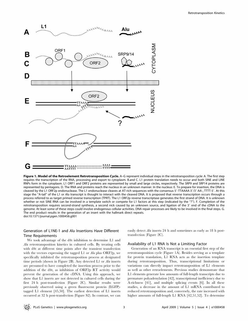
process begins with the generation of RNA (Figure 1A). Active L1
elements express two proteins from a bicistronic mRNA:
ORF1p[15] and ORF2p (Figure 1B and C). Both L1 proteins
are needed for L1 retrotransposition [16]. In contrast to L1,
ORF2p expression is sufficient for SINE retrotransposition
[4,9,17], while ORF1p may enhance the process [17]. ORF1p
possesses nucleic acid chaperone activity [18,19], an essential
property for L1 retrotransposition [19,20]. ORF2p is a multifunc-
tional protein with endonuclease and reverse transcriptase
activities [21,22]. Both proteins are proposed to interact in cis
[10,11] with the L1 RNA to form a cytoplasmic RNP complex
interacting with polyribosomes [20,23]. SINE RNA is predomi-
nantly found in the cytoplasm as an RNP complex [12,24,25]
(Figure 1C) and uses L1 protein(s) in trans for its mobilization. The
endonuclease of the L1 ORF2p generates the first nick within the
L1 endonuclease recognition sequence generating single stranded
DNA that primes the reverse transcription [22,26]. Both L1 and
Alu are proposed to undergo integration through a target-primed
reverse transcription (TPRT) reaction [27].
To generate a new insertion, L1 and SINE elements must
return to the nucleus either together or independently (Figure 1D).
Reported data suggest that retrotransposition-competent L1 RNPs
may transit through the nucleolus [28]. The 39 poly-A stretch or
‘‘A-tail’’ of LINEs, SINEs and processed pseudogenes is required
for the priming of reverse transcription (Figure 1E) [4,29]. Unlike
the post-transcriptionally generated A-tail of pol II RNAs
(mRNA), SINE A-tails are included within their sequence and
play an important role in SINE retrotransposition [30,31]. The
details of the final integration and ligation of the L1 or Alu inserts
into the host DNA remain unclear. Recent reports indicate that
cellular factors, such as DNA repair enzymes, may aid in the L1
retrotransposition process [32,33]. The final inserted sequence is
typically flanked by direct repeats (Figure 1G). Non-autonomous
retrotransposed inserts, such as Alu, SVA, hYs and retropseudo-
genes share these hallmarks with L1 inserts, strongly suggesting
that these elements use the L1 ORF2p endonuclease generated
nick for their integration [34–36].
To date, all known SINEs are ancestrally derived from RNA pol
III transcribed RNA genes, reviewed in [37]. The vast majority
are derived from different tRNA genes and only two (Alu and the
rodent B1) originated from the 7SL RNA gene, a component of
the signal recognition particle (SRP) [38]. Other examples of pol
III transcribed repeats include the four hY genes (hY1, hY3, hY4
and hY5) that likely contributed directly or indirectly to the
generation of almost 1000 copies in the human genome by
retrotransposition [36,39]. In contrast to SINEs, an internal RNA
pol II promoter drives LINE transcription with the unusual ability
to start transcription upstream of its location. Like other pol II
RNAs, L1 transcription is regulated by different mechanisms,
including promoter methylation [40], transcriptional attenuation
due to A-richness [41], premature polyadenylation [42], and the
generation of different splice variants [6]. Additional studies
suggest that at least some portion of the L1 mRNAs are capped
[43] and that the capping enhances L1 translation [44].
Previously, an L1 element tagged with a green fluorescent
protein (EGFP) retrotransposition cassette was used to detect L1
retrotransposition ‘‘near real time’’ [45]. The earliest detection of
an L1 retrotransposition event was 48 h post-transfection. In this
manuscript, we evaluate the timing of retrotransposition (defined
as the time required for a retroelement from the initial
transcription step to complete an insertion) of tagged Alu and
L1. We demonstrate that Alu elements only require about half of
the amount of time as L1 to generate an insert. Our data
demonstrate that the type of RNA polymerase dictates the
retrotransposition timing, but does not determine the retrotrans-
position rate (defined as the number of inserts a given element can
generate, i.e. the ‘‘efficiency’’ of an element). After evaluating
several potential time limiting steps, we show that the RNA
polymerase type is an important early factor contributing to the
divergent retrotransposition kinetics between LINEs and SINEs.
Results
The Use of an HIV Reverse Transcriptase Inhibitor d4t as aSuppressor of L1 and Alu Retrotransposition in Culture
Reverse transcriptase (RT) domains of multiple sources can be
grouped into a family of shared sequence homology [NCBI cdd
pfam00078.12] [46], including the RT of the human immunode-
ficiency virus and L1 ORF2 protein. Endogenous RT activity is
inhibited by two antiretroviral agents nevirapine and efavirenz
[47]. L1 retrotransposition in a culture assay system can be
suppressed by the addition of a variety of HIV RT inhibitors
[48,49]. This system utilizes a tagged vector designed to allow
expression of the reporter gene only when the retroelement goes
through its reverse transcriptase-dependent amplification process
(Figure 2A). Therefore, only the newly inserted element will
express the reporter gene (e.g. neo).
Using the established L1 and Alu retrotransposition tissue
culture assays [4,16], we evaluated the dose of, 29,39-didehydro-39-
deoxy-thymidine (d4t) required to abolish retrotransposition of L1
and L1 ORF2p driven Alu without adversely affecting cell growth
and viability. Treatment of transiently transfected HeLa cells
showed that both L1 and Alu activities presented a d4t activity
inhibitory concentration 50 (IC50) of about 2 mM (Figure S1). For
our subsequent experiments we utilized d4t treatments at 50 mM
(25 fold the IC50) to inhibit SINE and LINE retrotransposition in
tissue culture. We selected this dose for its efficient inhibition of
retrotransposition and lack of observed negative effects, deter-
mined by colony formation of an unrelated plasmid that expresses
a functional neomycin resistance gene and integrates into genomic
DNA by random integration rather than by an L1-dependent
mechanism (data not shown).
Author Summary
SINE retroelement amplification has been extremelysuccessful in the human genome. Although these non-autonomous elements parasitize factors from LINEs, boththe human Alu and the cumulative rodent SINEs havegenerated over one million copies in their respective hosts.Alu-induced mutagenesis is responsible for the majority ofthe documented instances of human retroelement inser-tion-induced disease. Our data indicate that SINEs requirea shorter period of time to complete insertion than L1s,possibly contributing to the ability of Alu elements toeffectively parasitize L1 components. We demonstrate thatRNA polymerase changes the timing Alu requires tocomplete retrotransposition and creates the need for theL1 ORF1protein in addition to ORF2p. We postulate thatthe way cells manage pol III and pol II (mRNA) transcriptsaffects the timing of a transcript going through theretrotransposition pathway. We propose a model thathighlights some of the critical differences of LINE and SINEtranscripts that likely play a crucial role in their retrotrans-position process.
Retrotransposition Kinetics
PLoS Genetics | www.plosgenetics.org 2 April 2009 | Volume 5 | Issue 4 | e1000458
Generation of L1NE-1 and Alu Insertions Have DifferentTime Requirements
We took advantage of the d4t inhibition to determine L1 and
Alu retrotransposition kinetics in cultured cells. By treating cells
with d4t at different time points after the transient transfection
with the vectors expressing the tagged L1 or Alu plus ORF2p, we
specifically inhibited the retrotransposition process at designated
time periods (shown in Figure 2B). Any detected L1 or Alu inserts
are presumed to have completed the insertion process prior to the
addition of the d4t, as inhibition of ORF2p RT activity would
prevent the generation of the cDNA. Using this approach, we
show that L1 inserts are not detected in cultured cells during the
first 24 h post-transfection (Figure 2C). Similar results were
previously observed using a green fluorescent protein (EGFP)-
tagged L1 element [45,50]. The earliest detection of L1 inserts
occurred at 32 h post-transfection (Figure S2). In contrast, we can
easily detect Alu inserts 24 h and sometimes as early as 18 h post-
transfection (Figure 2C).
Availability of L1 RNA Is Not a Limiting FactorGeneration of an RNA transcript is an essential first step of the
retrotransposition cycle (Figure 1A). Besides serving as a template
for protein translation, L1 RNA acts as the insertion template
during retrotransposition. Thus, transcriptional limitations or
variations can directly impact retrotransposition of L1 elements
as well as other retroelements. Previous studies demonstrate that
L1 elements generate low amounts of full-length transcripts due to
premature polyadenylation [42], transcriptional inefficiency due to
A-richness [41], and multiple splicing events [6]. In all these
studies, a decrease in the amount of L1 mRNA contributed to
reduced retrotransposition and, conversely, the rate increased with
higher amounts of full-length L1 RNA [42,51,52]. To determine
Figure 1. Model of the Retroelement Retrotransposition Cycle. A–G represent individual steps in the retrotransposition cycle: A. The first steprequires the transcription of the RNA, processing and export to cytoplasm. B.and C. L1 protein translation needs to occur and both SINE and LINERNPs form in the cytoplasm. L1 ORF1 and ORF2 proteins are represented by small and large circles, respectively. The SRP9 and SRP14 proteins arerepresented by pentagons. D. The RNA and proteins reach the nucleus in an unknown manner. In the nucleus: E. To prepare for insertion, the DNA iscleaved by the L1 ORF2p endonuclease. The L1 endonuclease cleaves at AT-rich sequences with the consensus 59-TTAAAA-39/39-AAqTTTT-59. At thisstage the ‘‘A-tail’’ of the L1 or Alu transcript is thought to interact with the cleaved DNA. It is proposed that reverse transcription occurs through aprocess referred to as target primed reverse transcription (TPRT). The L1 ORF2p reverse transcriptase generates the first strand of DNA. It is unknownwhether or not SINE RNA can be involved in a template switch or compete for L1 factors at this step (indicated by the ‘‘?’’). F. Completion of theretrotransposition requires second-strand synthesis, a second nick caused by an unknown source, and ligation of the 39 end of the cDNA to thegenome. At least some of these steps could involve endogenous cellular activities. DNA repair processes are likely to be involved in the final steps. G.The end product results in the generation of an insert with the hallmark direct repeats.doi:10.1371/journal.pgen.1000458.g001
Retrotransposition Kinetics
PLoS Genetics | www.plosgenetics.org 3 April 2009 | Volume 5 | Issue 4 | e1000458

Figure 2. Alu and L1 Exhibit Different Retrotransposition Kinetics. A. Assay design. A schematic of the constructs used for the L1 and Alutissue culture assay are shown on the top. RNA transcription is driven by a CMV promoter for the L1 construct or the internal pol III Alu promoter. Therestriction sites used in the construction of the other pol III driven vectors are shown. The L1 construct contains a full-length retrocompetent L1element with its ORF1 and ORF2. The L1 vector is tagged with the mneoI indicator cassette containing an inverted neomycin resistance gene (neo,light gray box) disrupted by an intron [16]. The Alu vector contains a neoTET cassette with a tetrahymena self-splicing intron interrupting the neo gene[4]. In both constructs, the introns will only splice out from a transcript generated by the L1 or Alu promoter. The spliced RNA is reverse transcribed,followed by integration of the cDNA into the genome. The new insert contains a functional neomycin gene. G418 resistance will be obtained only ifretrotransposition occurs. B. Schematic of treatment timeline. HeLa cells were seeded and transfected the next day with the appropriate constructs.After the three hour incubation with the transfection cocktail (3h*) the first set of cells was treated with d4t and G418 containing media (0 h). Notethat at this time point the plasmid DNA has already been in contact with the cells for 3 h. The second set of cells was treated after 3 hours (3 h), andso forth until completing all the time points (shown as arrows above). Cells were stained after 2 weeks of growth under selection. C. Alu inserts aredetected at 24 h, while L1 requires at least 48 hours to generate inserts. HeLa cells were transiently transfected with L1mneo (black bar) orAluYa5neoTET+ORF2p expression vector (gray bar) and d4t plus G418 treatment started 3, 6, 18, 24, and 48 h post-transfection (x axis). Inset showsrepresentative G418R foci results of the retrotransposition assay. Bars represent the relative % mean G418R colonies6standard deviation shown aserror bars for each construct. The 48 h data were used to define 100%. The mean of the observed G418 resistant colonies is shown in parenthesesabove each column.doi:10.1371/journal.pgen.1000458.g002
Retrotransposition Kinetics
PLoS Genetics | www.plosgenetics.org 4 April 2009 | Volume 5 | Issue 4 | e1000458

whether L1 RNA transcription and processing contributes to the
observed timing difference between L1 and Alu inserts, we
performed a time course to evaluate the generation of the spliced
RNA product in cells transiently transfected with L1mneo,
AluYa5neoTET, and L1neoTET (Figure 3). Because the Alu construct
is driven by RNA polymerase III, its tag (neoTET) contains a self
splicing intron disrupting the neomycin gene. Therefore, we
included an additional L1 construct that contains the exact same
self splicing (neoTET) tag present in the Alu vector to control for any
potential variations introduced by splicing dynamics. Full-length
spliced and unspliced transcripts from Alu and both L1 constructs
could be detected as early as 3 hours post-transfection (northern
blots shown in Figure S3). The mneo and neoTET tagged L1
constructs exhibited similar kinetics for the spliced transcript (only
RNA that will generate G418R colonies when retrotransposed),
peaking by 24 h and declining by 72 hours (Figure 3). Splicing
efficiency of the RNA produced by different expression vectors
was evaluated (Table S1). Equivalent splicing efficiency was
observed for the L1 and Alu transcripts sharing the same neomycin
cassette (neoTET or mneo). Alu-tag transcripts were only detected in
the cytoplasmic fraction at any of the time points evaluated (data
not shown), consistent with what has been previously reported for
the authentic Alu ‘‘untagged’’ RNA [53]. Despite early L1 mRNA
availability, no L1 inserts were observed at the 24 h time point.
Spliced Alu transcripts peak around 48 h, declining by 72 h, much
like L1 mRNA (Figure 3). However, in contrast to L1, numerous
Alu inserts are readily detectable by 24 h. These results
demonstrate that the full-length properly spliced L1 RNA is
generated in the same time period as the Alu RNA. Thus, it is
unlikely that RNA transcription or variation in the type of splicing
within the neo cassette account for the observed time difference
between the generation of Alu and L1 inserts.
Another difference between the Alu and L1 elements involves
the length of the transcript, which could alter the time required by
the reverse transcriptase to generate a full-length cDNA. In this
assay system full length inserts are not required to generate a
G418R colony. In both Alu and L1 assays, inserts are detected with
the retrotransposition of the minimal unit of a functional neomycin
gene, which is identical in length in both transcripts once the
intron is removed. Therefore, the timing differences observed
between these two elements should be independent of the
transcript length.
L1 ORF2p Activity Can Be Detected within 24 HoursWe next assessed whether the delay reflects the time required
for translation of the L1 proteins and the ability to reach the
nucleus (Figure 1B–E). ORF2 protein has been notoriously
difficult to observe by conventional techniques, such as western
blot analysis [28]. As an alternative, the ORF2p activities can be
evaluated.
Because Alu elements require ORF2p for retrotransposition,
evaluation of Alu retrotransposition serves as an alternate method
to detect ORF2p activity. Therefore, we exploited the trans-
complementation assay to monitor the ability of L1 to trans-
mobilize Alu, using AluYa5neoTET as a reporter construct. We
determined the Alu insertion kinetics in cells cotransfected with the
AluYa5neoTET plus the L1 no tag vector. Multiple Alu inserts were
detected as early as 24 h post-transfection (Figure 4), corroborat-
ing the availability of the ORF2p expressed from the L1 vector in
the nucleus by 24 h. Equivalent results were observed when using
a blasticidin tagged L1 to drive Alu retrotransposition (data not
shown). Under our experimental conditions, endogenous L1
present in HeLa cells does not significantly contribute to the
generation of the G418R colonies as the Alu vector was unable to
generate any inserts without L1 supplementation at 24, 32 and
48 h post-transfection (vector control, Figure 4). A few solitary
colonies (2 and 1) were observed at the 42 and 72 h time points.
This observation clearly demonstrates that a full-length L1 vector
generates enough ORF2p to reach the nucleus within 24 h and to
mobilize a tagged Alu element in our assay system. Our
observations are in agreement with previously published data
demonstrating that cells transiently transfected with L1 exhibit
extensive double strand breaks at 24 h post-transfection [33]. The
observed DNA breaks are dependent on the endonuclease activity
of the L1 ORF2p. Our data strongly suggest that translation and
nuclear localization of ORF2p is unlikely to be the main limiting
step for the observed differences between the L1 and Alu time
requirements.
In addition, pre-transfection of high amounts of ORF2p or any
of the L1 factors (proteins and/or RNPs) in trans did not alter L1
retrotransposition timing (Figure S4). This is not surprising
considering that L1 RNA exhibits a strong cis-preference for its
own translated proteins for retrotransposition [10,11]. Pre-
transfection with ORF2p showed a few more Alu inserts at early
time points (data not shown). However, this slight increase was not
statistically significant (Student’s paired t-test, p = 0.297).
The RNA Polymerase Dictates SINE and LINERetrotransposition Timing
Transcripts generated from RNA polymerase II and III
promoters differ in their capping, 39 end processing, folding
structures, post-transcriptional processing, interaction with trans-
lation factors and degradation pathways, reviewed in [54–56]. In
addition, these two transcriptional complexes can be observed in
different spatial locations in the nucleus indicating discrete
transcriptional sites [57,58]. To evaluate the timing of retrotrans-
position of other pol III-driven genes we generated ‘‘tagged’’
versions of 6 human genes (7SL, U6, hY1, hY3, hY4 and hY5) by
cloning the genes with at least 300 bp of their upstream enhancer
sequence 59 of the neoTET cassette (details in materials and
methods). Although the ‘‘functional’’ genes are not SINEs per se,
we selected these as examples of pol III-driven genes. The human
genome contains multiple examples of retrotransposed copies with
sequence homology to these genes [36,59]. Thus, these serve as
our best examples of other human pol III-driven constructs. We
also included in our analysis the pol III-driven B2 element as a
known active rodent SINE [9,60]. In our d4t-assay system, all
tagged pol III-driven elements generated inserts by 24 h post-
transfection when supplemented with just L1 ORF2p (Figure 5).
To better understand the RNA polymerase influence on
retrotransposition, we also evaluated the time requirement of
two pol II-driven (CMV) constructs: ORF1mneo and pol II Alu
(Figure 6A). We selected ORF1mneo because it generates a
transcript of L1 ORF1, which has previously been used to reflect
retropseudogene activity [10]. The ORF1mneo vector can retro-
transpose when a source of ORF2p is supplied in trans [10]. The
pol II Alu (pCMVYa5mneo) contains an Alu tagged with the ‘‘mneo’’
cassette from the L1-tagged construct [61], which contains pol III
terminators (4 Ts) that would generate truncated transcripts if the
internal pol III A and B boxes in the Alu sequence are used for
transcription. The ‘‘normal A-tail’’ at the end of the Alu sequence
and 59 of the neo cassette (Figure 6A) was not included in order to
prevent potential internal priming for TPRT in the cDNA
extension step (Figure 1E), which would circumvent inclusion of
the neo reporter gene in the retrotransposed copy. Thus, only the
Alu body sequence was utilized in the construct. Just like the L1
construct, the A-tail used in the TPRT step is generated from the
transcript polyadenylation by the RNA polymerase II from the
Retrotransposition Kinetics
PLoS Genetics | www.plosgenetics.org 5 April 2009 | Volume 5 | Issue 4 | e1000458

Retrotransposition Kinetics
PLoS Genetics | www.plosgenetics.org 6 April 2009 | Volume 5 | Issue 4 | e1000458

Figure 4. Detection of L1 ORF2p Trans-mobilization Activity at 24 h. HeLa cells were transiently transfected with AluYa5neoTET plus L1 no tagor empty vector (control). The d4t and G418 treatment was started at 24, 32, 36, 48 and 72 h post-transfection (x axis). Bars represent the relative %mean G418R colonies6standard deviation shown as error bars for each construct. The 72 h data were used to define 100%. The mean of theobserved G418 resistant colonies is shown in parentheses above each column. Note that for the control only 2 and 1 colonies were observed at 42and 72 hours, respectively. The data demonstrate that functional ORF2p generated by the L1 no tag ‘‘wildtype’’ vector must reach the nucleus by24 h for Alu retrotransposition to occur.doi:10.1371/journal.pgen.1000458.g004
Figure 5. RNA Pol III Transcripts Share Similar Retrotransposition Kinetics. Other pol III genes known to generate retrotransposed copiesparallel Alu kinetics by generating inserts within 24 h. HeLa cells were transiently transfected with the ORF2p expression vector plus the taggedvector of the rodent SINE B2 (vertical lines), and the 7SL (light gray), U6 (dark gray), hY1 (black), hY3 (white), hY4 (dotted) or hY5 (slanted-lines) RNAgenes. The above Inset shows representative G418R foci results of the B2 retrotransposition assay. Cells were treated with d4t+G418 at 0, 24, 48 and72 h post-transfection. The 72 h data were used to define 100%. Bars represent the % relative mean G418R colonies6standard deviation shown aserror bars for each construct (n = 3).doi:10.1371/journal.pgen.1000458.g005
Figure 3. Availability of Spliced L1 RNA Is Not a Limiting Factor. Transcription and retrotransposition kinetics were evaluated for the differentconstructs. HeLa cells were transiently transfected with L1mneo, AluYa5neoTET+ORF2p or L1neoTET (with the same self-splicing neo cassette used forthe Alu construct). Cells were either harvested for RNA quantitation (left y axis, black square) or treated with d4t plus G418 treatment for colonyquantitation (right y axis, gray circles) at the indicated time points post-transfection (x axis). RNA was quantitated relative to b-actin as control (detailsin Materials and Methods). Note that the colony numbers reflect the actual cumulative inserts that occurred from transfection to the d4t treatmenttime point. The data demonstrate that all constructs generate the spliced tagged transcripts at early time points in a similar manner; however theobserved inserts between Alu and L1 differ at 24 h.doi:10.1371/journal.pgen.1000458.g003
Retrotransposition Kinetics
PLoS Genetics | www.plosgenetics.org 7 April 2009 | Volume 5 | Issue 4 | e1000458

Figure 6. The RNA Polymerase Dictates the Retrotransposition Kinetics of Alu. A. Schematic of pol II-driven ORF1 and Alu vectors. TheORF1mneo construct was selected as a representative of retropseudogene activity. The constructs use the CMV promoter (CMVp, black box) togenerate pol II transcripts. The full mneoI indicator cassette from the L1 vector, consisting of the neomycin interrupted by an inverted intron (hatchedbox), its SV40 promoter (SV40p) and complete polyadenylation signal (pA signal) is located downstream of the L1 ORF1 (ORF1mneo) or a consensusAluYa5 (AluYa5mneo ‘‘pol II Alu’’) (arrow indicates where the Alu ‘‘normal A-tail’’ would have been located). B. Spliced RNA pol II generated transcriptsof tagged ORF1 and Alu are readily available by 24 hours. Poly-A selected RNA extracts from different post-transfection time points (24, 48 and 72 h)were evaluated by Northern blot analysis using an RNA strand specific probe to the neomycin resistance gene. The unspliced (open arrowhead) andspliced (black arrow) transcripts from the pol II-vectors AluYa5mneo and ORF1mneo are shown. b-actin is indicated by an *. C. The tagged ORF1transcript mimics tagged L1 insertion kinetics. Retrotransposition assays were performed using the ORF1mneo vector supplemented with an L1(black) or ORF2p expression (gray) vector. Cells were treated with d4t plus G418 at 24 and 48 h post-transfection. Bars represent the relative % meanG418R colonies6standard deviation shown as error bars for each construct (n = 3). The 72 h data were used to define 100%. The mean of theobserved G418 resistant colonies is shown in parentheses above each column. Only one colony (1) was observed at the 24 h time point. D.Transcription from a pol II promoter alters the retrotransposition requirements of a tagged Alu element. The retrotransposition capability of the pol II-driven Alu (AluYa5mneo) supplemented with ORF1p and ORF2p expression vectors was evaluated in HeLa cells. Cells were treated with d4t plus G418at 24 and 48 h post-transfection. The 72 h data were used to define 100%. Bars represent the relative % mean G418R colonies6standard deviationshown as error bars for each construct (n = 6). The total number of G418 resistant colonies for all experiments combined is shown in parenthesesindicated by a ‘‘t’’. No colonies were ever observed at the 24 h time point. E. Transcription and retrotransposition kinetics of pol II driven ORF1 andAlu. HeLa cells were transiently transfected with ORF1mneo (top panel) or AluYa5mneo (lower panel) and either harvested for RNA quantitation (left yaxis, black square) or treated with d4t plus G418 treatment for colony quantitation (right y axis, gray circles) at the indicated time points post-transfection (x axis). RNA was quantitated relative to b-actin as control. The data demonstrate that the generation of spliced pol II and pol III Alutranscripts are equivalent; however pol II Alu inserts are not detected at 24 h.doi:10.1371/journal.pgen.1000458.g006
Retrotransposition Kinetics
PLoS Genetics | www.plosgenetics.org 8 April 2009 | Volume 5 | Issue 4 | e1000458
SV40pA signal at the 39 end of the neo cassette (Figure 6A).
Spliced and unspliced transcripts were detected from both
constructs by 24 h (Figure 6B). The tagged ORF1p transcript
driven by an ORF2p generated one single insert at 24 hours
(Figure 6C), while the total number of colonies generated were 136
and 226 for 48 h and 72 h respectively. It is possible that the
endogenous L1 expression in HeLa cells [6] affected the timing.
However, our data on Alu retrotransposition indicates that effects
from endogenous L1 expression under our experimental condi-
tions are negligible (Figure 4). Most likely, the single G418R colony
observed at 24 hours is due to a rare event that escaped d4t
inhibition. A quantitative time course evaluation of the spliced
RNA product in cells transiently transfected with ORF1mneo and
AluYa5mneo further indicates that the availability of spliced
product is not limiting retrotransposition timing (Figure 6E).
No pol II-generated Alu inserts were ever observed when
supplemented with ORF2p under any conditions tested, repre-
senting a rate of less than 16106 cells/mg of plasmid. However,
retrotransposition of the pol II-driven Alu transcript occurred
when it was supplemented with both ORF2p and ORF1p
expression plasmid (Figure 6D). Under these conditions, G418R
colonies were observed at 48 h post-transfection, much like L1 and
retropseudogene behavior. No colonies were ever observed at the
24 h time point in 5 independent experiments using triplicates for
each time point. Swapping the RNA pol III for an RNA pol II
promoter changed the retrotransposition requirements of the
tagged Alu to reflect those observed for pseudogenes and LINEs.
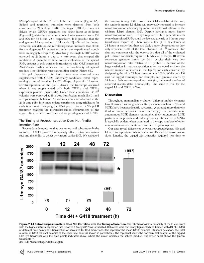
The Timing of Retrotransposition Does Not PredictInsertion Rate
Recent data demonstrate that one amino acid substitution in the
mouse L1 ORF1 protein dramatically affects retrotransposition
rate and the ability to detect new inserts earlier [50]. We evaluated
the insertion timing of the most efficient L1 available at the time,
the synthetic mouse L1 (L1m syn) previously reported to increase
retrotransposition efficiency by more than 200 fold relative to the
wildtype L1spa element [52]. Despite having a much higher
retrotransposition rate, L1m syn required 48 h to generate inserts
even when spliced RNA could be detected as early as 3 hours post-
transfection (Figure 7). There were a few (1 to 2) colonies at
24 hours or earlier but these are likely outlier observations as they
only represent 0.001 of the total observed G418R colonies. Our
data are consistent with the observation that all of the evaluated
pol II-driven constructs require 48 h, while all of the pol III-driven
constructs generate inserts by 24 h despite their very low
retrotransposition rates relative to L1 (Table 1). Because of the
large variation in retrotransposition rates, we opted to show the
relative number of inserts in the figures for each construct by
designating the 48 or 72 hour time point as 100%. While both U6
and Alu tagged transcripts, for example, can generate inserts by
24 hours, their retrotransposition rates (i.e., the actual number of
observed inserts) differ dramatically. The same is true for the
tagged L1 and ORF1 RNAs.
Discussion
Throughout mammalian evolution different mobile elements
have flourished within genomes. Retroelements such as LINEs and
SINEs have been particularly successful, generating more than one
third of human sequence mass. Interestingly, the parasitic non-
autonomous SINE elements outnumber their autonomous LINE
partners in the primate and rodent genomes. The success of SINEs
is especially evident when compared to the copy numbers of other
non-autonomous elements such as the retropseudogenes.
Our data reveal differences between retropseudogenes, Alu, and
L1 retrotransposition. When evaluating Alu and L1 retrotranspo-
sition kinetics, the tagged Alu transcript required less time to
Figure 7. L1 Retrotransposition Rate Does Not Correlate with the Timing of Insertion. The retrotransposition capability of the L1 constructwith the highest retrotransposition rate reported (L1m syn) [52] was evaluated. HeLa cells were transiently transfected and treated with d4t plus G418at different time points post-transfection or harvested for RNA extractions. Bars represent the mean G418R colonies6standard deviation. The totalnumber of G418 resistant colonies of the early time points is shown in parentheses. The top panel shows the northern blot analysis of the taggedL1m syn transcripts with the time points indicated above, where the arrow indicates the spliced product. The lower panel shows the b-actintranscripts (*).doi:10.1371/journal.pgen.1000458.g007
Retrotransposition Kinetics
PLoS Genetics | www.plosgenetics.org 9 April 2009 | Volume 5 | Issue 4 | e1000458

generate an insert. This timing difference can not be attributed to
differences in the time required to generate functional transcripts
or availability of L1 proteins. It is clear that full-length functional
L1 transcripts can be detected as early as 3 hours post-transfection
and are abundant by 24 h post-transfection. In addition, the
difference observed between Alu and L1 kinetics could not be
attributed to the type of detection cassette system (self splicing or
not) or to the differences in the retrotransposition rates. L1
colonies were rarely observed (Figure 7) at time points earlier than
48 h. These few observed G418R colonies possibly represent the
rare event that circumvented inhibition by d4t (in one experiment
a colony was observed even at the zero time point). In our assay,
production of L1 ORF2p is not limiting. Our data demonstrate
that enough ORF2p is generated from an L1 construct to drive Alu
insertions within 24 hours post-transfection, which indicates that
ORF2p is made and readily available for Alu transcript
mobilization. However, at this time we do not know if the ORF2p
reaches the nucleus as a ‘‘free’’ protein or as part of an RNP with
the L1 RNA or Alu RNA. As expected, due to the L1 cis-
preference [10], pre-transfections with ORF1p, ORF2p or other
L1 components, such as full-length transcripts or RNPs, did not
affect the L1 time requirement.
Although unexpected, it is not totally surprising that Alu and L1
present different retrotransposition time requirements. Previous data
show that, although Alu and L1 share the same insertion hallmarks,
the two elements can exhibit differences in their behavior. For
example, of two HeLa ‘‘cell lines,’’ only one supports Alu
retrotransposition while both support L1 retrotransposition [62]. In
addition, Alu and L1 are selectively inhibited by different APOBEC3
proteins [62]. This corroborates our observations that cellular
components differentiate between Alu and L1 retrotransposition.
Our data suggest that the observed time differences are
dependent on the type of RNA polymerase generating the
transcript. Multiple features that distinguish these two transcript
types may collectively or individually contribute to the observed
differences in the retrotransposition timing between L1 and Alu
elements. RNA capping, association with the translational
machinery and ORF1 requirement are plausible factors that
could influence SINE and LINE retrotransposition kinetics. As a
pol II product, L1 mRNA is likely capped. Experimental evidence
indicates that at least part of the L1 mRNA is capped [43] and that
capping enhances L1 translation in vitro [44]. In contrast, pol III
genes lack the 7-methylguanosine cap and are subjected to
different processing in a spatially separate location of the nucleus
[57,58]. L1 mRNA likely interacts with most, if not all, of the pol
II protein complexes that assemble with the transcription of
generic mRNAs, as evidenced by the premature polyadenylation
and splicing of L1 transcripts [6,42].
Even though both pol II and pol III produced RNAs form
complexes with various cellular proteins, the structure and
composition of these RNPs varies dramatically. As a rule, pol III
transcripts do not code for proteins and therefore interact with the
translational machinery in a different manner than mRNA. Most
known pol III transcripts fold to form a structured RNA and
associate with a variety of proteins to form RNPs. Specifically, Alu
interaction with SRP9 and SRP14 [12] is thought to transiently
provide proximity to the ribosomal complexes and translating L1
RNA, allowing the Alu transcript to efficiently compete for the L1
factors required for retrotransposition [26]. It is also likely that the
ability of the dimeric Alu to bind these proteins contributes to the
dramatic difference in retrotransposition rates observed between
Alu and other SINEs [9,13]. In contrast, the polyribosomes and
translation machinery assemble with the L1 mRNA in a more
stable complex to undergo translation. The cis-preference
displayed by L1 [20] suggests that the L1 RNA must dissociate
from the cellular translation machinery to form L1 RNPs as an
intermediate step in the retrotranspositional process. These L1
complexes are composed of L1 RNA, ORF1p [20], and likely
ORF2 protein [11]. All three components are shown to co-purify
in the polyribosomal fraction of the cytoplasm [11,23]. It is
plausible that ORF1p directly competes with the cellular
translation machinery for access to L1 mRNAs, transitioning the
L1 transcript away from the polyribosomal fraction and into the
retrotranspositionally competent RNPs. Because of their nature
and subcellular localization, SINEs completely avoid these two
potentially time consuming steps in their mobilization. Therefore,
SINE transcripts may enter their retrotransposition cycle as soon
as L1 ORF2p becomes available.
The pol II-driven Alu transcripts that are most likely to associate
with the cellular translational machinery, at least transiently,
require L1 ORF1 protein in addition to ORF2 protein for
retrotransposition in a manner reminiscent of retropseudogenes
[29]. The retrotransposition time of the pol II-driven Alu parallels
that of L1. At this stage it is unclear what the role of ORF1p is in
the trans-mobilization of retropseudogenes or the pol II Alu
transcript. However, it is consistent with the above-discussed
hypothesis implicating ORF1 protein in removing pol II RNAs
from their expected cycle of translation and degradation. Thus,
the pol II L1 and the pol III Alu transcript interactions with
different cellular components may dictate the timing difference
between L1 and Alu RNAs to form their respective retro-
transpositionally competent complexes.
The inefficient retrotransposition rate of the pol II-driven Alu
construct suggests that the presence of an Alu sequence within an
mRNA would not facilitate its retrotransposition by L1 factors.
Table 1. Relative retrotransposition rate of the differenttagged constructs in HeLa cells under the same transfectionconditions.
Mobileelement
Rate G418R
colonies (6106/mgat 72 h) mean6SD{
‘‘Relative’’rate*
Timerequirementfor insert (h)
L1¥ 7880.06321.0 5000 ,48
Alu (pol III)$ 1053.36160.8 500 ,24
7SL$ 66.7612.6 25 ,24
U6$ 67.1611.3 25 ,24
hY1$ 16.761.6 10 ,24
hY3$ 36.967.0 20 ,24
hY4$ 57.566.4 25 ,24
hY5$ 47.569.4 25 ,24
ORF1$ (pol II) 23.165.5 10 ,48
Alu (pol II)^ 1.360.9 1 ,48
{Rates were calculated by determining the number of G418 resistant coloniesgenerated at 72 h, per 1 million cells per mg of transfected plasmid. Ratesshould not be considered as absolute numbers, as results will vary for differentcell lines and conditions.
*Due to the intrinsic experimental variation, a rough approximation was used todetermine the relative retrotransposition rates. The lowest observed rate wasarbitrarily designated as ‘‘1.’’
¥Data from the JM101/L1.3 construct.$Retrotransposition of the element was driven by an optimized L1 ORF2expression plasmid (pBudORF2opt). Rates were much lower when a full-lengthwild type L1 was used as the driver for retrotransposition (not shown).
^Retrotransposition of the element was driven by cotransfection with L1 ORF1and ORF2 expression plasmids (pBudORF1opt and pBudORF2opt).
doi:10.1371/journal.pgen.1000458.t001
Retrotransposition Kinetics
PLoS Genetics | www.plosgenetics.org 10 April 2009 | Volume 5 | Issue 4 | e1000458

Although there is no available data on the SVA promoter, it is
unlikely that the pol III polymerase drives SVA transcription due
to the presence of numerous pol III terminators within its
sequence. Thus, it is questionable whether the truncated antisense
Alu-like sequences present in the SVA element contribute to the L1
trans-complementation of this retroposon as previously suggested
[35].
In addition to assisting its own retrotransposition, the cis-
preference exhibited by L1 may decrease cell damage by limiting
random retrotransposition of cellular mRNA. A previous study
demonstrated the co-localization of ORF1p and cellular proteins
to stress granules[63]. The authors suggest that the sequestering of
ORF1 protein in stress granules for degradation may prevent
promiscuous binding of ORF1p to non-L1 mRNAs. Thus, as a
side effect of L1 self-preference, retropseudogene formation is less
likely [5]. In addition, this ‘‘cis-preference’’ could help the L1
transcript ‘‘escape’’ the ribosomal complex and degradation
pathways. Once translation is completed, most transcripts decay
by several known mRNA degradation pathways, reviewed in [56].
In contrast, pol III transcripts are meant to perform their function
as RNA molecules in the cytoplasm or nucleus before degradation
by the exosome [64]. Essentially, the functional molecule of pol III
genes is the RNA, while for pol II genes the mRNA is an
intermediary prior to the generation of the functional protein. In
the case of L1, the ORF1p may play an additional role by
protecting the L1 RNA from degradation, increasing the chance of
returning to the nucleus where the involvement of ORF1p in the
L1 integration process has been previously suggested [18,23].
Thus, the requirement for both ORF1 and ORF2 proteins could
contribute to the longer time needed for L1 transcripts to generate
inserts. In addition, it is plausible that interactions with different
cellular components during insertion, mediated by ORF1p, may
contribute to the timing differences observed.
We postulate that the differences observed in retrotransposition
kinetics are dictated by the type of RNA polymerase generating
the transcript. We propose an initial model where the cytoplasmic
interactions of pol II (L1 and mRNA) and pol III transcripts and
pathways influence the amplification kinetics of LINEs and SINEs
(Figure 8). Overall, it is evident that the type of RNA polymerase
generating the transcript alters the timing of mobile element
insertion and remains a critical parameter in the classification of
different types of retroelements.
Materials and Methods
PlasmidsL1 related vectors. JM101/L1.3 referred to ‘‘L1mneo’’
contains a full-length copy of the L1.3 element and the mneoI
indicator cassette cloned in pCEP4 (Invitrogen) [61,65].
JM101/L1.3 no tag, referred as ‘‘L1 no tag’’ contains a full-
length copy of the L1.3 element cloned in pCEP4 [10].
TAM102/L1.3 contains the full-length copy of L1.3 element
and the mblastI indicator cassette cloned in pCEP4 [32].
ORF1mneo contains the L1.3 59UTR, L1.3 ORF1, and the
mneoI indicator cassette cloned in pCEP4 [10].
psmL1 contains the codon optimized L1spa sequence and the
neomycin indicator cassette cloned in pCEP4 [52].
L1neoTET contains the codon optimized L1RP driven by CMV
promoter and tagged with the self splicing intron neo cassette [4]
in pBluescript.
JM101/L1.3, JM101/L1.3 no tag, TAM102/L1.3 and
ORF1mneo were kind gifts from Dr. John Moran. psmL1 was a
kind gift from Dr. Jef Boeke [52]. pBudORF2opt [33]and
pBudORF1opt [66] have been previously described. The open
reading frames are cloned into the expression vector pBudCE4.1
(Invitrogen), under control of the CMV promoter.
SINE related vectors. Alu-neoTET containing a 7SL
upstream enhancer region - Alu core sequence followed by the
neoTET self-splicing indicator cassette and 44 A-stretch followed by
a pol III terminator [4] was a kind gift from Dr. Thierry
Heidmann.
The ‘‘SINE’’neoTET constructs listed below were created by
initially modifying the AluneoTET vector. The QuickChange site-
directed mutagenesis kit (Stratagene) was used to introduce an
AatII site (underlined) at the 39 end of the Alu element with a set of
complementary 59 phosphorylated primers to the following
sequence: 59- AGCCTGGGCGACAGAGCGAGTCGACGTC-
TCAAATCCCCTCAG -39 following the manufacturer’s recom-
mended protocol. The new construct, referred to as AluYa5neoTET
AatII, was then used to introduce the different individual elements
and their corresponding upstream enhancer sequences using the
BamHI (59 of 7SL promoter enhancer sequence) and the AatII sites
(schematic of the basic vector shown in Figure 2A). The BamHI
and AatII sites are underlined.
AluYa5neoTET, contains a larger amount of the upstream pol III
enhancer sequence of the 7SL gene (113 bp) and the AluYa5
consensus sequence from p7SLYa5BC1 [67].
7SLneoTET, contains a 413 bp fragment of the Human 7SL
RNA gene Accession # M20910 and its upstream pol III
enhancer sequence, PCR amplified with primers F7SL: 59-
GGATCCGCCCAGTGTGGGTGTGTCC-39 and R7SL: 59
GACGTCAAGAGACGGGGTCTCGCTATG-39
U6neoTET, contains a 512 bp fragment of the human small
nuclear RNA gene HUMUG6 Accession# M14486 and its
upstream pol III enhancer sequence PCR amplified with primers
FU6: 59-GGATCCGCAGACACTGCTCGGTAGTT-39 and
RU6: 59 GACGTCAAGAGACGGGGTCTCGCTATG-39
hY1neoTET, contains a 478 bp fragment of the human RNA
gene hY1 encoding Ro RNA Accession# V00584 and its
upstream pol III enhancer sequence PCR amplified with primers
FhY1: 59-GGATCCGTCCCACAGAGCTGTCCGGAGG-39
and RhY1: 59-GACGTCAAGACTAGTCAAGTGCAGTAGT-
GAG-39
hY3neoTET, contains a 430 bp fragment of the human RNA
gene hY3 encoding Ro RNA Accession# V00585 and its
upstream pol III enhancer sequence PCR amplified with primers
FhY3: 59-GGATCCCGCTCTAGACGTCCTGGCC-39 and
RhY3: 59-GACGTCAAGGCTAGTCAAGTGAAGCAG-39
hY4neoTET, contains a 494 bp fragment of the human RNA
gene hY4 encoding Ro RNA Accession# L32608 and its
upstream pol III enhancer sequence PCR amplified with primers
FhY4: 59-GGATCCACAGGCAGGGAFACGACAAA-39 and
RhY4: 59-GACGTCAAGCCAGTCAAATTTAGCAGTGG-39
hY5neoTET, contains a 667 bp fragment of the human RNA
gene hY5 encoding Ro RNA Accession# K01564 and its
upstream pol III enhancer sequence PCR amplified with primers
FhY5: 59-GGATCCTGATGATGAAACAAAGCC-39 and
RhY5: 59-GACGTCAACAGCAAGCTAGTCAAGC-39.
B2neoTET, contains 113 bp of the upstream pol III enhancer
sequence of the 7SL gene and the B2 sequence from p7SLB2BC1
[67].CMVYa5mneo (‘‘pol II Alu’’) the mneoI cassette including the SV40
polyadenylation signal from JM101/L1.3 was amplified by PCR
with the high fidelity phusion DNA polymerase (New England
Biolabs) using primers sets to introduce the 59 FseI and a 39 BglII
used for introduction into the compatible FseI-BamHI sites in
pGL3CMVYa5BC1-SV40pA [68]. The construct was then modified to
eliminate the internal polyA-stretch immediately downstream of
Retrotransposition Kinetics
PLoS Genetics | www.plosgenetics.org 11 April 2009 | Volume 5 | Issue 4 | e1000458

Figure 8. Model of SINE and LINE Cellular Interactions Potentially Contributing to Differences in Retrotransposition Kinetics. Wepresent a model of how pol II (L1 and mRNA) and pol III transcript interactions with their respective cellular components in the cytoplasm mayinfluence the retrotransposition timing of LINEs and SINEs. Structures are not drawn to scale. Transcription and processing are not the limiting stepsfor L1 and Alu, as both pol II and pol III spliced tagged transcripts are present in the cytoplasm as early as 3 h post-transfection. However, Alu requiresabout 24 h to generate an insert, while L1 requires about 48 h (*). Pol II transcript: LINE RNA and pol II-driven mRNAs reach the cytoplasm afterprocessing and modifications. The cytoplasmic pol II transcript has been spliced, polyadenylated and capped at its 59 end (shown as a black dot at theend of the RNA). The cap allows the recognition by several proteins involved in translation forming a large protein complex interacting with thetranscript. Capping also allows for the association with the PABP-1, elongation factors and the circularization of the mRNA (not shown). This largemulti-protein complex interacts with the translation machinery to generate the needed ORF1 and ORF2 proteins. The generated proteins willpreferentially bind to the L1 RNA that encoded them (cis-preference). Multiple ORF1p molecules (yellow circles) and possibly ORF2p (red circle) bindthe L1 transcript. We propose that the formation of the L1 RNP complex will allow the L1 RNA to separate from the translation machinery and evadethe normal degradation pathway en route to the nucleus. This process of detachment from the ribosomal complex and avoidance of the RNA decaypathways may increase the time requirement for L1 retrotransposition. These extra steps probably contribute to the extended time requirement forL1 to complete the retrotransposition process. The L1 RNA (likely as an RNP with ORF1 and ORF2) reaches the nucleus and generates a new insert(represented as a white box in the DNA). In our assay system, the insertion process of L1 elements requires about 48 hours for completion. CellularRNAs (e.g., mRNAs and the pol II Alu RNA) can occasionally use the L1 proteins to mediate their mobility in trans. However, retropseudogene (pol IImRNA) inserts are not efficiently generated in our experimental system and require ORF1p. We propose that the spurious interaction with ORF1pallows these mRNAs to be shunted to the nucleus to go through the retrotransposition process. In addition, it is likely that ORF1p also contributes tothe retrotransposition process in the nucleus. The ORF1mneo transcript generates inserts more efficiently than the other tested pol II transcripts,possibly because of close proximity to ORF1p in cis. Overall, the efficiency of a pol II transcript to generate inserts in tissue culture is likely correlatedwith its ability to interact with ORF1p. The role of ORF2p in the cytoplasm is unclear. Pol III transcript: SINE RNA reaches the cytoplasm with little or noprocessing and interacts with specific proteins. The cytoplasmic SINE RNP is stable and compact. In the case of Alu, the transcript forms a specificstructure which binds the SRP9 (green circle) and SRP14 (blue circle) proteins. It is hypothesized that these proteins may target SINE RNA to theribosomes, generating transient close proximity to nascent L1 proteins that might be essential for SINEs to efficiently use L1 in trans forretrotransposition. Although SINE RNPs may be targeted to the ribosomes, they are not functional components of the translational complex, makingthis interaction likely transitory. Whether pol III RNA gains access to the L1 retrotransposition machinery in the cytoplasm or in the nucleus remainsundetermined. Because SINE transcripts are not translated or functional components of the translational complex, the SINE RNPs are likely ‘‘free’’ tosequester the L1 proteins and immediately proceed with the retrotransposition cycle. We propose that the pol III SINE probably reaches the nucleusin a more efficient manner than the pol II transcripts, such as the L1 RNA, which must first dissociate from the translational complex and avoid thenormal mRNA degradation pathway. In our system, the insertion process of an Alu requires 24 hours or less for completion (* represented as a whitebox in the DNA).doi:10.1371/journal.pgen.1000458.g008
Retrotransposition Kinetics
PLoS Genetics | www.plosgenetics.org 12 April 2009 | Volume 5 | Issue 4 | e1000458

the Alu sequence and 59 to the selection cassette (Figure 6). The pol
II Alu transcripts generated from this construct are polyadenylated
from the SV40pA signal.
pIRES2-EGFP (BD Biosciences Clontech) was used as the
G418R expression plasmid for toxicity control.
All plasmid DNA was purified by alkaline lysis and twice
purified by cesium chloride buoyant density centrifugation. DNA
quality was also evaluated by the visual assessment of ethidium
bromide stained agarose gel electrophoresed aliquots. All new
constructs were sequence verified
Retrotransposition AssaysThe basic transient L1 [16] or Alu [4] retrotransposition assay
was performed as previously described with some minor
modifications. Briefly, HeLa cells (ATCC CCL2) were seeded in
T-75 flasks at a density of 56105 cells/flask or in 6 well plates at a
density of 2.5–56104/well. Transient transfections were per-
formed the next day with Lipofectamine Plus following the
manufacturer’s protocol (Invitrogen), with 3 mg of SINE-neoTET
vector plus 1 mg pBud-ORF2opt or 1 mg of L1 no tag. For L1
assays 1 mg of JM101/L1.3 was used. Inhibitory effects on cellular
growth or colony formation capabilities by the d4t treatment was
evaluated by transfecting cells in parallel with 0.3 mg of a plasmid
expressing neomycin resistance (pIRES2-EGFP; BD Biosciences
Clontech) as a ‘‘toxicity’’ control. Following removal of transfec-
tion cocktail, the cells were treated with the appropriate media
containing 400 mg/ml Geneticin/G418 (Fisher Scientific) alone or
in combination with 50 mM d4t for selection and/or reverse
transcriptase inhibition. After 14 days, cells were fixed and stained
for 30 minutes with crystal violet (0.2% crystal violet in 5% acetic
acid and 2.5% isopropanol). The inhibitor d4t- (29,39-Didehydro-
39-deoxy-thymidine; Sigma-Aldrich) was freshly added to the
selection media at the indicated time period. During the inhibitor
treatment period all cells in the experiment were refreshed daily
for the first week with the appropriate media. The rate of insertion
efficiency (retrotransposition rate) was determined as the number
of visible G418R-resistant colonies obtained at 72 h after transient
transfection of 16106 seeded HeLa cells with 1 mg of the neo
tagged construct.
Northern Blot AnalysisRNA extraction and poly(A) selection was performed as
previously described [42]. Total RNA was extracted using the
recommended protocol for TRIzol Reagent (Invitrogen) from two
75 cm2 cell culture flasks at 3, 6, 24, 48, and 72 hours post-
transfection. The PolyATract mRNA isolation system III
(Promega) was used to select polyadenylated RNA species
following the manufacturer’s protocol. After separation in a 1%
(L1) or a 2% (pol III constructs) agarose-formaldehyde gel, the
RNA was transferred to a Hybond-N nylon membrane (Amer-
sham Biosciences). The RNA was cross-linked to the membrane
using a UV-light (GS Gene linker, BioRad) and pre-hybridized in
30% formamide, 16 Denhardt’s solution, 1% SDS, 1 M NaCl,
100 mg/ml salmon sperm DNA, 100 mg/ml-1 yeast t-RNA at
60uC for at least 6 h. The 39 region of the neomycin gene was
amplified by PCR using the following primers T7neo (2): 59-
TAATACGACTCACTATAAGGACGAGGCAGCG-39 and
Neo northern (+): 50- GAAGAACTCGTCAAGAAGG-39. The
isolated PCR product was used as a DNA template to generate a32P-CTP (Amersham Biosciences) labeled single strand-specific
RNA probe using the MAXIscript T7 kit (Ambion) following the
manufacturer’s recommended protocol. We utilized material
included in the kit to generate the riboprobe for the b-actin.
The radiolabeled probe was purified by filtration through a
NucAway Spin column (Ambion). Hybridization with the probe
(final concentration of 4–126106 cpm/ml) was carried out
overnight in the pre-hybridization solution at 60uC. Two ten-
minute washes were performed at high stringency (0.16SSC,
0.1%SDS) at 60uC. The results of the northern blot assays were
evaluated using a Typhoon Phosphorimager (Amersham Biosci-
ences) and the ImageQuant software.
Supporting Information
Figure S1 Evaluation of D4t Inhibitory Concentration 50 (IC50)
on L1 and Alu Retrotransposition. HeLa cells were transiently
transfected with plasmids expressing a neomycin-tagged L1 (solid
line) or a marked Alu supplemented with an ORF2p expression
vector (dashed line). Cells were treated with different concentra-
tions of d4t and G418 for two weeks. Colonies were stained and
scored. The no treatment data were used to define 100%. The
relative % mean G418R colonies6standard deviation are shown
for each construct. The inhibitory concentration 50 (IC50) for L1
and Alu is ,2 mM d4t, the intercept (gray line) is shown.
Found at: doi:10.1371/journal.pgen.1000458.s001 (0.34 MB TIF)
Figure S2 The earliest detection of L1 inserts occurred at
32 hours post-transfection. HeLa cells were transiently transfected
with plasmids expressing a neomycin-marked L1 (black), marked
Alu supplemented with an ORF2p expression vector (gray) or a
control vector with neomycin resistance (white). Cells were treated
with G418 plus d4t at 24, 32, 42, 48 and 72 h post-transfection (x
axis). The 72 h data were used to define 100%. Bars represent the
relative % mean G418R colonies6standard deviation shown as
error bars for each construct.
Found at: doi:10.1371/journal.pgen.1000458.s002 (0.44 MB TIF)
Figure S3 Vector transcription kinetics. Cells were transiently
transfected with 5 mg of the tagged vectors. RNA was extracted at
different time points (3–72 h) after transfection. Poly-A selected
transcripts were evaluated by Northern blot analysis using an
RNA strand specific probe to the neomycin resistance gene or to
b-actin (indicated by an *). RNA is transcribed as early as 3 hours
post-transfection. L1mneo, AluYa5neoTET and L1neoTET are shown.
Transcripts containing the unspliced (open arrowhead) and spliced
(black arrow) neo indicator cassette are indicated. Only the spliced
transcripts are able to generate inserts conferring G418 resistance
and these transcripts were used for the RNA quantitation.
Exposures times varied due to the strong signal from the later
time periods.
Found at: doi:10.1371/journal.pgen.1000458.s003 (2.92 MB TIF)
Figure S4 Supplementation with L1 factors does not affect L1
insertion kinetics. The schematic of transfection and treatment
timeline is shown. Cells were stained after 2 weeks of treatment.
To ensure the early presence of the L1 factors, HeLa cells were
pre-transfected (T1) with plasmids expressing L1 ORF1p, ORF2p
ORF1p plus ORF2p, and a untagged L1 (as a source of L1 RNA
and or RNPs) 24 h before introducing the tagged L1 element (T2).
Cells were treated with d4t and G418 at 24, 48, 72 and 96 h post-
transfection (x axis). The 96 h data were used to define 100%. Bars
represent the relative % mean G418R colonies6standard
deviation shown as error bars for each construct.
Found at: doi:10.1371/journal.pgen.1000458.s004 (0.54 MB TIF)
Table S1 Evaluation of splicing efficiency of the tagged L1 and
Alu constructs at different time points.
Found at: doi:10.1371/journal.pgen.1000458.s005 (0.05 MB
DOC)
Retrotransposition Kinetics
PLoS Genetics | www.plosgenetics.org 13 April 2009 | Volume 5 | Issue 4 | e1000458

Acknowledgments
We are indebted and very grateful to Louisiana State University in Baton
Rouge and specifically Dr. Mark Batzer for hosting us in his laboratory
during our evacuation for hurricane Katrina. We thank the members of
Dr. Batzer’s laboratory for their support. We also thank Prescott Deininger
for his continuous support and insightful comments.
Author Contributions
Conceived and designed the experiments: ENK VPB AMRE. Performed
the experiments: ENK VPB BJW AMRE. Analyzed the data: ENK VPB
BJW AMRE. Contributed reagents/materials/analysis tools: BJW AMRE.
Wrote the paper: AMRE. Edited the article: ENK VPB BJW.
References
1. Lander ES, Linton LM, Birren B, Nusbaum C, Zody MC, et al. (2001) Initial
sequencing and analysis of the human genome. Nature 409: 860–921.
2. Waterston RH, Lindblad-Toh K, Birney E, Rogers J, Abril JF, et al. (2002)Initial sequencing and comparative analysis of the mouse genome. Nature 420:
520–562.3. Gibbs RA, Weinstock GM, Metzker ML, Muzny DM, Sodergren EJ, et al.
(2004) Genome sequence of the Brown Norway rat yields insights into
mammalian evolution. Nature 428: 493–521.4. Dewannieux M, Esnault C, Heidmann T (2003) LINE-mediated retrotranspo-
sition of marked Alu sequences. Nat Genet 35: 41–48.
5. Goncalves I, Duret L, Mouchiroud D (2000) Nature and structure of humangenes that generate retropseudogenes. Genome Res 10: 672–678.
6. Belancio VP, Hedges DJ, Deininger P (2006) LINE-1 RNA splicing and
influences on mammalian gene expression. Nucleic Acids Research 34:1512–1521.
7. Song M, Boissinot S (2006) Selection against LINE-1 retrotransposons resultsprincipally from their ability to mediate ectopic recombination. Gene 390:
206–213.
8. Chen JM, Stenson PD, Cooper DN, Ferec C (2005) A systematic analysis ofLINE-1 endonuclease-dependent retrotranspositional events causing human
genetic disease. Hum Genet 117: 411–427.
9. Dewannieux M, Heidmann T (2005) L1-mediated retrotransposition of murineB1 and B2 SINEs recapitulated in cultured cells. J Mol Biol 349: 241–247.
10. Wei W, Gilbert N, Ooi SL, Lawler JF, Ostertag EM, et al. (2001) Human L1
retrotransposition: cis preference versus trans complementation. Mol Cell Biol21: 1429–1439.
11. Kulpa DA, Moran JV (2006) Cis-preferential LINE-1 reverse transcriptase
activity in ribonucleoprotein particles. Nat Struct Mol Biol 13: 655–660.12. Hsu K, Chang DY, Maraia RJ (1995) Human signal recognition particle (SRP)
Alu-associated protein also binds Alu interspersed repeat sequence RNAs.Characterization of human SRP9. J Biol Chem 270: 10179–10186.
13. Bennett EA, Keller H, Mills RE, Schmidt S, Moran JV, et al. (2008) Active Alu
retrotransposons in the human genome. Genome Res 18: 1875–1883.14. Boeke J, Garfinkel DJ, Styles CA, Fink CR (1985) Ty elements transpose
through an RNA intermediate. Cell 40: 491–500.
15. Holmes SE, Singer MF, Swergold GD (1992) Studies on p40, the leucine zippermotif-containing protein encoded by the first open reading frame of an active
human LINE-1 transposable element. J Biol Chem 267: 19765–19768.
16. Moran JV, Holmes SE, Naas TP, DeBerardinis RJ, Boeke JD, et al. (1996) Highfrequency retrotransposition in cultured mammalian cells. Cell 87: 917–927.
17. Wallace N, Wagstaff BJ, Deininger PL, Roy-Engel AM (2008) LINE-1 ORF1
protein enhances Alu SINE retrotransposition. Gene 419: 1–6.18. Martin SL, Bushman FD (2001) Nucleic acid chaperone activity of the ORF1
protein from the mouse LINE-1 retrotransposon. Mol Cell Biol 21: 467–475.
19. Martin SL, Cruceanu M, Branciforte D, Wai-Lun LP, Kwok SC, et al. (2005)LINE-1 retrotransposition requires the nucleic acid chaperone activity of the
ORF1 protein. J Mol Biol 348: 549–561.
20. Basame S, Wai-Lun LP, Howard G, Branciforte D, Keller D, et al. (2006)Spatial Assembly and RNA Binding Stoichiometry of a LINE-1 Protein Essential
for Retrotransposition. J Mol Biol 357: 351–357.21. Mathias SL, Scott AF, Kazazian HH Jr, Boeke JD, Gabriel A (1991) Reverse
transcriptase encoded by a human transposable element. Science 254:
1808–1810.22. Feng Q, Moran JV, Kazazian HH Jr, Boeke JD (1996) Human L1
retrotransposon encodes a conserved endonuclease required for retrotranspo-
sition. Cell 87: 905–916.23. Kulpa DA, Moran JV (2005) Ribonucleoprotein particle formation is necessary
but not sufficient for LINE-1 retrotransposition. Hum Mol Genet 14:3237–3248.
24. Kremerskothen J, Zopf D, Walter P, Cheng JG, Nettermann M, et al. (1998)
Heterodimer SRP9/14 is an integral part of the neural BC200 RNP in primatebrain. Neurosci Lett 245: 123–126.
25. West N, Roy-Engel A, Imataka H, Sonenberg N, Deininger P (2002) Shared
Protein Components of SINE RNPs. J Mol Biol 321: 423–432.26. Boeke JD (1997) LINEs and Alus–the polyA connection. Nat Genet 16: 6–7.
27. Luan DD, Korman MH, Jakubczak JL, Eickbush TH (1993) Reverse
transcription of R2Bm RNA is primed by a nick at the chromosomal targetsite: a mechanism for non-LTR retrotransposition. Cell 72: 595–605.
28. Goodier JL, Ostertag EM, Engleka KA, Seleme MC, Kazazian HH Jr (2004) A
potential role for the nucleolus in L1 retrotransposition. Hum Mol Genet 13:1041–1048.
29. Esnault C, Maestre J, Heidmann T (2000) Human LINE retrotransposons
generate processed pseudogenes. Nat Genet 24: 363–367.
30. Roy-Engel AM, Salem AH, Oyeniran OO, Deininger L, Hedges DJ, et al.
(2002) Active alu element ‘‘A-Tails’’: size does matter. Genome Res 12:1333–1344.
31. Dewannieux M, Heidmann T (2005) Role of poly(A) tail length in Alu
retrotransposition. Genomics 86: 378–381.32. Morrish TA, Gilbert N, Myers JS, Vincent BJ, Stamato TD, et al. (2002) DNA
repair mediated by endonuclease-independent LINE-1 retrotransposition. Nat
Genet 31: 159–165.33. Gasior SL, Wakeman TP, Xu B, Deininger PL (2006) The human LINE-1
retrotransposon creates DNA double-strand breaks. J Mol Biol 357: 1383–1393.
34. Jurka J (1997) Sequence patterns indicate an enzymatic involvement inintegration of mammalian retroposons. Proc Natl Acad Sci U S A 94:
1872–1877.
35. Ostertag EM, Goodier JL, Zhang Y, Kazazian HH Jr (2003) SVA elements arenonautonomous retrotransposons that cause disease in humans. Am J Hum
Genet 73: 1444–1451.
36. Perreault J, Noel JF, Briere F, Cousineau B, Lucier JF, et al. (2005)Retropseudogenes derived from the human Ro/SS-A autoantigen-associated
hY RNAs. Nucleic Acids Res 33: 2032–2041.37. Kramerov DA, Vassetzky NS (2005) Short retroposons in eukaryotic genomes.
Int Rev Cytol 247: 165–221.
38. Walter P, Blobel G (1982) Signal recognition particle contains a 7S RNAessential for protein translocation across the endoplasmic reticulum. Nature 299:
691–698.
39. Perreault J, Perreault JP, Boire G (2007) The Ro Associated Y RNAs inMetazoans: Evolution and Diversification. Mol Biol Evol 24: 1678–1689.
40. Thayer RE, Singer MF, Fanning TG (1993) Undermethylation of specific LINE-
1 sequences in human cells producing a LINE-1-encoded protein. Gene 133:273–277.
41. Han JS, Szak ST, Boeke JD (2004) Transcriptional disruption by the L1
retrotransposon and implications for mammalian transcriptomes. Nature 429:268–274.
42. Perepelitsa-Belancio V, Deininger PL (2003) RNA truncation by prematurepolyadenylation attenuates human mobile element activity. Nat Genet 35:
363–366.
43. Athanikar JN, Badge RM, Moran JV (2004) A YY1-binding site is required foraccurate human LINE-1 transcription initiation. Nucleic Acids Res 32:
3846–3855.
44. Dmitriev SE, Andreev DE, Terenin IM, Olovnikov IA, Prassolov VS, et al.(2007) Efficient Translation Initiation Directed by the 900 Nucleotides-Long and
GC-Rich 59 UTR of the Human Retrotransposon LINE-1 mRNA is StrictlyCap-Dependent Rather Than IRES-Mediated. Mol Cell Biol 27: 4685–4697.
45. Ostertag EM, Prak ET, DeBerardinis RJ, Moran JV, Kazazian HH Jr (2000)
Determination of L1 retrotransposition kinetics in cultured cells. Nucleic AcidsRes 28: 1418–1423.
46. Marchler-Bauer A, Anderson JB, Cherukuri PF, DeWeese-Scott C, Geer LY, et
al. (2005) CDD: a Conserved Domain Database for protein classification.Nucleic Acids Res 33: D192–D196.
47. Sciamanna I, Landriscina M, Pittoggi C, Quirino M, Mearelli C, et al. (2005)
Inhibition of endogenous reverse transcriptase antagonizes human tumorgrowth. Oncogene 24: 3923–3931.
48. Kubo S, Seleme MC, Soifer HS, Perez JL, Moran JV, et al. (2006) L1
retrotransposition in nondividing and primary human somatic cells. Proc NatlAcad Sci U S A 103: 8036–8041.
49. Jones RB, Garrison KE, Wong JC, Duan EH, Nixon DF, et al. (2008)Nucleoside analogue reverse transcriptase inhibitors differentially inhibit human
LINE-1 retrotransposition. PLoS ONE 3: e1547.
50. Martin SL, Bushman D, Wang F, Li PW, Walker A, et al. (2008) A single aminoacid substitution in ORF1 dramatically decreases L1 retrotransposition and
provides insight into nucleic acid chaperone activity. Nucleic Acids Res 36:
5845–5854.51. Yang N, Zhang L, Zhang Y, Kazazian HH Jr (2003) An important role for
RUNX3 in human L1 transcription and retrotransposition. Nucleic Acids Res31: 4929–4940.
52. Han JS, Boeke JD (2004) A highly active synthetic mammalian retrotransposon.
Nature 429: 314–318.
53. Liu WM, Maraia RJ, Rubin CM, Schmid CW (1994) Alu transcripts:cytoplasmic localisation and regulation by DNA methylation. Nucleic Acids
Res 22: 1087–1095.54. Shilatifard A, Conaway RC, Conaway JW (2003) The RNA polymerase II
elongation complex. Annu Rev Biochem 72: 693–715.
55. Geiduschek EP, Kassavetis GA (2001) The RNA polymerase III transcriptionapparatus. J Mol Biol 310: 1–26.
Retrotransposition Kinetics
PLoS Genetics | www.plosgenetics.org 14 April 2009 | Volume 5 | Issue 4 | e1000458

56. Garneau NL, Wilusz J, Wilusz CJ (2007) The highways and byways of mRNA
decay. Nat Rev Mol Cell Biol 8: 113–126.57. Pombo A, Jackson DA, Hollinshead M, Wang Z, Roeder RG, et al. (1999)
Regional specialization in human nuclei: visualization of discrete sites of
transcription by RNA polymerase III. EMBO J 18: 2241–2253.58. Pombo A, Jones E, Iborra FJ, Kimura H, Sugaya K, et al. (2000) Specialized
transcription factories within mammalian nuclei. Crit Rev Eukaryot Gene Expr10: 21–29.
59. Ullu E, Weiner AM (1984) Human genes and pseudogenes for the 7SL RNA
component of signal recognition particle. EMBO J 3: 3303–3310.60. Kass DH, Kim J, Rao A, Deininger PL (1997) Evolution of B2 repeats: the
muroid explosion. Genetica 99: 1–13.61. Sassaman DM, Dombroski BA, Moran JV, Kimberland ML, Naas TP, et al.
(1997) Many human L1 elements are capable of retrotransposition. Nat Genet16: 37–43.
62. Hulme AE, Bogerd HP, Cullen BR, Moran JV (2007) Selective inhibition of Alu
retrotransposition by APOBEC3G. Gene 390: 199–205.
63. Goodier JL, Zhang L, Vetter MR, Kazazian HH Jr (2007) LINE-1 ORF1
Protein Localizes in Stress Granules with Other RNA-Binding Proteins,
Including Components of RNA Interference RNA-Induced Silencing Complex.
Mol Cell Biol 27: 6469–6483.
64. Houseley J, LaCava J, Tollervey D (2006) RNA-quality control by the exosome.
Nat Rev Mol Cell Biol 7: 529–539.
65. Dombroski BA, Scott AF, Kazazian HH Jr (1993) Two additional potential
retrotransposons isolated from a human L1 subfamily that contains an active
retrotransposable element. Proc Natl Acad Sci U S A 90: 6513–6517.
66. Wallace NA, Belancio VP, Deininger PL (2008) L1 mobile element expression
causes multiple types of toxicity. Gene 419: 75–81.
67. Roy AM, West NC, Rao A, Adhikari P, Aleman C, et al. (2000) Upstream
flanking sequences and transcription of SINEs. J Mol Biol 302: 17–25.
68. Roy-Engel AM, El Sawy M, Farooq L, Odom GL, Perepelitsa-Belancio V, et al.
(2005) Human retroelements may introduce intragenic polyadenylation signals.
Cytogenet Genome Res 110: 365–371.
Retrotransposition Kinetics
PLoS Genetics | www.plosgenetics.org 15 April 2009 | Volume 5 | Issue 4 | e1000458
Recommended