The Muscular SystemThe Muscular System
Objective:Objective:
The student will become familiar with the structure and function of the muscular system
The student will become familiar with the structure and function of the muscular system

Question of the day:Question of the day:
What moves you?What moves you?

Composition:Composition:
The musclar system makes up 40-50% of total body weight
The muscular system is – 75% water– 20% proteins
Actin and Myosin
– 5% miscellaneous carbohydrates, fats, and inorganic salts
The musclar system makes up 40-50% of total body weight
The muscular system is – 75% water– 20% proteins
Actin and Myosin
– 5% miscellaneous carbohydrates, fats, and inorganic salts
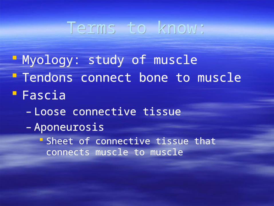
Terms to know:Terms to know:
Myology: study of muscle Tendons connect bone to muscle Fascia
– Loose connective tissue– Aponeurosis
Sheet of connective tissue that connects muscle to muscle
Myology: study of muscle Tendons connect bone to muscle Fascia
– Loose connective tissue– Aponeurosis
Sheet of connective tissue that connects muscle to muscle
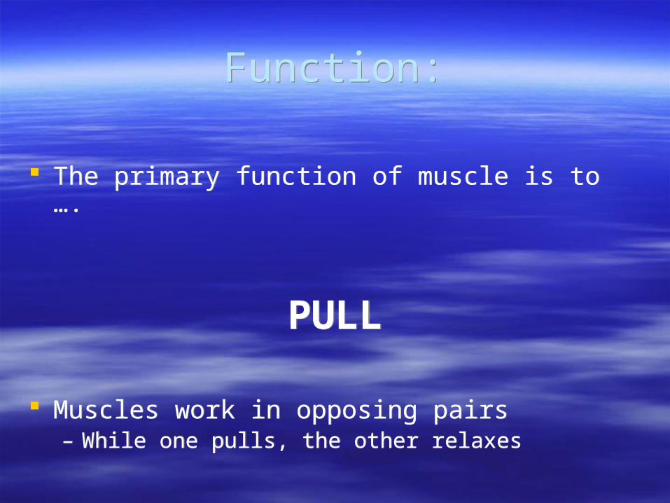
Function:Function:
The primary function of muscle is to ….
PULL
Muscles work in opposing pairs– While one pulls, the other relaxes
The primary function of muscle is to ….
PULL
Muscles work in opposing pairs– While one pulls, the other relaxes

Types of muscle tissue:Types of muscle tissue:
QuickTime™ and a decompressor
are needed to see this picture.
http://www.nlm.nih.gov/medlineplus/ency/images/ency/fullsize/19917.jpg
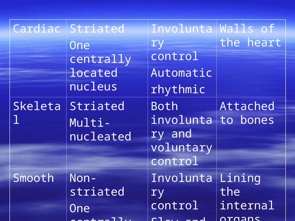
Cardiac Striated
One centrally located nucleus
Involuntary control
Automatic
rhythmic
Walls of the heart
Skeletal Striated
Multi-nucleated
Both involuntary and voluntary control
Attached to bones
Smooth Non-striated
One centrally located nucleus
Involuntary control
Slow and sustained
Lining the internal organs

Voluntary (skeletal) muscle function:
Voluntary (skeletal) muscle function:
1. Movement Physical movement of the body
2. Posture Maintain body position
3. Heat production Muscles give of 65% of their energy
production as heat Works to maintain a constant body
temperature
1. Movement Physical movement of the body
2. Posture Maintain body position
3. Heat production Muscles give of 65% of their energy
production as heat Works to maintain a constant body
temperature
Smooth (involuntary) Muscle function:
Smooth (involuntary) Muscle function:
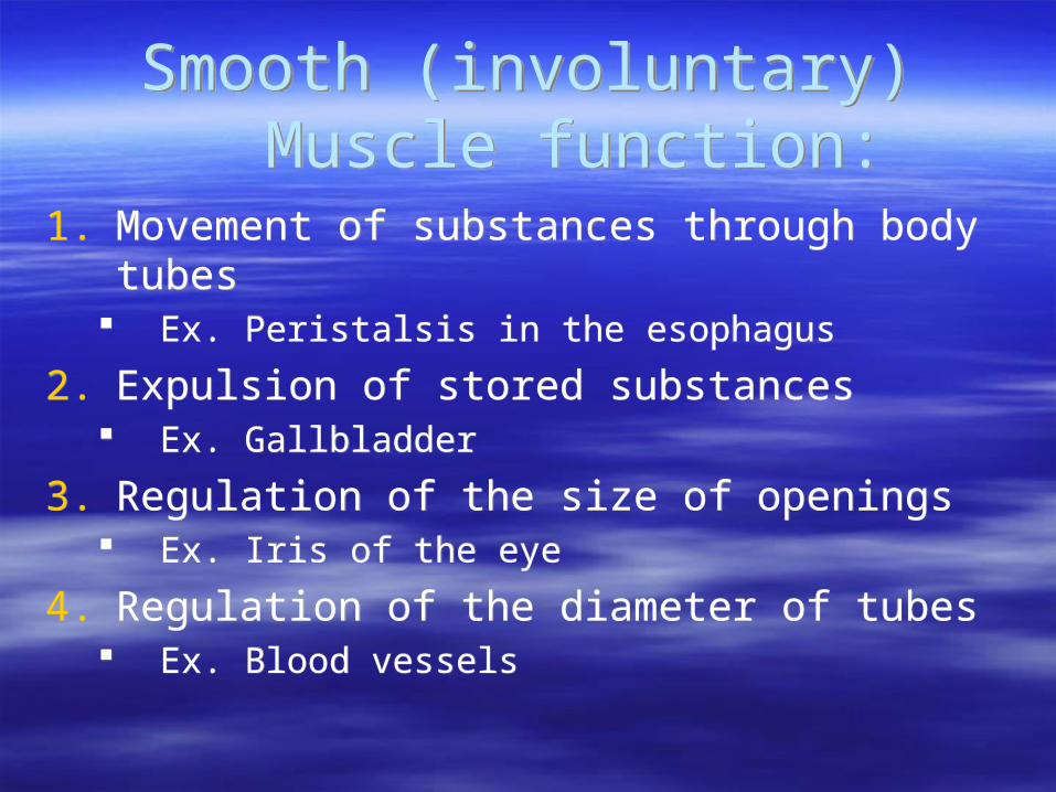
1. Movement of substances through body tubes Ex. Peristalsis in the esophagus
2. Expulsion of stored substances Ex. Gallbladder
3. Regulation of the size of openings Ex. Iris of the eye
4. Regulation of the diameter of tubes Ex. Blood vessels
1. Movement of substances through body tubes Ex. Peristalsis in the esophagus
2. Expulsion of stored substances Ex. Gallbladder
3. Regulation of the size of openings Ex. Iris of the eye
4. Regulation of the diameter of tubes Ex. Blood vessels

Characteristics of Muscle Tissue:Characteristics of Muscle Tissue:
1. Excitability (irritability) Abilitiy of muscle to be stimulated to evoke
response
2. Contractility Function of muscle as a contractile unit Muscle’s ability to contract/shorten
3. Extensibility The ability of a muscle to stretch
1. Excitability (irritability) Abilitiy of muscle to be stimulated to evoke
response
2. Contractility Function of muscle as a contractile unit Muscle’s ability to contract/shorten
3. Extensibility The ability of a muscle to stretch

Characteristics of Muscle Tissue: (cont.)
Characteristics of Muscle Tissue: (cont.)
4. Elasticity The ability of the muscles to return to normal
shape after being stretched or muscle contraction
5. Conductivity The ability to conduct an electric impulse
along the entire length of the muscle
4. Elasticity The ability of the muscles to return to normal
shape after being stretched or muscle contraction
5. Conductivity The ability to conduct an electric impulse
along the entire length of the muscle

Fascia:Fascia:
Loose connective tissue that “wraps” muscle tissue
Superficial fascia: subcutaneous layer that holds skin to
muscles Deep fascia: more fibrous than superficial
hold muscles together
Loose connective tissue that “wraps” muscle tissue
Superficial fascia: subcutaneous layer that holds skin to
muscles Deep fascia: more fibrous than superficial
hold muscles together
Functions of Fascia:Functions of Fascia:
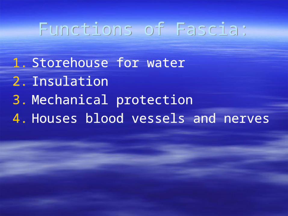
1. Storehouse for water
2. Insulation
3. Mechanical protection
4. Houses blood vessels and nerves
1. Storehouse for water
2. Insulation
3. Mechanical protection
4. Houses blood vessels and nerves
QuickTime™ and a decompressor
are needed to see this picture.
Muscle StructureMuscle Structure
http://www.ivy-rose.co.uk/Topics/Muscles/Epimysium.jpg
Epimyseum: tissue that wraps the entire muscle (found deep to the fascia)
Perimyseum: smaller units of muscle– Holds the fascicles together
Fascicle: collections of the smaller unit– Group of individually wrapped fibers
Endomysium: wrapping of the smaller unit– Each muscle fiber (myofiber) is surrounded by a
plasma membrane individually wrapped in a sheet of endomysium
Sarcolemma is comparable to the plasma membrane of a muscle fiber
Sarcoplasm– Liquid environment within a muscle cell that contains
calcium ions surrounding the myofibrils Myofibrils are made up of 2 types of filaments
(myofilaments)– Actin and Myosin– Adjacent myofilmaments line up with each other so that
the Z-line of one sarcomere lines up with the Z-line of an adjacent myofibrils
T-tubules– Are extensions of the sarcoplasmic reticulum that run
transversely along muscle fibers– Hold substances needed for muscle contraction

QuickTime™ and a decompressor
are needed to see this picture.
http://ab.mec.edu/abrhs/science/AnatPhys_Labs/images/sarcomere.gif
QuickTime™ and a decompressor
are needed to see this picture.
http://www.edcenter.sdsu.edu/cso/paper/image005.jpg
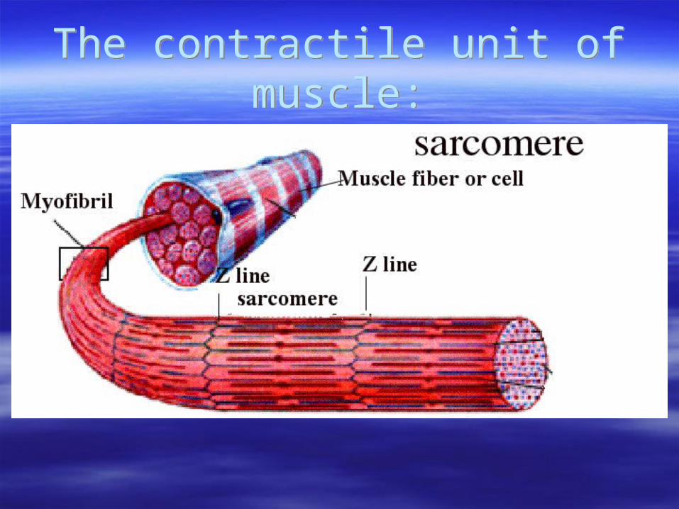
The contractile unit of muscle:The contractile unit of muscle:
Think about it … Think about it …
Tendons are continuous with the epimysium surrounding the larger muscle
Tendons connect muscle to bone
Tendons are continuous with the epimysium surrounding the larger muscle
Tendons connect muscle to bone
Why do we have all of this?Why do we have all of this?
All of these components are necessary to create movement
Muscle generates movement Muscle is living tissue
– Requires blood supply and nerve innervation
All of these components are necessary to create movement
Muscle generates movement Muscle is living tissue
– Requires blood supply and nerve innervation

How do muscles create movement?
How do muscles create movement?
Well actually they don’t …
the Brain does.
Well actually they don’t …
the Brain does.
Movement:Movement:
Movement is initiated in the brain which is part of the Central Nervous System (CNS) with the initiation of an action potential
The impulse travels down the spinal cord out to the motor neuron which carries the electrical impulse to the muscle
Between the motor neuron and muscle is a synapse
Movement is initiated in the brain which is part of the Central Nervous System (CNS) with the initiation of an action potential
The impulse travels down the spinal cord out to the motor neuron which carries the electrical impulse to the muscle
Between the motor neuron and muscle is a synapse

QuickTime™ and a decompressor
are needed to see this picture.

What is needed to create movement?
What is needed to create movement?
Mitochondria– Power house of the cell – Produce energy in the form of ATP with the help
of myoglobin
T-tubules– extension of the sarcoplasmic reticulum– Hold substances needed for muscle contraction
Mitochondria– Power house of the cell – Produce energy in the form of ATP with the help
of myoglobin
T-tubules– extension of the sarcoplasmic reticulum– Hold substances needed for muscle contraction
MyofilamentsMyofilaments
Actin and myosin Myosin has a hook-like binding site while
actin has a bulb shaped binding site These binding sites are not readily available
– The binding sites on actin and myosin are covered by tropomyosin and troponin This is to keep the binding sites from attaching to one
another during a relaxed state

Sliding Filament Mechanism:Sliding Filament Mechanism:1. Brain says move
2. Impulse travels down spinal cord
3. Impulse travels out spinal nerve to periphery (the muscle you want to move)
4. The impulse travels down the motor neuron
5. The impulse reaches the presynaptic bulb of the motor neuron where (ACh is stored in vesicles, the sarcolemma is semipermeable and polarized due to Na+ and K+ ions gathering outside the sarcollema)
1. Brain says move
2. Impulse travels down spinal cord
3. Impulse travels out spinal nerve to periphery (the muscle you want to move)
4. The impulse travels down the motor neuron
5. The impulse reaches the presynaptic bulb of the motor neuron where (ACh is stored in vesicles, the sarcolemma is semipermeable and polarized due to Na+ and K+ ions gathering outside the sarcollema)

6. The impulse crosses the neuromuscular junction (NMJ) with the help of ACh
The junction of the motor neuron and muscle fiber
7. As soon as the ACh hits the sarcolemma the action potential is stimulated and the permeability of the sarcolemma is changed or depolarized.
The wave of depolarization continues down the length of the muscle fiber.
8. Wave of depolarization releases Ca+ ions from the sarcoplasmic reticulum
Actin and myosin have on their surface binding sites so that the myofibrils can bind together to create muscle contraction
Binding sites are protected by Troponin/Tropomyosin to prevent the myofibrils from binding to each other when the muscle is at rest
Ca+ ions push Troponin/Tropomyosin away from the binding sites on actin and myosin, making the binding sites readily available for the creation of cross-bridges
9. ATP (created by the mitochondria by/with myoglobin) allows for a power stroke
Pushes the Z-lines of the sarcomere closer together
10.AChE (Acetylcholine Esterase) is secreted which destroys ACh which allows the sarcolemma to go back to its resting state (repolarized)
9. ATP (created by the mitochondria by/with myoglobin) allows for a power stroke
Pushes the Z-lines of the sarcomere closer together
10.AChE (Acetylcholine Esterase) is secreted which destroys ACh which allows the sarcolemma to go back to its resting state (repolarized)
What can interfere with muscle contraction?
What can interfere with muscle contraction?
Oxygen debt: can’t get enough oxygen into the body so we start to build up lactic acid– Lactic acid is “poison” to the muscles – Slows the reclaiming of Ca+ ions so the muscles
won’t fully relax
Muscle Fatigue: is largely the result of the depletion of oxygen and/or glycogen
Oxygen debt: can’t get enough oxygen into the body so we start to build up lactic acid– Lactic acid is “poison” to the muscles – Slows the reclaiming of Ca+ ions so the muscles
won’t fully relax
Muscle Fatigue: is largely the result of the depletion of oxygen and/or glycogen

Rigor MortisRigor Mortis
“when muscles tighten after you die” When a person dies they no longer posses
oxygen so lactic acid builds up and the sarcoplasmic reticulum breaks down releasing Ca+ ions and the muscles contract
Rigor can last for 12-20 hours
“when muscles tighten after you die” When a person dies they no longer posses
oxygen so lactic acid builds up and the sarcoplasmic reticulum breaks down releasing Ca+ ions and the muscles contract
Rigor can last for 12-20 hours
3 Major Energy systems for Muscle contraction:
(Anaerobic processes)
3 Major Energy systems for Muscle contraction:
(Anaerobic processes)
1. Available ATP
2. Phosphagen system
3. Glycolysis
1. Available ATP
2. Phosphagen system
3. Glycolysis

Available ATP:Available ATP:
Immediate energy source Good for the first 5-6 seconds of energy
expenditure
Immediate energy source Good for the first 5-6 seconds of energy
expenditure
Phosphagen system:Phosphagen system:
Contained in muscles High energy molecule creatine-phosphate or
phospho-creatine Capable of delivering lots of energy in a
short period of time 15 second energy bursts
Contained in muscles High energy molecule creatine-phosphate or
phospho-creatine Capable of delivering lots of energy in a
short period of time 15 second energy bursts
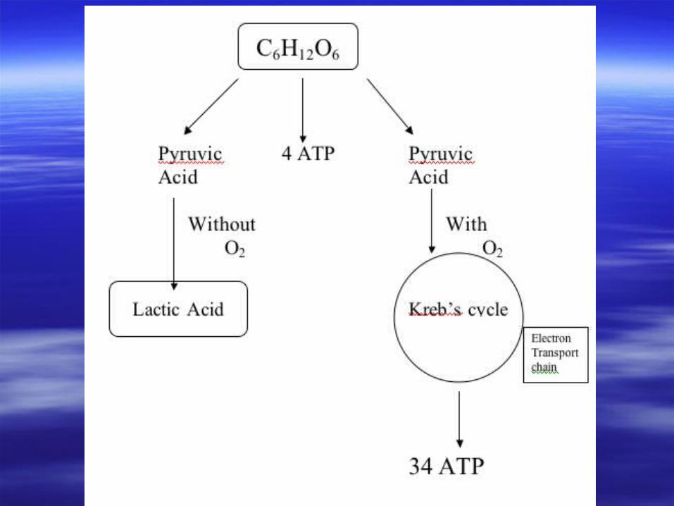
Glycolysis:Glycolysis:
“glucose splitting”– Breakdown of carbohydrates (glucose =
C6H12O6)
The Liver:– Stores glucose in the blood– Converts stored energy into usable energy
“glucose splitting”– Breakdown of carbohydrates (glucose =
C6H12O6)
The Liver:– Stores glucose in the blood– Converts stored energy into usable energy
Muscles contract to create movement:
Muscles contract to create movement:
The All-or-none principle states that when a stimulus is applied a muscle fiber will contract completely or not at all
No such thing as a partial contraction Strength of contraction depends on the
number of fibers stimulated
The All-or-none principle states that when a stimulus is applied a muscle fiber will contract completely or not at all
No such thing as a partial contraction Strength of contraction depends on the
number of fibers stimulated
How do muscles contact?How do muscles contact?
EMG (electromyogram) find diseases that damage muscle tissue, nerves, or the neuromuscular junctions (nmj)
EMG (electromyogram) find diseases that damage muscle tissue, nerves, or the neuromuscular junctions (nmj)
QuickTime™ and a decompressor
are needed to see this picture.
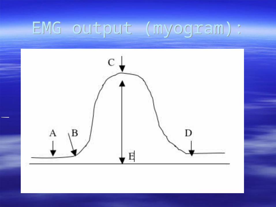
EMG output (myogram):EMG output (myogram):
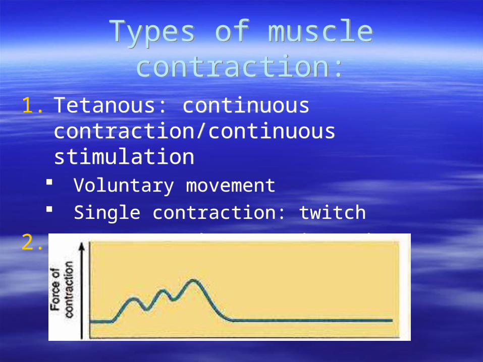
Types of muscle contraction:Types of muscle contraction:
1. Tetanous: continuous contraction/continuous stimulation
Voluntary movement Single contraction: twitch
2. Wave summation: staircasing effect
1. Tetanous: continuous contraction/continuous stimulation
Voluntary movement Single contraction: twitch
2. Wave summation: staircasing effect

Types of muscle contraction (cont.)Types of muscle contraction (cont.)
3. Isotonic: “iso” = same “tonic” = force Contraction that remains at the same force
throughout
4. Isometric: “iso” = same “metric” = length Contraction, no movement
3. Isotonic: “iso” = same “tonic” = force Contraction that remains at the same force
throughout
4. Isometric: “iso” = same “metric” = length Contraction, no movement

Types of muscle contraction (cont.):Types of muscle contraction (cont.):
5. Concentric: muscles shorten while generating force “positive” lift
6. Eccentric: the muscle elongates while under tension due to an opposing force being greater than the force generated by the muscle.
“negative” lift Rather than working to pull a joint in the
direction of the muscle contraction, the muscle acts to decelerate the joint at the end of a movement or otherwise control the repositioning of a load.
Movement:Movement:
Most muscles cross at least one joint– Some cross 2 or even 3 joints
When muscles contract they pull with equal force from both ends– One end of the muscle must be stable to create
movement
Most muscles cross at least one joint– Some cross 2 or even 3 joints
When muscles contract they pull with equal force from both ends– One end of the muscle must be stable to create
movement

Terminology:Terminology:
Origin: immovable end of the muscle – Where the muscle “starts”
Insertion: moveable end of the muscle – The end of the muscle that the force is applied
to
Belly: “meaty” part of the muscle– Where the actual muscle contraction occurs
Origin: immovable end of the muscle – Where the muscle “starts”
Insertion: moveable end of the muscle – The end of the muscle that the force is applied
to
Belly: “meaty” part of the muscle– Where the actual muscle contraction occurs

Levers:Levers:
The body moves through a system of levers Muscle pull works around these levers Levers also give the body a mechanical
advantage– This means that it takes less force to create
movement therefore making the muscles job “easier”
The body moves through a system of levers Muscle pull works around these levers Levers also give the body a mechanical
advantage– This means that it takes less force to create
movement therefore making the muscles job “easier”

Classes of levers:Classes of levers:
QuickTime™ and a decompressor
are needed to see this picture.
http://www.biologyreference.com/images/biol_03_img0301.jpg

Group Actions:Group Actions:
Agonist– “prime mover”– The muscle that initiates the movement
Synergist– Muscles that work together to generate movement
Antagonist– Muscles that work against the prime movers
Fixators/Stabilizers– Muscles that stabilize a joint or bone so that another
muscle can work more efficiently
Agonist– “prime mover”– The muscle that initiates the movement
Synergist– Muscles that work together to generate movement
Antagonist– Muscles that work against the prime movers
Fixators/Stabilizers– Muscles that stabilize a joint or bone so that another
muscle can work more efficiently

How are muscles named?How are muscles named?
600-700 muscles in the body1. Direction of fibers2. Location3. Size of the muscle4. Number of heads of origin5. Shape 6. Origins and insertions7. Action
600-700 muscles in the body1. Direction of fibers2. Location3. Size of the muscle4. Number of heads of origin5. Shape 6. Origins and insertions7. Action
Terminology:Terminology:
Hypertrophy: muscle growth (size)
Atrophy: muscle shrinking
Myopathy: muscle disease
Myoma: muscle tumor
Myolatia: muscle softening
Myocitis: inflammation of muscle tissue
Hypertrophy: muscle growth (size)
Atrophy: muscle shrinking
Myopathy: muscle disease
Myoma: muscle tumor
Myolatia: muscle softening
Myocitis: inflammation of muscle tissue
Disorders of the muscular system:
Disorders of the muscular system:
Fibrosis: increase in the amout of fibrous connective tissue where it is not supposed to be decreasing the ability of the muscles to contract and expand the way they’re supposed to
Muscular Dystrophy: disintigration of muscle fibers can lead to complete atrophy and loss of motor function
Fibrosis: increase in the amout of fibrous connective tissue where it is not supposed to be decreasing the ability of the muscles to contract and expand the way they’re supposed to
Muscular Dystrophy: disintigration of muscle fibers can lead to complete atrophy and loss of motor function

Disorders (cont.)Disorders (cont.)
Myesthemia Gravis: autoimmune disorder that results in the weakening of the the muscles caused by faulty nmj
Abnormal contractions:– Muscle spasm: a single muscle group does an
uncontrolled contraction– Tonic spasm: cramp
Myesthemia Gravis: autoimmune disorder that results in the weakening of the the muscles caused by faulty nmj
Abnormal contractions:– Muscle spasm: a single muscle group does an
uncontrolled contraction– Tonic spasm: cramp
Recommended