156 INŻYNIERIA MATERIAŁOWA MATERIALS ENGINEERING ROK XXXVII
The influence of surface topography elaborated by prism optics based laser interference modification on cell differentiationKrzysztof Czyż1*, Jan Marczak†, Roman Major2, Aldona Mzyk2, Antoni Rycyk1,
Antoni Sarzyński1, Marek Strzelec1, Przemysław Wachulak1
1Institute of Optoelectronics, Military University of Technology, Warszawa, Poland, 2Institute of Metallurgy and Materials Science, Polish Academy of Sciences, Kraków, Poland, *[email protected]
The direct laser interference lithography, based on bi-prism, tetragonal pyramid and an axicon was used as a tool for periodical structuring of different bio-scaffolds. The simple, source laser system with stable Nd:YAG laser resonator, amplifying system (energy up to 1 J) and prism-based interferometer optics gave possibility to create periodical, linear, dotted and even circular surface structures, with high degree of periods, width and depth control. The general idea consists of the imitation of the structure and function of tissues and adoption of these solutions for the material science. It is well known that the influ-ence of the artificial material on the cellular processes related to the differentiation, proliferation and growth are dependent on the chemical composition and surface topography as well as mechanical properties of applied materials. The most recent studies focus on the cell interaction with flat scaffolds. However, cells are found covering a highly rough surface in the blood vessels. The work was related to reconstruction of the structure and function and topography of luminal side of the blood vessels. The most important aspect was to recreate cellular niches which plays specific function on the activation and differentiation of stem cells. The mechanical stresses generated at any certain site depend on the surface micropatterns of the material as well as the other signals that cell receives from chemical stimuli. This work is one of the first attempts to determine correlation of surface micro-topography with biochemical stimulation by adsorbed on the materials surface molecules and both factors potential influence on cellular response. The study was aimed at showing the mechanism of human endothelial progenitor cells adhesion, morphology and function control in response to surface patterning. It was found that micro-channels enhanced intercellular connections forming tight junctions and the appropriate progenitor cell differentiation.
Key words: direct laser interference lithography, laser micromachining, prism optics, bioengineering, progenitor endothelial cells.
Inżynieria Materiałowa 4 (212) (2016) 156÷162DOI 10.15199/28.2016.4.2© Copyright SIGMA-NOT MATERIALS ENGINEERING
1. INTRODUCTION
Direct Laser Interference Lithography (DLIL) is increasingly ap-plied for periodical shaping of material surfaces and for changing their properties [1]. The DLIL can serve as a universal tool, among others to improve tribological properties [2], such as friction, wear and hardness, or optical properties [3], including changes of absorp-tion and improvement of antireflectivity. DLIL applications in bio-engineering for shaping biocompatible scaffolds for tissues using patterned materials with enhanced mechanical properties [4] and selectively influencing the growth and development of cells [5] are particularly interesting. DLIL technique uses two or more superim-posed laser beams that create an intense interference field which is further reproduced on the processed surface. It does not require any preparation or treatment of the area, and the only requirement is a sufficient absorption of material and high enough power den-sity of radiation. Creation of the periodical interference structures with a high degree of order on the surface of almost all materials is possible in a single technological step. There are a lot of methods for dividing a laser beam into an arrangement of many interfering beams [6]. The prism-based method proposed in this paper is one of the simplest solutions [7, 8].
The direct laser interference lithography and direct laser writing techniques have been previously successfully tested in the struc-turing of diamond-like carbon layers [9, 10] and the topographical response of smooth muscle cells and endothelial cells to the pat-tern and form of structuring [11, 12]. Stem cells, which were used herein, are defined as self-renewable and producing differentiated, specialized cells [13÷15]. Each stem cell is capable for prolifera-tion, self-renewal and differentiation through divisions, which are asymmetrical and therefore give rise to two-derived cells, which differ from each other. One of them is the same as the original stem cell, so that supports the population, while the other matures. Each
cell, which grows and matures, has its own niche. Each niche is a spatial structure of cells and extracellular material. It forms a mi-croenvironment that keeps the cells and gives signals to the main cell proliferation, maturation, or to self-renewal. During cell divi-sion, stem cells divide asymmetrically on the stem cell, which re-turns to the niche and progenitor, which is the initial form of the tissue to be reconstructed.
In the work progenitor induced cells were used. Induced en-dothelial cells (i-cells) are purified human endothelial cells derived from pluripotent stem cells. I-cells can be used immediately or maintained over multiple passages. These cells are suitable for use in vascular biology research including angiogenesis, artherosclero-sis, inflammation and other research areas. In cell therapy are used to rebuild tissue. The use of appropriate procedures of de-differen-tiation of the cell makes possible to obtain the recipient’s cells, de-differentiate them and re-introduce the appropriately directed differ-entiated cells to rebuild damaged tissue. I-cell medium supplement has been specially formulated with VascuLife VEGF. Endothelial Cells were cultured within several bioengineered platforms mim-icking the in vivo microenvironment and formed vascular architec-ture to enable tissue engineering and discovery applications [16]. Assays for measuring migration, invasion, and vascular sprouting in response to cytokines and inhibitors are also reported along with basic characterization data. I-cell exhibit characteristic gene and protein expression like CD 31, CD 62E or ZO-1. Anty CD-31 is a cell adhesion molecule which is required for leukocyte transen-dothelial migration (TEM) under most inflammatory conditions. It localizes to the lateral border recycling compartment (LBRC) and recycles from the LBRC to the junction in resting endothelial cells and Cell junction. CD 62E is cell-surface glycoprotein having a role in immunoadhesion [17÷19]. It mediates in the adhesion of blood neutrophils in cytokine-activated endothelium through interaction with PSGL1/SELPLG. Additionally, it may have a role in capillary

NR 4/2016 INŻYNIERIA MATERIAŁOWA MATERIALS ENGINEERING 157
morphogenesis. ZO-1 — this gene encodes a protein located on a cytoplasmic membrane surface of intercellular tight junctions [20, 21]. The encoded protein may be involved in signal transduction at cell–cell junctions. Two transcript variants encoding distinct iso-forms have been identified for this gene.
2. PRISM-BASED LASER INTERFERENCE
Method of prism-based interference consists of formation of a di-vergent beam, e.g. using a negative lens, division of this beam in the prism, and focusing the resulting multiple beams at the surface of the irradiated material. The number of interfering beams depends on the prism geometry (Fig. 1a). Structure period for two beams (bi-prism) is defined by a simple equation [7]:
Λ =
Dd
D d,
(1)
where λ is a wavelength, D is a distance between source and sam-ple, d is a physical interpretation of distance between two virtual sources (Fig. 1a), namely:
d a nbi-prism = −2 1( ) (2)
where a is a distance between the source and the prism, n is a me-dium refractive index, and α is a refraction angle of the prism.
Structure period increases 2 3 3/ times in the case of a py-ramidal triangular prism, and, respectively 2 times in the case of a pyramidal tetragonal prism with the same refraction angle α as in equation (2).
The use of parallel beam makes the structure period independent from the distance between sample and bi-prism, staying dependent only on wavelength and refractive angle (Fig. 1b) of the prism in the form of:
Λ =
−
2sin(arcsin( sin ) )n (3)
The distance of the best overlapping – X can be found using sim-ple mathematical transformations. The lowest theoretical structure period for bi-prism with refractive angle α = 0.7 rad, wavelength of 1064 nm and refractive index of bi-prism n = 1.5 nm is around 900 nm. Larger refractive angle of prism causes total internal re-flection of light, which precludes the desired interference of beams.
Figure 2 shows a comparison of theoretical changes of fabricated structure with experimental results for pyramidal tetragonal prism. Good consistency between experimental results and theory allows for precise adjustment of experimental setup according to the re-quirements of specific tests. It should be noted that the decrease of period of interference structure below 20 μm requires only insertion of prism with a higher refractive angle.
3. EXPERIMENTAL RESULTS
3.1. DLIL patterning
DLIL experimental stand (Fig. 3) comprises of Nd:YAG laser oscil-lator with stable resonator and Q-switch modulator, single-channel laser amplifier, set of harmonic converters and prism interferom-eter. Output energy from the oscillator is 30 mJ, and pulse duration is 8÷10 ns. Inserted diaphragm assures generation of the fundamen-tal transverse mode. After leaving the oscillator, the laser pulse is
Fig. 1. Scheme of radiation incidence on bi-prism in the form of: a) divergent beam, b) parallel beamRys. 1. Schemat padania promieniowania na pryzmat dwuścienny w for-mie: a) wiązki rozbieżnej, b) wiązki równoległej
Fig. 2. Comparison of theoretical and experimental period of struc-
ture, created as a result of irradiation by pyramidal tetragonal prism for constant source–prism distance and variable source–sample dis-tance. Refractive angle α = 5°, n = 1.45, λ = 1064 nmRys. 2. Porównanie teoretycznego i eksperymentalnego okresu struktury wytworzonej w wyniku naświetlania przez pryzmat czworościenny przy stałej odległości źródło–pryzmat i zmiennej odległości źródło–próbka. Kąt załamania α = 5°, n = 1,45, λ = 1064 nm
Fig. 3. Scheme of experimental arrangement based on the prism in-
terferometerRys. 3. Schemat układu eksperymentalnego opartego na interferometrze pryzmatycznym

158 INŻYNIERIA MATERIAŁOWA MATERIALS ENGINEERING ROK XXXVII
amplified in a set of two amplifiers, which results in maximum out-put energy of 1 J.
Harmonics subassembly converts the fundamental Nd:YAG la-ser wavelength to the wavelengths of 532 nm and 355 nm, allow-ing for the decrease in the period of the structures by two or three fold. The negative lens forms divergent laser beam, and the prism, positioned downstream the lens, divides the beam and projects the interference pattern on the sample (Fig. 1a). Change of the sam-ple position reflects in the change of structure period, according to equation (1). Configuration without the lens (Fig. 1b) was also used in several experiments.
Figure 4 presents a microscopic image of a linear structure with a period of 20 μm, created using two-beam DLIL with bi-prism. Dark areas represent a polymer substrate, whereas light lines refer to the diamond-like carbon (DLC) coating. The structure is charac-terised by a good contrast, constant period over the entire patterning area and homogeneity.
The depth of the pattern (grid) is dependent on the laser fluence in one pulse or in more precise micromachining on the number of subsequent laser shots. The last case needs the preservation of phase between shots, which is easily obtained in the-prism based inter-ferometer. More complex 3D surface structures can be obtained by using more than two laser beams, or by multiple sample irradiation using different interference fields (hierarchical structures).
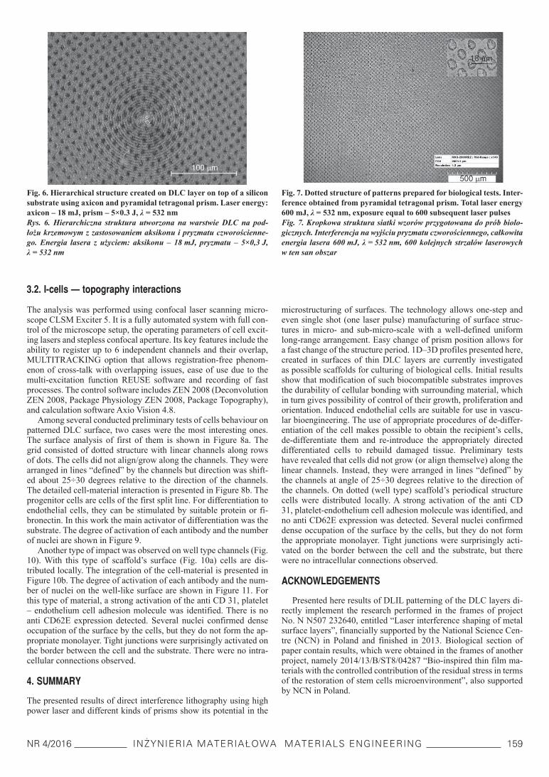
Figure 5a shows a standard dot structure on the DLC surface over silicon as a result of interference of 4 laser beams using pyra-midal tetragonal prism. Comparison of period with the overall di-mensions shows the advantage of the DLIL in creation of ten thou-sand dots over the area of 1 mm2 (in principle over 10×10 mm2) in one or several subsequent laser shots. A period around 12 microme-tres can be seen in Figure 5b. In the case presented in Figure 5c, the structure has been fabricated by five laser pulses with equal energy irradiating the same region of the sample, which is fairly difficult to achieve experimentally. The purpose was to create a dotted peri-odical structure on DLC surface with a thickness of 200 nm with-out perforation into the silicon substrate. Therefore, the contrast of a microscopic image is low. The desired grid has been obtained due to the precise selection of pulse energies and a very homogeneous cross-section of laser beam.
Axicon is a conical optical element with radial symmetry. It is used here to create circular structures due to its properties of divid-ing the laser beam into multiple beams and focusing them on the surface where interference occur. In the experiments quartz axicon
Fig. 4. Linear grid created in DLC layer on top of a polymer sub-
strate. Configuration with bi-prism (Fig. 1b). Laser energy 50 mJ, λ = 1064 nmRys. 4. Liniowy wzór utworzony w warstwie DLC na podłożu polimero-wym. Układ z dwuściennym pryzmatem (rys. 1b). Energia lasera 50 mJ, λ = 1064 nm
Fig. 5. Dotted periodical structure created on DLC layer on top of
a silicon substrate using pyramidal tetragonal prism. Laser energy 250 mJ, λ = 532 nm (a, b); laser energy 5×15 mJ, λ = 532 nm (c)Rys. 5. Kropkowa struktura periodyczna utworzona na warstwie DLC na podłożu krzemowym z zastosowaniem prymatu czworościennego. Energia lasera 250 mJ, λ = 532 nm (a, b); energia lasera 5×15 mJ, λ = 532 nm (c)
was used, which, in combination with the pyramidal tetragonal prism, gives the ability of creating hierarchical structures, such as one shown in Figure 6.
Dotted micropatterns that have been fabricated in order to test in-duced endothelial cells adhesion, proliferation and morphology (de-scribed later in Chapter 3.2) are shown in Figure 7. The total laser output energy was 600 mJ (λ = 532 nm) and number of subsequent laser pulses was around 600. The visible stability of the pattern grid and no blurring of the dots allowed precise control of the features dimensions. A period around 18 micrometres can be seen in a small inset in the upper-right corner of Figure 7.
a)
b)
c)
NR 4/2016 INŻYNIERIA MATERIAŁOWA MATERIALS ENGINEERING 159
3.2. I-cells — topography interactions
The analysis was performed using confocal laser scanning micro-scope CLSM Exciter 5. It is a fully automated system with full con-trol of the microscope setup, the operating parameters of cell excit-ing lasers and stepless confocal aperture. Its key features include the ability to register up to 6 independent channels and their overlap, MULTITRACKING option that allows registration-free phenom-enon of cross-talk with overlapping issues, ease of use due to the multi-excitation function REUSE software and recording of fast processes. The control software includes ZEN 2008 (Deconvolution ZEN 2008, Package Physiology ZEN 2008, Package Topography), and calculation software Axio Vision 4.8.
Among several conducted preliminary tests of cells behaviour on patterned DLC surface, two cases were the most interesting ones. The surface analysis of first of them is shown in Figure 8a. The grid consisted of dotted structure with linear channels along rows of dots. The cells did not align/grow along the channels. They were arranged in lines “defined” by the channels but direction was shift-ed about 25÷30 degrees relative to the direction of the channels. The detailed cell-material interaction is presented in Figure 8b. The progenitor cells are cells of the first split line. For differentiation to endothelial cells, they can be stimulated by suitable protein or fi-bronectin. In this work the main activator of differentiation was the substrate. The degree of activation of each antibody and the number of nuclei are shown in Figure 9.
Another type of impact was observed on well type channels (Fig. 10). With this type of scaffold’s surface (Fig. 10a) cells are dis-tributed locally. The integration of the cell-material is presented in Figure 10b. The degree of activation of each antibody and the num-ber of nuclei on the well-like surface are shown in Figure 11. For this type of material, a strong activation of the anti CD 31, platelet – endothelium cell adhesion molecule was identified. There is no anti CD62E expression detected. Several nuclei confirmed dense occupation of the surface by the cells, but they do not form the ap-propriate monolayer. Tight junctions were surprisingly activated on the border between the cell and the substrate. There were no intra-cellular connections observed.
4. SUMMARY
The presented results of direct interference lithography using high power laser and different kinds of prisms show its potential in the
microstructuring of surfaces. The technology allows one-step and even single shot (one laser pulse) manufacturing of surface struc-tures in micro- and sub-micro-scale with a well-defined uniform long-range arrangement. Easy change of prism position allows for a fast change of the structure period. 1D–3D profiles presented here, created in surfaces of thin DLC layers are currently investigated as possible scaffolds for culturing of biological cells. Initial results show that modification of such biocompatible substrates improves the durability of cellular bonding with surrounding material, which in turn gives possibility of control of their growth, proliferation and orientation. Induced endothelial cells are suitable for use in vascu-lar bioengineering. The use of appropriate procedures of de-differ-entiation of the cell makes possible to obtain the recipient’s cells, de-differentiate them and re-introduce the appropriately directed differentiated cells to rebuild damaged tissue. Preliminary tests have revealed that cells did not grow (or align themselve) along the linear channels. Instead, they were arranged in lines “defined” by the channels at angle of 25÷30 degrees relative to the direction of the channels. On dotted (well type) scaffold’s periodical structure cells were distributed locally. A strong activation of the anti CD 31, platelet-endothelium cell adhesion molecule was identified, and no anti CD62E expression was detected. Several nuclei confirmed dense occupation of the surface by the cells, but they do not form the appropriate monolayer. Tight junctions were surprisingly acti-vated on the border between the cell and the substrate, but there were no intracellular connections observed.
ACKNOWLEDGEMENTS
Presented here results of DLIL patterning of the DLC layers di-rectly implement the research performed in the frames of project No. N N507 232640, entitled “Laser interference shaping of metal surface layers”, financially supported by the National Science Cen-tre (NCN) in Poland and finished in 2013. Biological section of paper contain results, which were obtained in the frames of another project, namely 2014/13/B/ST8/04287 “Bio-inspired thin film ma-terials with the controlled contribution of the residual stress in terms of the restoration of stem cells microenvironment”, also supported by NCN in Poland.
Fig. 6. Hierarchical structure created on DLC layer on top of a silicon
substrate using axicon and pyramidal tetragonal prism. Laser energy: axicon – 18 mJ, prism – 5×0.3 J, λ = 532 nmRys. 6. Hierarchiczna struktura utworzona na warstwie DLC na pod-łożu krzemowym z zastosowaniem aksikonu i pryzmatu czworościenne-go. Energia lasera z użyciem: aksikonu – 18 mJ, pryzmatu – 5×0,3 J, λ = 532 nm
Fig. 7. Dotted structure of patterns prepared for biological tests. Inter-ference obtained from pyramidal tetragonal prism. Total laser energy 600 mJ, λ = 532 nm, exposure equal to 600 subsequent laser pulsesFig. 7. Kropkowa struktura siatki wzorów przygotowana do prób biolo-gicznych. Interferencja na wyjściu pryzmatu czworościennego, całkowita energia lasera 600 mJ, λ = 532 nm, 600 kolejnych strzałów laserowych w ten san obszar

160 INŻYNIERIA MATERIAŁOWA MATERIALS ENGINEERING ROK XXXVII
Fig. 10. Surface top view of the wells (a), substrate influence on the cell behaviour (b) – cumulative picture of images shown in Figure 11
Rys. 10. Widok z góry kropek na powierzchni (a), wpływ podłoża na zachowanie się komórek (b) – zbiorczy obraz zdjęć pokazanych na rysunku 11
Fig. 11. Separated registration channels: a) anti CD62E endothelium-leucocyte adhesion molecule, b) anti CD31-platelet-endothelium cell adhe-sion molecule, c) tight junctions, d) DAPI nucleiFig. 11. Rozseparowane kanały fluorescencyjne oddziaływania komórek progenitorowych: a) molekuła anty CD62E śródbłonkowo-leukocytarna, b) płytkowo-śródbłonkowa molekuła adhezyjna, c) złącza ścisłe międzykomórkowe, d) jądra komórkowe
a)
a) b)
b) c) d)
Fig. 8. Surface top view of the channels (a), substrate influence on the cell behaviour (b) – cumulative picture of images shown in Figure 9
Rys. 8. Widok z góry kanalików na powierzchni (a), wpływ podłoża na zachowanie się komórek (b) – zbiorczy obraz zdjęć pokazanych na rysunku 9
Fig. 9. Separated registration channels: a) anti CD62E endothelium-leucocyte adhesion molecule, b) anti CD31-platelet-endothelium cell adhe-sion molecule, c) tight junctions, d) DAPI nucleiRys. 9. Rozseparowane kanały fluorescencyjne oddziaływania komórek progenitorowych: a) molekuła anty CD62E śródbłonkowo-leukocytarna, b) płytkowo-śródbłonkowa molekuła adhezyjna, c) złącza ścisłe międzykomórkowe, d) jądra komórkowe
a)
a) b) c) d)
b)

NR 4/2016 INŻYNIERIA MATERIAŁOWA MATERIALS ENGINEERING 161
REFERENCES
[1] Lu C., Lipson R. H.: Interference lithography: a powerful tool for fabricating periodic structures. Laser and Photonics Review 4 (2010) 568÷580.
[2] Roch T., Weihnacht V., Scheibe H. J., Roch A. and Lasagni A. F.: Direct Laser Interference Patterning of tetrahedral amorphous car-bon films for tribological applications. Diam. Rel. Mater. 33 (2013) 20÷26.
[3] Xu J., Wang Z., Zhang Z., Wang D., Weng Z.: Fabrication of moth-eye structures on silicon by direct six-beam laser interference lithog-raphy. J. Appl. Physics 115 (2014) 203101.
[4] Dahotre N. B., Paital S. R., Samant A. N., Daniel C.: Wetting behav-iour of laser synthetic surface microtextures on Ti–6Al–4V for bioap-plication. Phil. Trans. R. Soc. A 368 (2010) 1863÷1889.
[5] Yu F., Li P., Shen H., Mathur S., Lehr C. M., Bakowsky U., Mücklich F.: Laser interference lithography as a new and efficient technique for micropatterning of biopolymer surface. Biomaterials 15 (2005) 2307÷2312.
[6] Burrow G. M., Gaylord T. K.: Multi-beam interference advances and applications: nano-electronics, photonic crystals, metamaterials, sub-wavelength structures, optical trapping, and biomedical structures. Micromachines 2 (2011) 221÷257.
[7] Sidharthan R., Chollet F., Murukeshan V. M.: Periodic patterning using multi-facet prism based laser interference lithography. Laser Physics 19 (2009) 505÷510.
[8] Zhou Q., Yang W., He F., Stoian R., Hui R., Cheng G.: Femtosecond multi-beam interference lithography based on dynamic wavefront en-gineering. Opt. Express. 21 (2013) 9851÷9861.
[9] Marczak J., Firak J., Rycyk A., Sarzyński A., Strzelec M., Kusiński J., Major R., Zasada D.: Utilisation of direct interference lithography in creation of periodical structures on the Surface of different materi-als. Inżynieria Materiałowa Materials Engineering 35 (2014) 13÷18, in Polish.
[10] Marczak J., Czyż K., Kusiński J., Onyszczuk T., Rycyk A., Sarzński A., Strzelec M.: Selected methods of laser periodical surface struc-turing. Inżynieria Materiałowa Materials Engineering 35 (2014) 523÷526, in Polish.
[11] Marczak J., Kusiński J., Major R., Rycyk A., Sarzyński A., Strzelec M., Czyż K.: Laser interference patterning of DLC layers for directed migration and growth of smooth muscle cell depositions. Opt. Appl. 44 (2014) 575÷586.
[12] Czyż K., Marczak J., Major R., Mzyk A., Rycyk A., Sarzyński A., Strzelec M.: Selected laser methods for surface structuring of biocom-patible diamond-like carbon layers. Diam. Relat. Mater. 67 (2016) 26÷40.
[13] Lutolf M. P., Gilbert P. M., Blau H. M.: Designing materials to direct stem-cell fate. Nature 462 (2009) 433÷441.
[14] Blau H. M., Sacco A., Gilbert P. M.: Skeletal muscle stem cells. In: Essentials of Stem Cell Biology, 2nd Edition (R. Lanza et al., eds.), Academic Press Inc., San Diego, CA (2009) 249÷257.
[15] Sacco A., Glbert P. M., Blau H. M.: Self-renewal, stem cell. In: En-cyclopedia of Stem Cell Research (Svendsen and Ebert, eds.), SAGE Publications, Thousand Oaks, CA (2008) 493÷496.
[16] Trembecka-Wójciga K., Major R., Lackner J. M., Major B.: Biomedi-cal inspired surface modification. Inżynieria Materiałowa 6 (2014) 560÷563.
[17] Wu F., Zhao Y. Jiao T., Shi D., Zhu X., Zhang M., Shi M., Zhou H.: CXCR2 is essential for cerebral endothelial activation and leukocyte recruitment during neuroinflammation. J. Neuroinflammation 12 (98) (2015) 1÷15.
[18] Kirui D. K., Mai J., Palange A. L., Qin G., van de Ven A. L., Liu X., Shen H., Ferrari M.: Transient mild hyperthermia induces E-selectin mediated localization of mesoporous silicon vectors in solid tumors. PLoS One 9:e86489 (2014) 1÷10.
[19] Hsu C. C., Hen L. F., Lin M. T. Tian Y. F.: Honokiol protected against heatstroke-induced oxidative stress and inflammation in diabetic rats. Int. J. Endocrinol. 2014:134575 (2014) 1÷10.
[20] Huber T. B., Schmidts M., Gerke P., Schermer B., Zahn A., Hartleben B., Sellin L., Walz G., Benzing T.: The carboxyl terminus of Neph family members binds to the PDZ domain protein zonula occludens-1. The Journal of Biological Chemistry 278 (2003) 13417÷13421.
[21] Yamamoto T., Harada N., Kawano Y., Taya S., Kaibuchi K.: In vivo interaction of AF-6 with activated Ras and ZO-1. Biochemical and Biophysical Research Communications 259 (1999) 103÷107.

162 INŻYNIERIA MATERIAŁOWA MATERIALS ENGINEERING ROK XXXVII
Wpływ topografii powierzchni wytworzonej przez laserową modyfikację interferencyjną na różnicowanie komórek
Krzysztof Czyż1*, Jan Marczak†, Roman Major2, Aldona Mzyk2, Antoni Rycyk1, Antoni Sarzyński1, Marek Strzelec1, Przemysław Wachulak1
1Instytut Optoelektroniki, Wojskowa Akademia Techniczna, Warszawa, 2Instytut Metalurgii i Inżynierii Materiałowej Polskiej Akademii Nauk w Krakowie, *[email protected]
Inżynieria Materiałowa 4 (212) (2016) 156÷162DOI 10.15199/28.2016.4.2© Copyright SIGMA-NOT MATERIALS ENGINEERING
Słowa kluczowe: bezpośrednia laserowa litografia interferencyjna, mikroobróbka laserowa, optyka pryzmatyczna, bioinżynieria, progenitorowe komórki śródbłonka..
1. CEL PRACY
W bioinżynierii występuje wpływ składu chemicznego, topografii powierzchni oraz właściwości mechanicznych materiału podłoża na procesy komórkowe związane z różnicowaniem, proliferacją i wzro-stem są zależne od składu chemicznego, topografii powierzchni oraz właściwości mechanicznych zastosowanych materiałów. Ce-lem przedstawionych w artykule eksperymentów było zastosowanie bezpośredniej laserowej litografii interferencyjnej do kształtowania periodycznych struktur na powierzchni biozgodnych warstw amor-ficznego węgla (DLC — diamond like carbon). W pracach wyko-rzystano wcześniejsze wyniki eksperymentów zespołu autorów do-tyczące topograficznej odpowiedzi proliferacji i wzrostu komórek mięśni gładkich i komórek śródbłonka na periodyczne wzory i for-my kształtowania powierzchni DLC. Choć najnowsze publikowane badania koncentrują się raczej na oddziaływaniu komórek z pła-skimi rusztowaniami/podłożami przygotowanymi do ich wzrostu, w mikroskali na powierzchni naturalnych naczyń krwionośnych wy-stępują duże nierówności. Uwzględniono to w przedstawionych wy-nikach badań podstawowych związanych z rekonstrukcją struktury, funkcjonowania i topografii wewnętrznej strony naczynia krwiono-śnego. Najważniejszym aspektem było odtworzenie nisz komórko-wych odgrywających istotną rolę w aktywacji i różnicowaniu ko-mórek macierzystych. Naprężenia mechaniczne powstające w tak określonych obszarach zależą od mikrowzoru powierzchni oraz od innych sygnałów, które komórki otrzymują w wyniku stymula-cji chemicznej. Praca stanowi jedną z pierwszych prób określenia związków pomiędzy mikrotopografią powierzchni a biochemiczną stymulacją przez zaabsorbowane na powierzchni molekuły oraz potencjalnym wpływem obu tych czynników na odpowiedź komór-kową. Opisane badania były ukierunkowane na wstępne ukazanie mechanizmu adhezji, morfologii i kontroli funkcjonowania ludzkich komórek progenitorowych śródbłonka w odpowiedzi na periodycz-ne ukształtowanie powierzchni podłoża.
2. MATERIAŁ I METODYKA BADAŃ
Źródłem promieniowania w stanowisku eksperymentalnym do lase-rowej litografii interferencyjnej był system składający się z genera-tora Nd:YAG, pracującego w stabilnym rezonatorze z Q-switchem, dwustopniowego wzmacniacza Nd;YAG i zestawu przetworników częstotliwości generacji (rys. 3). Czas trwania impulsów laserowych wynosił 8÷10 ns, a maksymalna energia wyjściowa do 1 J. Zestaw przetworników umożliwiał konwersję podstawowej długości fali la-sera Nd:YAG (1064 nm) na jej drugą (532 nm) i trzecią (355 nm) harmoniczną. Pole interferujących wiązek laserowych było tworzo-ne z użyciem optyki pryzmatycznej pracującej z wiązką rozbieżną — soczewką rozpraszającą lub z wiązką równoległą (rys. 1a, b). Wykorzystano symetryczne pryzmaty dwuścienne, czworościenne
i aksikony — stożkowe elementy optyczne o symetrii promieniowej.Rusztowania dla hodowli komórek stanowiły biozgodne war-
stwy DLC naniesione na podłoża krzemowe. Badano wzrost i pro-liferację progenitorowych, indukowanych komórek śródbłonka. Były to oczyszczone, ludzkie komórki otrzymane z pluripotencjal-nych komórek macierzystych.
Badania wytworzonych struktur periodycznych prowadzono z wykorzystaniem cyfrowego mikroskopu optycznego 3D model kH8700 firmy Hirox, Japonia. Analizy oddziaływania indukowanych komórek śródbłonka z podłożem prowadzono za pomocą konfokal-nego, laserowego mikroskopu skaningowego CLSM Exciter 5, wy-korzystując możliwość rejestracji sygnałów fluorescencyjnych barw-ników z aż do sześciu niezależnych kanałów rejestracji obrazów.
3. WYNIKI I ICH DYSKUSJA
Dość prosty schemat system laserowej litografii interferencyjnej, opar-ty na dwuściennych oraz czworościennych pryzmatach optycznych, a także stożkowym aksikonie, umożliwił tworzenie na powierzchni DLC periodycznych topografii struktur w kształcie siatek linii, kro-pek, a nawet symetrycznych okręgów z zachowaniem bardzo dużej powtarzalności okresu (rys. 4÷7). Regulacja energii impulsów lasero-wych i zachowanie jej dużej stabilności pozwoliło również na pełną kontrolę wymiarów struktur (szerokość, głębokość) zarówno przez dobór energii w jednym impulsie, jak i naświetlanie wielokrotne.
Wśród wielu wstępnych prób zachowania się komórek na struktu-rowanej powierzchni DLC najbardziej interesujące okazały się dwa przypadki. W pierwszym z nich siatka wzorów składała się z kropek z nałożoną wzdłuż ich rzędów siatką linii (rys. 8a). Wzrost komórek odbywał się liniowo, ale pod kątem około 25÷30° względem kierunku kanalików w powierzchni. Stopień aktywacji odpowiednich antyciał i liczbę jąder komórkowych pokazano na rysunku 9. Zupełnie innego rodzaju oddziaływanie zaobserwowano w przypadku powierzchnio-wych kanalików w kształcie periodycznych okrągłych studni (rys. 10, 11). Zidentyfikowano silną aktywację molekuły anty CD 31 (adhezja płytka–śródbłonek), brak tworzenia monowarstwy komórkowej (tylko skupiska jąder komórkowych) i brak połączeń międzykomórkowych.
4. PODSUMOWANIE
Przedstawione wyniki bezpośredniej laserowej litografii inter-ferencyjnej z użyciem lasera Nd:YAG dużej mocy i optyki pry-zmatycznej udowodniły przydatność tej metody w periodycznym mikrostrukturowaniu powierzchni DLC z dobrze zdefiniowanym uporządkowaniem na szerokich obszarach. Wstępne wyniki badań wzrostu komórek macierzystych na tak uformowanych, biozgod-nych powierzchniach pokazały z kolei możliwość poprawy trwało-ści wiązań komórkowych z otaczającym materiałem, co umożliwia kontrolę wzrostu, proliferacji i orientacji komórek.
Recommended