THE JOURNAL OFBIO~OOICAL CREMI~TRX Vol.246, No.9,Issue of May 10, pp. 2734-2744,1971
P&led in U.S.A.
The Glucose Oxidase Mechanism
INTERPRETATION OF THE pH DEPENDENCE*
(Received for publication, December 2, 1970)
MICHAEL K. WEIBEL$ AND HAROLD J. BRIGHTS
From the Department of Biochemistry, School of Medicine, University of Pennsylvania, Philadelphia, Penn- sylvania 19104
SUMMARY
The pH dependence of the steady state parameters of the glucose oxidase (EC 1.1.3.4, from Aspergillus niger) reaction was determined by 0 2-monitored experiments over the entire pH range from 3 to 10 at 29, with D-glucose as substrate. The data were fitted to a three-parameter steady state rate equation and the significance of the steady state parameters was examined by stopped flow half-reaction and turnover measurements at the extremes of the pH range used. The major conclusions from these studies can be summarized as follows.
1. At low pH, in the presence of halide, the maximum turn- over number (kcat) is determined entirely by the rate of flavin reduction (k,) in the reductive half-reaction. Further- more, substrate combines only with an unprotonated form of the oxidized enzyme and the reductive half-reaction can be represented as follows.
K, H+,?L, s Eo + S kt - Eo-s
At d E, + &lactons H+ k-1
Since kcat and kz are both specifically decreased by halides at low pH values, it is probable that the turnover rate in the low pH range is also limited by kz in the absence of halide. The steady state absorption spectrum of E. - S is indistinguish- able from the spectrum of E,,. This finding, together with the fact that removal of the l-hydrogen from D-glucose is a rate- limiting process in flavin reduction is consistent with both a hydride transfer mechanism and with a flavin-glucose adduct mechanism in which this adduct is relatively unstable and never accumulates significantly as a kinetic intermediate.
2. The importance of k2 as a limiting first order process in turnover diminishes as the pH is raised. Thus, at pH 10 the major first order process in turnover is the breakdown of a species of oxidized enzyme, E’,,, in the oxidative half-reaction. The rate of this process at pH 10.0 is 214 set-l, whereas kz has a value of 800 se0.
3. The reduced enzyme exists in two kinetically significant
* This project was supported by Grant GM 11040 from the National Institutes of Health, United States Public Health Serv- ice.
$ Recipient of National Institutes of Health Postdoctoral Fellowship 5 F02 GM-40938.
$ Research Career Development Awardee 5 K3 GM-34960, United States Public Health Service.
states of ionization, E,. and E,-. The rapid reoxidation of E, with 02, to regenerate &, is predominant at pH values less than 7. At pH values greater than 7, a much less rapid reac- tion of E,- with OZ, leading to the formation of Pow, becomes increasingly important. The species Elo- is unreactive with glucose and it is the conversion of a protonated form of El,,- to E. which principally governs keat at pH values greater than 7.
We present a complete kinetic scheme describing the effects of pH and discuss the possible chemical significance of the species E’,,.
The oxidation of aldoses to the corresponding lactones cata- lyzed by glucose oxidase (EC 1.1.3.4) is shown in Equation I.
EQUATION I
The first rapid reaction studies were reported by Gibson, Swo- boda, and h/Iassey (1) and by Nakamura and Ogura (2). Re- cently, Bright and Appleby (3) have described the pH de- pendence of individual steps of the reaction in studies (with the enzyme from Penicillium notatum) which were based on the kinetic mechanism elaborated by Gibson et al. (I) for the Asper- gillus niger enzyme. With P-n-glucose, at 25” and pH 5.5, the general mechanism of Gibson et al. reduced to’
G-+-S- k E, f P (2)
Ev + 02 k4 - E,,’ ----+ ?a E, + H,Ot (3)
SCHEMII: 1
1 The following abbreviations are used: ET, total catalytically active enzyme expressed in terms of substrate-reducible FAD; Eo, catalytically active enzyme in which FAD is in the oxidized form; E,, catalytically active enzyme in which FAD is in the reduced form; S, p-n-glucose. It should be noted that whereas previously (3) the mechanism for glucose oxidation at 25” was analyzed in terms of three steps and four essential ionizations, in these studies the mechanism has been considerably extended and modified. It has proved convenient to initially analyze turnover experiments in terms of the three parameters kred, k,,, and k,,t, and then examine the makeup of these parameters. As far as possible, the rate and equilibrium constant notation used pre- viously (4) has been retained.
2734
by guest on August 21, 2018
http://ww
w.jbc.org/
Dow
nloaded from
Issue of XIay 10, 1971 M. K. Weibel ancl H. J. Bright 273.5
In contrast, Nakamura and Ogura deduced the following scheme for both the A. n@er and P. rwtutuna enzymes (2, 4) at pR 5.5 and 25”.
kl Ic2 EofS ; Eo-s - (4)
k-1 E, + P
h E, + 02 - Ea + Hz02 (5)
&!HEME 2
Tsoth mechanisms require a single first order step involving an oxidized species of the enzyme. This species is identified as ,Vo in Scheme 1 and as & - S in Scheme 2. The two mech- anisms are homeomorphic with respect to their steady state turnover behavior.
From a combination of stopped flow and 02-monitored experi- ments, we show that the pH profiles of the A. niger enzyme are in substantial agreement with those of P. n&turn reported by Bright and Appleby in the presence of 0.2 M KC1 (3), but that the entire pH profile should be interpreted as a hybrid of Schemes 1 and 2.
EXPERIMENTAL PROCEDURE
Materials
Glucose oxidase (A. niger), analytical grade was obtained from $Iann Research Laboratories. The enzyme was further purified by chromatography on Whatman DE-23 DEAE- cellulose. A column, 2.5 X 30 cm, was equilibrated with 0.05 M potassium phosphate, pH 6.0, and approximately 200 mg of the protein were applied in 20 ml. The column was washed with 500 ml of the same buffer and then subjected to a 2-liter linear potassium phosphate gradient (pH 6.0, from 0.05 to 0.30 M).
A sample from the center of the yellow enzyme band was homo- geneous by standard disc gel electrophoresis and showed negligible catalase activity. A sensitive measurement of trace catalase activity in the purified glucose oxidase preparations indicated it to be present at noninterfering levels. O2 evolution as a result of catalase in the purified enzyme was measured in the following manner. An anaerobic solution of 10 pM glucose oxidase was incubated in the 02 monitor at pH 7.0 in the pres- ence of 1 OV4 M EDTA. Hz02 (low3 M) was added to the anaerobic enzyme solution and 02 evolution was measured. The rate of O2 evolution was several orders of magnitude smaller than the rate of O2 uptake caused by 10 PM glucose oxidase under turn- over conditions, where the highest attainable concentration of O2 is about 10e3 M. Furthermore, addition of CN- to both turnover and stopped flow experiments was without effect.
The purified enzyme was stored either as a precipitate in ammonium sulfate (60 g/100 ml) solution or in 0.10 M phos- phate, pH 6.0, at 4”. Enzyme was routinely recovered and concentrated, or subjected to buffer exchange, by means of the Schleicher and Schuell collodion bag apparatus (Schleicher and Schuell Company, Keene, New Hampshire).
Anhydrous dextrose, chemically pure, was obtained from Pfanstiehl Laboratories, Waukegan, Illinois, and all solutions were allowed 24 hours at 25” to mutarotate to equilibrium. Under all conditions of turnover used, mutarotation is very slow compared with the total time required for the last experimental
point to be taken. All concentrations of glucose are expressed as the amount of /? anomer present.
The following buffers were used and contained 10e4 M EDTA: 0.01 M potassium citrate for the pH range 3.0 to 5.0, with the exception of fluoride solutions in which species such as HF;- are sufficient buffering agents; 0.01 M imidazole-HCl for the pH range 6.0 to 8.0; 0.01 M Tris-HCl for the pH range 7.0 to 9.0; 0.01 M L-leucine or L-alanine for pH 10.0. Buffer adjustments were made with H&O4 or KOH. All chemicals, including buffers, were of “analyzed” or “certified” grade. Water was commercially deionized and solutions routinely millipored before use. Control of ionic strength was not necessary at the low buffer concentrations used. No specific buffer effects were observed.
Methods
Enzyme Concentration-All enzyme concentrations are ex- pressed as molarity of catalytically active bound FAD. Active flavin was determined spectrophotometrically on a Cary model 15 with a differential molar extinction coefficient of 1.31 x 10“ X+ cm-r ( f 5 yO) at 450 mp. This value was determined else- where (5) and represents the differential molar extinction co- efficient between oxidized and reduced bound FAD of the en- zyme from A. niger in 0.01 M potassium acetate, pH 5.5. This value is in good agreement with published spectra (6) and a reported extinction coefficient value of 1.4 X lo4 M-~ cm-’ for oxidized enzyme at 450 rnp (7). Typically the absorbance of solutons containing 1 to 10 pM bound FAD was measured in a. standaird, l-ml cuvette with 0.01 M potassium acetate, pH 5.5 A 1 .O M glucose solution, 10 ~1, was added via a Hamilton syringe, the solution mixed, and the cuvette top sealed with parafilm. The absorbance was then recorded for the reduced form and appropriate dilution corrections made. Accuracy was limited only by pipetting procedures and was within 1 yO.
Enzyme stability at the extremes of the pH range of interest was checked by means of OZ-monitored turnover experiments. With 0.10 M glucose and partially air-saturated solutions, rates of 02 uptake were determined throughout a 15-min experiment in which greater than 90% of the 02 was eventually consumed. Plots of ET/v versus l/O2 were linear, indicating that ET was conserved throughout the entire experiment. Since turnover data for kinetic analysis were gathered in less than 5 min in each case, none of the results is complicated by irreversible or slowly reversible effects of pH on the enzyme.
Oxygen-monitored Turnover-The Clark electrode, power source, and amplifier assembly were obtained from Yellow Springs Instrument Company, Yellow Springs, Ohio. The elec- trode chamber was thermostated at 25” and a IO-inch Beckman recorded was used. A 3-ml aliquot of the appropriate glucose solution was equilibrated by rapid stirring for 5 min with air or by bubbling O2 into the chamber. The value used for 02 solu- bility in pure water at 25” and total pressure of 760 mm was 1.21 x 10m3 M (8). 02 concentrations in dissolved air were referenced to this value and appropriate barometric corrections were made when significant. Low buffer and glucose concentra- tions were used when possible in order that the 02 activities measured by the electrode reflect actual oxygen concentrations. No corrections were made for salt effects on the solubility of O2 in the halide experiments as the maximum concentration of salt was 0.1 M and would only amount to a maximum correction of
by guest on August 21, 2018
http://ww
w.jbc.org/
Dow
nloaded from

2736 pH Depen,dence qf Glucose Oxidase Reaction Vol. 246, No. 9
3%) (9). Additions of enzyme were made to the system through a groove in the electrode holder with Hamilton syringes. Since the data obtained are integral in nature, integrated rate equa- tions for turnover were used. It was found that a simple three parameter equation (Equation 6) was adequate to fit all data at the 1% level for a given pH. Equation 6 is the integrated form of Equation 7.
lETI. = (l/khd)‘h ([ii$/([f% - @&I - [&It)))
+ Wk,,)~ln ([OM[OZ~~) + (l/&t). (IO210 - IOzlJ (6)
ET/V = l/(k,,d’[SI) + l/(kox.[Ozl) + l/kat (7)
These equations are based on a minimal mechanism involving two bimolecular steps and one monomolecular step in the turn- over sequence. The integral data from measurements at 10 or more glucose concentrations, in which 02 had been 80% depleted, were fitted by Equation 6 by optimizing the three rate parameters. A nonlinear regression program developed by Marguardt (lo), SHARE 3049, was revised to accommodate weighting and adapted to the problem. Confidence interval algorithms are built into the program and are indicated in the appropriate figures by vertical bars. Studies with fluoride at low pH were essentially zero order in 02 and linear plots of ET/v versus l/[S] were hand calculated.
Stopped Flow Turnover Experiments-These experiments were carried out at pH 10 at 25” with the Gibson-Durrum stopped flow apparatus. Bound FAD was measured spectrophotometri- tally at 450 rnp with slit widths less than 1.0 mm. The experi- ments were performed by a pH jump on the enzyme. The enzyme solutions were unbuffered at pH 7.8 and saturated with OZ. The glucose solution was made anaerobic by bubbling deoxygenated Nz through the solution for 15 min. The integral traces of percentage of transmission versus time were converted to absorbance versus time curves, then integrated sectionally and totally by numerical quadrature. Appropriate treatment of the areas obtained appears elsewhere (5). The two principal equations used are 8 and 9.
where E,, is the sum of all oxidized enzyme species and l//c’ is Oxygen-monitored Turnover-Fig. I, 2, and 3 show, respcc- the sum of the reciprocals of all first order rate constants as- tively, the pH profiles of kr&, k,,, and kcat as obtained from sociated with oxidized enzyme species. These yield all param- three parameter fits of 02-monitored turnover experiments. eters obtained from conventional steady state turnover monitored The integral data were fit to Equation 6 at the l$& level with 02 by product formation or substrate depletion. and glucose concentrations varied 5- and IO-fold, respectively.
Differential spectra of the intermediates associated with ICcat were obtained by observing initial absorbance of stopped flow experiments as a function of wave length. The steady state concent’rations of these intermediates could be readily cal- culated from known values of rate constants and substrate concentrations.
Stopped Flow Half-reactionsAl half-reactions were forced to pseudo first order reactions by maintaining S or O2 >> enzyme. The reductive half-reactions were carried out as follows. The
The pH profile for kred (Fig. I), the apparent bimolecular rate constant for the combination of glucose with enzyme, shows two distinct limbs in the absence of halide. The low pII limb is profoundly influenced by halide resulting in the upward shift of an apparent pK value. The chloride effect was noted in the case of the P. notatum enzyme, but not documented (3). The high pH limb is not characteristic of a simple ionization and may reflect nonspecific charge effects associated with several ionizations.
enzyme was slightly buffered at pH 7.8 with 0.001 M L-leucine and was made anaerobic by purging the interior of the reservoir syringe with Nz while stirring internally with a small magnetic bar. After 30 min, 10 PM L-amino acid oxidase (5 ~1 per ml of glucose oxidase solution) were added and the syringe sealed onto the valve block of the st>pped flow instrument. &oxygenated Nz was bubbled through the buffered glucose solution syringe for 5 min and 10 PM glucose oxidase (5 ~1 per ml of glucose solution) were added. The solution was then sealed to the valve block. This procedure achieves and maintains anaerobiosis of the solutions extremely well.
The reoxidative half-reactions were carried out as follows. The enzyme side was unbuffered at pH 7.8 and the reservoir syringe swept with Nz as before. Mannose (1.0 M, 1 ~1 per ml of enzyme solution) was then added and the enzyme was slowly reduced. The 02 side was buffered and initially saturated with either air or 02. Variable 02 concentrations were achieved by carefully mixing air- or 02-saturated solutions with N2-saturated solutions by means of a three-way Hamilton valve. Escess mannose on the millimolar level does not interfere with the reoxidative half-reaction as it reduces E, at least two orders of magnitude more slowly than 0% oxidizes E, under the expcri- mental conditions.
Electron Paramagnetic Resonance Experiment--8 turnover reaction of several seconds duration was initiated by mixing 20 PM unbuffered glucose oxidase with 0.05 M glucose, pII 10.0. Both solutions were initially air saturated and mixed rapidly in a simple fashion through a “T” joint. The mixed solution was delivered directly into a standard 3-mm quartz tube immersed in liquid Nz by means of a small diameter, short polyethylene needle. This procedure effectively quenched the reaction by rapid freezing under conditions where the principal rate-limiting process of turnover is the combination of ER with OZ. We estimate that 0.5 pm of G;- could have been detected in this experiment, which represents about 57; of the concentration of ER present. Electron paramagnetic resonance spectra were ob- tained with a Varian E-3 instrument equipped with a variable temperature regulator. The cavity was regulated at 100°K. The following settings were employed: field center at 3280 gauss; field width f 100 gauss; microwave power 20 milliwatts; mod- ulation amplitude 0.1 to 5.0 gauss at 100 kHz; microwave fre- quency, 9.17 GHz. At the highest sensitivity, no signal corre- sponding to any free radical could be detected.
RESULTS
by guest on August 21, 2018
http://ww
w.jbc.org/
Dow
nloaded from

Issue of May 10, 1971 M. K. Weibel and H. J. Bright 2737
I
I
8 I
!.O 4.0 6.0 8.0 10.0
PH
Fro. 1. The pH profile of kred (0, no halide; l , in the presence of 0.1 M Cl-). Evaluation of kred was carried out by nonlinear regression on Equation 6, using &monitored turnover data at the indicated pH values. Confidence intervals indicated by horizontal bars. Temperature 25” ; other conditions given under “Experi- mental Procedure.”
)C
I- 2.0 4.0 6.0 8.0 10.0
PH
FIG. 2. The pH profile of k,,. Evaluation of k,, was carried out by nonlinear regression on Equation 6, using 02-monitored turnover data at the indicated pH values. indicated by horizontal bars.
Confidence intervals The solid line represents computed
values of k,, using Equation 13 and constants from Table I. Tem- perature 25”; cedure.”
other conditions given under “Experimental Pro-
Fig. 2 shows the pH profile of k,,, the apparent bimolecular
rate constant for the combination of 02 with reduced enzyme.
Although the behavior of lc,, resembles a simple titration, the
limiting value at high pH values does not approach zero. Anal- ogous studies of the P. notatum enzyme were not extended to pH 10 and the leveling off of k,, at high pH was not seen. No halide effects were observed on this parameter at any pII.
Fig. 3 portrays the pH profile of kcat. This parameter rep-
I 2.0 4.0 6.0 6.0 IO.0
PH
FIG. 3. The pH profile of kGat (0, no halide; 0, in the presence of 0.1 M Cl-). Evaluation of kcat was carried out by nonlinear regression on Equation 6, using Op-monitored turnover data at the indicated pH values. Confidence intervals indicated by hovi- zontal bars. The solid line represents computed values of k,,t using Equation 14 and constants from Table I. Temperature 25’; other conditions given under “Experimental Procedure.”
0.75
;" : 0.50
-G 0‘ '0 ;
:: 0.25 9
0
II
II II
KCI , M
FIG. 4. The Cl- dependence of kred at pH 3.0. Evaluation of kred at each Cl- concentration was carried out by nonlinear regres- sion on Equation 6 using 0%.monitored turnover data. Confidence intervals indicated by horizontal bars. Temperature 25’; 0.01 M citrate buffer; other conditions as given under “Experimental Procedure.”
resents the limiting turnover velocity, v/ET, at infinite concen- trations of both 02 and glucose. It may reflect more than one first order process in turnover and has the form (21 /ki)-l, where the lci are individual rate constants for all kinetically significant
first order processes. It should be noted that kcat reaches a constant value of about 1000 see-1 at low pH in the absence of halide and falls off significantly at high pH. Halide specifically affects this constant at low pH and the effect of 0.10 RI Cl- is indicated in Fig. 3. At low pH, this parameter has rather large confidence intervals because of its small contribution to the turnover equation, but is well correlated. The effect of halide
on hat, which will prove to be important in the interpretation of the kinetic mechanism, was not investigated previously (3).
by guest on August 21, 2018
http://ww
w.jbc.org/
Dow
nloaded from

2735 pH Dependence of Glucose Oxidase Reaction Vol. 246 Yo 9 11 *
0
A
If
I ‘h
P n
0.02 0.05
KCI ,M
0.10
FIG. 5. The Cl- dependence of k,,t at pH 3.0. Evaluation of keat at each Cl- concentration was carried out by nonlinear regres- sion on Equation 6 using 02.monitored turnover data. Confidence intervals indicated by horizontal bars. Temperature 25’; 0.01 M citrate buffer; other conditions given under “Experimental Pro- cedure.”
FIG. 6. Comparison of stopped flow reductive half-reaction data (l/kred.obs, X) with 02.monitored turnover data (ET/V, 0) at pH 4.0, 0.1 M total fluoride. The turnover data were analyzed according to Equation 10. Temperature 25”; other conditions given under “Experimental Procedure.”
The parameters kred and kcat were also decreased in the pres-
ence of F- and Br- (measured by Oz-monitored turnover at
pH 4.0 and 0.10 M total halide), the order of effectiveness being F- >> Cl- = Br-. As previously reported (3), sulfate showed no effect. Figs. 4 and 5 show the effect of Cl- on the profiles of bed and hat, respectively, at pH 3.0. It should be noted in Fig. 5 that it is not possible to isolate /c,,~ in the low Cl- experi- ments as evidenced by the initial uncorrelated interval. This is attributed to the large initial decrease of kred, relative to that
of Lt, which causes the turnover equation to be principally controlled by k& with the substrate concentrations used.
Stopped Flow reductive Ha!&Reactions-Fig. 6 shows a com- parison of the stopped flow reductive half-reaction and On-moni-
FIG. 7. Comparison of stopped flow reductive half-reaction data (l/kred.abs, X in the presence of 0.01 M oL-leucine, 0 in the presence of 0.01 M Dkphenylalanine), stopped flow Eo-monitored
(S
m turnover data E&/[Os]o, l
> andOs-monitored turnover dat,a
(ET/vo~+,, - - -) it pH 10.0. The stopped flow and 02.monitored turnover data were analyzed according to Equations 8 and 7, respectively, and were both obtained in the presence of 0.01 M DL-leucine. Temperature 25”; other conditions given under “Experimental Procedure.” From the ordinate intercepts, kz and keat are 800 f 200 set-1 and 170 see-l, respectively. From the reciprocals of the slopes, kred is 1.1 X 10’ M-’ set-I.
I 0 2.0 4.0 6.0 8.0 10.0
+ x r0-5,(tfl-‘).s-’
FIG. 8. Stopped flow EO-monitored turnover data, at pH 10 and with several glucose concentrations, plotted according to Equa- tion 9. Temperature 25’; 0.01 M DL-leucine; other conditions given under “Experimental Procedure.” The ordinate intercept of unity shows that no first order process in turnover involves a species of reduced enzyme. The value of k,, from the reciprocal of the slope is 1.2 X 10⁶ M-I set-I.
tored turnover experiments at pH 4.0 with 0.10 M total fluoride. Under these conditions, the turnover reaction is zero order in 02 and Equation 7 reduces to Equation 10.
ET/V = l/(kred[f% + l/k,,t (10)
The fact that an ordinate intercept is observed for the half-
by guest on August 21, 2018
http://ww
w.jbc.org/
Dow
nloaded from

Issue of May 10, 1971 M. K. Weibel and H. J. Bright
10.0 - e’ -
s v)
FIG. 9. Stopped flow oxidative half-reaction at pH 3.2. Errors in mixing solutions containing dissolved Nz and 02 are indicated by vertical bars. Temperature 25’; 0.01 M citrate; other condi- tions given under “Experimental Procedure.” The value of k,, from the reciprocal of the slope is 1.8 X lo6 M-I see-*.
reaction plot and is identical with that of turnover indicates that keat is associated with an odxidieed form of the enzyme and occurs in the reductive half-reaction. The simplest explanation is that the reductive half-reaction can be represented as Equa- tion 4.
At high pH, the same comparison (pH 10.0, no halide), shown in Fig. 7, indicates that the slope of the stopped flow reductive half-reaction data is identical with that of both OQ-monitored turnover and stopped flow &-monitored turnover. However, the ordinate intercept of the half-reaction plot corresponds to 800 f 200 see-l which is considerably greater than the values corresponding to the ordinate intercepts of the turnover data. This indicates that at high pH there are one or more first order processes, in addition to kz, which are kinetically significant in turnover.
Stopped FZQW E,-Monitored Turnover-The stopped flow turnover data of Fig. 7 obtained at pH 10.0 are plotted ac- cording to Equation 8 and agree well with the line corresponding to 02-monitored turnover data plotted according to Equation 7. Both yield slopes of l//&d and the same ordinate intercept (corresponding to 170 see+). The stopped flow turnover inter- cept contains only those first order processes involving oxidized forms of the enzyme. The ordinate intercept of the turnover plot would contain all first order processes involving both oxidized and reduced intermediates. The fact that these two intercepts are equal indicates that there are no significant first order pro- cesses in turnover involving reduced enzyme. Furthermore, because of the agreement between the stopped flow turnover and 02-monitored turnover experiments, it must be concluded that E0 - Sand the other species of oxidized enzyme which partici- pate in kinetically significant first order processes have the same extinction coefficient as E. at 450 rnp, where the stopped flow- turnover reactions are monitored.
The oxidative constant k,, may also be obtained from the stopped flow turnover data from plots of Equation 9. Such a plot is shown in Fig. 8 for several glucose concentrations. The slope of this plot is l/k,, and the intercept is unity if there are no significant first order processes involving reduced inter- mediates (5).
FIG. 10. Stopped flow oxidative half-reaction at pH 10.0. Er- rors in mixing solutions containing dissolved Nz and 02 are indi- cated by vertical bars. Temperature 25”; 0.01 M nn-leucine; other conditions given under “Experimental Procedure.” The value of k,, from the reciprocal of the slope is 1.5 X 10⁶ M-* set-I.
1.0
a I.
< 00 0
i= 0.5- 8 0 P X
E
5 !? 8 0 0 xX t 0
E X 0
, 9x
350 400 450 500
mu
FIG. 11. Comparison of statically (0) and kinetically (X) determined difference absorption spectra at pH 3.0. The static difference spectrum is that of W6 M fully oxidized enzyme minus lo+ M fully reduced enzyme recorded on a Cary 15 spectrophotom- eter. The kinetic difference spectrum was obtained by the stopped flow method and consists of the initial steady state ab- sorbance during turnover (due chiefly to Eo-S) minus the ab- sorbance of fully reduced enzyme. The stopped flow experiments involved the mixing of 2 X 10-G M glucose oxidase in 1.5 M n-glucose (initially air-equilibrated) with an equal volume of solution containing 1.5 M n-glucose and saturated with OZ. For the sake of comparison, the two difference spectra have been normalized at 450 rnp. Temperat,ure 25”; 0.01 M citrate; other conditions given under “Experimental Procedure.”
Stopped Flow Oxidative Half-reaction-Figs. 9 and 10 are double reciprocal plots of pH jump oxidative half-reaction data at pH 3.1 and 10.0, respectively. The plot for low pH yields a k,, value (1.8 x lo6 M-I set+) which is identical with that from 02-monitored turnover experiments. A possible intercept
by guest on August 21, 2018
http://ww
w.jbc.org/
Dow
nloaded from
pH Dependence of Glucose Osidase Reaction Vol. 246, No. 9
Q Q
I 1 t
350 400 450 500
mu
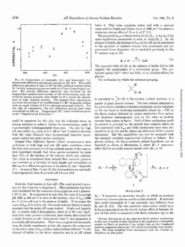
FIG. 12. Comparison of statically (0) and kinetically (X) determined difference absorption spectra at pH 10.0. The static difference spectrum is that of 1(Y6 M fully oxidized enzyme minus 10m5 M fully reduced enzyme recorded on a Cary 15 spectrophotom- eter. The kinetic difference spectrum was obtained by the stopped flow method and consists of the initial steady state ab- sorbance during turnover (due chiefly to E’,) minus the absorb- ance of fully reduced enzyme. The stopped flow experiments involved the mixing of air-equilibrated 2 X l(rs M glucose oxidase with an equal volume of 0.2 M n-glucose saturated with 02. For the sake of comparison, the two difference spectra have been normalized at 450 mF. Temperature 25”; other conditions given under “Experimental Procedure.”
could be conceived for this plot, but the indicated errors on mixing solutions to achieve various 02 concentrations make it experimentally indistinguishable from zero. The plot for high pH also yields a k,, value (1.5 X lo5 M-’ see-i) which is identical with the value obtained from Os-monitored turnover experi- ments carried out under similar conditions.
Sfopped Flow Difference Spectra-These measurements were performed at both high and low pH under conditions where the first order processes involving oxidized species of the enzyme were important enough that these species accounted for more than 75% of the fraction of the enzyme which was oxidized. The initial A absorbance from stopped flow turnover patterns was recorded as a function of wave length and normalized at 450 mp to a difference spectrum of E, minus E, (see “Appendix A”). As seen in Figs. 11 and 12, the intermediates are spectrally indistinguishable from E,, at both pH 3.0 and 10.0.
DISCUSSION
Reductive Half-reaction at Low pH-The most general expres- sion for this sequence is Equation 4. This mechanism has been well established for the substrates 2-deoxyglucose and n-glucose- I-*H (1, 10). It is important to note that the value of ki at high pH (800 + 200 see-r) is close to the limiting value of heat (1000 see-i) at low pH and in the absence of halide. If we assign the value of kcat to ks at low pH, this would indicate that ks is nearly constant over the entire pH range studied. Bright and Appleby (3), in studies with 2-deoxyglucose, where kz is the only signif- icant first order process in turnover, have shown that except for a slight decrease at pH values greater than 7, this parameter is essentially pH independent. These facts, coupled with the results from the halide experiments at low pH, support the assignment of the entire value of kcat (with a value of about 1000 set? in the absence of halide) to the flavin reduction step ks at pH values
below 5. This value correlates rather well with a minimal value used by Bright and Gibson for kz of 1000 set-l to achieve a deuterium isotope effect of 15 on kz at 3” (11).
The parameter kred is interpreted as (k&z) /(k-r + k,), or, if the rapid equilibrium assumption is used, as (k,k,)/(k-,). In the absence of halide, the decrease of kred at low pH can be attributed to the partition of oxidized enzyme into protonated and un- protonated forms (Equation 11) as concluded previously for the P. notatum enzyme (3).
Kl E. + H+ ; &a+ (11)
The apparent value of pK1 in the absence of halide (2.5 to 3.0) suggests the participation of a carboxylate group. The pro- tonated species E,Hf either has little, or no, binding affinity for glucose.
The mechanism by which the substrate grouping
\,a
is converted to \, ,C=X in flavoprotein oxidase reactions is a
matter of great current interest. The two extreme alternatives for a heterolytic oxidation-reduction mechanism can be visualized on the one hand as involving nucleophilic attack by -XH on the flavin nucleus followed by proton abstraction from carbon and electronic rearrangement, and on the other as hydride transfer from carbon to flavin. Both of these mechanisms could be assisted in principle by the general base catalysis which we find associated with kred and both mechanisms have been dis- cussed by us (3, 11) and by others (see Reference 12 for a recent discussion). The two possibilities can now be compared with the currently available experimental evidence as follows. For glucose oxidase, the flavin-substrate adduct hypothesis can be depicted2 as shown in Mechanism I, where H-A represents either Hz0 or an acidic enzyme residue with pK, > 10.
Eo - S kr
K,
I
~L’IIGCHANISM I
A E,+P
E. - S represents an encounter complex in which no covalent interaction between glucose and flavin has occurred. It is known that model compounds of I are spectrally very different from E. and E, (14). The two pertinent observations which have to be accommodated are the kinetic isotope effect of between lo- and 15-fold which is associated with flavin reduction (kz) in the
2 Recent discussions of the substrate-flavin adduct mechanism in the case of model studies (Reference 13) emphasize nucleophilic attack on flavin at position C-4a, rather than at the 2-carbonyl position suggested previously (11) which is probably very unreac- tive. We have benefited from discussions with Dr. Gordon A. Hamilton on this point.
by guest on August 21, 2018
http://ww
w.jbc.org/
Dow
nloaded from

Issue of ;\‘Iay 10, 1971 M. K. Weibel and H. J. Bright 2741
case of D-glucose-l-2H (11) and the fact that the steady state absorption spectrum of E0 - S plus I (see Fig. 11) is indistinguish- able from the spectrum of E,,. Mechanism I can only be recon- ciled with these observations if we assume rapid equilibrium between E0 - S and I, with k,/lc_, 2 0.1 and krl 2 lo4 see-I. The expression for kz (which has a value of about 1000 set-I) is then krkrr/k-r, with kr 2 lo6 see-1 and k-r 2 lo5 set+. Inter- mediate I would therefore not accumulate to any extent during turnover and the experimentally determined value of k2 would exhibit the full kinetic isotope effect originating in proton re- moval from glucose, which is controlled by lcrl. Thus, although we can place limitations on the possibility of a glucose-flavin adduct mechanism, our experimental data can neither establish nor rule out this mechanism.
The second alternative, namely hydride transfer from glucose, assisted by general base catalysis, is depicted in Mechanism II.
steady state spectral data of Fig. 11, since Intermediates IV and V may reasonably be expected to have the spectral properties of E,,.
At low pH in the presence of halide, the inhibitory effect we observe is attributed to a general shutdown of active enzyme by binding of halide. This inactivation is presumed to be the result of the induction by halide of a minor conformational change which affects both the binding of substrate and flavin reduction. With the Cl- profiles of kobs and Ic,.t at pH 3.0, appropriate binding plots of S/[Cl-] versus S (where S = (kred, C1-)/kred and represents a saturation function assuming kred, Cod- = 0) appeared nonlinear. This may reflect multiple binding or nonequivalent and interacting halide effects. It is reasonable to argue that protonation of the protein is required for halide binding as halide effects disappear at pH values greater than 6. All halide effects were observed to be reversible to the
IT IV
/p L-c +
Ho Ho “\ P-
\
H\c/o- Ft
O- HO’ L -\
H\c/o-
0- HO’ L
* E-c, 0- HO/‘\ . .
H+ II
H+ II Ii
Ei P- /p O-
“\ / H O-
OH+ HO/\ e “$,H Ho/\
/O tC 3
\ c’
OH -r,/ L L
III v k*
MECHANI~~M II
It is postulated in Mechanism II that the carboxylate anion important in both binding glucose and assisting hydride trans- fer within the E0 - S complex. The bracketed Intermediates II and III are E,, - X complexes in which all binding interactions are satisfied with the exception of the carboxylate-glucose-l-hydroxyl interaction. Intermediate 11 is not accumulated to any signif- icant degree and favorable enzyme-substrate interaction in Intermediate 111 cannot occur because the carboxyl anion is protonated. Intermediates IV and V are significant kinetic intermediates with identical spectrophotometric properties corresponding to those of E,. Intermediate IV represents the carboxylate-glucose-1-hydroxyl interaction, and Intermedi- ate V is the result of proton transfer from the glucose-1-hydroxyl to carboxylate. (It is of interest in this regard that the product D-glucono-d-&tone, which lacks a I-hydroxyl group, is an extremely weak inhibitor (1) .) Hydride transfer is then assumed to proceed to the electron sink of the flavin moiety. I f one assigns pK values of 2.5 for the carboxyl and 12.3 for the I- hydroxyl of glucose (151, the equilibrium V/IV is about lo-‘4
Since the observed value of lcz is lo3 set@, which is based on a concentration of (IV+V) for the intermediate undergoing the first order process kz, the real value of kz would be 1Ol3 set-I. This is within the fundamental vibrational frequency range for a carbon-hydrogen-stretching motion. This interpretation is supported by the large substrate deuterium isotope effects (11, 16) and minor solvent deuterium isotope effects reported previously (11).3 Furthermore, Mechanism II is consistent with the
extent that complete recovery of enzyme which had been sub- jected to halides, as well as complete reduction of Eo in stopped flow turnover and half-reactions, could be achieved.
The more pronounced effect of halide on kred, as compared to the effect on keat, is evident from the halide parameter pro- files in Figs. 4 and 5. This is attributed to the preferred bind- ing of halide to EcH+ (Equation II), resulting in a concerted action of H+ and halide in decreasing kred.
Oxidative Half-reaction at Low pH-The bimolecular oxidative constant, k,,, does not show any halide effects. However, its pH profile displays two distinct limbs. The apparent lack of any halide effect at low pH is attributed to the fact that the oxidation of reduced flavin by 0% is probably not catalyzed by the attached protein. This conclusion is suggested by the work of Gibson and Hastings (17) who obtained a bimolecular reoxidation rate constant with the model system FMNH2/02 which is almost identical with that for reduced glucose oxidase under comparable conditions.
The stopped flow- oxidativ-e half-reaction measurements at low pH agree quite well with those obtained from 02-monitored turnover and no significant first order process was observed. Oxidative half-reactions in which reduced enzyme was incubated in *Hz0 for several hours to exchange the hydrogen in positions N(1) and N(5) of the flavin showed no isotope effects at pH 3.0.
Reductive Half-reaction at High pH-The high pH drift of k red, the apparent bimolecular rate constant for the combination of glucose with E,,, appears to reflect multiple ionizations. If rapid equilibrium is assumed for the Michaelis scheme of Equa- 3 D. S. Page, unpublished results.
by guest on August 21, 2018
http://ww
w.jbc.org/
Dow
nloaded from

2742 pH Dependence of Glucose Oxidase Reaction Vol. 246, No. 9
tion 4, then the constant kred is interpreted as klkz/i&. Any one of the three rate constants may be responsible for the drift. However, kz and kred have decreased by about the same amount at pH 10.0. Both parameters decrease to about 75% of their values at pH 5.0 and show a comparable slow drift in the pH range 5 to 8. Therefore, if we use kred as an empirical marker for the drift in kz, we can calculate the contribution of lcz to kcat at high pH values (see “Appendix B”). Upon dissection of the kz contribution from kcat, the profile of the residual param- eter shows a pK in the vicinity of 9. As shown earlier, the species associated with the profile of the residual part of koat is not part of the reductive sequence but must be associated with the oxidative half-reaction.
Oxidative Half-reaction at High pH-Three features of the high pH experiments must be accounted for in any explanation. (a) The apparent pK of 7.5 associated with the k,, profile and the fact that there are two distinct species reacting with 02 as evidenced by the finite values of k,, at the pH extremes of the profile. (b) The apparent pK of 9 associated with the residual kcat profile. (c) The necessity for a first order process involving some species of oxidized enzyme (E’o) in the oxidative half- reaction which decays to Eo. This step can not be an effective equilibrium process since kred, which is associated with the Eo species, does not portray a pH dependence which resembles the pH dependence of the residual part of lccat. A minimal mech- anism consistent with all aspects of the high pH experiments is as follows.
Only those states of ionization of the enzyme which directly determine the rate of the subsequent chemical process are shown in Scheme 3. Furthermore, high pH effects on lcz are neglected in this mechanism, but will be accounted for in the parameter analysis (see “Appendix B”). The species of Hf& (see Equa- tion 11) is included in Scheme 3 for the sake of completeness, although this species is not kinetically significant at pH values above 5.
Utilizing the steady state assumption, and rapid equilibrium for both the proton transfers and Michaelis complex, the turn- over equation is
ET/V = k-t/(kJcs[SI)
+ 0 + K4,app /[H+l)/(@~ + k’&mJ[H+l)KM)
+ @‘J-L, spp AH+]) (1 + W[H+l)/ (ks(k4 + k’.J-L,.,,/[H+l)) + l/h
(12)
It should be noted that Equation 12 is indeterminant at H+ -+ 0 because of the third term. This is equivalent to locking all enzyme into E’o after one turnover. Comparing Equation
TABLE I Rate and acid dissociation constants for reoxidative half-reaction
(see Scheme 3)
Derived as shown in the text.
k, (M-I set-I) .................. 1.95 f 0.10 x 10⁶
k’4 (M-1 set-I). ................. 1.52 f 0.30 X 015
ks (see-l). .................... 2.52 f 0.10 X lo3
Kd.,,, (M)...................... 3.95 f 0.60 X 1O-s
K, (M). / 8.71 f 0.40 x 10⁻¹⁰
12 with the empirical three parameter turnover equation (Equa- tion 7) we find
k,, = (a + k’aK4,.,,/[H+]/(l + K+,,/[H+l) (13)
and
k cat = kzks(k4 + k’dG,app/[H+I)/
((k&‘&,spp/W+I) 0 + Kd[H+l) (14)
+ kj(kc + k’4K4.&[H+l))
The expression for k,, contains three parameters of which k4 and lc14 are estimated as the limiting low and high pH values of the k,, profile. Kw~ is initially estimated from the apparent pK of the k,, profile. With nonlinear regression, Equation 13 was fit between pH 5 and 10. With the values of k4, k’4, and K 4.aPp and fixed empirical values of lcz calculated from the drift in k& where kz = I&,,, . (k,..&/k&,,,&, the values of KS and ks were obtained by nonlinear regression on Equation 14. The parameters thus obtained were all heavily correlated in the regression analysis and are given in Table I. Fits of the data with the above parameters and empirical kz values yield a stand- ard deviation of about 4yo for the k,, profile and about 1% for the kcat profile. The calculated profiles of k,, and kcat are shown shown in Figs. 2 and 3, respectively by the solid line.
It is difficult to reconcile the large difference of magnitude in K 4,app and Kg as resulting from the same ionizable group of the reduced and oxidized isoalloxazine moiety, respectively. K4,app
is close to the accepted pK of 6 to 7 for the N(1) hydrogen of reduced isoalloxazine (18). However, the anion would be expected to undergo oxidation by O2 faster than the neutral species, rather than slower as is observed.
A preferred interpretation is that the same ionizable group is involved in Kl,app and Kg, but that K4,app is the product of an intramolecular equilibrium process coupled to a proton transfer equilibrium. This would disguise the true pK4 in the following manner. For the system,
K4 K; ER s Ek- e ER- (15)
the apparent equilibrium for ER + ER is of the form ER-/
ER = K&‘4/[H+]. Thus K4,app = KJC14 and if we assume Kd to be equal to Kg, then Kr4, the intramolecular equilibrium, is in the vicinity of 45. Returning to Scheme 3, the replacement of the E R F! ER equilibrium with the sequence of Equation 15 yields the same form of steady state rate equation. Although a new intermediate, E’R has been added to the scheme, K’4 is
by guest on August 21, 2018
http://ww
w.jbc.org/
Dow
nloaded from

Issue of May 10, 1971 ill’. K. Weibel and H. J. Bright 2743
large enough that significant amounts of El,-, in comparison to E, and ER, are never accumulated during turnover. This is apparent when the two different steady state rate equations are compared. The new term @4/H+). (1 +P4) appears wherever the previous term K Q&H+ appeared in Equation 12. The approximation (1 +Klq) N K’4 is valid as K’J is at least 45.
It is appropriate here to compare the results of these studies with those obtained previously with the P. wfatum enzyme (3). The studies are in agreement with respect to the conclusion that glucose must bind with a form of the enzyme which has an essential ionizable group in the basic state (see Scheme 3). Simi- larly, the fact that O2 combines with a species of reduced enzyme containing an ionizable group in the acidic state was recognized previously. However, the greater precision of the present data, together with the wider pH range used, have shown additionally that the basic form of the reduced enzyme (E’R in Scheme 3) has low, rather than zero, reactivity toward 02. The major discrepancy between this and the earlier report concerns the extent and consequences of the contribution of flavin reduction (controlled by kz) to turnover. It should be noted that the magnitude of kz (around 1000 see-I) in the absence of halide is such that it is difficult to evaluate this constant by the stopped flow half-reaction technique. This point was discussed pre- viously (11) in considering the kinetics of oxidation of n-glucose- l-2H. For this substrate as well as 2-deoxyglucose (l), kf is the only kinetically significant first order process in turnover. How- ever, the expedience of including I? in the low pH experiments (Fig. 6) clearly shows that flavin reduction is the only signif- icant first order process in turnover under these conditions. If this is also true for the P. not&urn enzyme, then the assign- ment of a second ionization at the lcs step (4) in the oxidative half-reaction (to account for the acidic limb of the bell-shaped kccat profile in the presence of 0.2 M Cl-) would be incorrect. The high pH behavior of lc,,$ is dominated by pK, as was con- cluded previously (3). The value of pK6, however, is larger than that deduced directly from the kcat profile because of the failure to recognize the small contribution of i& to koat at high pH values.
It is of interest to speculate about the chemical significance of the steps of the oxidative half-reaction. The pK of the N(3) hydrogen of free oxidized isoalloxazine is estimated to be 10 (19, 20). The pK of this hydrogen in the reduced form of isoalloxazine is not known, but it is reasonable to assume it is also close to 10. Our pK value of 9.1 (pK1 and pK5) is close to the pK value of 9.4 for the N(3) ionization reported for n-amino acid oxidase (20). In light of this information, we may postu- late the following processes for each of the steps in the pH-de- pendent shunt.
It is well known that the free isoalloxazine system in the fully reduced state is folded about N(5) and N(10) whereas in the oxidized state the molecule is planar. We therefore might suppose that a hydrogen bond, which is formed between protein and the hydrogen of N(3) in oxidized flavin is broken when the flavin becomes reduced. We propose Kd to be concerned with dissociation of the N(3) proton. K’d represents attainment of a low energy-folded structure of the reduced isoalloxazine moiety, possibly involving interaction of the C(4) oxygen with protein. The subsequent reoxidation would be from a more stable structure than ER and hence, as is observed, a slower reoxidation rate would be anticipated. This would be particu- larly true if the transition state for electron transfer is similar
to the planar structure of oxidized isoalloxazine. After oxidation, K6 represents the reprotonation of N(3) and kg the subsequent rapid formation of the hydrogen bond between protein and the N(3) proton.
The proposal of an interaction between the protein and position N(3) is similar to the iminol-tyrosine interaction involv- ing the N(3) flavin position which was proposed by Theorell to account for fluorescence properties of an AMN-apoprotein complex (21). Swoboda has recently studied the binding of FAD to glucose oxidase apoenzyme (22). His data indicate possibly two first order processes subsequent to the initial bi- molecular encounter. However, these processes are quite slow (0.1 to 0.8 min+) compared with the k~ step we propose. A fast monomolecular process of about 2500 set+ such as we report would not be within the time resolution of the FAD-binding experiments. Fluorescence-monitored stopped flow half-re- actions at high pH will be necessary to establish whether the ks step is important in the binding and subsequent quenching of fluorescence of FAD bound to the glucose oxidase apoprotein.
As in the low pH studies, the stopped flow reoxidative half- reaction at high pH showed no isotope effect upon exchanging the hydrogens of reduced flavin with solvent deuterium. Bright and Gibson did not detect any isotope effect in the oxidative half-reaction of glucose oxidase in their studies at pH 5.5 (11). Gray and Jones have reported large primary isotope effects for hydrogen abstraction from the imine position of ethylenimine by methyl radicals (23). The lack of a primary isotope affecting the oxidative half-reaction at all pH values supports the con- tention that this process is electron transfer to oxygen followed by rapid proton dissociation from the isoalloxazine positions N(1) and N(5) rather than hydrogen abstraction.
Recently, Massey et al. have examined several flavoprotein reactions for the presence of superoxide radical (6,) with eryth- rocuprein superoxide dismutase (24). They did not detect 6,- in the glucose oxidase reaction at pH 8.5 by this method. Knowles et al. have shown directly by rapid freezing electron paramagnetic resonance studies the production of &- during catalysis of the oxidation of xanthine by molecular 6, in the presence of xanthine oxidase (25). Our rapid freezing electron paramagnetic resonance experiments did not detect &- at pH 10.0 in the glucose oxidase reaction under conditions where the predominant species controlling turnover is reduced enzyme. Therefore, it appears that dl, if produced, may be reduced to the level of peroxide before it can diffuse from the solvent cage of enzyme-bound flavin.
APPENDIX A
Define the fraction of total enzyme in the reduced form as o( = [E,.]/[ET] and the fraction of total enzyme in the oxidized form (assuming only two such species) as ([E,] + [E”,])/[ET]. At pH 3, E”, is predominantly E,S, whereas at pH 10 E”, is predominantly E’o. Furthermore, let pl be the mole fraction [E,]/([E,] + [E”,,]) and qz be the mole fraction [E”O]/([E,,] + [E”,]). Then the initial steady state absorbance in a stopped flow turnover experiment is given as follows
After O2 is depleted and turnover is complete A, = [ET] 4. Consequently, AA = [ET]&CX).((Q~Q, + ql ~“0) - e,). I f E”,, = e0 then AA is proportional to (Q - +) and can be nor- malized to the static difference spectrum Eo - E, at all wave
by guest on August 21, 2018
http://ww
w.jbc.org/
Dow
nloaded from
2744 pH Dependence of Glucose Oxidase Reaction Vol. 246, No. 9
lengths. If a high proportion of oxidized enzyme is in the form EN0 and its spectrum is significantly different from that of E,,, then the normalized stopped flow difference spectrum will deviate from that of E0 - E,. The deviation will depend on both q&2 and cNoleo.
APPENDIX B
The simplest model for the high pH drift of kred, the apparent bimolecular rate constant for the combination of glucose and oxidized enzyme, is the following:
kl k20 k(O$
K;[;+ s i-,:r-;;-K;;H~Eoo
kl k21 k(02)
E Ol
t S /(E,S), - E,, -E
k-1 Ol
IF I? IF K.1) H
+ I*, Ki :I H
+ Kill H
+
21 <I <I
kl k2. k(02) Eoi + S c (EoS)i & Eri -Eo.
It is assumed that only the electron transfer parameter k~ is affected by the ionization state of the enzyme. pH effects on k(Os) will be ignored as it separates independently from the parameters kred and kz in the turnover equation to be derived.
The above mechanism contains i ionizations, therefore
v = ([En,] + [&I + . [&l)k(On) = k[Ozl 2 -Gj (1) j-0
and
[ET] = C all species = 2 ([&,I + [EaSi] + [Evil) i-0
(2)
Considering a turnover loop for a specific ionization state and assuming steady state and rapid equilibria,
[EoS~] = k[Eri1[02]/k2i (3)
[EON] = k-~k[E~~l[O~l/k~k2~[S] (4)
Substitution of Equations 3 and 4 into Equation 2 and dividing the result by Equation 1 yields
[EP]/v = 2 (k-l/‘klk2i[SI). ([ETil![EvTI) j=!l
(5)
+ 2 (l/kzi). ([EviII[Er,I) + llklO21 i-0
The quotient (E,,/E,,) defines the fraction of reduced enzyme pi in the jth ionization state. The first two terms of Equation 5 have the same functional form aside from a constant factor k_l/(kl. [S]). The leading term is correlated with l/kred, the experimental bimolecular rate constant for oxidized enzyme and glucose obtained from turnover data, and the following term
is l/kz, an apparent first order flavin reduction step. l/ks at high pH is only one of the contributions to the turnover param- eter l/k,,$ which includes all first order contributions to turn- over. Since both kred and the apparent flavin reduction rate constant ks are controlled by the same function qj/kzi with re- spect to pH, differences in kred reflect similar changes in kz. This will allow dissection of the kz contribution from the kcat turn- over parameter without resorting to difficult direct measure- ments of the stopped flow reductive half-reaction. In addition, for small changes in the values of kzi value and a finite number of ionizations with different pK values in the pH range 5 to 9, the dependence of the term
2 qjlkti i-0
as a function of pH would produce a continuous curve such as that noted for krcd at high pH.
REFERENCES
[l.
[a.
3.
4.
5.
6. 7.
8. 9.
10.
11.
12.
13.
14.
15. 16.
17.
18.
19.
20. 21.
22. 23. 24.
25.
GIBSON, Q. H., SWOBOD~, B. E. P., AND MASSEY, V., J. Biol. Chem., 239, 3927 (1964).
NAKAMURA, T., AND OGURA, Y., J. Biochem. (Tokyo), 52, 214 (1962).
BRIGHT, H. J., AND APPLEBY, M., J. Biol. Chem., 244, 3625 (1969).
NSKAMURA, T., AND OGURA, Y., J. Biochem. (Tokyo), 63, 308 (1968).
DUKE, R. F., WEIBEL, M. K., PAGE, D. S., BULGRIN, V. G., AND LUTHY. J.. J. Amer. Chem. Sot.. 91.3904 (1969).
MASSEY, V., kid GIBSON, Q. H., Fed.‘Pr&., 23,\ 18 (1964). SWOBODA, B. E. P., AND MASSEY, V., J. Biol. Chem., 240,2209
(1965). Chemistry and Physics Handbook, Ed. 34, 1953-1954, p. 1532. ROBINSON, J., AND COOPER, J. M., Anal. Biochem., 33, 390
(1970). MARQUARDT, D. W., J. Sot. Industr. Appl. Math., 11, 431
(1963) BRIGHT, H. J., AND GIBSON, Q. H., J. Biol. Chem., 242, 994
(1967). HEMMERICH, P., NAGELSCHNEIDER, G., AND VEEGER, C.,
Fed. Eur. Biochem. Sot. Lett., 8, G9 (1970). BROWN, L. E., AND HAMILTON, G. A., J. Amer. Chem. Sot.,
92, 7225 (1970). WALKER, W. H., HEMMERICH, P., AND MASSEY, V., EUY. J.
Biochem., 13, 258 (1970). EIGEN, M., Angew. Chem. Znt. Ed. Engl., 3, 1 (1964). HAUG~, J..G.,-FOULDS, G., AND BENTLEY, R., Biochirn. Bio-
vhvs. Acta. 169.398 (1968). G;B&N, Q. H., AND HASTINGS, J. W., Biochem. J., 83, 368
(1962). DIDLEY,K. H., EHRENBEIZG, A., HEMMERICH, P., AND MULLER,
F., Helv. Chim. Acta, 47, 1354 (1964). WALAAS, E., AND WALAAS, O., Acta Chem. Sand., 10, 122
(1956). MASSEY, V., AND GANTHER, H., Biochemistry, 4, 1161 (1965). THEORELL, H., in 0. HOFFMAN-OSTENHOF (Editor), Proceed-
ings of the Fourth International Congress on Biochemistry, Vol. VIII, Pergamon Press, London, 1960, p. 167.
SWOBOD~, B. E. P., Biochtm. Biophys. Acta, 175,365,380 (1969). GRAY, P., AND JONES, A., Faraday Sot. Trans., 61, 2161 (1965). MASSEY, V., STRICKLAND, S., MAYHEW, S. G., HOWELL, L. G.,
ENGEL, P: C., MATTHEWS,.R. G., SCHUMA~, M., AND SULLI- VAN. P. A.. Biochem. Biovhus. Res. Commun.. 36,891 (1969).
KNOW&, PI F., GIBSON, i. It., PICIC, F. M., A&D BRAY, ‘R. Cl, Biochem. J., 111, 53 (1969).
by guest on August 21, 2018
http://ww
w.jbc.org/
Dow
nloaded from

Michael K. Weibel and Harold J. BrightDEPENDENCE
The Glucose Oxidase Mechanism: INTERPRETATION OF THE pH
1971, 246:2734-2744.J. Biol. Chem.
http://www.jbc.org/content/246/9/2734Access the most updated version of this article at
Alerts:
When a correction for this article is posted•
When this article is cited•
to choose from all of JBC's e-mail alertsClick here
http://www.jbc.org/content/246/9/2734.full.html#ref-list-1
This article cites 0 references, 0 of which can be accessed free at
by guest on August 21, 2018
http://ww
w.jbc.org/
Dow
nloaded from
Recommended