PEK
ING
UN
ION
MED
ICA
L C
OLL
EGE
C
HEN
The Circulatory System
YONG-MEI CHEN (陈咏梅)
Dept. of Anatomy, Histology & Embryology
Peking Union Medical College
Tel:69156461
E-mail address: [email protected]

PEK
ING
UN
ION
MED
ICA
L C
OLL
EGE
C
HEN
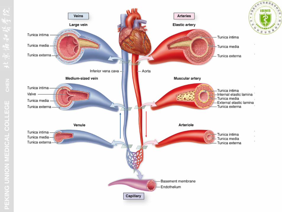
Blood vascular system + Lymphatic vascular system
heart capillary
artery
vein
Composition:
lymphatic vascular system
lymph
↔ tissue fluid ↔ cell
PEK
ING
UN
ION
MED
ICA
L C
OLL
EGE
C
HEN

PEK
ING
UN
ION
MED
ICA
L C
OLL
EGE
C
HEN
General structure of blood vessels
Specific structure of blood vessels
including artery, vein & capillary
Structure of heart
Structure of lymphatic vessels
Content

PEK
ING
UN
ION
MED
ICA
L C
OLL
EGE
C
HEN
1. General structure of blood vessels--- 3 concentric layers
I. Blood vascular system

PEK
ING
UN
ION
MED
ICA
L C
OLL
EGE
C
HEN
(1) Tunica intima
1) Endothelium + basal lamina
2) Subendothelial layer: LCT
3) Internal elastic lamina –
a fenestrated sheet of elastin
PEK
ING
UN
ION
MED
ICA
L C
OLL
EGE
C
HEN
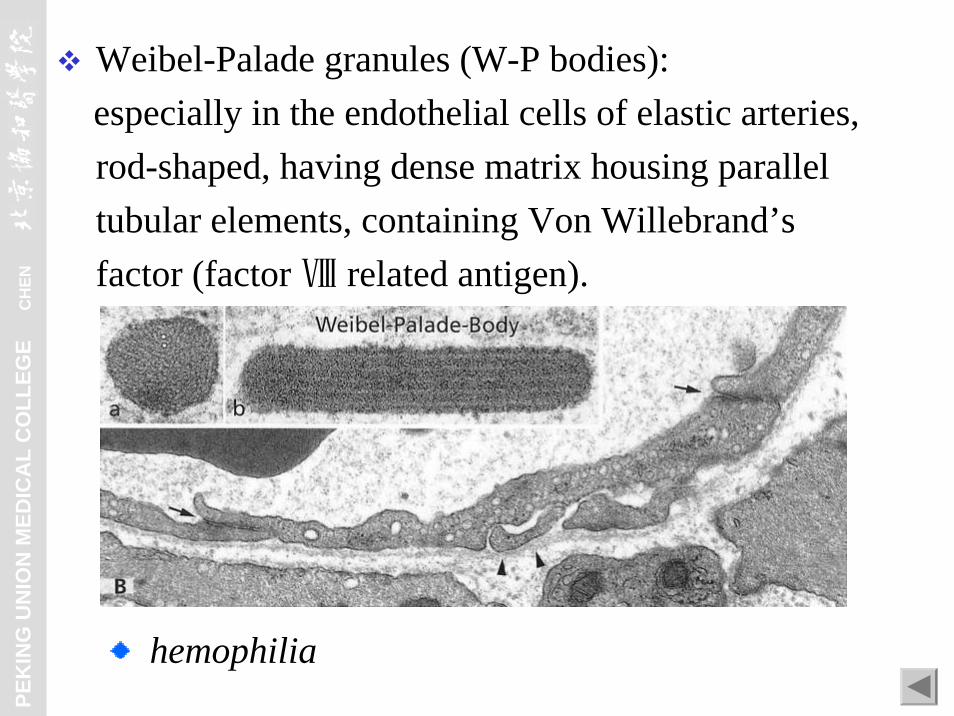
Weibel-Palade granules (W-P bodies): especially in the endothelial cells of elastic arteries, rod-shaped, having dense matrix housing parallel tubular elements, containing Von Willebrand’s factor (factor Ⅷ related antigen).
hemophilia

PEK
ING
UN
ION
MED
ICA
L C
OLL
EGE
C
HEN
Aorta Femoral artery
Myoendothelial junctions

PEK
ING
UN
ION
MED
ICA
L C
OLL
EGE
C
HEN
(2) Tunica media
1) Various numbers of
layers intermingled
with fibroelastic C.T.
2) External elastic lamina
smooth muscle cell

PEK
ING
UN
ION
MED
ICA
L C
OLL
EGE
C
HEN
Muscular A. Elastic A.

PEK
ING
UN
ION
MED
ICA
L C
OLL
EGE
C
HEN
(3) Tunica adventitia
vasa vasorumlymphatic vesselinnervation
fibroelastic C.T.

PEK
ING
UN
ION
MED
ICA
L C
OLL
EGE
C
HEN
1) Composition:
2. Specific structure of blood vessels(1) Capillaries
a. Pericytes:
Morphology: having long processes, basal lamina fusing with that of endothelial cell. Functions: contractility & participating in regeneration.

PEK
ING
UN
ION
MED
ICA
L C
OLL
EGE
C
HEN
b. Endothelial cells + basal lamina

PEK
ING
UN
ION
MED
ICA
L C
OLL
EGE
C
HEN
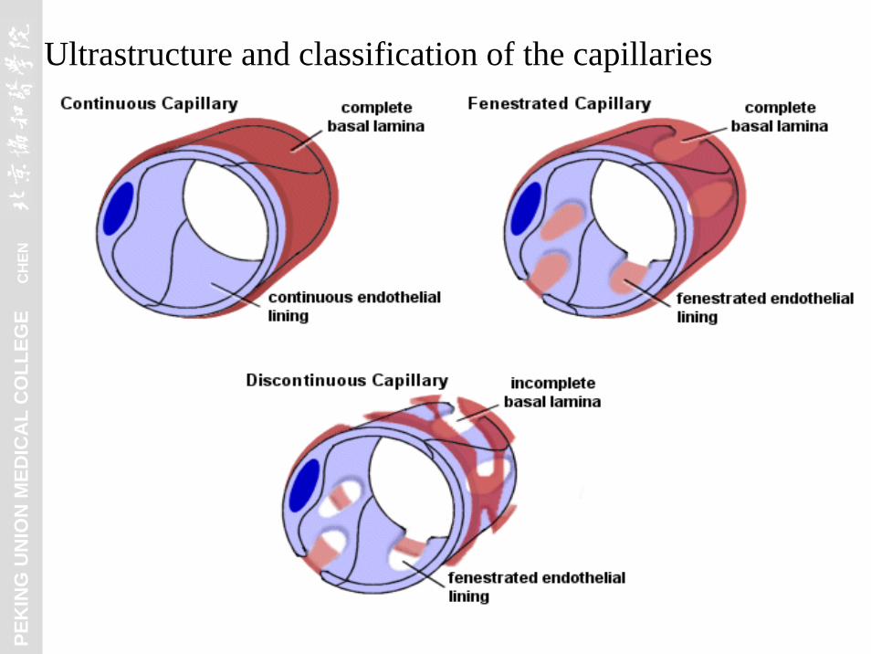
2) Ultrastructure and classification of the capillaries

PEK
ING
UN
ION
MED
ICA
L C
OLL
EGE
C
HEN
a. Continuous Capillaries:Morphology: continuous endothelium, tight junctions,
continuous basal lamina, pinocytotic vesicles.

PEK
ING
UN
ION
MED
ICA
L C
OLL
EGE
C
HEN
Distribution: muscle, lung, CNS, C.T., exocrine gland, etc.
PEK
ING
UN
ION
MED
ICA
L C
OLL
EGE
C
HEN
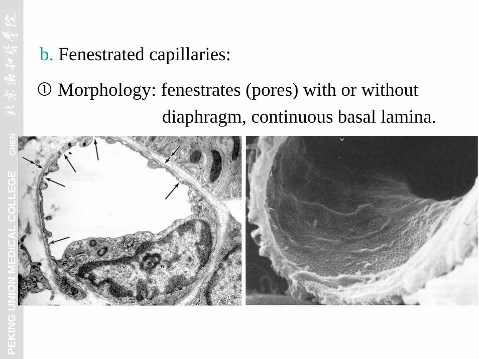
b. Fenestrated capillaries:
Morphology: fenestrates (pores) with or without diaphragm, continuous basal lamina.

PEK
ING
UN
ION
MED
ICA
L C
OLL
EGE
C
HEN
Distribution: stomach, intestine, endocrine gland, kidney, etc.
PEK
ING
UN
ION
MED
ICA
L C
OLL
EGE
C
HEN
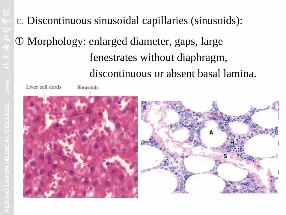
c. Discontinuous sinusoidal capillaries (sinusoids):
Morphology: enlarged diameter, gaps, large fenestrates without diaphragm, discontinuous or absent basal lamina.

PEK
ING
UN
ION
MED
ICA
L C
OLL
EGE
C
HEN
Distribution: liver, spleen, bone marrow, etc.

PEK
ING
UN
ION
MED
ICA
L C
OLL
EGE
C
HEN
3) Functions:
b. Metabolic functions
a.
c. Antithrombogenic function
Permeability. exchange vessels
Activation angiotensin I ⎯⎯→ angiotensin II
Inactivation bradykinin, serotonin, etc.
Lipolysis lipoproteins
Production of vasoactive factors e.g, endothelins & NO
ACE
PEK
ING
UN
ION
MED
ICA
L C
OLL
EGE
C
HEN

PEK
ING
UN
ION
MED
ICA
L C
OLL
EGE
C
HEN
4) Microcirculation: a. Composition:
b. Functions: blood pressure & blood flow regulation,and thermoregulation in particular areas.

PEK
ING
UN
ION
MED
ICA
L C
OLL
EGE
C
HEN
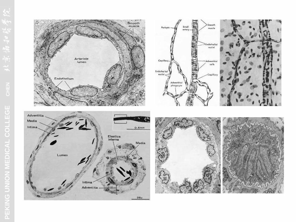
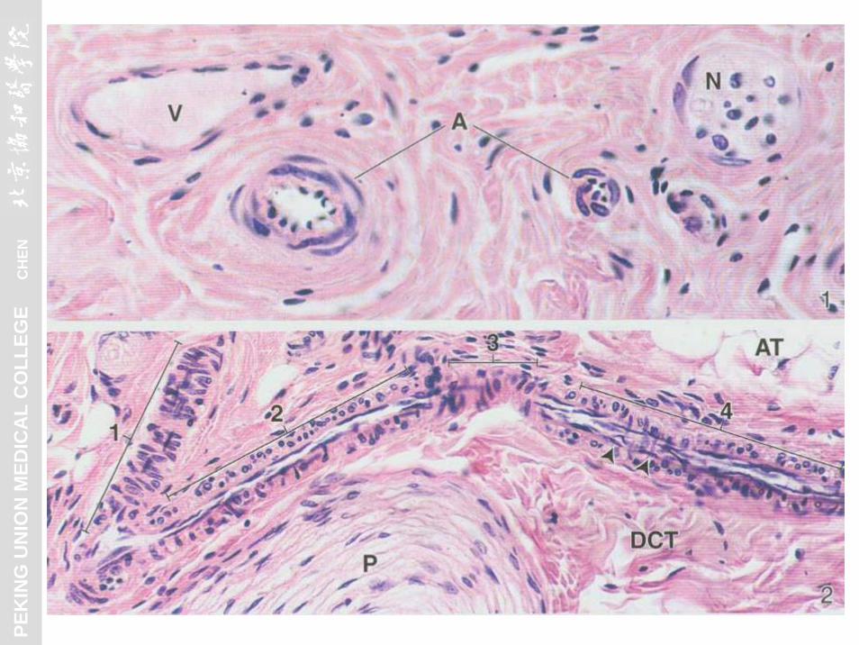
(2) Arteries
1) Arterioles (Peripheral resistance vessels)
Diameter < 0.5 mm (including all 3 layers)1-2 concentric smooth muscle layers in media, thin adventitia
PEK
ING
UN
ION
MED
ICA
L C
OLL
EGE
C
HEN
PEK
ING
UN
ION
MED
ICA
L C
OLL
EGE
C
HEN
PEK
ING
UN
ION
MED
ICA
L C
OLL
EGE
C
HEN
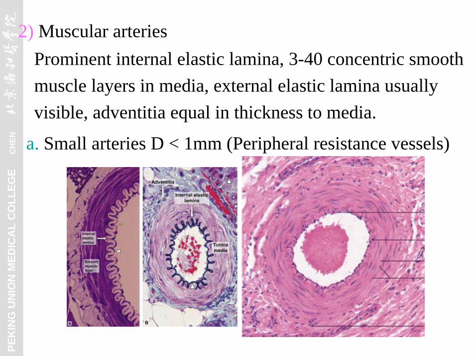
2) Muscular arteriesProminent internal elastic lamina, 3-40 concentric smooth muscle layers in media, external elastic lamina usually visible, adventitia equal in thickness to media.
a. Small arteries D < 1mm (Peripheral resistance vessels)
PEK
ING
UN
ION
MED
ICA
L C
OLL
EGE
C
HEN
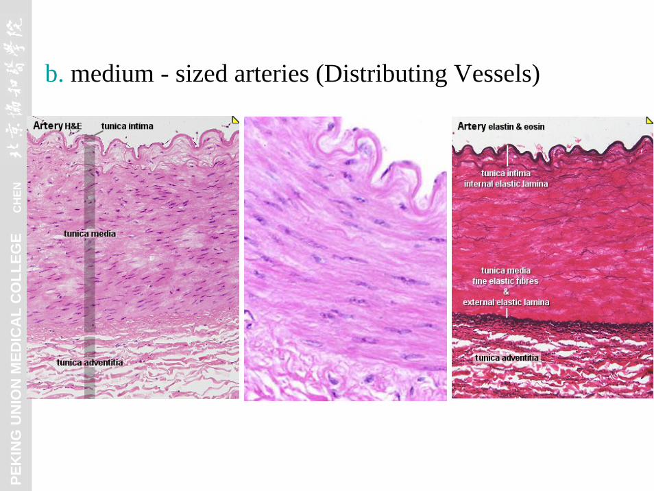
b. medium - sized arteries (Distributing Vessels)

PEK
ING
UN
ION
MED
ICA
L C
OLL
EGE
C
HEN
3) Elastic Arteries (Conducting Vessels) yellowish color
Relatively thick intima, 40-70 elastic laminae in media, thin adventitia.

PEK
ING
UN
ION
MED
ICA
L C
OLL
EGE
C
HEN
4) Specialized arteries and age changes in arteries:
Newborn Adult

PEK
ING
UN
ION
MED
ICA
L C
OLL
EGE
C
HEN
Specific receptors:
a. Carotid bodies & aortic bodies: chemoreceptors,sensing O2, CO2 tension & pH
b. Carotid sinuses: baroreceptors, sensing BP
type I cell
type II cell

PEK
ING
UN
ION
MED
ICA
L C
OLL
EGE
C
HEN
(3) Veins (Capacitance vessels) Comparing with their corresponding arteries, veins have valves and squashed, larger lumen, thinner wall, and poorly demarcated layers.

PEK
ING
UN
ION
MED
ICA
L C
OLL
EGE
C
HEN
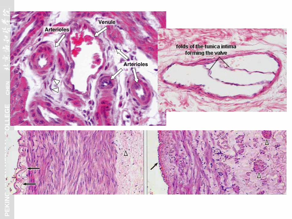
1) Venules diameter < 1mm
pericytes in postcapillary venules, discontinuous or no smooth muscle in media.

PEK
ING
UN
ION
MED
ICA
L C
OLL
EGE
C
HEN
venule
arteriole

PEK
ING
UN
ION
MED
ICA
L C
OLL
EGE
C
HEN
Permeabilitypostcapillary venule

PEK
ING
UN
ION
MED
ICA
L C
OLL
EGE
C
HEN
2) Small to Medium-Sized veins diameter: 1-9mm
2-4 1ayers of smooth muscle cells in media, intermixed with fibroelastic C.T., thicker adventitia with or without some longitudinal arranged smooth muscle cells.one- way valves in veins > 2mm diameter

PEK
ING
UN
ION
MED
ICA
L C
OLL
EGE
C
HEN

PEK
ING
UN
ION
MED
ICA
L C
OLL
EGE
C
HEN
Venous valves: paired, semilunar folds of the intima.

PEK
ING
UN
ION
MED
ICA
L C
OLL
EGE
C
HEN
3) Large veinsrelatively thin media, thickest adventitia with many longitudinal bundles of smooth muscles.
PEK
ING
UN
ION
MED
ICA
L C
OLL
EGE
C
HEN
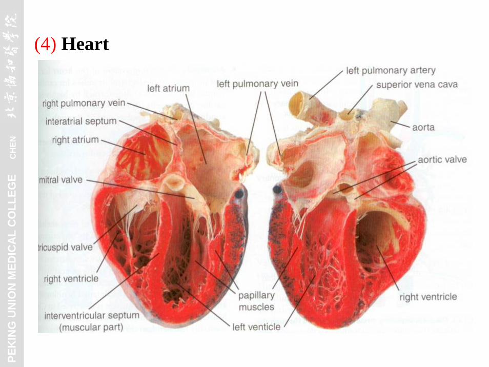
(4) Heart

PEK
ING
UN
ION
MED
ICA
L C
OLL
EGE
C
HEN
(4) Heart

PEK
ING
UN
ION
MED
ICA
L C
OLL
EGE
C
HEN
1) Tunics: a. Endocardium
Endothelial cells + basal lamina
Subendothelial layer
Subendocardial layer
Branches of Purkinje fibers in it

PEK
ING
UN
ION
MED
ICA
L C
OLL
EGE
C
HEN
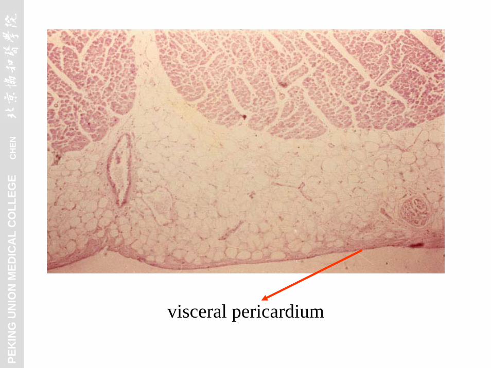
c. Epicardium: CT + mesothelium
b. Myocardium
Subepicardial layer
PEK
ING
UN
ION
MED
ICA
L C
OLL
EGE
C
HEN
visceral pericardium

PEK
ING
UN
ION
MED
ICA
L C
OLL
EGE
C
HEN

PEK
ING
UN
ION
MED
ICA
L C
OLL
EGE
C
HEN
2) Fibrous skeleton (cardiac skeleton) Including annuli fibrosi, the trigona fibrosa, and the septum membranaceum.
PEK
ING
UN
ION
MED
ICA
L C
OLL
EGE
C
HEN
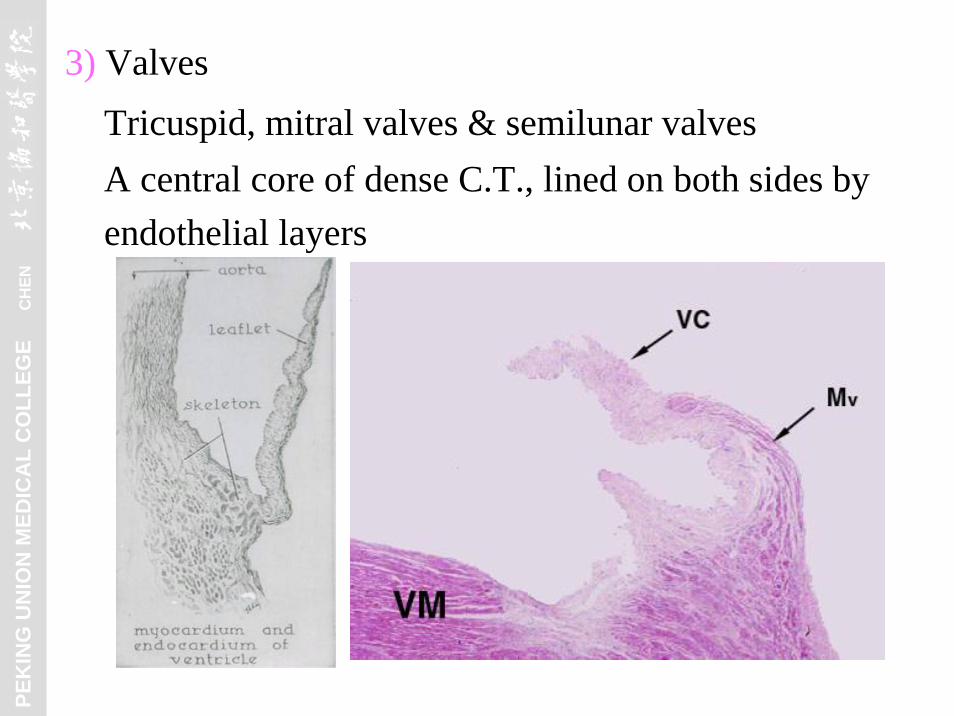
3) ValvesTricuspid, mitral valves & semilunar valves A central core of dense C.T., lined on both sides by endothelial layers

PEK
ING
UN
ION
MED
ICA
L C
OLL
EGE
C
HEN
4) Impulse Conducting System
b. Atrioventricular (AV) node, in subendocardial layer
c. AV Bundle of His, in subendocardial layer
a. Sinoatrial (SA) node: pacemaker, in subepicardial layer
PEK
ING
UN
ION
MED
ICA
L C
OLL
EGE
C
HEN
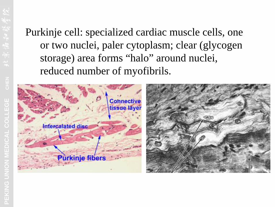
Purkinje cell: specialized cardiac muscle cells, one or two nuclei, paler cytoplasm; clear (glycogen storage) area forms “halo” around nuclei, reduced number of myofibrils.

PEK
ING
UN
ION
MED
ICA
L C
OLL
EGE
C
HEN

PEK
ING
UN
ION
MED
ICA
L C
OLL
EGE
C
HEN
II. Lymphatic Vascular System
Blind ended vessels, endothelial cells have no fenestrate, no tight junction, and little or no basal lamina.
1. Lymphatic capillaries

PEK
ING
UN
ION
MED
ICA
L C
OLL
EGE
C
HEN

PEK
ING
UN
ION
MED
ICA
L C
OLL
EGE
C
HEN
2. Lymphatic vessels
PEK
ING
UN
ION
MED
ICA
L C
OLL
EGE
C
HEN
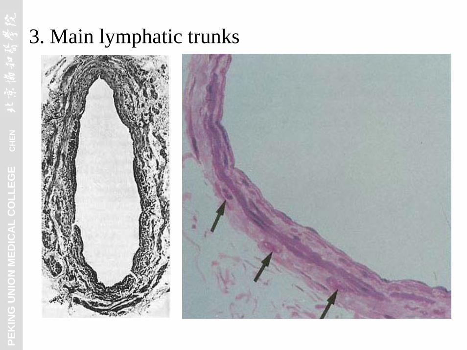
3. Main lymphatic trunks

PEK
ING
UN
ION
MED
ICA
L C
OLL
EGE
C
HEN
General structure of blood vessels--- 3 concentric layers
Summary

PEK
ING
UN
ION
MED
ICA
L C
OLL
EGE
C
HEN
PEK
ING
UN
ION
MED
ICA
L C
OLL
EGE
C
HEN
PEK
ING
UN
ION
MED
ICA
L C
OLL
EGE
C
HEN
Ultrastructure and classification of the capillaries

PEK
ING
UN
ION
MED
ICA
L C
OLL
EGE
C
HEN
Purkinje cell
Recommended