Tension Free
Transvaginal Tape
R.K.Mishra
World Laparoscopy Hospital

History
Tension-free vaginal tape, or TVT, as it
is commonly referred to, was first
introduced in 1995 by Ulmsten in Europe
and subsequently started being used in
the United States in 1998.
World Laparoscopy Hospital

TVT
World Laparoscopy Hospital

TVT
World Laparoscopy Hospital

Procedure
World Laparoscopy Hospital

Procedure
World Laparoscopy Hospital

World Laparoscopy Hospital

Procedure
Paraurethral dissection is performed after an initial midline incision on the anterior vaginal mucosa at the level of the midurethra. Note the small suprapubic abdominal incisions bilaterally.
World Laparoscopy Hospital

Procedure
After bilateral dissection of the paraurethral space, the rigid catheter guide is inserted into the urinary catheter. The handle of the guide is deflected to the ipsilateral side and the needle is inserted into the paraurethral space.
World Laparoscopy Hospital

Procedure
The tip of the needle
is angulated laterally
and the endopelvic
fascia is perforated
just behind the
inferior surface of the
pubic symphysis.
World Laparoscopy Hospital

Procedure
After perforation of the
endopelvic fascia, the
tip of the needle is
guided through the
retropubic space
along the backside of
the pubic symphysis.
World Laparoscopy Hospital

Procedure
After perforation of the
rectus fascia, a hand is
used to palpate the
needle tip
suprapubically
and guide the needle
to the abdominal
incision.
World Laparoscopy Hospital

Procedure
After the technique is
repeated on the other
side, the TVT sling is
in place with the tape
lying flat against the
posterior surface of the
midurethra.
World Laparoscopy Hospital

Procedure
The needles are detached
and an instrument is
placed between the tape
and the urethra. Gentle
traction on each end
brings the tape in contact
with the urethra and
correct tension is
adjusted with an
intraoperative cough stress
test.
T
World Laparoscopy Hospital

Procedure
The incisions are closed. The completed procedure allows fixation of the tape below the midurethra with the ends just below the skin level.
World Laparoscopy Hospital

Anaesthesia
Initially, the bladder is emptied with an 18 French Foley catheter.
The catheter balloon will help the surgeon identify the bladder neck to help direct the local anaesthesia injections.
The local anaesthesia should be injected bilaterally via a long spinal needle in the skin and abdominal wall just above the pubic symphysis, downward posterior to the pubic bone through the space of Retzius.
The anaesthesia should be injected along the intended course of the needle.
World Laparoscopy Hospital

Anaesthesia
The vaginal speculum should be inserted to expose the anterior vaginal wall.
The local anaesthesia is injected sub-urethral, starting approximately 1.0 cm from the external urethral meatus and moving proximally.
The local anaesthesia is injected on each side of the urethra toward the bladder neck in to the retropubic space.
The surgeon should then wait three to four minutes for the anaesthesia to take effect.
World Laparoscopy Hospital

Abdominal/Suprapubic Incisions &
Single Vaginal Incision
A vaginal incision with blunt dissection produces a space lateral to the urethra, which becomes the starting position for each of the TVT sling needles.
Two abdominal incisions are performed first at the intended exit points with the needles just superior to the pubic symphysis.
World Laparoscopy Hospital

Procedure
The key steps for this part
of the procedure are:
Determining the location
of the 1 cm bilateral
abdominal incisions
Determining the location
of the 1.5 cm mid-urethral
anterior vaginal incision
World Laparoscopy Hospital

Single Vaginal Incision
The anterior vaginal wall overlaying the mid urethra is elevated with Allis clamps and incised vertically in the midline. The incision should begin approximately 1 cm from the external urethra meatus and extend proximally for 1.5 cm. The incision should be long enough to accommodate the width of the TVT sling.
World Laparoscopy Hospital
Procedure
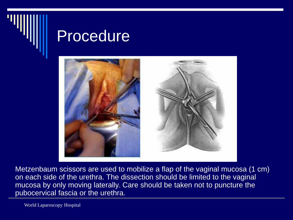
Metzenbaum scissors are used to mobilize a flap of the vaginal mucosa (1 cm) on each side of the urethra. The dissection should be limited to the vaginal mucosa by only moving laterally. Care should be taken not to puncture the pubocervical fascia or the urethra.
World Laparoscopy Hospital

TVT Sling and Device
Placement
During this step, the surgeon insert the TVT needle into the vaginla incision, through the periurethral fascia, into the retropubic space (Space of Retzius) and upward until the needle comes through the abdominal incisions. Correct placement, positioning and movement of the introducer needle during the procedure are critical.
World Laparoscopy Hospital
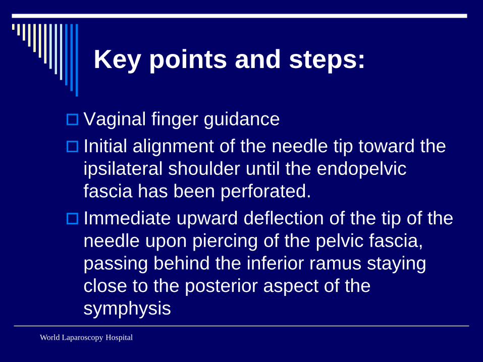
Key points and steps:
Vaginal finger guidance
Initial alignment of the needle tip toward the
ipsilateral shoulder until the endopelvic
fascia has been perforated.
Immediate upward deflection of the tip of the
needle upon piercing of the pelvic fascia,
passing behind the inferior ramus staying
close to the posterior aspect of the
symphysis
World Laparoscopy Hospital

Insertion of Needle
Two hands are required to pass the needle. The surgeon should concentrate on the role of each
hand. Position the needle tip through the vaginal incision directed lateral to the urethra. When
passing the needle, the vaginal mucosa is between the surgeon's finger and the tip of the needle.
World Laparoscopy Hospital

Insertion of Urethral Guide
Palpate the inferior ramus laterally and the urethra medially with the straight catheter guide inside. Once the endopelvic fascia has been penetrated beneath the inferior ramus, the handle of the needle is directed downward and pressure is applied upward by the hand in the vagina. The force advancing the needle actually comes form the palm or the thumb of the vaginal hand and the vaginal finger guiding it. The second hand is used to direct the back end of the handle. It determines the angle and steers the needle. The second hand does not torque or advance the needle.
World Laparoscopy Hospital

Insertion of Needle
Once the needle tip has been passed through the abdominal incision the handle can be disconnected. The needle should not be pulled completely though to the abdomen until cystoscopy has verified its position.
World Laparoscopy Hospital

Laparoscopic View of TVT
needles
World Laparoscopy Hospital

Cystoscopic Evaluation
After each pass of needle, cystoscopy is performed with needle in place extending from the vagina to the abdomen. In the event of a perforation, the needle will be easy to identify so it can be removed and re-introduced The surgeon must have cystoscopy privileges to perform this critical step of the procedure. During cystoscopy, the bladder should be distended to at least 250-300 cc of fluid. Using a 70-degree lens, the cystoscope is rotated and the bladder is inspected for perforations, which often occur at the one o'clock and 11 o'clock positions on the anterior wall of the bladder. The bladder neck should also be inspected.
After bladder integrity has been confirmed the TVT needles are pulled through the tissues and placed on the abdomen
World Laparoscopy Hospital

Laparoscopic View
World Laparoscopy Hospital

"looser is better than tighter."
The cough test should be conducted with a full bladder (250cc or saline). The surgeon should coordinate the procedure with the anesthesiologist so the patient is awake to cooperate during the tension test
The patient is asked to cough again and the tape is adjusted until only one or two drops of urine leak
World Laparoscopy Hospital
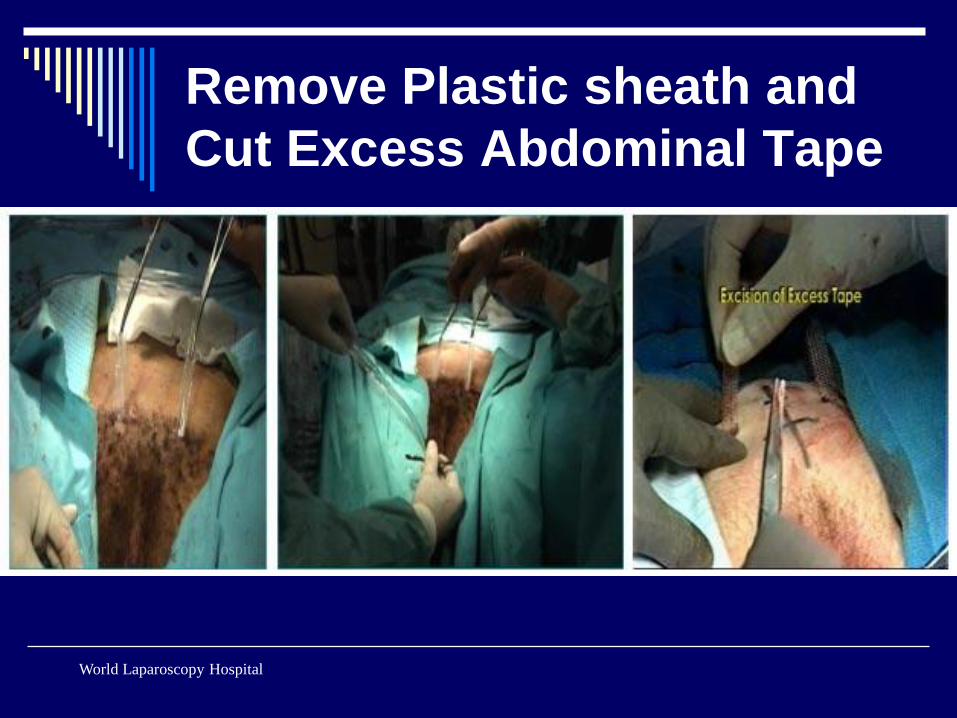
Remove Plastic sheath and
Cut Excess Abdominal Tape
World Laparoscopy Hospital
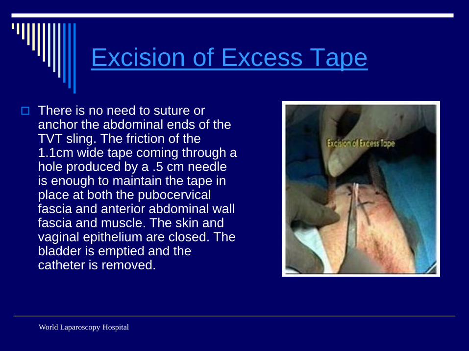
Excision of Excess Tape
There is no need to suture or anchor the abdominal ends of the TVT sling. The friction of the 1.1cm wide tape coming through a hole produced by a .5 cm needle is enough to maintain the tape in place at both the pubocervical fascia and anterior abdominal wall fascia and muscle. The skin and vaginal epithelium are closed. The bladder is emptied and the catheter is removed.
World Laparoscopy Hospital
CONTRAINDICATIONS
As with any suspension surgery, these procedures should not be performed in pregnant patients.
Additionally, because the PROLENE* polypropylene mesh will not stretch significantly, it should not be
Performed in patients with future growth potential including women with plans for future pregnancy.
World Laparoscopy Hospital

WARNINGS & PRECAUTIONS
Patients who are on anti-coagulation therapy.
Patients who have a urinary tract infection.
Patient should be counselled that future pregnancy may negate the effects of the surgical procedure and the patient may again become incontinent.
In case of pregnancy, delivery via caesarean section should be considered.
Post-operatively, refrain from heavy lifting and/or exercise (e.g. cycling, jogging) for at least three to four weeks and to refrain from intercourse for one month. The patients can usually return to other normal activity after one or two weeks.
World Laparoscopy Hospital
ADVERSE REACTIONS
Punctures or lacerations or injury to vessels, nerves, bladder, urethra, or bowel may occur during
Instrument passage and may require surgical repair.
Improper placement of the TVT device may result in incomplete or no relief from urinary incontinence or
May cause urinary tract obstruction.
World Laparoscopy Hospital
TVT VS TOT
Although the TVT complication rate is very low, there is a greater chance of damage to abdominal viscera and the bladder because of the entry into the anterior abdominal cavity.
By contrast, the TOT approach avoids almost all major structures. The only potential area of concern is that around the obturator vessels.
However, the location of these vessels is on the opposite side of where the needles are placed.
Obturator vessels are at least 3.5 cm away from the "safe zone" where the needle is inserted.
Additionally, the time to perform the procedure is on average 10 minutes, indicating that this procedure is even easier than the TVT with regard to placement.
World Laparoscopy Hospital
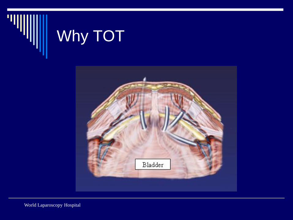
Why TOT
World Laparoscopy Hospital
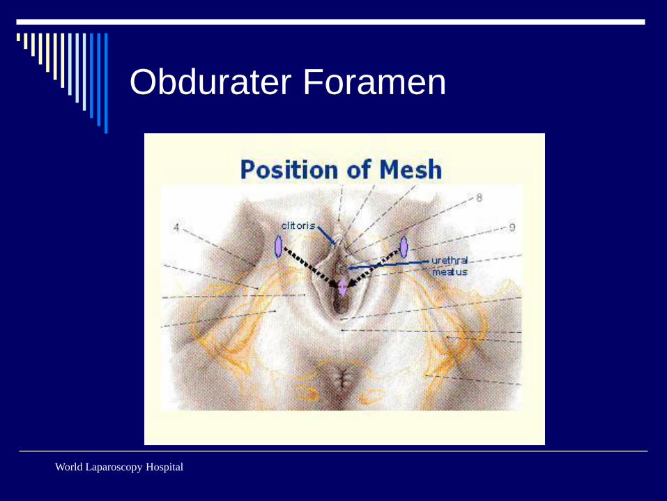
Obdurater Foramen
World Laparoscopy Hospital

Obturator Anatomy
World Laparoscopy Hospital

Pubourethral Ligament.
World Laparoscopy Hospital

TOT Sling
World Laparoscopy Hospital
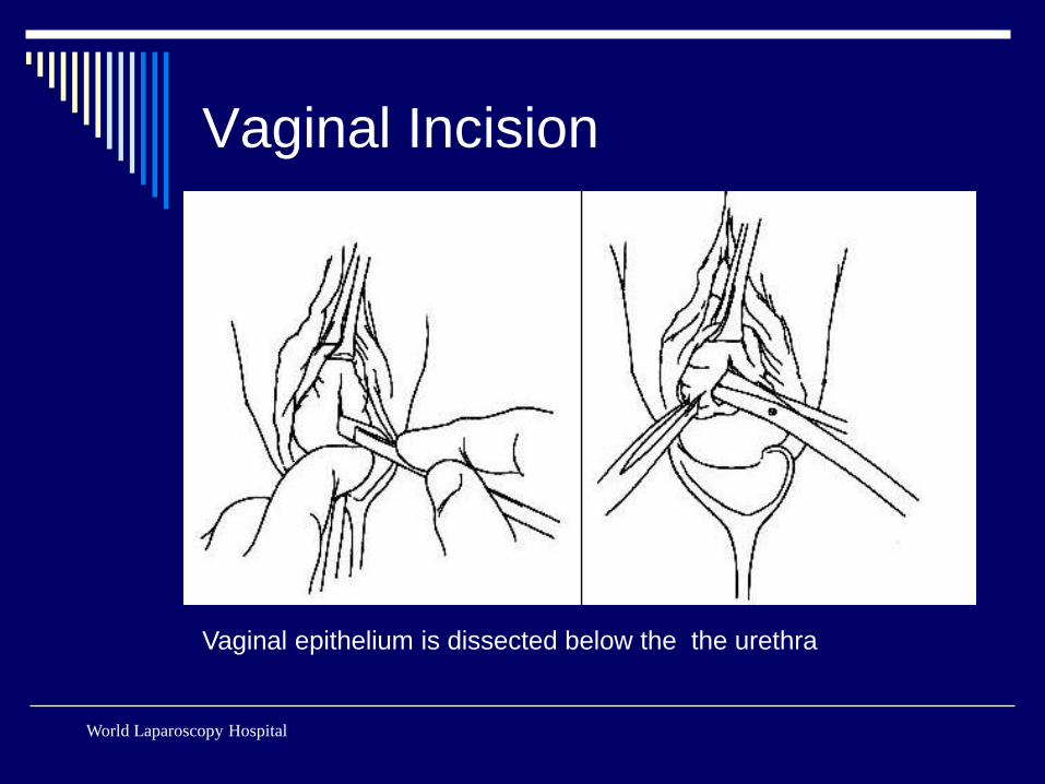
Vaginal Incision
Vaginal epithelium is dissected below the the urethra
World Laparoscopy Hospital

Groin Incision
Area of groin incision located 1cm inferior to adductor longus
tendinous insertion at the level of clitoris.
World Laparoscopy Hospital

Insertion of Needle
The needle is passed through the groin incision, through the obturator membrane
and muscles and brought into the vaginal incision.
World Laparoscopy Hospital

Tape is pulled through the
groin incision
Connected tape is then brought back through the groin incision.
World Laparoscopy Hospital
Ending the Procedure
Needle and tape is passed on the opposite
side. Tape is then adjusted with an intra-
operative cough test and adjusted until no
leakage occurs. Excess mesh is cut off at
the groin incisions and these are closed
with steri-strips and vaginal incision is
closed with absorbable suture.
World Laparoscopy Hospital

Thank You
Batch May 2008 at India Habitat Centre
World Laparoscopy Hospital
Recommended