The Texas Medical Center Library The Texas Medical Center Library
DigitalCommons@TMC DigitalCommons@TMC
The University of Texas MD Anderson Cancer Center UTHealth Graduate School of Biomedical Sciences Dissertations and Theses (Open Access)
The University of Texas MD Anderson Cancer Center UTHealth Graduate School of
Biomedical Sciences
8-2013
STUDYING AGGREGATE FORMATION BY AMYOTROPHIC STUDYING AGGREGATE FORMATION BY AMYOTROPHIC
LATERAL SCLEROSIS-ASSOCIATED MUTANT SOD1 PROTEIN IN LATERAL SCLEROSIS-ASSOCIATED MUTANT SOD1 PROTEIN IN
DROSOPHILA MODEL DROSOPHILA MODEL
Michael McCarthy
Follow this and additional works at: https://digitalcommons.library.tmc.edu/utgsbs_dissertations
Part of the Medicine and Health Sciences Commons, and the Molecular Biology Commons
Recommended Citation Recommended Citation McCarthy, Michael, "STUDYING AGGREGATE FORMATION BY AMYOTROPHIC LATERAL SCLEROSIS-ASSOCIATED MUTANT SOD1 PROTEIN IN DROSOPHILA MODEL" (2013). The University of Texas MD Anderson Cancer Center UTHealth Graduate School of Biomedical Sciences Dissertations and Theses (Open Access). 381. https://digitalcommons.library.tmc.edu/utgsbs_dissertations/381
This Thesis (MS) is brought to you for free and open access by the The University of Texas MD Anderson Cancer Center UTHealth Graduate School of Biomedical Sciences at DigitalCommons@TMC. It has been accepted for inclusion in The University of Texas MD Anderson Cancer Center UTHealth Graduate School of Biomedical Sciences Dissertations and Theses (Open Access) by an authorized administrator of DigitalCommons@TMC. For more information, please contact [email protected].

STUDYING AGGREGATE FORMATION BY AMYOTROPHIC LATERAL
SCLEROSIS-ASSOCIATED MUTANT SOD1 PROTEIN IN DROSOPHILA MODEL
By
Michael Jonathann McCarthy B.S.
APPROVED:
Sheng Zhang PhD
Joseph Alcorn PhD
Eric Swindell PhD
Kartik Venkatachalam PhD
Eric Wagner PhD
APPROVED:
Dean, The University of Texas
Graduate School of Biomedical Sciences at Houston
ii
STUDYING AGGREGATE FORMATION BY AMYOTROPHIC LATERAL
SCLEROSIS-ASSOCIATED MUTANT SOD1 PROTEIN IN DROSOPHILA MODEL
A
THESIS
Presented to the Faculty of
The University of Texas
Health Science Center at Houston
and
The University of Texas
MD Anderson Cancer Center
Graduate School of Biomedical Sciences
in Partial Fulfillment
of the Requirements
for the Degree of
MASTER OF SCIENCE
by
Michael Jonathann McCarthy B.S.
Houston, Texas
August 2013
iii
ABSTRACT
A common pathological hallmark of most neurodegenerative disorders is the presence
of protein aggregates in the brain. Understanding the regulation of aggregate formation is
thus important for elucidating disease pathogenic mechanisms and finding effective
preventive avenues and cures. Amyotrophic Lateral Sclerosis (ALS), also known as Lou
Gehrig’s disease, is a selective neurodegenerative disorder predominantly affecting motor
neurons. The majority of ALS cases are sporadic, however, mutations in superoxide
dismutase 1 (SOD1) are responsible for about 20% of familial ALS (fALS). Mutated SOD1
proteins are prone to misfold and form protein aggregates, thus representing a good candidate
for studying aggregate formation.
The long-term goal of this project is to identify regulators of aggregate formation by
mutant SOD1 and other ALS-associated disease proteins. The specific aim of this thesis
project is to assess the possibility of using the well-established Drosophila model system to
study aggregation by human SOD1 (hSOD1) mutants. To this end, using wild type and the
three mutant hSOD1 (A4V, G85R and G93A) most commonly found among fALS, I have
generated 16 different SOD1 constructs containing either eGFP or mCherry in-frame
fluorescent reporters, established and tested both cell- and animal-based Drosophila hSOD1
models. The experimental strategy allows for clear visualization of ectopic hSOD1
expression as well as versatile co-expression schemes to fully investigate protein aggregation
specifically by mutant hSOD1.
I have performed pilot cell-transfection experiments and verified induced expression
of hSOD1 proteins. Using several tissue- or cell type-specific Gal4 lines, I have confirmed

iv
the proper expression of hSOD1 from established transgenic fly lines. Interestingly, in both
Drosophila S2 cells and different fly tissues including the eye and motor neurons, robust
aggregate formation by either wild type or mutant hSOD1 proteins was not observed. These
preliminary observations suggest that Drosophila might not be a good experimental organism
to study aggregation and toxicity of mutant hSOD1 protein. Nevertheless this preliminary
conclusion implies the potential existence of a potent protective mechanism against mutant
hSOD1 aggregation and toxicity in Drosophila.
Thus, results from my SOD1-ALS project in Drosophila will help future studies on
how to best employ this classic model organism to study ALS and other human brain
degenerative diseases.

v
TABLE OF CONTENTS
CHAPTER ONE ..................................................................................................................................... 1
Introduction ............................................................................................................................................ 1
Amyotrophic Lateral Sclerosis ........................................................................................................... 2
Genetic basis of ALS .......................................................................................................................... 4
Superoxide dismutase (SOD) ............................................................................................................. 4
ALS-associated mutation in SOD1 .................................................................................................... 5
SOD1-based ALS models in mice ...................................................................................................... 5
Mutant SOD1 induces a gained of toxicity ........................................................................................ 6
CHAPTER TWO .................................................................................................................................... 8
Pathogenic Mechanisms of ALS: A Literature Review ......................................................................... 8
Protein aggregates: a common pathological feature of many neurodegenerative diseases ................ 9
Astrocytes and Glia Cells: A brief introduction ............................................................................... 11
Astrocytes and Glia Cells: Role in ALS ........................................................................................... 15
Mitochondria dysfunction in ALS .................................................................................................... 17
Aberrant Calcium Signaling in ALS ................................................................................................ 22
Oxidative Stress ................................................................................................................................ 23
CHAPTER THREE .............................................................................................................................. 25
The Goal of the Project and the Research Design ................................................................................ 25
The goal of this project ..................................................................................................................... 26
Drosophila, an excellent model organism to study human diseases ................................................ 26
Experimental Design ........................................................................................................................ 28
Cell line- and whole animal-based ALS/SOD1 models in Drosophila ............................................ 28
Selection of hSOD1 mutations for the study .................................................................................... 29
Tagging with fluorescent reporters for convenient monitoring of SOD1 protein expression and
aggregate formation .......................................................................................................................... 29
Cell-based model .............................................................................................................................. 30
Animal-based model ......................................................................................................................... 31
vi
CHAPTER FOUR ................................................................................................................................ 36
Molecular Cloning, Methods and Experimental Procedures ................................................................ 36
Molecular Cloning to Generate Fluorescent-tagged hSOD1 Fusion Protein ................................... 37
pMK33-hSOD1 constructs for cell line-based models ..................................................................... 37
pUAST-hSOD1 constructs for transgenic animals ........................................................................... 37
Summary of cloning procedures ....................................................................................................... 38
Summary of Protocols ...................................................................................................................... 40
Generating UAS-hSOD1-eGFP and UAS-hSOD1-mCherry transgenic flies .................................. 41
Targeted overexpression of UAS-hSOD1-eGFP or UAS-hSOD1-mCherry using Gal4 system ..... 45
Dissection and staining of adult fly brain and ventral nerve cord .................................................... 46
Imaging of adult fly eyes .................................................................................................................. 46
CHAPTER FIVE .................................................................................................................................. 47
Cloning Results and Assessing hSOD1 Aggregation Formation in Drosophila Cell Lines ................ 47
Results of the Molecular Cloning ..................................................................................................... 48
Pilot transfection assays ................................................................................................................... 50
Co-transfection assays ...................................................................................................................... 51
CHAPTER SIX .................................................................................................................................... 53
Studying hSOD1Aggregation and Toxicity in Transgenic Drosophila Models ................................... 53
Confirmation of hSOD1 transgene expression in transgenic fly lines ............................................. 54
Eye-specific over-expression of hSOD1 transgenes ........................................................................ 56
Motor neuron-specific expression of hSOD1 ................................................................................... 62
Glia-specific over-expression of hSOD1 transgenes ........................................................................ 68
Summary and Discussion ................................................................................................................. 69
CHAPTER SEVEN .............................................................................................................................. 71
Perspectives .......................................................................................................................................... 71
Toxicity and aggregation of human SOD1 protein in Drosophila ................................................... 72
Glia-specific toxicity of human SOD1 protein in Drosophila?........................................................ 73
Future studies based on the established SOD1 models in Drosophila ............................................. 74
Cellular toxicity in ALS: a link between dysregulated neuronal excitability, aggregates and others?
.......................................................................................................................................................... 76
ALS and athleticism: a causative or an incidental link? ................................................................... 78
Vita ....................................................................................................................................................... 96

vii
LIST OF TABLES
Table 1 experiment design and project completion table……………………………………48
Table 2: Summary of established transgenic fly lines………………………………………49
LIST OF FIGURES
Figure 1. Healthy motor neuron and neuromuscular junction………………………….…… 3
Figure 2. Astrocytes play an active role in synaptic cleft traffic and neuron response…......13
Figure 3. ETC and localization of SOD1 in mitochondria…………………………….……19
Figure 4. Neuromuscular junction deterioration…………………………………….…..…..21
Figure 5. The UAS-GAL4 binary expression system.....…………………….…...…..……..32
Figure 6 hSOD1 expression in S2 cells after copper induction…………………..……..…..52
Figure 7 Western blot of transgenic expression in Drosophila………………………..……55
Figure 8 hSOD1 expression under GMR eye specific GAL4 driver…………………..…....58
Figure 9 hSOD1 expression under GMR eye specific GAL4 driver………………..….…...59
Figure 10 Co-expression of WT & G85R hSOD1 in the Eye………………………….…....61
Figure 11 Percent Survival of hSOD1 wild type & mutant expression in motor neurons….63
Figure 12 hSOD1 expression under the D42 motor neuron GAL4 driver…………………..65
Figure 13 hSOD1 expression under the D42 motor neuron GAL4 driver……………….….67

1
CHAPTER ONE
Introduction

2
Amyotrophic Lateral Sclerosis
Amyotrophic Lateral Sclerosis (ALS) is a neurodegenerative disease that
predominantly and selectively affects upper and lower motor neurons in the brain and spinal
cord. ALS, in the US is more commonly known as Lou Gerig’s disease and is diagnosed at a
rate of about 5,600 new cases a year, affecting a total of 30,000 Americans at any given time
("ALS association," 2013). The pathology of this disorder is characterized by a deterioration
of the neuromuscular junction (NMJ), caused by the retraction and progressive degeneration
of motor neuron axons (healthy NMJ is seen in figure 1). This results in sensory loss, muscle
weakness, muscle atrophy and eventually paralysis. In human patients ALS symptoms onset
is varied but usually begins with sporadic weakness or stiffness and then progresses to
systemic weakness and loss of function. Paralysis and a complete loss of mobility occur at
advanced stages of disease and eventually dysfunction of respiratory motor neurons occurs.
Death is usually the result of paralyzed respiratory muscles and occurs within 1 to 5 years of
diagnosis, but has been document to take up to 20 years after diagnosis (Bruijn, Miller, &
Cleveland, 2004). Although, in later stages total loss of motor function is very common,
control of eye muscles and movement as well as bladder function often remains unaffected.
3
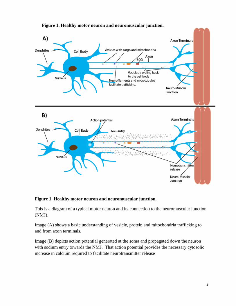
Figure 1. Healthy motor neuron and neuromuscular junction.
This is a diagram of a typical motor neuron and its connection to the neuromuscular junction
(NMJ).
Image (A) shows a basic understanding of vesicle, protein and mitochondria trafficking to
and from axon terminals.
Image (B) depicts action potential generated at the soma and propagated down the neuron
with sodium entry towards the NMJ. That action potential provides the necessary cytosolic
increase in calcium required to facilitate neurotransmitter release
Figure 1. Healthy motor neuron and neuromuscular junction.
A)
B)
4
Genetic basis of ALS
Most cases of ALS are sporadic with no known genetic component, 5 to 10% of ALS
is familial. Among the familial Amyotrophic Lateral Sclerosis (fALS) cases, around 20 –
40% are caused by one of over 150 identified mutations in the superoxide dismutase 1
(SOD1) gene and about 50% of fALS are due to a hexanucleotide repeat expansion mutation
in an intron of the C9orf72 gene. Interestingly around 44 mutations in the TDP-43 gene,
which codes for an RNA binding protein, are responsible for about 5% of all sporadic and
familial ALS (Da Cruz & Cleveland, 2011; Kwiatkowski et al., 2009; Mackenzie,
Rademakers, & Neumann, 2010). However, a total of 46 identified mutations so far in the
Fused in Sarcoma or Translocated in liposarcoma (FUS/TLS) gene, another RNA binding
protein, is seen in around 4% of familial ALS, but rarely seen in sporadic ALS (Da Cruz &
Cleveland, 2011; Vance et al., 2009).
Superoxide dismutase (SOD)
Among the many ALS-associated genes, SOD1 is the first identified and by far most
extensively studied. In mammalians, there are three different superoxide dismutase enzymes
and they are found at three different cellular locations. ALS-associated superoxide dismutase
1 (SOD1) is a cytosolic enzyme that causes the dismutation of the highly reactive anion
known as superoxide, to H2O2, which then can be further reduced to water (H2O) and
molecular oxygen (O2). Superoxide dismutase 2 (SOD2) resides in the mitochondria while
superoxide dismutase 3 (SOD3) is an extracellular protein. Notably, mutations in SOD2

5
have also been found to cause motor neuron disease and to exacerbate ALS caused by SOD1
mutations (van Zundert, Izaurieta, Fritz, & Alvarez, 2012).
ALS-associated mutation in SOD1
SOD1 itself encodes a small protein of 153 amino acids (a.a.) long and is fully
functional as a homodimer. Remarkably, over 150 mutations distributed throughout this
small protein have been identified in ALS patients. Among the most well characterized
mutations in SOD1, an alanine (A) to valine (V) change at position 4 (A4V) is the most
common mutation in fALS patients, and a glycine (G) to alanine (A) change at position 93
(G93A) was employed to create the first established mouse ALS model (van Zundert et al.,
2012). Notably, SOD1 proteins harboring either of these two mutations are still
enzymatically active as superoxide dismutase, although they become physically unstable and
prone to aggregation. Another commonly modeled SOD1 mutation is a glycine (G) to
arginine (R) change at position 85, referred to as G85R, which is commonly referred to as
enzymatically inactive, although detailed studies suggest it retains a very low level of
activity. However, SOD1 protein with this G85R mutation is more unstable as compared to
other SOD1 mutants and aggressively forms aggregates (Bruijn et al., 1997; Wang et al.,
2009).
Thus, far only one mutation in SOD1, D90A, has been seen to cause fALS in a
recessive manner, while all other mutations cause fALS in a dominant manner. The recessive
mutation of SOD1 is predominantly seen in a small subset of the Scandinavian population
and appears to be recessive only in that population (Robberecht et al., 1996).
SOD1-based ALS models in mice
6
Modeling of SOD1 mutations related to ALS in mice has been successful in that it
produces animals with consistent phenotypes that accurately reflect the disease course in
humans. One exception is that in ALS mouse models, the onset of weakness and paralysis
presents very consistently in the hind limbs, in contrast to the sporadic or non-uniform onset
of the symptoms in human patients. Also, among the reported ALS mouse models, different
disease relevant mutations of SOD1 cause consistent and nearly uniform symptoms. With
variation in disease course mostly limited to the age of onset and length of disease course,
making findings in one model relevant to other models. Two exceptions are notable, first
G127X mutation in SOD1 induces symptoms at low expression levels, although still
accumulates in CNS tissue and results in both protoaggregates and aggregates. It is in
contrast with other more common ALS models that mostly rely on overexpression of mutated
SOD1 (Jonsson et al., 2004). Second, A4V mutation in SOD1 appears to require either a
high level of protein expression, a common feature of mutant SOD1models, or co-expression
with wild type SOD1(Deng et al., 2006).
Mutant SOD1 induces a gained of toxicity
The pathogenic mechanisms underlying ALS are still far from clear (see Chapter 2
below). The reason SOD1 mutations are so tightly linked to ALS is also not yet known.
However, SOD1-associated ALS in general is believed to be the result of a gain of toxicity in
mutant SOD1, because many mutant forms of the protein remain catalytically active. In
addition, for those mutations that do not retain the endogenous SOD1 function, they do not
correlate with correspondingly altered severity of the symptoms or an earlier onset of the
disease (Boillee, Vande Velde, & Cleveland, 2006; Bruijn et al., 2004; Ince et al., 2011;
Pasinelli & Brown, 2006). Moreover, mice with SOD1 knock out do not develop motor

7
neuron degeneration symptoms, but instead have been found to have increased incidence of
tumor formation, although this does not impact survival or accelerate aging biomarkers
(Boillee, Vande Velde, et al., 2006; Muller, Lustgarten, Jang, Richardson, & Van Remmen,
2007; Reaume et al., 1996).

8
CHAPTER TWO
Pathogenic Mechanisms of ALS: A Literature Review
9
Protein aggregates: a common pathological feature of many neurodegenerative diseases
Amyotrophic Lateral Sclerosis, Parkinson’s, Alzheimer’s, transmissible spongiform
encephalopathy’s and Huntington’s are neurodegenerative diseases that affect different areas
of the brain and involve different gene mutations. Nevertheless, they all involve the gradual
loss of neurons. At the pathological level, almost all brain disease share a common feature:
the formation and presence of aberrant protein deposits (aggregates).
It is generally believed that protein aggregates are derived from accumulation of mis-
folded proteins. The protein aggregates are composed primarily of unique proteins in
different diseases. For example, in Parkinson ’s disease (PD), the Lewy body is mainly
composed of PD-associated α-Synuclein protein; In Huntington’s disease (HD), the
inclusions contain the cleaved and mis-folded disease gene product Huntingtin; In
Alzheimer’s disease (AD), the extracellular plaques are predominantly consisted of secreted
Aβ peptides derived from processed AD gene product APP, while the intracellular tangles are
from the hyper-phosphorylated Tau protein. Similarly, the protein products of the ALS-
associated genes such as mutant SOD1, TDP43 or FUS have also been found in neuronal
aggregates from ALS patients (Bruijn et al., 2004). The close link between protein aggregates
and brain degenerative diseases has promoted speculations of a causative relationship
between abnormal protein deposition and neurodegenration.
Protein aggregation is a simple term for a highly complex process that involves a
dynamic equilibrium between soluble monomers or oligomers of accumulated protein and
various states of higher order insoluble structures termed aggregates. The path from

10
accumulated protein to aggregate is dependent on cellular environment, interaction with other
proteins and concentration of the accumulated protein (Aguzzi & O'Connor, 2010). In the
case of SOD1 associated fALS, there is an aberrant accumulation of mutant SOD1 protein
accompanied by a lack of degradation by the ubiquitin mediated degradation pathway,
resulting in aggregated inclusions consisting of mis-folded mutant SOD1 protein, as depicted
in figure 4 below (Bruijn et al., 2004). It has also been suggested the protein half-life plays a
role in SOD1 mutations ability to aggregate aggressively and might be an indicator of
aggregate formation (Wang et al., 2003).
However, there is a growing body of empirical evidence to suggest the actual
formation of protein fibrils may be either neuroprotective or the end stage by-product of the
real culprit. That is, earlier stage prefibrillar oligomers or protofibrils have been implicated
as the true source of toxic element responsible for neuronal cell death (Caughey & Lansbury,
2003). It has been suggested that accumulated mutant proteins could overload protease
machinery, overtax heat shock proteins and damage mitochondria, increasing the risk of cell
death due to oxidative stress and thus inducing apoptosis. As an example of the complexity
of the field, one study that illustrates the toxicity of pre-aggregate oligomers of mutant SOD1
in ALS focused on the G127X mutation that will produce a truncated SOD1 protein. This
study found that mutant G127X SOD1 will cause ALS, even with fewer aggregates and at
lower expression levels than other SOD1 mutants, which suggests that an overtaxing or
sequestering of chaperones does not occur in some cases (Jonsson et al., 2004). Further, it
has been shown that mis-folded mutant G127X SOD1 can exist at higher steady state levels
between synthesis, mis-folded accumulation and degradation/aggregation in the central

11
nervous system, than elsewhere in an organism, suggesting a possible reason for CNS
susceptibility to disease (Jonsson et al., 2004).
The above findings regarding the G127X mutation of SOD1 underscore the
possibility that oligomers and protofibrils may be more damaging than fully formed
aggregates. Sorting out the true cause of neurodegeneration and understanding the cellular
regulation process of forming aggregates will be critical to developing effective treatments
for ALS.
Astrocytes and Glia Cells: A brief introduction
Although ALS is a motor neuron disease, increasing evidence suggest an essential
role of neuronal supporting cells especially astrocytes and glia in the disease pathogenesis
(Nagai et al., 2007; Perea & Araque, 2005; Rao & Weiss, 2004; Wallis, Zagami, Beart, &
O'Shea, 2012). Glia cells fall into two basic classifications based on their location they are
either central glia or peripheral glia. Central glia cell types include microglia, astrocytes,
oligodendrocytes and ependymal cells. Schwann cells are the only type of peripheral glia
cells; they sheath axons and cover the cell bodies of neurons (Hammond, 2008).
Astrocytes were first believed to only be “brain stuffing” or the glue and structure,
which provided the physical support that facilitated the organization and connections of
neurons and to supply nutrients to neurons via liaison with and maintenance of the blood
brain barrier (Volterra & Meldolesi, 2005). In the late 1980s it was discovered that
astrocytes expressed voltage gated ion channels as well as receptors for neurotransmitters,
suggesting they played a more sophisticated role in neural tissue than previously thought
(Volterra & Meldolesi, 2005).
12
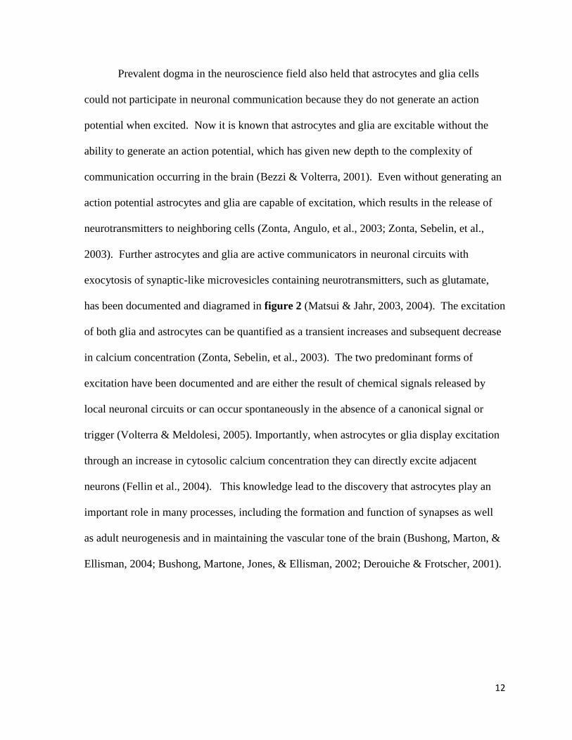
Prevalent dogma in the neuroscience field also held that astrocytes and glia cells
could not participate in neuronal communication because they do not generate an action
potential when excited. Now it is known that astrocytes and glia are excitable without the
ability to generate an action potential, which has given new depth to the complexity of
communication occurring in the brain (Bezzi & Volterra, 2001). Even without generating an
action potential astrocytes and glia are capable of excitation, which results in the release of
neurotransmitters to neighboring cells (Zonta, Angulo, et al., 2003; Zonta, Sebelin, et al.,
2003). Further astrocytes and glia are active communicators in neuronal circuits with
exocytosis of synaptic-like microvesicles containing neurotransmitters, such as glutamate,
has been documented and diagramed in figure 2 (Matsui & Jahr, 2003, 2004). The excitation
of both glia and astrocytes can be quantified as a transient increases and subsequent decrease
in calcium concentration (Zonta, Sebelin, et al., 2003). The two predominant forms of
excitation have been documented and are either the result of chemical signals released by
local neuronal circuits or can occur spontaneously in the absence of a canonical signal or
trigger (Volterra & Meldolesi, 2005). Importantly, when astrocytes or glia display excitation
through an increase in cytosolic calcium concentration they can directly excite adjacent
neurons (Fellin et al., 2004). This knowledge lead to the discovery that astrocytes play an
important role in many processes, including the formation and function of synapses as well
as adult neurogenesis and in maintaining the vascular tone of the brain (Bushong, Marton, &
Ellisman, 2004; Bushong, Martone, Jones, & Ellisman, 2002; Derouiche & Frotscher, 2001).
13
Figure 2 Astrocytes play an active role in synaptic cleft traffic and neuron response. Glutamate released from the pre-synaptic neuron travels across the cleft to activate the
AMPA and NMDA receptors on the post synaptic cleft.
If glutamate is not cleared from the synaptic cleft by the EAAT2 glutamate reuptake
transporter in the neighboring astrocytes, it will remain in the cleft and bind more receptors
that are present or inserted into the membrane. Potentially inducing excessive signaling and
excitoxicity.
Figure 2. Astrocytes play an active role in synaptic cleft traffic and neuron response.

14
Astrocytes have been shown to be involved in almost every functional aspect of the
central nervous system from neurogenesis and synaptogenesis, to acting as local integration
units and liaisons between synaptic and non-synaptic communication (Denise et al., 2004;
Mauch et al., 2001; Sanai et al., 2004; Seri, Garcia-Verdugo, McEwen, & Alvarez-Buylla,
2001). The role of glia cells in neuronal signaling was also underestimated, now it is known
that glia cells can regulate synaptic function and structure (Mauch et al., 2001; Ullian,
Sapperstein, Christopherson, & Barres, 2001).
There are two documented examples where glia-cell signaling, to the neuron, directly
regulates synaptic function and structure. The first includes Bergmann glial cells, where a
calcium concentration increases resulting from glutamate activated AMPA receptors,
maintains the extensive network of synapses that innervate Purkinje cells(Volterra &
Meldolesi, 2005). The absence of this glia signal alters the hyperinnervation and synaptic
current of the postsynaptic targets. (Iino et al., 2001) The second example is the binding of
ephrin A3, which is released from the surface of astrocytic processes, to its receptor EphA4,
which is expressed on dendritic spine membranes. This event initiates intracellular signals
that control the shape, size and number of dendritic spines. Without this signal spines will
grow too long and become very disorganized (Murai, Nguyen, Irie, Yamaguchi, & Pasquale,
2003). This information sets new precedents for understanding how information flow and
trafficking in neuronal networks is established by excitation of both neurons and glia and
astrocytes. It also serves to bring attention to the ability of astrocytes/glia to dynamically and
extensively respond to the state of neurons and synaptic communication.
15
Astrocytes and Glia Cells: Role in ALS
Astrocytes inclusions, as they relate to ALS were described by Beckman et al. in a
study that attempts to establish a timeline for motor neuron degeneration. The inclusion
resembled the lewy bodies found in Parkinson’s and were found to be the first abnormalities
in G85R mutant hSOD1 expressing mice(Beckman, Estevez, & Crow, 2001). This occurred
in the mutant mice at 8 to 10 months of age and was seen before inclusions were detected in
motor neurons. Astrocyte localization was confirmed with immunoreactivity to antibodies
specific to glial fibrillary acid protein (GFAP), which is not expressed in neurons. A two fold
increase in SOD1 concentration was seen in the spinal cords of dissected mice along with a
10 fold increase in SOD1-G85R containing astrocyte inclusions at end stage and this was
accompanied by a marked increase in astrocytosis.
Aggregates of SOD1 were then seen only in a few neurons before onset of symptoms,
but were diffuse and small, though they were reactive to SOD1 and ubiquitin antibodies. At
later stages of disease large round and irregular shaped SOD1 containing inclusions were
observed that were immunoreactive to SOD1 antibodies at their core and periphery. These
inclusions were reactive to ubiquitin, however only at the periphery. In the final stage of
disease spinal cord extracted showed a 60% loss of motor axons and a 10 fold increase in
mutant SOD1 astrocyte inclusions over motor neuron inclusions. Indicating that the astroglia
are a major focus of damage for ALS associated SOD1 mutation G85R (Beckman et al.,
2001).
Microglia cells are myeloid-lineage immune cells of the central nervous system.
They and astrocytes are the primary inflammatory response mediators. Microglia has been

16
shown in some cases to cross the blood brain barrier (BBB) and or the blood spinal cord
barrier to communicate and interact directly with the immune system (Boillee, Yamanaka, et
al., 2006; Skaper & Facci, 2012; Weydt & Moller, 2005). Mast cells are bone marrow
derived innate immune system cells that are capable of crossing the blood brain barrier and
blood spinal cord barrier in the absence of inflammation. They are capable of producing
many inflammatory and neurotrophic mediators, key to ALS are tumor necrosis factor-α
(TNFα) and nerve growth factor, to which glia cells may respond (Skaper & Facci, 2012).
Neuron death can be caused by microglia activation and secretion of toxic factors
such as TNFα and nitric oxide (NO) can be released from astrocytes expressing mutated
SOD1 proteins, which encourages pro-apoptotic pathways in motor neurons (Volterra &
Meldolesi, 2005). Release of toxic factors from astrocytes and glia ultimately kill motor
neurons through the activation of BAX mediated cell death agents (Nagai et al., 2007).
Importantly, it has been shown that among the many non-neuronal cells astrocytes are the
only cell type that are capable of releasing such toxic factors and motor neurons are the only
type of neuronal cells that are killed by these toxins (Nagai et al., 2007).
Motor neuron death also involves excitotoxicity which may be mediated by
astrocytes, as they are the major mediators of neuronal support. Astrocytes are responsible
for the regulation of ion concentrations in the extracellular space, as well as modulation and
clearance of neurotransmitters, such as glutamate, from the synaptic cleft (Benediktsson,
Schachtele, Green, & Dailey, 2005; Hirrlinger, Hulsmann, & Kirchhoff, 2004; Volterra &
Meldolesi, 2005).

17
In ALS there are reports of the focal loss of the EAAT2 transporter (normal function
seen in figure 2) in astrocytes, which is the dominant transporter responsible for glutamate
reuptake. Glutamate is the major excitatory neurotransmitter (Parpura et al., 1994) and with
the reuptake of glutamate inhibited, glutamate is left in the synaptic cleft free to bind to any
available receptors. This will result in the activation of a cation channels that will depolarize
the neuron and can ultimately result in repetitive neuronal excitability, calcium entry and
eventually excitotoxicity through repetition. In addition to glutamate excitotoxicity as a
result of the loss of EAAT2 transporter in astrocytes, the release of D-serine from glia, which
causes chronic activation of NMDAR, has also been linked to mutant SOD1 and ALS
(Sasabe et al., 2007).
Mitochondria dysfunction in ALS
Mitochondria are classically described as the power house of the cell and as such it is
the primary source of ATP production. To achieve this vital role, it employs a very complex
system that utilizes three partitioned domains which are the matrix, inner membrane space
and the outer membrane. Using a set of intramembranous protein complexes in located in
their inner membrane, mitochondria are able to establish and maintain and electron transport
chain (ETC) for the purpose of stripping electrons and collecting protons (H+ atoms) from
molecules as seem in figure 3.
Notably, SOD1 is the major cytosolic protein present to prevent oxidative damage
and reduce superoxide leakage form mitochondria. It has also been shown that SOD1 is
present in the mitochondria, primarily in the inter membrane space but also in the matrix and
on the cytosolic face of the outer membrane. (Carri & Cozzolino, 2011). The presence of
18
SOD1 is thought to provide protection to mitochondrial proteins from oxidation thus
preserving mitochondrial homeostasis (Aquilano, Vigilanza, Rotilio, & Ciriolo, 2006;
Kloppel, Michels, Zimmer, Herrmann, & Riemer, 2010; O'Brien, Dirmeier, Engle, & Poyton,
2004). Accordingly, mutations in SOD1 and its abnormal aggregation have been suspected
to disrupt this important cellular protective mechanism and contribute to the disease
pathogenesis.
Axonal transport of mitochondria has been shown to be dysfunctional in ALS and
could indeed be related to the problems caused by mutant SOD1 presence in mitochondria
(Carri & Cozzolino, 2011; De Vos, Grierson, Ackerley, & Miller, 2008; Magrane &
Manfredi, 2009). Mutant SOD1 has been shown to localize to the inner membrane space and
matrix of the mitochondria with the aid of its copper chaperone (CCS), which binds to
Mia40. They then enter the mitochondria (depicted in figure 3) through the Erv1/Mia40
oxidative folding mechanism of import (Carri & Cozzolino, 2011). Once inside the
mitochondria, mutant SOD1 accumulates in and forms oligomers (Deng et al., 2006; Ferri et
al., 2006) (Furukawa, Fu, Deng, Siddique, & O'Halloran, 2006).
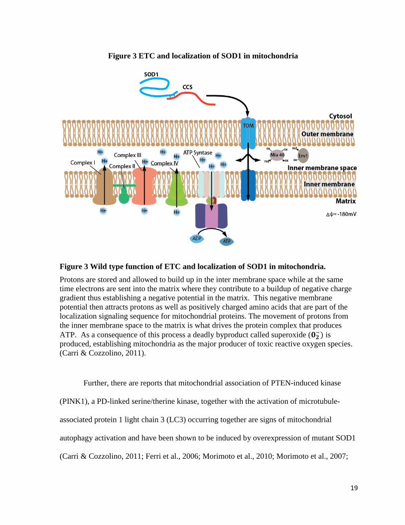
19
Figure 3 Wild type function of ETC and localization of SOD1 in mitochondria.
Protons are stored and allowed to build up in the inter membrane space while at the same
time electrons are sent into the matrix where they contribute to a buildup of negative charge
gradient thus establishing a negative potential in the matrix. This negative membrane
potential then attracts protons as well as positively charged amino acids that are part of the
localization signaling sequence for mitochondrial proteins. The movement of protons from
the inner membrane space to the matrix is what drives the protein complex that produces
ATP. As a consequence of this process a deadly byproduct called superoxide ( is
produced, establishing mitochondria as the major producer of toxic reactive oxygen species.
(Carri & Cozzolino, 2011).
Further, there are reports that mitochondrial association of PTEN-induced kinase
(PINK1), a PD-linked serine/therine kinase, together with the activation of microtubule-
associated protein 1 light chain 3 (LC3) occurring together are signs of mitochondrial
autophagy activation and have been shown to be induced by overexpression of mutant SOD1
(Carri & Cozzolino, 2011; Ferri et al., 2006; Morimoto et al., 2010; Morimoto et al., 2007;
Figure 3 ETC and localization of SOD1 in mitochondria
20
Pizzasegola et al., 2009). PINK1 itself has been implicated in mitophagy a cellular process
of removing dysfunctional mitochondria. (Deas, Wood, & Plun-Favreau, 2011) This process
may be activated as a result of disruption of mutant SOD1 to the electron transport chain
(ETC) (Cozzolino et al., 2009) and can lead to mislocalization of mitochondria from the
neural muscular junction as seen in figure 4 (Nguyen, Garcia-Chacon, Barrett, Barrett, &
David, 2009). Indeed, mitochondrial mislocalization in the axon has been seen in ALS and is
suggested to be a cause of axonal degeneration (Han et al., 2012; Tsuda et al., 2008).
Additionally, mutant SOD1 can aggregate in the matrix or inter membrane space and
measures to remove or decrease aggregated mutant SOD1 in mitochondria has been shown to
be effective at protecting the cell from mutant SOD1 induced toxicity (Carri & Cozzolino,
2011).

21
Figure 4 Aggregates of SOD1 can inhibit axonal travel and mitochondria localization.
Image (A) depicts aggregates of mutant SOD1 may inhibit vesicle travel and mitochondria
localization to axon terminal. Thus contributing to axonal detachment and retraction from
the neuromuscular junction (NMJ)>\.
Image (B) shows an action potential which may be ineffective because of axon retraction
from the neuromuscular junction, exacerbating the degeneration process.
A)
B)
Figure 4. Neuromuscular junction deterioration

22
Aberrant Calcium Signaling in ALS
One study has shown that human mutant SOD1-G93A in SH-SY5Y cells showed a
decreased ability to clear calcium from the cytosol to intracellular storage sites, such as the
mitochondria and endoplasmic reticulum, or to the extracellular space. It has been postulated
that this inability to clear calcium from the cytosol could account for the lack of calcium
oscillation seen in these mutant SOD1 transfected cells (Jaiswal et al., 2009). This leads to a
calcium buildup as a result of a storage and handling deficit, which is seen as an increase in
cytosolic calcium concentration a hallmark of excitotoxicity and deadly to the neuron.
It has been shown with thapsigargin treatment, which induces endoplasmic reticulum
(ER) stress, of SH-SY5Y cells transfected with G93A mutation of hSOD1 that selective
release of calcium from the (ER) only slightly increases cytosolic calcium concentration over
the similarly tested wild-type hSOD1 expressing cells. This indicates that dysfunction of
stores of calcium in the ER only slightly contributes to motor neuron dysfunction and
confirms the mitochondria as major players with regard to calcium accumulation and
dysfunction (Jaiswal et al., 2009). When mitochondrial ability to store calcium becomes
dysfunctional as seen in G93A mutant SOD1 transfected cells, neurons are then subject to
cytosolic calcium accumulation. This then puts greater stress on the neuron and leaves it
susceptible to calcium mediated cell death pathways resulting in neruodegeneration (Jaiswal
et al., 2009).
Cytosolic increases in calcium concentration, which are seen in ALS, may be the
result of oxidative stress to mitochondria (see below) and can lead to an increase in the
production of nitric oxide and peroxynitrite both of which could be deadly to the cell (Rao &
Weiss, 2004; Raoul et al., 2002).
23
Oxidative Stress
As noted, SOD1 is the major cytosolic protein to prevent oxidative damage and
reduce superoxide leakage form mitochondria. It has been proposed that mutant SOD1 could
reverse its protective catalytic activity, instead of scavenging and dismutation of superoxide
( ) to hydrogen peroxide (H2O2) and molecular oxygen (O2), it might be able revert to
produce toxic superoxide (Beckman et al., 2001; S. I. Liochev & Fridovich, 2000; Stefan I.
Liochev & Fridovich, 2003). This could be the result of mutations that leave SOD1 defective
in binding co-factor Zinc (Stefan I. Liochev & Fridovich, 2003). In such a state SOD1 could
actually take electrons from other cellular antioxidants and then donate them to molecular
oxygen, thus producing superoxide, making SOD1 the source of the oxidative
stress(Beckman et al., 2001). The end result is postulated to be the production of
peroxynitrite (ONOO) from superoxide and nitric oxide (Beckman & Koppenol, 1996). This
would cause the production of tyrosine nitration by reaction with peroxynitrite. It is
suggested that the critical protein affected by nitro-tyrosine residues is microtublues, thus
interfering with their ability to polymerize, which in turn could inhibit proper subcellular
trafficking and localization of mitochondria and neurotransmitter vesicle travel. (Beckman et
al., 2001) However, the clinical and experimental evidence does not support that hypothesis
(Boillee, Vande Velde, et al., 2006; Bruijn et al., 2004; Stefan I. Liochev & Fridovich, 2003).
Similar hypothesis regarding aberrant neuronal chemistry caused by the inability of
mutant SOD1 to complex with Cu and Zn leaving excess cytosolic Cu and Zn ions to run
amuck in the neurons have also not been supported by experimental and clinical data(Boillee,
Vande Velde, et al., 2006). Additionally, mice transfected with SOD1quad
mutants, which
have completely null copper-binding sites, did not show signs of any of the above mentioned
24
aberrant chemistry that would result in toxic superoxide or nitration species nor is disease
course altered (Wang et al., 2003).
ALS as a result of mutant SOD1 is not related to superoxide buildup and thus not a
consequence of oxidative damage or stress cause by superoxide. Individual mutations have
been documented to be throughout the SOD1 gene and yet all cause the same neuronal
degeneration with minimal differentiation. Sporadic and familial ALS, along with other
neurodegenerative diseases have signs consistent with a deficit in protein homeostasis, such
as aggregation and mislocalization of mutated and wild type RNA regulatory proteins, like
TDP-43. Thus, questions regarding aberrant protein accumulation and aggregation must be
solved in order to comprehensively address neurodegeneration. Elucidating genes which are
involved in the regulation of the aggregate formation process could contribute to a more
complete understanding of neurodegeneration as a whole.

25
CHAPTER THREE
The Goal of the Project and the Research Design

26
The goal of this project
The goal of this project is to test the feasibility of using Drosophila model system to
study the regulation of aggregates formed by the human SOD1 (hSOD1) protein. Abnormal
protein aggregation is a prominent pathological hallmark of ALS. Their formation, by the
ALS-associated disease proteins such as mutant SOD1, TDP43, FUS1, has been linked to the
dysfunction and degeneration of motor neurons during disease pathogenesis. In particular,
mutations in the small SOD1 gene have been established to be the cause of 20% of familial
ALS. The long-term goal of the project is to systematically identify and analyze the
regulators of aggregates formation by the mutated human SOD1 and other ALS-associated
protein. This will enable further dissection of the molecular networks that regulate
intracellular protein mis-folding and clearance in general, as well as provide potential targets
for therapeutic interventions against this fatal disease.
Drosophila, an excellent model organism to study human diseases
Drosophila, one of the best-studied genetic organisms, has emerged as an excellent
model system to study human brain degenerative diseases. Although a simple model
organism, the fruit fly offers several important advantages. For example, as a relatively
simple invertebrate, it is small in size, easy to maintain and care for, and can be raised in
large quantities at low cost. Second, it has high fecundity and short generation time, which is
usually not longer than 13 days from egg to reproductive-competent mature adult. These
features make Drosophila an ideal system to be subjected to large-scale and detailed genetic
analysis. In fact, over the last one hundred years, the fruit fly has been one of the most
extensively characterized and genetically manipulated multi-cellular organisms. In that time
27
a large arsenal of sophisticated experimental tools have been developed, such as the easily-
applied transgenic techniques and targeted mutagenesis. Utilized by many and important to
this study is the UAS-Gal4 binary expression system for targeted overexpression or
knockdown of a target gene. Further, clonal mosaic systems for analyzing lethal mutations
and differentiating cell-autonomous effect. Along with the recently developed genome-wide
dsRNA libraries and RNAi transgenic lines for convenient knock-down of every single gene
in the fly genome in both cultured cells and the whole animals. Moreover, Drosophila has a
well-documented developmental process as well as extensively studied and highly-conserved
signaling pathways. Notably, many important signaling pathways such as Wingless/Wnt,
Notch, Hedgehog and Toll/innate immunity, were first discovered and elucidated in the fruit
flies and still bear their original colorful names from this simple species. Furthermore, the
fly genome also contains homologs of many human neurodegenerative disease genes, such as
PD genes Parkin and Pink1, HD gene Huntingtin, AD gene App and Tau and ALS gene
SOD1 and TDP43. Importantly to this study, the fruit fly also has a relatively simple yet
conserved nervous system that bears many similarities with that in humans. These include,
but are not limited to, diverse neuronal cell types such as sensory and motor neurons, the
existence of glia cells, as well as the similar neuronal transmitters such as histamine,
dopamine and glutamate signaling. Over the last decade, many human brain degenerative
diseases including AD, PD and HD have been successfully modeled in the fruit fly (Bilen &
Bonini, 2005; Marsh & Thompson, 2006). Finally, of particular relevance to this study,
Drosophila and human copper chaperons have been shown to be highly conserved and
functionally interchangeable (Hua, Georgiev, Schaffner, & Steiger, 2010). Thus, wild type
28
and mutant hSOD1 expressed in Drosophila is likely to complex with copper as it would in
mammalians.
Experimental Design
Cell line- and whole animal-based ALS/SOD1 models in Drosophila
To study the regulation of the aggregate formation process by mutant SOD1, we have
decided to develop both cell line- and whole animal-based human SOD1 models in
Drosophila. The cell line-based model allows quick analysis of hSOD1 protein expression
and aggregation potential in vitro. Further, the cell-based model can very easily scaled up
such as to 384-well plate format that can be used for to high-throughput cell-based screens
such as RNAi or bioactive compounds. The whole-animal based hSOD1 model enables us to
analyze the in vivo effect of SOD1 in their ability to develop aggregates and induce toxicity
in different tissues and cell types such as neuron verse glia. If both models are successful,
we can further establish stable cell line models that express mutant human SOD1. Such a
cell model is amenable to high-throughput screens such as genome-wide RNAi to quickly
investigate every gene in the fly genome for their role in regulating mutant hSOD1
aggregation and toxicity. Extending this study to a screen for chemical compounds that
could directly yield potential drug candidates would be quick and simple. Subsequently, the
whole-animal models can facilitate in vivo validation of any identified hits from the cell-
based screens.
29
Selection of hSOD1 mutations for the study
For mutant hSOD1, we will focus on the three most-characterized ALS mutations:
A4V, G85R and G93A. Human SOD1 mutation A4V it the most prominent mutation of
fALS associated SOD1, and has been shown to be particularly unstable and aggressive at
forming aggregates ("ALS association," 2013; Boillee, Vande Velde, et al., 2006). However,
it remains catalytically active as does the well characterized G93A mutation which was used
to establish the first hSOD1 associated ALS mouse model (Bruijn et al., 2004; Spalloni et al.,
2004). Furthermore like mutation A4V of SOD1 the G85R is also aggressive at forming
aggregates, but is catalytically dysfunctional (Boillee, Vande Velde, et al., 2006; Watson,
Lagow, Xu, Zhang, & Bonini, 2008). The mutations focused on for this study provide a good
representative sample of the over 150 ALS associated hSOD1 mutations. As the control, we
will include the wild type human SOD1 (WT-hSOD1) in all the studies to eliminate
background effect and false positives.
Tagging with fluorescent reporters for convenient monitoring of SOD1 protein
expression and aggregate formation
To facilitate convenient visualization of gene expression and subsequent aggregate
formation, both wild type (WT) and mutant hSOD1 will be fused to either eGFP or mCherry
fluorescent proteins with a short flexible linker mainly composed of small amino acid glycine
and serine (GS). The use of the two different fluorescent reporters allows easy distinction of
mutant hSOD1 and control WT- hSOD1 proteins co-expressed in the same cell. For
example, in the cell-based model, plasmids containing wild-type hSOD1 fused to mCherry
reporter will be co-transfected with mutant hSOD1 with eGFP reporter. With this co-
transfection scheme, the presence of red-colored WT-hSOD1 will serve as an internal control
30
for the protein expression level of transfected DNA, which is a critical factor in protein
aggregation. More importantly, it also allows clear distinction of whether the effect on
aggregate formation from a study is specific for mutant hSOD1, which will be the targets of
interest for follow up study. Or are the observed effects on aggregate formation non-specific
and affect both mutant and WT-hSOD1. Such an observation would likely mean we have a
false positive due to background effect and should be disregarded.
Cell-based model
We have selected the pMK33 vector as cloning template mainly for the following
reasons. First, pMK33 contains a copper-inducible metallothionein promoter, which allows
for temporal control of SOD1 expression by the addition or removal of copper from the
medium. This is preferred over constitutive expression such as by actin promoter which
might not be feasible for potentially toxic proteins such as SOD1. In addition, insoluble
aggregates may be impervious to regulation, thus for an accurate assessment of a gene’s
ability to regulate aggregate formation, it must be knocked down significantly before
aggregates form. Thus, in a RNAi or compound screen, before inducing hSOD1 expression,
we are able to first incubate the cells in the reagents such as dsRNA. This can be done for a
significant amount of time so as to efficiently knockdown the expression level of the target
gene. Moreover, it is important to consider that eGFP can take hours to fold completely and
fluoresce(Chudakov, Matz, Lukyanov, & Lukyanov, 2010). Lastly, this vector contains a
hygromycin resistance gene, which will allow for convenient establishment of stably
transfected cell-line through hygromycin selection.

31
Animal-based model
For the animal-based model, we will use the pUAST transgenic vector as cloning
template, which can be conveniently combined with the Gal4 system to achieve targeted
expression of studied genes in almost any temporal- and spatially-controlled manner in the
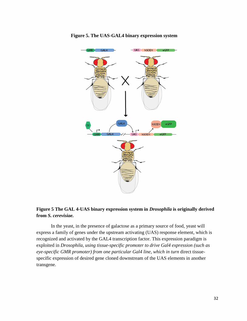
transgenic flies diagramed in figure 5.
32
Figure 5 The GAL 4-UAS binary expression system in Drosophila is originally derived
from S. cerevisiae.
In the yeast, in the presence of galactose as a primary source of food, yeast will
express a family of genes under the upstream activating (UAS) response element, which is
recognized and activated by the GAL4 transcription factor. This expression paradigm is
exploited in Drosophila, using tissue-specific promoter to drive Gal4 expression (such as
eye-specific GMR promoter) from one particular Gal4 line, which in turn direct tissue-
specific expression of desired gene cloned downstream of the UAS elements in another
transgene.
Figure 5. The UAS-GAL4 binary expression system
33
The strategy for this system is as the following: the gene of interest is placed under
the UAS response element promoter site (normally in the vectors pUAST for expression in
somatic tissues or pUASP for expression in germ lines) and inserted into the fly genome at
random. Expression of the GAL4 transcription factor is placed under the control of a
Drosophila tissue specific response element. For example, GMR is an eye specific
Drosophila response element recognized by a transcription factor that is primarily expressed
in the eye, thus the GAL4 gene downstream of this response element will be expressed
specifically in the eye. In the presence of a UAS-transgene, it can then find the correct UAS
site and express the gene downstream of the UAS site with tissue specificity. In this way the
expression of a transgenic gene can be accurately controlled. Importantly, a vast number of
tissue-, cell-type and-or temporal-specific Gal4 lines have been established in Drosophila.
Which are easily available from several national and international Drosophila stock centers.
Important to this study are the motor neuron specific D42-Gal4, pan-neuronal specific Elav-
and Appl-Gal4, the latter is more specific for expression in adult stage on, and glial-specific
Repo and GCM-Gal4 lines. Thus, the employment of pUAST enables us to easily target
hSOD1 transgene expression to any tissue of interest and examine their effects.
Rationale
Extensive studies have demonstrated that aggregates formation are tightly linked to
pathogenesis of ALS and other neuronal degenerative diseases. In ALS, mutant SOD1
proteins are known to be more prone to mis-folding and form protein aggregates. For
example, in previous mammalian cell-based ALS models, in both neuron-like non-dividing
NSC34 cells and dividing COS1 cells of monkey origin, GFP-tagged mutant human SOD1
including mutations A4V, G85R and G93A, but not wild type hSOD1, developed aggregates

34
(Corcoran, Mitchison, & Liu, 2004; Matsumoto, Stojanovic, Holmberg, Kim, & Morimoto,
2005). Thus, a detailed understanding of how cells respond to and regulate aggregates
formation by mutant SOD1 will help elucidate the molecular mechanisms that control protein
mis-folding and aggregation, and also potentially identify therapeutic targets for treating this
deadly neurodegenerative disorder.
Nevertheless, to our knowledge, except in Caenorhabditis elegans, few such
systematic studies have been conducted in multicellular model organisms. In a reported C.
elegans study, a genome-wide RNAi screen was carried out in a worm ALS model with pan-
neuronal expression of human G85R SOD1 mutant tagged with a yellow fluorescent fusion
(YFP) reproter, from which more than 80 dsRNA hits that caused enhanced aggregates and
diminished locomotion of the animal were isolated (Wang et al., 2009). We thus hypothesize
that similar studies, in additional model organisms that include more strigent controls to
screen for both suppressors and enhancers of mutant SOD1aggregates, will not only provide
a more comprehensive picture of the aggregation regulators in the cell, but also help narrow
down the conserved central players that control mutant SOD1 misfolding and accumulation.
Previous studies have demonstrated that mutant human SOD1 can also form protein
aggregates and induce animal toxicity in Drosophila, another well-established model
organism (Wang et al., 2009; Watson et al., 2008). Accordingly , we have planned to use the
tried and trusted versatile tools in this excellent animal model to systematic study the
regulation of aggregate formation by ALS associated human SOD1mutants. Our study is an
important and logical next step, as most signlaing pathways are highly conserved between the
fly and human, while a screen in the fly will be more cost effective and less time consuming
than that in a mouse model. In addition, establishing, in parrallel, a whole-animal and also a

35
cell-based ALS model in Drosophila will allow for more extensive controls and easy
validation of observations and results found in either model. With the previous experience in
aggregates-centered screens on Huntington’s disease and resources that are readily available
in our lab, we have expected that this ALS project will allow us to thoroughly and accurately
evaluate the feasibility of employing Drosophila model to study the regulators of aggregates
by mutant hSOD1, laying important foundation for more comprehensive studies in the future.

36
CHAPTER FOUR
Molecular Cloning, Methods and Experimental Procedures
37
Molecular Cloning to Generate Fluorescent-tagged hSOD1 Fusion Protein
The four genetic variations of human SOD1 used in the project are A4V, G85R, and
G93A and wild type (WT) control, as discussed in the previous chapter. The cDNA clones
containing each of the mutations and wild type hSOD1 were generously provided by Dr.
Daryl Bosco (University of Massachusettes Medical School, Worcester). The sequences of
each SOD1 construct were verified by DNA sequencing after every cloning step.
Each of the four genetic variations of hSOD1 will be fused in frame enhanced green
fluorescent protein (eGFP) and to the red fluorescent protein; mCherry, a derivative of eGFP,
thus a total of 8 fusion combinations.
pMK33-hSOD1 constructs for cell line-based models
The pMK33 vector is 8.5kbp in size and contains multiple restriction sites. For this
project we utilized XhoI and SpeI, which correspondingly match an Xho1 C-terminal
restriction site on PCR amplified hSOD1 cDNA and a SpeI site at the N-terminal of the PCR
amplified eGFP and mCherry sequences (See details below in Summary of cloning
procedures).
pUAST-hSOD1 constructs for transgenic animals
To generate transgenic fly models for SOD1, we used the transgenic vector pUAST,
which is 9Kbp in size by itself. It contains unique XhoI and XbaI cloning sites for the same
aforementioned hSOD1 gene construct insertions (Also see details below in Summary of
cloning procedures). The vector also contains a mini-white gene, which restores the
38
Drosophila red eye phenotype in the otherwise white eye color w1118
flies, so as to allow
easily determination of the success of transgenic events. It also allows for the use of the
UAS-GAL4 binary expression system to control tissue specific ectopic gene expression.
Amplified and digested hSOD1 will be fused in frame at its N-terminus, through a PCR-
introduced Agel restriction site on the GS linker (see below), with the C-terminus of the
chosen fluorescent marker.
Thus, as there are 8 different combinations of hSOD1 varieties with eGFP and
mCherry fusions. In order to establish both the cell line- and the whole animal-based models
to study regulators of aggregate formation by ALS-associated human SOD1 (hSOD1)
mutants, we would need to clone them into the selected vectors. A pUAST vector was
utilized to establish the whole animal-based model, and the pMK33 vector utilized to
establish the cell line-based model, thus for a total of 16 constructs, as summarized in Table
1 of the results section.
Summary of cloning procedures
The cloning strategy involved seven steps. (1) Both wild type and mutant hSOD1 as
well as the fluorescent reporters eGFP and mCherry were amplified by PCR, which used
primers with added restriction enzyme sites such as XhoI or AgeI at both ends to facilitate
cloning. A GS linker sequence encoding a short stretch of small amino acid glycine (G) and
serine (S) was also added to one end of the primers, so as to fuse the SOD1 open reading
frame with the eGFP or mCherry reporter; (2) The amplified SOD1 and eGFP/mCherry were
cut with restriction enzymes and then purified with a gel extraction kit from Qiagen and
stored at -20C until further use; (3) The cloning vector pUAST (for generating transgenic

39
flies) and pMK33 (cell transfection) were similarly cut with matching restriction enzymes
and gel purified; (4) Next, a triple ligation of the digested vector, hSOD1 and eGFP or
mCherry fragments was performed with T4 ligase ; (5) These ligations were transformed into
competent E. coli bacteria cells and grown on selection plates for 16 hours, and correct
clones were identified by PCR; (6) Extraction and purification of the plasmids was
accomplished with Qiagen mini-prep kits; (7) Plasmid DNA were diagnosed with digestion
by restriction enzymes and separation by agarose gel. The correct plasmids were further
verified by Sanger DNA sequencing (Lone Star labs).
List of primers:
SOD1 forward primer: 5’-CTCGAGGAATTCCTAGAAATAATTTTGTTTAA-‘3
SOD1 reverse primer with GS linker:
5’-GTGTAATTGGGATCGCCCAAGGCGGATCCTCCGGAGGTACCGGT-3’
eGFP forward primer: 5’-GTGAGCAAGGGCGAGGAG-3’
eGFP reverse primer: 5’-CCCTGAACCTGAAACATAACTAGT-3’
mCherry forward primer: 5’-ATAACCGGTGTGAGCAAGGCGAGGAGG-3’
mCherry reverse primer: 5’- GGCTGATTATGATCTAGAGTCGCG-3’

40
Summary of Protocols
PCR protocol: Either Taq or Phusion PCR polymerase was employed with the appropriate
buffer. In general a master mix of PCR reagents was made and included in this order:
deionize water, 10x or 5x buffer, dNTP at 200μM each, 0.5 μM of primers, 1μl of
polymerase and 50 ng/μl of template DNA. A final volume of 25μl per PCR reaction, the
reaction cycle included three stages, the first was denaturing at 98°C for 30 seconds and the
second stage was 25-30 cycles which included a denaturation step at 98°C for 10 seconds,
anneal step at 56°C for 23 seconds and extension step at 72°C for 1 minute and 20 seconds
and then a final stage of extension at 72°C for 5 minutes. The final product was then purified
using a Qiagen DNA purification Kit. Although, some steps were not necessary and no
modifications were made to the standard protocol.
Restriction enzyme: All restriction enzymes used in the project required a buffer (NEB
buffer number 4) containing 50mM Potassium Acetate, 20mM Tris-acetate, 10mM
Magnesium Acetate, 1mM DTT and a pH of 7.9 at 25°C. A master mix was used containing
2 μl of New England Biolabs(NEB) buffer number 4, 0.3μl of each restriction enzyme, 1ng
of template and 20μl deionized water per reaction. After digestion the entire reaction mixture
was loaded into a 2% agarose gel with ethidium bromide to highlight DNA under ultra violet
light and separated with 135mv for 12 minutes. The gel was then visualized under UV light
and the DNA bands of the correct size were excised from the gel and place into eppendorf
tubes. The DNA was then extracted from the agarose gel following the protocols from the
Qiagen Gel Purification Kit which.

41
DNA Ligation protocol: T4 DNA ligase was used for all ligations with a buffer containing
50mM Tris-HCl, 10mM MgCl2, 1mM ATP, 10mM DTT, at a pH of 7.5 at 25°C. The
components for a 20ul reaction were; 10x T4 DNA ligase buffer at 2μl, vector DNA 50ng
and insert DNA 50ng, Nuclease-free water up to 20μl and 1μl of T4 DNA ligase. The
ligation was then place in an 18°C incubator overnight but can also be performed at 25°C for
one hour.
E. Coli Transformation protocol: Transformation is a common molecular biology
technique employed to select and amplify the newly generated DNA constructs after ligation
of digested insert and vector DNA fragments. This technique exploits the natural ability of
some bacteria, specifically E.Coli used in this study, to accept and amplify foreign plasmids
under certain treatment. In my study, I used a standard transformation protocol involved 7
basic steps; (1) add ~11 μl of ligation sample to ~170μl of competent E. Coli. (2) Ice for 20
mins to allow bacteria to settle and plasmids to settle on bacteria outer membrane surface.
(3) Heat shock for no longer than 90 seconds at 42°C. This will open pores in the bacteria
membrane and allow plasmids to enter. (4) Immediately ice for 20 minutes to allow bacteria
to rest and the pores formed to close. It is important not to disturb the bacteria during this
time as they are in a very fragile state. (5) Add ~150μl of liquid broth and (6) incubate with
constant agitation at 37°C for 40 to 50 minutes. (7) Spread entire volume on an agar plate
that contains antibiotic. The plasmids used in this project contained an ampicillin resistance
gene so that antibiotic was incorporated into the mixture when making the plate.
Generating UAS-hSOD1-eGFP and UAS-hSOD1-mCherry transgenic flies

42
After sequence verification, the SOD1-eGFP or SOD1-mCherry constructs in pUAST
vector were microinjected into w1118
fly embryos following standard protocol. The flies were
allowed to mature and then crossed with w1118
virgins or males, and the resulting F1
progenies was screened for restoration of red eye color from the white eye flies of the
parental w1118
background, which indicated the success of transgenic events. The independent
transgenic lines were then crossed to double balancer flies to map the chromosomal location
of the transgenic inserts and also to balance the transgene in the genome. Double balancer
flies contain chromosomes that have been inverted and rearranged so that homologous
recombination cannot occur. This will help stabilize the inserted gene so it is not loss
through generations of mating. The balancer flies employed in this project were double
balancer flies that contained second and third chromosome balancers. They were
distinguished by two phenotypes and two markers. For the second chromosome the
phenotype was a curly wing called cyo and a marker called sp that is recognized by a change
in follicle grouping near the first leg on both sides. The third chromosome phenotype was a
change in bristle length on dorsal side of the thorax and it is called TM6C and a marker that is
seen as a change in eye shape and is called dropper (Dr).
Drosophila Genetics:
A total of 4 transgenic UAS-hSOD1-wild type fused to eGFP lines were tested during
this project along with 3 lines for UAS-hSOD1 mutation A4V and G93A. Two lines of
transgenic UAS-hSOD1-G85R fused to eGFP and two lines fused to mCherry were also
tested. The transgene were mapped and their chromosomal locations were determined
according to standard procedures using balancer stocks. For transgenic lines inserted on the
second chromosome, they were subsequently balanced with SM5a, Cyo ; those on the third
43
chromosome were balanced with TM6c balancers. The GAL4 lines selected to direct
expression of the hSOD1 transgenic flies in this project were GMR-GAL4 (eye specific),
D42 -GAL4 (motor neuron-specific) and GCM-GAL4 (glia cell during early development).
Genotypes for GMR-GAL4 drivers:
The genotype of the progeny transgenic flies for UAS-hSOD1-WT-eGFP on the third
chromosome after cross with GMR-Gal4 was: GMR-GAL4/+; UAS-hSOD1-WT-eGFP /+;
+/+. The genotype of the progeny transgenic flies for UAS-hSOD1-WT-eGFP on the second
chromosome after cross with GMR was: GMR-GAL4/ UAS-hSOD1-WT-eGFP; + /+; +/+.
The genotype of the transgenic, UAS-hSOD1-A4V-eGFP on the third chromosome, after
cross with GMR-Gal4 was: GMR-GAL4/+; UAS-hSOD1-A4V-eGFP /+; +/+. For the
second chromosome it was GMR-GAL4/ UAS-hSOD1- A4V -eGFP; + /+; +/+.
The genotype of the transgenic hSOD1-G85R-eGFP on the third chromosome, after cross
with GMR-Gal4 was: GMR-GAL4/+; UAS- hSOD1-G85R-eGFP /+; +/+. For the second
chromosome it was GMR-GAL4/ UAS-hSOD1- G85R -eGFP; + /+; +/+.
The genotype of the transgenic UAS-hSOD1-G85R-wCherry on the third chromosome, after
cross with GMR-Gal4 was: GMR-GAL4/+; UAS-hSOD1-G85R-mCherry /+; +/+. For the
second chromosome it was GMR-GAL4/ UAS-hSOD1- G85R-mCherry; + /+; +/+.
The genotype of the transgenic UAS-hSOD1-G93A-eGFP on the third chromosome, after
cross with GMR-Gal4 was: GMR-GAL4/+; UAS-hSOD1-G93A-eGFP /+; +/+. For the
second chromosome it was GMR-GAL4/ UAS-hSOD1- G93A -eGFP; + /+; +/+.

44
Genotypes for using motor neuron-specific D42-GAL4 drivers:
The genotype of the progeny transgenic flies for UAS-hSOD1-WT-eGFP on the third
chromosome after cross with motor neuron-specific D42-Gal4 line was: D42-Gal4 /+; UAS-
hSOD1-WT-eGFP /+; +/+. The genotype of the progeny transgenic flies for UAS-hSOD1-
WT-eGFP on the second chromosome after cross with D42-Gal4 line was: D42-Gal4/ UAS-
hSOD1-WT-eGFP; + /+; +/+.
The genotype of the transgenic, UAS-hSOD1-A4V-eGFP on the third chromosome, after
cross with D42-Gal4 was: D42-Gal4/+; UAS-hSOD1-A4V-eGFP /+; +/+. For the second
chromosome it was D42-Gal4/ UAS-hSOD1- A4V -eGFP; + /+; +/+.
The genotype of the transgenic hSOD1-G85R-eGFP on the third chromosome, after cross
with D42-Gal4 line was: D42-Gal4/+; UAS-hSOD1-G85R-eGFP /+; +/+. For the second
chromosome it was D42-Gal4/ UAS-hSOD1- G85R -eGFP; + /+; +/+.
The genotype of the transgenic UAS-hSOD1-G85R-wCherry on the third chromosome, after
cross with motor neuron-specific D42-Gal4 was: D42-GAL4/+; UAS- hSOD1-G85R-
mCherry /+; +/+. For the second chromosome it was D42-GAL4/ UAS-hSOD1- G85R-
mCherry; + /+; +/+.
The genotype of the transgenic UAS-hSOD1-G93A-eGFP on the third chromosome, after
cross with motor neuron-specific D42-Gal4 was: D42-GAL4/+; UAS-hSOD1-G93A-eGFP
/+; +/+. For the second chromosome it was D42-GAL4/ UAS-hSOD1- G93A -eGFP; + /+;
+/+.

45
Genotypes for GCM-GAL4 drivers:
The genotype of the progeny transgenic flies for UAS-hSOD1-WT-eGFP on the third
chromosome after cross with GCM-GAL4 was: GCM-GAL4/+; UAS-hSOD1-WT-eGFP /+;
+/+. The genotype of the progeny transgenic flies for UAS-hSOD1-WT-eGFP on the second
chromosome after cross with GCM was: GCM-GAL4/ UAS-hSOD1-WT-eGFP; + /+; +/+.
The genotype of the transgenic UAS-hSOD1-G85R-eGFP on the third chromosome, after
cross with GCM-GAL4 was: GCM-GAL4/+; UAS-hSOD1-G85R-eGFP /+; +/+. For the
second chromosome it was GCM-GAL4/ UAS-hSOD1- G85R -eGFP; + /+; +/+.
The genotype of the transgenic hSOD1-G85R-wCherry on the third chromosome, after cross
with GCM-GAL4 was: GCM-GAL4/+; UAS-hSOD1-G85R-mCherry /+; +/+. For the second
chromosome it was GCM-GAL4/ UAS-hSOD1- G85R-mCherry; + /+; +/+.
Targeted overexpression of UAS-hSOD1-eGFP or UAS-hSOD1-mCherry using Gal4
system
The UAS-hSOD1-eGFP or UAS-hSOD1-mCherry transgenic flies were crossed to
selected tissue-specific GAL4 drivers. Enabling us to check and verify correct transgene
expression via western analysis, for phenotypic characterization and imaging documentation.
For western analysis, 10 adult flies aged for 1 day expressing wild type hSOD1 and mutant
G85R-hSOD1 fused to eGFP and mCherry, driven by the D42 (motor neuron) and GMR (eye
specific) driver were homogenized in RIPO buffer. The samples were separated with
electrophoresis of a polyacrylamide gel and then transferred to a nitrocellulose membrane.

46
To detect proteins present on the membrane, anti-eGFP and mCherry antibodies were used.
The membrane was then imaged with a LI-COR imager on western blot membranes.
Dissection and staining of adult fly brain and ventral nerve cord
Adult flies expressing A4V- or G93A-hSOD1 mutation with eGFP tag were dissected
in 1X PBS to remove their brain and ventral nerve cord. The samples were then fixed in 4%
formaldehyde for 45 minutes, then stained with phlloidain and DAPI and placed on a slide to
image with a fluorescent microscope.
Imaging of adult fly eyes
Flies expressing hSOD1 under the GMR-Gal4 eye specific driver were fixed to a
slide with a fast drying clear liquid polymer and imaged with a fluorescent microscope.
Images were taken under green or red UV illumination to visualize eGFP or mCherry signals
and under bright light to image overall eye morphology.

47
CHAPTER FIVE
Cloning Results and Assessing hSOD1 Aggregation Formation in
Drosophila Cell Lines
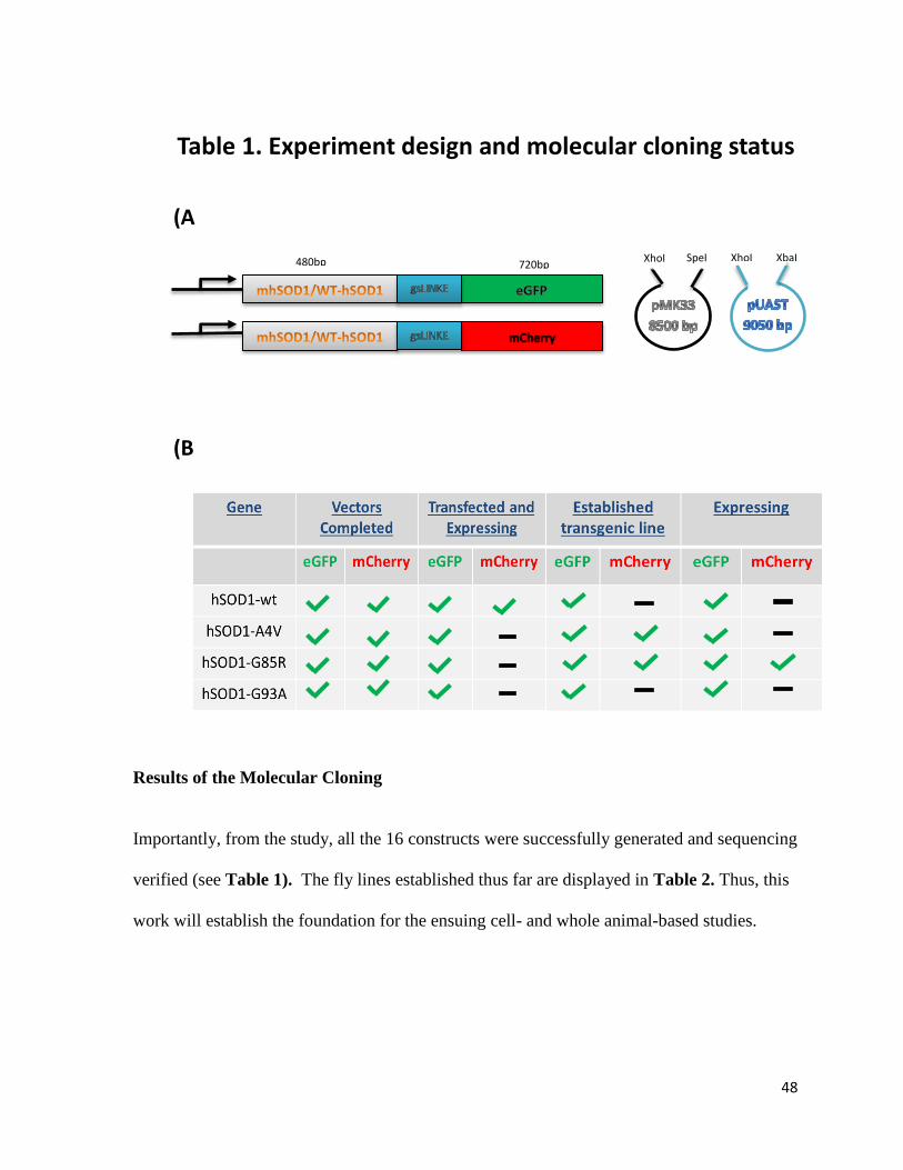
48
Results of the Molecular Cloning
Importantly, from the study, all the 16 constructs were successfully generated and sequencing
verified (see Table 1). The fly lines established thus far are displayed in Table 2. Thus, this
work will establish the foundation for the ensuing cell- and whole animal-based studies.
XhoI SpeI
480bp 720bp
XhoI XbaI
(A
)
(B
)
Table 1. Experiment design and molecular cloning status

49
Table 2: Summary of established transgenic fly lines

50
To investigate establishing a cell-based model to study aggregate formation by
mutant human SOD1 proteins, we first performed transient transfection assay to examine
their expression from the established pMK33 constructs. Further, we were able to test
whether the mutant hSOD1 proteins form robust aggregates in the cell. In the established
pMK33 constructs, the expression of wild type and mutant hSOD1-eGFP or mCherry fusions
were controlled under a copper-inducible metalothrenine protomor.
The S2 cells selected for this project are a macrophage lineage Drosophila cell line
that is commonly utilized for fruit fly related in vitro studies. An advantaged of these cells is
their relative robustness and hardiness when grown in S2 medium that has been supplimented
with antibiotic and serum. For this study the cells were specifically cultured in Schneider S2
growth medium from invitrogen, which was supplamented with the antibiotics streptomycin
and penicillian and with fetal bovine serum.
The established pMK33 constructs were transfected into Drosophila S2 cells. 48
hours after transfection, expression of fusion proteins were induced with aqueous copper
sulfate solution at a final of concentration 250 mM.
Pilot transfection assays
In this study, S2 cells were first transfected with the pMK33 vector construct to
assess proper expression of hSOD1 fused to eGFP under copper induction and they were
assess visually for any obvious formation of mutant hSOD1-eGFP aggregates. For this test
transfection, all four pMK33 constructs containing eGFP-tagged wild type or each of the
three selected hSOD1 mutants were used separately. 48 hours after SOD1 expression was
induced with copper at a final concentration of 250mM, the cells were visualized under a

51
fluorescent micrscope. Which revealed clear expression of each of the tested hSOD1-eGFP
fusion, and the protein were predominantly localized to the cytosol, as expected. These
observations suggested the successful transfection of the plasmids into the S2 cells and the
proper induciton of SOD1 protein expression. However, after 24, 48 and 72 hours of
induction, we did not observe obvious aggregation by either wild type or any of the three of
hSOD1 mutations.
Co-transfection assays
A second transfection strategy involved co-transfection of wild type hSOD1 fused to
mCherry and mutant G85R-hSOD1 fused to eGFP. The purpose of this stragey was to show
a clear contrast between any presence of aggregated mutant G85R-hSOD1 (revealed by the
green color of its eGFP tag) in the red background of a soulble wild type hSOD1 (revealed
by its mCherry tag). In addition, there exist reports suggesting that co-expression of wild
type and mutant hSOD1 may promote more aggressive aggregation and disease phenotype
(Deng et al., 2006; Graffmo et al., 2013; Jonsson et al., 2004). However, when examined
under fluorsecent microscopy 18 hours (Figure 6) or 24 hours (data not shown) after
induciton of protein expression, we again did not obseve obvious aggregate formation in
either of the tested conditions.
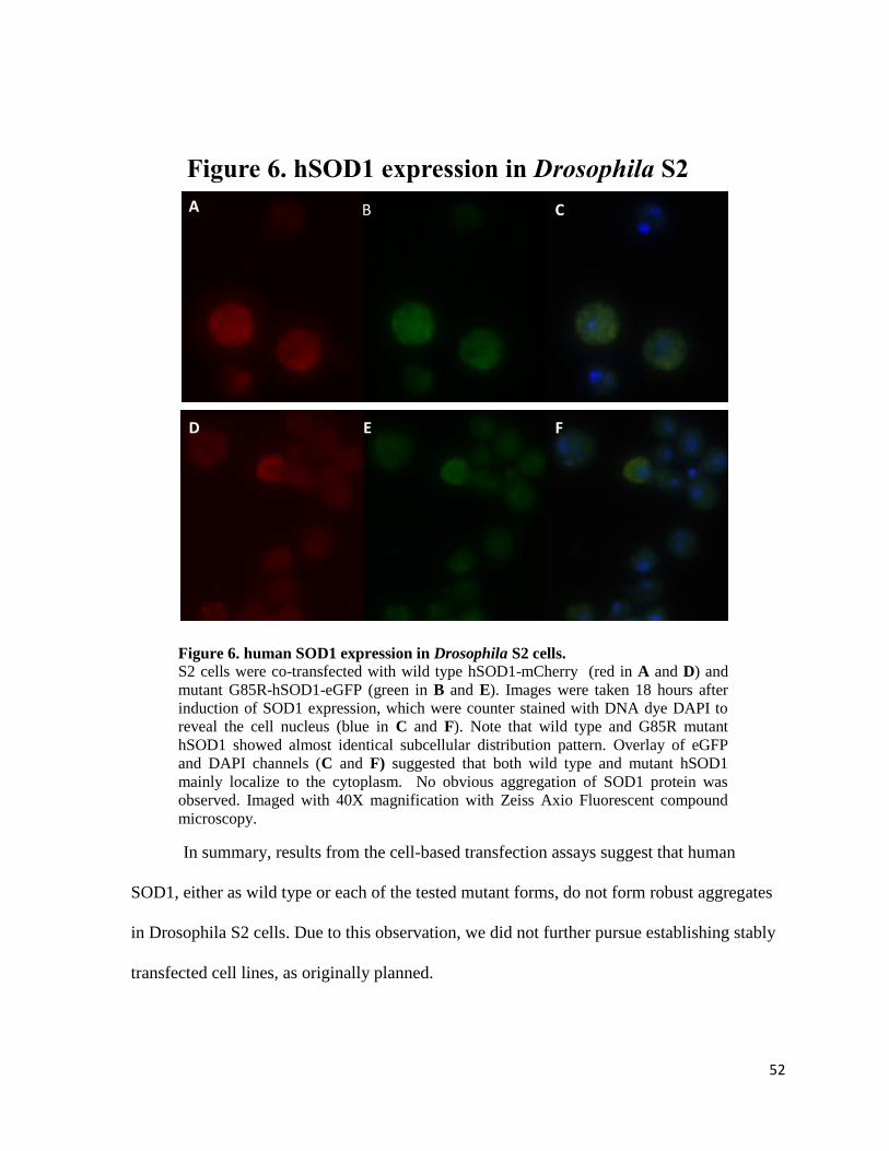
52
In summary, results from the cell-based transfection assays suggest that human
SOD1, either as wild type or each of the tested mutant forms, do not form robust aggregates
in Drosophila S2 cells. Due to this observation, we did not further pursue establishing stably
transfected cell lines, as originally planned.
Figure 6. human SOD1 expression in Drosophila S2 cells. S2 cells were co-transfected with wild type hSOD1-mCherry (red in A and D) and
mutant G85R-hSOD1-eGFP (green in B and E). Images were taken 18 hours after
induction of SOD1 expression, which were counter stained with DNA dye DAPI to
reveal the cell nucleus (blue in C and F). Note that wild type and G85R mutant
hSOD1 showed almost identical subcellular distribution pattern. Overlay of eGFP
and DAPI channels (C and F) suggested that both wild type and mutant hSOD1
mainly localize to the cytoplasm. No obvious aggregation of SOD1 protein was
observed. Imaged with 40X magnification with Zeiss Axio Fluorescent compound
microscopy.
A B C
D E F
Figure 6. hSOD1 expression in Drosophila S2
cells
53
CHAPTER SIX
Studying hSOD1Aggregation and Toxicity in Transgenic
Drosophila Models

54
In this chapter of the study, we applied the UAS-Gal4 system to overexpress human
SOD1 transgenes in different Drosophila tissues. We then analyzed the ability of WT and
mutant hSOD1to form aggregates in vivo and their effects on animal behavior and survival.
Confirmation of hSOD1 transgene expression in transgenic fly lines
To first confirm the proper expression of the hSOD1-eGFP or hSOD1-mCherry
fusion proteins from the established UAS-hSOD1 transgenic flies, we crossed the wild type
and mutants A4V, G85R and G93A SOD1 lines with D42 (motor neuron) and GMR (eye)
GAL4 drivers. These GAL4 driver lines provide tissue specific control over transgenic
hSOD1 expression. From each crosses, ten adult progenies containing both the GAL4 driver
and UAS transgene were collected and homogenized to extract whole proteins, followed by
western blot analysis to determine the success of protein expression from these transgenic
lines.
Figure 7 shows the results of the flies expressing wild type (WTSOD1-eGFP) and
mutant G85R (G85R-h SOD1-eGFP) hSOD1 fused to eGFP (Figure 2A), as well as samples
of the same mutant fused to mCherry (G85R-hSOD1- mCherry, Figure 2B). Expression is
controlled by either the motor neuron driver D42 GAL4 or the eye specific GMR driver.
Expression of the fusion transgenes were detected with an anti-eGFP or anti-mCherry
antibodies, respectively. hSOD1 encodes a small 16 kDa protein, whereas eGFP and
mCherry alone encodes 26kDa and 30kDa proteins, respectively. Accordingly, the hSOD1-
eGFP and hSOD1-mCherry fusion proteins should be at around 42 kDa and 46kDa sizes,
respectively. Indeed, Western blot analysis confirmed the correct expression of these three
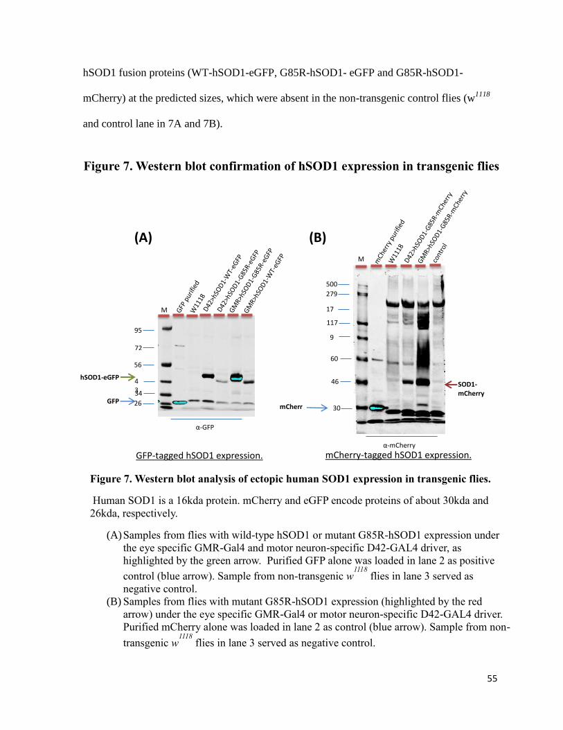
55
hSOD1 fusion proteins (WT-hSOD1-eGFP, G85R-hSOD1- eGFP and G85R-hSOD1-
mCherry) at the predicted sizes, which were absent in the non-transgenic control flies (w1118
and control lane in 7A and 7B).
M
170 117
95 60
500
46
30
279
SOD1-mCherry
95
72
43 34 26
56
GFP mCherry
GFP-tagged hSOD1 expression. mCherry-tagged hSOD1 expression.
hSOD1-eGFP
α-GFP
α-mCherry
M
(A) (B)
Figure 7. Western blot analysis of ectopic human SOD1 expression in transgenic flies.
Human SOD1 is a 16kda protein. mCherry and eGFP encode proteins of about 30kda and
26kda, respectively.
(A) Samples from flies with wild-type hSOD1 or mutant G85R-hSOD1 expression under
the eye specific GMR-Gal4 and motor neuron-specific D42-GAL4 driver, as
highlighted by the green arrow. Purified GFP alone was loaded in lane 2 as positive
control (blue arrow). Sample from non-transgenic w1118
flies in lane 3 served as
negative control.
(B) Samples from flies with mutant G85R-hSOD1 expression (highlighted by the red
arrow) under the eye specific GMR-Gal4 or motor neuron-specific D42-GAL4 driver.
Purified mCherry alone was loaded in lane 2 as control (blue arrow). Sample from non-
transgenic w1118
flies in lane 3 served as negative control.
Figure 7. Western blot confirmation of hSOD1 expression in transgenic flies
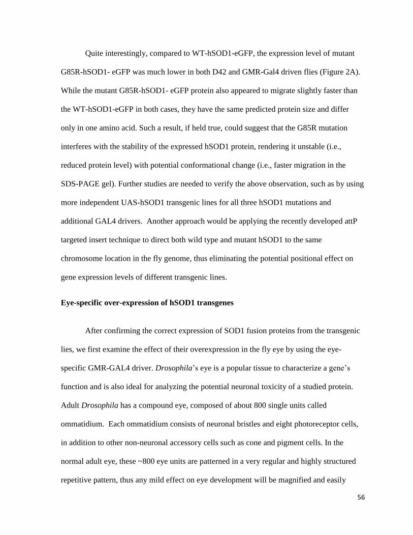
56
Quite interestingly, compared to WT-hSOD1-eGFP, the expression level of mutant
G85R-hSOD1- eGFP was much lower in both D42 and GMR-Gal4 driven flies (Figure 2A).
While the mutant G85R-hSOD1- eGFP protein also appeared to migrate slightly faster than
the WT-hSOD1-eGFP in both cases, they have the same predicted protein size and differ
only in one amino acid. Such a result, if held true, could suggest that the G85R mutation
interferes with the stability of the expressed hSOD1 protein, rendering it unstable (i.e.,
reduced protein level) with potential conformational change (i.e., faster migration in the
SDS-PAGE gel). Further studies are needed to verify the above observation, such as by using
more independent UAS-hSOD1 transgenic lines for all three hSOD1 mutations and
additional GAL4 drivers. Another approach would be applying the recently developed attP
targeted insert technique to direct both wild type and mutant hSOD1 to the same
chromosome location in the fly genome, thus eliminating the potential positional effect on
gene expression levels of different transgenic lines.
Eye-specific over-expression of hSOD1 transgenes
After confirming the correct expression of SOD1 fusion proteins from the transgenic
lies, we first examine the effect of their overexpression in the fly eye by using the eye-
specific GMR-GAL4 driver. Drosophila’s eye is a popular tissue to characterize a gene’s
function and is also ideal for analyzing the potential neuronal toxicity of a studied protein.
Adult Drosophila has a compound eye, composed of about 800 single units called
ommatidium. Each ommatidium consists of neuronal bristles and eight photoreceptor cells,
in addition to other non-neuronal accessory cells such as cone and pigment cells. In the
normal adult eye, these ~800 eye units are patterned in a very regular and highly structured
repetitive pattern, thus any mild effect on eye development will be magnified and easily

57
detected. Moreover, because it is a sensitive neuronal tissue that is dispensable for animal
viability, it is ideal to monitor the neurodegenerative consequences of a manipulated gene on
this neuronal tissue over an extended period of time. For example, gradual damage to the eye
cells has been often shown to present as an easily recognizable loss of red eye color, due to
the loss of internal pigment cells. Important to this study, as the eye is transparent, if the
fluorescent-tagged mutant hSOD1 protein forms aggregates in the eye cells, they can be
easily visualized directly under the fluorescent dissection microscope. This can be
performed without further dissection, as has been shown for aggregates-forming mutant
Huntington protein (Zhang, Binari, Zhou, & Perrimon, 2010), thus greatly facilitating the
monitoring for aggregates formation inside the eye.
Figures 8-10 shows the adult Drosophila’s eyes at differt ages expressing
fluorescent-tagged wild type and mutant hSOD1 from the transgenic flies, driven by the eye-
specific GMR-Gal4 driver.
Figure 8 consists of images of eyes of 30-day-old adult flies expressing wild type
hSOD1 and each of the mutant hSOD1 tagged with eGFP, along with eyes expressing mutant
G85R-hSOD1 with mCherry tag. When examined under bright field microscopy, all of these
eyes have regular well-patterned morphology, suggesting that overexpression of wild type or
either of the hSOD1 mutants does not affect normal eye development. Notably, there is no
clear loss of eye pigmentation in any of the hSOD1-expressing flies, even at age of day 70
(Figure 9 for wild type and G85R-SOD1 mutation, the same was seen for A4V and G93A
SOD1 mutants (data not shown). Indicating that there is no significant cellular degeneration
in these hSOD1-expressing flies, which further suggest that overexpressed hSOD1, either
wild type or any of the mutant forms, are well-tolerated by Drosophila eye cells.
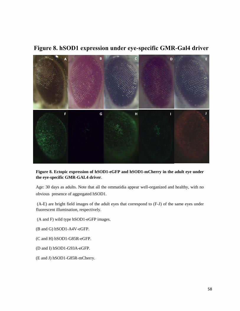
58
A B
C
D
E
F
G
H
I J
Figure 8. Ectopic expression of hSOD1-eGFP and hSOD1-mCherry in the adult eye under
the eye-specific GMR-GAL4 driver.
Age: 30 days as adults. Note that all the ommatidia appear well-organized and healthy, with no
obvious presence of aggregated hSOD1.
(A-E) are bright field images of the adult eyes that correspond to (F-J) of the same eyes under
fluorescent illumination, respectively.
(A and F) wild type hSOD1-eGFP images.
(B and G) hSOD1-A4V-eGFP.
(C and H) hSOD1-G85R-eGFP.
(D and I) hSOD1-G93A-eGFP.
(E and J) hSOD1-G85R-mCherry.
Figure 8. hSOD1 expression under eye-specific GMR-Gal4 driver
59
Figure 9. Ectopic expression of hSOD1-eGFP and hSOD1-mCherry in the adult
eye under the eye-specific GMR-GAL4 driver.
Age: 70 days as adults. Note that the ommatidia appear well-organized and healthy,
and there was no clear presence of aggregated hSOD1 in all genotypes.
(A-C) are bright field images of adult eyes that correspond to (D-F) the same eyes
under fluorescent illumination.
(A and D) hSOD1-WT-eGFP.
(B and E) hSOD1-G85R-eGFP.
(C and F) hSOD1-G85R-mCherry.
D
B C
F
A
E
Figure 9. hSOD1 expression under eye-specific GMR-GAL4
driver

60
When these hSOD1-expressing flies were visualized under fluoresecnt illumination to
examine the expression of eGFP- or mCherry-tagged hSOD1, there was clear presence of the
targeted proteins in the eyes. This observation is consistent with the confirmaiton by Western
blot analyses, although their expression levels varied (Figures 3-5). For example, the level of
A4V-hSOD1 expression was much lower as compared to that of other hSOD1 lines (Figure
3). It is possible that such variation is potentially due to positional effect of different
inserational sites, or it could suggest a reduced stablibility of the A4V mutant hSOD1
protein. Further studies are needed to clarify this potentially interesting issue.
These exmainations also suggested that there were almost no visible aggregate
formation in any of the hSOD1-expressing eyes (Figure 8-10), even in older flies at day 70
(Figure 4). Such an observation suggests that both wild type and either of the human SOD1
mutants do not easily form aggregates in Drosophila eye cells, which is in sharp contrast to
the prominent aggregates formation by mutant Hutintingtin protein expressed under similar
condition (Zhang et al., 2010),
To evaluate if co-expression of wild type hSOD1 could enhance the aggregation of
mutant hSOD1 in Drosophila, as has been shown in mouse models (Deng et al., 2006), we
simultaneously expressed wild type hSOD1-eGFP and mutant G85R-hSOD1-mCherry in the
eye (Figure 10). There was no observable enhancement of aggregation, although additional
quantifition by Western blot remained needed to confirm this conclusion.
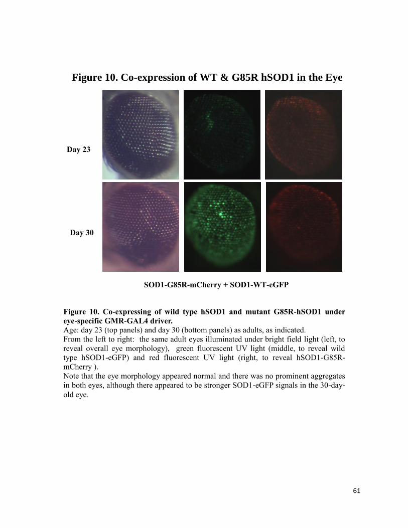
61
Day 23
SOD1-G85R-mCherry + SOD1-WT-eGFP
Day 30
Figure 10. Co-expressing of wild type hSOD1 and mutant G85R-hSOD1 under
eye-specific GMR-GAL4 driver. Age: day 23 (top panels) and day 30 (bottom panels) as adults, as indicated.
From the left to right: the same adult eyes illuminated under bright field light (left, to
reveal overall eye morphology), green fluorescent UV light (middle, to reveal wild
type hSOD1-eGFP) and red fluorescent UV light (right, to reveal hSOD1-G85R-
mCherry ).
Note that the eye morphology appeared normal and there was no prominent aggregates
in both eyes, although there appeared to be stronger SOD1-eGFP signals in the 30-day-
old eye.
Figure 10. Co-expression of WT & G85R hSOD1 in the Eye
62
Motor neuron-specific expression of hSOD1
Considering that ALS is a motor neuron degenerative disease, we further selected the
motor neuron-specific GAL 4 driver D42 to target hSOD1 transgene expression. This D42
driver will direct transgenic hSOD1 primarily in the motor neurons (Sanyal, 2009). Several
assays can be performed to investigate the effects of hSOD1 expression on animal behavior,
viability as well as the subcellular distribution and potential aggregation of wild type and
mutant hSOD1 proteins. For example, the animals can then be monitored for locomotion
defects such as the presentation or onset of paralysis. These animals can also be dissected to
obtain the adult brain and ventral nerve cord and examined under fluorescent compound
microscopy to determine the cellular localization and potential presence and localization of
aggregation of ectopically expressed hSOD1 proteins. Stained with phlloidian to visualize
the cytoskeleton and DAPI for the nuclei, the tissue can then be imagined with fluorescent
microscopy. The fluorescent tagged hSOD1 proteins in this expression system should label
the motor neurons and show, in addition to the pattern of expression, the formation of
aggregates in the motor neurons if they occur.
The flies expressing hSOD1 directed by the D42 motor neuron GAL4 driver were
aged for more than 90 days to assess their effects on animal locomotion and viability. To do
this, the D42>UAS-hSOD1 flies were transferred to new vials with fresh food every 3 days
to prevent animal loss related to texture changes in the food, and the number of the survival
flies were counted at that time. Close observations of flies expressing mutation G85R under
the D42 motor neuron driver showed no clear signs of paralysis or early decrease in mobility
compared to hSOD1 wild type expression directed by the same driver. These observations
are supported by survivorship data collected on G85R mutant hSOD1 fused to eGFP and

63
mCherry with respect to wild type hSOD1 fused to eGFP both expressed in motor neurons of
the flies. As presented in Figure 11 a total of 1,172 flies were counted, the mutant G85R-
hSOD1 expressing flies, driven by D42-GAL4, could live up to 104 days as adults, similar as
that of the wild type w1118
control (data not shown). Further, the overall trends of survival
curve between wild type SOD1 and mutant G85R-hSOD1expressing flies closely resembled
each other (Figure 6). Similarly, gross observations of mutant A4V- and G93A-hSOD1
expressing flies with D42 motor neuron revealed life span and mobility similar as that in
WT-SOD1 flies (data not shown). Together, they suggest that animal mobility and life span
are not significantly affected by mutant hSOD1 expression in Drosophila motor neurons.
0
20
40
60
80
100
120
1 6 12 18 24 30 36 42 48 54 60 66 71 77 83 89 95 101 107
Pe
rce
nt
surv
ival
Days
SOD1-wt-eGFP
SOD1-G85R-eGFP #1-4
SOD1-G85R-eGFP #2-3-2
SOD1-G85R-mCherry #1-1
SOD1-G85R-mCherry #1-2
Figure 11. Survival curve of adult flies expressing hSOD1 in motor
neurons

64
We next carried out preliminary examination to check whether there existed protein
aggregates formed by mutant SOD1 in these animals. Due to time restraints, we were only
able to analyze flies expressing A4V-hSOD1 (Figures 12 and 13) and G93A-hSOD1
mutants (data not shown). From these flies, adult brain and ventral nerve cord were dissected
and co-stained with phalloidin to visualize the actin cytoskeleton and DAPI dye for the
nuclei. The image in Figure 7 is of a dissected adult fly brain and ventral nerve cord viewed
at 10x magnification under fluorescent UV light to reveal of the expression of eGFP-tagged
A4V-hSOD1. The image revealed areas of higher level expression of mutant A4V-hSOD1-
eGFP. The brighter green areas in the image are actually the soluble A4V-hSOD1-eGFP
expressed in motor neurons, (which, due to a high fluorescent background, might be
confused as aggregation in a ubiquitous soluble A4V-hSOD1-eGFP expression context).
When examined under fluorescent compound microscopy, there was clear presence of eGFP-
tagged A4V- and G93A-hSOD1 protein in motor neurons that was enriched in the cell body
and also distributed along the whole axonal tract (Figure 7 and data not shown). However,
there was no prominent aggregate formation by mutant A4V-SOD1 protein in these neurons,
even in flies of 42 days of age (Figure 12).
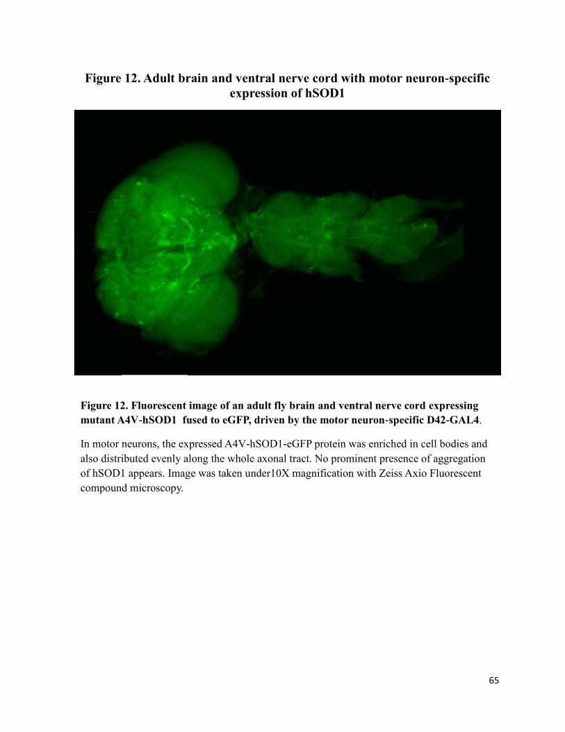
65
Figure 12. Fluorescent image of an adult fly brain and ventral nerve cord expressing
mutant A4V-hSOD1 fused to eGFP, driven by the motor neuron-specific D42-GAL4.
In motor neurons, the expressed A4V-hSOD1-eGFP protein was enriched in cell bodies and
also distributed evenly along the whole axonal tract. No prominent presence of aggregation
of hSOD1 appears. Image was taken under10X magnification with Zeiss Axio Fluorescent
compound microscopy.
Figure 12. Adult brain and ventral nerve cord with motor neuron-specific
expression of hSOD1

66
Upon closer examination, we found a few structures resembling hSOD1-derived
aggregates scattered along axon tracts of motor neurons (Figure 13). Some of these
aggregates-like structures, enriched with eGFP-tagged mutant SOD1, also had accumulation
of cytoskeleton protein F-Actin (Figure 8, top panels) while others did not (Figure 8, bottom
panels). In the future, additional studies are needed to quantify these structures and determine
whether such SOD1-enriched structures are indeed aggregates, for example by anti-ubiquitin
antibody staining to see if they are modified by ubiquitination, a hallmark of aggregates from
mis-folded proteins, or biochemical assay to examine their solubility in strong detergent such
as SDS. However, from our studies both in motor neurons (Figures 7 and 8) and in the eye
(Figures 3-5), it is clear that neither forms of the examined hSOD1 mutants develop robust
aggregates in fly cells.
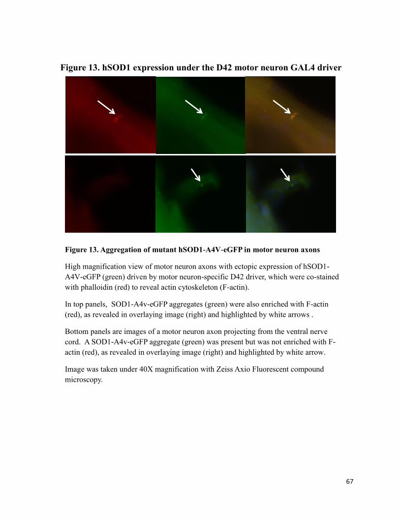
67
Figure 13. Aggregation of mutant hSOD1-A4V-eGFP in motor neuron axons
High magnification view of motor neuron axons with ectopic expression of hSOD1-
A4V-eGFP (green) driven by motor neuron-specific D42 driver, which were co-stained
with phalloidin (red) to reveal actin cytoskeleton (F-actin).
In top panels, SOD1-A4v-eGFP aggregates (green) were also enriched with F-actin
(red), as revealed in overlaying image (right) and highlighted by white arrows .
Bottom panels are images of a motor neuron axon projecting from the ventral nerve
cord. A SOD1-A4v-eGFP aggregate (green) was present but was not enriched with F-
actin (red), as revealed in overlaying image (right) and highlighted by white arrow.
Image was taken under 40X magnification with Zeiss Axio Fluorescent compound
microscopy.
Figure 13. hSOD1 expression under the D42 motor neuron GAL4 driver

68
Glia-specific over-expression of hSOD1 transgenes
ALS has been described a non-cell autonomus disease and as such neuron
degeneration depends not only on mutant protein expression in the neuron but also in the
surronding astrocyte/glia cells. Considering this, we next tested the effect of wild type and
mutant hSOD1 expression in Drosophila glia cells. To do this, we crossed the established
UAS-hSOD1 transgenic lines with glia cell-specific Gal4 driver line gcm-Gal4 (specific
promoter from the glia cell missing gene), which is expressed in almost all glial cells
including cortex, as soon as they are born. Cortex glia cells in Drosophila are also referred to
as cell-body-associated glia and are very similar to vertabrate astrocytes (Freeman &
Doherty, 2006). For this cross, a total of five line were selected for transgenic hSOD1
directed expression with the GCM GAL 4 driver. These line were two different G85R
mutant lines fused to eGFP along with two lines of G85R fused to mCherry and one line of
WT-hSOD1 fused to eGFP. Our observation were that very few, if any, flies expressing
hSOD1 in under the GCM GAL4 driver, either mutant G85R or wild type survived to adult
hood. Such a very interesting observation still needs furhter confirmation by using more
independent hSOD1 lines, both for wild type and for different hSOD1 mutants. Also further
testing should include additional glia-specific Gal4 driver such as repo-Gal4 as well as
controls such as by pan-neuronal GAL4 driver elav-Gal4.
However, it does raise an intriging possibility that high level of hSOD1 expresssion in
glia cells is lethal to Drosophila.

69
Summary and Discussion
We generated multiple UAS-hSOD1 transgenic lines and used available tissue-
specific Gal4 lines, to carry out preliminary analyses of the toxicity and aggregation property
of the wild type and mutant human SOD1 proteins in different fly tissue and cell types.
Although the study is still not completed yet and more controls and assasys are needed to
validate our preliminary observations. It is apparent at this point that human SOD1 protein,
either as wild type or the three mutated forms we examined, does not form robust aggregates
in different test settings. Included in our study is expression in cultured Drosophila S2 cells,
in adult eyes over an extended peroid of time (up to 70 days) or in motor neurons.
The original purpose of this project is to assess Drosophila as a model system to
study aggregates by mutant hSOD1, and if successful, its long term goal is to identify
regulators of aggregate formation by mutant hSOD1. To faciliate these goals, our expression
paradigm incorperates the fusion of a green or red fluorsecent protein to SOD1 proteins to
allow easy visualization of SOD1 protein expression and their aggregation. However, based
on our preliminary results so far, hSOD1 does not easily form aggregates in either the S2 cell
model or whole animal model in Drosophila. We will need to take serious re-consideration
regarding the viability of pursueing hSOD1 mutants in this model system.
Nevertheless, results so far also raised several interesting questions. For example,
although both mutant Huntingtin and mutant SOD1 proteins form aggregates in human cells,
they clearly behave very differently in term of aggrgates formation in Drosophila system.

70
Such a different behavior suggests that protein properties, and potentially aggregating
dynamics, of mutant Huntingtin and mutant SOD1 are very different. Moreover, why even
mutant forms of human SOD1 protein do not form robust aggregates even when expressed at
a high level over an extended period of time? One possibility is that protein quality control
machineries in Drosophila cells, those involved in protein re-folding and in clearance of mis-
folded proteins, are more effective than their counterparts in mammalian systems in dealing
with mutant SOD1 proteins. If this hypothesis is true, it raises a possibility that we can
identify the machinery(ies) in Drosophila that can correct or clear the mis-floded mutant
human SOD1 proteins, and harness its working mechanism to treat ALS.
Another very intriguing observation is the lethality caused by the overexpression of
either wild type or mutant G85R-hSOD1 specifically in glia. Notably, overexpression of
these proteins in the eye or motor neurons do not lead any obvious detrimental effect.
Although furhter studies are required to confirm the above observations, they raise a very
intersting possibility that glia cells are especially vulnearble to SOD1 expresion. Moreover,
existing findings from mammalian studies already support that nonautonomous effect from
astrocyte and glia contribute significantly to the ALS pathogenesis. Together, they present a
question whether the lethality associaited with glia-specific expresssion of SOD1 share some
mechanistic similarity with the pathogensis underlying ALS.

71
CHAPTER SEVEN
Perspectives
72
Toxicity and aggregation of human SOD1 protein in Drosophila
The results obtained during the study of this thesis project were consistent with a
previously established ALS models using hSOD1 in Drosophila (Watson et al., 2008). In that
study wild type hSOD1 and hSOD1 with A4V and G85R mutations were examined for
neuronal toxicity in Drosophila. Expression of human SOD1 in Drosophila was directed by
the GMR (eye specific) and D42 (motor neuron) Gal 4 drivers. The flies were examined for
deficits in motor function and life span, as well subject to Western analysis, motor neurons
counts and electrophysiology. It was found that although progressive climbing difficulty
was recorded with both wild type and mutant hSOD1 expression in motor neurons, there was
no significant loss of motor neurons nor obvious signs of paralysis (Watson et al., 2008).
Toxicity of wild type hSOD1 was confirmed with electrophysiology studies and showed
similar defect to that seen in G85R flies. The results suggest the giant fiber circuit is
defective when given high frequency stimulation, revealing defects in synaptic transmission
of older flies expressing wild type and G85R mutant hSOD1 in motor neurons (Watson et al.,
2008). Further, their life span data for motor neuron directed expression of WT and mutant
hSOD1 flies revealed similar survivorship curves as their control flies which expressed GFP
only under the same D42 driver (Watson et al., 2008). Finally, they found that focal
accumulation of hSOD1 protein did occur when directed by the eye-specific GMR-Gal4 and
motor neuron-specific D42 drivers, and that in motor neurons it was accompanied by a stress
response in surrounding glia cells (Watson et al., 2008). Interestingly, the study did observe
the same change to migration rate of mutation G85R of hSOD1 in western analysis (Watson
et al., 2008). Interestingly, the study did observe the same change to migration rate of G85R-
hSOD1 mutation in Western analysis (Watson et al., 2008). Notably, they concluded that
73
neuronal toxicity was not the result of hSOD1 aggregates and hSOD1 expression did not
cause a loss of neurons (Watson et al., 2008). Our current study, with respect to a lack of
obvious paralysis and unaffected life span in mutant hSOD1-expressing flies as compared
with ones with wild type hSOD1 expression, is consistent with this previous work.
The previously established model by Watson et al, however, did not employ hSOD1
fused to eGFP or mCherry fluorescent reporters, in contrast to our model. Thus, this suggests
that the fusion of a fluorescent protein to wild type and mutant hSOD1 does not increase or
decrease its toxicity in Drosophila. In addition, fusing fluorescent proteins to hSOD1 does
not appear to adversely affect the solubility and subcellular localization of hSOD1 in
Drosophila. Although the Waston et al 2008 study reported the toxicity of SOD1 protein in
the fly, as manifested as a decrease in geotaxis response (Watson et al., 2008), we have yet to
confirm this observation using our SOD1 model. However, we did observe the presence of
aggregates-like structures in neurons of SOD1-expressing animals when examined under
fluorescent microscopy (Figure 7). Further studies are needed to clarify this observation.
Glia-specific toxicity of human SOD1 protein in Drosophila?
Interestingly, in our current study, when wild type or mutant hSOD1 transgenic flies
were crossed with a glia cell specific GAL4 driver, gcm-GAL4, a surprising lethality
phenotype emerged that suggests expression of hSOD1 in this tissue could be lethal. ALS
caused by SOD1 mutations in general has been described as a non-cell autonomous disease
(Boillee, Vande Velde, et al., 2006; Boillee, Yamanaka, et al., 2006; Da Cruz & Cleveland,
2011; Nagai et al., 2007; Tamura, Sone, Yamashita, Wanker, & Okazawa, 2009; Wallis et
al., 2012). Non-cell autonomy is a term here to describe the nature of toxicity as dependent
74
on the involvement of surrounding cell or tissues. Glia and astrocyte cells are known as
support cells for neurons but recently their true nature of involvement with functions of
neurons and synaptic transmission has been revealed to be very complex (Volterra &
Meldolesi, 2005). It is now known that astrocytes are indeed excitable and express in their
membrane many of the same receptors and channels expressed by neurons (Volterra &
Meldolesi, 2005). Perhaps most notably astrocytes play a major role in sequestering
glutamate from the synaptic cleft, to avoid it remaining in the cleft and causing
excytotoxicity of neurons. The loss of the astrocyte glutamate reuptake transporter EAAT2
has been seen in ALS and is believed to be a major contributor of glutamate related
excytotoxicity (Howland et al., 2002; Wallis et al., 2012). In addition astrocytes release
trophic factors to neurons such as TNFα, which induces insertion of AMPA receptors into the
plasma membrane (Yin et al., 2012). AMPA receptors are the key metabotropic excitatory
receptors for which the agonist is glutamate. However, in advanced stages of the disease
progression the increased in TNFα release by astrocytes becomes toxic to neurons (Yin et al.,
2012).
Future studies based on the established SOD1 models in Drosophila
More work can be carried out to further test the above hypotheses using our hSOD1
models. First, in the whole animal models, it should include ubiquitous expression of both
wild type and mutant hSOD1, to more comprehensively determine the presence of an
aggregation phenotype. In doing so, we can also determine whether the hSOD1-induced
lethality phenotype observed under the glia cell gcm-GAL4 driver is truly glia-specific.

75
Second, of the current SOD1 model in S2 cells, which does not form robust
aggregates, it is possible that Drosophila S2 cells are expressing a gene alone or of a pathway
that is capable of completely suppressing aggregate formation by mutant hSOD1. In such a
scenario, our S2 cell-based SOD1 models could be used to conduct the proposed genome-
wide RNAi screen to reveal such a protective gene or pathway(s), potentially leading to a
new and exciting research direction.
Further, Drosophila S2 cells are macrophage lineage cells, which are immune system
cells, not neuronal cells. Since ALS is a neurodegenerative disease, switching to a
Drosophila neuronal cell line may yield different results with regard to aggregate formation.
With regard to copper homeostasis, Drosophila S2 cells contain copper chaperones that are
powerful regulators of free copper (Southon, Burke, Norgate, Batterham, & Camakaris,
2004). hSOD1 has been shown to accumulate and aggregate in neurons, astrocytes and
mitochondria (Cozzolino et al., 2009; Jaiswal et al., 2009; Nguyen et al., 2009). Aggregation
of any protein in a cell is a concentration dependent process, which means it inherently
requires accumulation of significant amount of mis-folded proteins and thus the expression
level of the protein will be an important factor in aggregation process. On this aspect, our S2
cell-based model employs copper induction of hSOD1 expression, while an enzymatically
active hSOD1 by itself is required to complex with copper and zinc, thus titration of aqueous
copper may be necessary to optimize its hSOD1expression. Another possibility would be to
codon optimize the inserted hSOD1 gene, to the codons preferred by the Drosophila species.
This has been suggested by the advising committee over this project and is postulated to
result in a noticeable difference in phenotype presentation.

76
Cellular toxicity in ALS: a link between dysregulated neuronal excitability, aggregates
and others?
The underlying cellular and molecular mechanisms underlying ALS are still far from
clear. Nevertheless, it is quite intriguing to observe a link between dysregulated neuronal
excitability, aggregates formation and other cellular factors in ALS pathogenesis.
To facilitate normal neuronal communication neurons must generate action potentials
to allow an influx of calcium, which then signals the release of neurotransmitter to the
synaptic cleft. Action potentials are propagated by and result from the opening of voltage
gated sodium channels, which allow for an influx of positively charged sodium ions, thereby
depolarizing the neuron. This sodium current allows for a spike in voltage, which is
propagated down the axon to the synaptic cleft. That action causes the necessary increase in
calcium concentration, which then facilitates the binding of vesicles to the inner membrane
of the neuron, subsequently the vesicle fuses with the membrane and opens to the synaptic
cleft to release its neurotransmitter contents. The neurotransmitter molecules travels across
the synaptic cleft from the presynaptic neuron to the post synaptic neuron and eventually
cause either an inhibitory postsynaptic potential or and excitatory post synaptic potential
(IPSP or an EPSP).
All neurons maintain a negatively polarized resting membrane potential that is
caused by negatively charged proteins and DNA. The resting membrane potential is
maintained at around -65mV by high intracellular potassium concentration. However, for an
action potential to occur the membrane must depolarize to a certain threshold potential and
then an all or nothing response in the form of an action potential is generated. An increase in
intracellular chloride, mediated by GABA channels, can prevent current from cation channels

77
depolarizing the membrane. This action will then inhibit the neuron from reaching the
depolarized threshold necessary to generate an action potential. Increases in depolarizing
threshold have been seen in ALS motor neuron axons and it has been suggested to be
attributed to dysfunction in either expression of or conductance of potassium channels.
(Cheah et al., 2012; Shibuya et al., 2011) However, an accumulation of negatively charged
proteins could be the underlying cause of that dysfunction and subsequent increase in
excitability. hSOD1 has a net negative charge of -6 and mutated SOD1 maintains the same
charge or in some cases decreases, but only slightly, while in other cases such as G127X the
net negative charge increases. (Sandelin, Nordlund, Andersen, Marklund, & Oliveberg, 2007)
Mutated proteins, such as SOD1, at some point lose functionality and accumulate while
remaining soluble and then aggregate. In this quasi-stable state of soluble accumulation the
negatively charged proteins may be able to aberrantly contribute to the global negative
charge of a cell. This may result in changes to cation conductance and produce excessive
inhibition of action potentials. Thus, the neuron in cooperation with astrocytes struggles to
overcome excessive inhibition and in the process becomes over excitable.
In support of the above hypothesis, in addition to the dysfunction seen of potassium
channel in ALS models, the following findings are noteworthy. First, it has been shown that
some mutant forms of SOD1 develop less soluble yet toxic aggregates, then become more
toxic when soluble yet mis-folded, but then becomes less toxic when correctly folded by
chaperones.(Brotherton, Li, & Glass, 2012). The less soluble but more aggregated SOD1
mutants become very toxic when solubilized until correctly folded by chaperones (Brotherton
et al., 2012). This evidence supports that mutant SOD1 is most toxic when mis-folded and
accumulated while still in electrochemical contact within the cell. Second, an increase in

78
persistent inward sodium conductance has been seen in ALS and causes increased
excitability. (Kiernan, 2009; Meehan et al., 2010; Shibuya et al., 2011) Third, TNFα release
by astrocytes is increased which actually induces AMPA receptor insertion into the
membrane, while later with disease progression it becomes toxic. (Boillee, Vande Velde, et
al., 2006; Yin et al., 2012) Overexpression of AMPA receptors will markedly increase
excitability and has been well established to play a prominent role in ALS. Fourth,
astrocytes have been shown to release d-serine which is a stronger agonist for NMDA
receptors than glycine and can actually increase their ability to conduct cations. (Sasabe et
al., 2007) NMDA up regulation in this manner has been seen in ALS and leads to an
increase in excitability, which in turn leads to an increased cytoplasmic calcium
concentration (Sasabe et al., 2007; Spalloni, Nutini, & Longone, 2013) Fifth, the loss of
EAAT2 glutamate transporter in astrocytes results in an increase in the concentration of the
main excitatory neurotransmitter glutamate in the synaptic cleft, which in turn will cause
excessive activation of NMDA and AMPA receptors (Howland et al., 2002). Consequently,
again leading to increased calcium concentration in the neuron, a sign of excytotoxicity, and
a result of increased excitability.
ALS and athleticism: a causative or an incidental link?
Some studies have shown a correlation between prolonged strenuous activity, such as
athletics or occupations that require intense physical activity and onset of ALS (Scarmeas,
Shih, Stern, Ottman, & Rowland, 2002; Sutedja et al., 2009). One most famous case of ALS
is Lou Gehrig, an outstanding baseball player who fell victim to the ALS during the peak of
his career, and ALS has often or better known by general public, as Lou Gehrig’s disease.
Notably, in ALS, the changes to neurons with regard to sodium and potassium channel
79
conductance are seen well before symptoms start and are suggested to appear early in life. If
there is a higher degree of excitability in motor neurons that occurs well before onset of
symptoms, it could translate to an increase in stamina. High stamina can lead to success
when engaged in physically demanding activities such as athletics. Just as a general increase
in energy level that may be the underlying reason for a lower body mass index (O'Reilly et
al., 2013). There is a remote possibility that it may not be exercise or environmental factors
that contribute to ALS; it may be that the underlying cellular mechanisms of ALS make
patients more likely to be active at a younger age.
80
REFERENCES
Aguzzi, A., & O'Connor, T. (2010). Protein aggregation diseases: pathogenicity and
therapeutic perspectives. Nat Rev Drug Discov, 9(3), 237-248. doi: 10.1038/nrd3050
ALS association. (2013). from www.ALS.org
Aquilano, K., Vigilanza, P., Rotilio, G., & Ciriolo, M. R. (2006). Mitochondrial damage due
to SOD1 deficiency in SH-SY5Y neuroblastoma cells: a rationale for the redundancy
of SOD1. Faseb Journal, 20(10), 1683-+. doi: 10.1096/fj.05-5225fje
Beckman, J. S., Estevez, A. G., & Crow, J. R. (2001). Superoxide dismutase and the death of
motoneurons in ALS. Trends Neurosci, 24(11), S15-S20. doi: 10.1016/s0166-
2236(00)01981-0
Beckman, J. S., & Koppenol, W. H. (1996). Nitric oxide, superoxide, and peroxynitrite: The
good, the bad, and the ugly. American Journal of Physiology-Cell Physiology, 271(5),
C1424-C1437.
Benediktsson, A. M., Schachtele, S. J., Green, S. H., & Dailey, M. E. (2005). Ballistic
labeling and dynamic imaging of astrocytes in orcranotypic hippocampal slice
cultures. Journal of Neuroscience Methods, 141(1), 41-53. doi:
10.1016/j.jneumeth.2004.05.013
Bezzi, P., & Volterra, A. (2001). A neuron-glia signalling network in the active brain. Curr
Opin Neurobiol, 11(3), 387-394. doi: 10.1016/s0959-4388(00)00223-3
Bilen, J., & Bonini, N. M. (2005). Drosophila as a model for human neurodegenerative
disease Annual Review of Genetics (Vol. 39, pp. 153-171).

81
Boillee, S., Vande Velde, C., & Cleveland, D. W. (2006). ALS: a disease of motor neurons
and their nonneuronal neighbors. Neuron, 52(1), 39-59. doi:
10.1016/j.neuron.2006.09.018
Boillee, S., Yamanaka, K., Lobsiger, C. S., Copeland, N. G., Jenkins, N. A., Kassiotis, G.,
Kollias, G., & Cleveland, D. W. (2006). Onset and progression in inherited ALS
determined by motor neurons and microglia. Science, 312(5778), 1389-1392. doi:
10.1126/science.1123511
Brotherton, T. E., Li, Y., & Glass, J. D. (2012). Cellular toxicity of mutant SOD1 protein is
linked to an easily soluble, non-aggregated form in vitro. Neurobiol Dis, 49C, 49-56.
doi: 10.1016/j.nbd.2012.08.010
Bruijn, L. I., Becher, M. W., Lee, M. K., Anderson, K. L., Jenkins, N. A., Copeland, N. G.,
Sisodia, S. S., Rothstein, J. D., Borchelt, D. R., Price, D. L., & Cleveland, D. W.
(1997). ALS-linked SOD1 mutant G85R mediates damage to astrocytes and promotes
rapidly progressive disease with SOD1-containing inclusions. Neuron, 18(2), 327-
338. doi: 10.1016/s0896-6273(00)80272-x
Bruijn, L. I., Miller, T. M., & Cleveland, D. W. (2004). Unraveling the mechanisms involved
in motor neuron degeneration in ALS. Annu Rev Neurosci, 27, 723-749. doi:
10.1146/annurev.neuro.27.070203.144244
Bushong, E. A., Marton, M. E., & Ellisman, M. H. (2004). Maturation of astrocyte
morphology and the establishment of astrocyte domains during postnatal
hippocampal development. International Journal of Developmental Neuroscience,
22(2), 73-86. doi: 10.1016/j.ijdeveneu.2003.12.008

82
Bushong, E. A., Martone, M. E., Jones, Y. Z., & Ellisman, M. H. (2002). Protoplasmic
astrocytes in CA1 stratum radiatum occupy separate anatomical domains. Journal of
Neuroscience, 22(1), 183-192.
Carri, M. T., & Cozzolino, M. (2011). SOD1 and mitochondria in ALS: a dangerous liaison.
J Bioenerg Biomembr, 43(6), 593-599. doi: 10.1007/s10863-011-9394-z
Caughey, B., & Lansbury, P. T. (2003). Protofibrils, pores, fibrils, and neurodegeneration:
separating the responsible protein aggregates from the innocent bystanders. Annu Rev
Neurosci, 26, 267-298. doi: 10.1146/annurev.neuro.26.010302.081142
Cheah, B. C., Lin, C. S., Park, S. B., Vucic, S., Krishnan, A. V., & Kiernan, M. C. (2012).
Progressive axonal dysfunction and clinical impairment in amyotrophic lateral
sclerosis. Clin Neurophysiol, 123(12), 2460-2467. doi: 10.1016/j.clinph.2012.06.020
Chudakov, D. M., Matz, M. V., Lukyanov, S., & Lukyanov, K. A. (2010). Fluorescent
Proteins and Their Applications in Imaging Living Cells and Tissues. Physiological
Reviews, 90(3), 1103-1163. doi: 10.1152/physrev.00038.2009
Corcoran, L. J., Mitchison, T. J., & Liu, Q. (2004). A novel action of histone deacetylase
inhibitors in a protein aggresome disease model. Curr Biol, 14(6), 488-492. doi:
10.1016/j.cub.2004.03.003
Cozzolino, M., Pesaresi, M. G., Amori, I., Crosio, C., Ferri, A., Nencini, M., & Carri, M. T.
(2009). Oligomerization of Mutant SOD1 in Mitochondria of Motoneuronal Cells
Drives Mitochondrial Damage and Cell Toxicity. Antioxidants & Redox Signaling,
11(7), 1547-U1543. doi: 10.1089/ars.2009.2545
83
Da Cruz, S., & Cleveland, D. W. (2011). Understanding the role of TDP-43 and FUS/TLS in
ALS and beyond. Curr Opin Neurobiol, 21(6), 904-919. doi:
10.1016/j.conb.2011.05.029
De Vos, K. J., Grierson, A. J., Ackerley, S., & Miller, C. C. J. (2008). Role of axonal
transport in neurodegenerative diseases Annu Rev Neurosci (Vol. 31, pp. 151-173).
Deas, E., Wood, N. W., & Plun-Favreau, H. (2011). Mitophagy and Parkinson's disease: The
PINK1-parkin link. Biochimica Et Biophysica Acta-Molecular Cell Research,
1813(4), 623-633. doi: 10.1016/j.bbamcr.2010.08.007
Deng, H. X., Shi, Y., Furukawa, Y., Zhai, H., Fu, R., Liu, E., Gorrie, G. H., Khan, M. S.,
Hung, W. Y., Bigio, E. H., Lukas, T., Dal Canto, M. C., O'Halloran, T. V., &
Siddique, T. (2006). Conversion to the amyotrophic lateral sclerosis phenotype is
associated with intermolecular linked insoluble aggregates of SOD1 in mitochondria.
Proc Natl Acad Sci U S A, 103(18), 7142-7147. doi: 10.1073/pnas.0602046103
Denise, A., Garcia, R., Doan, N. B., Imura, T., Bush, T. G., & Sofroniew, M. V. (2004).
GFAP-expressing progenitors are the principal source of constitutive neurogenesis in
adult mouse forebrain. Nat Neurosci, 7(11), 1233-1241. doi: 10.1038/nn1340
Derouiche, A., & Frotscher, M. (2001). Peripheral astrocyte processes: Monitoring by
selective immunostaining for the actin-binding ERM proteins. Glia, 36(3), 330-341.
doi: 10.1002/glia.1120
Fellin, T., Pascual, O., Gobbo, S., Pozzan, T., Haydon, P. G., & Carmignoto, G. (2004).
Neuronal synchrony mediated by astrocytic glutamate through activation of
extrasynaptic NMDA receptors. Neuron, 43(5), 729-743. doi:
10.1016/j.neuron.2004.08.011

84
Ferri, A., Cozzolino, M., Crosio, C., Nencini, M., Casciati, A., Gralla, E. B., Rotilio, G.,
Valentine, J. S., & Carri, M. T. (2006). Familial ALS-superoxide dismutases
associate with mitochondria and shift their redox potentials. Proc Natl Acad Sci U S
A, 103(37), 13860-13865. doi: 10.1073/pnas.0605814103
Freeman, M. R., & Doherty, J. (2006). Glial cell biology in Drosophila and vertebrates.
Trends Neurosci, 29(2), 82-90. doi: 10.1016/j.tins.2005.12.002
Furukawa, Y., Fu, R. G., Deng, H. X., Siddique, T., & O'Halloran, T. V. (2006). Disulfide
cross-linked protein represents a significant fraction of ALS-associated Cu, Zn-
superoxide dismutase aggregates in spinal cords of model mice. Proc Natl Acad Sci U
S A, 103(18), 7148-7153. doi: 10.1073/pnas.0602048103
Graffmo, K. S., Forsberg, K., Bergh, J., Birve, A., Zetterstrom, P., Andersen, P. M.,
Marklund, S. L., & Brannstrom, T. (2013). Expression of wild-type human
superoxide dismutase-1 in mice causes amyotrophic lateral sclerosis. Hum Mol Genet,
22(1), 51-60. doi: 10.1093/hmg/dds399
Hammond, C. (2008). Cellular and Molecular Neurophysiology: Elsevier.
Han, S. M., Tsuda, H., Yang, Y., Vibbert, J., Cottee, P., Lee, S. J., Winek, J., Haueter, C.,
Bellen, H. J., & Miller, M. A. (2012). Secreted VAPB/ALS8 major sperm protein
domains modulate mitochondrial localization and morphology via growth cone
guidance receptors. Dev Cell, 22(2), 348-362. doi: 10.1016/j.devcel.2011.12.009
Hirrlinger, J., Hulsmann, S., & Kirchhoff, F. (2004). Astroglial processes show spontaneous
motility at active synaptic terminals in situ. European Journal of Neuroscience, 20(8),
2235-2239. doi: 10.1111/j.1460-9568.2004.03689.x

85
Howland, D. S., Liu, J., She, Y., Goad, B., Maragakis, N. J., Kim, B., Erickson, J., Kulik, J.,
DeVito, L., Psaltis, G., DeGennaro, L. J., Cleveland, D. W., & Rothstein, J. D.
(2002). Focal loss of the glutamate transporter EAAT2 in a transgenic rat model of
SOD1 mutant-mediated amyotrophic lateral sclerosis (ALS). Proc Natl Acad Sci U S
A, 99(3), 1604-1609. doi: 10.1073/pnas.032539299
Hua, H., Georgiev, O., Schaffner, W., & Steiger, D. (2010). Human copper transporter Ctr1
is functional in Drosophila, revealing a high degree of conservation between
mammals and insects. Journal of Biological Inorganic Chemistry, 15(1), 107-113.
doi: 10.1007/s00775-009-0599-0
Iino, M., Goto, K., Kakegawa, W., Okado, H., Sudo, M., Ishiuchi, S., Miwa, A., Takayasu,
Y., Saito, I., Tsuzuki, K., & Ozawa, S. (2001). Glia-synapse interaction through
Ca2+-permeable AMPA receptors in Bergmann glia. Science, 292(5518), 926-929.
doi: 10.1126/science.1058827
Ince, P. G., Highley, J. R., Kirby, J., Wharton, S. B., Takahashi, H., Strong, M. J., & Shaw,
P. J. (2011). Molecular pathology and genetic advances in amyotrophic lateral
sclerosis: an emerging molecular pathway and the significance of glial pathology.
Acta Neuropathol, 122(6), 657-671. doi: 10.1007/s00401-011-0913-0
Jaiswal, M. K., Zech, W. D., Goos, M., Leutbecher, C., Ferri, A., Zippelius, A., Carri, M. T.,
Nau, R., & Keller, B. U. (2009). Impairment of mitochondrial calcium handling in a
mtSOD1 cell culture model of motoneuron disease. BMC Neurosci, 10, 64. doi:
10.1186/1471-2202-10-64
Jonsson, P. A., Ernhill, K., Andersen, P. M., Bergemalm, D., Brannstrom, T., Gredal, O.,
Nilsson, P., & Marklund, S. L. (2004). Minute quantities of misfolded mutant
86
superoxide dismutase-1 cause amyotrophic lateral sclerosis. Brain, 127, 73-88. doi:
10.1093/brain/awh005
Kiernan, M. C. (2009). Hyperexcitability, persistent Na+ conductances and
neurodegeneration in amyotrophic lateral sclerosis. Exp Neurol, 218(1), 1-4. doi:
10.1016/j.expneurol.2009.03.039
Kloppel, C., Michels, C., Zimmer, J., Herrmann, J. M., & Riemer, J. (2010). In yeast
redistribution of Sod1 to the mitochondrial intermembrane space provides protection
against respiration derived oxidative stress. Biochem Biophys Res Commun, 403(1),
114-119. doi: 10.1016/j.bbrc.2010.10.129
Kwiatkowski, T. J., Jr., Bosco, D. A., LeClerc, A. L., Tamrazian, E., Vanderburg, C. R.,
Russ, C., Davis, A., Gilchrist, J., Kasarskis, E. J., Munsat, T., Valdmanis, P.,
Rouleau, G. A., Hosler, B. A., Cortelli, P., de Jong, P. J., Yoshinaga, Y., Haines, J. L.,
Pericak-Vance, M. A., Yan, J., Ticozzi, N., Siddique, T., McKenna-Yasek, D., Sapp,
P. C., Horvitz, H. R., Landers, J. E., & Brown, R. H., Jr. (2009). Mutations in the
FUS/TLS Gene on Chromosome 16 Cause Familial Amyotrophic Lateral Sclerosis.
Science, 323(5918), 1205-1208. doi: 10.1126/science.1166066
Liochev, S. I., & Fridovich, I. (2000). Copper- and zinc-containing superoxide dismutase can
act as a superoxide reductase and a superoxide oxidase. J Biol Chem, 275(49), 38482-
38485. doi: 10.1074/jbc.M007891200
Liochev, S. I., & Fridovich, I. (2003). Mutant Cu,Zn superoxide dismutases and familial
amyotrophic lateral sclerosis: evaluation of oxidative hypotheses. Free Radical
Biology and Medicine, 34(11), 1383-1389. doi: 10.1016/s0891-5849(03)00153-9
87
Mackenzie, I. R. A., Rademakers, R., & Neumann, M. (2010). TDP-43 and FUS in
amyotrophic lateral sclerosis and frontotemporal dementia. Lancet Neurology, 9(10),
995-1007. doi: 10.1016/s1474-4422(10)70195-2
Magrane, J., & Manfredi, G. (2009). Mitochondrial Function, Morphology, and Axonal
Transport in Amyotrophic Lateral Sclerosis. Antioxidants & Redox Signaling, 11(7),
1615-U1618. doi: 10.1089/ars.2009.2604
Marsh, J. L., & Thompson, L. M. (2006). Drosophila in the study of neurodegenerative
disease. Neuron, 52(1), 169-178. doi: 10.1016/j.neuron.2006.09.025
Matsui, K., & Jahr, C. E. (2003). Ectopic release of synaptic vesicles. Neuron, 40(6), 1173-
1183. doi: 10.1016/s0896-6273(03)00788-8
Matsui, K., & Jahr, C. E. (2004). Differential control of synaptic and ectopic vesicular
release of glutamate. Journal of Neuroscience, 24(41), 8932-8939. doi:
10.1523/jneurosci.2650-04.2004
Matsumoto, G., Stojanovic, A., Holmberg, C. I., Kim, S., & Morimoto, R. I. (2005).
Structural properties and neuronal toxicity of amyotrophic lateral sclerosis-associated
Cu/Zn superoxide dismutase 1 aggregates. Journal of Cell Biology, 171(1), 75-85.
doi: 10.1083/jcb.200504050
Mauch, D. H., Nagler, K., Schumacher, S., Goritz, C., Muller, E. C., Otto, A., & Pfrieger, F.
W. (2001). CNS synaptogenesis promoted by glia-derived cholesterol. Science,
294(5545), 1354-1357. doi: 10.1126/science.294.5545.1354
Meehan, C. F., Moldovan, M., Marklund, S. L., Graffmo, K. S., Nielsen, J. B., & Hultborn,
H. (2010). Intrinsic properties of lumbar motor neurones in the adult G127insTGGG
superoxide dismutase-1 mutant mouse in vivo: evidence for increased persistent
88
inward currents. Acta Physiol (Oxf), 200(4), 361-376. doi: 10.1111/j.1748-
1716.2010.02188.x
Morimoto, N., Nagai, M., Miyazaki, K., Ohta, Y., Kurata, T., Takehisa, Y., Ikeda, Y.,
Matsuura, T., Asanuma, M., & Abe, K. (2010). Induction of Parkinsonism-Related
Proteins in the Spinal Motor Neurons of Transgenic Mouse Carrying a Mutant SOD1
Gene. Journal of Neuroscience Research, 88(8), 1804-1811. doi: 10.1002/jnr.22341
Morimoto, N., Nagai, M., Ohta, Y., Miyazaki, K., Kurata, T., Morimoto, M., Murakami, T.,
Takehisa, Y., Ikeda, Y., Kamiya, T., & Abe, K. (2007). Increased autophagy in
transgenic mice with a G93A mutant SOD1 gene. Brain Res, 1167, 112-117. doi:
10.1016/j.brainres.2007.06.045
Muller, F. L., Lustgarten, M. S., Jang, Y., Richardson, A., & Van Remmen, H. (2007).
Trends in oxidative aging theories. Free Radic Biol Med, 43(4), 477-503. doi:
10.1016/j.freeradbiomed.2007.03.034
Murai, K. K., Nguyen, L. N., Irie, F., Yamaguchi, Y., & Pasquale, E. B. (2003). Control of
hippocampal dendritic spine morphology through ephrin-A3/EphA4 signaling. Nat
Neurosci, 6(2), 153-160. doi: 10.1038/nn994
Nagai, M., Re, D. B., Nagata, T., Chalazonitis, A., Jessell, T. M., Wichterle, H., &
Przedborski, S. (2007). Astrocytes expressing ALS-linked mutated SOD1 release
factors selectively toxic to motor neurons. Nat Neurosci, 10(5), 615-622. doi:
10.1038/nn1876
Nguyen, K. T., Garcia-Chacon, L. E., Barrett, J. N., Barrett, E. F., & David, G. (2009). The
Psi(m) depolarization that accompanies mitochondrial Ca2+ uptake is greater in

89
mutant SOD1 than in wild-type mouse motor terminals. Proc Natl Acad Sci U S A,
106(6), 2007-2011. doi: 10.1073/pnas.0810934106
O'Brien, K. M., Dirmeier, R., Engle, M., & Poyton, R. O. (2004). Mitochondrial protein
oxidation in yeast mutants lacking manganese- (MnSOD) or copper- and zinc-
containing superoxide dismutase (CuZnSOD) - Evidence that MnSOD and CuZnSOD
have both unique and overlapping functions in protecting mitochondrial proteins from
oxidative damage. Journal of Biological Chemistry, 279(50), 51817-51827. doi:
10.1074/jbc.M405958200
O'Reilly, E. J., Wang, H., Weisskopf, M. G., Fitzgerald, K. C., Falcone, G., McCullough, M.
L., Thun, M., Park, Y., Kolonel, L. N., & Ascherio, A. (2013). Premorbid body mass
index and risk of amyotrophic lateral sclerosis. Amyotrophic Lateral Sclerosis and
Frontotemporal Degeneration, 14(3), 205-211. doi: 10.3109/21678421.2012.735240
Parpura, V., Basarsky, T. A., Liu, F., Jeftinija, K., Jeftinija, S., & Haydon, P. G. (1994).
GLUTAMATE-MEDIATED ASTROCYTE NEURON SIGNALING. Nature,
369(6483), 744-747. doi: 10.1038/369744a0
Pasinelli, P., & Brown, R. H. (2006). Molecular biology of amyotrophic lateral sclerosis:
insights from genetics. Nat Rev Neurosci, 7(9), 710-723. doi: 10.1038/nrn1971
Perea, G., & Araque, A. (2005). Glial calcium signaling and neuron-glia communication.
Cell Calcium, 38(3-4), 375-382. doi: 10.1016/j.ceca.2005.06.015
Pizzasegola, C., Caron, I., Daleno, C., Ronchi, A., Minoia, C., Carri, M. T., & Bendotti, C.
(2009). Treatment with lithium carbonate does not improve disease progression in
two different strains of SOD1 mutant mice. Amyotrophic Lateral Sclerosis, 10(4),
221-228. doi: 10.1080/17482960902803440
90
Rao, S. D., & Weiss, J. H. (2004). Excitotoxic and oxidative cross-talk between motor
neurons and glia in ALS pathogenesis. Trends Neurosci, 27(1), 17-23. doi:
10.1016/j.tins.2003.11.001
Raoul, C., Estevez, A. G., Nishimune, H., Cleveland, D. W., deLapeyriere, O., Henderson, C.
E., Haase, G., & Pettmann, B. (2002). Motoneuron death triggered by a specific
pathway downstream of Fas: Potentiation by ALS-linked SOD1 mutations. Neuron,
35(6), 1067-1083. doi: 10.1016/s0896-6273(02)00905-4
Reaume, A. G., Elliott, J. L., Hoffman, E. K., Kowall, N. W., Ferrante, R. J., Siwek, D. F.,
Wilcox, H. M., Flood, D. G., Beal, M. F., Brown, R. H., Scott, R. W., & Snider, W.
D. (1996). Motor neurons in Cu/Zn superoxide dismutase-deficient mice develop
normally but exhibit enhanced cell death after axonal injury. Nature Genetics, 13(1),
43-47. doi: 10.1038/ng0596-43
Robberecht, W., Aguirre, T., VandenBosch, L., Tilkin, P., Cassiman, J. J., & Matthijs, G.
(1996). D90A heterozygosity in the SOD1 gene is associated with familial and
apparently sporadic amyotrophic lateral sclerosis. Neurology, 47(5), 1336-1339.
Sanai, N., Tramontin, A. D., Quinones-Hinojosa, A., Barbaro, N. M., Gupta, N., Kunwar, S.,
Lawton, M. T., McDermott, M. W., Parsa, A. T., Verdugo, J. M. G., Berger, M. S., &
Alvarez-Buylla, A. (2004). Unique astrocyte ribbon in adult human brain contains
neural stem cells but lacks chain migration. Nature, 427(6976), 740-744. doi:
10.1038/nature02301
Sandelin, E., Nordlund, A., Andersen, P. M., Marklund, S. S., & Oliveberg, M. (2007).
Amyotrophic lateral sclerosis-associated copper/zinc superoxide dismutase mutations

91
preferentially reduce the repulsive charge of the proteins. J Biol Chem, 282(29),
21230-21236. doi: 10.1074/jbc.M700765200
Sanyal, S. (2009). Genomic mapping and expression patterns of C380, OK6 and D42
enhancer trap lines in the larval nervous system of Drosophila. Gene Expr Patterns,
9(5), 371-380. doi: 10.1016/j.gep.2009.01.002
Sasabe, J., Chiba, T., Yamada, M., Okamoto, K., Nishimoto, I., Matsuoka, M., & Aiso, S.
(2007). D-Serine is a key determinant of glutamate toxicity in amyotrophic lateral
sclerosis. Embo Journal, 26(18), 4149-4159. doi: 10.1038/sj.emboj.7601840
Scarmeas, N., Shih, T., Stern, Y., Ottman, R., & Rowland, L. P. (2002). Premorbid weight,
body mass, and varsity athletics in ALS. Neurology, 59(5), 773-775.
Seri, B., Garcia-Verdugo, J. M., McEwen, B. S., & Alvarez-Buylla, A. (2001). Astrocytes
give rise to new neurons in the adult mammalian hippocampus. Journal of
Neuroscience, 21(18), 7153-7160.
Shibuya, K., Misawa, S., Arai, K., Nakata, M., Kanai, K., Yoshiyama, Y., Ito, K., Isose, S.,
Noto, Y., Nasu, S., Sekiguchi, Y., Fujimaki, Y., Ohmori, S., Kitamura, H., Sato, Y.,
& Kuwabara, S. (2011). Markedly reduced axonal potassium channel expression in
human sporadic amyotrophic lateral sclerosis: an immunohistochemical study. Exp
Neurol, 232(2), 149-153. doi: 10.1016/j.expneurol.2011.08.015
Skaper, S. D., & Facci, L. (2012). Mast cell-glia axis in neuroinflammation and therapeutic
potential of the anandamide congener palmitoylethanolamide. Philos Trans R Soc
Lond B Biol Sci, 367(1607), 3312-3325. doi: 10.1098/rstb.2011.0391

92
Southon, A., Burke, R., Norgate, M., Batterham, P., & Camakaris, J. (2004). Copper
homoeostasis in Drosophila melanogaster S2 cells. Biochemical Journal, 383, 303-
309. doi: 10.1042/bj20040745
Spalloni, A., Albo, F., Ferrari, F., Mercuri, N., Bernardi, G., Zona, C., & Longone, P. (2004).
Cu/Zn-superoxide dismutase (GLY93-->ALA) mutation alters AMPA receptor
subunit expression and function and potentiates kainate-mediated toxicity in motor
neurons in culture. Neurobiol Dis, 15(2), 340-350. doi: 10.1016/j.nbd.2003.11.012
Spalloni, A., Nutini, M., & Longone, P. (2013). Role of the N-methyl-d-aspartate receptors
complex in amyotrophic lateral sclerosis. Biochim Biophys Acta, 1832(2), 312-322.
doi: 10.1016/j.bbadis.2012.11.013
Sutedja, N. A., Fischer, K., Veldink, J. H., van der Heijden, G. J., Kromhout, H., Heederik,
D., Huisman, M. H., Wokke, J. J., & van den Berg, L. H. (2009). What we truly know
about occupation as a risk factor for ALS: a critical and systematic review.
Amyotroph Lateral Scler, 10(5-6), 295-301. doi: 10.3109/17482960802430799
Tamura, T., Sone, M., Yamashita, M., Wanker, E. E., & Okazawa, H. (2009). Glial cell
lineage expression of mutant ataxin-1 and huntingtin induces developmental and late-
onset neuronal pathologies in Drosophila models. PLoS One, 4(1), e4262. doi:
10.1371/journal.pone.0004262
Tsuda, H., Han, S. M., Yang, Y., Tong, C., Lin, Y. Q., Mohan, K., Haueter, C., Zoghbi, A.,
Harati, Y., Kwan, J., Miller, M. A., & Bellen, H. J. (2008). The amyotrophic lateral
sclerosis 8 protein VAPB is cleaved, secreted, and acts as a ligand for Eph receptors.
Cell, 133(6), 963-977. doi: 10.1016/j.cell.2008.04.039
93
Ullian, E. M., Sapperstein, S. K., Christopherson, K. S., & Barres, B. A. (2001). Control of
synapse number by glia. Science, 291(5504), 657-661. doi:
10.1126/science.291.5504.657
van Zundert, B., Izaurieta, P., Fritz, E., & Alvarez, F. J. (2012). Early pathogenesis in the
adult-onset neurodegenerative disease amyotrophic lateral sclerosis. Journal of
Cellular Biochemistry, 113(11), 3301-3312. doi: 10.1002/jcb.24234
Vance, C., Rogelj, B., Hortobagyi, T., De Vos, K. J., Nishimura, A. L., Sreedharan, J., Hu,
X., Smith, B., Ruddy, D., Wright, P., Ganesalingam, J., Williams, K. L., Tripathi, V.,
Al-Saraj, S., Al-Chalabi, A., Leigh, P. N., Blair, I. P., Nicholson, G., de Belleroche,
J., Gallo, J.-M., Miller, C. C., & Shaw, C. E. (2009). Mutations in FUS, an RNA
Processing Protein, Cause Familial Amyotrophic Lateral Sclerosis Type 6. Science,
323(5918), 1208-1211. doi: 10.1126/science.1165942
Volterra, A., & Meldolesi, J. (2005). Astrocytes, from brain glue to communication elements:
the revolution continues. Nat Rev Neurosci, 6(8), 626-640. doi: 10.1038/nrn1722
Wallis, N., Zagami, C. J., Beart, P. M., & O'Shea, R. D. (2012). Combined excitotoxic-
oxidative stress and the concept of non-cell autonomous pathology of ALS: insights
into motoneuron axonopathy and astrogliosis. Neurochem Int, 61(4), 523-530. doi:
10.1016/j.neuint.2012.02.026
Wang, J., Farr, G. W., Hall, D. H., Li, F., Furtak, K., Dreier, L., & Horwich, A. L. (2009). An
ALS-linked mutant SOD1 produces a locomotor defect associated with aggregation
and synaptic dysfunction when expressed in neurons of Caenorhabditis elegans. PLoS
Genet, 5(1), e1000350. doi: 10.1371/journal.pgen.1000350

94
Wang, J., Slunt, H., Gonzales, V., Fromholt, D., Coonfield, M., Copeland, N. G., Jenkins, N.
A., & Borchelt, D. R. (2003). Copper-binding-site-null SOD1 causes ALS in
transgenic mice: aggregates of non-native SOD1 delineate a common feature. Hum
Mol Genet, 12(21), 2753-2764. doi: 10.1093/hmg/ddg312
Watson, M. R., Lagow, R. D., Xu, K., Zhang, B., & Bonini, N. M. (2008). A drosophila
model for amyotrophic lateral sclerosis reveals motor neuron damage by human
SOD1. J Biol Chem, 283(36), 24972-24981. doi: 10.1074/jbc.M804817200
Weydt, P., & Moller, T. (2005). The role of microglial cells in amyotrophic lateral sclerosis.
Phys Med Rehabil Clin N Am, 16(4), 1081-1090, xi. doi: 10.1016/j.pmr.2005.08.021
Yin, H. Z., Hsu, C. I., Yu, S., Rao, S. D., Sorkin, L. S., & Weiss, J. H. (2012). TNF-alpha
triggers rapid membrane insertion of Ca(2+) permeable AMPA receptors into adult
motor neurons and enhances their susceptibility to slow excitotoxic injury. Exp
Neurol, 238(2), 93-102. doi: 10.1016/j.expneurol.2012.08.004
Zhang, S., Binari, R., Zhou, R., & Perrimon, N. (2010). A genomewide RNA interference
screen for modifiers of aggregates formation by mutant Huntingtin in Drosophila.
Genetics, 184(4), 1165-1179. doi: 10.1534/genetics.109.112516
Zonta, M., Angulo, M. C., Gobbo, S., Rosengarten, B., Hossmann, K. A., Pozzan, T., &
Carmignoto, G. (2003). Neuron-to-astrocyte signaling is central to the dynamic
control of brain microcirculation. Nat Neurosci, 6(1), 43-50. doi: 10.1038/nn980
Zonta, M., Sebelin, A., Gobbo, S., Fellin, T., Pozzan, T., & Carmignoto, G. (2003).
Glutamate-mediated cytosolic calcium oscillations regulate a pulsatile prostaglandin
release from cultured rat astrocytes. Journal of Physiology-London, 553(2), 407-414.
doi: 10.1113/jphysiol.2003.046706

95

96
Vita
Michael Jonathann McCarthy was born in Detroit, Michigan on September 12, the
Son of Catherine McCarthy and Edmund McCarthy. After completing her work at Henry
Ford II High School, Sterling Heights, Michigan, he entered Oakland University in
Rochester, Michigan. He received the degree of Bachelors of Science with a major in
biochemistry in December. For the next year, he worked as a teaching assistant at Oakland
University. In September 2011 he entered The University of Texas Health Science Center at
Houston Graduate School of Biomedical Sciences.
8750 Birkhill dr
Sterling Heights
Michigan, 48314
Recommended