Nanomed J, Vol. 1, No. 2, Winter 2014 79
Received: May. 29, 2013; Accepted: Aug. 2, 2013
Vol. 1, No. 2, Winter 2014, page 79-87
Received: Apr. 22, 2014; Accepted: Jul. 12, 2014
Vol. 1, No. 5, Autumn 2014, page 298-301
Online ISSN 2322-5904
http://nmj.mums.ac.ir
Original Research
Sliver nanoparticles accelerate skin wound healing in mice (Mus musculus)
through suppression of innate immune system
Mohammad Saeed Heydarnejad1*
, Samira Rahnama2, Mohsen Mobini-Dehkordi
3, Parisa
Yarmohammadi2, Hoori Aslnai
4
1Research Institute of Biotechnology, Shahrekord University, Iran
2Department of Biology, Shahrekord University, Iran
3Department of Genetics, Shahrekord University, Iran
4Deptarment of Biology, Islamic Azad University of Shahrekord, Iran
Abstract
Objective(s): This study aimed to find the effects of silver nanoparticles (Ag-NPs) (40 nm) on
skin wound healing in mice Mus musculus when innate immune system has been suppressed.
Materials and Methods: A group of 50 BALB/c mice of about 8 weeks (weighting 24.2±3.0
g) were randomly divided into two groups: Ag-NPs and control group, each with 25 mice.
Once a day at the same time, a volume of 50 microliters from the nanosilver solution
(10ppm) was applied to the wound bed in the Ag-NPs group while in the untreated (control)
group no nanosilver solution was used but the wound area was washed by a physiological
solution. The experiment lasted for 14. Transforming growth factor beta (TGF-β),
complement component C3, and two other immune system factors involving in inflammation,
namely C-reactive protein (CRP) and rheumatoid factor (RF) in sera of both groups were
assessed and then confirmed by complement CH50 level of the blood.
Results: The results show that wound healing is a complex process involving coordinated
interactions between diverse immunological and biological systems and that Ag-NPs
significantly accelerated wound healing and reduce scar appearance through suppression of
immune system as indicated by decreasing levels of all inflammatory factors measured in this
study.
Conclusion: Exposure of mice to Ag-NPs can result in significant changes in innate immune
function at the molecular levels. The study improves our understanding of nanoparticle
interaction with components of the immune system and suggests that Ag-NPs have strong
anti-inflammatory effects on skin wound healing and reduce scarring.
Keywords: Innate immune system, Mus musculus, Silver nanoparticles (Ag-NPs), Skin
wound
*Corresponding author: Mohammad Saeed Heydarnejad, Research Institute of Biotechnology, Shahrekord
University, PO B 115, Shahrekord 88186, Iran.
Tel & Fax: +983814424419, Email: [email protected] or [email protected]

Acceleration of wound healing by silver nanoparticles
80 Nanomed J, Vol. 1, No. 2, Winter 2014
Introduction Silver nanoparticles (Ag-NPs) are clusters
of silver atoms that range in diameter from
1 to 100 nm and are attracting interest as
antibacterial and antimicrobial agents for
applications in medicine. They have also
increasingly been used for coatings on
various textiles and certain implants, for
the treatment of wounds and burns, as a
water disinfectant, and in air-freshener
sprays (1, 2). Recent evidence suggests
that Ag-NPs have potent anti-
inflammatory effects (3, 4, 5) and
accelerate wound healing (6, 7). Skin
wound healing proceeds through an
overlapping pattern of events including
coagulation, inflammation, proliferation,
matrix and tissue remodeling. The ultimate
goal for wound healing is a speedy
recovery with minimal scarring and
maximal function (8). For this efficient
and highly controlled repair process to
take place, numerous cell-signaling events
are required (3).The use of antimicrobial
prophylaxis is important in reducing the
wound’s microbial load (9). Indeed, when
associated with a heavy bacterial burden,
the rate of wound healing is reduced. A
cascade of reactions including an essential
innate immune system of host is initiated
following skin injury which finally leads
to restoration of tissue integrity and
function (10). The repair process of skin
wound starts immediately during which
various growth factors such as
transforming growth factor beta (TGF-β)
will release (11). TGF-β is the growth
factor affecting all cell types that are
involved in all stages of wound healing
(12). TGF-β is released by macrophages
and platelets. It acts as a potent chemo-
attractant for monocytes, macrophages,
neutrophils, lymphocytes, and fibroblasts.
TGF-β stimulates release of other growth
factors and induces its own
autoexpression. In addition, TGF-β plays
an important role in tissue fibrosis and
post-injury scarring (3).The complement
system is composed of a set of blood
proteins that circulate and work with each
other to boost immunity and to promote
inflammatory responses and host defense.
They primary perform the function of
destroying viruses and bacteria.
The primary components of the
complement system are nine in number
and they are designated from C1 to C9.
This system is known to have a profound
inflammatory response. The molecules of
the complement system interact in two
different enzymatic cascades: The classical
and the alternative pathways. The classical
pathway is activated by an antibody bound
to a foreign particle. The first component
is the complement 1 (C1), which can also
be activated by IgM and IgG
immunoglobulin. C4, then C2 are cleaved
to activate C3 and C5 (Bonaparte, et al,
2008). The alternative pathway of the
complement system is an innate
component of the immune system's natural
defense against infections.
The alternative pathway is one of three
complement pathways that opsonize and
kill pathogens. The pathway is triggered
when the C3b protein directly binds the
microbe (13).
Two other factors involve in inflammation
are rheumatoid factor (RF) and C-reactive
protein (CRP). Rheumatoid factor (RF) is
the autoantibody (antibody directed
against an organism's own tissues) that is
most relevant in rheumatoid arthritis (14).
It is defined as an antibody against the Fc
portion of IgG. RF and IgG join to form
immune complexes that contribute to the
disease process. In fact, RF is most
relevant in rheumatoid arthritis. However,
the presence of RF in serum can also
indicate the occurrence of suspected
autoimmune activity unrelated to
rheumatoid arthritis, such as that
associated with tissue or organ rejection.
In such instances, It is hypothesized that
RF may serve as one of several serological
markers for autoimmunity.
Skin wound healing is a complex
biological process that requires cellular
interactions between varieties of cells,
including immune cells. C-reactive protein

Heydarnejad MS, et al
Nanomed J, Vol. 1, No. 2, Winter 2014 81
(CRP) is a protein found in the blood, the
levels of which rise in response to
inflammation. Its physiological role is to
bind to phosphocholine expressed on the
surface of dead or dying cells (and some
types of bacteria) in order to activate the
complement system.
CRP binds to phosphocholine on microbes
and damaged cells and enhances
phagocytosis by macrophages. Thus, CRP
participates in the clearance of necrotic
and apoptotic cells. In other words, CRP is
used mainly as a marker of inflammation
(15). Measuring and charting CRP values
can prove useful in determining disease
progress or the effectiveness of treatments.
CH50 is a blood test called total
complement activity.
It is performed to check the level of the
compliment system. The compliment
system helps the ability of phagocytic cells
and antibodies to clear any pathogens from
an organism. Indeed, CH50 is a blood test
that helps us determine whether protein
abnormalities and deficiencies in the
complement system are responsible for
any increase in autoimmune activity.
The test basically helps to closely monitor
autoimmune disease activity. The titer of
the complement classical pathway is
expressed by CH50 (16).The study aims to
address the effects of Ag-NPs on skin
wound healing in mice Mus musculus
when innate immune system has been
suppressed.
Materials and Methods Mice holding
Animal test was performed with
compliance of the local ethics committee.
A group of 50 BALB/c mice of about 8
weeks (weighting 24.2±3.0 g) were
purchased from Medical Faculty of
Shahrekord University and then
transferred to the laboratory.
The animals were in a single group and
maintained on commercial pellet diet,
given deionized water ad libitum and kept
in plastic cages in a 20±2 ◦C, 50–70%
relative humidity room with a 12-h
light/dark cycle.
The photoperiod was provided by
fluorescent tubes (Thorn, 36 W, white
light), and all lighting was excluded during
the scotophase. A timer was used to turn
the lights on and off.
After 2 weeks acclimation, the mice were
randomly divided into two groups: Ag-
NPs and the control group, each with 25
mice.
The animals were kept fasting over night
before treatment. The mice were examined
daily for infections.
Preparation of Ag-NPs
Silver nanoparticles (Ag-NPs) were
purchased from Nano Pars Co., Iran with a
purity of 95%.
The final concentration of solution was 10
ppm. The mean diameter of Ag-NPs
averaged 40 nm (and ranged from 35 to 45
nm), according to the manufacturer.
Excisional wound model & experiment
Anesthesia for experimentation was
achieved with an intramuscular injection
of 10 ml ketamine, 0.5 ml acepromazine, 2
ml Diazepam and about 0.5 ml Xylazine
solution at a dose of 50 mg/Kg. A 2.0 ×
2.0 cm2 full-thickness excisional wound
was created after anesthesia.
The dorsal area of each mouse was
carefully shaved and the skin was
disinfected with iodine.
Then injury was made with some up to 10
mm in diameter, following anesthesia.
Once a day at the same time, a volume of
50 microliters from the nanosilver solution
(10 ppm) was applied to the wound bed in
the Ag-NPs group daily at a given time. In
the untreated (control) group no nanosilver
solution was used but the wound area was
washed by a physiological solution.
Each group of mouse was housed
separately. The experiment lasted for 14
days. Samplings (n= 8) were conducted on
days 2,7 and 14, during which the animals
were sacrificed. The blood was obtained
Acceleration of wound healing by silver nanoparticles
82 Nanomed J, Vol. 1, No. 2, Winter 2014
directly from heart. The serum was
obtained by centrifugation of the whole
blood at 3000 rpm for 15 min.
Enzyme-linked immunesorbent assay
(ELISA) were used to measure serum
concentrations of all complement
components. Level of TGF- β was also
measured using commercial ELISA kit
(Boster mouse TGF-β ELISA) from R&D
Systems, China.
To determine wound surface area the
Image Analysis tool was used. Statistical
analyses were performed using Student's
paired t-test, one-way ANOVA and Tukey
post-hoc test. A p value of 0.05 was
considered significant.
The results showed the average value ±
standard deviation.
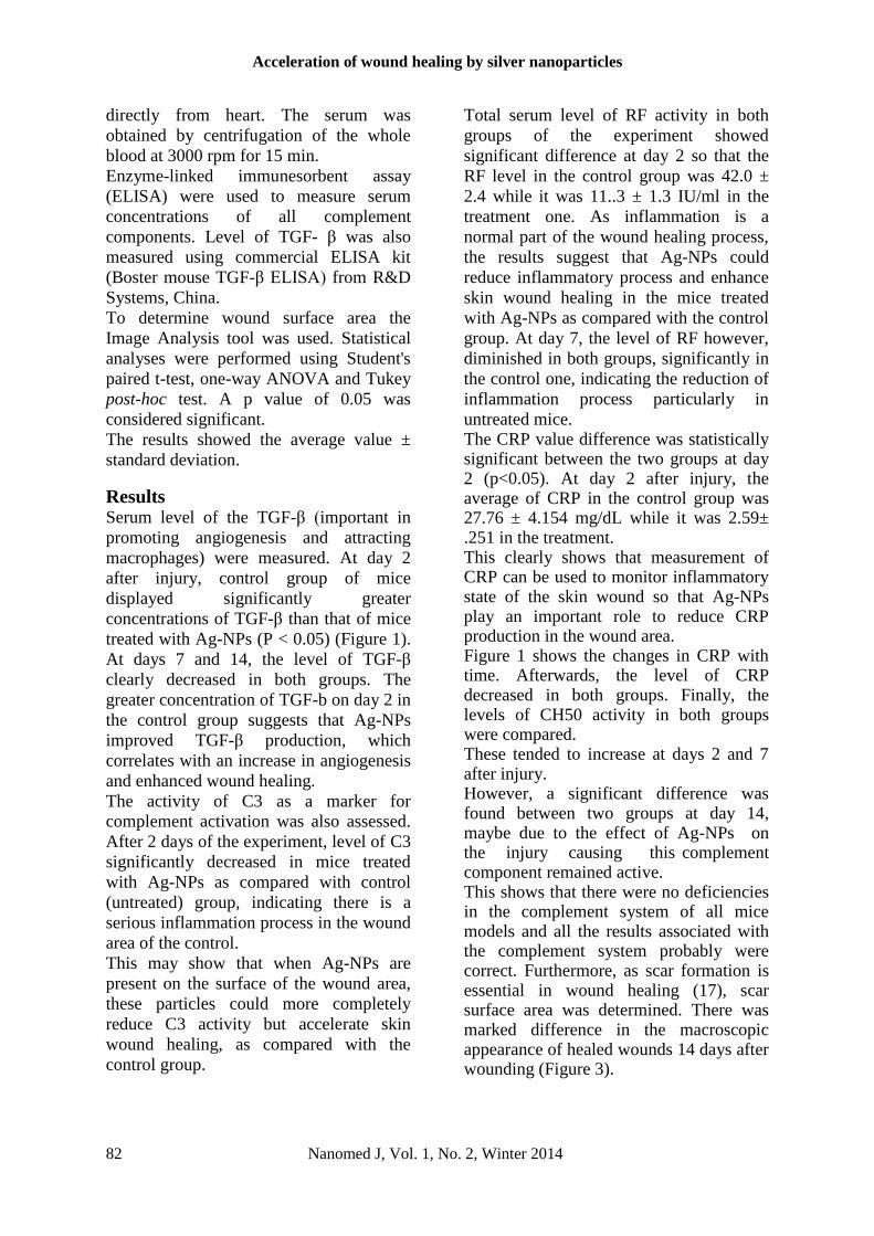
Results Serum level of the TGF-β (important in
promoting angiogenesis and attracting
macrophages) were measured. At day 2
after injury, control group of mice
displayed significantly greater
concentrations of TGF-β than that of mice
treated with Ag-NPs (P < 0.05) (Figure 1).
At days 7 and 14, the level of TGF-β
clearly decreased in both groups. The
greater concentration of TGF-b on day 2 in
the control group suggests that Ag-NPs
improved TGF-β production, which
correlates with an increase in angiogenesis
and enhanced wound healing.
The activity of C3 as a marker for
complement activation was also assessed.
After 2 days of the experiment, level of C3
significantly decreased in mice treated
with Ag-NPs as compared with control
(untreated) group, indicating there is a
serious inflammation process in the wound
area of the control.
This may show that when Ag-NPs are
present on the surface of the wound area,
these particles could more completely
reduce C3 activity but accelerate skin
wound healing, as compared with the
control group.
Total serum level of RF activity in both
groups of the experiment showed
significant difference at day 2 so that the
RF level in the control group was 42.0 ±
2.4 while it was 11..3 ± 1.3 IU/ml in the
treatment one. As inflammation is a
normal part of the wound healing process,
the results suggest that Ag-NPs could
reduce inflammatory process and enhance
skin wound healing in the mice treated
with Ag-NPs as compared with the control
group. At day 7, the level of RF however,
diminished in both groups, significantly in
the control one, indicating the reduction of
inflammation process particularly in
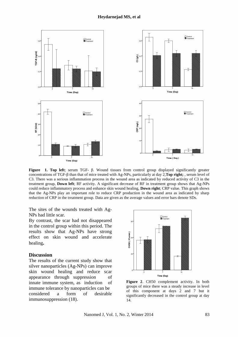
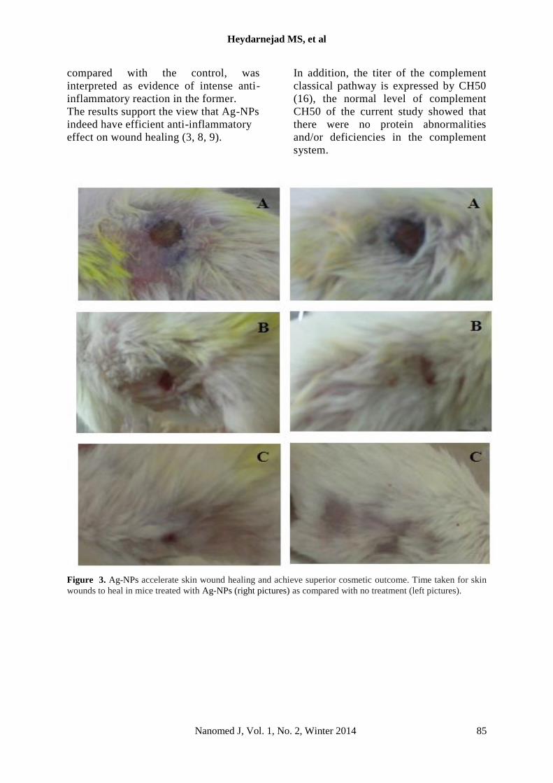
untreated mice. The CRP value difference was statistically significant between the two groups at day 2 (p<0.05). At day 2 after injury, the average of CRP in the control group was 27.76 ± 4.154 mg/dL while it was 2.59± .251 in the treatment. This clearly shows that measurement of CRP can be used to monitor inflammatory state of the skin wound so that Ag-NPs play an important role to reduce CRP production in the wound area. Figure 1 shows the changes in CRP with time. Afterwards, the level of CRP decreased in both groups. Finally, the levels of CH50 activity in both groups were compared. These tended to increase at days 2 and 7 after injury. However, a significant difference was found between two groups at day 14, maybe due to the effect of Ag-NPs on the injury causing this complement component remained active. This shows that there were no deficiencies in the complement system of all mice models and all the results associated with the complement system probably were correct. Furthermore, as scar formation is essential in wound healing (17), scar surface area was determined. There was marked difference in the macroscopic appearance of healed wounds 14 days after wounding (Figure 3).
Heydarnejad MS, et al
Nanomed J, Vol. 1, No. 2, Winter 2014 83
Figure 1. Top left; serum TGF- β. Wound tissues from control group displayed significantly greater
concentrations of TGF-β than that of mice treated with Ag-NPs, particularly at day 2,Top right; , serum level of
C3. There was a serious inflammation process in the wound area as indicated by reduced activity of C3 in the
treatment group, Down left; RF activity. A significant decrease of RF in treatment group shows that Ag-NPs
could reduce inflammatory process and enhance skin wound healing, Down right; CRP value. This graph shows
that the Ag-NPs play an important role to reduce CRP production in the wound area as indicated by sharp
reduction of CRP in the treatment group. Data are given as the average values and error bars denote SDs.
The sites of the wounds treated with Ag-
NPs had little scar.
By contrast, the scar had not disappeared
in the control group within this period. The
results show that Ag-NPs have strong
effect on skin wound and accelerate
healing.
Discussion The results of the current study show that
silver nanoparticles (Ag-NPs) can improve skin wound healing and reduce scar
appearance through suppression of
innate immune system, as induction of
immune tolerance by nanoparticles can be considered a form of desirable
immunosuppression (18).
Figure 2. CH50 complement activity. In both
groups of mice there was a steady increase in level
of this component at days 2 and 7 but it
significantly decreased in the control group at day
14.

Acceleration of wound healing by silver nanoparticles
84 Nanomed J, Vol. 1, No. 2, Winter 2014
Silver nanoparticles (Ag-NPs) appear to
possess potent antimicrobial activity to
reduce infections. In fact, Ag-NPs with
strong biocidal effects are toxic to
microorganisms.
They can kill bacteria that cause diseases
through the contamination of food, water,
and wounds (19).The mechanism of Ag-
NPs on microorganisms is not
completely clear, however, Ag-NPs
interact with a wide range of molecular
processes within microorganisms from
inhibition of growth to loss of infectivity
to cell death (20). For instance, free
radicals derived from the surface of Ag-
NPs had been suggested for the
antimicrobial activity (21). Skin wound
healing is a dynamic process involving the
coordinated interaction of blood cells,
proteins, proteases, growth factors, and
extracellular matrix components.
Wound healing is an essential
physiological process mediated by a
variety of factors responsible for the
regeneration and reorganization of
damaged tissue toward its normal
architecture (22).
The wound healing response can
be subdivided into a sequence of specific
events, made up of three stages:
the inflammatory stage, the proliferative
stage, and the maturation stage.
The immune system is primarily active
during the inflammatory stage.
Typically, the wound healing involves
steps that include inflammation around the
site of injury, angiogenesis and the
development of granulation tissue, repair
of the connective tissue and epithelium,
and ultimately remodeling that leads to a
healed wound (9). The decreased activity
of inflammatory factors measured in the
current study, i.e, TGF-β, Complement
system, CRP, RF is agreement with other
studies in which Ag-NPs have anti-
inflammatory effects and improve wound
healing e.g. Chaloupka et al. (23). In
addition, the results of this study show that
Ag-NPs could suppress complement
activity.
In fact, activation of the complement
cascade can be harmful if particles enter
the systemic circulation because this may
lead to hypersensitivity reactions and
anaphylaxis (24, 25, 26). TGF- β plays an
important role in wound healing and tissue
remodeling. It is the growth factor
affecting all cell types that are involved in
all stages of wound healing (12).
Because level of TGF-β in the control
group of the present study was found to
be increase, thus this factor served as a
chemo-attractant for neutrophils,
macrophages and fibroblasts.
In addition, scar appearance and the extent
of inflammation at the wound site were
decreased in the mice treated with Ag-NPs
so that the wound area was little and
transient compared with the control
group. Similarly, Tian et al. (3) in
investigating the wound-healing
properties of Ag-NPs found rapid healing
and improved cosmetic appearance in an
animal model. This is because TGF- β
plays an important role in tissue fibrosis
and post-injury scarring.
In addition, Wright et al.(27) have
proved that Ag-NPs inflammatory effects
on skin wound healing significantly
suppress inflammatory cytokines and
induced apoptosis of inflammatory
cells. Complement system is the first line
of the innate immunity (28, 29).
It is comprised a group of proteins that
functions primarily to fight infection.
The central results of activation of
pathways are to deposit the opsonin C3
on bacterial to promote inflammation,
phagocytosis and to lyse bacteria.
As the most common complement
protein in serum is C3 (30), the reduced
level of C3 at day 2 after injury in the
current study confirm that this
complement component with a
chemotactic response to neutrophils
releases inflammatory agents into the
wound area (31). Similarly, a reduction
in detectable levels of two other
therapeutic modalities namely RF and
CRP in mice treated with Ag-NPs, when
Heydarnejad MS, et al
Nanomed J, Vol. 1, No. 2, Winter 2014 85
compared with the control, was
interpreted as evidence of intense anti-
inflammatory reaction in the former.
The results support the view that Ag-NPs
indeed have efficient anti-inflammatory
effect on wound healing (3, 8, 9).
In addition, the titer of the complement
classical pathway is expressed by CH50
(16), the normal level of complement
CH50 of the current study showed that
there were no protein abnormalities
and/or deficiencies in the complement
system.
Figure 3. Ag-NPs accelerate skin wound healing and achieve superior cosmetic outcome. Time taken for skin
wounds to heal in mice treated with Ag-NPs (right pictures) as compared with no treatment (left pictures).
Acceleration of wound healing by silver nanoparticles
86 Nanomed J, Vol. 1, No. 2, Winter 2014
Conclusion Exposure of mice to Ag-NPs can result in
significant changes in innate immune
function at the molecular levels.
Furthermore, the immunosuppression
effects of Ag-NPs have been observed
with decreased serum levels of TGF- β,
C3, RF, CRP following confirmed by
CH50 in the blood.
The study improves our understanding of
nanoparticle interaction with components
of the immune system and suggests that
Ag-NPs have strong anti-inflammatory
effects on skin and reduce scarring.
Acknowledgments
This work was funded by the Research
Institute of Biotechnology, Shahrekord
University, Iran.
References
1. Chen X, Schluesener HJ. Nanosilver: a
Chen X, Schluesener HJ. Nanosilver: a
nanoproduct in medical application.
Toxicol Lett. 2008; 176: 1–12.
2. Mei N, Zhang YB, Chen Y, Guo XQ,
Ding W., Ali SF, et al. Silver
nanoparticle-induced mutations and
oxidative stress in mouse lymphoma cells.
Environ Mol Mutagen. 2012; 53: 409-
419.
3. Nadworny PL, Wang J, Tredget
EE, Burrell RE. Anti-inflammatory
activity of nanocrystalline silver in a
porcine contact ermatitis model.
Nanomed. 2008; 4: 241–251.
4. Sibbald, RG, Contreras-Ruiz, J, Coutts
P, Fierheller M, Rothman A, Woo K.
Bacteriology, inflammation, and healing: a
study of nanocrystalline silver dressings in
chronic venous leg ulcers. Ad Skin
Wound Care. 2007; 20: 549–558.
5. Tian J, Wong KK, Ho CM, Lok CN, Yu,
WY, Che CM, et al. Topical delivery of
silver nanoparticles promotes wound
healing. Chem Med Chem 2007; 2: 129–
136.
6. Wright JB, Lam K, Buret AG, Olson,
ME, Burrell RE. Early healing events in a
porcine model of contaminated wounds:
effects of nanocrystalline silver on matrix
metalloproteinases, cell apoptosis, and
healing. Wound Rep Regen. 2002; 10:
141–151.
7. Huang Y, Li X, Liao Z, Zhang G, Liu Q,
Tang J, et al. A randomized comparative
trial between Acticoat and SD-Ag in the
treatment of residual burn wounds,
including safety analysis. Burns. 2007; 33:
161-166.
8. Hendi A. Silver nanoparticles mediate
differential responses in some of liver and
kidney functions during skin wound
healing. J King Saud Uni. 2011; 23: 47-
52.
9. Atiyeh BS, Costagliola M, Hayek SN,
Dibo SA. Effect of silver on burn wound
infection control and healing: Review of
the literature. Burns. 2007; 33: 139: 148.
10. Cordeiro MF. Beyond mitomycin: TGF- β
and wound healing. Prog. Retin. Eye Res.
2002; 21: 75–89.
11. Werner S, Grose R. Regulation of wound
healing by growth factors and cytokines.
Physiol Rev. 2003; 83: 835–870.
12. Faler BJ, Macsata RA, Plummer D,
Mishra L, Sidawy AN. Transforming
growth factor-beta and wound healing.
Perspect Vasc Surg Endovasc Ther. 2006;
18: 55–62.
13. Bonaparte BS, Hair PS, Banthia D,
Marshall DM, Krishna NK. Human
astrovirus coat protein inhibits serum
complement activation via C1, the first
component of the classical pathway. J
Virol. 2008;82: 817-827.
14. RAS; Rheumatoid Arthritis Symptoms".
Retrieved 2011.
15. Karodi R, Jadhav M, Rub R, Bafna A.
Evaluation of the wound healing activity
of a crude extract of Rubia cordifolia L.
(Indian madder) in mice. Inter J Appl Res
Nat Prod. 2009; 2: 12-18
16. Booth NA, Campbell RD, Fothergill JE.
The purification and characterization of
bovine C4, the fourth component of
complement. Biochem J. 1979; 177: 959–
965.
17. Goodman G. "Postacne scarring: A review
of its pathophysiology and treatment".
Dermatologic surgery: official publication
for American Society for Dermatologic
Surgery. 2000; 26 (9): 857–871.
18. Zolnik BS, Gonzalez-Fernandez A,
Sadrieh N, Dobrovolskaia MA.
Minireview: Nanoparticles and the
Immune System. Endocrinol. 2010;151:
458– 465.
19. Faunce T, Watal A. Nanosilver and global
public health: international regulatory
issues. Nanomed. 2010; 5: 617–632.
Heydarnejad MS, et al
Nanomed J, Vol. 1, No. 2, Winter 2014 87
20. Lara HH, Garza-Treviño EN, Ixtepan-
Turrent L, Singh DK. Silver nanoparticles
are broad-spectrum bactericidal and
virucidal compounds. J Nanobiotechnol.
2011; 9: 1-8.
21. Danilczuk M, Lund A, Sadlo J, Yamada
H, Michalik J. Conduction electron spin
resonance of small silver particles.
Spectrochim Acta A Mol Biomol
Spectrosc. 2006; 63: 189-191.
22. Martin P. Wound healing–aiming for
perfect skin regeneration. Science 1997;
276:75–81.
23. Chaloupka K, Malam Y, Seifalian AM.
Nanosilver as a new generation of
nanoproduct in biomedical applications.
Trends Biotechnol. 2010; 28: 580-588.
24. Chanan-Khan A, Szebeni J, Savay S,
Liebes L, Rafique NM, Alving CR, et al.
Complement activation following first
exposure to pegylated liposomal
doxorubicin (Doxil): possible role in
hypersensitivity reactions. Ann Oncol.
2003; 14: 1430–1437
25. Szebeni J. Complement activation-related
pseudoallergy: a new class of drug-
induced acute immune toxicity. Toxicol.
2005; 216: 106–121
26. Szebeni. J, Alving CR, Rosivall L, Bunger
R, Baranyi L, Bedocs P, et al. 2007.
Animal models of complement-mediated
hypersensitivity reactions to liposomes
and other lipid-based nanoparticles. J
Liposome Res. 2007;17: 107–117
27. Wright BI, Lam K, Buret AG. Early
healing events in a porcine model of
contaminated wounds: Effect of
nanosilver in MMP’s cell apoptosis and
healing, J Dermatol. 1991; 124: 519–526.
28. Frank S, Madlener M, Werner S.
Transforming growth factors beta1, beta2,
and beta3 and their receptors are
differentially regulated during normal and
impaired wound healing. J Biol Chem.
1996; 271: 10188–10193.
29. Nakao A, Imamura T, Souchelnytskyi S,
Kawabata M, Ishisaki A, Oeda E, et al.
TGF-beta receptor-mediated signalling
through Smad2, Smad3, and Smad4.
EMBO J. 1997; 16: 5353–5362.
30. Lambris JD. The multifunctional role of
C3, the third component of complement.
Immunol Today. 1998; 9: 387- 393.
31. Barbul A, Shawe T, Rotter SM. Wound
healing in nude mice: a study on the
regulatory role of lymphocytes fibroplasia.
Surgery. 1989; 105: 764-769.
32.
Recommended