RENAL CELL CANCER (RCC) WITH
UNUSUAL PRESENTATION:
A CASE STUDY
BY
UCHECHI EUNICE OKANI
NURS 6035 Practicum I
Texas Woman’s University

Introduction
• Renal cell carcinoma (RCC)
is the commonest cancer
in the kidney
• Accounts for 3% of all
• adult malignancies
• Notorious for:
- Lack of early warning
signs ***
- Diverse clinical
manifestations ***
• ~ 25% present with distance
metastasis or advanced disease
(Atkins, 2009).

Importance of Topic
• PCPs responsibilities to improve outcome:
• Early recognition of presentations ***
• Early diagnosis
• Well directed work-up
• Adequate treatment/referrals
• Screening high risk individuals
• Missing early clues = late diagnosis, morbidity,
mortality, unnecessary health care cost.
Importance of Topic cont.
• This presentation will describe:
• Case of RCC with an unusual presentation
• Quick overview of RCC:
Epidemiology
risk factors
clinical presentations
differentials
diagnostic evaluations
management
screening
follow-up.

Case presentation
• HPI:
• 64-year-old Caucasian female with RCC with unusual presentation. Patient noted lesion in between her teeth on the lower left mandible.
• She went to dentist
• Referred to oral surgeon
• Mandibular gingival lesion excisional biopsy was performed 11/30/10

Case presentation cont.
• Pathology = clear cell carcinoma.
• Result sent to Baylor College of Dentistry
for consultation
• Confirmed histologic features = clear cell
carcinoma, highly suggestive of metastatic
renal cell carcinoma (unusual!!)

Case presentation cont.
•CT abdomen 12/2/10 revealed:
complex mass within right kidney
involving mid to upper pole, measuring
5 x 6 cm in size transversely and 6-7
cm in length. No enlarged
retroperitoneal lymph nodes. •
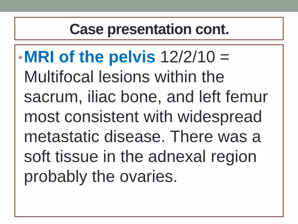
Case presentation cont.
•MRI of the pelvis 12/2/10 =
Multifocal lesions within the
sacrum, iliac bone, and left femur
most consistent with widespread
metastatic disease. There was a
soft tissue in the adnexal region
probably the ovaries.
Case presentation cont.
• Referred to surgeon (Urologist)
• Open right nephrectomy
• Pathology:
• Multifocal RCC, clear cell type, 5.8 cm
greatest dimension (5.8 cm - mid upper
pole and 0.8 cm - lower pole),
• Fuhrman grade 3 of 4, focal extension into
perinephric fat, extension into segmental
renal vein, negative resection margins.

•Case presentation cont.
•Past medical History
•Peripheral arterial disease (PAD)
•Past Surgical History
•Two back surgeries (any connection?)
•Aortobifemoral bypass on 11/08/06.
•Resection of mandibular gingival lesion on 11/30/10 (metastatic lesion)

Case presentation cont.
•Social History
•Married
• five children
•Remote history of smoking cigarette (risk factor).
•Alcohol use - minimal (~ a glass of wine per month)
•No illicit drug use.

Case presentation cont.
• Family History
• Father had lung cancer and had surgery. Father died of heart attack.
• Maternal grandmother died of stomach cancer.
(FH: some risk factor).
• Health Maintenance:
• Never had a colonoscopy
(wake up PCP!!)

Case presentation cont.
Allergies
• Demerol, Codeine, Penicillin, and Sulfa
• Medications
• Aspirin
• Calcium citrate
• Furosemide
• Isosorbide mononitrate
• Levothyroxine
• Metoprolol tartrate
• Morphine
• Ramipril
• Simvastatin
• Spironolactone
• Vitamin D-3 with Aloe

Case presentation cont.
• Review of Systems
• Notable for occasional mild nausea, constipation, and
back pain
• ** Negative genitourinary symptoms
• Vitals
• Height=66in, Weight=173.2lb, BMI = 28.0 Temp=98.0,
Pulse=71, Resp =16, BP 149/97

Case presentation cont.
• Physical Exam
• General: Well developed, well-nourished, anxious looking,
no obvious respiratory distress.
• Abdomen revealed a well healed oblique scar located in
the right flank.
• Oral exam reveals no notable lesion.
Case presentation cont.
• Laboratory Data
• Normal Complete blood count (CBC) with differentials
• Normal Comprehensive metabolic panel (CMP)
• Assessment
• Clear cell carcinoma of the kidney metastatic to gingiva
and bone
• Status post right radical nephrectomy and resection of
mandibular gingival metastatic lesion.

Case presentation cont.
• Plan:
• Patient was started on a cytokine plus VEGF inhibitor.
• The cytokine is alpha-interferon subcutaneous injection 9
million units three times a week.
• The VEGF inhibitor is Bevacizumab 15 mg/kg
intravenous infusion over 1 hour every 2 weeks.
Pathophysiology
•The proximal renal tubular epithelium
is the tissue of origin for renal cell
carcinoma (RCC). RCC occurs in
sporadic (nonhereditary) and
hereditary forms.
•Both forms are associated with
structural alterations of the short arm
of chromosome 3 (3p).

Pathophysiology cont.
• Genetic studies led to cloning of genes
whose alteration results in tumor formation.
These genes are either tumor suppressors
(VHL, TSC) or oncogenes (MET) (Curti &
Harris, 2011)
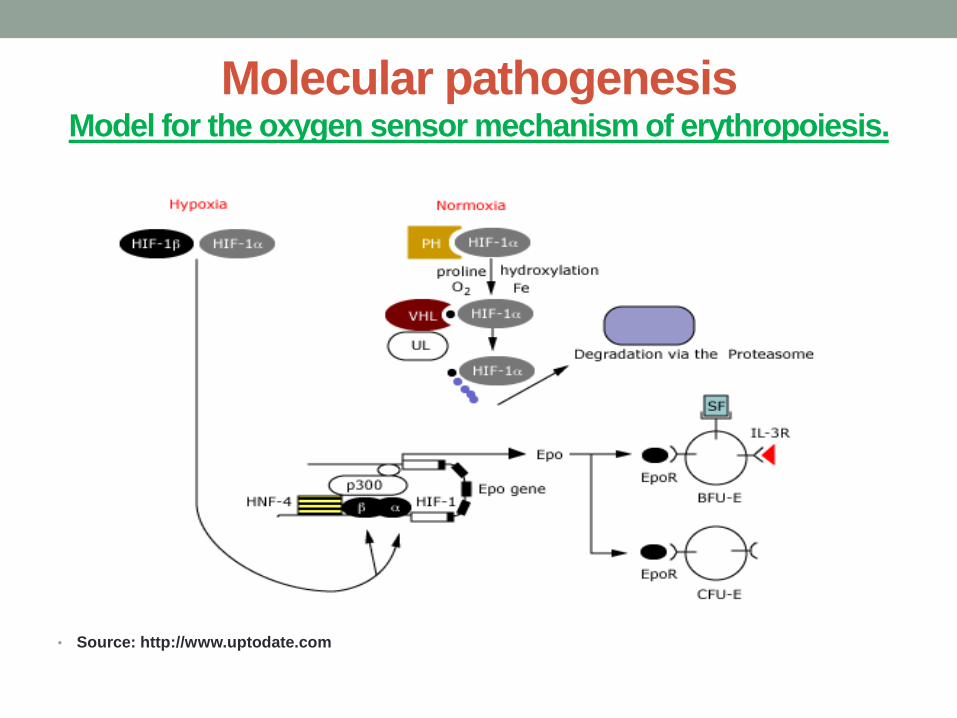
Molecular pathogenesis Model for the oxygen sensor mechanism of erythropoiesis.
• Source: http://www.uptodate.com

4 hereditary syndromes associated with RCC:
• a. Von Hippel-Lindau (VHL) syndrome,
• b. Hereditary papillary renal carcinoma (HPRC),
• c. Familial renal oncocytoma (FRO) associated
with Birt-Hogg-Dube syndrome (BHDS),
• d. Hereditary renal carcinoma (HRC)
(Curti & Harris, 2011)
Hereditary syndromes
Von Hippel-Lindau syndrome
• autosomal dominant syndrome
• 40% of patients develop RCC
• RCC is major cause of death among VHL
patients.
• Deletions of 3p occur commonly in RCC
associated with VHL
• The VHL (tumor suppressor) gene mutation
(Curti & Harris, 2011)

• Hereditary syndromes cont.
• Hereditary papillary renal carcinoma
• inherited disorder with an autosomal dominant
inheritance pattern
• Familial renal oncocytoma is characterized by
development of bilateral, multifocal oncocytoma
or oncocytic neoplasms in the kidney
(Curti & Harris, 2011)
• Hereditary syndromes
• Birth-Hogg-Dube syndrome: hereditary
cutaneous syndrome with benign tumors of the
hair follicle
• Hereditary renal carcinoma: an inherited
medical condition characterized by an increased
tendency to develop oncocytomas - benign
kidney tumors with low malignant potential.
(Curti & Harris, 2011)
Epidemiology
• No significant difference between Whites and Blacks in USA
• Mortality
• Survival rate increased from 34% (1954) to 69% (2002)
• Gender: men > women
• Age: Older > younger
• median age ~ 64
• Uncommon in < age 40
• Rare rare in children
(Atkins & Choeiri, 2011; Curti & Harris, 2011)

RISK FACTORS FOR RENAL CELL CANCER
•
• Smoking:
• Obesity
• Hypertension
• Exposure to toxic compounds
• Von Hippel-Lindau (VHL) disease
• Analgesic abuse nephropathy
• Acquired cystic disease
• Alcohol:
• Chronic hepatitis C
• Cytotoxic Chemotherapy

Clinical Presentations for RCC
• Occult
• Hematuria
• Abdominal or flank mass
• Scrotal Varicocele
• Symptoms associated with metastasis to brain, liver, bone, lymph nodes, and the lungs
• Inferior vena cava involvement
• Paraneoplastic syndromes : erythrocythosis and hypercalcemia
• Anemia
• Hepatic dysfunction
• Fever,
• Thrombocytosis
(Curti & Harris, 2011)

Differential Diagnosis
• Abscess
• Angiomyolipoma (benign)
• Acute pyelonephritis
• Chronic Pyelonephritis
• Distant primary lesion metastasis
• Non-Hodgkin’s lymphoma
• Metastasis from melanoma
• Oncocytoma (benign)
• Renal adenoma (benign)
• Renal cyst
• Renal infarction
• Sarcoma
• Wilms Tumor
• (Curti & Harris, 2011)

DIAGNOSTIC EVALUATION
• Recommended Tests
• CBC with differential
• Urinalysis (UA)
• Renal profile
• Electrolytes
• Liver function tests (LFTs)
• Calcium
• Erythrocyte sedimentation rate (ESR)
• Prothrombin time (PT)
• Activated partial thromboplastin time (aPTT)
(Curti & Harris, 2011)
Radiographic testing
• Ultrasound - less sensitive but is useful for
distinguishing simple benign cysts from
more complex cysts or solid tumor
• Abdominal and pelvic CT – useful for
patient with unexplained hematuria and
other suspicious presentations.
(Atkins, 2009)
TISSUE DIAGNOSIS AND STAGING WORKUP
• Biopsy of metastatic sites
• Tissue also obtained for histology and treatment from Nephrectomy or partial nephrectomy
Metastatic work-up
• Abdominal CT
• Bone scan
• CT of the chest
• MRI
• Position-emission tomography (PET) scan
(Curti & Harris, 2011)
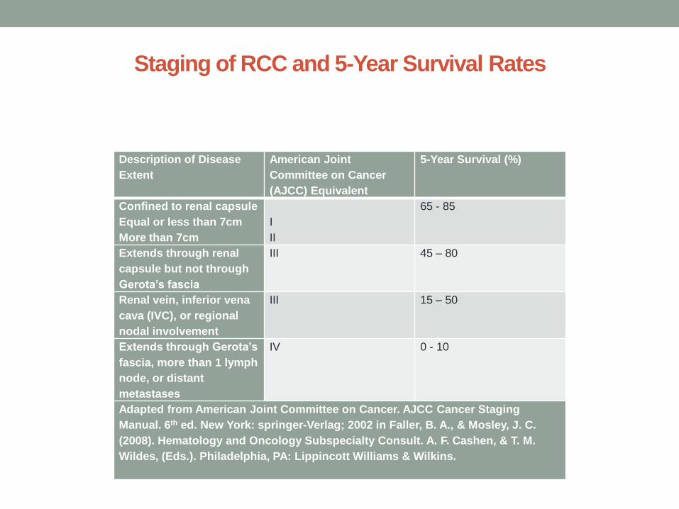
Staging of RCC and 5-Year Survival Rates
Description of Disease
Extent
American Joint
Committee on Cancer
(AJCC) Equivalent
5-Year Survival (%)
Confined to renal capsule
Equal or less than 7cm
More than 7cm
I
II
65 - 85
Extends through renal
capsule but not through
Gerota’s fascia
III 45 – 80
Renal vein, inferior vena
cava (IVC), or regional
nodal involvement
III 15 – 50
Extends through Gerota’s
fascia, more than 1 lymph
node, or distant
metastases
IV 0 - 10
Adapted from American Joint Committee on Cancer. AJCC Cancer Staging
Manual. 6th ed. New York: springer-Verlag; 2002 in Faller, B. A., & Mosley, J. C.
(2008). Hematology and Oncology Subspecialty Consult. A. F. Cashen, & T. M.
Wildes, (Eds.). Philadelphia, PA: Lippincott Williams & Wilkins.
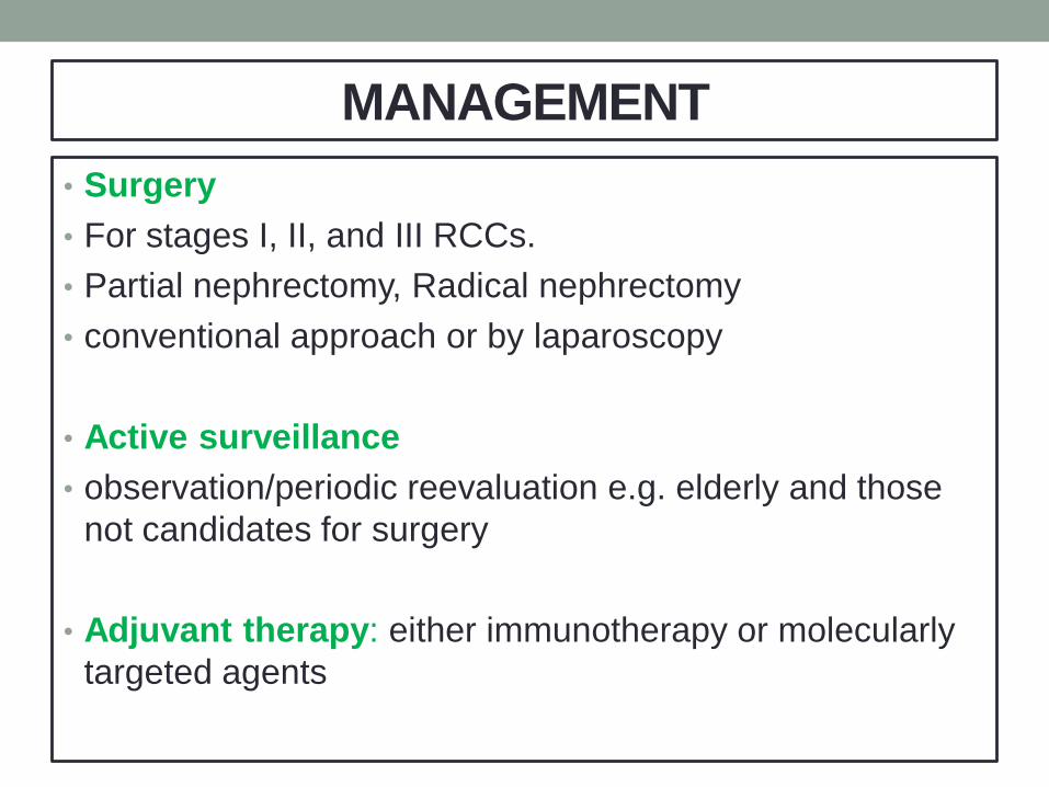
MANAGEMENT
• Surgery
• For stages I, II, and III RCCs.
• Partial nephrectomy, Radical nephrectomy
• conventional approach or by laparoscopy
• Active surveillance
• observation/periodic reevaluation e.g. elderly and those
not candidates for surgery
• Adjuvant therapy: either immunotherapy or molecularly
targeted agents

Management cont.
• Adjuvant therapy (post-op management)
• Cytokines: interferon
• Adjuvant/salvage (for metastatic disease)
Cytokines (designed to boost patient’s immune system to fight the cancer cells , thus called immunotherapy)
a. Interferon (for stage III)
(Patient is treated by this)
or
b. interlukin (if metastatic)

Management cont.
• Molecularly targeted Therapy
• Action:
• Designed to disrupt the signaling pathway
through which the cancer supports and
perpetuates itself.
• Cells have life spans which ends through
programmed cell death (apoptosis). Cancer cells
try making anti-apoptotic proteins.
Management cont.
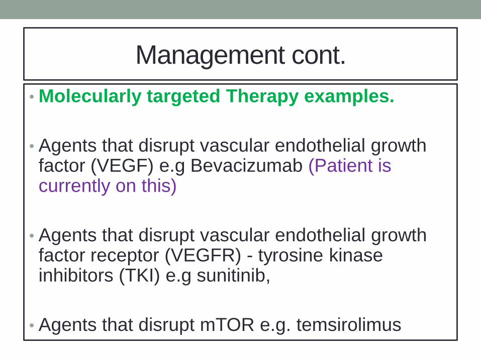
• Molecularly targeted Therapy examples.
• Agents that disrupt vascular endothelial growth factor (VEGF) e.g Bevacizumab (Patient is currently on this)
• Agents that disrupt vascular endothelial growth factor receptor (VEGFR) - tyrosine kinase inhibitors (TKI) e.g sunitinib,
• Agents that disrupt mTOR e.g. temsirolimus

SCREENING
High risk individuals need periodic monitoring with
ultrasound, MRI, CT
Examples:
VHL syndrome
end stage renal disease
prior kidney irradiation
strong family history of RCC
• No recommendation to screen asymptomatic individuals
without risk for RCC
(Atkins, 2009).
Long-Term Follow up and Monitoring
• Active surveillance
• Counseling about active surveillance,
frank discussion of cancer progression and
limitations of treatments if metastasis occur.
(Curti & Harris, 2011)
Long-Term Follow up and Monitoring cont.
• For Stage I and II RCC
Follow up Q 6 months x 2 years, then yearly
x 5 years (complete history and physical
exam, chest radiographs, LFTs, blood urea
nitrogen [BUN], creatinine levels, and
calcium levels)
(Curti & Harris, 2011)
Long-Term Follow up and Monitoring cont.
• For metastatic disease
• Abdominal CT between 4th and 6th month after treatment, then prn
• Close observation prn for asymptomatic patients with metastatic disease
• CT scan/MRI recommended for surveillance in cases of end-stage renal disease
(Curti & Harris, 2011)

Discussion
• What risk factors for RCC did this patient
have?
• How could patient’s risks be lowered by PCP?
• How could the PCP have contributed to earlier
diagnosis of patient’s disease?
• Who needs to be screened for RCC?

References
• Atkins, B. M. (2009). Clinical manifestations, evaluation, and staging of renal cell carcinoma
Uptodate. Retrieved from http://www.uptodate.com/contents/clinical-manifestations-
evaluation-and-staging-of-renal-cell-
carcinoma?source=search_result&search=renal+cell+cancer&selectedTitle=1%7E150
•
• Atkins, B. M. (2010). Overview of the treatment of renal cell carcinoma. Uptodate.
• Retrieved from http://www.uptodate.com/contents/overview-of-the-treatment-of-renal-
• cell-carcinoma?source=search_result&search=kidney+cancer&selectedTitle=2%7E150
• Atkins, B. M., and Choeiri, T. K. (2011). Epidemiology, pathology, and pathogenesis of renal
• cell carcinoma. Uptodate. Retrieved from
• http://www.uptodate.com/contents/epidemiology-pathology-and-pathogenesis-of-renal-
• cell-carcinoma?source=search_result&search=kidney+cancer&selectedTitle=4%7E150
• Chan-Smutko, G., Plon, S. E., and Iliopoulos, O. (2009). Molecular biology and
pathogenesis of von Hippel-Lindau disease. Uptodate. Retrieved from
http://www.uptodate.com/contents/molecular-biology-and-pathogenesis-of-von-hippel-
lindau-disease?source=see_link
• Curti, B. and Harris, J. E. (2011). Renal Cell Carcinoma. Medscape. Retrieved from
http://emedicine.medscape.com/article/281340-overview

References • Faller, B. A., & Mosley, J. C. (2008). Hematology and Oncology Subspecialty Consult. A. F. Cashen,
& T. M. Wildes, (Eds.). Philadelphia, PA: Lippincott Williams & Wilkins.
• George, D., and Jonasc, E. (2011). Immunotherapy of renal cell carcinoma. Uptodate.
• Retrieved from http://www.uptodate.com/contents/immunotherapy-of-renal-cell-
carcinoma?source=see_link&anchor=H34#H34
• Irishhealth (2011). Kidney cancer. Retrieved from http://www.irishhealth.com/article.html?id=1267
• Linehan, M. W., Rini, B., I., & Yang, J. C. (2008). DeVita, Hellman, and Rosenberg’s Cancer: Principles & Practice of Oncology. V. T. DeVita, T. S. Lawrence, & S. A. Rosenberg, (Eds.). Philadelphia, PA:
• Lippincott Williams & Wilkins.
• Newton, S., Hickey, M. & Marrs, J. (2009). Oncology Nursing Advisor: A Comprehensive Guide to Clinical Practice. St. Louis, MO: Mosby
• Thompson, J. A. (2009). Metastatic renal cell carcinoma: current standards of care [corrected]
• [published erratum appears in CLIN J ONCOL NURS 2010 Feb;14(1):21]. Clinical
Journal of Oncology Nursing, 13(6); 6-12. Retrieved from CINAHL database.
• Wood, L. S. (2009). Renal cell carcinoma: screening, diagnosis, and prognosis. Clinical journal
of oncology nursing 13(6); 1092-1095. Retrieved from CINAHL database.
Recommended