Recurrent Spontaneous Coronary Artery Dissection in a Patient with
Fibromuscular Dysplasia
Craig Basman, MD; Tannaz Shoja, MD; Aditya Mangla, DO; Jaffar Raza, MD; Suresh Jain, MD; Zoran Lasic, MD
Clinical presentation
• A 39 y/o African American woman with no significant past medical history who presented with sudden onset chest pain, nausea and diaphoreses while driving to work in 2007.
• The patient’s electrocardiogram revealed transient anterior ST elevations (V1‐4). Bedside echocardiogram revealed an ejection fraction of 50%, with mild apical hypokinesis.
• She was taken directly to the cardiac catheterization laboratory.

Cardiac Catheterization• Coronary angiogram showed a long, diffuse 75% lesion in the mid‐distal LAD (red
arrow in Figure 1).• IVUS revealed the presence of spontaneous coronary artery dissection (SCAD)
extending from mid‐distal LAD (Figure 2). LAD had no evidence of atherosclerotic changes on IVUS.
1 2
True lumen
False lumen
Cardiac Catheterization• A sirolimus eluting stents (Cypher RX), 2.5 x 28mm and 2.5 x 18mm were then
deployed in the distal LAD and the mid LAD respectively (Figure 3). Following second stent deployment dissection propagated proximally (arrow in Figure 4) requiring additional Cypher RX 3.0 x 13mm stent placement to contain the dissection (Figure 5). IVUS was then used to ensure containment of the dissection and adequate stent strut apposition.
43 5

Post‐Procedure• Final angiographic
appearance demonstrated complete dissection coverage and excellent flow in the LAD territory (Figure 6).
• The patient was medically optimized and discharged 2 days post‐procedure on aspirin and clopidogrel in addition to other guideline directed medical therapy (GDMT).
6

Recurrent Event• Patient discontinued both aspirin and clopidogrel in spite of
medical advice 3 years after initial presentation. She was event free until eight years after initial presentation, when she presented with NSTEMI. Subsequent angiogram demonstrated SCAD in a distal, small size segment of LCx not amenable to angioplasty. Localized coronary spasm was ruled out with intracoronary NTG administration. LAD stent sites were patent. Patient was discharged on aspirin and clopidogrel in addition to GDMT for NSTEMI.
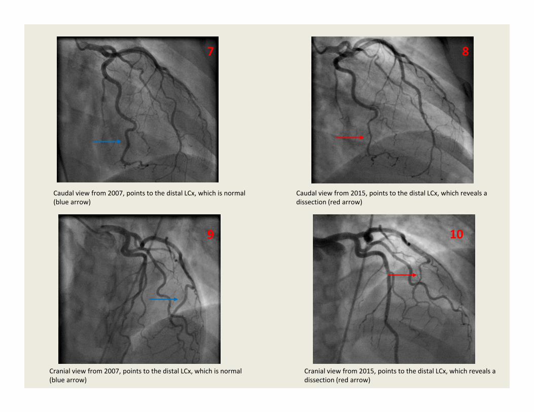
• Caudal and cranial views from 2007 and 2015 are lined up for comparison in Figures 7‐10 (next slide); red arrow points to dissection area and blue arrow to the same segment in non‐dissected artery.
87
Caudal view from 2007, points to the distal LCx, which is normal (blue arrow)
Caudal view from 2015, points to the distal LCx, which reveals a dissection (red arrow)
Cranial view from 2007, points to the distal LCx, which is normal (blue arrow)
Cranial view from 2015, points to the distal LCx, which reveals a dissection (red arrow)
9 10
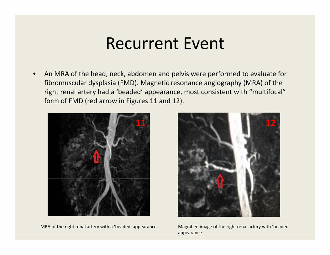
Recurrent Event• An MRA of the head, neck, abdomen and pelvis were performed to evaluate for
fibromuscular dysplasia (FMD). Magnetic resonance angiography (MRA) of the right renal artery had a ‘beaded’ appearance, most consistent with “multifocal” form of FMD (red arrow in Figures 11 and 12).
MRA of the right renal artery with a ‘beaded’ appearance. Magnified image of the right renal artery with ‘beaded’ appearance.
11 12
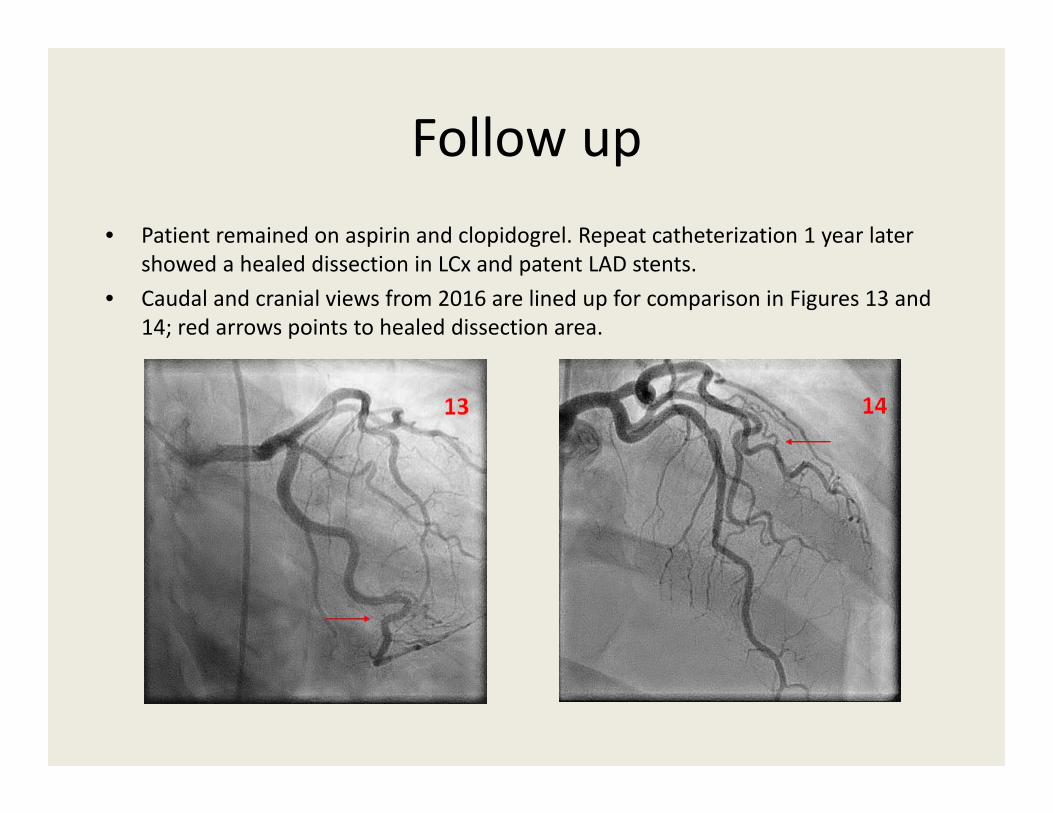
Follow up• Patient remained on aspirin and clopidogrel. Repeat catheterization 1 year later
showed a healed dissection in LCx and patent LAD stents.• Caudal and cranial views from 2016 are lined up for comparison in Figures 13 and
14; red arrows points to healed dissection area.

Conclusions• Spontaneous Coronary Artery Dissection is frequently
associated with Fibromuscular Dysplasia.
• Conservative therapy is generally favored, except in patients with active coronary ischemia, when interventional approach is warranted.
• We present a patient with recurrent SCAD in two distinct coronary arteries. Recent recommendations suggest need for dual antiplatelet therapy for 1 year after initial event followed by lifelong aspirin administration. Non‐adherence to antiplatelet therapy might have elevated our patient’s risk for recurrent event.
Recommended