source: https://doi.org/10.7892/boris.46036 | downloaded: 18.2.2022
Reconstituted High-Density Lipoprotein ModulatesActivation of Human LeukocytesRolf Spirig1,2 ., Alexander Schaub 2 , Alain Kropf2, Sylvia Miescher2, Martin O. Spycher2, Robert Rieben
1 Laboratory of Cardiovascular Research, Department of Clinical Research, University of Bern, Bern, Switzerland, 2 CSL Behring AG, Bern, Switzerland
Abstract
An anti-inflammatory effect of reconstituted High Density Lipoprotein (rHDL) has been demonstrated in atherosclerosis andin sepsis models. An increase of adhesion molecules as well as tissue factor expression on endothelial cells in response toinflammatory or danger signals are attenuated by the treatment with rHDL. Here we show the inhibitory effect of rHDL onthe activation of human leukocytes in a whole blood assay as well as on monocyte-derived human dendritic cells (DC).Multiplex analysis of human whole blood showed that phytohaemagglutinin (PHA)-induced secretion of the cytokines IL-1b,IL-1RA, IL-2R, IL-6, IL-7, IL-12(p40), IL-15 and IFN-a was inhibited. Furthermore, an inhibitory effect on the production of thechemokines CCL-2, CCL-4, CCL-5, CXCL-9 and CXCL-10 was observed. Activation of granulocytes and CD14+ monocytes byPHA is inhibited dose-dependently by rHDL shown as decreased up-regulation of ICAM-1 surface expression. In addition, wefound a strong inhibitory effect of rHDL on toll-like receptor 2 (TLR2)- and TLR4-mediated maturation of DC. Treatment ofDC with rHDL prevented the up-regulation of cell surface molecules CD80, CD83 and CD86 and it inhibited the TLR-drivenactivation of inflammatory transcription factor NF-kB. These findings suggest that rHDL prevents activation of crucial cellularplayers of cellular immunity and could therefore be a useful reagent to impede inflammation as well as the link betweeninnate and adaptive immunity.
Citation: Spirig R, Schaub A, Kropf A, Miescher S, Spycher MO, et al. (2013) Reconstituted High-Density Lipoprotein Modulates Activation of HumanLeukocytes. PLoS ONE 8(8): e71235. doi:10.1371/journal.pone.0071235
Editor: Lucia Gabriele, Istituto Superiore di Sanita, Italy
Received February 14, 2013; Accepted June 28, 2013; Published August 14, 2013
Copyright: � 2013 Spirig et al. This is an open-access article distributed under the terms of the Creative Commons Attribution License, which permitsunrestricted use, distribution, and reproduction in any medium, provided the original author and source are credited.
Funding: This work was supported by the EU 6th Framework Integrated Research Project ‘‘Reprogramming the Immune System for the Establishment ofTolerance’’ (RISET) and the Swiss National Science Foundation (grants no. 3200B0-116618 and 32003B_135272). The funders had no role in study design, datacollection and analysis, decision to publish, or preparation of the manuscript.
Competing Interests: Although Rolf Spirig, Alexander Schaub, Alain Kropf, Sylvia Miescher and Martin Spycher are employees of CSL Behring (Switzerland), thisdoes not alter our adherence to all the PLOS ONE policies on sharing data and materials.
* E-mail: [email protected]
. These authors contributed equally to this work.
Introduction
A beneficial effect of treatment with reconstituted High Density
Lipoprotein (rHDL), containing plasma derived apolipoprotein A-
I (apoA-I) and phosphatidylcholine (PC), was described in models
for atherosclerosis, myocardial infarction, stroke and endotoxemia,
and in clinical trials demonstrating effects on atherosclerotic
plaques [1,2]. Protective properties of rHDL on the endothelium
have been described to be mediated by inhibiting up-regulation of
inflammatory adhesion molecules like ICAM-1 (CD54), VCAM-1
(CD106) and E-selectin (CD62E) on endothelial cells (EC) [3] as
well as reduced thrombin induced tissue-factor (TF) expression [4],
and increasing bioavailability of NO [5]. A study in humans
showed that rHDL reduces plasma levels of TNF-a and expression
of CD11b on monocytes [2]. Protection against cardiac ischemia/
reperfusion (I/R) injury was demonstrated by a reduced cardiac
content of TNF-a and enhanced secretion of prostaglandin I2 and
-E2 in a Langendorff perfusion model [6]. In myocardial infarction
in rats, infusion of rHDL showed an increased phosphorylation of
the MAP kinase family member extracellular-signal-related kinase
(ERK) [7].
Physiologically HDL is involved in lipid homeostasis, especially
in reversed cholesterol transport. Furthermore, HDL has been
suggested to reduce atherosclerosis by suppression of hematopoi-
etic stem cell proliferation, leukocytosis and monocytosis as
another anti-atherogenic effect taking place prior the anti-
inflammatory effect of this molecule [8]. A disturbed cholesterol
efflux is associated with development of diseases such as
atherosclerosis or acute coronary syndrome (ACS). Interestingly,
cholesterol accumulation in cellular membranes has been shown to
activate toll-like receptors (TLR) in macrophages [9]. Further-
more, endogenous TLR agonists are rapidly released under
conditions of inflammation and tissue damage [10–13]. Recent
studies have highlighted the involvement of TLR2 and TLR4 in
the early inflammatory process of I/R injury in vivo [14,15],
atherosclerosis [16] or ACS [17].
rHDL can be considered a substance which attenuates the pro-
inflammatory effects of many mediators of innate immunity. We
hypothesized, therefore, that rHDL might influence inflammatory
responses of important cellular players of innate immunity, namely
neutrophils and monocytes, as well as professional antigen
presenting cells (APC), which are crucially involved in linking
the innate with the adaptive immune system.
Granulocytes and in particular neutrophils are the most
abundant leukocytes in the human body and provide a crucial
first line of defense against infections [18]. On the other hand,
activation of neutrophils may be detrimental in situations of acute
and chronic inflammation with release of proteolytic enzymes as
well as oxygen free radicals increasing damage in diseases such as
myocardial infarction [19] or in graft rejection [20]. Furthermore,
PLOS ONE | www.plosone.org 1 August 2013 | Volume 8 | Issue 8 | e71235
*. 1
neutrophils are a rich source of various proinflammatory cytokines
and chemokines as e.g. CXCL-8 [21]. Monocytes are a second
important cell type involved in inflammation. They have been
described to have antigen-presenting activity [22] and to release a
whole set of pro- and anti-inflammatory cytokines, thus being
major contributors to cellular inflammation in diseases such as I/R
injury [23–25]. In addition, monocytes can differentiate into
dendritic cells (DC) which are very potent APC and pivotal for the
initiation of T-cell mediated immune responses, as seen for
example in allograft rejection as well as in tolerance induction
[26]. In addition, DC are producers of various cytokines and
chemokines and a major source of IL-12, which acts as the third
signal in antigen presentation and co-stimulation required for
successful T cell priming [27].
In this study, we show that rHDL interferes with the activation
of human granulocytes, CD14+ monocytes and monocyte-derived
DC (MoDC) at multiple levels by reducing immunostimulatory
properties, secretion of proinflammatory cytokines and chemo-
kines. Furthermore, rHDL inhibits TLR-induced activation of the
transcription factor NF-kB.
Materials and Methods
Ethics statementThe volunteer blood donors signed an informed consent. In
this document the donors are informed about the use of the
donation. In addition, the donation is coded and transferred
anonym to the research laboratory. The annual blood donation
volume is defined and every second year the hemoglobin value is
tested. There is a biannual screening for virus markers for HIV,
HCV and HBV. The medical dossier is archived for 40 years.
This internal donation system for research purposes is under the
supervision of the medical services and was approved by an in-
house ethical committee (CSL Behring) headed by the medical
direction.
Preparation of reconstituted High Density Lipoprotein(rHDL; CSL111)
rHDL (CSL111) was prepared as described in detail by Lerch
et al. [28]. In brief, rHDL with a molar ratio of apoA-I to soybean
phosphatidylcholine (PC) of 1:150 was prepared. Cholic acid
sodium salt (3.08 kg) was dissolved in 25 liters of a buffer
containing 10 mmol/l Tris-HCl, 10 mmol/l NaCl, 1 mmol/l
EDTA, pH 8.0. In this buffer 4.2 kg PC were dissolved for 6 h at
room temperature. The lipid solution was sterile-filtered (0.22 mm)
and then mixed with 1 kg of apoA-I in 200 liters 10 mmol/l NaCl,
and incubated for at least 2 h at 0–2uC. After the incubation the
mixture was diafiltered with a Pellicon using Biomax cassettes
(NMWR = 10 kDa; Millipore) with at least 5 vol of a 1% sucrose
solution. The protein concentration was then increased to
approximately 2.5%, and the pH was adjusted to 7.5 with either
0.2 mol/l NaOH or 0.2 mol/l HCl. The protein concentration
was determined by the Biuret method, sucrose was added to a final
concentration of 10% and the concentration of the lipoprotein
solution was adjusted to 2% protein concentration. After a final
sterile filtration (0.22 mm) the rHDL was filled in bottles of 1 g
rHDL (protein weight) and lyophilized.
Stimulation and FACS analysis of leukocytes in wholeblood
Heparinized whole blood from healthy volunteers was collected
into pyrogen-free tubes, to which 5 mg/ml phytohemagglutinin-M
(PHA, Calbiochem, Massachusetts, USA) was added for leukocyte
stimulation. Simultaneously, rHDL was added to the whole blood
at concentrations ranging from 0.04 to 1 mg/ml and incubated
overnight at 37uC, 5% CO2 in a humidified atmosphere. Addition
of substances resulted in a 1:2 dilution of human whole blood. The
following day all manipulations were performed at 4uC or on ice.
The cells were directly stained with antibodies specific for CD14,
ICAM-1 and CD45 (BD Biosciences) for 30 min on ice. Red blood
cells (RBC) were lysed by a 30 min incubation with EC Lysis
Buffer (Qiagen AG, Basel, Switzerland) and gentle mixing. With
the majority of RBC lysed, the tubes were centrifuged (4006g,
10 min, 4uC) and the cells resuspended in 300 ml PBS. Data
acquisition and analysis was performed on a FACSCanto II flow
cytometer employing the BD FACSDiva software (both BD
Biosciences AG).
For analysis, leukocytes were identified by gating on the pan-
leukocyte surface marker CD45 which enables to exclude
remaining non-lysed RBC from analysis. Then granulocytes were
separated using granularity (side scatter; SSC) and monocytes
using CD14 as marker which is expressed on the majority of
monocytes (.90% of the whole population). This procedure
allowed the assessment of ICAM-1 expression on the cell surface of
each of these cell populations (all antibodies from BD Biosciences).
To compare the levels of up-regulation of the indicated surface
molecules, the median fluorescence intensity (MFI) ratios were
calculated by dividing the median fluorescence of PHA-treated
gated cells populations i.e. granulocytes and CD14+ monocytes by
the median fluorescence of non-treated cells (indicated as fold
increase MFI).
Measurement of cytokines and chemokines by Luminexmultiplex array system
For the analysis of cytokine/chemokine production, superna-
tants were harvested after overnight stimulation as described
above. The cytokine/chemokine levels in these supernatants were
measured by using a commercial human cytokine magnetic 25-
plex panel (Cat. LHC0009M, Invitrogen Life Technologies,
Paisley, UK) according to manufacturer9s instructions. The panel
consists of the following analytes: IL-1b, IL-1RA, IL-2, IL-2R, IL-
4, IL-5, IL-6, IL-7, IL-10, IL-12p40, IL-13, IL-15, IL-17A, TNF-
a, IFN-a, IFN-c, GM-CSF, CCL-2 (MCP-1), CCL-3 (MIP-1a),
CCL-4 (MIP-1b), CCL-5 (RANTES), CCL-11 (Eotaxin), CXCL-8
(IL-8), CXCL-9 (MIG), CXCL-10 (IP-10).
Generation and stimulation of human monocyte-derivedDC (MoDC)
Human peripheral blood mononuclear cells (PBMC) were
isolated from buffy coats obtained from healthy blood donors
(Regional Red Cross Blood Donation Center, Bern, Switzerland)
by density gradient centrifugation over Ficoll-Paque (Amersham,
Uppsala, Sweden). Monocytes were isolated from PBMC as
described previously [29–31] by spontaneous aggregation and
rosetting [32]. The purified monocytes were incubated for 6 days
at a concentration of 106 cells/ml in RPMI 1640 medium
(Invitrogen Life Technologies) containing 10% fetal calf serum
(FCS; Amimed/BioConcept), 1% [2mM] L-Glutamine (Invitro-
gen), 1% [100 U/ml] Penicillin/Streptomycin (Invitrogen),
10 ng/ml GM-CSF (R&D Systems Europe Ltd, Abingdon, Oxon,
UK), and 10 ng/ml IL-4 (R&D) to generate MoDC as described
initially by Sallusto and Lanzavecchia [33]. The cells were kept at
37uC in a 5% CO2 humidified atmosphere. On day 3, the culture
medium was replaced with fresh medium. For induction of
maturation 100 ng/ml LPS (Sigma), 5 mg/ml lipoteichoic acid
(LTA, Sigma) or 20 mg/ml hyaluronic acid (HA, Sigma) were
added to the cultures for the indicated time periods. Concentra-
Inhibition of Human Leukocyte Activation
PLOS ONE | www.plosone.org 2 August 2013 | Volume 8 | Issue 8 | e71235
tions of the TLR agonists were chosen according previous
published literature [31,34,35] . Treatment of HA with polymyxin
B (Sigma), an inhibitor of LPS, did not affect biological activity of
HA [34], indicating that the observed effects of HA were not due
to potential LPS contamination. MoDC were pre-incubated with
different concentrations of rHDL (0.016, 0.08, 0.04, 0.2, 1.0 mg/
ml) for 30–60 minutes. After this period, maturation of MoDC
was induced by LPS, HA or LTA. Cells were not washed before
addition of TLR agonists.
FACS analysis of MoDC and cell viabilityCells were incubated with FITC-labeled monoclonal antibody
(mAb) against CD80, CD83 and CD86 (BD, Franklin Lakes, NJ,
USA) or Isotype Control IgG1 (BD).
For determination of viability, propidium iodide (PI; Invitro-
gen; 5 mg/ml) was added to stained cells immediately before
analysis by flow cytometry. As control for cell viability staining,
cells were treated with PBS containing 0.1% BSA (Sigma) and
0.1% saponin (Sigma) and then stained with PI. To compare the
levels of up-regulation of the indicated surface molecules, the
median fluorescence intensity (MFI) ratios were calculated by
dividing the median fluorescence of TLR-treated MoDC by the
median fluorescence of non-treated MoDC (indicated as fold
increase MFI). Measurements were performed with a BD
FACScan flow cytometer and the obtained data were analyzed
using FlowJo (Tree Star Inc., Ashland, OR, USA).
Detection of NF-kB activation by a transcription factorELISA
The production of NF-kB p65 was measured using an NF-kB
assay kit (Active Motif, Rixensart, Belgium) according the
manufacturer9s instructions. In brief, cell extract (10 mg of total
protein, generated with the provided lysis buffer) of LTA activated
DC, with or without additional pretreatment by rHDL (40 mg/ml)
and untreated cells, was added to each well coated with consensus-
binding site oligonucleotides of NF-kB p65. A primary antibody
specific for an epitope on the bound and active form of the
transcription factor was then added, followed by incubation with
secondary HRP-conjugated antibody.
Statistical analysisData are presented as mean 6 standard deviation (SD)
representing experiments with 3 to 5 different donors. Paired two
tailed students t-tests were performed for evaluation of signifi-
cance. Differences were considered statistically significant at p-
values less than 0.05. Data were analyzed using GraphPad Prism
software 5.04 (GraphPad, San Diego, CA).
Results
Secretion of proinflammatory cytokines is prevented byrHDL in whole blood
The biological effect of rHDL on the inhibition of an
inflammatory reaction was first assessed in a human whole blood
assay and screened by multiplex technology on production of
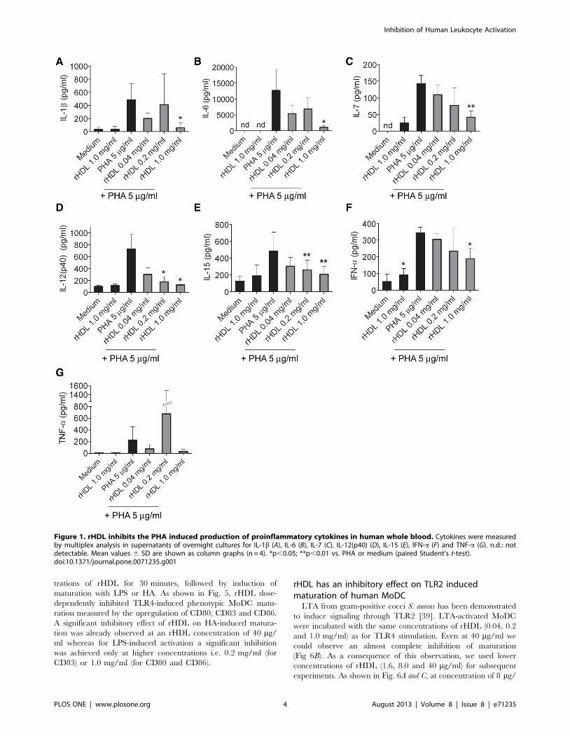
various cytokines and chemokines. Over-night stimulation of
whole blood with phytohemagglutinin (PHA; 5 mg/ml) led to a
considerable secretion of IL-1b, IL-6, IL-12(p40), IL-15, TNF-a,
and IFN-a (Fig. 1). No secretion of the pro-inflammatory cytokine
IL-17A was detected (Lower limit of detection (LLOD): 51.15 pg/
ml). Induction of IFN-c by treatment with PHA was only detected
in one donor (LLOD: 14.5 pg/ml) whereas GM-CSF was secreted
in three donors out of four (LLOD: 23.98 pg/ml; data not shown).
Co-incubation with rHDL reduced PHA-induced secretion of IL-
1b, IL-6, IL-12(p40), IL-15 and IFN-a (Fig. 1). At an rHDL
concentration of 1 mg/ml we observed significantly reduced
secretion for all of these cytokines. No secretion of cytokines was
induced by treating the cells with rHDL at 1 mg/ml in the
absence of PHA, except for IFN-a, where a low, but significant
increase was observed. No significant effect of rHDL on PHA-
induced secretion of TNF-a was observed. There might have been
a trend towards inhibition of TNF-a with 1 mg/ml rHDL.
Surprisingly, there was a high variation in the TNF-a levels
measured in PHA treated blood samples of the different donors
when treated with 0.2 mg/ml of rHDL.
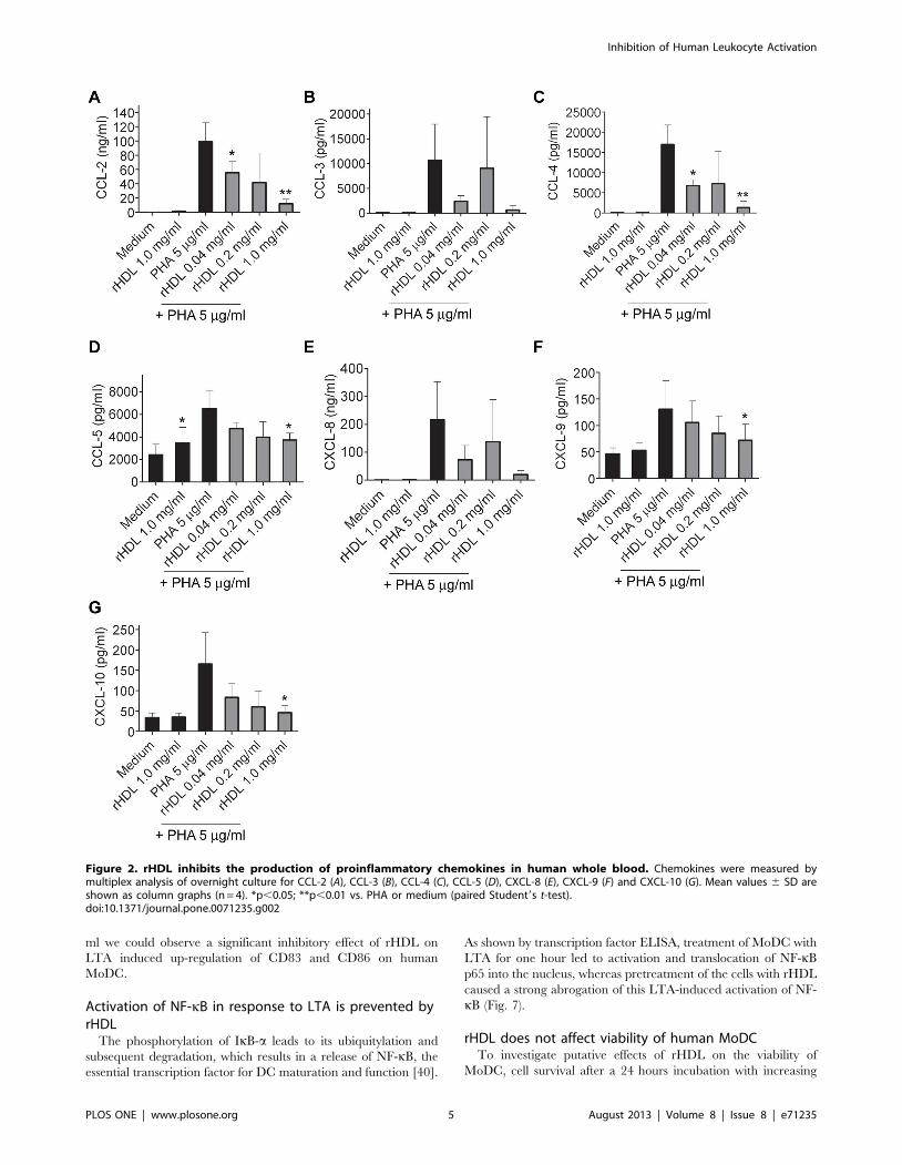
Influence on secretion of chemokines by rHDL in wholeblood
PHA stimulation of whole blood led to a secretion of all
measured chemokines. A significant inhibitory effect on the
production of the chemokines CCL-2, CCL-4, CCL-5, CXCL-9
and CXCL-10 was observed at a concentration of 1 mg/ml of
rHDL (Fig. 2). No significant effect of rHDL on the secretion of
CCL-3, CCL-11 and CXCL-8 could be detected. However, there
was trend towards inhibition of CCL-3 and CXCL-8 by 1 mg/ml
of rHDL (Fig. 2).
Influence on secretion of anti-inflammatory cytokines byrHDL in whole blood
The following cytokines that were measured in this study have
been considered to possess anti-inflammatory properties: IL-1RA,
IL-2R, IL-4, IL-5, IL-10 and IL-13. PHA induced secretion of
IL-1RA and IL-2R was significantly reduced by rHDL (Fig. 3).
No secretion of IL-4 and IL-5 was detected in the supernatants
(LLOD IL-4: 58.49 pg/ml; LLOD IL-5: 3.27 pg/ml) of PHA
activated cells. Secretion of IL-10 and IL-13 was detected in two
out of four donors, all other values were below detection (LLOD
IL-10: 30.1 pg/ml; LLOD IL-13: 28.15 pg/ml).
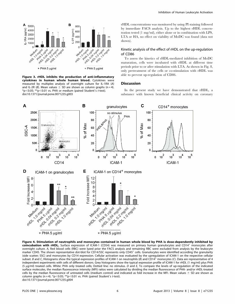
Upregulation of ICAM-1 on CD14+ monocytes andgranulocytes in whole blood is inhibited by rHDL in adose-dependent manner
Many of the cytokines the excretion of which was affected by
rHDL are produced by cells of myeloid origin, namely granulo-
cytes, monocytes, macrophages or DC. Therefore, we analyzed
the expression of ICAM-1 (CD54) as a marker of cellular
activation [36] on CD14+ monocytes and granulocytes (Fig. 4A).
Incubation with rHDL dose-dependently inhibited the up-
regulation of ICAM-1 on human granulocytes (Fig 4B and D)
and CD14+ monocytes (Fig. 4C and E).
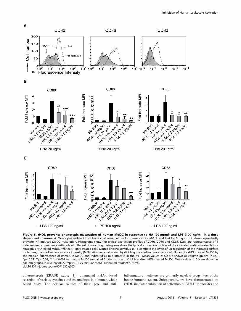
Phenotypic maturation induced by endogenous as wellas exogenous TLR4 agonists of human MoDC isprevented by rHDL
The frequency of myeloid DC in the peripheral human blood
is very low (about 0.5% of all mononuclear cells) [37]. To
investigate the effect of rHDL on DC maturation we worked
therefore with monocyte-derived DC. As PHA also acts as a
polyclonal stimulus on lymphocytes, we have used TLR agonists
to more specifically activate myeloid cells. It has been described
that LPS derived from E. coli leads to maturation of human
MoDC via TLR4, after the formation of a TLR4-signaling
complex containing MD-2, CD14 and TLR4. HA has been
described as endogenous TLR4 agonist, inducing the formation
of a unique TLR4 complex consisting of MD-2, CD44 and
TLR4 [38]. MoDC were pre-incubated with different concen-
Inhibition of Human Leukocyte Activation
PLOS ONE | www.plosone.org 3 August 2013 | Volume 8 | Issue 8 | e71235
trations of rHDL for 30 minutes, followed by induction of
maturation with LPS or HA. As shown in Fig. 5, rHDL dose-
dependently inhibited TLR4-induced phenotypic MoDC matu-
ration measured by the upregulation of CD80, CD83 and CD86.
A significant inhibitory effect of rHDL on HA-induced matura-
tion was already observed at an rHDL concentration of 40 mg/
ml whereas for LPS-induced activation a significant inhibition
was achieved only at higher concentrations i.e. 0.2 mg/ml (for
CD83) or 1.0 mg/ml (for CD80 and CD86).
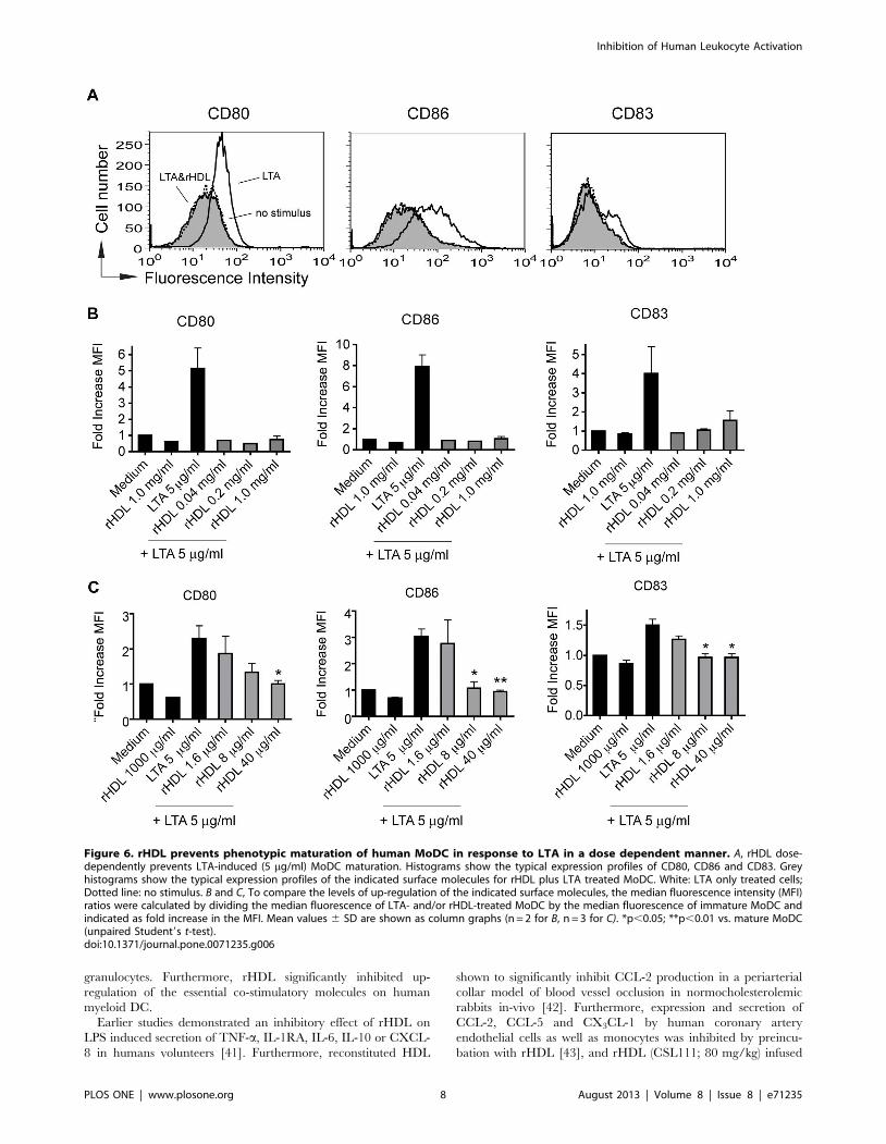
rHDL has an inhibitory effect on TLR2 inducedmaturation of human MoDC
LTA from gram-positive cocci S. aureus has been demonstrated
to induce signaling through TLR2 [39]. LTA-activated MoDC
were incubated with the same concentrations of rHDL (0.04, 0.2
and 1.0 mg/ml) as for TLR4 stimulation. Even at 40 mg/ml we
could observe an almost complete inhibition of maturation
(Fig 6B). As a consequence of this observation, we used lower
concentrations of rHDL (1.6, 8.0 and 40 mg/ml) for subsequent
experiments. As shown in Fig. 6A and C, at concentration of 8 mg/
Figure 1. rHDL inhibits the PHA induced production of proinflammatory cytokines in human whole blood. Cytokines were measuredby multiplex analysis in supernatants of overnight cultures for IL-1b (A), IL-6 (B), IL-7 (C), IL-12(p40) (D), IL-15 (E), IFN-a (F) and TNF-a (G). n.d.: notdetectable. Mean values 6 SD are shown as column graphs (n = 4). *p,0.05; **p,0.01 vs. PHA or medium (paired Student’s t-test).doi:10.1371/journal.pone.0071235.g001
Inhibition of Human Leukocyte Activation
PLOS ONE | www.plosone.org 4 August 2013 | Volume 8 | Issue 8 | e71235
ml we could observe a significant inhibitory effect of rHDL on
LTA induced up-regulation of CD83 and CD86 on human
MoDC.
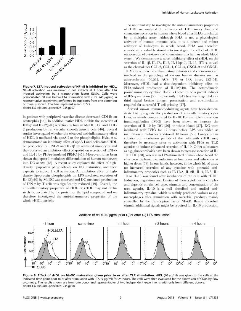
Activation of NF-kB in response to LTA is prevented byrHDL
The phosphorylation of IkB-a leads to its ubiquitylation and
subsequent degradation, which results in a release of NF-kB, the
essential transcription factor for DC maturation and function [40].
As shown by transcription factor ELISA, treatment of MoDC with
LTA for one hour led to activation and translocation of NF-kB
p65 into the nucleus, whereas pretreatment of the cells with rHDL
caused a strong abrogation of this LTA-induced activation of NF-
kB (Fig. 7).
rHDL does not affect viability of human MoDCTo investigate putative effects of rHDL on the viability of
MoDC, cell survival after a 24 hours incubation with increasing
Figure 2. rHDL inhibits the production of proinflammatory chemokines in human whole blood. Chemokines were measured bymultiplex analysis of overnight culture for CCL-2 (A), CCL-3 (B), CCL-4 (C), CCL-5 (D), CXCL-8 (E), CXCL-9 (F) and CXCL-10 (G). Mean values 6 SD areshown as column graphs (n = 4). *p,0.05; **p,0.01 vs. PHA or medium (paired Student9s t-test).doi:10.1371/journal.pone.0071235.g002
Inhibition of Human Leukocyte Activation
PLOS ONE | www.plosone.org 5 August 2013 | Volume 8 | Issue 8 | e71235
rHDL concentrations was monitored by using PI staining followed
by immediate FACS analysis. Up to the highest rHDL concen-
tration tested (1 mg/ml), either alone or in combination with LPS,
LTA or HA, no effect on viability of MoDC was found (data not
shown).
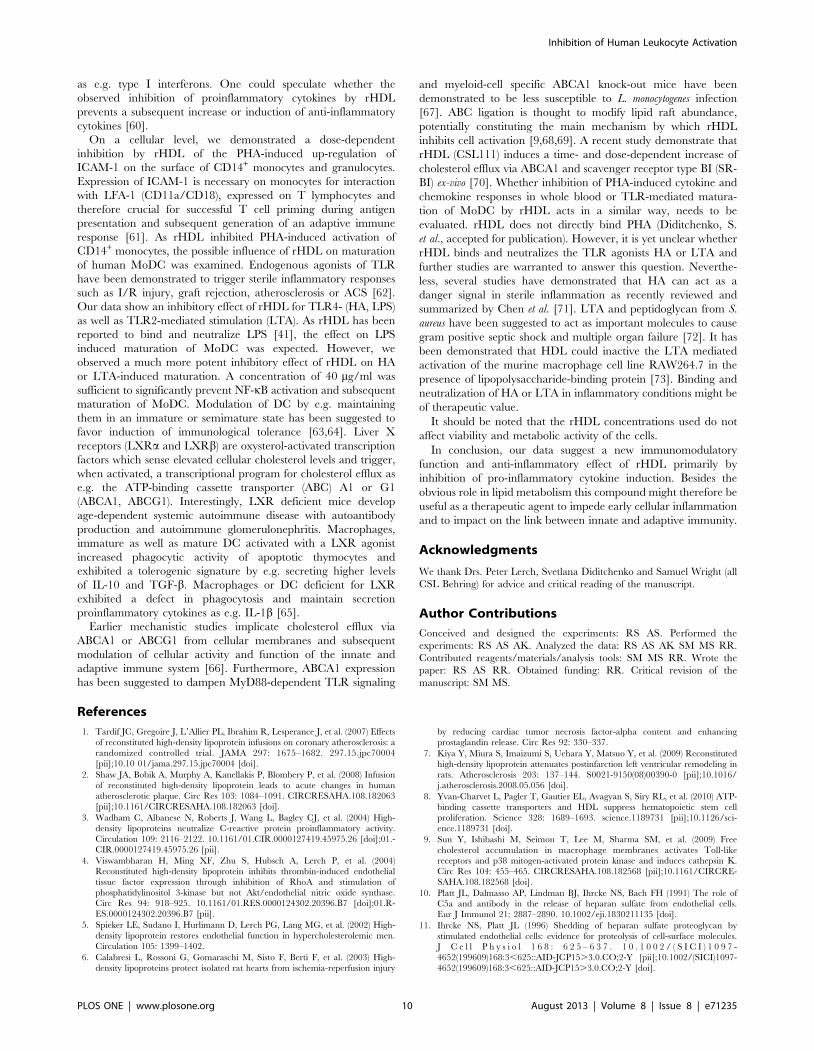
Kinetic analysis of the effect of rHDL on the up-regulationof CD86
To assess the kinetics of rHDL-mediated inhibition of MoDC
maturation, cells were incubated with rHDL at different time
periods prior to or after stimulation with LTA. As shown in Fig. 8,
only pretreatment of the cells or co-stimulation with rHDL was
able to prevent up-regulation of CD86.
Discussion
In the present study we have demonstrated that rHDL, a
substance with known beneficial clinical activity on coronary
Figure 3. rHDL inhibits the production of anti-inflammatorycytokines in human whole human blood. Cytokines weremeasured by multiplex analysis of overnight culture for IL-1RA (A)and IL-2R (B). Mean values 6 SD are shown as column graphs (n = 4).*p,0.05; **p,0.01 vs. PHA or medium (paired Student9s t-test).doi:10.1371/journal.pone.0071235.g003
Figure 4. Stimulation of neutrophils and monocytes contained in human whole blood by PHA is dose-dependently inhibited bycoincubation with rHDL. Surface expression of ICAM-1 (CD54) was measured on primary human granulocytes and CD14+ monocytes afterovernight culture. A, Red blood cells (RBC) were lyzed prior the FACS analysis and remaining RBC were excluded from analysis by the leukocytemarker CD45. The shown representative dot-blot for CD14/SSC represents only CD45+ cells. Granulocytes were identified according the granularity(side scatter; SSC) and monocytes by CD14 expression. Cellular activation was evaluated by the upregulation of ICAM-1 on the respective cellularsubset. B and C, Histograms show the typical expression profiles of ICAM-1 on neutrophils (B) and CD14+ monocytes (C). Data are representative of 4independent experiments with cells of different donors. Grey histograms show the typical expression profile of ICAM-1 for rHDL (1 mg/ml) plus PHA(5 mg/ml) treated cells. White: PHA only treated cells; Dotted line: no stimulus. D and E, To compare the levels of up-regulation of the indicatedsurface molecules, the median fluorescence intensity (MFI) ratios were calculated by dividing the median fluorescence of PHA- and/or rHDL-treatedcells by the median fluorescence of untreated cells (medium control) and indicated as fold increase in the MFI. Mean values 6 SD are shown ascolumn graphs (n = 4). *p,0.05; **p,0.01 vs. PHA (paired Student9s t-test).doi:10.1371/journal.pone.0071235.g004
Inhibition of Human Leukocyte Activation
PLOS ONE | www.plosone.org 6 August 2013 | Volume 8 | Issue 8 | e71235
atherosclerosis (ERASE study; [1]), attenuated PHA-induced
secretion of various cytokines and chemokines, in a human whole
blood assay. The cellular sources of these pro- and anti-
inflammatory mediators are primarily myeloid progenitors of the
innate immune system. Subsequently, we have demonstrated an
rHDL-mediated inhibition of activation of CD14+ monocytes and
Figure 5. rHDL prevents phenotypic maturation of human MoDC in response to HA (20 mg/ml) and LPS (100 ng/ml) in a dosedependent manner. A, Monocytes isolated from buffy coat were cultured in presence of GM-CSF and IL-4 for 6 days. rHDL dose-dependentlyprevents HA-induced MoDC maturation. Histograms show the typical expression profiles of CD80, CD86 and CD83. Data are representative of 5independent experiments with cells of different donors. Grey histograms show the typical expression profiles of the indicated surface molecules forrHDL plus HA treated MoDC. White: HA only treated cells; Dotted line: no stimulus. B, To compare the levels of up-regulation of the indicated surfacemolecules, the median fluorescence intensity (MFI) ratios were calculated by dividing the median fluorescence of HA- and/or rHDL-treated MoDC bythe median fluorescence of immature MoDC and indicated as fold increase in the MFI. Mean values 6 SD are shown as column graphs (n = 5).*p,0.05; **p,0.01; ***p,0.001 vs. mature MoDC (unpaired Student9s t-test). C, LPS- and/or rHDL-treated MoDC. Mean values 6 SD are shown ascolumn graphs (n = 5). *p,0.05; **p,0.01 vs. mature MoDC (unpaired Student9s t-test).doi:10.1371/journal.pone.0071235.g005
Inhibition of Human Leukocyte Activation
PLOS ONE | www.plosone.org 7 August 2013 | Volume 8 | Issue 8 | e71235
granulocytes. Furthermore, rHDL significantly inhibited up-
regulation of the essential co-stimulatory molecules on human
myeloid DC.
Earlier studies demonstrated an inhibitory effect of rHDL on
LPS induced secretion of TNF-a, IL-1RA, IL-6, IL-10 or CXCL-
8 in humans volunteers [41]. Furthermore, reconstituted HDL
shown to significantly inhibit CCL-2 production in a periarterial
collar model of blood vessel occlusion in normocholesterolemic
rabbits in-vivo [42]. Furthermore, expression and secretion of
CCL-2, CCL-5 and CX3CL-1 by human coronary artery
endothelial cells as well as monocytes was inhibited by preincu-
bation with rHDL [43], and rHDL (CSL111; 80 mg/kg) infused
Figure 6. rHDL prevents phenotypic maturation of human MoDC in response to LTA in a dose dependent manner. A, rHDL dose-dependently prevents LTA-induced (5 mg/ml) MoDC maturation. Histograms show the typical expression profiles of CD80, CD86 and CD83. Greyhistograms show the typical expression profiles of the indicated surface molecules for rHDL plus LTA treated MoDC. White: LTA only treated cells;Dotted line: no stimulus. B and C, To compare the levels of up-regulation of the indicated surface molecules, the median fluorescence intensity (MFI)ratios were calculated by dividing the median fluorescence of LTA- and/or rHDL-treated MoDC by the median fluorescence of immature MoDC andindicated as fold increase in the MFI. Mean values 6 SD are shown as column graphs (n = 2 for B, n = 3 for C). *p,0.05; **p,0.01 vs. mature MoDC(unpaired Student9s t-test).doi:10.1371/journal.pone.0071235.g006
Inhibition of Human Leukocyte Activation
PLOS ONE | www.plosone.org 8 August 2013 | Volume 8 | Issue 8 | e71235
in patients with peripheral vascular disease decreased CD11b on
neutrophils [44]. In addition, native HDL inhibits the secretion of
IFN-c and IL-12(p40) secretion by human MoDC [45] and CCL-
2 production by rat vascular smooth muscle cells [46]. Several
studies investigated whether the observed anti-inflammatory effect
of HDL is mediated via apoA-I or the phospholipids. Hyka et al.
demonstrated an inhibitory effect of apoA-I and delipidated HDL
on production of TNF-a and IL-1b by activated monocytes and
they observed an inhibitory effect of apoA-I on secretion of TNF-aand IL-1b by PHA-stimulated PBMC [47]. Moreover, it has been
shown that apoA-I modulates differentiation of human monocytes
into DC in-vitro [48]. A recent study explored the effect of high-
density lipoprotein phospholipids on DC maturation and their
capacity to induce T cell activation. An inhibitory effect of high-
density lipoprotein phospholipids on LPS mediated secretion of
IL-12(p40) by MoDC was observed and DC mediated production
of IFN-c by T cells was significantly reduced [49]. Overall, the
anti-inflammatory properties of HDL or rHDL may not exclu-
sively be mediated by the protein or the lipid compound and we
therefore investigated the anti-inflammatory properties of the
whole rHDL particle.
As an initial step to investigate the anti-inflammatory properties
of rHDL we analyzed the influence of rHDL on cytokine and
chemokine secretion in human whole blood after PHA stimulation
by a multiplex assay. Although PHA is not a physiological
activator of human immune cells, it is a potent and robust
activator of leukocytes in whole blood. PHA was therefore
considered a valuable stimulus to investigate the effect of rHDL
on secretion of cytokines and chemokines in a human whole blood
system. We demonstrate a novel inhibitory effect of rHDL on the
secretion of IL-1b, IL-2R, IL-7, IL-12(p40), IL-15, IFN-a as well
as the chemokines CCL-2, CCL-4, CCL-5, CXCL-9 and CXCL-
10. Many of these proinflammatory cytokines and chemokines are
involved in the pathology of various human diseases such as
atherosclerosis [50,51], ACS [17] or I/R injury [52–54].
Moreover, rHDL had a dose-dependent inhibitory effect on
PHA-induced production of IL-12(p40). The heterodimeric
proinflammatory cytokine IL-12 is known to be a potent inducer
of IFN-c secretion [55]. Importantly, IL-12 has been regarded as a
third signal besides antigen presentation and co-stimulation
required for successful T cell priming [27].
Several known immunomodulating agents have been demon-
strated to upregulate the production of anti-inflammatory cyto-
kines, as mainly demonstrated for IL-10. For example intravenous
immunoglobulins (IVIG) have been shown to increase the
secretion of IL-10 by DC [56] or whole blood [57]. DC were
incubated with IVIG for 12 hours before LPS was added as
maturation stimulus for additional 48 hours [56]. Longer prein-
cubation or incubation periods of the cells with rHDL may
therefore be necessary prior to activation with PHA or TLR
agonists to induce enhanced secretion of IL-10. Other substances
as e.g. glucocorticoids have been shown to increase secretion of IL-
10 in DC [58], whereas in LPS-stimulated human whole blood the
effect was biphasic, i.e. induction at low doses and inhibition at
higher doses [59]. In our hands, however, in the whole blood assay
no increased secretion of any cytokine with potential anti-
inflammatory properties such as IL-1RA, IL-2R, IL-4, IL-5, IL-
10 or IL-13 was found after incubation of the cells with rHDL.
Induction, regulation and kinetics of these cytokines is complex
and depends on the cell type, stimulus and concentration of the
used agonist. IL-10 is a well described and studied anti-
inflammatory cytokine, which is mainly produced various as e.g.
macrophages after stimulation with microbial products mainly
controlled by the transcription factor NF-kB. Beside microbial
stimuli, additional signals might be required for IL-10 production,
Figure 7. LTA induced activation of NF-kB is inhibited by rHDL.NF-kB activation was measured in cell extracts at 1 hour after LTAinduced activation by a transcription factor ELISA. Cells werepreincubated 30 min before LTA stimulation with rHDL (40 mg/ml). Arepresentative experiment performed in duplicates from one donor outof three is shown. The bars represent mean 6 SD.doi:10.1371/journal.pone.0071235.g007
Figure 8. Effect of rHDL on MoDC maturation given prior to or after TLR stimulation. rHDL (40 mg/ml) was given to the cells at theindicated time point prior to or after stimulation with LTA (5 mg/ml) for 24 hours. The cells were then evaluated for the expression of CD86 by flowcytometry. The results shown are from one donor and representative of two independent experiments with cells from different donors.doi:10.1371/journal.pone.0071235.g008
Inhibition of Human Leukocyte Activation
PLOS ONE | www.plosone.org 9 August 2013 | Volume 8 | Issue 8 | e71235
as e.g. type I interferons. One could speculate whether the
observed inhibition of proinflammatory cytokines by rHDL
prevents a subsequent increase or induction of anti-inflammatory
cytokines [60].
On a cellular level, we demonstrated a dose-dependent
inhibition by rHDL of the PHA-induced up-regulation of
ICAM-1 on the surface of CD14+ monocytes and granulocytes.
Expression of ICAM-1 is necessary on monocytes for interaction
with LFA-1 (CD11a/CD18), expressed on T lymphocytes and
therefore crucial for successful T cell priming during antigen
presentation and subsequent generation of an adaptive immune
response [61]. As rHDL inhibited PHA-induced activation of
CD14+ monocytes, the possible influence of rHDL on maturation
of human MoDC was examined. Endogenous agonists of TLR
have been demonstrated to trigger sterile inflammatory responses
such as I/R injury, graft rejection, atherosclerosis or ACS [62].
Our data show an inhibitory effect of rHDL for TLR4- (HA, LPS)
as well as TLR2-mediated stimulation (LTA). As rHDL has been
reported to bind and neutralize LPS [41], the effect on LPS
induced maturation of MoDC was expected. However, we
observed a much more potent inhibitory effect of rHDL on HA
or LTA-induced maturation. A concentration of 40 mg/ml was
sufficient to significantly prevent NF-kB activation and subsequent
maturation of MoDC. Modulation of DC by e.g. maintaining
them in an immature or semimature state has been suggested to
favor induction of immunological tolerance [63,64]. Liver X
receptors (LXRa and LXRb) are oxysterol-activated transcription
factors which sense elevated cellular cholesterol levels and trigger,
when activated, a transcriptional program for cholesterol efflux as
e.g. the ATP-binding cassette transporter (ABC) A1 or G1
(ABCA1, ABCG1). Interestingly, LXR deficient mice develop
age-dependent systemic autoimmune disease with autoantibody
production and autoimmune glomerulonephritis. Macrophages,
immature as well as mature DC activated with a LXR agonist
increased phagocytic activity of apoptotic thymocytes and
exhibited a tolerogenic signature by e.g. secreting higher levels
of IL-10 and TGF-b. Macrophages or DC deficient for LXR
exhibited a defect in phagocytosis and maintain secretion
proinflammatory cytokines as e.g. IL-1b [65].
Earlier mechanistic studies implicate cholesterol efflux via
ABCA1 or ABCG1 from cellular membranes and subsequent
modulation of cellular activity and function of the innate and
adaptive immune system [66]. Furthermore, ABCA1 expression
has been suggested to dampen MyD88-dependent TLR signaling
and myeloid-cell specific ABCA1 knock-out mice have been
demonstrated to be less susceptible to L. monocytogenes infection
[67]. ABC ligation is thought to modify lipid raft abundance,
potentially constituting the main mechanism by which rHDL
inhibits cell activation [9,68,69]. A recent study demonstrate that
rHDL (CSL111) induces a time- and dose-dependent increase of
cholesterol efflux via ABCA1 and scavenger receptor type BI (SR-
BI) ex-vivo [70]. Whether inhibition of PHA-induced cytokine and
chemokine responses in whole blood or TLR-mediated matura-
tion of MoDC by rHDL acts in a similar way, needs to be
evaluated. rHDL does not directly bind PHA (Diditchenko, S.
et al., accepted for publication). However, it is yet unclear whether
rHDL binds and neutralizes the TLR agonists HA or LTA and
further studies are warranted to answer this question. Neverthe-
less, several studies have demonstrated that HA can act as a
danger signal in sterile inflammation as recently reviewed and
summarized by Chen et al. [71]. LTA and peptidoglycan from S.
aureus have been suggested to act as important molecules to cause
gram positive septic shock and multiple organ failure [72]. It has
been demonstrated that HDL could inactive the LTA mediated
activation of the murine macrophage cell line RAW264.7 in the
presence of lipopolysaccharide-binding protein [73]. Binding and
neutralization of HA or LTA in inflammatory conditions might be
of therapeutic value.
It should be noted that the rHDL concentrations used do not
affect viability and metabolic activity of the cells.
In conclusion, our data suggest a new immunomodulatory
function and anti-inflammatory effect of rHDL primarily by
inhibition of pro-inflammatory cytokine induction. Besides the
obvious role in lipid metabolism this compound might therefore be
useful as a therapeutic agent to impede early cellular inflammation
and to impact on the link between innate and adaptive immunity.
Acknowledgments
We thank Drs. Peter Lerch, Svetlana Diditchenko and Samuel Wright (all
CSL Behring) for advice and critical reading of the manuscript.
Author Contributions
Conceived and designed the experiments: RS AS. Performed the
experiments: RS AS AK. Analyzed the data: RS AS AK SM MS RR.
Contributed reagents/materials/analysis tools: SM MS RR. Wrote the
paper: RS AS RR. Obtained funding: RR. Critical revision of the
manuscript: SM MS.
References
1. Tardif JC, Gregoire J, L9Allier PL, Ibrahim R, Lesperance J, et al. (2007) Effects
of reconstituted high-density lipoprotein infusions on coronary atherosclerosis: a
randomized controlled trial. JAMA 297: 1675–1682. 297.15.jpc70004
[pii];10.10 01/jama.297.15.jpc70004 [doi].
2. Shaw JA, Bobik A, Murphy A, Kanellakis P, Blombery P, et al. (2008) Infusionof reconstituted high-density lipoprotein leads to acute changes in human
atherosclerotic plaque. Circ Res 103: 1084–1091. CIRCRESAHA.108.182063
[pii];10.1161/CIRCRESAHA.108.182063 [doi].
3. Wadham C, Albanese N, Roberts J, Wang L, Bagley CJ, et al. (2004) High-
density lipoproteins neutralize C-reactive protein proinflammatory activity.
Circulation 109: 2116–2122. 10.1161/01.CIR.0000127419.45975.26 [doi];01.-
CIR.0000127419.45975.26 [pii].
4. Viswambharan H, Ming XF, Zhu S, Hubsch A, Lerch P, et al. (2004)
Reconstituted high-density lipoprotein inhibits thrombin-induced endothelial
tissue factor expression through inhibition of RhoA and stimulation of
phosphatidylinositol 3-kinase but not Akt/endothelial nitric oxide synthase.
Circ Res 94: 918–925. 10.1161/01.RES.0000124302.20396.B7 [doi];01.R-
ES.0000124302.20396.B7 [pii].
5. Spieker LE, Sudano I, Hurlimann D, Lerch PG, Lang MG, et al. (2002) High-
density lipoprotein restores endothelial function in hypercholesterolemic men.
Circulation 105: 1399–1402.
6. Calabresi L, Rossoni G, Gomaraschi M, Sisto F, Berti F, et al. (2003) High-
density lipoproteins protect isolated rat hearts from ischemia-reperfusion injury
by reducing cardiac tumor necrosis factor-alpha content and enhancing
prostaglandin release. Circ Res 92: 330–337.
7. Kiya Y, Miura S, Imaizumi S, Uehara Y, Matsuo Y, et al. (2009) Reconstituted
high-density lipoprotein attenuates postinfarction left ventricular remodeling in
rats. Atherosclerosis 203: 137–144. S0021-9150(08)00390-0 [pii];10.1016/j.atherosclerosis.2008.05.056 [doi].
8. Yvan-Charvet L, Pagler T, Gautier EL, Avagyan S, Siry RL, et al. (2010) ATP-
binding cassette transporters and HDL suppress hematopoietic stem cell
proliferation. Science 328: 1689–1693. science.1189731 [pii];10.1126/sci-
ence.1189731 [doi].
9. Sun Y, Ishibashi M, Seimon T, Lee M, Sharma SM, et al. (2009) Free
cholesterol accumulation in macrophage membranes activates Toll-like
receptors and p38 mitogen-activated protein kinase and induces cathepsin K.
Circ Res 104: 455–465. CIRCRESAHA.108.182568 [pii];10.1161/CIRCRE-
SAHA.108.182568 [doi].
10. Platt JL, Dalmasso AP, Lindman BJ, Ihrcke NS, Bach FH (1991) The role ofC5a and antibody in the release of heparan sulfate from endothelial cells.
Eur J Immunol 21: 2887–2890. 10.1002/eji.1830211135 [doi].
11. Ihrcke NS, Platt JL (1996) Shedding of heparan sulfate proteoglycan by
stimulated endothelial cells: evidence for proteolysis of cell-surface molecules.
J C e l l P h y s i o l 1 6 8 : 6 2 5 – 6 3 7 . 1 0 . 1 0 0 2 / ( S I C I ) 1 0 9 7 -
4652(199609)168:3,625::AID-JCP15.3.0.CO;2-Y [pii];10.1002/(SICI)1097-
4652(199609)168:3,625::AID-JCP15.3.0.CO;2-Y [doi].
Inhibition of Human Leukocyte Activation
PLOS ONE | www.plosone.org 10 August 2013 | Volume 8 | Issue 8 | e71235
12. Platt JL, Vercellotti GM, Lindman BJ, Oegema TR Jr., Bach FH, et al. (1990)Release of heparan sulfate from endothelial cells. Implications for pathogenesis
of hyperacute rejection. J Exp Med 171: 1363–1368.
13. Luke HJ, Prehm P (1999) Synthesis and shedding of hyaluronan from plasma
membranes of human fibroblasts and metastatic and non-metastatic melanomacells. Biochem J 343 Pt 1: 71–75.
14. Leemans JC, Stokman G, Claessen N, Rouschop KM, Teske GJ, et al. (2005)
Renal-associated TLR2 mediates ischemia/reperfusion injury in the kidney.J Clin Invest 115: 2894–2903. 10.1172/JCI22832 [doi].
15. Wu H, Chen G, Wyburn KR, Yin J, Bertolino P, et al. (2007) TLR4 activation
mediates kidney ischemia/reperfusion injury. J Clin Invest 117: 2847–2859.10.1172/JCI31008 [doi].
16. Michelsen KS, Doherty TM, Shah PK, Arditi M (2004) TLR signaling: an
emerging bridge from innate immunity to atherogenesis. J Immunol 173: 5901–5907. 173/10/5901 [pii].
17. Wyss CA, Neidhart M, Altwegg L, Spanaus KS, Yonekawa K, et al. (2010)
Cellular actors, Toll-like receptors, and local cytokine profile in acute coronarysyndromes. Eur Heart J 31: 1457–1469. ehq084 [pii];10.1093/eurheartj/
ehq084 [doi].
18. Nathan C (2006) Neutrophils and immunity: challenges and opportunities. NatRev Immunol 6: 173–182. nri1785 [pii];10.1038/nri1785 [doi].
19. Bell D, Jackson M, Nicoll JJ, Millar A, Dawes J, et al. (1990) Inflammatory
response, neutrophil activation, and free radical production after acute
myocardial infarction: effect of thrombolytic treatment. Br Heart J 63: 82–87.
20. Yasunami Y, Kojo S, Kitamura H, Toyofuku A, Satoh M, et al. (2005)
Valpha14 NK T cell-triggered IFN-gamma production by Gr-1+CD11b+ cells
mediates early graft loss of syngeneic transplanted islets. J Exp Med 202: 913–918. jem.20050448 [pii];10.1084/jem.20050448 [doi].
21. Mantovani A, Cassatella MA, Costantini C, Jaillon S (2011) Neutrophils in the
activation and regulation of innate and adaptive immunity. Nat Rev Immunol11: 519–531. nri3024 [pii];10.1038/nri3024 [doi].
22. Randolph GJ, Jakubzick C, Qu C (2008) Antigen presentation by monocytes
and monocyte-derived cells. Curr Opin Immunol 20: 52–60. S0952-7915(07)00181-1 [pii];10.1016/j.coi.2007.10.010 [doi].
23. Hansson GK, Robertson AK, Soderberg-Naucler C (2006) Inflammation and
atherosclerosis. Annu Rev Pathol 1: 297–329. 10.1146/annurev.-pathol.1.110304.100100 [doi].
24. Sung FL, Zhu TY, Au-Yeung KK, Siow YL, O K (2002) Enhanced MCP-1
expression during ischemia/reperfusion injury is mediated by oxidative stressand NF-kappaB. Kidney Int 62: 1160–1170. kid577 [pii];10.1111/j.1523-
1755.2002.kid577.x [doi].
25. Yue Tl TL, Chen J, Bao W, Narayanan PK, Bril A, et al. (2001) In vivo
myocardial protection from ischemia/reperfusion injury by the peroxisomeproliferator-activated receptor-gamma agonist rosiglitazone. Circulation 104:
2588–2594.
26. Banchereau J, Steinman RM (1998) Dendritic cells and the control of immunity.Nature 392: 245–252. 10.1038/32588 [doi].
27. Curtsinger JM, Mescher MF (2010) Inflammatory cytokines as a third signal for
T cell activation. Curr Opin Immunol 22: 333–340. S0952-7915(10)00047-6[pii];10.1016/j.coi.2010.02.013 [doi].
28. Lerch PG, Foertsch V, Hodler G, Bolli R (1996) Production and characteriza-
tion of a reconstituted high density lipoprotein for therapeutic applications. VoxSang 71: 155–164.
29. Obregon C, Dreher D, Kok M, Cochand L, Kiama GS, et al. (2003) Human
alveolar macrophages infected by virulent bacteria expressing SipB are a majorsource of active interleukin-18. Infect Immun 71: 4382–4388.
30. Obregon C, Rothen-Rutishauser B, Gitahi SK, Gehr P, Nicod LP (2006)
Exovesicles from human activated dendritic cells fuse with resting dendritic cells,allowing them to present alloantigens. Am J Pathol 169: 2127–2136. S0002-
9440(10)62672-7 [pii];10.2353/ajpath.2006.060453 [doi].
31. Spirig R, van Kooten C, Obregon C, Nicod L, Daha M, et al. (2008) Thecomplement inhibitor low molecular weight dextran sulfate prevents TLR4-
induced phenotypic and functional maturation of human dendritic cells. J
Immunol 181: 878–890. 181/2/878 [pii].
32. Mentzer SJ, Guyre PM, Burakoff SJ, Faller DV (1986) Spontaneous aggregationas a mechanism for human monocyte purification. Cell Immunol 101: 312–319.
33. Sallusto F, Lanzavecchia A (1994) Efficient presentation of soluble antigen by
cultured human dendritic cells is maintained by granulocyte/macrophagecolony-stimulating factor plus interleukin 4 and downregulated by tumor
necrosis factor alpha. J Exp Med 179: 1109–1118.
34. Spirig R, Djafarzadeh S, Regueira T, Shaw SG, von Garnier C, et al. (2010)Effects of TLR agonists on the hypoxia-regulated transcription factor HIF-
1alpha and dendritic cell maturation under normoxic conditions. PLoS ONE 5:e0010983. 10.1371/journal.pone.0010983 [doi].
35. Termeer C, Benedix F, Sleeman J, Fieber C, Voith U, et al. (2002)
Oligosaccharides of Hyaluronan activate dendritic cells via toll-like receptor 4.J Exp Med 195: 99–111.
36. Roebuck KA, Finnegan A (1999) Regulation of intercellular adhesion molecule-
1 (CD54) gene expression. J Leukoc Biol 66: 876–888.
37. Dzionek A, Fuchs A, Schmidt P, Cremer S, Zysk M, et al.(2000) BDCA-2,BDCA-3, and BDCA-4: three markers for distinct subsets of dendritic cells in
human peripheral blood. J Immunol 165: 6037–6046.
38. Taylor KR, Yamasaki K, Radek KA, Di NA, Goodarzi H, et al. (2007)
Recognition of hyaluronan released in sterile injury involves a unique receptor
complex dependent on Toll-like receptor 4, CD44, and MD-2. J Biol Chem 282:
18265–18275. M606352200 [pii];10.1074/jbc.M606352200 [doi].
39. Schwandner R, Dziarski R, Wesche H, Rothe M, Kirschning CJ (1999)Peptidoglycan- and lipoteichoic acid-induced cell activation is mediated by toll-
like receptor 2. J Biol Chem 274: 17406–17409.
40. Akira S, Takeda K (2004) Toll-like receptor signalling. Nat Rev Immunol 4:
499-511. 10.1038/nri1391 [doi];nri1391 [pii].
41. Pajkrt D, Doran JE, Koster F, Lerch PG, Arnet B, et al. (1996)Antiinflammatory effects of reconstituted high-density lipoprotein during human
endotoxemia. J Exp Med 184: 1601–1608.
42. Nicholls SJ, Dusting GJ, Cutri B, Bao S, Drummond GR, et al. (2005)
Reconstituted high-density lipoproteins inhibit the acute pro-oxidant and
proinflammatory vascular changes induced by a periarterial collar innormoch o le s t e ro l emic ra bb i t s . C i r cu l a t i on 111 : 154 3–1550 .
01.CIR.0000159351.95399.50 [pii];10.1161/01.CIR.0000159351.95399.50[doi].
43. Bursill CA, Castro ML, Beattie DT, Nakhla S, van der Vorst E, et al. (2010)
High-density lipoproteins suppress chemokines and chemokine receptors in vitroand in vivo. Arterioscler Thromb Vasc Biol 30: 1773–1778. ATV-
BAHA.110.211342 [pii];10.1161/ATVBAHA.110.211342 [doi].
44. Murphy AJ, Woollard KJ, Suhartoyo A, Stirzaker RA, Shaw J, et al. (2011)
Neutrophil activation is attenuated by high-density lipoprotein and apolipopro-
tein A-I in in vitro and in vivo models of inflammation. Arterioscler ThrombVasc Biol 31: 1333–1341. ATVBAHA.111.226258 [pii];10.1161/ATV-
BAHA.111.226258 [doi].
45. Perrin-Cocon L, Diaz O, Carreras M, Dollet S, Guironnet-Paquet A, et al.
(2012) High-density lipoprotein phospholipids interfere with dendritic cell Th1
functional maturation. Immunobiology 217: 91–99. S0171-2985(11)00171-9[pii];10.1016/j.imbio.2011.07.030 [doi].
46. Tolle M, Pawlak A, Schuchardt M, Kawamura A, Tietge UJ, et al. (2008) HDL-associated lysosphingolipids inhibit NAD(P)H oxidase-dependent monocyte
chemoattractant protein-1 production. Arterioscler Thromb Vasc Biol 28: 1542–
1548. ATVBAHA.107.161042 [pii];10.1161/ATVBAHA.107.161042 [doi].
47. Hyka N, Dayer JM, Modoux C, Kohno T, Edwards CK, III, et al. (2001)
Apolipoprotein A-I inhibits the production of interleukin-1beta and tumornecrosis factor-alpha by blocking contact-mediated activation of monocytes by T
lymphocytes. Blood 97: 2381–2389.
48. Kim KD, Lim HY, Lee HG, Yoon DY, Choe YK, et al. (2005) ApolipoproteinA-I induces IL-10 and PGE2 production in human monocytes and inhibits
dendritic cell differentiation and maturation. Biochem Biophys Res Commun338: 1126–1136. S0006-291X(05)02310-7 [pii];10.1016/j.bbrc.2005.10.065
[doi].
49. Perrin-Cocon L, Diaz O, Carreras M, Dollet S, Guironnet-Paquet A, et al.(2012) High-density lipoprotein phospholipids interfere with dendritic cell Th1
functional maturation. Immunobiology 217: 91–99. S0171-2985(11)00171-9[pii];10.1016/j.imbio.2011.07.030 [doi].
50. Veillard NR, Kwak B, Pelli G, Mulhaupt F, James RW, et al. (2004) Antagonism
of RANTES receptors reduces atherosclerotic plaque formation in mice. CircRes 94: 253–261. 10.1161/01.RES.0000109793.17591.4E [doi];01.R-
ES.0000109793.17591.4E [pii].
51. Goossens P, Gijbels MJ, Zernecke A, Eijgelaar W, Vergouwe MN, et al. (2010)Myeloid type I interferon signaling promotes atherosclerosis by stimulating
macrophage recruitment to lesions. Cell Metab 12: 142–153. S1550-4131(10)00232-9 [pii];10.1016/j.cmet.2010.06.008 [doi].
52. Braunersreuther V, Pellieux C, Pelli G, Burger F, Steffens S, et al. (2010)
Chemokine CCL5/RANTES inhibition reduces myocardial reperfusion injuryin atherosclerotic mice. J Mol Cell Cardiol 48: 789–798. S0022-2828(09)00319-
8 [pii];10.1016/j.yjmcc.2009.07.029 [doi].
53. Dewald O, Zymek P, Winkelmann K, Koerting A, Ren G, et al. (2005) CCL2/
Monocyte Chemoattractant Protein-1 regulates inflammatory responses critical
t o h e a l i n g m y o c a r d i a l i n f a r c t s . C i r c R e s 9 6 : 8 8 1 – 8 8 9 .01.RES.0000163017.13772.3a [pii];10.1161/01.RES.0000163017.13772.3a
[doi].
54. Freitas MC, Uchida Y, Lassman C, Danovitch GM, Busuttil RW, et al. (2011)
Type I interferon pathway mediates renal ischemia/reperfusion injury.
Transplantation 92: 131–138. 10.1097/TP.0b013e318220586e [doi].
55. Trinchieri G (2003) Interleukin-12 and the regulation of innate resistance and
adaptive immunity. Nat Rev Immunol 3: 133–146. 10.1038/nri1001[doi];nri1001 [pii].
56. Bayry J, Lacroix-Desmazes S, Carbonneil C, Misra N, Donkova V, et al. (2003)
Inhibition of maturation and function of dendritic cells by intravenousimmunoglobulin. Blood 101: 758–765. 10.1182/blood-2002-05-1447
[doi];2002-05-1447 [pii].
57. Lories RJ, Casteels-Van DM, Ceuppens JL, Van Gool SW (2004) Polyclonalimmunoglobulins for intravenous use induce interleukin 10 release in vivo and in
vitro. Ann Rheum Dis 63: 747–748. 10.1136/ard.2003.007138 [doi];63/6/747[pii].
58. Woltman AM, de Fijter JW, Kamerling SW, Paul LC, Daha MR, et al. (2000)
The effect of calcineurin inhibitors and corticosteroids on the differentiation ofhuman dendritic cells. Eur J Immunol 30: 1807–1812. 10.1002/1521-
4141(200007)30:7,1807::AID-IMMU1807.3.0.CO;2-N [pii];10.1002/1521-4141(200007)30:7<1807::AID-IMMU1807>3.0.CO;2-N [doi].
59. Franchimont D, Martens H, Hagelstein MT, Louis E, Dewe W, et al. (1999)
Tumor necrosis factor alpha decreases, and interleukin-10 increases, the
Inhibition of Human Leukocyte Activation
PLOS ONE | www.plosone.org 11 August 2013 | Volume 8 | Issue 8 | e71235
sensitivity of human monocytes to dexamethasone: potential regulation of the
glucocorticoid receptor. J Clin Endocrinol Metab 84: 2834–2839.60. Saraiva M, O9Garra A (2010) The regulation of IL-10 production by immune
cells. Nat Rev Immunol 10: 170–181. nri2711 [pii];10.1038/nri2711 [doi].
61. Dougherty GJ, Murdoch S, Hogg N (1988) The function of human intercellularadhesion molecule-1 (ICAM-1) in the generation of an immune response.
Eur J Immunol 18: 35–39. 10.1002/eji.1830180107 [doi].62. Rock KL, Latz E, Ontiveros F, Kono H (2010) The sterile inflammatory
response. Annu Rev Immunol 28: 321–342. 10.1146/annurev-immunol-
030409-101311 [doi].63. Morelli AE, Thomson AW (2007) Tolerogenic dendritic cells and the quest for
transplant tolerance. Nat Rev Immunol 7.64. Palucka K, Banchereau J, Mellman I (2010) Designing vaccines based on biology
of human dendritic cell subsets. Immunity 33: 464–478. S1074-7613(10)00367-5[pii];10.1016/j.immuni.2010.10.007 [doi].
65. Gonzalez N, Bensinger SJ, Hong C, Beceiro S, Bradley MN, et al. (2009)
Apoptotic cells promote their own clearance and immune tolerance throughactivation of the nuclear receptor LXR. Immunity 31: 245–258. S1074-
7613(09)00318-5 [pii];10.1016/j.immuni.2009.06.018 [doi].66. Norata GD, Pirillo A, Ammirati E, Catapano AL (2012) Emerging role of high
density lipoproteins as a player in the immune system. Atherosclerosis 220: 11–
21. S0021-9150(11)00567-3 [pii];10.1016/j.atherosclerosis.2011.06.045 [doi].67. Zhu X, Westcott MM, Bi X, Liu M, Gowdy KM, et al. (2012) Myeloid cell-
specific ABCA1 deletion protects mice from bacterial infection. Circ Res 111:
1398–1409. CIRCRESAHA.112.269043 [pii] ;10.1161/CIRCRE-
SAHA.112.269043 [doi].
68. Murphy AJ, Woollard KJ, Hoang A, Mukhamedova N, Stirzaker RA, et al.
(2008) High-density lipoprotein reduces the human monocyte inflammatory
response. Arterioscler Thromb Vasc Biol 28: 2071–2077. ATV-
BAHA.108.168690 [pii];10.1161/ATVBAHA.108.168690 [doi].
69. Murphy AJ, Westerterp M, Yvan-Charvet L, Tall AR (2012) Anti-atherogenic
mechanisms of high density lipoprotein: Effects on myeloid cells. Biochim
Biophys Acta 1821: 513–521. S1388-1981(11)00154-5 [pii];10.1016/j.bba-
lip.2011.08.003 [doi].
70. Chen Z, O9Neill EA, Meurer RD, Gagen K, Luell S, et al. (2012) Reconstituted
HDL elicits marked changes in plasma lipids following single-dose injection in
C57Bl/6 mice. J Cardiovasc Pharmacol Ther 17: 315–323. 1074248411426144
[pii];10.1177/1074248411426144 [doi].
71. Chen GY, Nunez G (2010) Sterile inflammation: sensing and reacting to
damage. Nat Rev Immunol 10: 826-837. nri2873 [pii];10.1038/nri2873 [doi].
72. De Kimpe SJ, Kengatharan M, Thiemermann C, Vane JR (1995) The cell wall
components peptidoglycan and lipoteichoic acid from Staphylococcus aureus act
in synergy to cause shock and multiple organ failure. Proc Natl Acad Sci U S A
92: 10359–10363.
73. Grunfeld C, Marshall M, Shigenaga JK, Moser AH, Tobias P, et al. (1999)
Lipoproteins inhibit macrophage activation by lipoteichoic acid. J Lipid Res 40:
245–252.
Inhibition of Human Leukocyte Activation
PLOS ONE | www.plosone.org 12 August 2013 | Volume 8 | Issue 8 | e71235
Recommended