Baiba Spriņģe
QUANTITATIVE ASSESSMENT
OF MANDIBULAR RESIDUAL RIDGE
ON CONE BEAM COMPUTED
TOMOGRAPHY IMAGE
Summary of Doctoral Thesis
for obtaining the degree of a Doctor of Medicine
Speciality Prosthodontics
Riga, 2015

The Doctoral Thesis was developed in the Department of Prosthetic Dentistry
of Rīga Stradiņš University, Latvia
Scientific supervisor:
Dr. med., Professor Una Soboļeva,
Department of Prosthetic Dentistry,
Rīga Stradiņš University, Latvia
Official reviewers:
Dr. med., Professor Ilga Urtāne,
Department of Orthodontics, Rīga Stradiņš University, Latvia
Dr. habil. med., Professor Pēteris Apse,
SIA “ADENTA”, Latvia
Dr. med., Assistant Professor Olev Salum,
Department of Stomatology, University of Tartu, Estonia
Defence of the Doctoral Thesis will take place at the public session of the
Doctoral Committee of Medicine on 9 November 2015 at 15.00 in Hippocrates
Lecture Theatre, 16 Dzirciema Street, Rīga Stradiņš University.
The Doctoral Thesis will be available in the library of RSU and on the RSU
website: www.rsu.lv
The Doctoral Thesis was co-financed by ESF project “Support for the Acquisition
of Doctoral Study Programs and Scientific Degree at Rīga Stradiņš University”.
Agreement No 2009/0147/1DP/1.1.2.1.2/09/IPIA/VIAA/009
Secretary of the Doctoral Committee:
Dr. habil. med., Professor Ingrīda Čēma
3
TABLE OF CONTENTS
ABBREVIATIONS ............................................................................................ 4
INTRODUCTION .............................................................................................. 5
1. MATERIALS AND METHODS .................................................................... 8
1.1. Dental implant planning in the anterior area of edentulous
mandible on CBCT and OPG images .............................................. 9
1.2. The influence of reduced general BMD on the quantitative
changes of mandibular residual ridge ............................................ 12
1.3. The relationship between the amount of mandibular
residual ridge and patients` satisfaction with conventional
complete dentures ........................................................................... 14
2. RESULTS ..................................................................................................... 18
2.1. Dental implant planning in the anterior area of edentulous
mandible on CBCT and OPG images ............................................. 18
2.2. The influence of reduced general BMD on the quantitative
changes of mandibular residual ridge ............................................. 20
2.3. The relationship between the amount of mandibular
residual ridge and patients` satisfaction with conventional
complete dentures ........................................................................... 23
3. DISCUSSION ............................................................................................... 27
3.1. Dental implant planning in the anterior area of edentulous
mandible on CBCT and OPG images ............................................. 27
3.2. The influence of reduced general BMD on the quantitative
changes of mandibular residual ridge ............................................. 31
3.3. The relationship between the amount of mandibular
residual ridge and patients` satisfaction with conventional
complete dentures ........................................................................... 36
4. CONCLUSIONS .......................................................................................... 41
4.1. Dental implant planning in the anterior area of edentulous
mandible on CBCT and OPG images ............................................. 41
4.2. The influence of reduced general BMD on the quantitative
changes of mandibular residual ridge ............................................. 41
4.3. The relationship between the amount of mandibular
residual ridge and patients` satisfaction with conventional
complete dentures ........................................................................... 42
5. PRACTICAL RECOMMENDATIONS ....................................................... 43
6. SCIENTIFIC PUBLICATIONS AND PRESENTATIONS ......................... 44
7. REFERENCES ............................................................................................. 49
8. ACKNOWLEDGEMENTS .......................................................................... 55
4
ABBREVIATIONS
ACP The American College of Prosthodontists
cm Centimeter
CT Computed tomography
Dx Dextra (right)
DXA Dual energy X-ray absorptiometry
BMD Bone mineral density
CBCT Cone beam computed tomography
HU Hounsfield units
L2–L4 2nd
–4th
lumbar spine vertebrae
mm Millimeter
OPG Orthopantomogram
p Significance level
WHO World Health Organization
r Pearson correlation coefficient
RRR Residual ridge resorption
RSU Rīga Stradiņš University
SD Standard deviation
Sin Sinistra (left)
CI Confidence interval
VAS Visual analogue scale
5
INTRODUCTION
The major problem in the treatment of the edentulous jaw is the alveolar
bone resorption, particularly in the mandible, where the support of conventional
complete denture in comparison with the maxilla is anatomically smaller and
the amount of alveolar bone for denture users decreases 3 to 4 times faster
(Atwood, 1971; Tallgren, 2003; Pan et al., 2010). Reduced mandibular alveolar
bone quantity decreases not only the support of conventional complete
dentures, but also the stability and retention, which, in turn, contributes to
patients` dissatisfaction with dentures. To decrease the rate of the resorption of
alveolar residual ridge and improve the result of prosthodontic treatment as
well as the quality of life, it is advisable to place 2 to 4 dental implants in
anterior area of mandible.
As the reduction of the sagittal alveolar bone width is observed first to
the reduction of the alveolar bone height, it is essential to recognize the "knife-
edge" shaped mandible. To achieve the desirable sagittal width parameters for
tooth implant placement in alveolar bone, osteotomy often is performed. As a
result, mandibular alveolar bone height may be reduced significantly,
prohibiting further dental implant planning. For edentulous mandibular alveolar
bone amount estimation different radiologic examination methods are used, for
example, orthopantomogram (OPG). However, the estimation of edentulous
mandibular alveolar bone amount, regarding analysis of the both height and
width parameters together on three-dimensional images, such as cone beam
computed tomography (CBCT), is little described in the literature.
It is postulated in the literature that anatomical, functional, prosthetic,
genetic and metabolic components may be important causative factors for
residual ridge resorption (RRR) (Atwood, 1962; Kim et al., 2012). Although
there are conflicting opinions in the literature about the relationship between
6
osteoporosis and alveolar bone amount, there are studies that have shown an
association between edentulous mandibular “knife-edge” shaped alveolar bone
and reduced general bone mineral density (BMD) (Nishimura et al., 1992).
However, there are only few studies where edentulous mandibular alveolar
bone quantity is determined using CBCT examination method that provides an
objective assessment of bone height and also sagittal width parameters.
In the literature reduced residual ridge, especially in the mandible, is
associated with patients` dissatisfaction with conventional complete dentures
(Huumonen et al., 2012). However, these studies have mainly analyzed residual
ridge volume, derived solely from the bone height parameters. In order to carry
out an objective analysis of the mandibular residual ridge volume impact on
patients` satisfaction with conventional complete dentures, appropriate three-
dimensional examinations should be performed. The amount of mandibular
RRR as well as patients` satisfaction with complete dentures may also be
affected by the time. Therefore it would be important to compare the
quantitative parameters of mandibular residual ridge and patients` satisfaction
with conventional complete dentures in dynamics within a specified time
period for which currently no data are available in the literature.
The purpose of the study
To assess the amount of mandibular residual ridge on CBCT image.
The objectives of the study
1. To compare the possibilities of dental implant planning in anterior
area of edentulous mandible on CBCT and digital OPG images.
2. To determine the impact of reduced general BMD on mandibular
residual ridge quantity.
3. To clarify the relationship between the amount of mandibular
residual ridge and patients` satisfaction with conventional complete
dentures.
4. To evaluate the changes between mandibular residual ridge amount
7
and patients` satisfaction with conventional complete dentures
during 3 year period.
The hypothesis of the study
1. When planning dental implants in the anterior area of the edentulous
mandible there is a significant difference between precise bone
measurement options, performed on CBCT or digital OPG images.
2. Reduced general BMD results in significant narrowing of
mandibular residual ridge.
3. Reduced amount of mandibular residual ridge is related to patients`
dissatisfaction with conventional complete dentures.
4. Using the conventional complete dentures for 3 years, mandibular
residual ridge height decreases, as well as the patients` satisfaction.

8
1. MATERIALS AND METHODS
Initially, to perform 3 different studies, 65 women, aged from 54 to
87 years, were invited to recurrent appointments to the Prosthodontic Clinic at
the Institute of Stomatology, Rīga Stradiņš University. In this institution 3 years
ago they had received conventional complete dentures for both jaws and had
participated in our previous study.
Therefore this patient group had specific inclusion criteria: at least 3
years since the beginning of menopause; at least 5 years of being edentulous; a
minimum of 3 years of experience with the use of complete dentures. None of
the patients had experienced diseases or factors affecting bone metabolism that
may be associated with the resorption of mandibular residual ridge or
secondary osteoporosis. None of the patients were using bisphosphonates at
the time of the study.
Control appointments attended 53 patients and for each of them digital
OPG examination was designated. Later 45 women wanted to find out about
the dental implant retained mandibular overdenture treatment, which was why
CBCT examination for each of them was indicated.
Since not all 45 patients were able to plan dental implants in the anterior
area of edentulous mandible due to insufficient bucco-lingual bone volume,
reasons for the RRR were evaluated and the relationship between reduced
general BMD and reduction of mandibular residual ridge amount was analyzed.
To determine general BMD dual energy X-ray absorptiometry (DXA) for all
patients was indicated.
To understand why the most part of the patients expressed a desire to
change the existing mandibular conventional complete dentures to dental
implant retained overdentures, for all 45 patients subjective satisfaction with
the existing mandibular conventional complete dentures using Likert visual
analogue scale (VAS) was evaluated. Also the relationship between VAS
9
scores and different mandibular residual ridge measurements, performed on
digital OPG as well as on sagittal CBCT images, was analyzed.
Permission for this study was obtained from the Ethics Commission of
Rīga Stradiņš University.
1.1. Dental implant planning in the anterior area of edentulous
mandible on CBCT and OPG images
This study included 37 women, aged from 54 to 85 years (72.08 ± 8.53).
For each patient digital OPG (Pantomograph Trophycan C) and CBCT
(i-CAT, Next generation, Imaging Sciences) examinations were performed.
In this study it was assumed that it was not possible to place dental
implants in the anterior region of edentulous mandible if the sagittal width of
mandibular residual ridge was less than 5.0 mm, but height – less than
11.0 mm.
For each patient linear measurements in the mandibular midline and
6 mm mesial from the mesial border of both mental foramina were performed
using digital OPG and sagittal CBCT images.
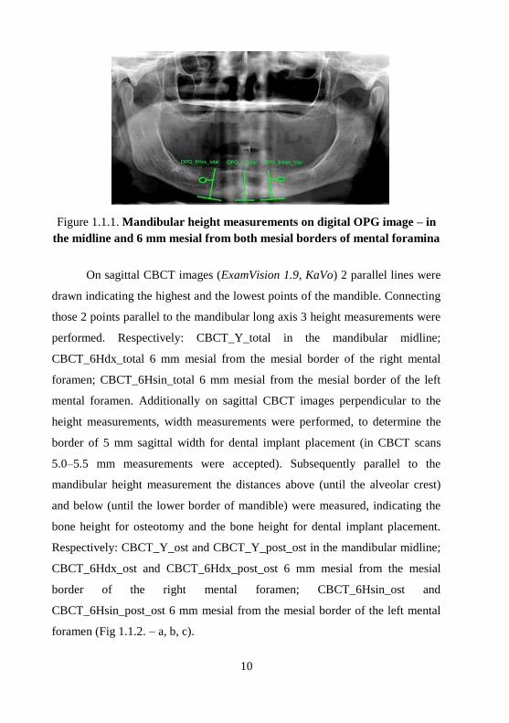
Using digital OPG images (KODAK Dental Imaging Software 6.3)
parallel line to the mandibular long axis, which connected the most prominent
points of the lower mandibular border was drawn. Perpendicular to this line
3 height measurements were performed connecting the lower border of
mandible to the alveolar crest. Respectively: OPG_Y_total in the mandibular
midline; OPG_6Hdx_total 6 mm mesial from the mesial border of the right
mental foramen; OPG_6Hsin_total 6 mm mesial from the mesial border of the
left mental foramen (Fig 1.1.1.).
10
Figure 1.1.1. Mandibular height measurements on digital OPG image – in
the midline and 6 mm mesial from both mesial borders of mental foramina
On sagittal CBCT images (ExamVision 1.9, KaVo) 2 parallel lines were
drawn indicating the highest and the lowest points of the mandible. Connecting
those 2 points parallel to the mandibular long axis 3 height measurements were
performed. Respectively: CBCT_Y_total in the mandibular midline;
CBCT_6Hdx_total 6 mm mesial from the mesial border of the right mental
foramen; CBCT_6Hsin_total 6 mm mesial from the mesial border of the left
mental foramen. Additionally on sagittal CBCT images perpendicular to the
height measurements, width measurements were performed, to determine the
border of 5 mm sagittal width for dental implant placement (in CBCT scans
5.0–5.5 mm measurements were accepted). Subsequently parallel to the
mandibular height measurement the distances above (until the alveolar crest)
and below (until the lower border of mandible) were measured, indicating the
bone height for osteotomy and the bone height for dental implant placement.
Respectively: CBCT_Y_ost and CBCT_Y_post_ost in the mandibular midline;
CBCT_6Hdx_ost and CBCT_6Hdx_post_ost 6 mm mesial from the mesial
border of the right mental foramen; CBCT_6Hsin_ost and
CBCT_6Hsin_post_ost 6 mm mesial from the mesial border of the left mental
foramen (Fig 1.1.2. – a, b, c).

11
a b c
Figure 1.1.2. Mandibular height measurements on CBCT sagittal image in
the midline (a), 6 mm mesial from the right (b) and from the left (c) mental
foramina
During both radiologic examinations all patients were positioned
according to the protocol of Dentomaxillofacial Diagnostic Radiology division,
department of Therapeutic Dentistry, Institute of Stomatology, Rīga Stradiņš
University. All CBCT images were collected, processed and reconstructed in
0.3 mm voxel matrix.
All measurements were performed twice by 1 measurer (author of the
study), with at least 2 weeks between measurements.
The data were analyzed using descriptive and analytical statistical
methods. T-test was used to determine if the mean measurements on digital
OPG and CBCT images were significantly different. The significance level was
accepted as p < 0.05. After the method of Dahlberg (Dahlberg coefficient)
measurement error for one measurer between repeated measurements was
calculated.
12
1.2. The influence of reduced general BMD on the quantitative
changes of mandibular residual ridge
This study included 38 women, aged from 54 to 83 years (70.08 ± 6.21).
For all patients CBCT (i-CAT, Next generation, Imaging Sciences) and
DXA (Lunar DEXA DPX-NT, GE Medical Systems) examinations were
performed.
General BMD was determined in both femoral necks (total mean) and
the L2–L4 lumbar area using DXA examinations. According to the World
Health Organization`s (WHO) criteria, the worst possible T-score (the number
of standard deviations above or below the mean for a healthy 30 year old adult
of the same sex and ethnicity as the patient) from both areas was taken into
account (WHO Technical Report, 1994).
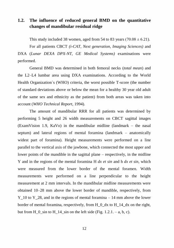
The amount of mandibular RRR for all patients was determined by
performing 5 height and 26 width measurements on CBCT sagittal images
(ExamVision 1.9, KaVo) in the mandibular midline (landmark – the nasal
septum) and lateral regions of mental foramina (landmark – anatomically
widest part of foramina). Height measurements were performed on a line
parallel to the vertical axis of the jawbone, which connected the most upper and
lower points of the mandible in the sagittal plane – respectively, in the midline
Y and in the regions of the mental foramina H dx et sin and h dx et sin, which
were measured from the lower border of the mental foramen. Width
measurements were performed on a line perpendicular to the height
measurement at 2 mm intervals. In the mandibular midline measurements were
obtained 10–28 mm above the lower border of mandible, respectively, from
Y_10 to Y_28, and in the regions of mental foramina – 14 mm above the lower
border of mental foramina, respectively, from H_0_dx to H_14_dx on the right,
but from H_0_sin to H_14_sin on the left side (Fig. 1.2.1. – a, b, c).

13
a b c
Figure 1.2.1. Mandibular height and width measurements on CBCT
sagittal images in the mandibular midline (a), right (b) and left (c) regions
of mental foramina
During CBCT examination all patients were positioned according to the
same protocol of Dentomaxillofacial Diagnostic Radiology division,
department of Therapeutic Dentistry, Institute of Stomatology, Rīga Stradiņš
University. All CBCT images were collected, processed and reconstructed
in 0.3 mm voxel matrix.
All measurements were performed twice by 1 measurer (author of the
study), with at least 2 weeks between measurements.
The data were analyzed using descriptive and analytical statistical
methods. An analysis of variance and linear, and multivariate regression
analyses were used to define the relationship between reduced BMD and
amount of mandibular RRR. After the method of Dahlberg (Dahlberg
coefficient) measurement error for one measurer between repeated
measurements was calculated.
14
1.3. The relationship between the amount of mandibular
residual ridge and patients` satisfaction with conventional
complete dentures
Since patients` satisfaction with mandibular conventional complete
dentures was analyzed both cross-sectional and prospectively in 3 year
dynamics, 2 study subgroups were created. The cross-sectional study subgroup,
in which the relationship between patients` satisfaction with mandibular
conventional complete dentures and mandibular residual ridge amount,
assessed on both digital OPG and CBCT images, was analyzed, was defined as
a “group A” and included 37 patients, aged from 54 to 85 years (72.08 ± 8.53)".
In turn, the prospective study subgroup, in which the changes between patients`
satisfaction with mandibular conventional complete dentures and mandibular
residual ridge amount, evaluated on digital OPG images, during 3-year period,
were analyzed, was defined as a "group B" and included 25 patients, aged from
56 to 79 years (69.02 ± 5.42).
For all patients digital OPG (Pantomograph Trophycan C) and CBCT
(i-CAT, Next generation, Imaging Sciences) examinations were performed.
Also patients` subjective satisfaction with the existing mandibular conventional
complete dentures using Likert 100 mm or 10 point VAS (Awad and Feine,
1998) was evaluated.
Patients` subjective satisfaction with the existing mandibular
conventional complete dentures for both groups “A” and “B” using VAS was
assessed with 5 questions: 1. Are you satisfied with the mandibular complete
denture? 2. Do you feel comfortable while using your mandibular denture? 3.
Do you feel pain while using your mandibular denture? 4. Does soreness occur
while using your mandibular denture? 5. Is your mandibular denture stable?
(Lauriņa, 2008) For each patient points from the 1st (VAS_1) and the worst
estimated 2nd
, 3rd
, 4th
or 5th
(VAS_worst) question were taken into account. To

15
assess the changes of these indicators in dynamics, patients from "group B"
were interviewed 2 times – for the first time at least 2 months after denture
delivery, but the second time – 3 years after the use of complete denture.
In addition, no patient had any relining procedures for complete
mandibular dentures, since receiving them 3 years ago.
The amount of mandibular RRR for all patients was determined by
performing measurements on digital OPG (KODAK Dental Imaging Software
6.3) and CBCT (ExamVision 1.9, KaVo) images in the mandibular midline
(landmark – the nasal septum) and lateral regions of mental foramina (landmark
–foramen midline).
On digital OPG images parallel line to the mandibular long axis, which
connected the most prominent points of the lower mandibular border, was
drawn. Perpendicular to this line 3 height measurements were performed
connecting the lower border of mandible to the alveolar crest. Respectively:
OPG_Y_total in the mandibular midline; OPG_Hdx_total on the mandibular
right side; OPG_Hsin_total on the mandibular left side (Fig 1.3.1.).
Figure 1.3.1. Mandibular height measurements on digital OPG image in the
midline and lateral regions
16
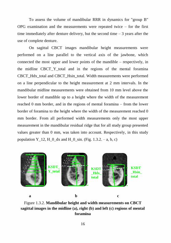
To assess the volume of mandibular RRR in dynamics for "group B"
OPG examination and the measurements were repeated twice – for the first
time immediately after denture delivery, but the second time – 3 years after the
use of complete denture.
On sagittal CBCT images mandibular height measurements were
performed on a line parallel to the vertical axis of the jawbone, which
connected the most upper and lower points of the mandible – respectively, in
the midline CBCT_Y_total and in the regions of the mental foramina
CBCT_Hdx_total and CBCT_Hsin_total. Width measurements were performed
on a line perpendicular to the height measurement at 2 mm intervals. In the
mandibular midline measurements were obtained from 10 mm level above the
lower border of mandible up to a height where the width of the measurement
reached 0 mm border, and in the regions of mental foramina – from the lower
border of foramina to the height where the width of the measurement reached 0
mm border. From all performed width measurements only the most upper
measurement in the mandibular residual ridge that for all study group presented
values greater than 0 mm, was taken into account. Respectively, in this study
population Y_12, H_0_dx and H_0_sin. (Fig. 1.3.2. – a, b, c)
a b c
Figure 1.3.2. Mandibular height and width measurements on CBCT
sagittal images in the midline (a), right (b) and left (c) regions of mental
foramina
KSDT_
Y_total
KSDT
_Hdx_
total
KSDT
_Hsin_
total

17
During both radiologic examinations all patients were positioned
according to the same protocol of Dentomaxillofacial Diagnostic Radiology
division, department of Therapeutic Dentistry, Institute of Stomatology, Rīga
Stradiņš University. All CBCT images were collected, processed and
reconstructed in 0.3 mm voxel matrix.
All measurements were performed twice by 1 measurer (author of the
study), with at least 2 weeks between measurements.
The data were analyzed using descriptive and analytical statistical
methods. T-test was used to determine if the mean measurements on digital
OPG and CBCT scans were significantly different. The significance level was
accepted as p < 0.05. For assessment of the correlation between VAS scores
and radiological measurements from mandibular residual ridge Pearson
correlation coefficient (r) was used. After the method of Dahlberg (Dahlberg
coefficient) measurement error for one measurer between repeated
measurements was calculated.

18
2. RESULTS
2.1. Dental implant planning in the anterior area of edentulous
mandible on CBCT and OPG images
Based on the method of Dahlberg (Dahlberg coefficient) it was
estimated that the measurement error for measurements performed on CBCT
images was 0.00–0.60, indicating that there was no measurement error in the
results. However the measurement error on digital OPG images was 1.44–3.21.
There was statistically significant difference (p = 0.000) between mean
measurements, performed on CBCT and digital OPG images in mandibular
midline (OPG_Y_total and CBCT_Y_total) and mesial from the right
(OPG_6Hdx_total and CBCT_6Hdx_total) and left (OPG_6Hsin_total and
CBCT_6Hsin_total) mental foramina (Table 2.1.1.).
Table 2.1.1.
Comparison of mandibular residual ridge mean height measurements on
OPG and CBCT images in the mandibular midline and 6 mm mesial from
both mental foramina
Measurement Mean value (mm) SD p N
OPG_Y_total 22.83 4.80 0.000 37
CBCT_Y_total 20.57 3.47
OPG_6Hdx_total 21.67 5.18 0.000 37
CBCT_6Hdx_total 18.92 4.52
OPG_6Hsin_total 21.28 5.03 0.000 37
CBCT_6Hsin_total 18.79 4.04
N – number of patients
SD – standard deviation
p – level of significance (p < 0,05)

19
According to 5.0–5.5 mm width measurement border on CBCT images
it was not possible to place dental implants without osteotomy for 100%
patients in the mandibular midline as well as 6 mm mesial from both mental
foramina.
According to 11 mm height measurement border on digital OPG images
it was possible to place dental implants for 100% patients in the mandibular
midline as well as 6 mm mesial from both mental foramina. While according to
11 mm height measurement border on CBCT images after osteotomy it was not
possible to place dental implants for 10.8% patients 6 mm mesial from the right
mental foramen (CBCT_6Hdx_post_ost) and for 10.8% patients 6 mm mesial
from the left mental foramen (CBCT_6Hsin_post_ost). Besides for 8.1%
patients it was not possible to place dental implants neither 6 mm mesial from
the right mental foramen, nor 6 mm mesial from the left mental foramen.
(Table 2.1.2.)
Table 2.1.2.
Comparison of mandibular residual ridge mean and minimum height
measurements on OPG images before osteotomy and on CBCT images
after osteotomy in the mandibular midline and 6 mm mesial from both
mental foramina
Measurement
Mean
value
(mm)
SD p
Min.
value
(mm)
How
many %
can have
dental
implant
N
OPG_Y_total 22.83 4.80 0.000
13.00 100 37
KSDT_Y_post_ost 18.13 3.04 11.34 100 37
OPG_6Hdx_total 21.67 5.18 0.000
11.20 100 37
KSDT_6Hdx_post_ost 16.34 4.27 5.05 89 37
OPG_6Hsin_total 21.28 5.03 0.000
11.50 100 37
KSDT_6Hsin_post_ost 16.35 3.79 9.24 89 37
Min. – minimum; N – number of patients;
SD – standard deviation; p – level of significance (p < 0,05)

20
2.2. The influence of reduced general BMD on the quantitative
changes of mandibular residual ridge
Based on the method of Dahlberg (Dahlberg coefficient) it was
estimated that the measurement error for measurements performed on CBCT
images was 0.00–0.46, indicating that there was no measurement error in the
results.
The mean DXA T-score was −1.73 ± 1.30 (range from −4.2 to 1.0).
There were 11 patients with normal BMD, 14 with osteopenia and 13 with
osteoporosis.
There was no statistically significant relationship between general BMD
and mandibular residual ridge height measurements in the mandibular midline
(Y) as well as in both regions of mental foramina (H_dx, h_dx and H_sin,
h_sin) (Table 2.2.1.).
Table 2.2.1.
Relationship between general BMD (DXA) and mandibular residual ridge
height and sagittal width measurements (CBCT) in the mandibular
midline and both lateral regions of mental foramina
Measurement BMD
N Coefficient p 95% CI
Y −0.03 0.668 −0.16 0.09 38
H_dx −0.01 0.800 −0.10 0.08 38
h_dx −0.04 0.634 −0.24 0.15 38
H_sin 0.00 0.971 −0.10 0.11 38
h_sin 0.19 0.118 −0.05 0.43 38
BMD – bone mineral density
N – number of patients
p – level of significance (p < 0,05)
CI – confidence interval

21
There was also no statistically significant relationship between general
BMD and mandibular residual ridge width measurements in the mandibular
midline (Y_10 to Y_28) (Table 2.2.2.) as well as in both regions of mental
foramina (H_0_dx to H_14_dx and H_0_sin to H_14_sin) (Table 2.2.3.).
Table 2.2.2.
Relationship between general BMD (DXA) and mandibular residual ridge
sagittal width measurements (CBCT) in the mandibular midline
Measurement BMD
N Coefficient p 95% CI
Y_10 −0.01 0.933 −0.26 0.24 38
Y_12 0.05 0.524 −0.12 0.24 38
Y_14 0.03 0.621 −0.09 0.15 38
Y_16 0.05 0.376 −0.07 0.19 38
Y_18 0.58 0.379 −0.07 0.19 38
Y_20 −0.04 0.589 −0.19 0.11 38
Y_22 −0.08 0.431 −0.31 0.13 38
Y_24 −0.20 0.252 −0.54 0.14 38
Y_26 −0.56 0.150 −1.34 0.21 38
Y_28 −1.42 0.198 −3.64 0.78 38
BMD – bone mineral density
N – number of patients
p – level of significance (p < 0,05)
CI – confidence interval

22
Table 2.2.3.
Relationship between general BMD (DXA) and mandibular residual ridge
sagittal width measurements (CBCT) in both lateral regions of mental
foramina
BMD – bone mineral density
N – number of patients
p – level of significance (p < 0,05)
CI – confidence interval
Regarding the multivariate regression analysis, there was no statistically
significant relationship between worst general BMD T-score and mandibular
residual ridge height and width measurements performed at one site – in the
Measurement BMD
N Coefficient p 95% CI
H_0_dx −0.01 0.894 −0.26 0.23 38
H_2_dx 0.03 0.532 −0.08 0.16 38
H_4_dx −0.02 0.662 −0.15 0.09 38
H_6_dx −0.00 0.987 −0.13 0.13 38
H_8_dx −0.04 0.588 −0.21 0.12 38
H_10_dx −0.06 0.573 −0.28 0.16 38
H_12dx 0.05 0.766 −0.33 0.45 38
H_14_dx 0.33 0.685 −1.33 2.01 38
H_0_sin −0.06 0.600 −0.29 0.17 38
H_2_sin 0.00 0.895 −0.13 0.15 38
H_4_sin −0.02 0.641 −0.14 0.09 38
H_6_sin −0.05 0.458 −0.18 0.08 38
H_8_sin −0.09 0.367 −0.29 0.11 38
H_10_sin −0.11 0.438 −0.40 0.17 38
H_12_sin −0.22 0.503 −0.89 0.44 38
H_14_sin − − − 38
23
mandibular midline as well as the right and left regions of the mental
foramen.
Regarding patient age, it was estimated that it had no statistically
significant relationship with worst general BMD T-score or mandibular residual
ridge height and width measurements.
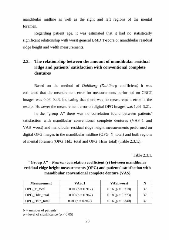
2.3. The relationship between the amount of mandibular residual
ridge and patients` satisfaction with conventional complete
dentures
Based on the method of Dahlberg (Dahlberg coefficient) it was
estimated that the measurement error for measurements performed on CBCT
images was 0.03–0.43, indicating that there was no measurement error in the
results. However the measurement error on digital OPG images was 1.44–3.21.
In the “group A” there was no correlation found between patients`
satisfaction with mandibular conventional complete dentures (VAS_1 and
VAS_worst) and mandibular residual ridge height measurements performed on
digital OPG images in the mandibular midline (OPG_Y_total) and both regions
of mental foramen (OPG_Hdx_total and OPG_Hsin_total) (Table 2.3.1.).
Table 2.3.1.
“Group A” – Pearson correlation coefficient (r) between mandibular
residual ridge height measurements (OPG) and patients` satisfaction with
mandibular conventional complete denture (VAS)
Measurement VAS_1 VAS_worst N
OPG_Y_total −0.01 (p = 0.917) 0.16 (p = 0.318) 37
OPG_Hdx_total −0.00 (p = 0.967) 0.18 (p = 0.273) 37
OPG_Hsin_total 0.01 (p = 0.942) 0.16 (p = 0.340) 37
N – number of patients
p – level of significance (p < 0,05)
24
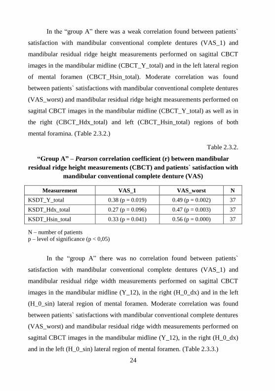
In the “group A” there was a weak correlation found between patients`
satisfaction with mandibular conventional complete dentures (VAS_1) and
mandibular residual ridge height measurements performed on sagittal CBCT
images in the mandibular midline (CBCT_Y_total) and in the left lateral region
of mental foramen (CBCT_Hsin_total). Moderate correlation was found
between patients` satisfactions with mandibular conventional complete dentures
(VAS_worst) and mandibular residual ridge height measurements performed on
sagittal CBCT images in the mandibular midline (CBCT_Y_total) as well as in
the right (CBCT_Hdx_total) and left (CBCT_Hsin_total) regions of both
mental foramina. (Table 2.3.2.)
Table 2.3.2.
“Group A” – Pearson correlation coefficient (r) between mandibular
residual ridge height measurements (CBCT) and patients` satisfaction with
mandibular conventional complete denture (VAS)
Measurement VAS_1 VAS_worst N
KSDT_Y_total 0.38 (p = 0.019) 0.49 (p = 0.002) 37
KSDT_Hdx_total 0.27 (p = 0.096) 0.47 (p = 0.003) 37
KSDT_Hsin_total 0.33 (p = 0.041) 0.56 (p = 0.000) 37
N – number of patients
p – level of significance (p < 0,05)
In the “group A” there was no correlation found between patients`
satisfaction with mandibular conventional complete dentures (VAS_1) and
mandibular residual ridge width measurements performed on sagittal CBCT
images in the mandibular midline (Y_12), in the right (H_0_dx) and in the left
(H_0_sin) lateral region of mental foramen. Moderate correlation was found
between patients` satisfactions with mandibular conventional complete dentures
(VAS_worst) and mandibular residual ridge width measurements performed on
sagittal CBCT images in the mandibular midline (Y_12), in the right (H_0_dx)
and in the left (H_0_sin) lateral region of mental foramen. (Table 2.3.3.)
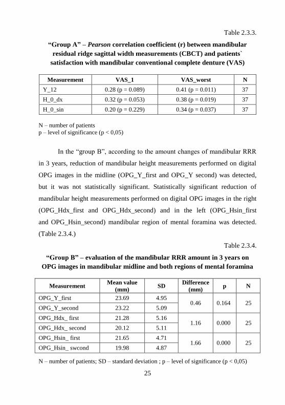
25
Table 2.3.3.
“Group A” – Pearson correlation coefficient (r) between mandibular
residual ridge sagittal width measurements (CBCT) and patients`
satisfaction with mandibular conventional complete denture (VAS)
Measurement VAS_1 VAS_worst N
Y_12 0.28 (p = 0.089) 0.41 (p = 0.011) 37
H_0_dx 0.32 (p = 0.053) 0.38 (p = 0.019) 37
H_0_sin 0.20 (p = 0.229) 0.34 (p = 0.037) 37
N – number of patients
p – level of significance (p < 0,05)
In the “group B”, according to the amount changes of mandibular RRR
in 3 years, reduction of mandibular height measurements performed on digital
OPG images in the midline (OPG_Y_first and OPG_Y second) was detected,
but it was not statistically significant. Statistically significant reduction of
mandibular height measurements performed on digital OPG images in the right
(OPG_Hdx_first and OPG_Hdx_second) and in the left (OPG_Hsin_first
and OPG_Hsin_second) mandibular region of mental foramina was detected.
(Table 2.3.4.)
Table 2.3.4.
“Group B” – evaluation of the mandibular RRR amount in 3 years on
OPG images in mandibular midline and both regions of mental foramina
Measurement Mean value
(mm) SD
Difference
(mm) p N
OPG_Y_first 23.69 4.95 0.46 0.164 25
OPG_Y_second 23.22 5.09
OPG_Hdx_ first 21.28 5.16 1.16 0.000 25
OPG_Hdx_ second 20.12 5.11
OPG_Hsin_ first 21.65 4.71 1.66 0.000 25
OPG_Hsin_ swcond 19.98 4.87
N – number of patients; SD – standard deviation ; p – level of significance (p < 0,05)
26
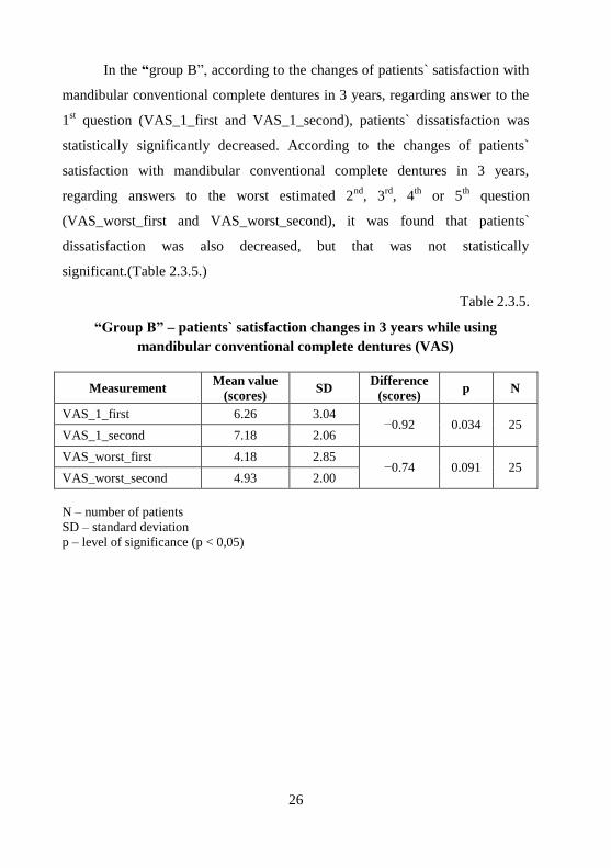
In the “group B”, according to the changes of patients` satisfaction with
mandibular conventional complete dentures in 3 years, regarding answer to the
1st question (VAS_1_first and VAS_1_second), patients` dissatisfaction was
statistically significantly decreased. According to the changes of patients`
satisfaction with mandibular conventional complete dentures in 3 years,
regarding answers to the worst estimated 2nd
, 3rd
, 4th
or 5th
question
(VAS_worst_first and VAS_worst_second), it was found that patients`
dissatisfaction was also decreased, but that was not statistically
significant.(Table 2.3.5.)
Table 2.3.5.
“Group B” – patients` satisfaction changes in 3 years while using
mandibular conventional complete dentures (VAS)
Measurement Mean value
(scores) SD
Difference
(scores) p N
VAS_1_first 6.26 3.04 −0.92 0.034 25
VAS_1_second 7.18 2.06
VAS_worst_first 4.18 2.85 −0.74 0.091 25
VAS_worst_second 4.93 2.00
N – number of patients
SD – standard deviation
p – level of significance (p < 0,05)

27
3. DISCUSSION
3.1. Dental implant planning in the anterior area of edentulous
mandible on CBCT and OPG images
Patients with mandibular conventional complete dentures often
complain about discomfort due to RRR and consecutive reduced support,
stability and retention of removable dentures (Batenburg et al., 1998; Tallgren,
2003; Hyland et al., 2009). To decrease the rate of RRR (Kordatzis et al., 2003;
Carlsson, 2004; Bodic et al., 2005), and to improve the result of prosthodontic
treatment as well as the quality of life (Reich et al., 2011), it is advisable to
place 2 to 4 dental implants in anterior area of edentulous mandibular bone
(Feine et al., 2002).
Studies show that two-dimensional OPG images are not precise enough
to identify or measure all anatomical structures of anterior mandibular alveolar
bone (Wismeijer et al., 1997; Kaya et al., 2008; Ngeow et al., 2009), while
three-dimensional CBCT images provide 100% precise identification of those
structures (Parnia et al., 2012). Therefore scientific literature affirms that for
dental implant planning in anterior area of mandibular alveolar bone three-
dimensional X-ray examination methods like CBCT are suggested
(Eufinger et al., 1997; Bou Serhal et al., 2002; Madrigal et al., 2008;
Monsour and Dudhia, 2008; Angelopoulos et al., 2008; Dreiseidler et al., 2009;
Georgescu et al., 2010). Also regarding recommendations of the American
Academy of Oral and Maxillofacial Radiology, CBCT should be considered for
preoperative cross-sectional imaging of potential implant sites, while OPG
should be used as the imaging modality of choice in the initial evaluation
of the dental implant patient (Tyndall et al., 2012). Following these
recommendations in our study, digital OPG as well as CBCT were performed
for each patient.
28
However, when CBCT is performed, the radiation dose should be taken
into account. Compared with digital OPG, CBCT represents about 3 to 7 times
greater radiation dose, depending on the selected device settings
(Ludlow et al., 2006; Dreiseidler et al., 2009; Holroyd and Gulson, 2009).
Results of scientific studies suggest that for dental implant supported
mandibular overdenture the most distal implant should be placed at least 1 mm
mesial from the most mesial point of the anterior loop of the inferior alveolar
nerve (Jensen et al., 2011). Unfortunately, due to variations of the anatomy of
inferior alveolar nerve and its anterior loop there are clear risks in implant
placement in this area. Accessory mental foramina (Naitoh et al., 2009;
Naitoh et al., 2010; Kalender et al., 2012; Imada et al., 2014), which cannot
always be localized on OPG in contradistinction to CBCT images
(Santos u.c., 2013; Imada u.c., 2014), are described in the literature. There are
also studies that show clinically insignificant length of anterior loop of inferior
alveolar nerve (Rosenquist, 1996; Benninger et al., 2011). The mean
(0.9–7.6 mm) and maximum (0.5–9.0 mm) length of anterior loop of inferior
alveolar nerve reflected in the literature are of a wide range (Uchida et al.,
2007; Uchida et al., 2009; Apostolakis and Brown, 2012; Parnia et al., 2012;
Rosa et al., 2013; Chen et al., 2013; Von Arx et al., 2013). To establish certain
methodology, in our study we calculated the arithmetic mean value of the
maximum lengths of the anterior loop of inferior alveolar nerve (5.0 mm) from
previously represented studies, where the loop was identified. Consequently, on
the ground of the recommendation about leaving 1 mm mesial from anterior
loop of inferior alveolar nerve (Jensen u.c., 2011), the measurement site for
distal dental implant placement in mandibular overdenture case in our study
was selected 6.0 mm mesial from the mesial border of mental foramen. The
third measurement site was selected in the mandibular midline because it is a
good anatomical landmark.

29
Although there is no distinct definition in the scientific literature, what
“short” dental implants mean, most authors agree that the length of 10 mm is
the boundary between “standard” and “short” dental implants (Morand and
Irinakis, 2007; Telleman et al., 2011; Sun et al., 2011). As the literature has
conflicting opinions about the use of dental implants shorter than 10 mm,
showing worse (Telleman et al., 2011) or, on the contrary, equivalent
(Triplett et al., 1991; Sun et al., 2011) survival and success rates to “standard”
dental implants, in our study we chose the situation where 10 mm long
“standard” dental implants were planned for placement in the anterior area of
edentulous mandible. According to the classification of the mandibular RRR,
established by the American College of Prosthodontists, many patients from
this study corresponded to the 3rd and 4th class of RRR, denoting severe bone
atrophy and subsequent selection of 10 mm or shorter dental implant selection
(Batenburg et al., 1998). In our study 11 mm mandibular bone height was
accepted as minimum required for the placement of 10 mm long dental
implants, including recommended 1 mm distance (Dietrich u.c., 1993) to the
lower boundary of the mandible.
Dental implants with a diameter less than 3.75 mm are considered as
“narrow” dental implants (Arisan et al., 2010). Study results show that survival
and success rates for a “narrow” diameter dental implants are equivalent to that
of the "standard" dental implants, stating they can optimally support
overdentures (Cho et al., 2007; Arisan et al., 2010; Sohrabi et al., 2012). Since
majority of the patients from our study group had severe mandibular RRR, we
planned to select the smallest "standard" dental implants with 3.0 mm diameter
that had been recommended for overdenture support. In our study it was
assumed that the minimum width of the mandibular residual ridge required for
planning of 3 mm diameter dental implant placement was 5 mm, including
recommended 1 mm distance (Dietrich et al., 1993; Quirynen et al., 2003)
buccal as well as lingual.

30
Insufficient bucco-lingual width of mandibular residual ridge often is the
reason for osteotomy to flatten "knife-edge" shaped alveolar ridge and to
achieve the desired bone width parameters (Eufinger et al., 1997). After
osteotomy the initial bone height is reduced, which, in fact, may negate the
opportunity to place dental implants.
Madrigal et al has published a study of similar methodology
(Madrigal et al., 2008), which aimed to demonstrate the variations between
digital OPG and CBCT examination methods in evaluating the dimensions of
alveolar residual ridge and localizing anatomical structures in the anterior area
of mandible. This study included 50 patients with complete or partial
edentulism in the anterior area of mandible. For each patient digital OPG
(Ortofox Siemens AG) and CBCT (NewTom 9000QR) examinations were
performed. Radiologic measurements in the mandibular midline, 1 cm distal
from the midline and in the midline of both mental foramina were recorded.
As the aim of our study also was to compare the opportunities of two
different X-ray examination methods when planning dental implants in the
anterior area of edentulous mandible, similar linear measurements of
mandibular alveolar bone using digital OPG and CBCT sagittal images were
performed. In our study the mean values of mandibular height measurements,
performed on digital OPG images, were statistically significantly higher than
the values of sagittal CBCT images. This was on contrary to Madrigal study
results, of which may be be explained by the optical zoom of digital OPG
hardware.
Our results were also in agreement with Georgescu study, confirming
that mandibular measurements performed with the aid of digital OPG were
overestimated, comparing with those obtained by CBCT sagittal scans
(Georgescu et al., 2010).
According to the width parameters of edentulous mandibular alveolar
bone, to place dental implants in our study group in the mandibular midline and
31
6 mm mesial from both mental foramina, all patients needed to plan osteotomy.
While comparing height parameters of edentulous mandibular alveolar bone
before osteotomy on two-dimensional OPG images and after osteotomy on
sagittal three-dimensional CBCT images, it showed that the difference of those
mean measurements was statistically significant. After osteotomy in 10.8% of
cases, it was not possible to plan dental implant placement 6 mm mesial from
both mental foramina because of insufficient residual alveolar bone height.
However, using only OPG examination data, all cases were amenable for
planning of dental implant placement in the same area.
It was further shown that in calculating measurement error, there was a
significant difference in the accuracy of measurements between OPG and
CBCT imaging, in favorite to CBCT. That was also in agreement with the
literature, suggesting that the measurement error is greater for the
measurements performed on OPG than on CBCT scans (Hu et al., 2012).
Our study results clearly underlines the significance of CBCT
examination in prosthodontic treatment planning for edentulous jaws to obtain
accurate information about opportunities or problems regarding dental implant
placement.
3.2. The influence of reduced general BMD on the quantitative
changes of mandibular residual ridge
One hypothesis from the literature is that systemic factors, such as
osteoporosis, may play a more significant role in RRR than local factors
(Atwood, 1962; Kribbs, 1990). The premise is that osteoporosis may define the
final speed and contour of the resorption process when the impact of local
factors after the last tooth extraction have already disappeared (Devlin and
Ferguson, 1991; Bozic and Hren, 2005). Since the most pronounced RRR
appears due to local factors at a time 6 to 24 months after the last tooth
32
extraction (Atwood, 1971; Knezovic-Zlataric et al., 2002), we therefore in our
study included patients who had lost their last tooth at least 5 years prior this
study. The shortcomings of this study was the inability to identify the causes
and the dynamics of tooth loss, as it is possible that RRR for some patients was
induced by periodontitis or traumatic tooth extraction, consequently resulting in
greater amount of alveolar bone resorption. However, since several patients
before prosthodontics were treated at other dental clinics, such data could not
be collected due to the lack of objective information.
To establish a more homogeneous edentulous study group regarding
prosthetic factor impact on RRR, all patients 3 years prior this study in the
Prosthodontic Clinic at the Institute of Stomatology, Rīga Stradiņš University
received similar design conventional complete dentures, which were made in
the same dental laboratory. Although the opinions in the literature, whether the
use of dentures at night reinforces the amount of RRR (Campbell, 1960;
Carlsson, 2004) or not (Kovacić et al., 2010; Kranjčić et al., 2013), are
controversial, our patients were advised not to use the conventional complete
dentures during night.
To observe reduced general BMD, and exclude other metabolic factors
that could influence the amount of mandibular RRR, in our study we did not
include patients whose medical history showed any systemic diseases or
conditions, or use of medications that could cause mandibular RRR. In our
study population, based on a survey of patients, the possibility of secondary
osteoporosis as well as other osteoporosis-related risk factors, such as alcohol
abuse, smoking or diet-related disorders, were also excluded. However, since
the medical history was obtained by interviewing patients, there might be a risk
that some patients had an undisclosed disease that could affect our study
results. We also did not consider whether any patient had used biphosphonates
prior this study because it was not possible to collect reliable information.
However, it is very important to collect such information prior dental implant

33
planning because of the risk of jaw osteonecrosis. After the consumption of
antiresorptive drugs, there is a possibility that the DXA results could be
improved but the mandibular ridge parameters would still be reduced.
Previous data suggest that 30% of all Caucasian women after the age
of 50 have osteoporosis (Albright et al., 1941; WHO, 1994), therefore, we
only included menopausal patients who were at a higher risk for diminished
general BMD.
Various studies have investigated the relationship between BMD and
mandibular RRR. Although the majority of studies used DXA to determine
BMD (Klemetti et al., 1993a; Klemetti et al., 1993b; Balcikonyte et al., 2003;
Bozic and Hren, 2005; Ozola et al., 2011), some preferred a visual analysis of a
radiographic images (Hirai et al., 1993; Soikkonnen et al., 1996) or the
certification of osteoporotic fractures (Kribbs, 1990; Bollen et al., 2000). There
are also studies where BMD was determined in different regions of the
mandible by conventional radiography (Nishimura et al., 1992), single (SPA)
(Von Wowern, 1985), or dual (DPA) photon absorptiometry
(Buyukkaplan, 2012), quantitative computed tomography (QCT)
(Merheb et al., 2012) or CBCT (Helmi et al., 2009). However, there are still
conflicting opinions whether mandibular BMD correlates with skeletal BMD
(Cakur et al., 2009; Merheb et al., 2012). In our study, we used DXA to ensure
accurate evaluation of general BMD in the L2–L4 lumbar area and in both
femoral necks. Currently, this examination method is accepted as the “gold
standard” for the diagnostics of osteoporosis because of its precision, greater
functionality and lower radiation dose (National Osteoporosis Society, 1994).
To determine the RRR, Klemetti et al. described different methods,
including the clinical classification based on the degree of atrophy
(Klemetti et al., 1993a; Soikkonnen et al., 1996), radiographic measurements in
the region of mental foramen (Kribbs et al., 1989; Kribbs, 1990;
Hirai et al., 1993; Bollen et al., 2000; Balcikonyte et al., 2003), radiographic
34
comparison of the jaw bone at specific times (Von Wowern and Kollerup,
1992), measurements of the mandibular symphysis using computed
tomography (Klemetti et al., 1993b) and other radiographically detected indexes
at different sites of the mandible (Bozic and Hren, 2005). In our study we were
interested in developing new methodology for the mandibular residual ridge
measurements involving not only the height but also the width parameters of
the alveolar bone using CBCT images. This method seems objective and
repeatable, as there was no systemic errors found associated with the
measurements. In this study edentulous jaw width measurements were
performed every 2 mm to detect small changes in the mandibular bone width.
In the midline, this measurement was performed 10–28 mm above the lower
mandibular border, and in the lateral regions, it was performed 14 mm above
the lower border of the mental foramina. These specific intervals were selected
because edentulous jaw bone resorption in the bucco-lingual aspect occurs
mostly in the alveolar process. Landmarks for width measurements were
selected according to provisional measurements, respectively, in our study, the
highest mandibular height in the midline was 28 mm, but the highest alveolar
crest from the lower border of the mental foramen was 14 mm. There were no
patients without any alveolar process remaining where first width measurement
would be absent.
In a previous retrospective, cross-sectional study, (Nishimura et al.,
1992) the pattern of RRR was analyzed, characterizing the longitudinal
morphologic changes of the mandibular bony contour in 30 completely
edentulous male and female patients. They measured both the sagittal and
vertical dimensions of the mandibular bony contour at the symphysis area on
longitudinally taken superimposed lateral cephalographic tracings. They also
calculated the radiographic bone density (RD) of the second vertebra and the
center of the mandibular symphysis. Finally they suggested that osteopenic
changes in women might be associated with a long-term bone remodeling
35
pattern in the edentulous mandible, which results in the “knife-edge”
morphology.
In our study, we were interested in the same hypothesis; however, in
contrast, our results indicated that postmenopausal women with reduced BMD
do not have the “knife-edge” tendency in the mandibular residual ridge. The
difference in our study, which could influence the interpretation and
comparison of both study results, was that we developed a methodology for
accurate mandibular ridge measurements in the CBCT sagittal plane, not only
in the midline but also in both regions of the mental foramina. To detect BMD,
we used DXA, which is currently the gold standard. In addition, Nishimura et
al. performed a retrospective study, whereas our study was a cross-sectional.
Until now, there was only one study (Helmi et al., 2009) that
investigated the connection between BMD and mandibular RRR using CBCT
images. The authors found a statistically significant correlation between
diminished mandibular BMD and increased levels of RRR. However, this study
was limited to a small patient group, which consisted of 6 edentulous female
patients. In addition, BMD was estimated in the mandible in Hounsfield units
(HU) using CBCT. Unfortunately, CBCT does not allow the reliable and
accurate assessment of bone quality when focusing on the radiographic density
information that is expressed by HU. Because BMD not only depends on the
calcium content of bone alone but also on the structural characteristics, BMD
measurements detected by CBCT do not correlate with BMD measurements
detected by DXA (Hua, 2009). In this study, mandibular height measurements
were carried out similar to our study on CBCT images in the regions of the
mental foramina, however, the methodology was not specifically described.
Unfortunately, it is impossible to compare these results with ours because of the
different measurement methodologies and study populations.
In our study, we did not find a statistically significant relationship
between general BMD and the amount of mandibular RRR, which corresponds
36
to the majority of studies published in the literature (Kribbs et al., 1989;
Kribbs, 1990; Klemetti et al., 1993a; Balcikonyte et al., 2003; Bozic and
Hren, 2005). However, to confirm such results, it would be necessary to
perform the long-term study in dynamics of RRR by using the existing raw data
as a reference point.
3.3. The relationship between the amount of mandibular
residual ridge and patients` satisfaction with conventional
complete dentures
In the literature there are different opinions about the effect of residual
ridge amount on patients` satisfaction with conventional complete dentures,
however, most authors agree that the alveolar bone volume affects the outcome
of the prosthodontic treatment. But in the published studies, to characterize the
amount of mandibular alveolar bone, authors have analyzed only bone height
parameters, which reflect volume of bone resorption only partially
(Närhi et al., 1997; Pan et al., 2010). Since the amount of bone loss is initially
observed on the buccal and lingual surfaces, but only afterwards on the top of
the mandibular residual ridge (Atwood, 1963), three-dimensional X-ray
examination, such as CBCT, is required. This would allow to estimate objective
amount of bone loss and its impact on patients` satisfaction with mandibular
conventional complete dentures.
For our study "group A" specific selection criteria as well as new
methodology was established.
Since patients` satisfaction may be associated with the ability to adapt to
the dentures, all patients prior our study were using the conventional complete
dentures for at least 3 years. Also to establish a specific reference point, all
patients 3 years prior this study in the Prosthodontic Clinic at the Institute of
Stomatology, Rīga Stradiņš University received new dentures, which were
37
made in the same dental laboratory. Although the opinions in the literature,
whether the use of dentures at night reinforces the amount of RRR (Campbell,
1960; Carlsson, 2004) or not (Kovacić et al., 2010; Kranjčić et al., 2013), are
controversial, our patients were advised not to use the conventional complete
dentures during the night. Also all patients in our study had lost their last tooth
at least 5 years ago, which reduced the impact of local postextraction factors on
mandibular RRR (Devlin and Ferguson, 1991; Bozic and Hren, 2005) that, in
turn, could cause a sudden changes in patient VAS satisfaction scores.
To clarify patients` satisfaction with complete dentures, some authors
used multilevel scale where patients had to answer with 1 of 2 or more offered
multiple choice questions, for example – satisfied or dissatisfied (Garrett et al.,
1996; De Baat et al., 1997; Huumonen et al., 2012). However, most authors for
assessing an immeasurable value such as satisfaction used VAS
(Awad et al., 2003; Pan et al., 2010; Nuñez et al., 2013).
In order to determine the lowest satisfaction for each patient, in our
study points from the 1st (VAS_1) and one of the worst estimated 2
nd, 3
rd, 4
th or
5th
(VAS_worst) VAS question were taken into account. This method was
selected, because dissatisfaction with the use of complete dentures for each
patient could be induced by different factors, for example, because of the poor
stability of the mandibular denture or some soreness during the denture use.
To determine actual amount of mandibular residual ridge measurements
on digital OPG as well as on CBCT images for all “group A” patients were
performed.
Pan et al. (Pan et al., 2010) analyzed the relationship between
edentulous mandibular alveolar bone height and patients` satisfaction with
conventional complete dentures. To evaluate the volume of mandibular RRR
for 107 patients, 5 mandibular height measurements from OPG images, which
were divided into 4 different resorption classes, were performed. Patients`
satisfaction with complete dentures was assessed by 100 mm VAS scale 6
38
months after receiving prosthodontic treatment. The results of this and our
study were similar, showing no statistically significant correlation between
height measurements of mandibular residual ridge and patients` satisfaction
with mandibular conventional complete dentures. However, it should be noted
that the time interval for each study, the patients were interviewed and VAS
scores collected, i.e., 6 months and 3 years after receiving dentures, was
different. That is important, because longer period of time may be associated
with better adaptation and higher satisfaction scores and also possible changes
in complete dentures as well as in the prosthetic field in oral cavity.
As described in previous similar studies (Närhi et al., 1997;
Huumonen et al., 2012) mandibular alveolar bone height parameters were
evaluated on digital OPG images. The novelty of our study was that the height
measurements of the mandibular residual ridge in the same areas were
performed also on sagittal CBCT images that gave the actual information about
bone height, regarding longitudinal axis of the mandibular bone. This is
particularly relevant when the mandible has a labial or lingual inclination,
which cannot be detected on OPG images.
As a result, our study confirmed the hypothesis that there was no
correlation between the height measurements of edentulous mandibular
alveolar bone, measured on OPG images, and patients` satisfaction with
conventional complete mandibular dentures, which in turn was when the same
mandibular height measurements were performed on sagittal CBCT images.
Another novelty of our study was that the amount of mandibular RRR
was assessed not only regarding the height, but also the sagittal width
parameters. As a result, we found one more correlation between the width
measurements of mandibular residual ridge, and patients` satisfaction with
mandibular conventional complete dentures. This, in turn, suggests VAS
satisfaction scores may be related directly to the bone narrowing.

39
Correlations in our study were mainly assessed between the amount of
mandibular RRR and the worst rated question, which could be explained by the
fact that patients were able to be more objective and more critical in response to
specific but not general question. This tendency also appeared in Awad et al.
study, where approximately 80% of patients responded positively about their
dentures as a whole, however, the analysis of specific issues became more
negative (Awad and Feine, 1998).
Until now literature shows no data about the relationship in specific time
period between the amount of mandibular RRR changes and patients`
satisfaction changes.
Therefore, in order to establish as possible homogeneous study "group
B", specific selection criteria as well as a new methodology was developed.
Selection criteria discussed previous for the "group A" also
corresponded to the "group B". In addition it should be mentioned that no
patient had any relining procedures for complete mandibular dentures, since
receiving them 3 years ago.
Our study showed that with a statistically significant decrease of
edentulous mandibular alveolar bone height in both lateral regions of mental
foramina, also statistically significant improvement in patients` assessment of
the first question: "Are you satisfied with your mandibular complete denture?"
occurred. Perhaps such unexpected relationship could be explained by the fact
that, despite the volume reduction of mandibular residual ridge, patients during
3 years were more able to adapt to their mandibular conventional complete
dentures and, in general, less unsatisfied. It should be noted that the reduction
of mandibular height was relatively small (1.2 to 1.7 mm), which probably
caused no discomfort and did not affect patients` VAS satisfaction scores. In
addition, it should be mentioned that the measurement error in this situation
was not included in the statistical calculation of the results, as it was measured
separately by the method of Dahlberg. This, in turn, could affect the clinical
40
reliability of the results, showing different relationship between both
discussed parameters.
Indicator that was analyzed in the literature and compared in a specific
period of time, was the amount of edentulous alveolar bone for conventional
complete denture users. The results clearly showed that bone height
measurements after specific time period decreased, which was observed
particularly in the anterior area of mandible. Various studies suggested that
mandibular RRR for conventional complete denture users within 5 years is
2 times, while in the 7 and 25 years – 4 times faster than maxillary RRR
(Tallgren, 1969; Tallgren, 2003; Kovacić et al., 2010). Also, in our study
decrease of edentulous mandibular alveolar bone height for complete denture
users in the time period of 3 years was observed. However, in contrast to other
studies, mandibular height reduction in our study was more pronounced and
statistically significant in the lateral areas, but insignificant in the mandibular
midline.
The results of this study demonstrate the advantages of CBCT
examination for evaluating the amount of mandibular RRR and its further
relationship with patients` satisfaction with conventional complete dentures.
41
4. CONCLUSIONS
4.1. Dental implant planning in the anterior area of edentulous
mandible on CBCT and OPG images
1. Despite greater radiation dose, CBCT rather than digital OPG
examination method is indicated for dental implant planning in the
anterior area of severely resorbed edentulous mandible.
2. Digital OPG as the only examination method for dental implant
planning in the anterior area of severely resorbed edentulous
mandible is insufficient and can result in errors in treatment planning
as well as iatrogenic mistakes during surgery.
4.2. The influence of reduced general BMD on the quantitative
changes of mandibular residual ridge
1. Postmenopausal women with reduced general BMD do not have
diminished height of edentulous mandibular alveolar bone.
2. Buccolingual narrowing or a “knife-edge” tendency of the
mandibular residual ridge is not related to reduced general BMD in
postmenopausal women.
42
4.3. The relationship between the amount of mandibular residual
ridge and patients` satisfaction with conventional complete
dentures
1. Patients` satisfaction with mandibular conventional complete
dentures has no relationship with the amount of mandibular RRR, as
determined from digital OPG height measurements.
2. With increasing amount of mandibular RRR, regarding sagittal
CBCT height and width measurements, increases patients`
dissatisfaction with mandibular conventional complete dentures.
3. Despite the reduction of edentulous mandibular alveolar bone height
in the lateral regions, during 3 years also patients overall
dissatisfaction with mandibular conventional complete dentures
decreases.

43
5. PRACTICAL RECOMMENDATIONS
1. Despite greater radiation dose, CBCT examination method is indicated for
dental implant planning in the anterior area of severely resorbed edentulous
mandible, while digital OPG as only examination method in the same
situation is insufficient and can result in inappropriate treatment plan as well
as iatrogenic mistakes during surgery.
2. Reduced amount of edentulous mandibular residual ridge is not clinically
predisposing factor for reduced general BMD in postmenopausal women.
3. Patients with a pronounced amount of mandibular RRR can expect
dissatisfaction with mandibular conventional complete dentures, although
after 3 years, as a result of adaptation, such relevance may disappear.
44
6. SCIENTIFIC PUBLICATIONS
AND PRESENTATIONS
International peer-reviewed scientific publications (2)
1. Baiba Spriņģe, Anda Slaidiņa, Una Soboļeva, Aivars Lejnieks.
General bone mineral density and mandibular residual ridge
resorption // The International journal of prosthodontics 2014; 27(3):
270–276.
2. Baiba Ozola, Anda Slaidiņa, Lija Lauriņa, Una Soboļeva, Aivars
Lejnieks. The influence of bone mineral density and body mass
index on resorption of edentulous jaws // “Stomatologija”, Baltic
Dental and Maxillofacial Journal, 2011; 1: 19–24.
Latvian peer-reviewed scientific publications (3)
1. Baiba Spriņģe, Anda Slaidiņa, Una Soboļeva, Aivars Lejnieks.
Saistība starp bezzobu apakšžokļa rezorbciju un vispārējo kaulu
minerālblīvumu // RSU Zinātniskie raksti – Internā medicīna,
ķirurģija, medicīnas bāzes zinātnes, stomatoloģija, farmācija, 2011;
290–298.
2. Baiba Ozola, Una Soboļeva. Koniska stara 3D volumetriskā
datortomogrāfa pielietojums zobu protezēšanā // RSU Zinātniskie
raksti – Internā medicīna, ķirurģija, medicīnas bāzes zinātnes,
stomatoloģija, farmācija, 2010: 437–443.
3. Baiba Ozola, Una Soboļeva, Anda Slaidiņa, Lija Lauriņa, Aivars
Lejnieks. Bezzobu žokļu kaulu rezorbcijas saistība ar osteoporozi un
ķermeņa masas indeksu // RSU Zinātniskie raksti – Internā medicīna,
ķirurģija, medicīnas bāzes zinātnes, stomatoloģija, farmācija, 2009:
481–489.

45
Presentations in international scientific conferences (5)
1. Dental implant planning in the edentulous mandible – OPG or
CBCT? // 38th Annual Conference of European Prosthodontic
Association – EPA 2014 and 21st Scientific Congress of the Turkish
Prosthodontic and Implantology Association, oral presentation,
Turkey, 2014
2. Samazināta vispārējā kaulu minerālblīvuma ietekme uz bezzobu
apakšžokļa rezorbciju // Apvienotais Pasaules latviešu zinātnieku III
kongress, poster presentation, Latvia, 2011.
3. The association between resorption af mandibular residual ridge and
general bone mineral density // 4th International Conference –
Advanced digital technology in head and neck reconstruction – oral
presentation, Germany, 2011.
4. The impact of osteoporosis on radiomorphometric indices of the
edentulous jaws // 2nd Baltic Sea Region Conference in Medical
Sciences for Medical Students and Young Doctors – oral
presentation, Lithuania, 2007.
5. Impact of osteoporosis on residual ridge resorbtion of edentulous
jaws // 2nd Baltic Scientific Conference of Dentistry – poster
presentation, Latvia, 2007.
Presentations in Latvian scientific conferences (5)
1. Vispārējā kaulu minerālblīvuma ietekme uz bezzobu apakšžokļu
rezorbciju // Konference “Zobārstniecības izglītība, zinātne un
prakse neatkarīgajā Latvijā (1994–2014)”, poster presentation,
Latvia, 2014.
2. Vispārējā kaulu minerālblīvuma ietekme uz bezzobu apakšžokļu
rezorbciju – RSU 11. Zinātniskā konference, oral presentation,
Latvia, 2012.

46
3. Saistība starp bezzobu apakšžokļa resorbciju un vispārējo kaulu
minerālblīvumu // RSU 10. zinātniskā konference – oral presen-
tation, Latvia, 2011.
4. Bezzobu žokļu kaulu rezorbcijas saistība ar osteoporozi un ķermeņa
masas indeksu // RSU rezidentu 12. zinātniski praktiskā konference –
oral presentation, Latvia, 2009.
5. Osteoporozes ietekme uz bezzobu žokļu kaulu Rezorbciju // RSU
57. Medicīnas nozares studentu zinātniskā konference – oral
presentation, Latvia, 2008.
Theses in international scientific conferences (11)
1. Springe B., Soboleva U. Dental implant planning in the edentulous
mandible – OPG or CBCT? // 38th Annual Conference of European
Prosthodontic Association – EPA 2014 and 21st Scientific Congress
of the Turkish Prosthodontic and Implantology Association, 110.
2. E. Ņikitina, A. Slaidiņa, B. Springe, U. Soboļeva. I. Daukste, A.
Lejnieks. The bone mineral density influence on the edentulous
residual ridge resorption // Clinical Oral Implants Research, 2013, 24
(9), 69.
3. Nikitina E., Slaidina A., Springe B., Abeltins A., Soboleva U.,
Lejnieks A. Residual ridge resorption and osteoporosis //
Stomatologija, Baltic Dental and Maxillofacial Journal, 2012, Nr. 14
(8), 32.
4. E. Ņikitina, A. Slaidiņa, B. Springe, U. Soboļeva. I. Daukste,
A. Lejnieks. The Impact of Bone Mineral Density and Age on
Residual Ridge Resorption Detected by CBCT // Clinical Oral
Implants Research, 2012, Nr. 23 (7), 74.
5. Slaidiņa, E. Ņikitina, B. Spriņģe, U. Soboļeva, A. Ābeltiņš,
I. Daukste, A. Lejnieks. Bone mineral density an edentulous jaw
47
bone quality and quantity // PER/IADR, Helsinki, Denmark.
6. B. Ozola, A. Slaidiņa, U. Soboļeva, A. Lejnieks. Samazināta
vispārējā kaulu minerālblīvuma ietekme uz bezzobu apakšžokļa
rezorbciju // Apvienotais Pasaules latviešu zinātnieku III kongress,
2011: 66.
7. A.Slaidina, B. Ozola, A. Abeltins, U. Soboleva, A. Lejnieks.
Relationship Between Loss Of The General Bone Mineral Density
And Reduction Of The Residual Ridge In Edentulous Post-
menopausal Females: A 4-Year Pilot Study // 14th ICP biennial
meeting: Big island of Hawaii, 2011: 181–182.
8. B. Ozola, A. Slaidina, U. Soboleva, A. Lejnieks. The association
between resorption and mandibular residual ridge and general bone
mineral density // Advanced digital technology in head and neck
reconstruction, 4th international conference, 2011; 133–134.
9. B. Ozola, A. Slaidiņa, U. Soboļeva, A. Lejnieks. The impact of
osteoporosis on radiomorphometric indices of the edentulous jaws //
2nd Baltic Sea Region Conference in Medical Sciences for Medical
Students and Young Doctors, 2007; 53.
10. B. Ozola, A. Slaidiņa. Impact of osteoporosis on residual ridge
resorbtion of edentulous jaws // 2nd Baltic Scientific Conference in
Dentistry; Stomatologija – Baltic Dental and Maxillofacial Journal,
2007; 1 (4): 55.
11. A. Slaidiņa, U. Soboļeva, E. Ņikitina, B. Ozola, A. Lejnieks. The
impact of osteoporosis on radiomorphometric indices of the
edentulous jaws // Nordic–Baltic Oral Medicine meeting, 2007; 25.

48
Theses in Latvian scientific conferences (5)
1. E. Ņikitina, A. Slaidiņa, B. Springe, U. Soboļeva. I. Daukste,
A. Lejnieks. Kaula minerālblīvuma un vecuma ietekme uz bezzobu
žokļu alveolārā kaula rezorbciju // RSU 12. Zinātniskā konference,
2013: 292.
2. E. Ņikitina, A. Slaidiņa, B. Springe, U. Soboļeva., L. Lauriņa,
I. Daukste, A. Lejnieks. Kaulu minerālblīvuma ietekme uz bezzobu
žokļu kaula kvantitāti un kvalitāti // RSU 12. Zinātniskā konference,
2013: 300.
3. B. Spriņģe, A. Slaidiņa, U. Soboļeva, A. Lejnieks. Vispārējā kaulu
minerālblīvuma ietekme uz bezobu apakšžokļu rezorbciju // RSU
11. Zinātniskā konference, 2012: 312.
4. B. Ozola, A. Slaidiņa, U. Soboļeva, A. Lejnieks. Saistība starp
bezzobu apakšžokļa resorbciju un vispārējo kaulu minerālblīvumu //
RSU 10. zinātniskā konference, 2011; 94.
5. B. Ozola, A. Slaidiņa. Osteoporozes ietekme uz bezzobu žokļu kaulu
Rezorbciju // RSU 57. Medicīnas nozares studentu zinātniskā
konference, 2008; 15–16.
49
7. REFERENCES
1. Albright F., Smith P. H., Richardson A. M. Post-menopausal osteoporosis. Its
clinical features // Journal of the American Medical Association, 1941; 116: 2465–
2474.
2. Angelopoulos C., Thomas S. L., Hechler S.,et al. Comparison between digital
panoramic radiography and cone-beam computed tomography for the identification
of the mandibular canal as part of presurgical dental implant assessment // J Oral
Maxillofac Surg, 2008; 66 (10): 2130–2135.
3. Apostolakis D., Brown J. E. The anterior loop of the inferior alveolar nerve:
prevalence, measurement of its length and a recommendation for interforaminal
implant installation based on cone beam CT imaging // Clin Oral Implants Res,
2012; 23: 1022–1030.
4. Arisan V., Bölükbaşi N., Ersanli S., Ozdemir T. Evaluation of 316 narrow diameter
implants followed for 5–10 years: a clinical and radiographic retrospective
study // Clin Oral Implants Res, 2010; 21 (3): 296–307.
5. Awad M. A., Feine J. S. Measuring patient satisfaction with mandibular
prostheses // Community Dent Oral Epidemiol, 1998; 26 (6): 400–405.
6. Awad M. A., Lund J. P., Shapiro S. H., et al. Oral health status and treatment
satisfaction with mandibular implant overdentures and conventional dentures: a
randomized clinical trial in a senior population // International Journal of
Prosthodontics, 2003; 16: 390–396.
7. Balcikonyte E., Balciuniene I., Alekna V. Bone mineral density and radiographic
mandibular body height // Stomatologija, Baltic Dental and Maxillofacial Journal,
2003; 5: 137–140.
8. Batenburg R. H., Meijer H. J., Raghoebar G. M., Vissink A. Treatment concept for
mandibular overdentures supported by endosseous implants: a literature review // Int
J Oral Maxillofac Implants, 1998; 13 (4): 539–545.
9. Benninger B., Miller D., Maharathi A., Carter W. Dental implant placement
investigation: is the anterior loop of the mental nerve clinically relevant? // J Oral
Maxillofac Surg, 2011; 69 (1): 182–185.
10. Bodic F., Hamel L., Lerouxel E., et al. Bone loss and teeth-review // Joint Bone
Spine, 2005; 72: 215–222.
11. Bollen A. M., Taguchi A., Hujoel P. P., Hollender L. G. Case-control study on
self-reported osteoporotic fractures and mandibular cortical bone // Oral Surgery,
Oral Medicine, Oral Pathology, 2000; 90: 518–524.
12. Bou Serhal C., Jacobs R., Quirynen M., Van Steenberghe D. Imaging technique
selection for the preoperative planning of oral implants: a review of the
literature // Clin Implant Dent Relat Res, 2002; 4 (3): 156–172.
13. Bozic M., Hren N. I. Osteoporosis and mandibles // Dentomaxillofacial Radiology,
2005; 35: 178–184.
14. Buyukkaplan U. S. Comparison of mandibular bone mineral density in osteoporotic,
osteopenic and normal elderly edentulous subjects measured by the dual-energy
X-ray absorptiometry technique // Gerodontology, 2012; 29 (2): 1098–1102.
15. Cakur B., Dagistan S., Sahin A., et al. Reliability of mandibular cortical index and
mandibular bone mineral density in the detection of osteoporotic
women // DentoMaxilloFacial Radiolog, 2009; 38 (5): 255–261.
50
16. Campbell R. L. A comparative study of the resorption of the alveolar ridges in
denture-wearers and non-denture-wearers // J Am Dent Assoc, 1960; 60: 143–153.
17. Carlsson G. E. Responses of jawbone to pressure // Gerodontology, 2004; 21: 65–
70.
18. Chen J. C., Lin L. M., Geist J. R., et al. A retrospective comparison of the location
and diameter of the inferior alveolar canal at the mental foramen and length of the
anterior loop between American and Taiwanese cohorts using CBCT // Surg Radiol
Anat, 2013; 35 (1): 11–18.
19. Cho S. C., Froum S., Tai C. H., et al. Immediate loading of narrow-diameter
implants with overdentures in severely atrophic mandibles // Pract Proced Aesthet
Dent, 2007; 19 (3): 167–174.
20. De Baat C., Van Aken A. A., Mulder J., Kalk W. "Prosthetic condition" and patients'
judgment of complete dentures // J Prosthet Dent, 1997; 5 (78): 472–478.
21. Devlin H., Ferguson M. W. Alveolar ridge resorption and mandibular atrophy – a
review of the local and systemic factors // Br Dent J, 1991; 8: 101–104.
22. Dietrich U., Lippold R., Dirmeier T., et al. Statistische Prognose zur
Implantatprognose am Beispiel von 2017 IMZ-Implantaten unterschiedlicher
Indikation der letzten 13 Jahre // Zahnaerztl Implantol, 1993; 9: 9–18.
23. Dreiseidler T., Mischkowski R. A., Neugebauer J.,et al. Comparison of cone-beam
imaging with orthopantomography and computerized tomography for assessment in
presurgical implant dentistry // Int J Oral Maxillofac Implants, 2009; 24 (2): 216–
225.
24. Eufinger H., König S., Eufinger A. The role of alveolar ridge width in dental
implantology // Clin Oral Investig, 1997; 1 (4): 169–177.
25. Feine J. S., Carlsson G. E., Awad M. A., et al. The McGill consensus statement on
overdentures as first choise standard of care for edentulous patients //
Gerodontology, 2002; 19: 3–4.
26. Garrett N. R., Kapur K. K., Perez P. Effects of improvements of poorly fitting
dentures and new dentures on patient satisfaction // J Prosthet Dent, 1996; 4 (76):
403–413.
27. Georgescu C. E., Mihai A., Didilescu A. C., et al. Cone beam computed tomography
as a method of quantitative and qualitative analysis of alveolar crest in the frontal
mandibular area // Rom J Morphol Embryol, 2010; 51 (4): 713–717.
28. Helmi S. A., Muslich M., Poedji R. Relationship between the age, the bone density,
and the height of mandibular residual ridge in edentulous women // Padjadjaran
Journal of Dentistry, 2009; 21 (1): 25–31.
29. Hirai T., Ishijima T., Hashikawa Y., Yajima T. Osteoporosis and reduction of
residual ridge in edentulous patients // Journal of Prosthetic Dentistry, 1993; 69: 49–
56.
30. Holroyd J. R., Gulson A. D. The radiation protection implications of the use of cone
beam computed tomography (CBCT) // Health Protection Agency, Centre for
Radiation, Chemical and Environmental, 2009; 1–10.
31. Hu K. S., Choi D. Y., Lee W. J., et al. Reliability of two different presurgical
preparation methods for implant dentistry based on panoramic radiography and
cone-beam computed tomography in cadavers // J Periodontal Implant Sci, 2012; 42
(2): 39–44.
32. Hua Y., Nackaerts O., Duyck J., et al. Bone quality assessment based on cone beam
51
computed tomography imaging // Clinical Oral Implants Research, 2009;
20: 767–771.
33. Huumonen S., Haikola B., Oikarinen K., et al. Residual ridge resorption, lower
denture stability and subjective complaints among edentulous individuals // J Oral
Rehabil, 2012; 39 (5): 384–390.
34. Hyland R., Ellis J., Thomason M., et al. A qualitative study on patient perspectives
of how conventional and implant-supported dentures affect eating // Journal of
Dentistry, 2009; 37: 718–723.
35. Imada T. S., Fernandes L. M., Centurion B. S., et al. Accessory mental foramina:
prevalence, position and diameter assessed by cone-beam computed tomography and
digital panoramic radiographs // Clin Oral Implants Res, 2014; 25 (2): 94–99.
36. Jensen O. T., Cottam J., Ringeman J. Avoidance of the mandibular nerve with
implant placement: a new "mental loop" // J Oral Maxillofac Surg, 2011; 69 (6):
1540–1543.
37. Kalender A., Orhan K., Aksoy U. Evaluation of the mental foramen and accessory
mental foramen in Turkish patients using cone-beam computed tomography images
reconstructed from a volumetric rendering program // Clin Anat, 2012; 25
(5): 584–592.
38. Kaya Y., Sencimen M., Sahin S., et al. Retrospective radiographic evaluation of the
anterior loop of the mental nerve: comparison between panoramic radiography and
spiral computerized tomography // Int J Oral Maxillofac Implants., 2008; 23
(5): 919–925.
39. Kim J. H., Oh M. Y., Paek J., Lee J. Association between FGFR1OP2/wit3.0
polymorphisms and residual ridge resorption of mandible in Korean population //
Plos One, 2012; 7 (8): e42734.
40. Klemetti E., Vainio P. Effect of bone mineral density in skeleton and mandible on
extraction of teeth and clinical alveolar height // Journal of Prosthetic Dentistry,
1993a; 70: 21–25.
41. Klemetti E., Vainio P., Lassila V. Trabecular bone mineral density of mandible and
alveolar height in postmenopausal women // Scandinavian Journal of Dental
Research, 1993b; 101: 166–170.
42. Knezovic-Zlataric D., Celebik A., Lazic B. Resorptive changes of maxillary and
mandibular bone structures in removable denture wearers // Acta Stomatologica
Croatica, 2002; 36: 261–265.
43. Kordatzis K., Wright P. S., Meijer H. J. Posterior mandibular residual ridge
resorption in patients with conventional dentures and implant overdentures // Int J
Oral Maxillofac Implants, 2003; 18: 447–452.
44. Kovacić I., Celebić A., Zlatarić D. K., et al. Decreasing of residual alveolar ridge
height in complete denture wearers. A five year follow up study // Coll Antropol,
2010; 34 (3): 1051–1056.
45. Kranjčić J., Kostelić Stunić M., Celebić A., et al. Denture relining as an indicator of
residual ridge resorption // Med Glas (Zenica), 2013; 10 (1): 126–132.
46. Kribbs P. J., Smith D. E., Chesnut C. H. M., Kilcoyne R. F. Relationships between
mandibular and skeletal bone in an osteoporotic population // Journal of
Prosthodontic Dentistry, 1989; 62: 703–707.
47. Kribbs P. J. Comparison of mandibular bone in normal and osteoporotic
women // Journal of Prosthodontic Dentistry, 1990; 63 (2): 218–222.
52
48. Lauriņa L. Totāls zobu zaudējums: zobu protezēšanas pakalpojumu pieejamības,
specifisko diagnostikas metožu nepieciešamības un pacientu apmierinātības
novērtējums // Promocijas darbs,2008; 1–93.
49. Ludlow J. B., Davies-Ludlow L. E., Brooks S. L. et al. Dosimetry of 3 CBCT
devices of oral and maxillofacial radiology: CB Mercurey, New Tom 3G and
i-cat // Dentomaxillofacial Radiology, 2006; 35: 219–226.
50. Madrigal C., Ortega R., Meniz C., López-Quiles J. Study of available bone for
interforaminal implant treatment using cone-beam computed tomography // Med
Oral Patol Oral Cir Bucal, 2008; 13 (5): 307–312.
51. Merheb J., Temmerman A., Rasmusson L., et al. Relation of Jawbone density to
skeletal bone density // Clinical Oral Implants Research, 2012; 23 (7): 19.
52. Monsour P. A., Dudhia R. Implant radiography and radiology // Aust Dent J, 2008;
53 (1): 11–25.
53. Morand M., Irinakis T. The challenge of implant therapy in the posterior maxilla:
providing a rationale for the use of short implants // J Oral Implantol, 2007; 33
(5): 257–662.
54. Naitoh M., Hiraiwa Y., Aimiya H., et al. Accessory mental foramen assessment
using cone-beam computed tomography // Oral Surg Oral Med Oral Pathol Oral
Radiol Endod, 2009; 107 (2): 289–294.
55. Naitoh M., Nakahara K., Suenaga Y., et al. Comparison between cone-beam and
multislice computed tomography depicting mandibular neurovascular canal
structures // Oral Surg Oral Med Oral Pathol Oral Radiol Endod, 2010;109
(1): 25–31.
56. National Osteoporosis Society Priorities for Prevention. Osteoporosis: a
decision-making document for diagnosis and prevention. Bath // 1994.
57. Ngeow W. C., Dionysius D. D., Ishak H., Nambiar P. A radiographic study on the
visualivisualization of the anterior loop in dentate subjects of different age
groups // Journal of Oral Science, 2009; 51: 231–237.
58. Nishimura I., Hosokawa R., Atwood D. A. The knife-edge tendency in mandibular
residual ridges in women // The Journalof Prosthetic Dentistry, 1992; 67
(6): 820–826.
59. Nuñez M. C., Silva D. C., Barcelos B. A., Leles C. R. Patient satisfaction and oral
health-related quality of life after treatment with traditional and simplified protocols
for complete denture construction // Gerodontology, 2013; 12078 (10.1111): 1–7.
60. Närhi T. O., Ettinger R. L., Lam E. W. Radiographic findings, ridge resorption, and
subjective complaints of complete denture patients // Int J Prosthodont, 1997; 10
(2): 183–189.
61. Pan S., Dagenais M., Thomason J. M., et al. Does mandibular edentulous bone
height affect prosthetic treatment success? // Journal of dentistry, 2010; 38: 899–
907.
62. Parnia F., Moslehifard E., Hafezeqoran A., et al. Characteristics of anatomical
landmarks in the mandibular interforaminal region: a cone-beam computed
tomography study // Med Oral Patol Oral Cir Bucal, 2012; 17 (3): 420–425.
63. Quirynen M., Mraiwa N., van Steenberghe D., Jacobs R. Morphology and
dimensions of the mandibular jaw bone in the interforaminal region in patients
requiring implants in the distal areas // Clin Oral Implants Res, 2003; 14 (3): 280–
285.
53
64. Reich K. M.., Huber C. D., Lippnig W. R., et al. Atrophy of the residual alveolar
ridge following tooth loss in an historical population // Oral Diseases, 2011; 17:
33–44.
65. Rosa M. B., Sotto-Maior B. S., Machado Vde C., Francischone C. E. Retrospective
study of the anterior loop of the inferior alveolar nerve and the incisive canal using
cone beam computed tomography // Int J Oral Maxillofac Implants, 2013; 38
(2): 388–392.
66. Rosenquist B. Is there an anterior loop of the inferior alveolar nerve? // Int J
Periodontics Restorative Dent, 1996; 16 (1): 40–45.
67. Santos O., Pinheiro L. R., Umetsubo O. S., et al. Assessment of open source
software for CBCT in detecting additional mental foramina // Braz Oral Res, 2013;
27 (2): 128–135.
68. Sohrabi K., Mushantat A., Esfandiari S., Feine J. How successful are small-diameter
implants? A literature review // Clin Oral Implants Res, 2012; 23 (5): 515–525.
69. Soikkonnen K., Ainamo A., Xie Q. Height of the residual ridge and radiographic
appearance of bony structure in the jaws of clinically edentulous elderly
people // Journal of Oral Rehabilitation, 1996; 23: 470–475.
70. Sun H. L., Huang C., Wu Y. R., Shi B. Failure rates of short (≤ 10 mm) dental
implants and factors influencing their failure: a systematic review // Int J Oral
Maxillofac Implants, 2011; 26 (4): 816–825.
71. Tallgren A. Positional changes of complete dentures. A 7-year longitudinal
study // Acta Odontol Scand., 1969; 27 (5): 539–561.
72. Tallgren A. The continuing reduction of the residual alveolar ridges in complete
denture wearers: A mixed longitudinal study covering 25 yeras // The journal of
Prosthetic Dentistry, 2003; 89 (5): 427–435.
73. Telleman G., Raghoebar G. M., Vissink A., et al. A systematic review of the
prognosis of short (< 10 mm) dental implants placed in the partially edentulous
patient // J Clin Periodontol, 2011; 38 (7): 667–676.
74. Triplett R. G., Mason M. E., Alfonso W. F., McAnear J. T. Endosseous cylinder
implants in severely atrophic mandibles // Int J Oral Maxillofac Implants, 1991;
6: 264–269.
75. Tyndall D. A., Price J. B., Tetradis S., et al. Position statement of the American
Academy of Oral and Maxillofacial Radiology on selection criteria for the use of
radiology in dental implantology with emphasis on cone beam computed
tomography // Oral Surg Oral Med Oral Pathol Oral Radiol, 2012; 113 (6): 817–826.
76. Uchida Y., Noguchi N., Goto M., et al. Measurement of anterior loop length for the
mandibular canal and diameter of the mandibular incisive canal to avoid nerve
damage when installing endosseous implants in the interforaminal region: a second
attempt introducing cone beam computed tomography // J Oral Maxillofac Surg,
2009; 67 (4): 744–750.
77. Uchida Y., Yamashita Y., Goto M., Hanihara T. Measurement of anterior loop
length for the mandibular canal and diameter of the mandibular incisive canal to
avoid nerve damage when installing endosseous implants in the interforaminal
region // J Oral Maxillofac Surg, 2007; 65 (9): 1772–1779.
78. Von Arx T., Friedli M., Sendi P., et al. Location and dimensions of the mental
foramen: a radiographic analysis by using cone-beam computed tomography //
J Endod, 2013; 39 (12): 1522–1528.
54
79. Von Wowern N. In vivo measurement of bone mineral concent of mandibles by
dual-photon absorptiometry // Scandinavian Journal of Dental Research, 1985;
93: 162–168
80. Von Wowern N., Kollerup G. Symptomatic osteoporosis: a risc factor for residual
ridge reduction of the jaws // Journal of Prosthetic Dentistry, 1992; 67: 656–660.
81. WHO Study Group on Assessment of Fracture Risk and its Application to Screening
for Postmenopausal Osteoporosis. Assessment of fracture risk and its application to
screening for postmenopausal osteoporosis // Report of a WHO study
group – Geneva // 1994; 843: 1–129.
82. Wismeijer D., Van Waas M. A. J., Vermeeren, J. I. J. F. Patients’ perception of
sensory disturbances of the mental nerve before and after implant surgery: a
prospective study of 110 patients // British Journal of Oral and Maxillofacial
Surgery, 1997; 35: 254–259.
55
8. ACKNOWLEDGEMENTS
I would like to thank my scientific supervisor Dr. med., Professor
Una Soboļeva for introducing me to the scientific world, for the motivation as
well as the kind support and valuable advice during the process of Doctoral
Thesis.
I would also like to show my gratitude to my official scientific reviewers
Dr. med., Professor Ilga Urtāne, Dr. habil. med., Professor Pēteris Apse and
Dr. med., Assistant Professor Olev Salum for the invested time, sincere
responsiveness as well as professional advice and recommendations.
I also thank Dr. med. Irēna Rogovska for consultations and assistance
with the statistical data analysis.
I sincerely thank my colleagues from the Prosthodontic Clinic at the
Institute of Stomatology, Rīga Stradiņš University, particularly Dr. med.,
Assistant Professor Anda Slaidiņa and Dr. med., Assistant Professor
Lija Lauriņa for any kind of help.
Most importantly, none of this would have been possible without the
invaluable support, kind patience and true love of my husband Uģis and all
my family.
Recommended