Int. J. Environ. Res. Public Health 2011, 8, 2569-2583; doi:10.3390/ijerph8072569
International Journal of
Environmental Research and
Public Health ISSN 1660-4601
www.mdpi.com/journal/ijerph
Article
Pulsed Ultraviolet Light Reduces Immunoglobulin E Binding to
Atlantic White Shrimp (Litopenaeus setiferus) Extract
Sandra Shriver 1, Wade Yang
1,*, Si-Yin Chung
2 and Susan Percival
1
1 Department of Food Science & Human Nutrition, University of Florida, P.O. Box 110370,
359 FSHN Bldg. Newell Drive, Gainesville, FL 32611, USA;
E-Mails: [email protected] (S.S.); [email protected] (S.P.) 2 Southern Regional Research Center, Agricultural Research Service, U.S. Department of
Agriculture, 1100 Robert E. Lee Blvd., Bldg 001 SRRC, New Orleans, LA 70124, USA;
E-Mail: [email protected]
* Author to whom correspondence should be addressed; E-Mail: [email protected];
Tel.: +1-352-392-1991 ext. 507; Fax: +1-352-392-9467.
Received: 18 May 2011 / Accepted: 19 June 2011 / Published: 24 June 2011
Abstract: Pulsed ultraviolet light (PUV), a novel food processing and preservation
technology, has been shown to reduce allergen levels in peanut and soybean samples. In
this study, the efficacy of using PUV to reduce the reactivity of the major shrimp allergen,
tropomyosin (36-kDa), and to attenuate immunoglobulin E (IgE) binding to shrimp extract
was examined. Atlantic white shrimp (Litopenaeus setiferus) extract was treated with PUV
(3 pulses/s, 10 cm from light source) for 4 min. Tropomyosin was compared in the
untreated, boiled, PUV-treated and [boiled+PUV]-treated samples, and changes in the
tropomyosin levels were determined by sodium dodecyl sulfate-polyacrylamide gel
electrophoresis (SDS-PAGE). IgE binding of the treated extract was analyzed via
immunoblot and enzyme-linked immunosorbent assay (ELISA) using pooled human
plasma containing IgE antibodies against shrimp allergens. Results showed that levels of
tropomyosin and IgE binding were reduced following PUV treatment. However, boiling
increased IgE binding, while PUV treatment could offset the increased allergen reactivity
caused by boiling. In conclusion, PUV treatment reduced the reactivity of the major shrimp
allergen, tropomyosin, and decreased the IgE binding capacity of the shrimp extract.
OPEN ACCESS
Int. J. Environ. Res. Public Health 2011, 8
2570
Keywords: allergen; allergy; shrimp; pulsed ultraviolet light; PUV, tropomyosin;
IgE antibodies
1. Introduction
In the United States, approximately 6% of children and 3.7% of adults are affected by one or more
food allergies. Eight major food sources, including cow’s milk, shellfish, egg, fish, tree nuts, peanuts,
soybean, and wheat, account for approximately 85% of all food allergies [1]. Allergies to shellfish,
such as shrimp, affect 0.1% of American children and 2% of American adults, making shellfish
allergies the most common type of food allergy in adults [2].
The major heat-stable allergen of shrimp is a 36-kDa protein known as tropomyosin, also referred
to as Sa-II or Pen a 1 [3-5]. Tropomyosin plays an important role in muscular contraction, as well as in
the regulation of cellular structure and motility [6]. Although present in both vertebrates and
invertebrates, tropomyosin is known to elicit an allergic reaction only when it is derived from
invertebrate sources, such as crustaceans, arachnids, insects and mollusks [5]. Two additional shrimp
allergens have been identified: myosin light chain (20-kDa) [7] and arginine kinase (40-kDa) [8-10].
However, studies suggest that the majority of shrimp allergenicity can be attributed to tropomyosin
protein alone [3,5,11], where tropomyosin was recognized by 82% of patients with shrimp allergies
and was shown to inhibit IgE binding to whole-body shrimp extract in 85–95% of patients.
According to Jeong and others [6], the most frequent symptoms of shrimp-induced allergies include
itching, hives, swelling of the lips and tongue, pulmonary symptoms, gastrointestinal symptoms, and
anaphylactic shock. Methods such as oral and sublingual immunotherapy have been used in attempt to
prevent shrimp-induced allergies, yet the only completely effective approach to date is total avoidance [12].
Complete avoidance is often difficult and inconvenient, considering the prevalence of food allergens in a
multitude of products. Thus, researchers are seeking diverse ways, such as postharvest treatment
methods, to reduce the allergen reactivity of food products before they reach the consumer. When an
allergen enters the body, immunoglobulin E (IgE) antibodies elicit an immune response by binding to
specific epitopes on an allergen, which can be linear or conformational [13]. Postharvest methods have
the potential to alter these epitopes by disrupting or masking amino acid sequences (e.g., protein
fragmentation or genetic modification) or by altering the conformation of the protein (e.g., protein
denaturation, protein crosslinking, or aggregation). Postharvest methods, including power
ultrasound [14], gamma irradiation [15], and high hydrostatic pressure processing [16], have been
shown to alter allergen reactivity by modifying allergen structure. More recently, pulsed ultraviolet
light (PUV), an emerging technology, has been employed to reduce the allergen reactivity of peanut
products [17,18] and soy extracts [19].
Pulsed ultraviolet light is considered more effective in food processing (specifically microbial
inactivation) than conventional, or continuous, UV light, because of its instantaneous high-energy
pulses and greater capability to penetrate. In a PUV system, electrical energy is captured and stored in
a capacitor and is ultimately released in short pulses as ultraviolet, infrared, and visible light
(approximately 54%, 20%, and 26%, respectively) [20]. The resultant bursts can be several thousand

Int. J. Environ. Res. Public Health 2011, 8
2571
times more intense than continuous UV light [21]. Upon coming into contact with a sample, the light
interacts with molecules, which are excited and–upon returning to ground state–liberate energy as
photons or heat, which can induce chemical changes. Thus the efficacy of PUV has been attributed to
photochemical, photothermal, and photophysical reactions [21]. These effects may contribute to
changes in protein structure and reduction in IgE binding to allergens.
At a shorter exposure (e.g., seconds), PUV is normally regarded as nonthermal, as the temperature
rise of food is insignificant. However, at a longer exposure (e.g., minutes), PUV can generate
significant photothermal effects and incur considerable temperature rise and moisture loss to the
sample [17-19,22-25]. It has also been found that prolonged UV light treatment caused formation of
insoluble complexes in food, depolymerization of starch, peroxidation of unsaturated fatty acids,
carbohydrate crosslinking, protein crosslinking, and protein fragmentation [26-29]. The significant
photothermal effect of PUV after an extended exposure enables the PUV technology to be applied
directly to solid foods, e.g., whole almond [23], for allergen mitigation. Li [23] exposed the whole
almond kernels to PUV for 4–7 min. It was found the whole almond treated with PUV for 4 min had a
desirable and pleasurable roasted almond flavor and taste, and its IgE biding capacity was
pronouncedly reduced, although the exact mechanism of PUV interactons with the solids is
still unknown.
Based on the studies of PUV treatment on peanut and soybean allergens [17-19], we hypothesized
that PUV treatment of Atlantic white shrimp extract would alter the reactivity of the major shrimp
allergen, tropomyosin, and consequently reduce the overall allergenic potential of the shrimp extract.
Our objective was to examine the efficacy of PUV exposure on the inactivation of major shrimp
allergen by measuring changes in tropomyosin level and IgE binding.
2. Experimental Section
2.1. Materials
Frozen Atlantic white shrimp (Litopenaeus setiferus) were purchased de-headed and shelled from
Publix Supermarkets, Inc. (Lakeland, FL). Coomassie Plus (Bradford) Protein assay, bovine serum
albumin (BSA), StartingBlock/Tris-buffered saline/Tween-20 (StartingBlock), GelCode Blue gel
staining reagent, o-phenylenediamine dihydrochloride (OPD) and SuperSignal West Pico
Chemiluminescent (ECL) substrate were purchased from Thermo Fisher Scientific Inc. (Rockford, IL).
Electrophoresis equipment and reagents including pre-cast Tris-HCL minigels (4–15%),
Mini-PROTEAN®
Tetra cell tanks, Laemmli sample buffer, Tris-glycine transfer buffer,
Tris-glycine-SDS running buffer, nitrocellulose membrane (0.45 μm) and Trans-blot SD Semi-dry
Transfer cells were purchased from Bio-Rad Laboratories, Inc. (Hercules, CA). Pooled human plasma
from 3 patients with history of shrimp allergy was obtained from PlasmaLabs International (Everett,
WA) for immunoblotting and enzyme-linked immunosorbent assay (ELISA). The plasma-specific IgE
level for pooled human plasma with antibodies specific to shrimp was measured by the ImmunoCAP
method performed by PlasmaLabs and was determined to be 92 kU L−1
. A secondary-detection
antibody, mouse anti-human IgE conjugated to horseradish peroxidase (HRP), was obtained from
Invitrogen (Carlsbad, CA). Rat monoclonal anti-tropomyosin (IgG isotype) and rabbit polyclonal
anti-rat IgG-H&L conjugated to HRP antibodies were purchased from Abcam Inc. (Cambridge, MA).
Int. J. Environ. Res. Public Health 2011, 8
2572
Costar Enzyme Immunoassay (EIA) polystyrene 96-well plates (Corning, NY) and Immobilon P
blotting polyvinylidene fluoride (PVDF) membrane (0.45 μm) (Millipore Corporation, Bedford, MA)
were used for immunological assays.
2.2. Preparation of Atlantic White Shrimp Crude Protein Extract
Shrimp extract was prepared following the methods of Motoyama and others [30,31] with
modification. Briefly, shrimp (25 g) was ground in a food processor at low speed for 10 s. A volume of
0.6 M KCl in 0.01 M phosphate buffer (200 mL, pH 7) was added to the processed tissue, and the
mixture was homogenized at the high-speed setting (10,000 rpm) for 1 min using a BioSpec
BioHomogenizer (Bartlesville, OK). The buffer was carefully selected in order to solubilize the major
allergen while maintaining its native state. Protein concentration was measured with Bradford assay
using BSA protein standards. The extract was diluted to a concentration of 5 mg/mL with 0.6 M KCl
in 0.01 M phosphate buffer (pH 7). Samples were treated immediately or stored packaged on ice in a
styrofoam container placed at 4 C for no longer than one week.
2.3. Preparation of Boiled Shrimp Extract
A common way of preparing shrimp is by boiling, but literature shows the allergen immunoactivity
of shrimp is not reduced in general or in some cases is even increased by boiling [32-34].
Consumption of boiled or steamed shrimp resulted in more severe allergy skin test responses in some
patients than raw shrimp [33,34]. Liu and others [35] reported that although iELISA demonstrated that
the raw shrimp extracts had higher IgE binding than the boiled shrimp extracts, dot-blot results showed
higher IgE binding to the purified tropmyosin from boiled shrimp than raw shrimp. In this study,
boiled shrimp extracts were also prepared by heating in boiling water for 4 min and then tested for its
IgE binding capacity, which was compared to the PUV treated samples. A volume of 10 mL of crude
shrimp protein extract was placed in boiling water for 4 min in a loosely capped 15 mL centrifuge tube.
Following heat treatment, samples were cooled on ice, and protein concentration was measured.
2.4. Treatment of Shrimp Extract with PUV
Raw and boiled samples (10 mL, 5 mg/mL) were treated with a Xenon Steripulse-XL RS-3000
batch PUV sterilization unit (Wilmington, MA), as described by Chung and others [17], to examine
the effect of PUV on the immunoreactivity of both raw and boiled shrimp samples. The treatment of
boiling followed by PUV was to exemine if synergistic or antagonistic effect on allergen reactivity
existed between boiling and PUV treatments.
Pulses were emitted at a rate of 3/s with a pulse width of 360 s, and samples were positioned at a
distance of 10 cm from the quartz window of the PUV lamp in aluminum dishes with a diameter of
7.2 cm. Under these conditions, the maximum energy level of the emitted radiation was 0.27 J/cm2 per
pulse as per the factory calibration. Temperature of the samples was recorded using an Omega
OS423-LS non-contact infrared thermometer (Omega Engineering, Inc., Stamford, CT), and the
samples were cooled on ice following treatment. Changes in volume were measured, and protein
concentration was determined with Bradford assay.

Int. J. Environ. Res. Public Health 2011, 8
2573
2.5. Electrophoresis of Treated Shrimp Extract
Samples were analyzed under reducing conditions as described by Laemmli [36]. Briefly, a sample
containing protein (12 µg) was combined with sample buffer (62.5 mM Tris-HCL, pH 6.8, 2% SDS,
25% glycerol, 0.01% bromophenol blue, 0.05% -mercaptoethanol) in a microcentrifuge tube. The
mixture was heated in boiling water for 5 min. The sample was then subjected to electrophoresis
within a 4-15% Tris-glycine gel for 1.5 h at 150 V per the manufacturer’s recommendations.
Subsequently, the gel was stained with GelCode Blue reagent for 2 h and destained for 1 h with
deionized water. The protein bands were scanned with a Canon Pixma MP160 scanner.
2.6. Determination of IgE- and IgG-Binding to Tropomyosin with Western Blot
Following electrophoresis, proteins were transferred onto a PVDF blotting membrane at 15 V for
30 min. Nonspecific binding sites were blocked for 1 h at room temperature (RT) with StartingBlock
blocking buffer. The membrane was incubated overnight at 4 C with pooled human plasma containing
anti-shrimp IgE antibodies (1:80) diluted in blocking buffer. After washing with Tris buffered saline
containing 0.1% Tween 20 (TBST), the blot was then incubated in mouse anti-human IgE-HRP diluted
1:1,000 in blocking buffer for 1 h at RT. The blot was again washed, and incubated in SuperSignal
West Pico Chemiluminescent substrate for 5 min at RT. The protein bands were developed on an
X-ray film. To visualize changes in tropomyosin band intensity, the above procedure was replicated,
replacing the primary antibody with rat monoclonal anti-tropomyosin (IgG) (1:1,000) and the
secondary antibody with rabbit polyclonal anti-rat IgG-HRP (1:40,000).
2.7. Determination of IgE Binding to Treated Shrimp Extract with Dot Blot
A nitrocellulose membrane was blotted with 1.25 and 2.5 g of raw, boiled, PUV-treated, and
[boiled+PUV]-treated shrimp extract protein and allowed to dry at 4 C. Following blocking with
StartingBlock blocking buffer, the blot was incubated overnight at 4 C in a pooled human plasma
containing anti-shrimp IgE antibodies (1:80) diluted in blocking buffer. The blot was washed and then
incubated in mouse anti-human IgE-HRP diluted 1:1,000 in blocking buffer (1 h; RT). The blot was
again washed, and incubated in SuperSignal West Pico Chemiluminescent substrate for 5 min at RT.
The protein spots were developed on an x-ray film.
2.8. Determination of IgE Binding to Treated Shrimp Extracts with Indirect ELISA
Polystyrene 96-well plates were coated overnight at 4 C with untreated, boiled, PUV-treated, and
[boiled+PUV]-treated shrimp extract diluted in phosphate-buffered saline (PBS) to a concentration of
20 μg/mL (100 μL per well in triplicates). The plates were subsequently washed with TBST and
blocked with StartingBlock blocking buffer (200 μL per well) at room temperature for 2–3 h. Pooled
human plasma containing IgE antibodies specific for shrimp allergens was diluted in PBS (1:10) and
added in equal amounts to each well (100 μL). The plate was incubated at room temperature with
gentle shaking for 1 h. After washing with TBST, each well was then incubated with secondary
antibody, monoclonal mouse anti-human IgE conjugated to HRP (1:3,000), for 1 h (100 μL per well)
with gentle shaking. The wells were again washed, and an OPD substrate (0.5 mg/mL) dissolved in
Int. J. Environ. Res. Public Health 2011, 8
2574
0.1 M citrate buffer (pH 5.5) and 0.03% hydrogen peroxide was added to each well (100 μL per well).
The reaction was stopped at 15–30 min with 2.5 N sulfuric acid (100 μL per well), and absorbance was
measured at 490 nm using a Spectramax 340384
spectrophotometer (Molecular Devices, Inc.
Sunnyvale, CA).
2.9. Statistical Analysis
Statistical analysis was conducted using one-way analysis of variance (ANOVA) with the SAS 9.2
software package (Cary, N.C.). Significant differences (α = 0.05) between means of the untreated
(control) and treated (boiled, PUV-treated, and [boiled+PUV]-treated) samples for total IgE binding
were determined using least significant difference (LSD) and Duncan’s Multiple Range tests.
3. Results and Discussion
3.1. Optimal PUV Treatment Time for Shrimp Extract
To establish an appropriate treatment time, several time courses for PUV treatment were tested.
SDS-PAGE and Western blot analyses were used to determine the minimum exposure time at which
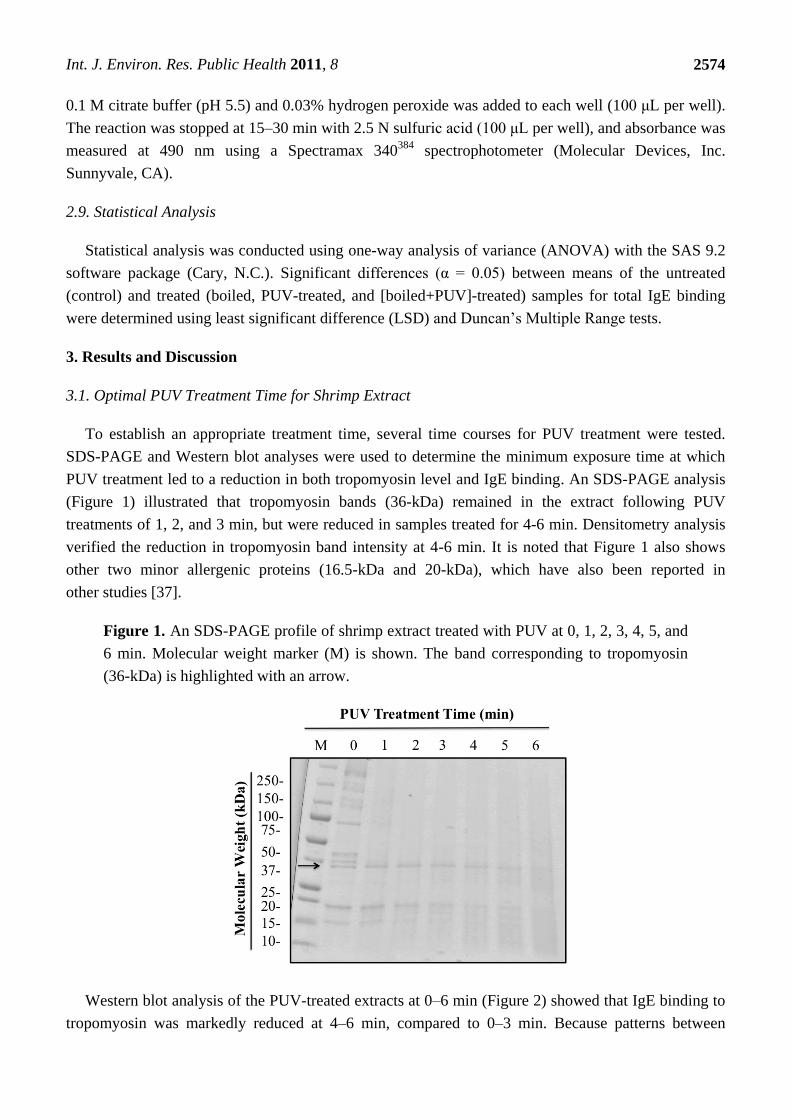
PUV treatment led to a reduction in both tropomyosin level and IgE binding. An SDS-PAGE analysis
(Figure 1) illustrated that tropomyosin bands (36-kDa) remained in the extract following PUV
treatments of 1, 2, and 3 min, but were reduced in samples treated for 4-6 min. Densitometry analysis
verified the reduction in tropomyosin band intensity at 4-6 min. It is noted that Figure 1 also shows
other two minor allergenic proteins (16.5-kDa and 20-kDa), which have also been reported in
other studies [37].
Figure 1. An SDS-PAGE profile of shrimp extract treated with PUV at 0, 1, 2, 3, 4, 5, and
6 min. Molecular weight marker (M) is shown. The band corresponding to tropomyosin
(36-kDa) is highlighted with an arrow.
Western blot analysis of the PUV-treated extracts at 0–6 min (Figure 2) showed that IgE binding to
tropomyosin was markedly reduced at 4–6 min, compared to 0–3 min. Because patterns between

Int. J. Environ. Res. Public Health 2011, 8
2575
4 and 6 min were similar and a higher moisture loss was observed at 6 min, 4 min was chosen as the
optimal treatment time for subsequent experimentation.
Figure 2. Western blot analysis of shrimp extract samples treated with PUV at 0 (control),
1, 2, 3, 4, 5, and 6 min using pooled human plasma from 3 individuals containing IgE
antibodies against shrimp. Tropomyosin bands (36-kDa) are highlighted within a box.
3.2. Changes in Tropomyosin Band Intensity of Untreated, Boiled, PUV-Treated, and
[Boiled+PUV]-Treated Shrimp Extracts
As illustrated in the SDS-PAGE profile (Figure 3), a 36-kDa band representing tropomyosin was
present following treatments with boiling, PUV, and boiling+PUV. A decrease in tropomyosin was
observed in both PUV- and [boiled+PUV]-treated samples (lanes 3 and 4), whereas the boiled extract
(lane 2) did not show a change in tropomyosin density compared to the control. These results were
verified using densitometry analysis.
Figure 3. An SDS-PAGE profile of untreated (1), boiled (2), PUV-treated (3), and
[boiled+PUV]-treated (4) shrimp extracts. Molecular weight marker is shown (M). An
arrow highlights the bands corresponding to tropomyosin (36-kDa).
Int. J. Environ. Res. Public Health 2011, 8
2576
Like Figure 1, Figure 3 also shows the behavior of other two allergenic proteins (16.5-kDa and
20-kDa) under boiling and PUV treatments. The 20-kDa protein followed a similar trend to
tropomyosin’s in terms of boiling and PUV effects, while boiling alone did not change much the
16.5-kDa protein, but PUV caused it to be almost undetectable as illustrated on lanes 3 and 4.
Resistance to thermal denaturation or degradation is characteristic of the tropomyosin protein, as it
is recognized for its heat stability [3]. However, instantaneous pulses of energy generated during
prolonged PUV treatment may cause more intense localized heating [38,39], which may contribute to
the reduction of tropomyosin. On the other hand, UV exposure, which is basically nonthermal, can
also cause protein crosslinking or fragmentation [27,29]. So, we believe both the nonthermal and
photothermal effects of PUV played a role in reducing the allergenic reactivity of shrimp proteins in
this study.
3.3. IgE- and IgG-binding to Untreated, Boiled, PUV-treated, and [Boiled+PUV]-Treated
Shrimp Extracts
3.3.1. Western Blot
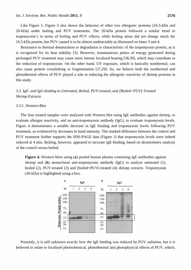
The four treated samples were analyzed with Western blot using IgE antibodies against shrimp, to
evaluate allergen reactivity, and an anti-tropomyosin antibody (IgG), to evaluate tropomyosin levels.
Figure 4 demonstrates a notable decrease in IgE binding and tropomyosin levels following PUV
treatment, as evidenced by decreases in band intensity. The marked difference between the control and
PUV treatment further supports the SDS-PAGE data (Figure 1) that tropomyosin levels were indeed
reduced at 4 min. Boiling, however, appeared to increase IgE binding, based on densitometry analysis
of the control versus boiled.
Figure 4. Western blots using (a) pooled human plasma containing IgE antibodies against
shrimp and (b) monoclonal anti-tropomyosin antibody (IgG) to analyze untreated (1),
boiled (2), PUV-treated (3) and [boiled+PUV]-treated (4) shrimp extracts. Tropomyosin
(36-kDa) is highlighted using a box.
Presently, it is still unknown exactly how the IgE binding was reduced by PUV radiation, but it is
believed to relate to localized photochemical, photothermal and photophysical effects of PUV, which,
Int. J. Environ. Res. Public Health 2011, 8
2577
as mentioned earlier, can cause protein modifications, including protein fragmentation, denaturation,
or crosslinking and affect IgE binding. Fragmented protein sections that are smaller than the resolution
limit of the gel may travel through the acrylamide pores more quickly, causing them to ultimately be
lost into the chamber buffer. Conversely, proteins that have been modified by crosslinking are often
too large to migrate through the gel or migrate with slight difficulty, thereby resulting in a smeared
appearance in SDS-PAGE and Western blots. Chung and others [40] have demonstrated the presence
of protein smears due to the crosslinking of peanut allergens caused by enzymatic reactions. Also, protein
band smearing has been observed in the allergens of peanuts that have been heated or roasted [41]. While
band smearing was not detected in the blot with IgE (Figure 4a), they can be seen in blots using an
anti-tropomyosin antibody. As shown in Figure 4b, tropomyosin bands and others were notably smeared
in PUV-treated samples, but not in boiled and control samples. This finding indicates that modification
or crosslinking of tropomyosin might have occurred during the PUV treatment.
Additionally, Taheri-Kafrani and others [42] have illustrated the smearing of milk allergens that have
been modified via the Maillard reaction, which is a non-enzymatic browning or protein-carbohydrate
reaction. Several studies [42-44] have linked glycation or Maillard reaction to the modification of
allergens and their subsequent changes in IgE binding. A study on squid tropomyosin [44] found that IgE
binding was suppressed with the progression of the Maillard reaction. However, in one study using
scallop tropomyosin [43], an increase in the allergen potency was displayed with the progression of
Maillard reaction. Whether Maillard reaction could occur in the shrimp extract during PUV treatment
is not known. Considering the high energy produced by PUV and the instantaneous heat absorbed by
the molecules [39] (i.e., proteins/carbohydrate in the shrimp extracts), it is possible that a Maillard
reaction may occur, but such a postulation needs to be further investigated.
3.3.2. Dot Blot
Dot blot analysis was performed to determine the allergen reactivity of the shrimp protein extract as
a whole–that is, tropomyosin and other possible allergens that were not detected via Western blot
analysis. IgE binding to whole shrimp extract was greatly reduced following PUV treatment (Figure 5).
However, there was an increase in IgE binding following boiling treatment. Interestingly, PUV
appeared to attenuate or negate the boiling effect, as shown in the [boiled+PUV]-treated sample; IgE
binding was notably reduced compared to the boiled-only sample. Of all the treatments, PUV alone
displayed the most reduction in IgE binding.
The finding that boiling or heating can lead to an increase in IgE binding (Figure 5) is not unusual,
because the effects of thermal processing on food allergens have been studied extensively, and, in
different studies, heating has been shown to either decrease or increase allergen potency. For example,
one study [41] described a 90-fold increase in IgE binding of roasted peanuts over raw peanuts,
whereas another study reported that roasting actually decreased the overall allergen reactivity of
hazelnuts [45]. It has also been noted that children with milk allergies show a tolerance to extensively
heated milk [46]. In this dot blot analysis (Figure 5), boiling caused an increase in IgE binding
possibly not just due to tropomyosin itself, but also due to other proteins present in the whole extract.
These proteins could include arginine kinase (40-kDa) [8,9] and myosin light chain (20-kDa) [7,38]
which have both been shown to play a minor role in shrimp allergy. Also, the minor 16.5 kDa protein
was also persistent during the boiling treatment (Figure 3). The effects of boiling on shrimp reactivity

Int. J. Environ. Res. Public Health 2011, 8
2578
are consistent with the results of Carnes and others [32] who also found that boiled shrimp extracts
were more immunoreactive in both in vivo skin prick trials and in vitro direct ELISA results.
Figure 5. Dot blot analysis of untreated, boiled, PUV-treated, and [boiled+PUV]-treated
shrimp extract using pooled human plasma containing IgE antibodies against shrimp.
3.3.3. Indirect ELISA
To support the dot blot data in Figure 4, IgE binding of the four treated shrimp extracts was also
determined using an indirect ELISA. Again, a significant decrease (α = 0.05) in IgE binding was seen
in the PUV-treated extract, compared to the control (Figure 6). An increase in IgE binding was
observed in the boiled extract while there was no significant change (α = 0.05) in IgE binding of the
[boiled+PUV]-treated extract, as compared to the control. The finding was in agreement with the data
of dot blot (Figure 4).
Figure 6. Indirect ELISA illustrating changes in IgE binding compared to untreated, boiled,
PUV-treated, and [boiled+PUV]-treated shrimp extracts using pooled human plasma
containing IgE antibodies against shrimp. A = absorbance of the sample; A0 = absorbance
of untreated sample. Data are expressed as mean ±SEM (n = 5). Results are relative values,
normalized to the untreated sample; untreated is standardized and set to 1. Values that are
significantly different (α = 0.05) from the untreated sample are annotated as **.
Int. J. Environ. Res. Public Health 2011, 8
2579
Reduction in IgE binding of PUV-treated extract was likely explained by changes in the amount of
detectable tropomyosin described above in SDS-PAGE (Figures 1 and 3) and Western blot analysis
(Figures 2 and 4). In the [boiled+PUV]-treated sample, IgE binding did not appear to change, compared
to the control. This was because tropomyosin increased after boilng but was offset to the control level
after the PUV treatment. That is to say, the increase and decrease in IgE binding, respectively, due to
boiling and PUV treatment, may negate each other, thus resulting in a negligible change in IgE binding
of the [boiled+PUV] extract. Such an effect may be deemed as an antagonistic effect.
3.4. Temperature and Volume Changes Following Treatment of Shrimp Extract
As mentioned earlier, PUV is considered a nonthermal method when used for brief periods of time
(several seconds); however, following a PUV treatment period of 4 min, a sample surface temperature
of 68.3 ± 2.5 C was detected using an infrared thermometer immediately after the PUV pulses
stopped and the treatment chamber door was opened. It must be noted that there was a 5–10 s delay in
probing the sample surface temperature, while the instantaneous temperatures of the sample during the
PUV treatment could be higher. Previous trials using thermocouples to monitor temperature during
PUV treatment were unsuccessful, because extended PUV exposure of the metal probe confounded the
readings. Fiber optic temperature sensing may be a way to go for recording accurately the sample
temperature during PUV treatment, which was not conducted in this study. For shorter PUV treatments,
a type-K thermocouple produced by Omega Engineering, Inc. (Stanford, CT), has been utilized [47]
for temperature measurement without confounding a complication in temperature reading. A study that
analyzed PUV treatment for decontamination of shell-eggs [47] reported an increased surface
temperature of 10.5 ± 1.2 C after 30 s at 9.5 cm distance from the quartz window of the PUV lamp.
Following boiling, PUV, and [boiling+PUV] treatments, moisture loss in each sample was
measured: boiled-only (5.83 ± 2.3%), PUV-only (29 ± 3.6%), and boiled with PUV (39.7 ± 2.5%).
Moisture loss was higher in the PUV-treated samples because the samples were not enclosed during
PUV treatment (the purpose was to ensure the maximum absorption of the PUV radiation). By contrast,
moisture loss was minimal in the boiled sample, because the samples were loosely capped during
boiling. Chung and others [17] also noted volume reductions of approximately 40% following PUV
treatment. To correct for moisture loss in the samples, protein measurements were taken after
treatments, and these values were used for subsequent experiments.
Excessive moisture loss is indicative of a sample temperature during the PUV treatment that was
well above the boiling point of water at atmosphere, which caused the water to evaporate. The
significant temperature increases due to the photothermal effect of PUV and the capability of PUV in
mitigating allergens may potentially be used in conjunction with food preparation to simultaneously
heat the food and reduce its allergenic potency. Li [23] took advantage of this unique feature of PUV
radiation to desirably roast the whole almond after 4–7 min exposure and yet considerably reduce its
IgE biding capacity. This can also be a potential method to cook the peeled whole shrimp and
significantly reduce its allergen, although this idea was not tested yet in this study.

Int. J. Environ. Res. Public Health 2011, 8
2580
4. Conclusions
A marked decrease in IgE binding of the shrimp extract following PUV treatment has been
demonstrated. The decrease was likely due to a reduction in the detectable level of tropomyosin in the
PUV-treated extract as shown in SDS-PAGE (Figure 1) and Western blot (Figure 2). Furthermore, the
appearance of protein band smearing, as illustrated in Figure 4, is likely due to the modification, such as
crosslinking, of tropomyosin and may contribute to the decrease in IgE binding. Boiling increased IgE
binding to whole shrimp sample; however, the effect of boiling was offset when it was combined with
PUV treatment. Overall, PUV was found to be capable of reducing the allergenic potency of shrimp
extracts. Further optimization is still needed before the PUV technology can be adopted. In vivo studies
are also needed to verify the reduction in allergenic potency of the PUV-treated shrimp extracts.
Acknowledgments
The authors gratefully acknowledge Melanie Correll, University of Florida, Department of
Agricultural and Biological Engineering, for her intellectual input concerning this manuscript.
References
1. Ellman, L.K.; Chatchatee, P.; Sicherer, S.H.; Sampson, H.A. Food hypersensitivity in two groups
of children and young adults with atopic dermatitis evaluated a decade apart. Pediatr. Allergy.
Immunol. 2002, 13, 295-298.
2. Sicherer, S.H.; Sampson, H.A. Food allergy. J. Allergy. Clin. Immunol. 2010, 125, S116-125.
3. Shanti, K.N.; Martin, B.M.; Nagpal, S.; Metcalfe, D.D.; Rao, P.V., Identification of tropomyosin
as the major shrimp allergen and characterization of its IgE-binding epitopes. J. Immunol. 1993,
151, 5354-5363.
4. Jeoung, B.J.; Reese, G.; Hauck, P.; Oliver, J.B.; Daul, C.B.; Lehrer, S.B. Quantification of the
major brown shrimp allergen Pen a 1 (tropomyosin) by a monoclonal antibody-based sandwich
ELISA. J. Allergy. Clin. Immunol. 1997, 100, 229-234.
5. Reese, G.; Ayuso, R.; Lehrer, S.B. Tropomyosin: an invertebrate pan-allergen. Int. Arch. Allergy.
Immunol. 1999, 119, 247-258.
6. Jeong, K.Y.; Hong, C.S.; Yong, T.S. Allergenic tropomyosins and their cross-reactivities. Protein
Pept. Lett. 2006, 13, 835-845.
7. Ayuso, R.; Grishina, G.; Bardina, L.; Carrillo, T.; Blanco, C.; Ibanez, M.D.; Sampson, H.A.;
Beyer, K. Myosin light chain is a novel shrimp allergen, Lit v 3. J. Allergy. Clin. Immunol. 2008,
122, 795-802.
8. Yu, C.J.; Lin, Y.F.; Chiang, B.L.; Chow, L.P. Proteomics and immunological analysis of a novel
shrimp allergen, Pen m 2. J. Immunol. 2003, 170, 445-453.
9. Garcia-Orozco, K.D.; Aispuro-Hernandez, E.; Yepiz-Plascencia, G.; Calderon-de-la-Barca, A.M.;
Sotelo-Mundo, R.R. Molecular characterization of arginine kinase, an allergen from the shrimp
Litopenaeus vannamei. Int. Arch. Allergy. Immunol. 2007, 144, 23-28.

Int. J. Environ. Res. Public Health 2011, 8
2581
10. Shiomi, K.; Sato, Y.; Hamamoto, S.; Mita, H.; Shimakura, K. Sarcoplasmic calcium-binding
protein: identification as a new allergen of the black tiger shrimp Penaeus monodon. Int. Arch.
Allergy. Immunol. 2008, 146, 91-98.
11. Daul, C.B.; Slattery, M.; Reese, G.; Lehrer, S.B. Identification of the major brown shrimp
(Penaeus aztecus) allergen as the muscle protein tropomyosin. Int. Arch. Allergy. Immunol. 1994,
105, 49-55.
12. Skripak, J.M.; Sampson, H.A. Towards a cure for food allergy. Curr. Opin. Immunol. 2008, 20,
690-696.
13. Tanabe, S. Epitope Peptides and Immunotherapy. Curr. Protein Pept. Sci. 2007, 8, 109-118.
14. Li, Z.; Lin, H.; Cao, L.M.; Jameel, K. Effect of high intensity ultrasound on the allergenicity of
shrimp. J. Zhejiang Univ. Sci. B 2006, 7, 251-256.
15. Zhenxing, L.; Hong, L.; Limin, C.; Jamil, K. The influence of gamma irradiation on the
allergenicity of shrimp (Penaeus vannamei). J. Food Eng. 2007, 79, 945-949.
16. Hildebrandt, S.; Schutte, L.; Stoyanov, S.; Hammer, G.; Steinhart, H.; Paschke, A. In vitro
determination of the allergenic potential of egg white in processed meat. J. Allergy. (Cairo) 2010,
2, 155-165.
17. Chung, S.Y.; Yang, W.; Krishnamurthy, K., Effects of pulsed UV-light on peanut allergens in
extracts and liquid peanut butter. J. Food Sci. 2008, 73, C400-404.
18. Yang, W.; Mwakatage, N.R.; Goodrich-Schneider, R.; Krishnamurthy, K.; Rababah, T.M.
Mitigation of major peanut allergens by pulsed ultraviolet light. Food Bioprocess Technol. 2011,
DOI 10.1007/s11947-011-0615-6.
19. Yang, W.W.; Chung, S.Y.; Ajayi, O.; Krishnamurthy, K.; Konan, K.; Goodrich-Schneider, R.,
Use of pulsed ultraviolet light to reduce the allergenic potency of soybean extracts. J. Food Eng.
2010, 6, 1-12.
20. Shriver, S.K.; Yang, W. Thermal and nonthermal methods for allergen control. Food Eng. Rev.
2011, 3, 26-43.
21. Krishnamurthy, K.; Demirci, A.; Irudayaraj, J. M. Inactivation of Staphylococcus aureus in milk
using flow-through pulsed UV-light treatment system. J. Food Sci. 2007, 72, M233-239.
22. Wakatage, N.R. Efficacy of Pulsed UV Light Treatment on Removal of Peanut Allergens. M.S.
Thesis, Department of Food and Animal Sciences, Alabama A&M University, Normal, AL, USA,
2008.
23. Li, Y. Effect of Pulsed Ultraviolet Light, High Hydrostatic Pressure and Non-thermal Plasma on
the Antigenicity of Almond. M.S. Thesis, Department of Agricultural and Biological Engineering,
University of Florida, Gainesville, FL, USA, 2011.
24. Nooji, J. Reduction of Wheat Allergen Potency by Pulsed Ultraviolet Light, High Hydrostactic
Pressure and Nonthermal Plasma. M.S. Thesis, Department of Food Science and Human
Nutrition, University of Florida, Gainesville, FL, USA, 2011.
25. Shriver, S.K. Effect of Selected Emerging Nonthermal Processing Methods on the Allergen
Reactivity of ATLANTIC White Shrimp (Litopenaeus Setiferus). M.S. Thesis, Department of Food
Science and Human Nutrition, University of Florida, Gainesville, FL, USA, 2011.
26. Fiedorowicz, M.; Tomasik, P.; Lii, C.Y. Degradation of starch by polarised light. Carbohydr.
Polymer. 2001, 45, 79-87.

Int. J. Environ. Res. Public Health 2011, 8
2582
27. Greenberg, J.R. Ultraviolet light-induced crosslinking of mRNA to proteins. Nucleic Acids Res.
1979, 6, 715-32.
28. Kramer, G.F.; Norman, H.A.; Krizek, D.T.; Mirecki, R.M. Influence of UV-B radiation on
polyamines, lipid peroxidation and membrane lipids in cucumber. Phytochemistry 1991, 30,
2101-2108.
29. Friso, G.; Barbato, R.; Giacometti, G.M.; Barber, J. Degradation of D2 protein due to UV-B
irradiation of the reaction centre of photosystem II. FEBS Letters 1994, 339, 217-221.
30. Motoyama, K.; Suma, Y.; Ishizaki, S.; Nagashima, Y.; Lu, Y.; Ushio, H.; Shiomi, K.
Identification of tropomyosins as major allergens in Antarctic krill and mantis shrimp and their
amino acid sequence characteristics. Mar. Biotechnol. 2008, 10, 709-718.
31. Motoyama, K.; Suma, Y.; Ishizaki, S.; Nagashima, Y.; Shiomi, K. Molecular cloning of
tropomyosins identified as allergens in six species of crustaceans. J. Agric. Food Chem. 2007, 55,
985-991.32.Carnes, J.; Ferrer, A.; Huertas, A.J.; Andreu, C.; Larramendi, C.H.; Fernandez-Caldas,
E. The use of raw or boiled crustacean extracts for the diagnosis of seafood allergic individuals.
Ann. Allergy Asthma Immunol. 2007, 98, 349-354.
32. Carnes, J.; Ferrer, A.; Huertas, A.J.; Andreu, C.; Larramendi, C.H.; Fernandez-Caldas, E. The use
of raw or boiled crustacean extracts for the diagnosis of seafood allergic individuals. Ann. Allergy
Asthma Immunol. 2007, 98, 349-354.
33. Daul, C.B.; Morgan, J.E.; Hughes, J.; Lehrer, S.B. Provocation-challenge studies in
shrimp-sensitive individuals. J. Allerg. Clin. Immunol. 1988, 81, 1180-1186.
34. Naqpal, S.; Rajappa, L.; Metcalfe, D.D.; Rao, P.V. Isolation and characterization of heat-stable
allergens from shrimp (Penaeus indicus). J. Allerg. Clin. Immunol. 1989, 83, 26-36.
35. Liu, G.-M.; Cheng, H.; Nesbit, J.B.; Su, W.-J.; Gao, M.-J.; Maleki, S.J. Effects of Boiling on the
IgE-Binding Properties of Tropomyosin of Shrimp (Litopenaeus vannamei). J. Food. Sci. 2010,
75, T1-T5.
36. Laemmli, U. K. Cleavage of structural proteins during the assembly of the head of
bacteriophage T4. Nature 1970, 227, 680-685.
37. Samson, K.T.R; Chen, F.H.; Miura, K.; Odajima, Y.; Likura, Y.; Rivas, M.N.; Minoguchi, K.;
Adachi, M. IgE Binding to Raw and Boiled Shrimp Proteins in Atopic and Nonatopic Patients
with Adverse Reactions to Shrimp. Int. Arch. Allergy. Immunol. 2004, 133, 225-232.
38. Muranov, K.; Maloletkina, O.; Poliansky, N.; Kleymenov, S.; Rozhkov, S.; Goryunov, A.;
Ostrovsky, M.; Kurganov, B. Mechanism of aggregation of UV-irradiated βL–crystallin. Exper.
Eye. Res. 2011, 92, 76-86.
39. Gomez-Lopez, V.; Ragaert, P.; Debevere, J.; Devlieghere, F. Pulsed light for food
decontamination: a review. Trends Food Sci. Technol. 2007, 18, 464-473.
40. Chung, S.-Y.; Kato, Y.; Champagne, E.T. Polyphenol oxidase/caffeic acid may reduce the
allergenic properties of peanut allergens. J. Sci. Food Agr. 2005, 2631-2637.
41. Maleki, S.J.; Chung, S.Y.; Champagne, E.T.; Raufman, J.P. The effects of roasting on the
allergenic properties of peanut proteins. J. Allergy. Clin. Immunol. 2000, 106, 763-768.

Int. J. Environ. Res. Public Health 2011, 8
2583
42. Taheri-Kafrani, A.; Gaudin, J.C.; Rabesona, H.; Nioi, C.; Agarwal, D.; Drouet, M.; Chobert, J.M.;
Bordbar, A.K.; Haertlft, T. Effects of heating and glycation of beta-lactoglobulin on its
recognition by IgE of sera from cow milk allergy patients. J. Agr. Food. Chem. 2009, 57,
4974-4982.
43. Nakamura, A.; Watanabe, K.; Ojima, T.; Ahn, D.H.; Saeki, H. Effect of maillard reaction on
allergenicity of scallop tropomyosin. J. Agr. Food. Chem. 2005, 53, 7559-7564.
44. Nakamura, A.; Sasaki, F.; Watanabe, K.; Ojima, T.; Ahn, D.H.; Saeki, H. Changes in allergenicity
and digestibility of squid tropomyosin during the Maillard reaction with ribose. J. Agr. Food.
Chem. 2006, 54, 9529-9534.
45. Hansen, K.S.; Ballmer-Weber, B.K.; Luttkopf, D.; Skov, P.S.; Wuthrich, B.; Bindslev-Jensen, C.;
Vieths, S.; Poulsen, L.K. Roasted hazelnuts--allergenic activity evaluated by double-blind,
placebo-controlled food challenge. Allergy 2003, 58, 132-138.
46. Nowak-Wegrzyn, A.; Bloom, K.A.; Sicherer, S.H.; Shreffler, W.G.; Noone, S.; Wanich, N.;
Sampson, H.A. Tolerance to extensively heated milk in children with cow's milk allergy.
J. Allergy Clin. Immunol .2008, 122, 342-347.
47. Keklik, N. M.; Demirci, A.; Puri, V. M. Inactivation of Listeria monocytogenes on unpackaged
and vacuum-packaged chicken frankfurters using pulsed UV-light. J. Food Sci. 2009, 74,
M431-M439.
© 2011 by the authors; licensee MDPI, Basel, Switzerland. This article is an open access article
distributed under the terms and conditions of the Creative Commons Attribution license
(http://creativecommons.org/licenses/by/3.0/).
Recommended