
1
Pulmonary Physiology
Pulmonary Physiology
• Control of Breathing• Mechanics/Work of Breathing• Ventilation• Gas transport (including pulmonary
circulation)• Gas Exchange (including diffusion of
gas/gas transfer)
2
• “When you can’t breathe, nothing else tt ”matters.”
Control of Breathing
• Keep PCO2 40 mmHg awake• Neural Control• Chemical Control
3
Neural Control
• Inspiratory inhibition reflex (Hering B ) i it t h j tBreuer); irritant, mechano, j receptors: stimulation in patients with, e.g., interstitial fibrosis, pulmonary embolism, atelectasis
• Stimulation of mechanoreceptors in airways: can cause tachypnea,airways: can cause tachypnea, bronchoconstriction
Chemical control
• CO2 stimulation • Hypoxemic stimulation• H+ stimulation

4
Chemical Control: CO2 stimulation
• Rise in PaCO2 = increase in [H+] concentration in ECF and ventrolateral surface of the medulla, stimulating gventilation (hyperventilation)
• In turn, dilation of CNS blood vessels by increased CO2leads to increased removal of CO2 and decrease in central CO2 stimulation; also, the hyperventilation results in lower CO2 to stimulate. Also, lower CO2 = brain vessel constriction =buildup in ECF CO2 again
• Chronically elevated PaCO2 = increased ECF [HCO3-] = elevated pH and [H+] so acute increase in PaCO willelevated pH and [H+] so acute increase in PaCO2 will induce less of a change in [H+] and therefore less stimulus to ventilation
Chemical Control: Hypoxemic Stimulation
• Peripheral chemoreceptors (carotid body, aorta) respond primarily to low PaO2; also can respond to PCO2 and [H+] as well as decreased blood flow to CO2 a d [ ] as e as dec eased b ood oand increased temperature
• Sleep depresses ventilatory stimulation; PaCO2 rises by several mmHg in sleep (most in REM sleep)
• Low PaO2=increased VE,then decreased arterial and CNS CO2 and [H+] = less central stimulus to 2 breathe, [HCO3-] reduced but [H+] restored in several days, and thus unopposed stimulus to breathe by low PO2 remains

5
Chemical Control: Hydrogen ion stimulation
• Metabolic acidosis stimulates; alkalosis inhibits breathing primarily through peripheral chemoreceptors, ~7.30 to 7 507.50
• Acute metabolic acidemia = increased ventilation; then decreased PaCO2 and CNS PCO2, decreased [H+], decreased [HCO3-], and after 24 hours normalization of CNS [H+].
• So, chronically low [HCO3-] = easier to stimulate by CO2. Conversely, met alkalosis = low VE; eventual increase in CNS [HCO3-] = more difficult to stimulate by CO2 to [ ] y 2induce a rise in [H+]
• Note: when WOB elevated, PCO2 not as potent a stimulus to breathe
Mechanics of breathing
• Total mechanical work of breathing=overcoming elastic resistive work+ flow resistive work; inelastic-resistive work+ flow-resistive work; in normal individual this applies to INSPIRATION.
• Severe airway obstruction:, may need expiratory work to overcome EXPIRATORY flow resistance
• Asthma= normal elastic resistance, high flow resistance; pulmonary fibrosis/stiff lungs p y g(ARDS)= normal flow resistance, high elastic resistance and need for work to overcome this.

6
Mechanics of breathing
• Elastic forces: recoil of lungs and recoil of chest wall =equilibrium at FRC (functional residual capacity)El t Δ P/ Δ V thi i th di t ibilit f th• Elastance= Δ P/ Δ V; this is the distensibility of the respiratory system (lungs, chest wall)
• Lung volume dependent• Compliance= Δ V/ Δ P • Healthy: Lung compliance~0.2 L/cmH20: eg, change
inspiratory pressure 5 cmH20, 1.0 L air is inspired,=1 L/5 cmH20=0.2 L/cmH20
Mechanics of breathing
• Emphysema: increased compliance due to loss of elastic recoil pressure: e g change inof elastic recoil pressure: e.g., change in inspiratory pressure 5 cmH20.0 L air, or 0.4 L/cmH20 compliance
• (Sounds like a good thing for inspiration, but less efficient expiration..)
• Pulmonary fibrosis: increased elastic recoil pressure (stiff lungs): 5 cmH20 inspiratorypressure (stiff lungs): 5 cmH20 inspiratory pressure change with 0.5 L air inspired; compliance=0.10 L/cmH20

7
Mechanics of Breathing
• Transpulmonary pressure: difference between pleural pressure (usually measured as esophageal pressure) and mouth pressure static=no airflowp
• Static compliance: relationship of transpulmonary pressure under static conditions (no airflow) to different degrees of lung inflation (volumes)
• Static inspiratory compliance=VT/Pplateau-PEEP• Volume vs pressure curve shifted up and to the left in
patient with chronic airflow limitation (eg emphysema); shifted down and to the right in obesity and pulmonary fib ifibrosis
Mechanics of Breathing
• Dynamic compliance: compliance determined during breathing
• Dynamic compliance=V /Pdynamic-PEEP• Dynamic compliance=VT/Pdynamic-PEEP
8
Mechanics of Breathing
• Inspiratory airway resistance=pressure difference across airways between mouth and alveoli=Pdynamic-P l t /fl ( l 4 H20/L/ )Pplateau/flow (normal=<4 cmH20/L/sec)
• Maximal inspiratory flow rate depends primarily upon muscular effort
• Expiration: higher volumes=higher flow rates, but once ~50% TLC, rate declines with greater effort because of dynamic airway compression
• Dynamic airway compression=more collapse of airways i i ti i h (l f l t /i din expiration in emphysema (loss of elastance/increased compliance) as effort increases=gas trapping
Mechanics/Work of Breathing
• Note that low and high respiratory rates cause increased mechanical work of breathing: Hi h t l l l d t i t t l• High rates=low lung volumes=need to increase total ventilation so that alveolar ventilation is maintained, since there is increased wasted (dead space) ventilation, so increased work to overcome flow resistance
• Slow rates=little flow resistive work because of low flow rates but must increase VT to maintain alveolar ventilation; thus must use increased work to overcome elastic resistanceelastic resistance

9
Mechanics/Work of Breathing
• Elastic resistance high (low compliance) = increased respiratory frequency; VT usually low, so rapid, shallow breathing=least workshallow breathing=least work
• eg, pulmonary fibrosis = low lung compliance; obesity, kyphoscoliosis = low chest wall compliance
• Flow resistance is high= decreased respiratory frequency ; generally deeper and slower breathing (eg, chronic airflow limitation)
• Note, however: with lung hyperinflation, volume , g yp ,pressure curve changes with decreasing compliance and the patient may breathe more rapidly and shallowly as well
Mechanics/Work of Breathing
• Metabolic work:• Oxygen consumption (VO2) in normals~1.0
ml/liter of ventilation; O2 cost of breathing increases in patients with respiratory disorders due to the increased work required

10
Ventilation
• PACO2 = VCO2/VA x K (the constant isPACO2 VCO2/VA x K (the constant is actually 863 mmHg, derived from ideal gas laws).
• The ratio of VCO2/VA for normal people at rest, at sea level, is about 1/21.6; thus,
l PACO 1/21 6 863 Hnormal PACO2 = 1/21.6 x 863 mmHg = ~40 mmHg.
Gas Transport: Pulmonary Circulation and Diffusion of Gas (Gas Transfer)
• Conduction of blood coming from the tissues through the alveolar capillaries so that O2 can be added and CO2
dremoved.• Pulmonary vessels=low pressures and low resistance to flow
(thin walled)• Resistance=driving pressure/flow (Q)• Most resistance in the arterioles and capillaries• Driving pressure=pressure at the beginning of the pulmonary
circulation (the pulmonary artery) and other end (left atrium); normally eg blood flow 6 L/min and mean driving pressure ofnormally, eg, blood flow 6 L/min and mean driving pressure of 9 mmHg, resistance is 9/6 or 1.5 mmHg/L/min (~10% of systemic pressure).

11
Gas Transport: Pulmonary Circulation and Diffusion of Gas (Gas Transfer)
• Pulmonary capillary blood volume increases during inspiration and exerciseduring inspiration and exercise
• Reduced when patients receive mechanical ventilation (intrathoracic pressure is raised, thus impeding venous return to the heart)
• Patients with increased pulmonary pressure (eg pulmonary hypertension, pulmonary
b li ) di d iembolism)=cardiodynamic consequences as well as disturbance of gas transfer
Gas Transport: Pulmonary Circulation and Diffusion of Gas (Gas Transfer)
• Transfer of O2 and CO2 between alveolar gas and pulmonary capillary blood is entirely passive, with the
t f diff i f l l ill b irate of diffusion of gas across alveolar-capillary barrier determined by (1) solubility of gas in liquid, (2) density of gas, (3) partial pressure difference between alveolar air and pulmonary capillary blood, and (4) surface area available for diffusion
• CO2 diffusion not a clinical problem because CO2 xmuch more soluble and diffusible than oxygen between air and bloodblood
• Total diffusing capacity includes uptake by hemoglobin and rate of flow

12
Gas Transport: Pulmonary Circulation and Diffusion of Gas (Gas Transfer)
• Low diffusion capacity not typically low PaO2, since so much redundancy:
• Complete exposure of alveolar PO2 to capillary blood=no decrease in end capillary PO2, even if there is less of it (low lung volume, low Hgb) and no change in AaDO2 (note that if less of it, lower O2CONTENT, not PaO2)
• But incomplete transfer = decrease in end capillary PO2 and widened AaDO2PO2 and widened AaDO2
“Diffusion Capacity” vs Diffusion
• Note that: decreased diffusing capacity/gas transfer abnormality can result from numerous abnormalities not having anything to do with diffusion block itselfhaving anything to do with diffusion block itself
• So when we say diffusion abnormality=cause of hypoxemia, we mean those abnormalities which involve some form of diffusion block, or other inability to transfer gas completely (eg, low PIO2+ increased circulatory time) so that insufficient transfer of alveolar PO2 occur
• Low alveolar volume, low Hgb, may result in low diffusing capacity as measured by transfer of CO, and low O2 content, but not low PaO2

13
Gas Transport: CO2
• CO2 in physical solution: most carried in RBC ith bi b t b d tRBCs either as bicarbonate, or bound to Hgb (carbaminoHgb)
• Some is dissolved in plasma
Gas Transport:Oxygen
• O2 combined with Hgb in RBCs, and dissolved O2 in physical solution in the plasmap y p
• Normal: 1 gm of Hgb able to combine chemically with 1.34 ml O2
• Thus: O2 capacity=1.34 ml O2 /gmHgb• If 15 gm Hgb/100 ml blood, O2 capacity=20 ml O2 /100
ml blood=200 ml O2 /liter blood• Dissolved O2 = .003 ml O2 /100 ml blood/mmHg2 2• CaO2 =SaO2 x [O2 capacity + dissolved O2]/l/mmHg
PaO2• If PaO2 =100 mmHg, O2 content = 200 ml O2 /liter blood
+ 3 mlO2/liter blood=~203 mlO2/liter blood x SaO2

14
Hypoxemia
• Low partial pressure of O2 in blood (PaO2) OR l O t tOR low O2 content
Gas Transport: Pulmonary Circulation and Diffusion of Gas (Gas Transfer)
• Hypoxemia: hypoventilation, low PIO2, diffusion b lit ( t b if t t) V/Qabnormality (must be severe if at rest), V/Q
mismatch, shunt (note that shunt and diffusion block manifest similarly in corresponding areas of lung; diffusion abnormality (if not block) does NOT equal shunt)
• Note that low V/Q does not=shunt• O2 saturation=O2 content/O2 capacity x 100O2 satu at o O2 co te t/O2 capac ty 00• Degree of O2 saturation depends on O2 tension

15
Physiologic Causes of Hypoxemia
Alveolar Hypoventilation
Decreased PIO2
Diffusion Abnormality
V/Q mismatchV/Q mismatch
Shunt

16
Physiologic Causes of Hypoxemia
Widening of AaDO2:gDiffusion AbnormalityV/Q mismatchShunt
No widening of AaDO2:H til tiHypoventilation?Low PIO2 (may slightly widen if impaired diffusion
Gas Exchange
• Alveolar Gas Equation:• PAO2= FIO2x (PB-PH20) – PCO2/R +
[PACO2 x FIO2 x 1-R/R] (full)• PAO2= FIO2x (PB-PH20) – PCO2/R
(simplified)• R=Respiratory Exchange Ratio: (gas R=CO2• R=Respiratory Exchange Ratio: (gas R=CO2
added to alveolar gas by blood/amount of O2 removed from alveolar gas by blood; low V/Q=low R); normal=0.8

17
Abnormal Ventilation,Abnormal Gas Exchange
18
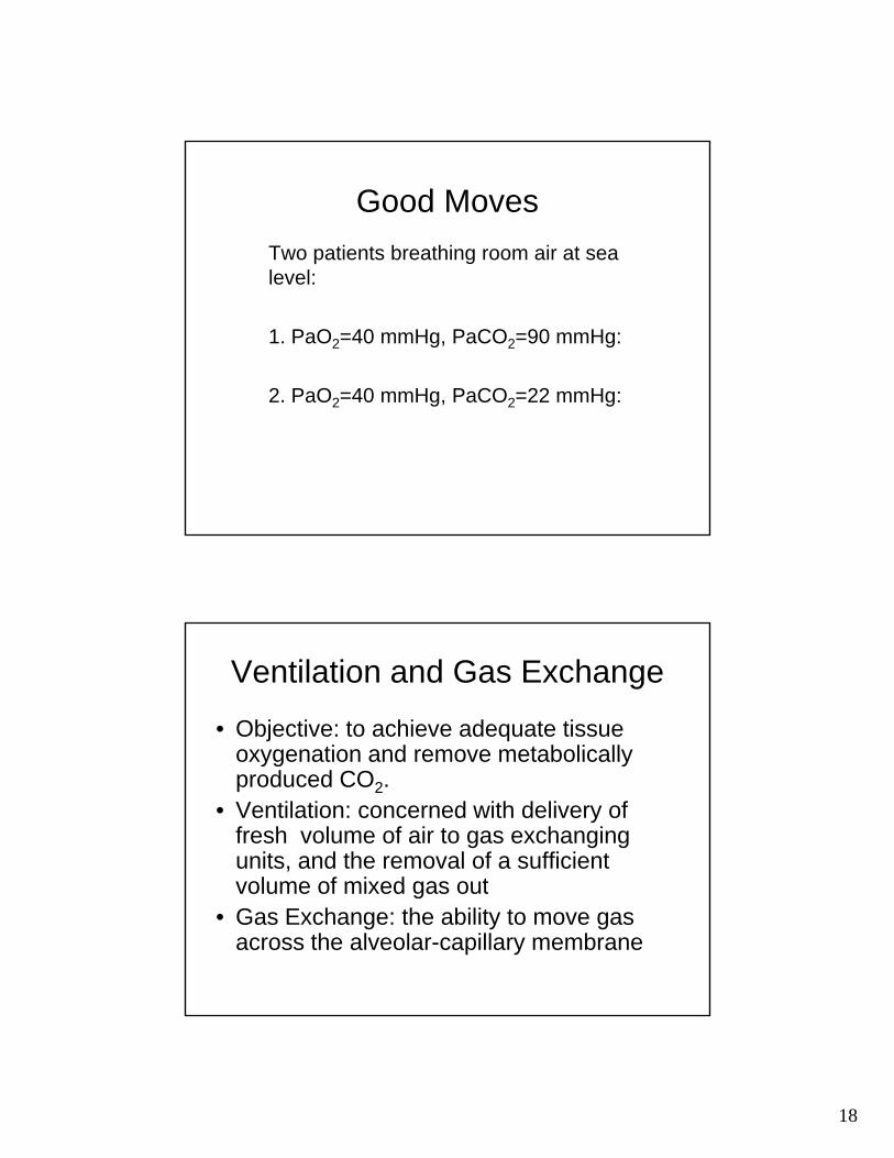
Good MovesTwo patients breathing room air at sea l llevel:
1. PaO2=40 mmHg, PaCO2=90 mmHg:
2. PaO2=40 mmHg, PaCO2=22 mmHg:2 g, 2 g
Ventilation and Gas Exchange
• Objective: to achieve adequate tissue oxygenation and remove metabolicallyoxygenation and remove metabolically produced CO2.
• Ventilation: concerned with delivery of fresh volume of air to gas exchanging units, and the removal of a sufficient volume of mixed gas outvolume of mixed gas out
• Gas Exchange: the ability to move gas across the alveolar-capillary membrane
19
Ventilation and Gas Exchange
• The failure of either or both results in i i d t i l bl d dimpaired arterial blood gases and ultimately respiratory failure.
• Ventilatory failure: Hypercapnic respiratory failure
• Gas exchange failure: Hypoxemic• Gas exchange failure: Hypoxemic respiratory failure
• Hypoxemia is the inevitable result of both
Hypoxemia
• Low partial pressure of O2 in blood (PaO2) OR l O t t (C O2 S O2 OOR low O2 content (CaO2:SaO2 x O2carrying capacity+.03 ml O2/l/mmHg PaO2)
20
Hypoxemia
• Hypoxemia is not synonymous with:
Hypoxemia
• Hypoxemia is not synonymous with:– Hypoxia (metabolic O2 deficiency; may be “stagnant”,Hypoxia (metabolic O2 deficiency; may be stagnant ,
“histocytoxic”, “hypoxic”, and “anemic”)

21
Hypoxemia
• Hypoxemia is not synonymous with:– Hypoxia (metabolic O2 deficiency; may be “stagnant”,Hypoxia (metabolic O2 deficiency; may be stagnant ,
“histocytoxic”, “hypoxic”, and “anemic”)– Low O2 carrying capacity (1.34 ml O2 /gm Hgb; if 15
gmHgb/100ml blood, then 20 ml O2 /100ml blood, or 200 ml O2 /liter of blood)
– Low O2 delivery (Ca O2 x C.O.)

22
Physiologic Causes of Hypoxemia
Alveolar Hypoventilation yp
Decreased PIO2
Diffusion Abnormality
V/Q i t hV/Q mismatch
Shunt
Ventilation• Minute Ventilation (VE)=tidal volume (VT) x
respiratory frequency (“dead space” volume notrespiratory frequency ( dead space volume not accounted for)
• Alveolar ventilation (VA)=that part of minute ventilation which participates in gas exchange (that volume of fresh gas entering the respiratory exchange zone each minute)Al l til ti l l l (tid l• Alveolar ventilation=alveolar volume (tidal volume-dead space volume) x respiratory frequency

23
Ventilation
• Alveolar PCO2 (PACO2)=VCO2/VA x KVCO2 CO2 prod ction• VCO2=CO2 production
• VA=alveolar ventilation • Normal: VCO2/VA=1/21.6; K=863 mmHg, so
PACO2=~40mmHg))• Alveolar PCO2=CO2 leaving lungs after gas
exchange; directly reflects arterial PCO2exchange; directly reflects arterial PCO2• e.g., halving alveolar ventilation with constant
CO2 production will double the alveolar PCO2• e.g., doubling the alveolar PCO2 reflects halved
alveolar ventilation
Hypoventilation
• Inability to inspire and expire a volume of air/gas sufficient to meet metabolicair/gas sufficient to meet metabolic demands
• Inabilty to bring a fresh volume of O2 with each breath to the gas exchanging unit, and inability to remove CO2 produced by metabolismmetabolism.
• Sine qua non: Increased arterial PCO2(PaCO2); decreased arterial PO2 (PaO2) breathing room air (parallel changes!!)
24
Hypoventilation/Alveolar hypoventilation
• All hypoventilation concerns either :i d d d /tid l l ti• increased dead space/tidal volume ratio (anatomic or physiologic), or
• Decreased MINUTE ventilation (decreased tidal volume, and/or decreased respiratory rate)
• Each may result in alveolar hypoventilation (PaCO2 elevated)
Alveolar Hypoventilation: 2 Clinical Pearls
• Does not widen the AaDO2
• The hypoxemia may be readily ameliorated with supplemental O2
• Challenge: Write a proof for this latter statement

25
Alveolar Gas Equation
• PAO2=PIO2 – PACO2/R2 2 2
• PAO2 =PIO2 – PACO2/R + [PCO2 x FIO2 x 1-R/R]
Alveolar Gas Equation
• PAO2=PIO2 – PACO2/R• PIO2: FIO2 (Patm-PH20)
26
Alveolar Gas Equation
• PAO2=PIO2 – PACO2/R• PIO2: FIO2 (Patm-PH20)• PACO2=PaCO2
Alveolar Gas Equation
• PAO2=PIO2 – PACO2/R• PIO2: FIO2 (Patm-PH20)• PACO2=PaCO2• R=Respiratory Exchange Ratio: (gas
R=CO2 added to alveolar gas by blood/amount of O2 removed fromblood/amount of O2 removed from alveolar gas by blood; low V/Q=low R); normal=0.8
27
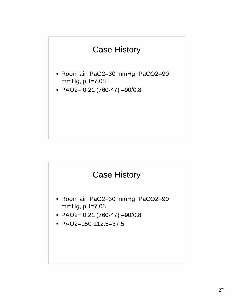
Case History
• Room air: PaO2=30 mmHg, PaCO2=90 mmHg, pH=7.08
• PAO2= 0.21 (760-47) –90/0.8
Case History
• Room air: PaO2=30 mmHg, PaCO2=90 mmHg, pH=7.08
• PAO2= 0.21 (760-47) –90/0.8• PAO2=150-112.5=37.5

28
Case History
• PaO2=30 mmHg, PaCO2=90 mmHg, pH=7.08
• PAO2= 0.21 (760-47) –90/0.8• PAO2=150-112.5=37.5• AaDO2=7.5 mmHg
Alveolar Hypoventilation
• CNS: central hypoventilation; infectious, t ti l d t d lltraumatic, vascular damage to medullary centers; pharmacologic and sleep suppression of ventilatory drive

29
Alveolar Hypoventilation
• CNS: central hypoventilation; infectious, t ti l d t d lltraumatic, vascular damage to medullary centers; pharmacologic and sleep suppression of ventilatory drive
• Peripheral nervous system/myoneural junction: poliomyelitis, Guillain-Barre,junction: poliomyelitis, Guillain Barre, myasthenia gravis
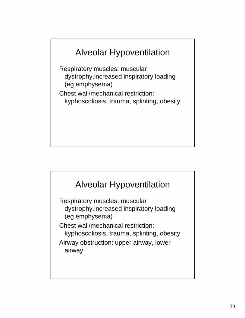
Alveolar Hypoventilation
Respiratory muscles: muscular dystrophy, ALS i d i i t l di (ALS, increased inspiratory loading (eg emphysema)
30
Alveolar Hypoventilation
Respiratory muscles: muscular d t h i d i i t l didystrophy,increased inspiratory loading (eg emphysema)
Chest wall/mechanical restriction: kyphoscoliosis, trauma, splinting, obesity
Alveolar Hypoventilation
Respiratory muscles: muscular d t h i d i i t l didystrophy,increased inspiratory loading (eg emphysema)
Chest wall/mechanical restriction: kyphoscoliosis, trauma, splinting, obesity
Airway obstruction: upper airway lowerAirway obstruction: upper airway, lower airway
31
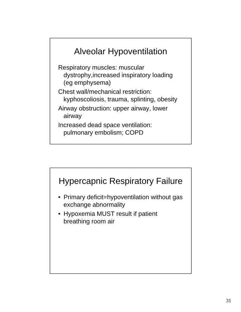
Alveolar Hypoventilation
Respiratory muscles: muscular d t h i d i i t l didystrophy,increased inspiratory loading (eg emphysema)
Chest wall/mechanical restriction: kyphoscoliosis, trauma, splinting, obesity
Airway obstruction: upper airway lowerAirway obstruction: upper airway, lower airway
Increased dead space ventilation: pulmonary embolism; COPD
Hypercapnic Respiratory Failure
• Primary deficit=hypoventilation without gas h b litexchange abnormality
• Hypoxemia MUST result if patient breathing room air

32
AaDO2 and Hypoxemia
• Widened in diffusion disorder, V/Q i t h d h tmismatch, and shunt
• Not widened in alveolar hypoventilation and decreased PIO2
• Normal 10-15 mmHg in young adult
Climbing Everest (Decreased PIO2)
• P atm= 250 mmHgP CO2 18 H R 1• PaCO2=18 mmHg; R=1
• PAO2=PIO2-PCO2/R• PAO2=.21 (250-47)-18/1=24.6 mmHg• Recent data: altitude 8400m, mean
PaO2=30 mmHg, Mean AaDO2 5.4PaO2 30 mmHg, Mean AaDO2 5.4 mmHg (wider than expected): Grocott et al, NEJM 360;2: 141
33
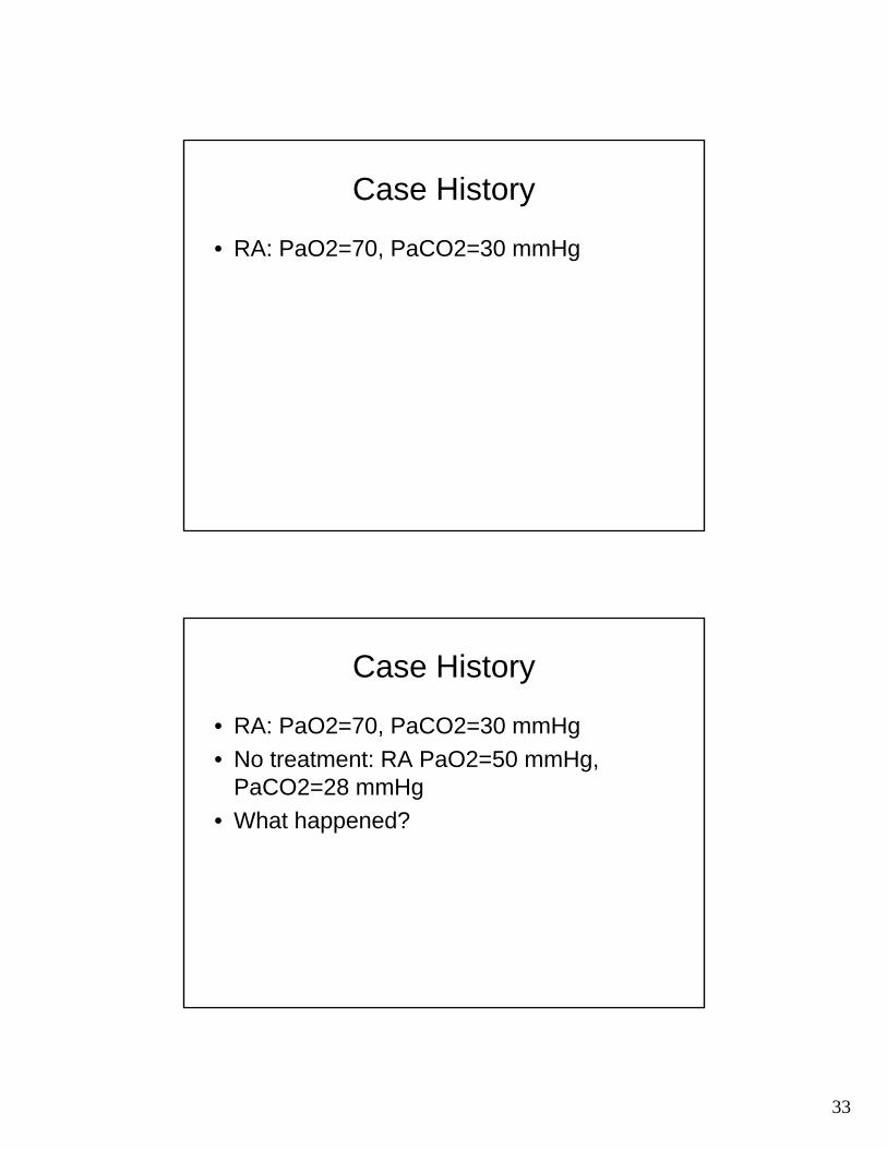
Case History
• RA: PaO2=70, PaCO2=30 mmHg
Case History
• RA: PaO2=70, PaCO2=30 mmHg• No treatment: RA PaO2=50 mmHg,
PaCO2=28 mmHg• What happened?

34
What happened?
• PAO2=PIO2 – PACO2/R• 0.21 FIO2, PaO2=50 mmHg,
PaCO2=28 mmHg• PAO2=0.21(713)-28/0.8=150-35=
115 mmHg115 mmHg• AaDO2=115-50= 65 mmHg
Hypoxemia
• No widening of AaDO2: hypoventilation, low PIO2low PIO2.
• Widened AaDO2: shunt, low V/Q, low diffusing capacity
• Hypoxemia of each may be overcome with supplemental O2 except: shunt.
• Note: no gas exchange=no amelioration of hypoxemia with O2, whether dead space, shunt, or no diffusion.

35
Low V/Q
• “Venous admixture”• Alveolar filling: pneumonia, pulmonary
edema (cardiogenic/non-cardiogenic)• COPD a common situation of low V/Q• Usually will involve some infinitely low V/Q
(shunt) and decreased diffusion(shunt) and decreased diffusion.
36
Low V/Q
• Low relationship of V to Q; NOT low til ti i ll l l ill itventilation in all alveolar capillary units
• That is, Low V/Q is NOT hypoventilation (unless all units are the same low V/Q)
Gas Transport: Pulmonary Circulation and Diffusion of Gas (Gas Transfer)
• Transfer of O2 between alveolar gas and pulmonary capillary blood is entirely passive, with the rate of diff i f l l ill b idiffusion of gas across alveolar-capillary barrier determined by (1) solubility of gas in liquid, (2) density of gas, (3) partial pressure difference between alveolar air and pulmonary capillary blood, and (4) surface area available for diffusion
• Total diffusing capacity includes uptake by hemoglobin and rate of flowg
37
Diffusion Abnormality
• Alveolar-capillary membrane thickening (pulmonary hypertension pulmonary(pulmonary hypertension, pulmonary vasculitis, pulmonary embolism)
• Alveolar-capillary membrane destruction (emphysema)
• Pulmonary interstitial thickening ( l fib i )(pulmonary fibrosis)
• Alveolar filling (pulmonary edema, pneumonitis)
Gas Transport: Pulmonary Circulation and Diffusion of Gas (Gas Transfer)
• Low gas transfer may also result from processes not clearly blocking diffusion, such as low Hgb, or increased rate of flow disallowing adequate gas transferdisallowing adequate gas transfer
• All diffusion abnormalities do not typically =low PaO2, or low O2 content, since so much redundancy:
• Complete exposure of alveolar PO2 to capillary blood=no decrease in end capillary PO2, even if there is less of it (low lung volume, low Hgb) and no change in AaDO2 (note that if less of it, lower O2 CONTENT, not PaO2)
• But incomplete transfer = decrease in end capillary PO2 and id d A DOwidened AaDO2

38
Gas Transport: Pulmonary Circulation and Diffusion of Gas (Gas Transfer)
• Diffusing capacity, measured diffusing capacity (DLCO), g p y g p y ( )and diffusion of gases differ
• DLCO: not same properties as oxygen; different methods of measurement
• Low diffusion will cause low measured diffusing capacity, possibly low PaO2, and widened AaDO2, low diffusing capability because of non-diffusion reasons (eg low Hgb) will cause low measured diffusing capacity and low O2 content but not decreased PaO2 or widened AaDO2content but not decreased PaO2 or widened AaDO2
Shunt
• Infinitely low V/Q (but NOT low V/Q)• Supplemental O2 will not raise PaO2 with
large shunt• Clinical examples: ARDS, other severe
pneumonia, cardiogenic pulmonary edemaedema
• May also be cardiogenic R-L shunt

39
• Shunt Fraction (Qs/Qt): Cc’O2-C O2/C ’O2 C O2 ( l 5%)CaO2/Cc’O2-CvO2 (normal <5%)
• Where CaO2 is arterial O2 content;• Cc’O2 is end capillary oxygen content;• CvO2 is mixed venous (pulmonary artery)
O2 contentO2 content

40
Hypoxemic Respiratory Failure
• Primary deficit=hypoxemia without h til ti til l t (?)hypoventilation, until late (?)
• Gas exchange abnormality: shunt, low V/Q, low diffusing capacity, all…

41
SUMMARY
• Hypoventilation: High PaCO2, Low PaO2, id i f A DOno widening of AaDO2
• Gas exchange abnormality: Low PaO2, normal or low PaCO2, widened AaDO2
• Hypoxemia of all hypoventilation and gas exchange abnormalities may beexchange abnormalities may be sufficiently overcome by supplemental O2unless gas exchange abnormality is absolute (eg shunt)
Good MovesPaO2=40 mmHg, PaCO2=90
HmmHg:Severe alveolar hypoventilation; no gas exchange abnormality: ventilate, give oxygen if necessary; find and treat cause (s) of hypoventilation
PaO2=40 mmHg, PaCO2=22 mmHg:Severe gas exchange abnormality: oxygenate; find and treat cause (s) of gas exchange problem (or low PIO2)
Recommended


