Pulmonary Hypertension: Pathophysiology
and Signaling Pathways
Bradley A. Maron and Joseph Loscalzo
Abstract Pulmonary hypertension (PH) is characterized by pathological changes
to cell signaling pathways within the alveolar-pulmonary arteriole–right ventricular
axis that results in increases in pulmonary vascular resistance and, ultimately, the
development of right ventricular (RV) dysfunction. Cornerstone histopathological
features of the PH vasculopathy include intimal thickening, concentric hypertro-
phy, and perivascular fibrosis of distal pulmonary arterioles. The presence of
plexogenic lesions is pathognomonic of pulmonary arterial hypertension (PAH);
when present, this severe form of remodeling is associated with subtotal oblitera-
tion of the blood vessel lumen. The extent of RV remodeling in PH correlates with
clinical symptom severity and portends a poor outcome. Currently available
PH-specific pharmacotherapies that aim to improve symptom burden by targeting
pulmonary vasodilatory/vasoconstrictor cell signaling pathways do not fully reverse
pulmonary vascular remodeling and, thus, are largely unsuccessful at maintaining
normal cardiopulmonary hemodynamics long term. Thus, determining the molecular
mechanisms that are responsible for pulmonary vascular remodeling in PH is of
great potential therapeutic value, particularly pathways that promote apoptosis-
resistant cellular proliferation, disrupt normal cellular bioenergetics to alter cell
function, and/or modulate severely abnormal responses to pulmonary vascular
injury. This chapter reviews current insights into PH pathophysiology and disease
mechanisms, and discusses novel cell signaling pathways that implicate microRNAs
and mitochondrial dysfunction in the development of the PH phenotype.
B.A. Maron
Cardiovascular Division, Department of Medicine, Brigham and Women’s Hospital, Harvard
Medical School, 77 Avenue Louis Pasteur, NRB Room 0630-AO, Boston, MA 02115, USA
J. Loscalzo (*)
Cardiovascular Division, Department of Medicine, Brigham and Women’s Hospital, Harvard
Medical School, 77 Avenue Louis Pasteur, NRB Room 0630-AO, Boston, MA 02115, USA
e-mail: [email protected]
M. Humbert et al. (eds.), Pharmacotherapy of Pulmonary Hypertension,Handbook of Experimental Pharmacology 218, DOI 10.1007/978-3-642-38664-0_2,
© Springer-Verlag Berlin Heidelberg 2013
31
Keywords Network medicine • Systems pharmacology • Complex diseases •
Pharmacogenetics
Contents
1 Introduction . . . . . . . . . . . . . . . . . . . . . . . . . . . . . . . . . . . . . . . . . . . . . . . . . . . . . . . . . . . . . . . . . . . . . . . . . . . . . . . . . . . 33
2 PH Pathophysiology . . . . . . . . . . . . . . . . . . . . . . . . . . . . . . . . . . . . . . . . . . . . . . . . . . . . . . . . . . . . . . . . . . . . . . . . . . 34
3 Cell Signaling Mechanisms in the Pathobiology of PH . . . . . . . . . . . . . . . . . . . . . . . . . . . . . . . . . . . . . 37
3.1 Endothelial Nitric Oxide Synthase in PH . . . . . . . . . . . . . . . . . . . . . . . . . . . . . . . . . . . . . . . . . . . . . 37
3.2 Endothelin-1 System . . . . . . . . . . . . . . . . . . . . . . . . . . . . . . . . . . . . . . . . . . . . . . . . . . . . . . . . . . . . . . . . . . . 40
3.3 Soluble Guanylyl Cyclase and Phosphodiesterase Inhibition in PH . . . . . . . . . . . . . . . . . 42
3.4 Phosphodiesterase Inhibition in PH . . . . . . . . . . . . . . . . . . . . . . . . . . . . . . . . . . . . . . . . . . . . . . . . . . . . 43
3.5 Prostacyclin Signaling in PH . . . . . . . . . . . . . . . . . . . . . . . . . . . . . . . . . . . . . . . . . . . . . . . . . . . . . . . . . . . 44
3.6 Mitochondrial Dysfunction . . . . . . . . . . . . . . . . . . . . . . . . . . . . . . . . . . . . . . . . . . . . . . . . . . . . . . . . . . . . . 46
3.7 Peroxisome Proliferator-Activated Receptor-γ . . . . . . . . . . . . . . . . . . . . . . . . . . . . . . . . . . . . . . . . 48
3.8 MicroRNA-Mediated Regulation of Cellular Responses to Hypoxia . . . . . . . . . . . . . . . . 50
4 Conclusions . . . . . . . . . . . . . . . . . . . . . . . . . . . . . . . . . . . . . . . . . . . . . . . . . . . . . . . . . . . . . . . . . . . . . . . . . . . . . . . . . . . 52
References . . . . . . . . . . . . . . . . . . . . . . . . . . . . . . . . . . . . . . . . . . . . . . . . . . . . . . . . . . . . . . . . . . . . . . . . . . . . . . . . . . . . . . . . 52
Abbreviations
BH4 Tetrahydrobiopterin
BMP-RII Bone morphogenetic protein receptor II
cAMP Cyclic adenosine monophosphate
cGMP Cyclic guanosine monophosphate
COX Cyclooxygenase
EGFR Epidermal growth factor receptor
eNOS Endothelial nitric oxide synthase
ET-1 Endothelin-1
ETA Endothelin-type A receptor
ETB Endothelin-type B receptor
FeNO Iron-nitrosyl
GTP Guanosine triphosphate
HAPE High altitude pulmonary edema syndrome
HHT Hereditary hemorrhagic telangiectasia
HIF Hypoxia-inducible factor
HIV Human immunodeficiency virus
IL Interleukin
ISCU1/2 Iron–sulfur cluster assembly proteins
KL Kruppel-like factor
LO Lipooxygenases
LV Left ventricle
MAPK Mitogen-activated protein kinase
miR MicroRNA
NO• Nitric oxide
NOX NADPH oxidase
32 B.A. Maron and J. Loscalzo
•O2� Superoxide
O2NOO� Peroxynitrate
ONOO� Peroxynitrite
PAEC Pulmonary artery endothelial cells
PAH Pulmonary arterial hypertension
PDE Phosphodiesterase inhibitor
PDGF Platelet-derived growth factor
PDK Pyruvate dehydrogenase kinase
PG Prostaglandin
PH Pulmonary hypertension
PKG Protein kinase G
PPAR-γ Peroxisome proliferator-activated receptor
PSMC Pulmonary artery smooth muscle cells
PTPC Permeability transition pore complex
ROS Reactive oxygen species
RV Right ventricle
sGC Soluble guanylyl cyclase
SOD Superoxide dismutase
TAPSE Tricuspid annular plane systolic excursion
TGF Transforming growth factor
TXA2 Thromboxane
VEGF Vascular endothelial growth factor
1 Introduction
Maladaptive changes to the phenotype of pulmonary arterioles resulting in pulmo-
nary vascular dysfunction, right ventricular (RV) pressure loading, and, ultimately,
right heart failure are a central pathophysiological mechanism leading to the
development of clinically evident pulmonary hypertension (PH). The “two-hit”
hypothesis of PH proposes that in the presence of a predisposing genetic and/or
molecular substrate, exposure to certain environmental or biological mediators of
vascular injury initiates a cascade of adverse cell signaling events culminating in
gross structural malformation and functional deterioration to pulmonary arterioles.
Although no single inciting event is known to trigger universally the development
of PH, pulmonary endothelial dysfunction and decreased levels of bioavailable
nitric oxide (NO•) are observed in early stages of many PH disease forms. Impor-
tantly, the pulmonary vascular bed hosts the greatest density of vascular tissue
within the human circulatory system (Barst and Rubin 2011); thus, even subtle
perturbations to signaling pathways that regulate structure and function of cells
within the alveolar-pulmonary circulation interface may translate into meaningful
changes to cardiopulmonary performance.
The cornerstone histopathological feature of PH is adverse remodeling of distal
pulmonary arterioles that is characterized by intimal thickening (Farber and
Pulmonary Hypertension: Pathophysiology and Signaling Pathways 33

Loscalzo 2004), dysregulated proliferation of apoptosis-resistant pulmonary artery
endothelial cells (PAECs) and pulmonary vascular smooth muscle cells (PSMCs)
(Abe et al. 2010), increased perivascular fibrosis, and, in certain forms of PH, the
genesis of plexogenic lesions (Archer et al. 2010). Subtotal luminal obliteration of
small- and medium-sized pulmonary arterioles, abnormal pulmonary vascular
reactivity, and increased pulmonary blood vessel tone contribute to elevations in
pulmonary vascular resistance and uncoupling of RV-pulmonary circulatory func-
tion (Rondelet et al. 2010). Enhanced understanding of cross talk between signaling
pathways in PAECs, PSMCs, lung fibroblasts, and RV myocytes that occurs in
response to injury has led to the development of PH-specific pharmacotherapies.
These treatments aim to improve pulmonary vascular tone by restoring nitric oxide
(NO•)- or prostacyclin-mediated signaling pathways, or through inhibition of
endothelin-1 (ET-1)-dependent and -independent activation of vascular calcium
channels that promotes vascular mitogenesis and vasoconstriction (Schneider
et al. 2007; McLaughlin et al. 2009). Despite this progress, however, clinical
outcome in PH remains poor, particularly among patients afflicted with pulmonary
arterial hypertension (PAH), in which mortality rates approach 10 % within 1 year
of diagnosis (Benza et al. 2010). This observation has stimulated novel dimensions
of investigation that emphasize abnormalities in mitochondrial function, cellular
metabolism, and microRNA (miR)-dependent responses to hypoxia as potentially
under-recognized mechanisms involved in the pathogenesis of PH.
2 PH Pathophysiology
In PH, pulmonary circulatory performance is impaired as a consequence of adverse
changes to the compliance of medium- and small-sized pulmonary arterioles that
occur in response to chronic pulmonary vascular injury. In the majority of patients,
these changes occur owing to hypoxic pulmonary vasoconstriction; vascular
congestion in the setting of left atrial hypertension (i.e., impaired left ventricular
[LV] function, mitral valve disease); or impedance to pulmonary blood flow as a
consequence of primary lung, cardiac, pulmonary, or vascular thromboembolic
disease (Maron and Loscalzo 2013). In PAH, the interplay between specific mole-
cular and genetic factors induces the effacement of pulmonary arterioles and
disrupts homeostatic mechanisms that control normal blood vessel tone and platelet
function. This results in the classic PAH phenotypic triad of microvascular throm-
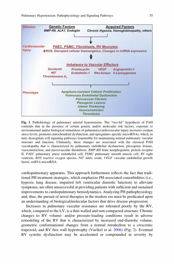
bosis, increased pulmonary vascular reactivity, and plexiform lesions (Fig. 1).
The contemporary definition of PH stipulates that the following hemodynamic
criteria be met: a sustained elevation in mean pulmonary artery pressure
(>25 mmHg) and pulmonary vascular resistance (>3 Wood units) in the setting
of a normal pulmonary capillary wedge pressure. These measures emphasize
pulmonary vascular dysfunction as the central determinate mitigating the diagnosis
of PH. This distinction departs from previous iterations of this definition by
identifying the pulmonary circulatory system as a specific entity within the larger
34 B.A. Maron and J. Loscalzo
cardiopulmonary apparatus. This approach furthermore reflects the fact that tradi-
tional PH treatment strategies, which emphasize PH-associated comorbidities (i.e.,
hypoxic lung disease, impaired left ventricular diastolic function) to alleviate
symptoms, are often unsuccessful at providing patients with sufficient and sustained
improvements to cardiopulmonary hemodynamics. Analyzing PH pathophysiology
and, thus, the pursuit of novel therapies in the modern era must be predicated upon
an understanding of biological/molecular factors that drive disease progression.
Increases in pulmonary vascular resistance are tolerated poorly by the RV,
which, compared to the LV, is a thin-walled and non-compacted structure. Chronic
changes to RV volume- and/or pressure-loading conditions result in adverse
remodeling of the RV that is characterized by increased end-diastolic volume,
geometric conformational changes from a normal tetrahedron to a crescentic
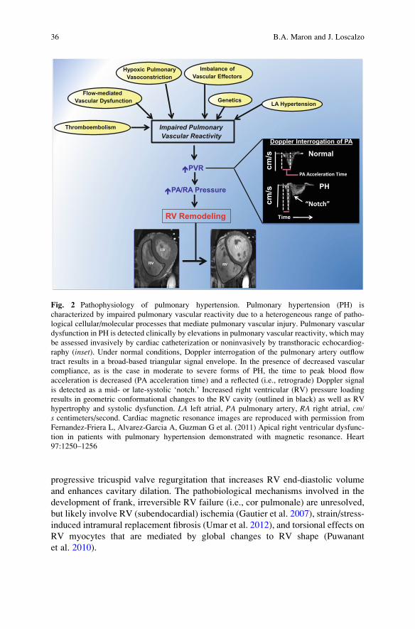
trapezoid, and RV free wall hypertrophy (Voelkel et al. 2006) (Fig. 2). Eventual
RV systolic dysfunction may be accelerated or compounded in severity by
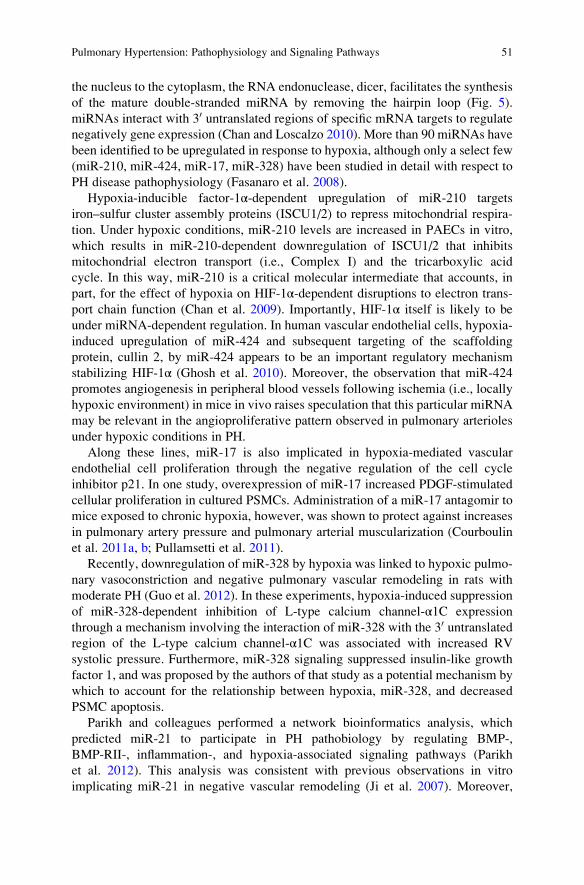
Genetic FactorsBMP-RII, ALK1, Endoglin
Acquired FactorsChronic Hypoxia, Hemoglobinopathy, others
PAEC, PSMC, Fibroblasts, RV MyocytesCardiovascular Injury �ROS, Disrupted cellular bioenergetics, Changes in miRNA expression
Stimulus
Imbalance to Vascular Effectors Serotonin
NO•
Thromboxane A2
VEGFRho-kinase
ProstacyclinEndothelin-1
Angiopoietin-15-Lipoxygenase
Phenotype Apoptosis-resistant Cellular ProliferationPulmonary Endothelial Dysfunction
Perivascular FibrosisPlexogenic LesionsIntimal ThickeningVasoconstriction
Thrombosis
Fig. 1 Pathobiology of pulmonary arterial hypertension. The “two-hit” hypothesis of PAH
contends that in the presence of certain genetic and/or molecular risk factors, exposure to
environmental and/or biological stimulators of pulmonary/cardiovascular injury increases oxidant
stress levels, promotes mitochondrial dysfunction, and upregulates specific microRNAs, which, in
turn, dysregulate cell signaling pathways responsible for maintaining normal pulmonary vascular
structure and function. Ultimately, these changes are associated with the classical PAH
vasculopathy that is characterized by pulmonary endothelial dysfunction, plexogenic lesions,
vasoconstriction, and microvascular thrombosis. BMP-RII bone morphogenetic protein receptor
II, PAEC pulmonary artery endothelial cell, PSMC pulmonary smooth muscle cell, RV right
ventricle, ROS reactive oxygen species, NO• nitric oxide, VEGF vascular endothelial growth
factor, miRNA microRNA
Pulmonary Hypertension: Pathophysiology and Signaling Pathways 35
progressive tricuspid valve regurgitation that increases RV end-diastolic volume
and enhances cavitary dilation. The pathobiological mechanisms involved in the
development of frank, irreversible RV failure (i.e., cor pulmonale) are unresolved,
but likely involve RV (subendocardial) ischemia (Gautier et al. 2007), strain/stress-
induced intramural replacement fibrosis (Umar et al. 2012), and torsional effects on
RV myocytes that are mediated by global changes to RV shape (Puwanant
et al. 2010).
Hypoxic Pulmonary Vasoconstriction
Imbalance of Vascular Effectors
Flow-mediatedVascular Dysfunction Genetics
Impaired Pulmonary Vascular Reactivity
LA Hypertension
Thromboembolism
PVR
PA/RA Pressure
Normal
PH
Doppler Interrogation of PA
PA Accelera�on Time
“Notch”
Time
cm/s
cm/s
RV Remodeling
Fig. 2 Pathophysiology of pulmonary hypertension. Pulmonary hypertension (PH) is
characterized by impaired pulmonary vascular reactivity due to a heterogeneous range of patho-
logical cellular/molecular processes that mediate pulmonary vascular injury. Pulmonary vascular
dysfunction in PH is detected clinically by elevations in pulmonary vascular reactivity, which may
be assessed invasively by cardiac catheterization or noninvasively by transthoracic echocardiog-
raphy (inset). Under normal conditions, Doppler interrogation of the pulmonary artery outflow
tract results in a broad-based triangular signal envelope. In the presence of decreased vascular
compliance, as is the case in moderate to severe forms of PH, the time to peak blood flow
acceleration is decreased (PA acceleration time) and a reflected (i.e., retrograde) Doppler signal
is detected as a mid- or late-systolic ‘notch.’ Increased right ventricular (RV) pressure loading
results in geometric conformational changes to the RV cavity (outlined in black) as well as RV
hypertrophy and systolic dysfunction. LA left atrial, PA pulmonary artery, RA right atrial, cm/s centimeters/second. Cardiac magnetic resonance images are reproduced with permission from
Fernandez-Friera L, Alvarez-Garcia A, Guzman G et al. (2011) Apical right ventricular dysfunc-
tion in patients with pulmonary hypertension demonstrated with magnetic resonance. Heart
97:1250–1256
36 B.A. Maron and J. Loscalzo
Pulmonary artery pressure is dependent partly upon RV systolic function; thus,
in the setting of diminished RV contractility, pulmonary artery pressure may be
normal despite severe pulmonary vascular disease. Along these lines, decremental
changes in RV systolic function in patients with PH are associated with worsening
symptomatology (e.g., dyspnea, fatigue, abdominal/peripheral edema), decreased
functional capacity, and increased mortality. This is the case in patients with even
mild heart failure (New York Heart Association Class II) and left atrial
hypertension-associated PH due to LV systolic dysfunction, in which an RV
ejection fraction �39 % is an independent predictor of early mortality (de Groote
et al. 1998). Similarly, in patients with PAH, decreases in tricuspid annular plane
systolic excursion (TAPSE), an echocardiographic measurement of RV systolic
function, correlate inversely with 1-year mortality rates (Forfia et al. 2006). In turn,
clinical benefits afforded to PAH patients by endothelin receptor antagonists and
prostacyclin replacement therapy (see Part II, Olschewski 2013; Clozel et al. 2013)
occur by virtue of their favorable effect on RV loading conditions, which promotes
reverse RV remodeling and restores RV-pulmonary vascular coupling (Oikawa
et al. 2005; Chin et al. 2008).
3 Cell Signaling Mechanisms in the Pathobiology of PH
3.1 Endothelial Nitric Oxide Synthase in PH
Nitric oxide (NO•) is a 30 Da lipophilic gaseous molecule, which may diffuse
through PAEC/PSMC membranes to participate in intercellular signaling. Nitric
oxide is synthesized in mammalian tissues via activation of three nitric oxide
synthase (NOS) isoforms, each of which are homodimeric enzymes containing a
calmodulin-binding domain that separates an N-terminal heme-binding domain and
a C-terminal reductase domain (Porter et al. 1990). Nitric oxide synthases catalyze
the formation of NO• from L-arginine in a reaction that consists of two distinct
monooxygenation steps. In the first monooxygenation step, two moles of electrons
are donated by one mole of NADPH to a heme-bound oxygen via flavin adenine
dinucleotide (FAD) and flavin mononucleotide (FMN). This allows for the
two-electron oxidation of a guanidine nitrogen of L-arginine to form one mole
each of omega-N-hydroxy-L-arginine and water (Delker et al. 2010). In the second
monooxygenation step, one-half mole of NADPH transfers one electron to a second
heme-bound oxygen, and omega-N-hydroxy-L-arginine undergoes a three-electronoxidation to form one mole each of NO• and L-citrulline (Griffith and Stuehr 1995).
Activation of endothelial NOS (eNOS), such as in response to vascular endothe-
lial shear stress, is modulated by various intracellular posttranslational
modifications, including S-nitrosylation (e.g., Cys94, Cys99), phosphorylation
(e.g., Ser1177, Ser65, Thr495), and palmitoylation, among others (Dudzinski
et al. 2006). The classical extracellular signaling pathways involved in eNOS
Pulmonary Hypertension: Pathophysiology and Signaling Pathways 37
activation include G-protein-coupled receptor signal transduction, which increases
intracellular Ca2+ levels and, subsequently, levels of Ca2+-calmodulin; Akt signal-
ing via sphingosine 1-phosphate; vascular endothelial growth factor (VEGF) via
phosphatase calcineurin; and hormonal stimuli (e.g., estrogen and insulin) (Murata
et al. 2002; Egom et al. 2011; Cerqueira et al. 2012). Decreased pulmonary vascular
eNOS activity is observed in numerous animal models of PH in vivo and in humans
with this disease (Steudel et al. 1998; Gangopahyay et al. 2011). Specifically, loss
of NO• bioavailability is linked to impaired endothelium-dependent and -independent
vasodilation, increased PSMC mitogenesis, and platelet aggregation. Proposed
mechanisms to account for diminished levels of functional eNOS in PH are
provided below.
3.1.1 Hypoxia and eNOS in PH
The mechanism(s) by which hypoxia influences eNOS gene expression is (are)
controversial, as PAEC exposure to PaO2 < 70 mmHg has been associated with
both increased and decreased eNOS protein expression levels. Fish and colleagues
demonstrated that hypoxia induces a decrease in acetylation and lysine 4 methyla-
tion of eNOS proximal promoter histones to decrease eNOS gene transcription
(Fish et al. 2010). In contrast, others have suggested that hypoxia-inducible factor-
1α (HIF-1α), a master transcription factor that modulates a wide range of cellular
processes in response to hypoxia, binds to a HIF response element near the
promoter region of eNOS to increase eNOS gene expression (Coulet et al. 2003).
However, in this scenario, hypoxia-mediated upregulation of eNOS expression
does not necessarily imply increased eNOS activity. To the contrary, tonic stimula-
tion of eNOS is associated with a paradoxical decrease in eNOS activity, likely
owing to the consumption and subsequent depletion of key cofactors (i.e., 5,6,7,8-
tetrahydrobiopterin [BH4]) necessary for normal eNOS function. Under these
conditions, eNOS is ‘uncoupled,’ resulting in the preferential generation of super-
oxide (•O2�) over NO• (see Sect. 3.3). Data from PH experiments in vivo support
this claim: eNOS deficiency and/or impaired eNOS function is a key factor in
disease pathogenesis. For example, eNOS knockout mice (eNOS�/�) exposed to
mild hypoxia demonstrate significantly increased RV systolic pressure and dimin-
ished markers of eNOS bioactivity as compared to wild-type controls (Fagan
et al. 1999). Diminished eNOS activity is also implicated in inflammatory
(monocrotaline), genetic (bone morphogenetic protein receptor II [BMP-RII] defi-
cient), and angioproliferative (VEGF inhibition with SU-5416) experimental
models of PAH in vivo (Tang et al. 2004).
Hypoxia may also decrease eNOS activity by inducing posttranslational modifi-
cation(s) of eNOS and/or caveolin-1, which decreases Ca2+ sensing by eNOS and
results in dissociation of eNOS from its regulatory proteins, heat shock protein
90 and calmodulin (Murata et al. 2002). Alternatively, hypoxia may decrease levels
of bioavailable NO• through eNOS-independent mechanisms. In red blood cells, for
example, hypoxia promotes increased levels of heme iron-nitrosyl (FeNO) that
38 B.A. Maron and J. Loscalzo

limits hemoglobin S-nitrosylation, which, in turn, is a key PaO2-sensitive mecha-
nism implicated in the regulation of pulmonary vascular tone (McMahon
et al. 2005).
3.1.2 Oxidant Stress and eNOS
Perturbations to the redox status of PAECs, PSMCs, RV myocytes, and lung
fibroblasts due to activation of reactive oxygen species-generating (ROS) enzymes,
such as NADPH oxidase (NOX), xanthine oxidase, and uncoupled eNOS, or via
disrupted electron transport chain function in mitochondria promote pulmonary
vasculopathy characterized by impaired NO•-dependent vasodilation, intimal
thickening, and perivascular fibrosis (Mittal et al. 2012). In humans, increases in
pulmonary vascular ROS generation may occur as a pathological response to
chronic hypoxia, or increased pulmonary vascular blood flow (e.g., secondary to
intracardiac shunt); or due to impaired antioxidant enzyme function, as is the case
in sickle cell anemia-associated PH in which glutathione peroxidase deficiency is
observed (Gizi et al. 2011). ROS may impair eNOS activity through the oxidation
of enzyme cofactors (i.e., BH4), or inactivate NO• such as in the case of •O2
� which
reacts with NO• to generate peroxynitrite (ONOO�). Additionally, the interaction
of •O2� with the stable NO• by-product nitrite (NO2
�) forms peroxynitrate
(O2NOO�) and, thus, decreases levels of NO2
�, which is a key substrate for
NOS-independent synthesis of NO• (2HNO2!N2O3 + H2O; N2O3!NO• + NO2•)
(Lundberg et al. 2011); (Spiegelhalder et al. 1976).
3.1.3 Genetic Mediators of eNOS in PH
BMP-RII is a serine–threonine kinase and member of the transforming growth
factor-β (TGF-β) superfamily of receptors (Rosenzweig et al. 1995). Approxi-
mately 70 % of familial PAH cases involve mutations in BMP-RII, and receptor
dysfunction is increasingly recognized as a contributor to non-PAH forms of PH
(Machado et al. 2006). Although BMP-RII is believed to contribute to remodeling
of pulmonary blood vessels through a wide range of signaling pathways, including
SMAD-dependent PSMC migration (Long et al. 2009), it was recently
demonstrated that two BMP-RII ligands, BMP2 and BMP4, are involved in
BMP-RII-dependent phosphorylation of eNOS at Ser-1177 to upregulate eNOS
activity (Gangopahyay et al. 2011). Similarly, abnormalities in the function of
endoglin, a key BMP receptor accessory protein in human PAECs, are linked to
the development of PAH when present in the setting of the clinical syndromes
hereditary hemorrhagic telangiectasia (HHT) type 1 and type 2. Mice heterozygous
for vascular endothelial endoglin expression (Eng+/�) develop PH spontaneously
in vivo due, in part, to increased pulmonary vascular ROS generation, eNOS
uncoupling, and decreased NOS-inhibitable NO• production (Toporsian
et al. 2010).
Pulmonary Hypertension: Pathophysiology and Signaling Pathways 39
Several eNOS polymorphisms are implicated in the development of PH and
other vascular diseases. For example, a single nucleotide polymorphism leading to
a substitution of aspartic acid for glutamic acid at position 298 (Glu298Asp) of
eNOS and an increased NOS4a allelic frequency of 27-bp variable number of
repeats increase susceptibility to developing the high altitude pulmonary edema
(HAPE) syndrome, including elevations in pulmonary artery pressure and pulmo-
nary vascular resistance. These changes may occur owing to function-limiting
changes in the conformation of eNOS, although the precise mechanism by which
to account for the phenomenon is unknown (Miyamoto et al. 1998; Droma
et al. 2002; McDonald et al. 2004).
3.2 Endothelin-1 System
Endothelin-1 (ET-1) is a 21-amino acid vasoactive peptide that contains two
disulfide bridges between Cys1-Cys15 and Cys3-Cys11 (Yeager et al. 2012),
which are necessary for endothelin converting enzyme-mediated proteolytic cleav-
age of ET-1 from its precursor, ‘Big ET-1.’ Endothelin-1 is constitutively expressed
in a wide range of mammalian cell types, including hepatic sinusoidal cells, renal
epithelial cells, and PAECs (Huggins et al. 1993). Endothelin-1 gene expression
levels are upregulated significantly in RV myocytes, PAECs, PSMCs, and lung
fibroblasts in the presence of stimuli associated with pulmonary vascular injury in
PH, including cytokines that mediate vascular inflammation (i.e., TGF-β, IL-6)(Olave et al. 2012), increased levels of pulmonary vascular ROS (An et al. 2007),
hypoxia (Yamashita et al. 2001), and decreased levels of bioavailable NO•
(Kourembanas et al. 1993). In fact, plasma ET-1 levels may be increased fourfold
in patients with PAH or PH due to left atrial hypertension, and anti-ET-1 immuno-
histochemical analysis demonstrates significantly increased immunoreactivity in
PAECs and PSMCs of plexiform lesions compared to blood vessels harvested
from normal controls (Giaid et al. 1993). Endothelin-1 is also released from sickled
red blood cells and interacts with the blood vessel wall to promote vasoconstriction
in a process that contributes to the systemic and pulmonary vasculopathy of sickle
cell anemia (Gladwin and Vichinsky 2008).
Endothelin-1 regulates pulmonary vascular tone through its interaction with the
vasoconstrictor endothelin-type A (ETA) and -type B (ETB) receptors in PSMCs
and vasodilatory ETB receptors in PAECs, which do not constitutively express ETA.
Endothelin-type A and ETB receptors are members of the superfamily of G-protein-
coupled receptors and are overall highly homologous (55 %), with the exception of
the cysteine-rich 35-amino acid sequence distal to the seventh transmembrane
domain, in which homology between receptors is only 75 % (Doi et al. 1999).
Since cysteine(s) in this region are believed to regulate G-protein coupling to both
ETA and ETB receptors, and, thus, are integral to receptor signal transduction, it has
been postulated that differences in this region between receptor subtypes may
account, in part, for their differential functions (Okamoto et al. 1997).
40 B.A. Maron and J. Loscalzo

In PSMCs, stimulation of ETA/B receptors by ET-1 induces Gi and Gq coupling
to modulate phospholipase C-mediated hydrolysis of phosphatidylinositol
4,5-bisphosphate to inositol 1,4,5-triphosphate (IP3). In turn, opening of IP3-sensitive
calcium (Ca2+) channels as well as ET-1-mediated opening of the store-operated
and nonselective Ca2+ channels induces an increase in intracellular Ca2+ flux
([Ca2+]i), Ca2+ waves, and Ca2+ oscillations that promotes vasoconstriction (Liu
et al. 2012). Importantly, ET-1-induced vasoconstriction persists following ET-1
dissociation from the ETA receptor, indicating that the [Ca2+]i flux response
mediated by ET-1 is robust, Ca2+-dependent hyperpolarization is delayed during
ET-1 signaling, or both (Zhang et al. 2003; Liu et al. 2012). The functional
consequence of ETA receptor signaling on vascular tone is noteworthy: relative to
norepinephrine, the concentration of ET-1 required to induce 50 % blood vessel
contraction (i.e., EC50) in pig coronary arteries, rat aorta, and rat pulmonary artery
is 0.52, 1.4, and 0.68, respectively (Huggins et al. 1993). ET-1 binding to ETA
receptors (Ki ¼ 0.6 nmol l�1) also promotes vascular smooth muscle cell
mitogenesis by activating various signaling intermediaries that regulate protein
synthesis, including protein kinase C; mitogen-activated protein kinase (MAPK);
p70S6K, which targets the ribosomal protein S6K to increase cellular protein
synthesis; and epidermal growth factor receptor (EGFR) via tyrosine phosphoryla-
tion (Iwasaki et al. 1999; Kapakos et al. 2010). Interestingly, upregulation of the
proto-oncogene transcription factor c-fos by ET-1 (or hypoxia) is linked to cellular
proliferation and fibrosis of PSMCs, lung fibroblasts, and myocytes in experimental
animal models of PH (Rothman et al. 1994; Nishimura et al. 2003; Recchia
et al. 2009), providing molecular evidence to account for the proliferative pheno-
typic overlap between plexogenic lesions of PAH and various solid tumors.
In contrast to PSMCs, ET-1 binding to the ETB receptor (Ki of 0.12 nmol l�1) in
PAECs results in the activation of eNOS and synthesis of NO•, which is required to
maintain normal pulmonary vascular tone and prevent vascular remodeling.
Endothelin-type B receptor-dependent activation of eNOS is believed to occur
via G-protein coupling to the ETB receptor that stimulates [Ca2+]i flux and
subsequent elevations in Ca2+ binding to calmodulin, which is a key allosteric
modulator of eNOS activity. Recent work from our laboratory has demonstrated
that pathophysiological levels of the mineralocorticoid hormone aldosterone akin to
those observed in humans with PH increase NOX4-dependent ROS generation in
PAECs in vitro, which is associated with ETB receptor dysfunction, impaired ETB
receptor-dependent activation of eNOS, and oxidation of NO• to ONOO� (Maron
et al. 2012).
Endothelin-type B receptor signal transduction also results in the synthesis of
vasodilatory prostaglandins (PG). In isolated guinea pig lungs exposed to ET-1, a
~50-fold increase in ETB receptor-dependent PGI2 synthesis is observed
(D’Orleans-Juste et al. 1991), although the mechanism by which ETB receptor
activation stimulates PGI2 synthesis is not well characterized. Internalization of
the ETB receptor/ET-1 complex and subsequent proteasomal degradation is the
chief mechanism by which ET-1 elimination occurs. This conclusion is supported
Pulmonary Hypertension: Pathophysiology and Signaling Pathways 41

in vivo by experiments involving the transgenic spotting lethal rat (sl/sl), whichlacks constitutively expressed vascular ETB receptors. Compared to wild-type rats,
these rats demonstrate significantly higher circulating levels of ET-1 and more
severe PH following monocrotaline injection to induce pulmonary vascular injury
(Nishida et al. 2004).
3.3 Soluble Guanylyl Cyclase and PhosphodiesteraseInhibition in PH
Nitric oxide is the primary biological activator of the heterodimeric (α1/β1 or α1/β2)enzyme soluble guanylyl cyclase (sGC) that catalyzes the conversion of cytosolic
GTP to cGMP, which is a critical secondary signaling molecule necessary for
activation of cGMP-dependent protein kinase (i.e., protein kinase G [PKG]) to
promote PSMC relaxation and inhibit platelet aggregation and thrombosis. Nitric
oxide binding to the heme (Fe2+) prosthetic group of sGC results in the formation of
a hexa-coordinated histidine–heme–NO• intermediate. Subsequent cleavage of the
heme–histidine bond leads to a dramatic upregulation of enzyme activity:
nanomolar concentrations of NO• may induce an appropriate 100-fold increase in
sGC activation (Evgenov et al. 2006). In PH, elevated levels of ROS accumulation
may impair sGC activity through the oxidation of heme from the ferrous (Fe2+) to
ferric (Fe3+) state that converts sGC to an NO•-insensitive state, presumably owing
to decreased affinity of NO• for oxidized heme. Alternatively, others have
demonstrated that sGC activity is influenced by the redox status of functional
sGC cysteinyl thiol(s) in a manner that is independent of the heme redox state
(Fernhoff et al. 2009; Yoo et al. 2012). Work from our laboratory has demonstrated
that pathophysiological concentrations of H2O2 induce the formation of higher
cysteinyl thiol oxidative states of Cys-122 on the β1 subunit of sGC in vascular
smooth muscle cells, including sulfenic acid, sulfinic acid, and the disulfide form.
Posttranslational oxidative modification of Cys122, in turn, functions as a redox
‘switch’ that regulates enzyme function, resulting in decreased NO•-sensing by sGC
and impaired enzyme activity (Maron et al. 2009). The importance of abnormal
sGC function in the pathogenesis of PH is well established. Transgenic mice
deficient in the sGC α1-subunit develop exaggerated elevations in RV systolic
pressure and muscularization of intraacinar pulmonary arterioles following expo-
sure to chronic hypoxia compared to wild-type mice (Vermeersch et al. 2007).
Moreover, hypoxia alone is associated with decreased mRNA and protein levels of
sGC as well as sGC-dependent cGMP formation (Hassoun et al. 2004).
Collectively, these observations implicate the pharmacotherapeutic potential of
heme-independent sGC activators in PH. Work from Ko and colleagues and drug
discovery experiments performed at Bayer HealthCare in the early 1990s identified
YC-1 [3-50-hydroxymethyl-20furyl)-1-benzyl indazole] and 5-substituted-2-
furaldehyde-hydrazone derivative compounds (i.e., BAY compounds), respectively,
42 B.A. Maron and J. Loscalzo

as synthetic heme-(in)dependent activators of sGC (Ko et al. 1994; Stasch
et al. 2006). BAY 58-2667, perhaps the best studied among these compounds,
activates sGC with a Km of 74 μM and a Vmax of 0.134 μmol min�1 mg�1 (Schmidt
et al. 2003), and although the precise mechanism by which this (and other) BAY
compounds activates heme-oxidized sGC is not fully resolved, one leading hypoth-
esis contends that BAY 58-2667 competes with the oxidized heme moiety for
binding to the sGC-activating motif to induce enzyme activation (Pellicena
et al. 2004) (see Part III, Sect. 1). The effect of these compounds on sGC-NO•
vasodilatory signaling and NO•-dependent vascular remodeling has been assessed in
PH in vivo. In one study, the administration of YC-1 to hypoxic mice decreased
PSMC proliferation and pulmonary artery pressure (Huh et al. 2011). The effect of
BAY 63-2521 (Riociguat™) on cardiopulmonary hemodynamics was also assessed
in a small cohort of patients with PAH, chronic thromboembolic PH, or PH from
interstitial lung disease. Drug therapy (3.0–7.5 mg day�1) over 12 weeks decreased
pulmonary vascular resistance by 215 dyne s cm�5, which was associated with an
increase in the median 6-min walk distance by 55.0 m from baseline (Ghofrani
et al. 2010).
3.4 Phosphodiesterase Inhibition in PH
In 1962, Butcher and Sutherland implicated phosphodiesterase enzymatic activity
in endogenous degradation of adenosine 30, 50 phosphate (cAMP) (Butcher and
Sutherland 1962). Eleven PDE isoforms have since been detected in mammalian
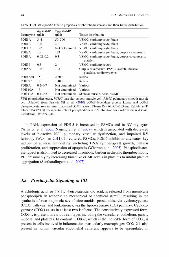
tissue (Table 1). The fields of PDE biochemistry and PH intersected following the
identification of cGMP-specific PDE type-5, at elevated concentrations in PSMCs,
platelets, and myocytes. Phosphodiesterase type-5 regulates cGMP bioactivity via
(1) the hydrolysis of cGMP to 50-GMP, and (2) allosteric binding of cGMP to
PDE-5 GAF1 domains, which induces a conformational change to the structure of
PDE-5 and positively feeds back to promote cGMP metabolism (Fig. 3). PDE-5-
cGMP binding ranges from Kd of 2.4 μM (pH ¼ 5.2) to 0.15 μM (pH ¼ 9.5) (Turko
et al. 1999). The pH-dependent manner by which this occurs is in concert with
reports demonstrating a regulatory role for the cyclic nucleotide ionizing residues
(i.e., pH-sensitive) Asp-289 and Asp-478 in modulating PDE-5 function
(McAllister-Lucas et al. 1995). The intracellular concentration of cGMP may also
be influenced by flux through membrane-bound cGMP-gated channels and the
multidrug transporter, although the contribution of these to total levels of bioactive
cGMP is negligible in pulmonary vascular tissue (Serre et al. 1995).
1 GAF is an acronym of the various tissues in which these domains were originally described:
cGMP-dependent phosphodiesterases (PDEs), nabaena adenylyl cyclases, and E. coli FhlA
(Francis et al. 2010).
Pulmonary Hypertension: Pathophysiology and Signaling Pathways 43
In PAH, expression of PDE-5 is increased in PSMCs and in RV myocytes
(Wharton et al. 2005; Nagendran et al. 2007), which is associated with decreased
levels of bioactive NO•, pulmonary vascular dysfunction, and impaired RV
lusitropy (Waxman 2011). In cultured PSMCs, PDE-5 inhibition attenuates key
indices of adverse remodeling, including DNA synthesis/cell growth, cellular
proliferation, and suppression of apoptosis (Wharton et al. 2005). Phosphodiester-
ase type-5 is also linked to decreased thrombotic burden in chronic thromboembolic
PH, presumably by increasing bioactive cGMP levels in platelets to inhibit platelet
aggregation (Suntharalingam et al. 2007).
3.5 Prostacyclin Signaling in PH
Arachidonic acid, or 5,8,11,14-eicosatetraenoic acid, is released from membrane
phospholipids in response to mechanical or chemical stimuli, resulting in the
synthesis of two major classes of eicosanoids: prostanoids, via cyclooxygenase
(COX) pathway, and leukotrienes, via the lipooxygenase (LO) pathway. Cycloox-
ygenase (COX) exists in at least two isoforms. The constitutively expressed form,
COX-1, is present in various cell types including the vascular endothelium, gastric
mucosa, and platelets. In contrast, COX-2, which is the inducible form of COX, is
present in cells involved in inflammation, particularly macrophages. COX-2 is also
present in normal vascular endothelial cells and appears to be upregulated in
Table 1 cGMP-specific kinetic properties of phosphodiesterases and their tissue distribution
Isoenzyme
Km cGMP
(μM)
Vmax cGMP
(μM) Tissue distribution
PDE1A 3–4 50–300 VSMC, cardiomyocyte, brain
PDE1B 1–6 30 VSMC, cardiomyocyte, brain
PDE1C 1–2 Not determined VSMC, cardiomyocyte, brain
PDE2A 10 123 VSMC, cardiomyocyte, brain, corpus cavernosum
PDE3A 0.02–0.2 0.3 VSMC, cardiomyocyte, brain, corpus cavernosum,
platelets
PDE3B 0.3 2 VSMC, cardiomyocytes
PDE5A 1–6 1–3 Corpus cavernosum, PSMC, skeletal muscle,
platelets, cardiomyocytes
PDE6A/B 15 2,300 Retina
PDE 6C 17 1,400 Retina
PDE9A 0.2–0.7 Not determined Various
PDE 10A 13 Not determined Various
PDE 11A 0.4–0.2 Not determined Skeletal muscle, heart, VSMC
PDE phosphodiesterase, VSMC vascular smooth muscle cell, PSMC pulmonary smooth muscle
cell. Adapted from Francis SH et al. (2010) cGMP-dependent protein kinase and cGMP
phosphodiesterases in nitric oxide and cGMP action. Pharm Rev 62:525–563 and Reffelman T,
Kloner RA (2003) Therapeutic role of phosphodiesterase 5 inhibition for cardiovascular disease.
Circulation 108:239–244
44 B.A. Maron and J. Loscalzo

response to stimuli associated with vascular injury, such as shear stress (Topper
et al. 1996). Key products of the COX pathway that are pertinent to the pathophysi-
ology of PH include the potent vasoconstrictor and stimulus for platelet aggrega-
tion, thromboxane A2 (TXA2), and prostaglandin I2 (prostacyclin), which exerts
opposing effects to TXA2, including vasodilation and inhibition of platelet activa-
tion. Prostacyclin is synthesized from PGH2 by prostacyclin synthase in a reaction
that occurs primarily in vascular endothelial cells. Early investigators speculated
that the pulmonary vasoconstriction phenotype in PAH was a consequence of
imbalanced TXA2/PGI2 synthesis. This hypothesis was supported by the observa-
tion that, compared to normal pulmonary blood vessels, pre-capillary pulmonary
arterioles harvested from patients with PAH, hepato-pulmonary PH, and
HIV-associated PH demonstrate significantly decreased PGI2 synthase mRNA
and protein expression levels (Tuder et al. 1999). In contrast, elevated levels of
eNOSPharmacological NO• Donor
NOS-independent NO synthesis
iNOS
NO•
NO•
α1β1
Fe2+
Cys-122
sGC
+
GTP cGMP
cGMP-gated Channel
PKG VSMCRELAXATION
PDE5Catalytic site Binding site
PDE5
+ +
Multidrug TransporterVSMC
Fig. 3 Influence of phosphodiesterase type-5 on the nitric oxide signaling axis. Nitric oxide (NO•)
derived from endothelial nitric oxide synthase (eNOS), pharmacological NO• donors, or via
induction of inducible NOS in vascular smooth muscle cells (VSMCs) activates soluble guanylyl
cyclase (sGC) to generate cGMP. Normal NO•–sGC signaling is regulated by the redox status of
the prosthetic heme ligand and functional cysteinyl thiol(s), such as Cys122, which are present
near the catalytically active region of the β1-subunit of sGC. The interaction of cGMPwith its most
relevant biological target, protein kinase G (PKG), results in VSMC relaxation. This vasodilatory
effect of this pathway is offset through the interaction of cGMP with phosphodiesterases (PDEs),
which decreased bioactive cGMP levels through hydrolysis of cGMP to form 50GMP, or by
allosteric binding of cGMP to PDE. In the case of PDE type-5 (PDE5), increased levels of
cGMP and/or PKG upregulates PDE5 activity. The contribution of gated cGMP channels and
the multidrug transported to the regulation of bioactive cGMP levels is negligible in pulmonary
vascular tissue. Adapted from Francis SH et al. (2010) cGMP-dependent protein kinase and cGMP
phosphodiesterases in nitric oxide and cGMP action. Pharm Rev 62:525–563, with permission
Pulmonary Hypertension: Pathophysiology and Signaling Pathways 45

TXA2 and increased pulmonary vascular sensitivity to TXA2 have been reported in
PH, such as in the lamb model of hypoxia-induced neonatal persistent pulmonary
hypertension (Hinton et al. 2007). These observations are consistent with other
reports in adults demonstrating that, compared to healthy controls, patients with
PAH or PH due to hypoxic lung disease demonstrate elevated levels of urinary
excretion of 11-dehydro-thromboxane B2, a stable metabolite of TXA2, and
decreased levels of the stable prostacyclin metabolite, 2,3-dinor-6-keto-prostaglan-
din F1α (Christman et al. 1992).
Shear stress, ET-1, hypoxia, and BMP-RII dysfunction are each associated with
overactivation of the TXA2 synthesis pathway relative to PGI2 in vascular tissue
(Zaugg et al. 1996; Song et al. 2005; Racz et al. 2010). Although the precise
mechanism by which to account for this imbalance is unresolved, abnormal
COX-2 function in the setting of vascular injury may play a role. It was recently
demonstrated that compared to control mice, COX-2 knockdown mice
administered monocrotaline to induce vascular inflammation exhibit a robust
increase in NOX4 gene expression, dihydroethidium fluorescence (indicative of
ROS accumulation), and ETA receptor expression in pulmonary arterioles, whereas
prostacyclin levels were decreased significantly (Seta et al. 2011). These findings
are consistent with reports in cultured COX-2-deficient PSMCs, in which hypoxia
results in a hypertrophic remodeling response and a vasoconstrictor phenotype
(Fredenburgh et al. 2008).
5-lipooxygenase (5-LO) catalyzes the conversion of arachidonic acid ultimately
into various leukotrienes that mediate cellular processes involved in vascular
remodeling and cellular responses to injury. Leukotriene B4, for example, exerts
both chemotactic and chemokinetic activity on polymorphonuclear leukocytes and
eosinophils (Ford-Hutchinson et al. 1980), and leukotrienes C4, D4, and E4 (each of
which contain a cysteine) are implicated in pulmonary vasoconstriction and
increased pulmonary vascular permeability. Although rats that overexpress 5-LO
do not develop PAH spontaneously, pulmonary vascular dysfunction and abnormal
cardiopulmonary hemodynamics are accelerated in the presence of pulmonary
vascular inflammation (Jones et al. 2004). This supports other observations
indicating that an inflammatory milieu is conducive to 5-LO-dependent synthesis
of the vasoactive cysteinyl leukotrienes (Listi et al. 2008). Molecular inhibition of
the 5-LO-activating protein (FLAP) has, in turn, been shown to prevent pulmonary
hypertension in rats exposed to chronic hypoxia (Voelkel et al. 1996).
3.6 Mitochondrial Dysfunction
Mitochondria regulate bioenergetics, cellular respiration, and the intracellular redox
status and, thus, have the potential to regulate PAEC/PSMC signaling pathways
linked to cell survival, proliferation, and ROS production. Hydrogen (hydride)
derived from dietary carbohydrate and fats is oxidized by molecular oxygen (O2)
via the tricarboxylic acid (TCA) cycle and β-oxidation pathways, respectively, to
46 B.A. Maron and J. Loscalzo
generate adenosine triphosphate (ATP). These biochemical events occur via the
electron transport chain, in which two electrons donated by NADH + H+ flow
sequentially from complex I to ubiquinone (coenzyme Q) to complex III (ubiquinol:
cytochrome c oxidoreductase) and then to cytochrome c. Electrons are then trans-
ferred to complex IV to reduce ½O2 and generate H2O (Wallace 2005). Protons are
pumped across the inner mitochondrial membrane to establish the significantly
negative electrochemical gradient (Δψm: ~ �200 mV) across that membrane,
which provides the electromotive force necessary for ATP synthesis.
Changes to mitochondrial membrane permeability, and, hence, the normal
Δψm, are antecedent to reversible structural and functional changes in
mitochondria and, if unchecked, commit the cell to apoptosis (Kroemer and Reed
2000; Michelakis et al. 2008). Numerous mechanisms to account for the relation-
ship between mitochondrial membrane permeability and changes to cell survival
have been proposed and include increased permeability of the voltage-sensitive
permeability transition pore complex (PTPC), alkalinization of the local pH, and
perturbations to the intramitochondrial redox status that results in oxidation of a key
thiol involved in regulating PTPC opening and/or oxidation of pyridine nucleotides
(i.e., NADH/NAD+) to favor PTPC opening (Woodfield et al. 1998; Zamzami and
Kroemer 2001). Collectively, these changes afford egress of apoptosis-associated
proteins (e.g., Bax, Bcl-2, others) from the intramitochondrial to extramito-
chondrial space, thereby activating programmed cell death signaling pathways
(Mossalam et al. 2012).
Pathological disruptions to mitochondria-dependent regulation of cell survival
are a central mechanism in the pathobiology of various angioproliferative diseases,
including solid tumor cancers. Apoptosis-resistant proliferation of PAECs/PSMCs
is likewise a prominent pathophenotypic feature of PH, particularly with respect to
plexigenic lesions in PAH. This observation has raised attention to the possibility
that mitochondrial dysfunction is an under-recognized pathobiological factor by
which to account for the phenotypic overlap between these two broad categories of
disease. Evidence in support of this concept is derived partly from observations
made in the fawn-hooded rat, a unique animal strain that develops PAH spontane-
ously. Pulmonary vascular smooth muscle cells harvested from these animals
demonstrate mitochondria that are decreased in size and fragmented prior to the
development of pulmonary vascular remodeling (Bonnet et al. 2006). The func-
tional effects of these changes are linked to a shift in mitochondrial metabolism
from oxidative phosphorylation toward glycolysis, impairment to electron flux, and
subsequent activation of hypoxia-inducible factor (HIF)-1α (Archer et al. 2008). In
turn, HIF-1α has been shown in endothelial cells cultured from patients with
idiopathic PAH to target carbonic anhydrase IX, which decreases levels of the
antioxidant enzyme manganese superoxide dismutase (SOD2) to increase vascular
ROS generation and decrease levels of NO• (Fijalkowska et al. 2010). Interestingly,
in these experiments, increased HIF-1α expression correlated inversely with low
numbers of mitochondria, indicating that negative control of mitochondrial biogen-
esis by HIF-1α may be one mechanism by which to account for abnormal cellular
respiration patterns observed in in vivo models of PAH. Conventional
Pulmonary Hypertension: Pathophysiology and Signaling Pathways 47
factors associated with PAH may also influence mitochondrial dysfunction directly.
For example, compared to healthy controls, stimulation of PSMCs with ET-1,
platelet-derived growth factor (PDGF), or IL-6 harvested from PAH patients results
in Kruppel-like factor 5 (KL-5)-mediated activation of cyclin B1 that hyperpolarizes
the mitochondrial inner membrane to inhibit apoptosis (Courboulin et al. 2011b).
Under normoxic conditions, electron transport chain complexes I or II generate•O2
� that is dismutated to form H2O2, which is a key signaling molecule required
for activation of Kv channels necessary to maintain the negative electrochemical
gradient of the mitochondria (Bonnet et al. 2006). At PaO2 < 70 mmHg, there is
decreased intramitochondrial H2O2 generation, opening of O2-sensitive Kv1.5
channels, and subsequent activation of L-type Ca2+ channels that promotes pulmo-
nary vasoconstriction (Archer et al. 2004). Human PSMCs in PAH, however, are
deficient in Kv1.5 channels, and data from experimental animal models of PAH
suggest that the effect of this deficiency is mitochondrial hyperpolarization, and,
consequently, tonic activation of L-type Ca2+ channels associated with vasocon-
striction and proliferation of PSMCs (Reeve et al. 2001).
Less well established is the role of abnormal mitochondrial bioenergetics in the
development of pulmonary vascular dysfunction and/or RV hypertrophy in
PH. There is increasing evidence suggesting that in cardiomyocytes, an abnormal
shift in cellular fuel utilization vis-a-vis the glucose–fatty acid cycle (i.e., Randle’scycle) (Randle et al. 1963) accounts for changes to myocardial structure
(i.e., hypertrophy) and function (i.e., impaired contractility) (Fig. 4). In the
monocrotaline and pulmonary artery banding rat models of PAH, for example,
decreased RV O2 consumption is observed and modulates a shift from oxidative
phosphorylation to glycolysis by a mechanism involving increased Glut-1 expres-
sion and upregulation of pyruvate dehydrogenase kinase (PDK) expression with
consequent increased phosphorylation of pyruvate dehydrogenase leading to its
inhibition (Piao et al. 2010). The functional effects of this process include impaired
RV systolic function and prolongation of the QT interval, which can be reversed by
PDK inhibition or through inhibition of fatty acid oxidation to induce an indirect
reciprocal shift in the mitochondrial fuel source back to glucose (oxidation) (Fang
et al. 2012).
3.7 Peroxisome Proliferator-Activated Receptor-γ
Peroxisome proliferator-activated receptor (PPAR-γ) is a transcription factor most
commonly associated with its regulatory effect on genes involved in fatty acid
storage and glucose metabolism in adipocytes (Kilroy et al. 2012); PPAR-γ, and itstranscription target apoE, are also key downstream targets of BMP-RII signal
transduction. In turn, loss of function to BMP-RII via somatic mutation or dissoci-
ation of BMP-RII-interacting proteins is associated with PSMC proliferation
in vitro and the development of PAH in vivo (Merklinger et al. 2005; Chan
et al. 2007; Song et al. 2008). In PSMCs, the antiproliferative effect of BMP-RII
48 B.A. Maron and J. Loscalzo

Fig. 4 Mitochondrial bioenergetics. (a) The “glucose–fatty acid cycle” or Randle Cycle describesa mechanism to maintain homeostatic control of circulating levels of glucose and fatty acids.
(b) Inhibition of glucose utilization by fatty acid oxidation is regulated most strongly by pyruvate
dehydrogenase (PDH) and, to a lesser extent, by 6-phosphofructo-1-kinase (PFK) and glucose
uptake. Phosphofructokinase inhibition owing to citrate accumulation or via pharmacological/
molecular strategies shifts glucose toward glycogen synthesis and pyruvate to gluconeogenesis or
the synthesis of TCA intermediates (i.e., anaplerosis). Overactivation of PDK in right ventricular
myocytes in an in vivo model of pulmonary hypertension has been associated with a shift from
glucose oxidation to glycolysis and subsequent myocardial dysfunction (Piao et al. 2010). LCFAlong-chain fatty acids, TAG triacylglycerol, Pyr pyruvate, Cyto cytosol, MITO mitochondria,
GLUT4 glucose transporter 4, HK hexokinase, Glc-6-P glucose-6-phosphate, Fru-6-P fructose
6-phosphate, CPT I carnitine palmitoyltransferase I, β-ox β-oxidation. Reproduced with permis-
sion from Hue et al. (2009) Am J Physiol Endocrinol Metab 297:E578–E591
Pulmonary Hypertension: Pathophysiology and Signaling Pathways 49
is modulated by phospho-ERK and PPAR-γ binding to DNA, which, in turn,
stimulates apoE synthesis and secretion (Hansmann et al. 2007). Transgenic
ApoE knockout mice (ApoE�/�) fed a high-fat diet demonstrate spontaneous
development of PH, which is reversible through pharmacological stimulation of
PPAR-γ with pioglitazone (Hansmann et al. 2007). In human PAECs, BMP-RII
signaling appears to induce a PPAR-γ/β–catenin complex that targets the gene
encoding apelin to modulate normal cellular responses to injury and, in PSMCs,
suppresses cellular proliferation (Falcetti et al. 2010; Alastalo et al. 2011).
3.8 MicroRNA-Mediated Regulation of Cellular Responsesto Hypoxia
MicroRNA (miRNA) are non-canonical and highly conserved noncoding
ribonucleic acid molecules (~20 nucleotides) that participate in a heterogeneous
range of cellular processes and are believed to regulate over 30 % of all mRNA
transcripts (Berezikov et al. 2005). MicroRNA transcription generates hairpin-
looped molecules known as primary miRNAs (pri-mRNA), which are processed
in the cell nucleus to form miRNA precursors (pre-miRNA). Once exported from
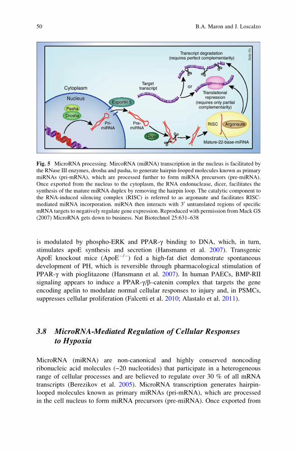
Fig. 5 MicroRNA processing. MircoRNA (miRNA) transcription in the nucleus is facilitated by
the RNase III enzymes, drosha and pasha, to generate hairpin-looped molecules known as primary
miRNAs (pri-mRNA), which are processed further to form miRNA precursors (pre-miRNA).
Once exported from the nucleus to the cytoplasm, the RNA endonuclease, dicer, facilitates the
synthesis of the mature miRNA duplex by removing the hairpin loop. The catalytic component to
the RNA-induced silencing complex (RISC) is referred to as argonaute and facilitates RISC-
mediated miRNA incorporation. miRNA then interacts with 30 untranslated regions of specific
mRNA targets to negatively regulate gene expression. Reproduced with permission fromMack GS
(2007) MicroRNA gets down to business. Nat Biotechnol 25:631–638
50 B.A. Maron and J. Loscalzo
the nucleus to the cytoplasm, the RNA endonuclease, dicer, facilitates the synthesis
of the mature double-stranded miRNA by removing the hairpin loop (Fig. 5).
miRNAs interact with 30 untranslated regions of specific mRNA targets to regulate
negatively gene expression (Chan and Loscalzo 2010). More than 90 miRNAs have
been identified to be upregulated in response to hypoxia, although only a select few
(miR-210, miR-424, miR-17, miR-328) have been studied in detail with respect to
PH disease pathophysiology (Fasanaro et al. 2008).
Hypoxia-inducible factor-1α-dependent upregulation of miR-210 targets
iron–sulfur cluster assembly proteins (ISCU1/2) to repress mitochondrial respira-
tion. Under hypoxic conditions, miR-210 levels are increased in PAECs in vitro,
which results in miR-210-dependent downregulation of ISCU1/2 that inhibits
mitochondrial electron transport (i.e., Complex I) and the tricarboxylic acid
cycle. In this way, miR-210 is a critical molecular intermediate that accounts, in
part, for the effect of hypoxia on HIF-1α-dependent disruptions to electron trans-
port chain function (Chan et al. 2009). Importantly, HIF-1α itself is likely to be
under miRNA-dependent regulation. In human vascular endothelial cells, hypoxia-
induced upregulation of miR-424 and subsequent targeting of the scaffolding
protein, cullin 2, by miR-424 appears to be an important regulatory mechanism
stabilizing HIF-1α (Ghosh et al. 2010). Moreover, the observation that miR-424
promotes angiogenesis in peripheral blood vessels following ischemia (i.e., locally
hypoxic environment) in mice in vivo raises speculation that this particular miRNA
may be relevant in the angioproliferative pattern observed in pulmonary arterioles
under hypoxic conditions in PH.
Along these lines, miR-17 is also implicated in hypoxia-mediated vascular
endothelial cell proliferation through the negative regulation of the cell cycle
inhibitor p21. In one study, overexpression of miR-17 increased PDGF-stimulated
cellular proliferation in cultured PSMCs. Administration of a miR-17 antagomir to
mice exposed to chronic hypoxia, however, was shown to protect against increases
in pulmonary artery pressure and pulmonary arterial muscularization (Courboulin
et al. 2011a, b; Pullamsetti et al. 2011).
Recently, downregulation of miR-328 by hypoxia was linked to hypoxic pulmo-
nary vasoconstriction and negative pulmonary vascular remodeling in rats with
moderate PH (Guo et al. 2012). In these experiments, hypoxia-induced suppression
of miR-328-dependent inhibition of L-type calcium channel-α1C expression
through a mechanism involving the interaction of miR-328 with the 30 untranslatedregion of the L-type calcium channel-α1C was associated with increased RV
systolic pressure. Furthermore, miR-328 signaling suppressed insulin-like growth
factor 1, and was proposed by the authors of that study as a potential mechanism by
which to account for the relationship between hypoxia, miR-328, and decreased
PSMC apoptosis.
Parikh and colleagues performed a network bioinformatics analysis, which
predicted miR-21 to participate in PH pathobiology by regulating BMP-,
BMP-RII-, inflammation-, and hypoxia-associated signaling pathways (Parikh
et al. 2012). This analysis was consistent with previous observations in vitro
implicating miR-21 in negative vascular remodeling (Ji et al. 2007). Moreover,
Pulmonary Hypertension: Pathophysiology and Signaling Pathways 51
hypoxia-mediated miR-21 upregulation in PAECs appears to contribute to the PH
vascular phenotype by decreasing BMP-RII, RhoB, and Rho kinase, which, under
normal conditions, are involved in pulmonary vasodilatory signaling. miR-21 was
likewise linked to disease expression in various PH animal models in vivo, and was
observed to be highly expressed in pulmonary vascular tissue in humans with PH.
4 Conclusions
Pulmonary hypertension describes a complex disorder characterized by
dysregulation of cell signaling pathways that maintain normal structure and func-
tion to distal pulmonary blood vessels. In severe forms of PH, this may result in an
obliterative vasculopathy, severely elevated pulmonary artery pressure and pulmo-
nary vascular resistance, and adverse RV remodeling. The development of success-
ful PH pharmacotherapies in the future that aim to modify disease progression will
likely hinge on the identification of novel molecular mechanisms that modulate
pulmonary vascular remodeling. This pursuit is expected to require enhanced
understanding of the processes by which miRNAs, mitochondria, and other molec-
ular factors regulate cellular bioenergetics, survival, and proliferation to contribute
to PH disease expression.
References
Abe K, Toba M et al (2010) Formation of plexiform lesions in experimental severe pulmonary
arterial hypertension. Circulation 121(25):2747–2754
Alastalo TP, Li M et al (2011) Disruption of PPARgamma/beta-catenin-mediated regulation of
apelin impairs BMP-induced mouse and human pulmonary arterial EC survival. J Clin Invest
121(9):3735–3746
An SJ, Boyd R et al (2007) NADPH oxidase mediates angiotensin II-induced endothelin-1
expression in vascular adventitial fibroblasts. Cardiovasc Res 75(4):702–709
Archer SL, Wu XC et al (2004) O2 sensing in the human ductus arteriosus: redox-sensitive K+
channels are regulated by mitochondria-derived hydrogen peroxide. Biol Chem 385
(3–4):205–216
Archer SL, Gomberg-Maitland M et al (2008) Mitochondrial metabolism, redox signaling, and
fusion: a mitochondria-ROS-HIF-1alpha-Kv1.5 O2-sensing pathway at the intersection of
pulmonary hypertension and cancer. Am J Physiol Heart Circ Physiol 294(2):H570–H578
Archer SL, Weir EK et al (2010) Basic science of pulmonary arterial hypertension for clinicians:
new concepts and experimental therapies. Circulation 121(18):2045–2066
Barst RJ, Rubin LJ (2011) Pulmonary hypertension. In: Fuster V, Walsh RA, Harrington RA (eds)
Hurst’s the heart, 13th edn. McGraw-Hill, New York
Benza RL, Miller DP et al (2010) Predicting survival in pulmonary arterial hypertension: insights
from the Registry to Evaluate Early and Long-Term Pulmonary Arterial Hypertension Disease
Management (REVEAL). Circulation 122(2):164–172
Berezikov E, Guryev V et al (2005) Phylogenetic shadowing and computational identification of
human microRNA genes. Cell 120(1):21–24
52 B.A. Maron and J. Loscalzo
Bonnet S, Michelakis ED et al (2006) An abnormal mitochondrial-hypoxia inducible factor-
1alpha-Kv channel pathway disrupts oxygen sensing and triggers pulmonary arterial hyperten-
sion in fawn hooded rats: similarities to human pulmonary arterial hypertension. Circulation
113(22):2630–2641
Butcher RW, Sutherland EW (1962) Adenosine 30,50-phosphate in biological materials.
I. Purification and properties of cyclic 30,50-nucleotide phosphodiesterase and use of this
enzyme to characterize adenosine 30,50-phosphate in human urine. J Biol Chem 237:1244–1250
Cerqueira FM, Brandizzi LI et al (2012) Serum from calorie-restricted rats activates vascular cell
eNOS through enhanced insulin signaling mediated by adiponectin. PLoS One 7(2):e31155
Chan SY, Loscalzo J (2010) MicroRNA-210: a unique and pleiotropic hypoxamir. Cell Cycle 9
(6):1072–1083
Chan MC, Nguyen PH et al (2007) A novel regulatory mechanism of the bone morphogenetic
protein (BMP) signaling pathway involving the carboxyl-terminal tail domain of BMP type II
receptor. Mol Cell Biol 27(16):5776–5789
Chan SY, Zhang YY et al (2009) MicroRNA-210 controls mitochondrial metabolism during
hypoxia by repressing the iron-sulfur cluster assembly proteins ISCU1/2. Cell Metab 10
(4):273–284
Chin KM, Kingman M et al (2008) Changes in right ventricular structure and function assessed
using cardiac magnetic resonance imaging in bosentan-treated patients with pulmonary arterial
hypertension. Am J Cardiol 101(11):1669–1672
Christman BW, McPherson CD et al (1992) An imbalance between the excretion of thromboxane
and prostacyclin metabolites in pulmonary hypertension. N Engl J Med 327(2):70–75
Clozel M, Maresta A, Humbert M (2013) Endothelin receptor antagonists. In: Humbert M,
Evgenov OV, Stasch JP (eds) Pharmacotherapy of pulmonary hypertension. Springer,
Heidelberg
Coulet F, Nadaud S et al (2003) Identification of hypoxia-response element in the human
endothelial nitric-oxide synthase gene promoter. J Biol Chem 278(47):46230–46240
Courboulin A, Paulin R et al (2011a) Role for miR-204 in human pulmonary arterial hypertension.
J Exp Med 208(3):535–548
Courboulin A, Tremblay VL et al (2011b) Kruppel-like Factor 5 contributes to pulmonary artery
smooth muscle proliferation and resistance to apoptosis in human pulmonary arterial hyper-
tension. Respir Res 12:128
D’Orleans-Juste P, Telemaque S et al (1991) Different pharmacological profiles of big-endothelin-3
and big-endothelin-1 in vivo and in vitro. Br J Pharmacol 104(2):440–444
de Groote P, Millaire A et al (1998) Right ventricular ejection fraction is an independent predictor
of survival in patients with moderate heart failure. J Am Coll Cardiol 32(4):948–954
Delker SL, Xue F et al (2010) Role of zinc in isoform-selective inhibitor binding to neuronal nitric
oxide synthase. Biochemistry 49(51):10803–10810
Doi T, Sugimoto H et al (1999) Interactions of endothelin receptor subtypes A and B with Gi, Go,
and Gq in reconstituted phospholipid vesicles. Biochemistry 38(10):3090–3099
Droma Y, Hanaoka M et al (2002) Positive association of the endothelial nitric oxide synthase
gene polymorphisms with high-altitude pulmonary edema. Circulation 106(7):826–830
Dudzinski DM, Igarashi J et al (2006) The regulation and pharmacology of endothelial nitric oxide
synthase. Annu Rev Pharmacol Toxicol 46:235–276
Egom EE, Mohamed TM et al (2011) Activation of Pak1/Akt/eNOS signaling following sphingo-
sine-1-phosphate release as part of a mechanism protecting cardiomyocytes against ischemic
cell injury. Am J Physiol Heart Circ Physiol 301(4):H1487–H1495
Evgenov OV, Pacher P et al (2006) NO-independent stimulators and activators of soluble
guanylate cyclase: discovery and therapeutic potential. Nat Rev Drug Discov 5(9):755–768
Fagan KA, Fouty BW et al (1999) The pulmonary circulation of homozygous or heterozygous
eNOS-null mice is hyperresponsive to mild hypoxia. J Clin Invest 103(2):291–299
Pulmonary Hypertension: Pathophysiology and Signaling Pathways 53
Falcetti E, Hall SM et al (2010) Smooth muscle proliferation and role of the prostacyclin
(IP) receptor in idiopathic pulmonary arterial hypertension. Am J Respir Crit Care Med 182
(9):1161–1170
Fang YH, Piao L et al (2012) Therapeutic inhibition of fatty acid oxidation in right ventricular
hypertrophy: exploiting Randle’s cycle. J Mol Med (Berl) 90(1):31–43
Farber HW, Loscalzo J (2004) Pulmonary arterial hypertension. N Engl J Med 351(16):1655–1665
Fasanaro P, D’Alessandra Y et al (2008) MicroRNA-210 modulates endothelial cell response to
hypoxia and inhibits the receptor tyrosine kinase ligand Ephrin-A3. J Biol Chem 283
(23):15878–15883
Fernhoff NB, Derbyshire ER et al (2009) A nitric oxide/cysteine interaction mediates the activa-
tion of soluble guanylate cyclase. Proc Natl Acad Sci USA 106(51):21602–21607
Fijalkowska I, Xu W et al (2010) Hypoxia inducible-factor1alpha regulates the metabolic shift of
pulmonary hypertensive endothelial cells. Am J Pathol 176(3):1130–1138
Fish JE, Yan MS et al (2010) Hypoxic repression of endothelial nitric-oxide synthase transcription
is coupled with eviction of promoter histones. J Biol Chem 285(2):810–826
Ford-Hutchinson AW, Bray MA et al (1980) Leukotriene B, a potent chemokinetic and
aggregating substance released from polymorphonuclear leukocytes. Nature 286
(5770):264–265
Forfia PR, Fisher MR et al (2006) Tricuspid annular displacement predicts survival in pulmonary
hypertension. Am J Respir Crit Care Med 174(9):1034–1041
Francis SH et al (2010) cGMP-dependent protein kinases and cGMP phosphodiesterases in nitric
oxide and cGMP action. Pharmacol Rev 62(3):525–563
Fredenburgh LE, Liang OD et al (2008) Absence of cyclooxygenase-2 exacerbates hypoxia-
induced pulmonary hypertension and enhances contractility of vascular smooth muscle cells.
Circulation 117(16):2114–2122
Gangopahyay A, Oran M et al (2011) Bone morphogenetic protein receptor II is a novel mediator
of endothelial nitric-oxide synthase activation. J Biol Chem 286(38):33134–33140
Gautier M, Antier D et al (2007) Continuous inhalation of carbon monoxide induces right ventricle
ischemia and dysfunction in rats with hypoxic pulmonary hypertension. Am J Physiol Heart
Circ Physiol 293(2):H1046–H1052
Ghofrani HA, Hoeper MM et al (2010) Riociguat for chronic thromboembolic pulmonary hyper-
tension and pulmonary arterial hypertension: a phase II study. Eur Respir J 36(4):792–799
Ghosh G, Subramanian IV et al (2010) Hypoxia-induced microRNA-424 expression in human
endothelial cells regulates HIF-alpha isoforms and promotes angiogenesis. J Clin Invest 120
(11):4141–4154
Giaid A, Yanagisawa M et al (1993) Expression of endothelin-1 in the lungs of patients with
pulmonary hypertension. N Engl J Med 328(24):1732–1739
Gizi A, Papassotiriou I et al (2011) Assessment of oxidative stress in patients with sickle cell
disease: the glutathione system and the oxidant-antioxidant status. Blood Cells Mol Dis 46
(3):220–225
Gladwin MT, Vichinsky E (2008) Pulmonary complications of sickle cell disease. N Engl J Med
359(21):2254–2265
Griffith OW, Stuehr DJ (1995) Nitric oxide synthases: properties and catalytic mechanism. Annu
Rev Physiol 57:707–736
Guo L, Qiu Z et al (2012) The microRNA-328 regulates hypoxic pulmonary hypertension by
targeting at insulin growth factor 1 receptor and L-type calcium channel-alpha1C. Hyperten-
sion 59(5):1006–1013
Hansmann G, Wagner RA et al (2007) Pulmonary arterial hypertension is linked to insulin
resistance and reversed by peroxisome proliferator-activated receptor-gamma activation.
Circulation 115(10):1275–1284
Hassoun PM, Filippov G et al (2004) Hypoxia decreases expression of soluble guanylate cyclase in
cultured rat pulmonary artery smooth muscle cells. Am J Respir Cell Mol Biol 30(6):908–913
54 B.A. Maron and J. Loscalzo

Hinton M, Gutsol A et al (2007) Thromboxane hypersensitivity in hypoxic pulmonary artery
myocytes: altered TP receptor localization and kinetics. Am J Physiol Lung Cell Mol Physiol
292(3):L654–L663
Huggins JP, Pelton JT et al (1993) The structure and specificity of endothelin receptors: their
importance in physiology and medicine. Pharmacol Ther 59(1):55–123
Huh JW, Kim SY et al (2011) YC-1 attenuates hypoxia-induced pulmonary arterial hypertension
in mice. Pulm Pharmacol Ther 24(6):638–646
Iwasaki H, Eguchi S et al (1999) Endothelin-mediated vascular growth requires p42/p44 mitogen-
activated protein kinase and p70 S6 kinase cascades via transactivation of epidermal growth
factor receptor. Endocrinology 140(10):4659–4668
Ji R, Cheng Y et al (2007) MicroRNA expression signature and antisense-mediated depletion
reveal an essential role of MicroRNA in vascular neointimal lesion formation. Circ Res 100
(11):1579–1588
Jones JE, Walker JL et al (2004) Effect of 5-lipoxygenase on the development of pulmonary
hypertension in rats. Am J Physiol Heart Circ Physiol 286(5):H1775–H1784
Kapakos G, Bouallegue A et al (2010) Modulatory role of nitric oxide/cGMP system in
endothelin-1-induced signaling responses in vascular smooth muscle cells. Curr Cardiol Rev
6(4):247–254
Kilroy G, Kirk-Ballard H et al (2012) The ubiquitin ligase Siah2 regulates PPARgamma activity in
adipocytes. Endocrinology 153(3):1206–1218
Ko FN, Wu CC et al (1994) YC-1, a novel activator of platelet guanylate cyclase. Blood 84
(12):4226–4233
Kourembanas S, McQuillan LP et al (1993) Nitric oxide regulates the expression of
vasoconstrictors and growth factors by vascular endothelium under both normoxia and
hypoxia. J Clin Invest 92(1):99–104
Kroemer G, Reed JC (2000) Mitochondrial control of cell death. Nat Med 6(5):513–519
Listi F, Caruso M et al (2008) Pro-inflammatory gene variants in myocardial infarction and
longevity: implications for pharmacogenomics. Curr Pharm Des 14(26):2678–2685
Liu XR, Zhang MF et al (2012) Enhanced store-operated Ca(2)+ entry and TRPC channel
expression in pulmonary arteries of monocrotaline-induced pulmonary hypertensive rats. Am
J Physiol Cell Physiol 302(1):C77–C87
Long L, Crosby A et al (2009) Altered bone morphogenetic protein and transforming growth
factor-beta signaling in rat models of pulmonary hypertension: potential for activin receptor-
like kinase-5 inhibition in prevention and progression of disease. Circulation 119(4):566–576
Lundberg JO, Weitzberg E et al (2011) The nitrate-nitrite-nitric oxide pathway in mammals. In:
Bryan NS, Loscalzo J (eds) Nitrite and nitrate in human health and disease. Humana, New
York
Machado RD, Aldred MA et al (2006) Mutations of the TGF-beta type II receptor BMPR2 in
pulmonary arterial hypertension. Hum Mutat 27(2):121–132
Maron BA, Loscalzo J (2013) Pulmonary hypertension in non-pulmonary arterial hypertension
patients. In: Creager MA, Beckman JA, Loscalzo J (eds) Vascular medicine: a companion to
Braunwald’s heart disease, 2nd edn. Elsevevier, Philadelphia, PA, pp 419–432
Maron BA, Zhang YY et al (2009) Aldosterone increases oxidant stress to impair guanylyl cyclase
activity by cysteinyl thiol oxidation in vascular smooth muscle cells. J Biol Chem 284
(12):7665–7672
Maron BA, Zhang YY et al (2012) Aldosterone inactivates the endothelin-B receptor via a
cysteinyl thiol redox switch to decrease pulmonary endothelial nitric oxide levels and modulate
pulmonary arterial hypertension. Circulation 126(8):963–974
McAllister-Lucas LM, Haik TL et al (1995) An essential aspartic acid at each of two allosteric
cGMP-binding sites of a cGMP-specific phosphodiesterase. J Biol Chem 270
(51):30671–30679
McDonald DM, Alp NJ et al (2004) Functional comparison of the endothelial nitric oxide synthase
Glu298Asp polymorphic variants in human endothelial cells. Pharmacogenetics 14
(12):831–839
Pulmonary Hypertension: Pathophysiology and Signaling Pathways 55

McLaughlin VV, Archer SL et al (2009) ACCF/AHA 2009 expert consensus document on
pulmonary hypertension a report of the American College of Cardiology Foundation Task
Force on Expert Consensus Documents and the American Heart Association developed in
collaboration with the American College of Chest Physicians; American Thoracic Society,
Inc.; and the Pulmonary Hypertension Association. J Am Coll Cardiol 53(17):1573–1619
McMahon TJ, Ahearn GS et al (2005) A nitric oxide processing defect of red blood cells created by
hypoxia: deficiency of S-nitrosohemoglobin in pulmonary hypertension. Proc Natl Acad Sci
USA 102(41):14801–14806
Merklinger SL, Jones PL et al (2005) Epidermal growth factor receptor blockade mediates smooth
muscle cell apoptosis and improves survival in rats with pulmonary hypertension. Circulation
112(3):423–431
Michelakis ED, Wilkins MR et al (2008) Emerging concepts and translational priorities in
pulmonary arterial hypertension. Circulation 118(14):1486–1495
Mittal M, Gu XQ et al (2012) Hypoxia induces K(v) channel current inhibition by increased
NADPH oxidase-derived reactive oxygen species. Free Radic Biol Med 52(6):1033–1042
Miyamoto Y, Saito Y et al (1998) Endothelial nitric oxide synthase gene is positively associated
with essential hypertension. Hypertension 32(1):3–8
Mossalam M, Matissek KJ et al (2012) Direct induction of apoptosis using an optimal
mitochondrially targeted P53. Mol Pharm 9(5):1449–1458
Murata T, Sato K et al (2002) Decreased endothelial nitric-oxide synthase (eNOS) activity
resulting from abnormal interaction between eNOS and its regulatory proteins in hypoxia-
induced pulmonary hypertension. J Biol Chem 277(46):44085–44092
Nagendran J, Archer SL et al (2007) Phosphodiesterase type 5 is highly expressed in the
hypertrophied human right ventricle, and acute inhibition of phosphodiesterase type 5 improves
contractility. Circulation 116(3):238–248
Nishida M, Okada Y et al (2004) Role of endothelin ETB receptor in the pathogenesis of
monocrotaline-induced pulmonary hypertension in rats. Eur J Pharmacol 496(1–3):159–165
Nishimura T, Vaszar LT et al (2003) Simvastatin rescues rats from fatal pulmonary hypertension
by inducing apoptosis of neointimal smooth muscle cells. Circulation 108(13):1640–1645
Oikawa M, Kagaya Y et al (2005) Increased [18F]fluorodeoxyglucose accumulation in right
ventricular free wall in patients with pulmonary hypertension and the effect of epoprostenol.
J Am Coll Cardiol 45(11):1849–1855
Okamoto Y, Ninomiya H et al (1997) Palmitoylation of human endothelinB. Its critical role in G
protein coupling and a differential requirement for the cytoplasmic tail by G protein subtypes.
J Biol Chem 272(34):21589–21596
Olave N, Nicola T et al (2012) Transforming growth factor-beta regulates endothelin-1 signaling
in the newborn mouse lung during hypoxia exposure. Am J Physiol Lung Cell Mol Physiol 302
(9):L857–L865
Olschewski H (2013) Prostacyclins. In: Humbert M, Evgenov OV, Stasch JP (eds) Pharmacother-
apy of pulmonary hypertension. Springer, Heidelberg
Parikh VN, Jin RC et al (2012) MicroRNA-21 integrates pathogenic signaling to control pulmo-
nary hypertension: results of a network bioinformatics approach. Circulation 125
(12):1520–1532
Pellicena P, Karow DS et al (2004) Crystal structure of an oxygen-binding heme domain related to
soluble guanylate cyclases. Proc Natl Acad Sci USA 101(35):12854–12859
Piao L, Fang YH et al (2010) The inhibition of pyruvate dehydrogenase kinase improves impaired
cardiac function and electrical remodeling in two models of right ventricular hypertrophy:
resuscitating the hibernating right ventricle. J Mol Med (Berl) 88(1):47–60
Porter TD, Beck TW et al (1990) NADPH-cytochrome P-450 oxidoreductase gene organization
correlates with structural domains of the protein. Biochemistry 29(42):9814–9818
Pullamsetti SS, Doebele C et al (2011) Inhibition of microRNA-17 improves lung and heart
function in experimental pulmonary hypertension. Am J Respir Crit Care Med 185(4):409–419
56 B.A. Maron and J. Loscalzo

Puwanant S, Park M et al (2010) Ventricular geometry, strain, and rotational mechanics in
pulmonary hypertension. Circulation 121(2):259–266
Racz A, Veresh Z et al (2010) Cyclooxygenase-2 derived thromboxane A(2) and reactive oxygen
species mediate flow-induced constrictions of venules in hyperhomocysteinemia. Atheroscle-
rosis 208(1):43–49
Randle PJ, Garland PB et al (1963) The glucose fatty-acid cycle. Its role in insulin sensitivity and
the metabolic disturbances of diabetes mellitus. Lancet 1(7285):785–789
Recchia AG, Filice E et al (2009) Endothelin-1 induces connective tissue growth factor expression
in cardiomyocytes. J Mol Cell Cardiol 46(3):352–359
Reeve HL, Michelakis E et al (2001) Alterations in a redox oxygen sensing mechanism in chronic
hypoxia. J Appl Physiol 90(6):2249–2256
Rondelet B, Dewachter L et al (2010) Sildenafil added to sitaxsentan in overcirculation-induced
pulmonary arterial hypertension. Am J Physiol Heart Circ Physiol 299(4):H1118–H1123
Rosenzweig BL, Imamura T et al (1995) Cloning and characterization of a human type II receptor
for bone morphogenetic proteins. Proc Natl Acad Sci USA 92(17):7632–7636
Rothman A, Wolner B et al (1994) Immediate-early gene expression in response to hypertrophic
and proliferative stimuli in pulmonary arterial smooth muscle cells. J Biol Chem 269
(9):6399–6404
Schmidt P, SchrammM et al (2003) Mechanisms of nitric oxide independent activation of soluble
guanylyl cyclase. Eur J Pharmacol 468(3):167–174
Schneider MP, Boesen EI et al (2007) Contrasting actions of endothelin ET(A) and ET
(B) receptors in cardiovascular disease. Annu Rev Pharmacol Toxicol 47:731–759
Serre V, Ildefonse M et al (1995) Effects of cysteine modification on the activity of the cGMP-
gated channel from retinal rods. J Membr Biol 146(2):145–162
Seta F, Rahmani M et al (2011) Pulmonary oxidative stress is increased in cyclooxygenase-
2 knockdown mice with mild pulmonary hypertension induced by monocrotaline. PLoS One
6(8):e23439
Song Y, Jones JE et al (2005) Increased susceptibility to pulmonary hypertension in heterozygous
BMPR2-mutant mice. Circulation 112(4):553–562
Song Y, Coleman L et al (2008) Inflammation, endothelial injury, and persistent pulmonary
hypertension in heterozygous BMPR2-mutant mice. Am J Physiol Heart Circ Physiol 295
(2):H677–H690
Spiegelhalder B, Eisenbrand G et al (1976) Influence of dietary nitrate on nitrite content of human
saliva: possible relevance to in vivo formation of N-nitroso compounds. Food Cosmet Toxicol
14(6):545–548
Stasch JP, Schmidt PM et al (2006) Targeting the heme-oxidized nitric oxide receptor for selective
vasodilatation of diseased blood vessels. J Clin Invest 116(9):2552–2561
Steudel W, Scherrer-Crosbie M et al (1998) Sustained pulmonary hypertension and right ventric-
ular hypertrophy after chronic hypoxia in mice with congenital deficiency of nitric oxide
synthase 3. J Clin Invest 101(11):2468–2477
Suntharalingam J, Hughes RJ et al (2007) Acute haemodynamic responses to inhaled nitric oxide
and intravenous sildenafil in distal chronic thromboembolic pulmonary hypertension
(CTEPH). Vascul Pharmacol 46(6):449–455
Tang JR, Markham NE et al (2004) Inhaled nitric oxide attenuates pulmonary hypertension and
improves lung growth in infant rats after neonatal treatment with a VEGF receptor inhibitor.
Am J Physiol Lung Cell Mol Physiol 287(2):L344–L351
Toporsian M, Jerkic M et al (2010) Spontaneous adult-onset pulmonary arterial hypertension
attributable to increased endothelial oxidative stress in a murine model of hereditary hemor-
rhagic telangiectasia. Arterioscler Thromb Vasc Biol 30(3):509–517
Topper JN, Cai J et al (1996) Identification of vascular endothelial genes differentially responsive
to fluid mechanical stimuli: cyclooxygenase-2, manganese superoxide dismutase, and endo-
thelial cell nitric oxide synthase are selectively up-regulated by steady laminar shear stress.
Proc Natl Acad Sci USA 93(19):10417–10422
Pulmonary Hypertension: Pathophysiology and Signaling Pathways 57
Tuder RM, Cool CD et al (1999) Prostacyclin synthase expression is decreased in lungs from
patients with severe pulmonary hypertension. Am J Respir Crit Care Med 159(6):1925–1932
Turko IV, Francis SH et al (1999) Studies of the molecular mechanism of discrimination between
cGMP and cAMP in the allosteric sites of the cGMP-binding cGMP-specific phosphodiesterase
(PDE5). J Biol Chem 274(41):29038–29041
Umar S, Lee JH et al (2012) Spontaneous ventricular fibrillation in right ventricular failure
secondary to chronic pulmonary hypertension. Circ Arrhythm Electrophysiol 5(1):181–190
Vermeersch P, Buys E et al (2007) Soluble guanylate cyclase-alpha1 deficiency selectively
inhibits the pulmonary vasodilator response to nitric oxide and increases the pulmonary
vascular remodeling response to chronic hypoxia. Circulation 116(8):936–943
Voelkel NF, Tuder RM et al (1996) Inhibition of 5-lipoxygenase-activating protein (FLAP)
reduces pulmonary vascular reactivity and pulmonary hypertension in hypoxic rats. J Clin
Invest 97(11):2491–2498
Voelkel NF, Quaife RA et al (2006) Right ventricular function and failure: report of a National
Heart, Lung, and Blood Institute working group on cellular and molecular mechanisms of right
heart failure. Circulation 114(17):1883–1891
Wallace DC (2005) A mitochondrial paradigm of metabolic and degenerative diseases, aging, and
cancer: a dawn for evolutionary medicine. Annu Rev Genet 39:359–407
Waxman AB (2011) Pulmonary hypertension in heart failure with preserved ejection fraction: a
target for therapy? Circulation 124(2):133–135
Wharton J, Strange JW et al (2005) Antiproliferative effects of phosphodiesterase type 5 inhibition
in human pulmonary artery cells. Am J Respir Crit Care Med 172(1):105–113
Woodfield K, Ruck A et al (1998) Direct demonstration of a specific interaction between
cyclophilin-D and the adenine nucleotide translocase confirms their role in the mitochondrial
permeability transition. Biochem J 336(Pt 2):287–290
Yamashita K, Discher DJ et al (2001) Molecular regulation of the endothelin-1 gene by hypoxia.
Contributions of hypoxia-inducible factor-1, activator protein-1, GATA-2, AND p300/CBP.
J Biol Chem 276(16):12645–12653
Yeager ME, Belchenko DD et al (2012) Endothelin-1, the unfolded protein response, and
persistent inflammation: role of pulmonary artery smooth muscle cells. Am J Respir Cell
Mol Biol 46(1):14–22
Yoo BK, Lamarre I et al (2012) Quaternary structure controls ligand dynamics in soluble
guanylate cyclase. J Biol Chem 287(9):6851–6859
Zamzami N, Kroemer G (2001) The mitochondrion in apoptosis: how Pandora’s box opens. Nat
Rev Mol Cell Biol 2(1):67–71
Zaugg CE, Hornstein PS et al (1996) Endothelin-1-induced release of thromboxane A2 increases
the vasoconstrictor effect of endothelin-1 in postischemic reperfused rat hearts. Circulation 94
(4):742–747
Zhang WM, Yip KP et al (2003) ET-1 activates Ca2+ sparks in PASMC: local Ca2+ signaling
between inositol trisphosphate and ryanodine receptors. Am J Physiol Lung Cell Mol Physiol
285(3):L680–L690
58 B.A. Maron and J. Loscalzo

http://www.springer.com/978-3-642-38663-3
Recommended