VPM 221VPM 221VPM 221VPM 221
P th l f thP th l f thPathology of the Pathology of the alimentary systemalimentary systemalimentary system alimentary system
and peritoneumand peritoneum
Dr. Enrique M. AburtoDr. Enrique M. AburtoDr. Enrique M. AburtoDr. Enrique M. Aburto
Fall 2008Fall 2008Fall 2008Fall 2008
VPM 221VPM 221
• TextbooksTextbooks– Pathologic Basis of Veterinary Medicine,
McGavin & Zachary, 4th ed. 2006– Jubb, Kennedy & Palmer’s Pathology of
Domestic Animals. Grant, 5th ed. 2007th– Veterinary Pathology, Jones, Hunt & King, 6th
ed. 1997Tumors in Domestic Animals Meuten 4th ed– Tumors in Domestic Animals, Meuten, 4th ed. 2002
IntroductionIntroduction
• MouthMouth• Esophagus
St h• Stomach• Intestines• Peritoneum• Liver & Pancreas (dealt with separately)Liver & Pancreas (dealt with separately)
Pathology of Alimentary System and Peritoneum:
The Big Picture▪StomatitidesStomatitides ▪Oral tumors ▪Ruminal tympany▪GDV▪Grain overloadG t i l▪Gastric ulcers▪Intestinal obstructions▪Enteritis & diarrheaEnteritis & diarrhea ▪Neonatal diarrhea▪Diarrhea in adults▪Peritonitis and abdominal fat necrosis

Alimentary systemAlimentary system
• Diseases are commonDiseases are common • Diagnostic procedures include
Cli i l d U/S l– Clinical exam –endoscopy, U/S, laparoscopy– Biopsy– Fecal exam– Necropsy & histopathology– Other lab tests
Portals of entry of pathogens
• Ingestion (most common)C &• Coughed up & swallowed
• Systemic blood-borne infections• Parasitic migration
Basic Reactions of GI tractBasic Reactions of GI tract
Cellular degeneration and necrosis
Inflammation
C ll lif ti d l iCell proliferation and neoplasia
Altered ph siolog (secretionAltered physiology (secretion, absorption &/or motility)
Predisposing Factors to GI Disease
▪ Direct exposure to environmentp▪ Management/husbandry factors▪ Contamination of feed and water▪ Contamination of feed and water▪ Loose suspension in abdomen

Protective MechanismsProtective Mechanisms
▪ Endogenous secretions ▪ Resident flora and fauna▪ Resident flora and fauna▪ Vomiting▪ Increased peristalsis & diarrhea ▪ Rapid epithelial turnoverRapid epithelial turnover▪ Local immune response
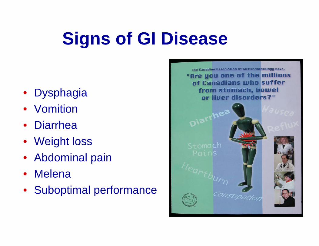
Signs of GI Diseaseg
• Dysphagia• Vomition• Vomition• Diarrhea
W i ht l• Weight loss• Abdominal pain
M l• Melena• Suboptimal performance
Diseases of Buccal Cavity
▪ Developmental abnormalities▪ Inflammatory diseases▪ NeoplasiaNeoplasia
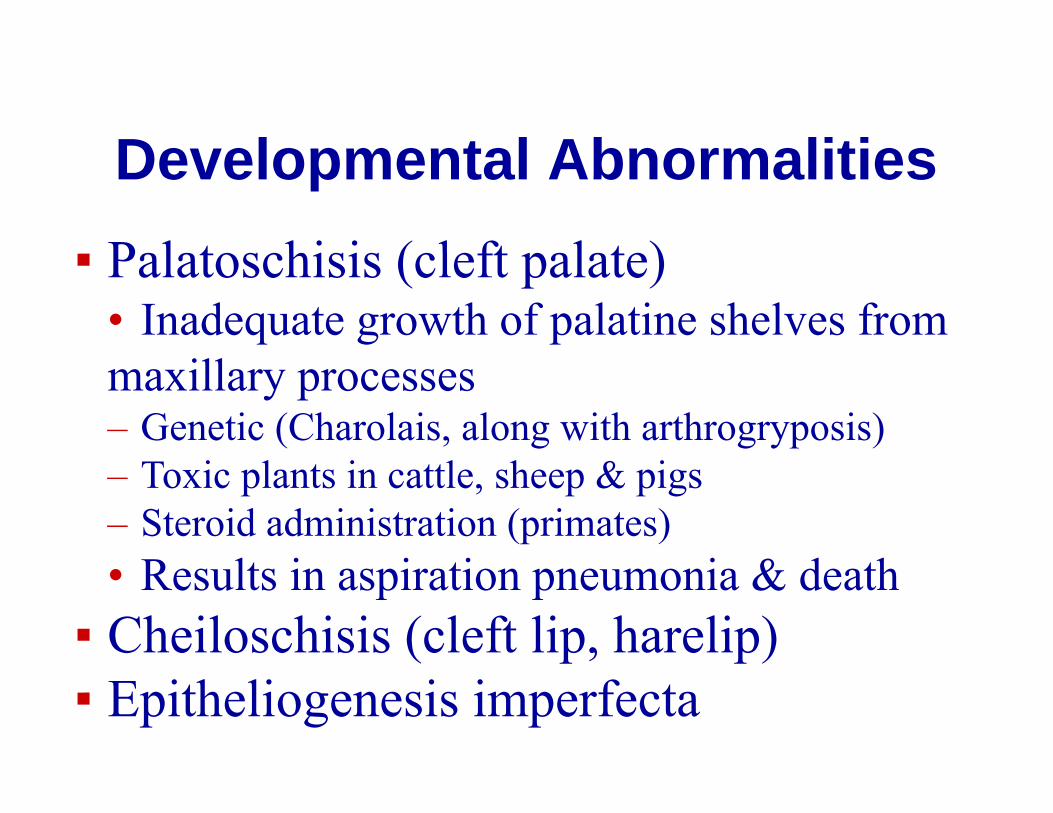
Developmental AbnormalitiesDevelopmental AbnormalitiesPalatoschisis (cleft palate)▪ Palatoschisis (cleft palate)• Inadequate growth of palatine shelves from maxillary processes– Genetic (Charolais, along with arthrogryposis)– Toxic plants in cattle, sheep & pigs– Steroid administration (primates)
R l i i i i & d h• Results in aspiration pneumonia & death▪ Cheiloschisis (cleft lip, harelip)( p p)▪ Epitheliogenesis imperfecta
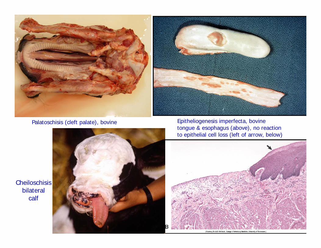
Epitheliogenesis imperfecta, bovine tongue & esophagus (above), no reaction to epithelial cell loss (left of arrow, below)
Palatoschisis (cleft palate), bovine
Cheiloschisisbilateral
calf
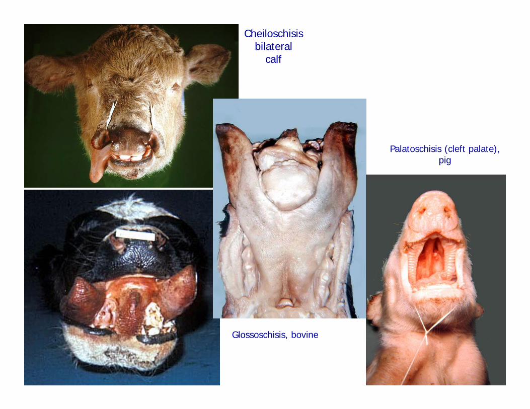
Cheiloschisisbilateral
calf
Palatoschisis (cleft palate), pig
Glossoschisis bovineGlossoschisis, bovine
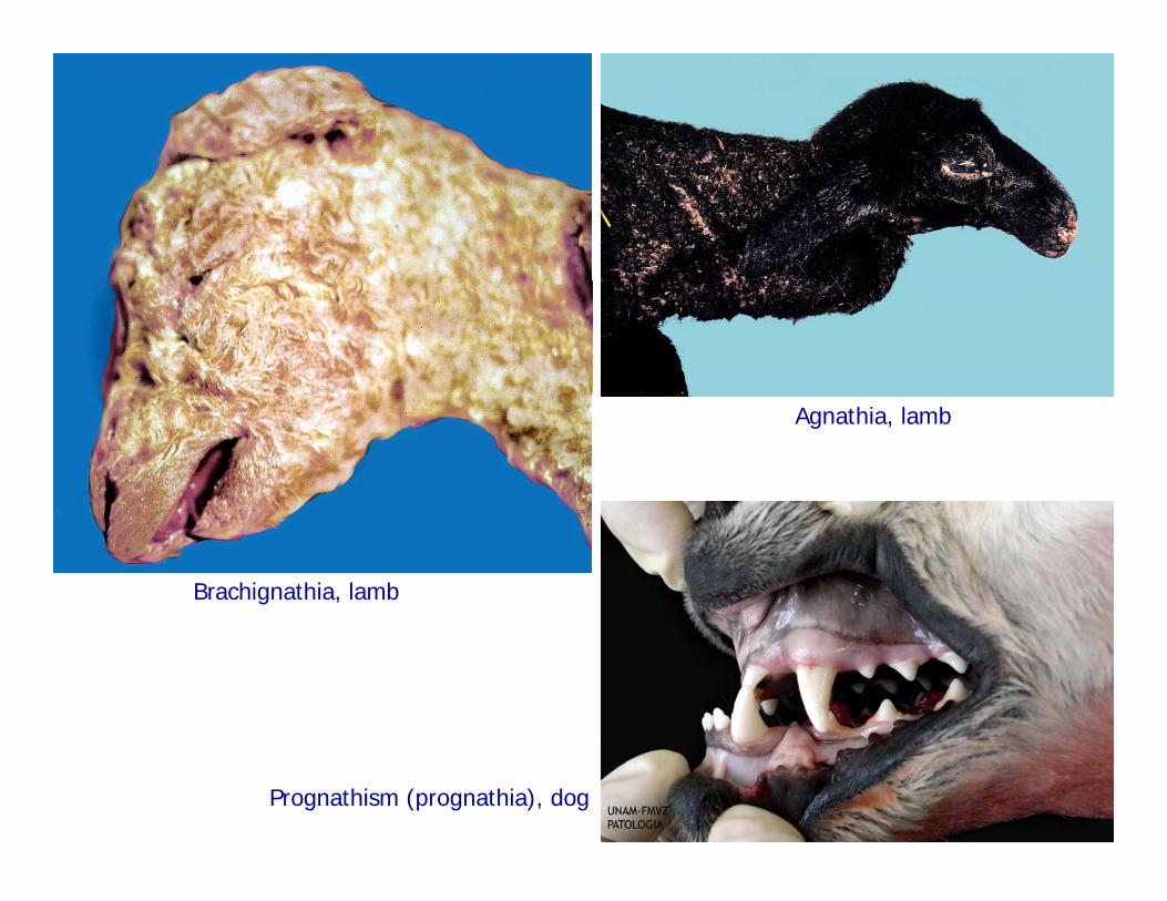
Agnathia, lamb
Brachignathia, lamb
Prognathism (prognathia), dog
Inflammation of the mouth – SomeInflammation of the mouth Some terminology
▪ Stomatitis – general term▪ Cheilitis - lipsp▪ Glossitis - tongue▪ Gingivitis - gingivaGingivitis - gingiva▪ Pharyngitis - pharynx
Tonsillitis tonsils▪ Tonsillitis - tonsils▪ Sialoadenitis – salivary glands
Causes of stomatitisCauses of stomatitis
▪ Infectious agents, local and septicemic▪ TraumaTrauma▪ Chemical injury
A i▪ Auto-immune▪ Systemic diseases y▪ Idiopathic
StomatitisStomatitis
Important indicator of local and some▪ Important indicator of local and some systemic diseases.▪ Morphologic manifestations include
• Inflammation (redness, papules,Inflammation (redness, papules, pseudomembranes, granuloma)• Degeneration (vesicles erosions ulcers)• Degeneration (vesicles, erosions, ulcers)• Often a combination of both
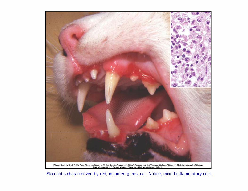
Stomatitis characterized by red, inflamed gums, cat. Notice, mixed inflammatory cells
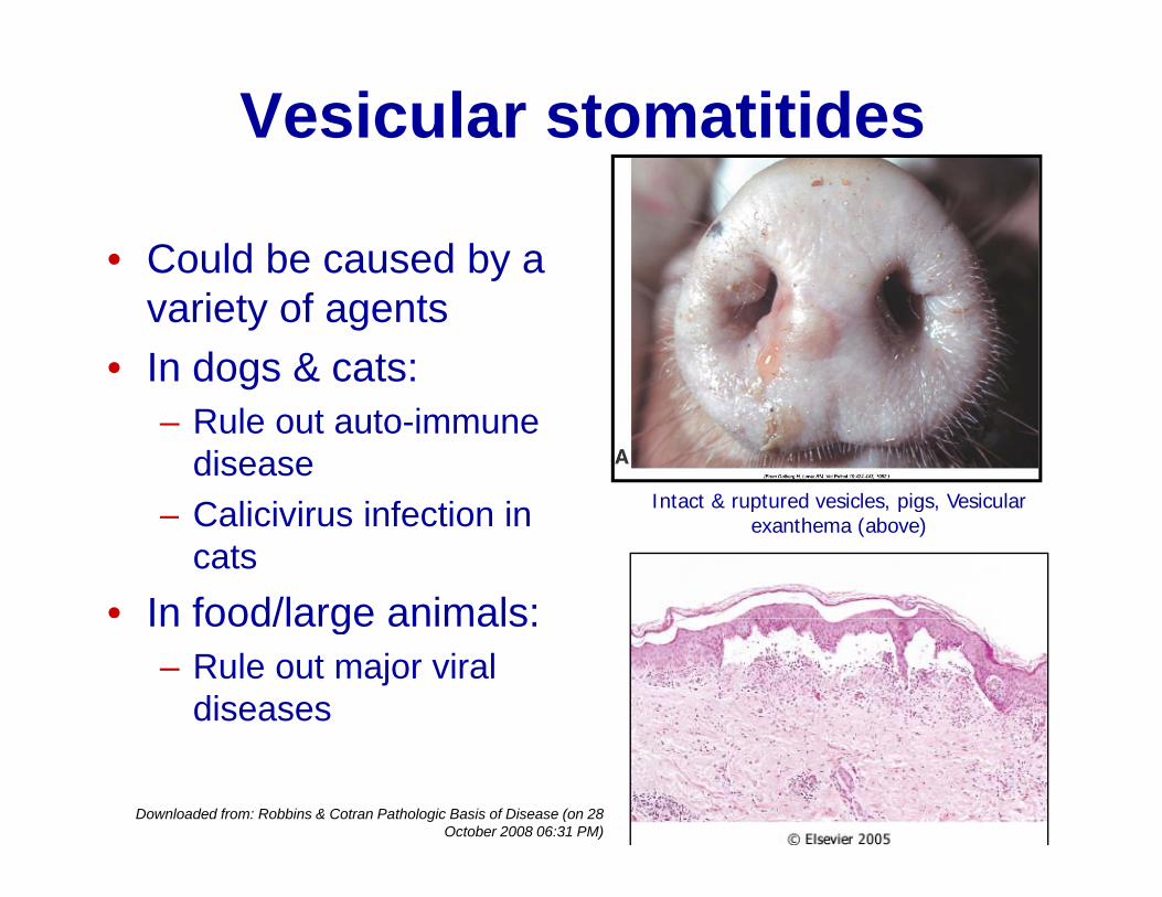
Vesicular stomatitides
• Could be caused by a yvariety of agents
• In dogs & cats:– Rule out auto-immune
disease C li i i i f ti i Intact & ruptured vesicles, pigs, Vesicular– Calicivirus infection in cats
• In food/large animals:
Intact & ruptured vesicles, pigs, Vesicular exanthema (above)
In food/large animals: – Rule out major viral
diseases
Downloaded from: Robbins & Cotran Pathologic Basis of Disease (on 28 October 2008 06:31 PM)

Pathogenesis of vesicularPathogenesis of vesicular stomatitides
▪ Epithelial damage (viral)intracellular edema & ballooning
degeneration vesicles bullae erosionsdegeneration vesicles bullae erosions ulcers cellular infiltration▪ Lesions in stratified epithelium▪ Lesions in stratified epithelium▪ No viral inclusion bodies
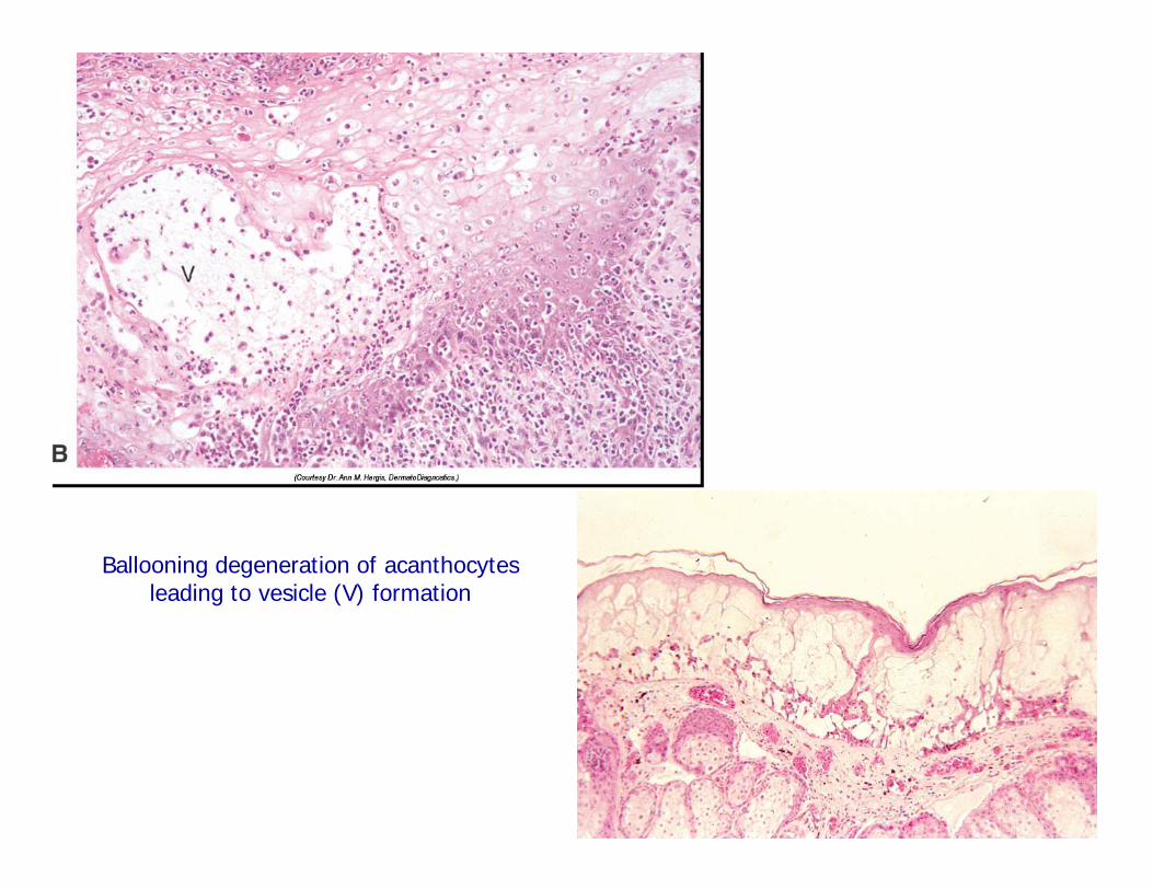
Ballooning degeneration of acanthocytes leading to vesicle (V) formation
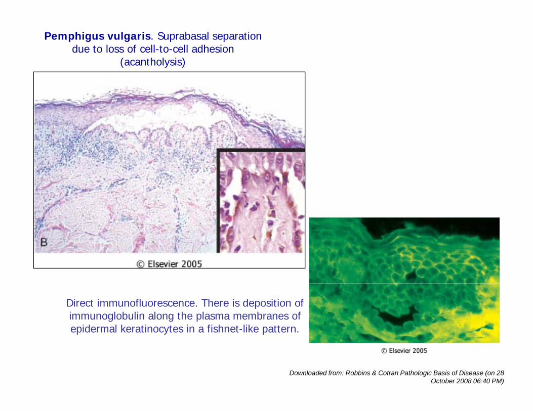
Pemphigus vulgaris. Suprabasal separation due to loss of cell-to-cell adhesion
(acantholysis)
Direct immunofluorescence. There is deposition of immunoglobulin along the plasma membranes of epidermal keratinocytes in a fishnet-like pattern.
Downloaded from: Robbins & Cotran Pathologic Basis of Disease (on 28 October 2008 06:40 PM)
Foot and Mouth DiseaseFoot and Mouth DiseaseFMD, Aphthous fever
(Most dreaded animal disease in the world)( )
▪ Caused by a picornavirus (7 antigenic types)y p ( g yp )▪ Affects mainly ruminants and pigs▪ Transmission by ingestion and by inhalationy g y▪ Infection of pharynx leads to viremia and widespread localization in epidermal sitesp p▪ Lesions only in sites subjected to mechanical injury▪ Clinical signs: drooling saliva, lameness, etc.g g▪ Highly contagious, high morbidity, low mortality except in neonates

FMD LesionsFMD Lesions▪ Mucosal hyperemia, vesicles/bullae on lips, oral mucosa, feet and teats.▪ Vesicles/bullae rupture erosions/ulcersp▪ Scab formation & scarification▪ Myocardial degeneration & myocarditis inMyocardial degeneration & myocarditis in
neonates acute heart failureComplications incl de▪ Complications include• Secondary bacterial infection
H f ti• Hoof separation
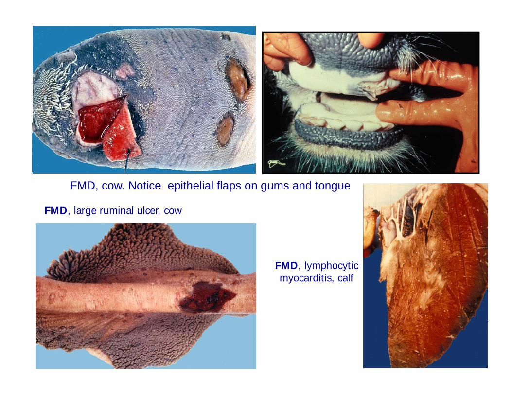
FMD, large ruminal ulcer, cow
FMD, cow. Notice epithelial flaps on gums and tongue
FMD, lymphocytic myocarditis, calf
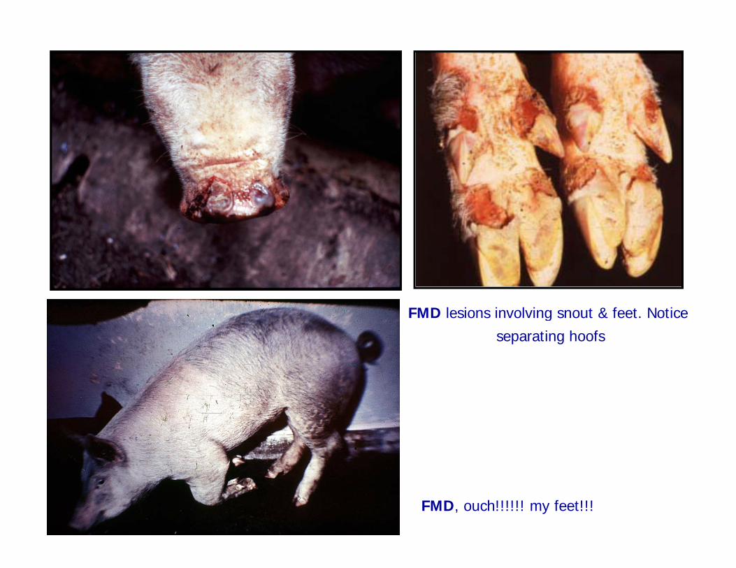
FMD lesions involving snout & feet. Notice g
separating hoofs
FMD, ouch!!!!!! my feet!!!
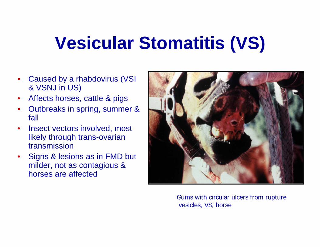
Vesicular Stomatitis (VS)Vesicular Stomatitis (VS)
C d b h bd i (VSI• Caused by a rhabdovirus (VSI & VSNJ in US)
• Affects horses, cattle & pigs• Outbreaks in spring summer &• Outbreaks in spring, summer &
fall • Insect vectors involved, most
likely through trans-ovarianlikely through trans ovarian transmission
• Signs & lesions as in FMD but milder, not as contagious & horses are affected
Gums with circular ulcers from rupturevesicles, VS, horse

Other vesicular stomatitidesOther vesicular stomatitides• Swine vesicular disease
– Caused by an enterovirus (Picornaviridae)
– Affects only swine (EuropeAffects only swine (Europe & Asia)
• Vesicular exanthema (VE)C– Caused by a calicivirus
– Affected mainly swine & sea lions in US until
di d i 1956eradicated in 1956
• Chemical irritationRuptured vesicles & ulceration, VE, pig uptu ed es c es & u ce at o , , p g
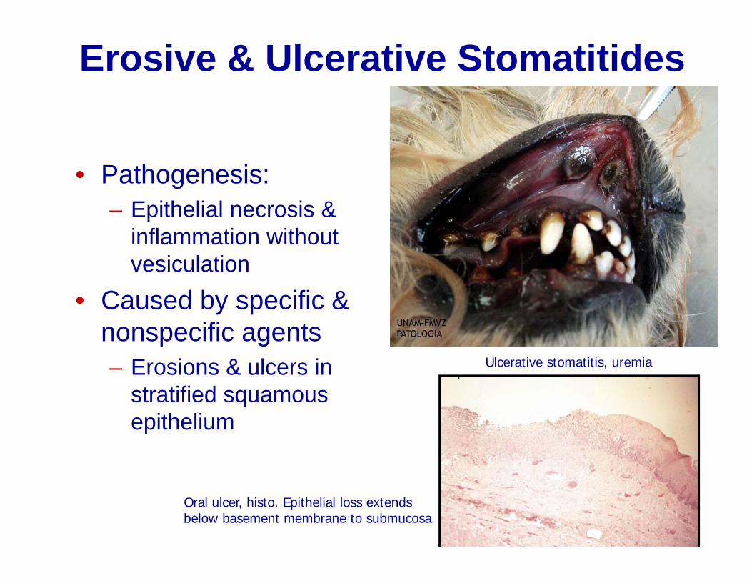
Erosive & Ulcerative Stomatitides
• Pathogenesis:Pathogenesis:– Epithelial necrosis &
inflammation without vesiculation
• Caused by specific & nonspecific agentsnonspecific agents– Erosions & ulcers in
stratified squamousUlcerative stomatitis, uremia
stratified squamous epithelium
Oral ulcer, histo. Epithelial loss extendsbelow basement membrane to submucosa
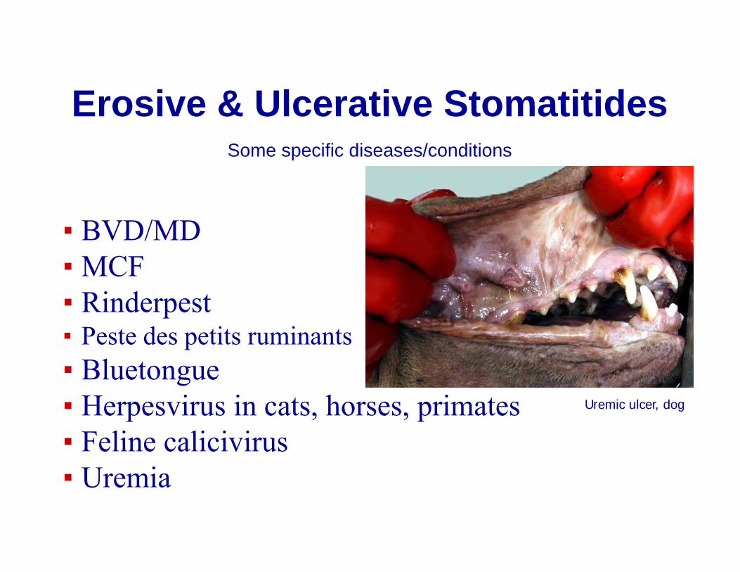
Erosive & Ulcerative StomatitidesErosive & Ulcerative StomatitidesSome specific diseases/conditions
▪ BVD/MD ▪ MCF▪ Rinderpest ▪ Peste des petits ruminants▪ Bluetongue▪ Herpesvirus in cats, horses, primates▪ Feline calicivirus
i
Uremic ulcer, dog
▪ Uremia
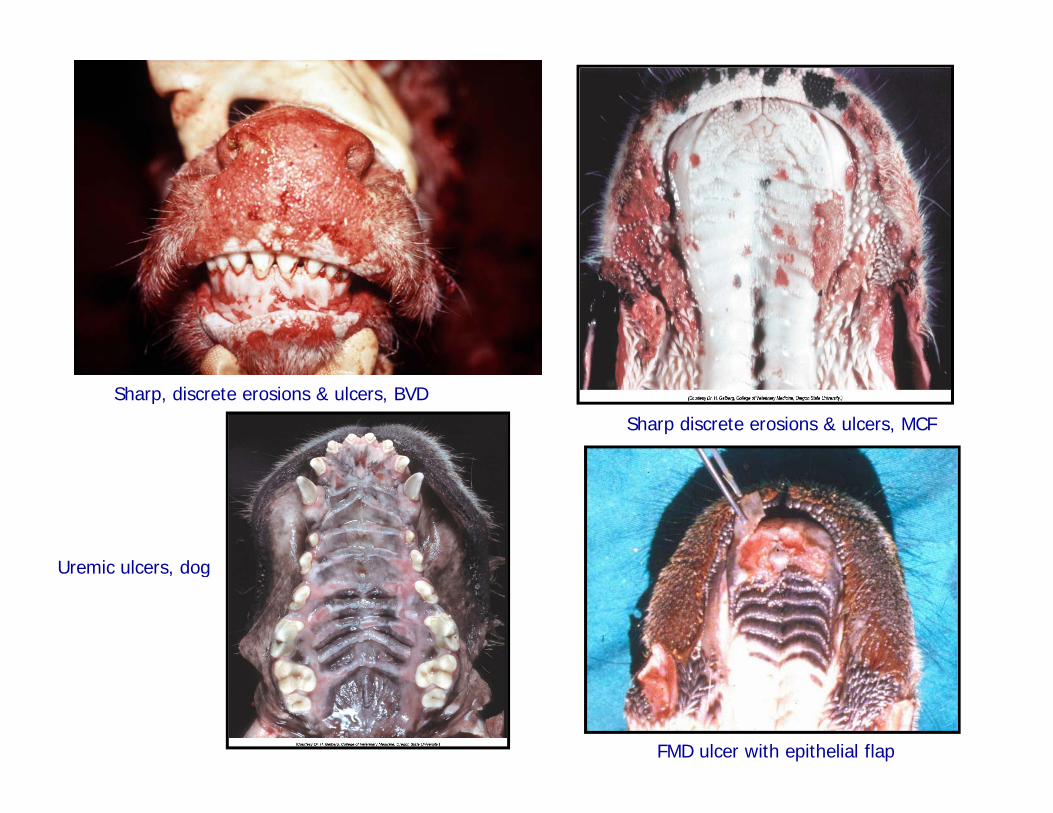
Sharp, discrete erosions & ulcers, BVD
Sharp discrete erosions & ulcers, MCF
U i l dUremic ulcers, dog
FMD ulcer with epithelial flap
Uremic StomatitisPathogenesis & Lesions
▪ PathogenesisPathogenesis▪ High blood & salivary urea bacterial infection
high ammonia caustic injuryhigh ammonia caustic injury▪ High serum blood urea nitrogen vascular damage thrombosis ischemia infarctiondamage thrombosis ischemia infarction▪ Increased urea decreased immune response▪ LesionsLesions▪ Ulcers most often near salivary ducts, around teeth with plaques & ventral surface of tongueteeth with plaques & ventral surface of tongue
(Affected animals often have ammoniacal odor to the breath).
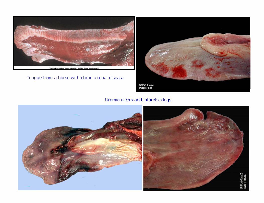
Tongue from a horse with chronic renal disease
Uremic ulcers and infarcts, dogs

Wh t i di i ?What is your diagnosis?
Recommended