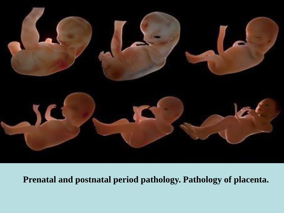
Prenatal and postnatal period pathology. Pathology of placenta.
Prenatal and postnatal period pathology. Pathology of placenta.I. Microspecimens:
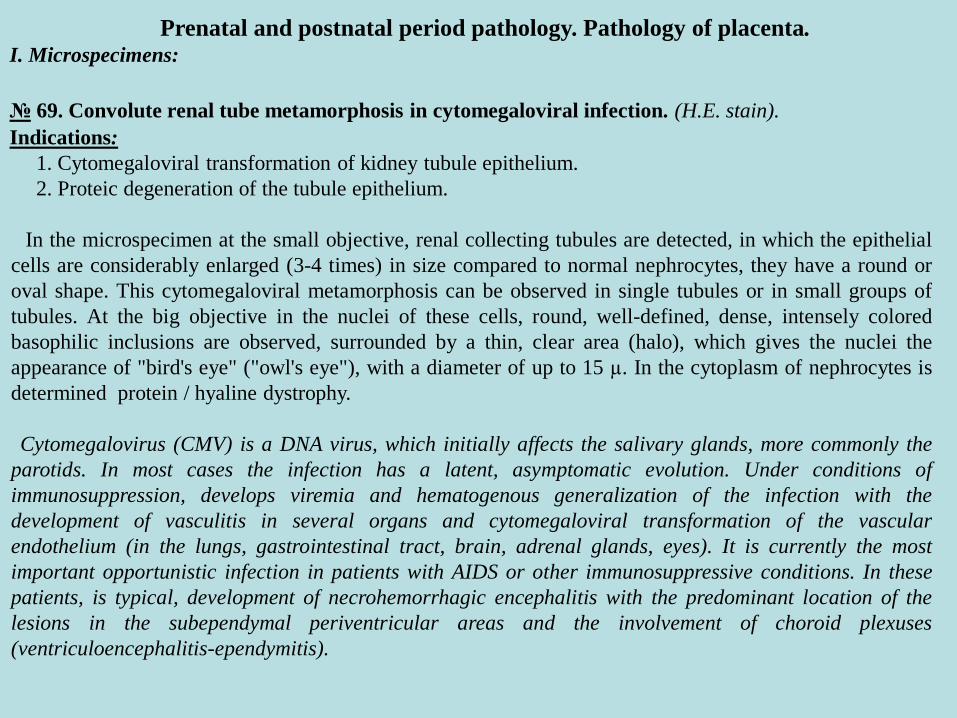
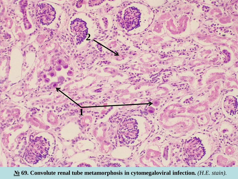
№ 69. Convolute renal tube metamorphosis in cytomegaloviral infection. (H.E. stain).
Indications:
1. Cytomegaloviral transformation of kidney tubule epithelium.
2. Proteic degeneration of the tubule epithelium.
In the microspecimen at the small objective, renal collecting tubules are detected, in which the epithelial
cells are considerably enlarged (3-4 times) in size compared to normal nephrocytes, they have a round or
oval shape. This cytomegaloviral metamorphosis can be observed in single tubules or in small groups of
tubules. At the big objective in the nuclei of these cells, round, well-defined, dense, intensely colored
basophilic inclusions are observed, surrounded by a thin, clear area (halo), which gives the nuclei the
appearance of "bird's eye" ("owl's eye"), with a diameter of up to 15 µ. In the cytoplasm of nephrocytes is
determined protein / hyaline dystrophy.
Cytomegalovirus (CMV) is a DNA virus, which initially affects the salivary glands, more commonly the
parotids. In most cases the infection has a latent, asymptomatic evolution. Under conditions of
immunosuppression, develops viremia and hematogenous generalization of the infection with the
development of vasculitis in several organs and cytomegaloviral transformation of the vascular
endothelium (in the lungs, gastrointestinal tract, brain, adrenal glands, eyes). It is currently the most
important opportunistic infection in patients with AIDS or other immunosuppressive conditions. In these
patients, is typical, development of necrohemorrhagic encephalitis with the predominant location of the
lesions in the subependymal periventricular areas and the involvement of choroid plexuses
(ventriculoencephalitis-ependymitis).
In newborns, especially in premature infants and in the early postnatal period, there is widespread a
severe form of infection. The morphological substrate consists in the cytomegalic metamorphosis of
endotheliocytes and epithelial cells from different parenchymal organs. The most serious complications are
encephalitis with periventricular necrosis, calcinosis, microcephaly, hydrocephalus.
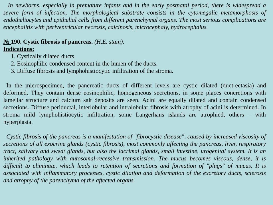
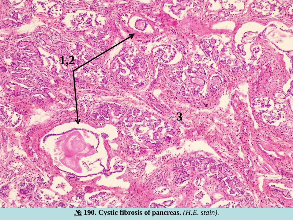
№ 190. Cystic fibrosis of pancreas. (H.E. stain).
Indications:
1. Cystically dilated ducts.
2. Eosinophilic condensed content in the lumen of the ducts.
3. Diffuse fibrosis and lymphohistiocytic infiltration of the stroma.
In the microspecimen, the pancreatic ducts of different levels are cystic dilated (duct-ectasia) and
deformed. They contain dense eosinophilic, homogeneous secretions, in some places concretions with
lamellar structure and calcium salt deposits are seen. Acini are equally dilated and contain condensed
secretions. Diffuse periductal, interlobular and intralobular fibrosis with atrophy of acini is determined. In
stroma mild lymphohistiocytic infiltration, some Langerhans islands are atrophied, others – with
hyperplasia.
Cystic fibrosis of the pancreas is a manifestation of "fibrocystic disease", caused by increased viscosity of
secretions of all exocrine glands (cystic fibrosis), most commonly affecting the pancreas, liver, respiratory
tract, salivary and sweat glands, but also the lacrimal glands, small intestine, urogenital system. It is an
inherited pathology with autosomal-recessive transmission. The mucus becomes viscous, dense, it is
difficult to eliminate, which leads to retention of secretions and formation of "plugs" of mucus. It is
associated with inflammatory processes, cystic dilation and deformation of the excretory ducts, sclerosis
and atrophy of the parenchyma of the affected organs.
Clinical manifestations may occur at birth or later in adolescence, and depend on the predominant location
of the lesions. Macroscopically the pancreas in cystic fibrosis is reduced in size, has a dense consistency,
nodular appearance, on section cysts of variable sizes are seen. It can be complicated by excretory
insufficiency, fat absorption disorder, steatorrhea, intestinal obstruction, A avitaminosis, cachexia.
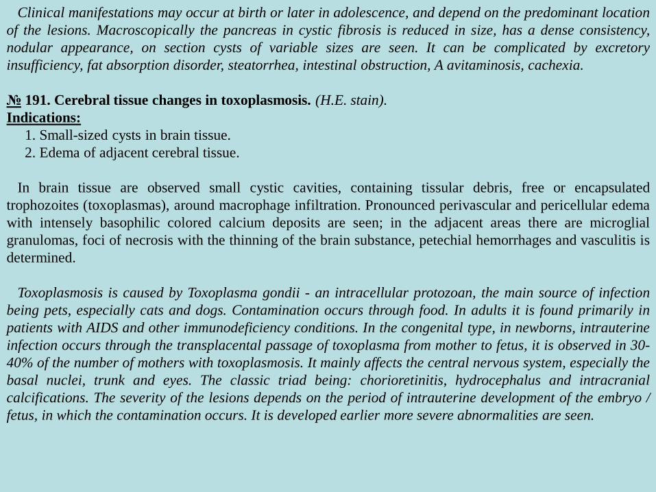
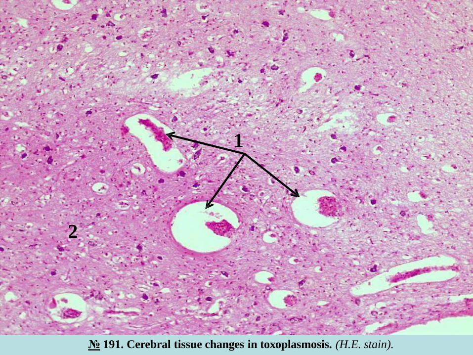
№ 191. Cerebral tissue changes in toxoplasmosis. (H.E. stain).
Indications:
1. Small-sized cysts in brain tissue.
2. Edema of adjacent cerebral tissue.
In brain tissue are observed small cystic cavities, containing tissular debris, free or encapsulated
trophozoites (toxoplasmas), around macrophage infiltration. Pronounced perivascular and pericellular edema
with intensely basophilic colored calcium deposits are seen; in the adjacent areas there are microglial
granulomas, foci of necrosis with the thinning of the brain substance, petechial hemorrhages and vasculitis is
determined.
Toxoplasmosis is caused by Toxoplasma gondii - an intracellular protozoan, the main source of infection
being pets, especially cats and dogs. Contamination occurs through food. In adults it is found primarily in
patients with AIDS and other immunodeficiency conditions. In the congenital type, in newborns, intrauterine
infection occurs through the transplacental passage of toxoplasma from mother to fetus, it is observed in 30-
40% of the number of mothers with toxoplasmosis. It mainly affects the central nervous system, especially the
basal nuclei, trunk and eyes. The classic triad being: chorioretinitis, hydrocephalus and intracranial
calcifications. The severity of the lesions depends on the period of intrauterine development of the embryo /
fetus, in which the contamination occurs. It is developed earlier more severe abnormalities are seen.
In cerebral toxoplasmosis, microcephaly, hydrocephalus, multiple cystic cavities, calcifications, abscesses
are observed. Ocular complications: microphthalmia, cataracts, calcifications in the retina and vascular
membrane is determined. Intrauterine death of the fetus can occur, and in the postnatal period - cachexia,
paralysis, mental retardation, blindness, the association of secondary infection with the development of
purulent meningoencephalitis are seen.
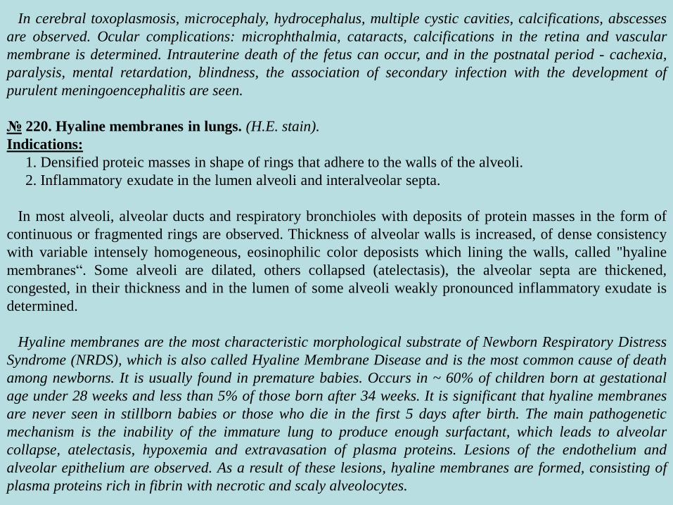
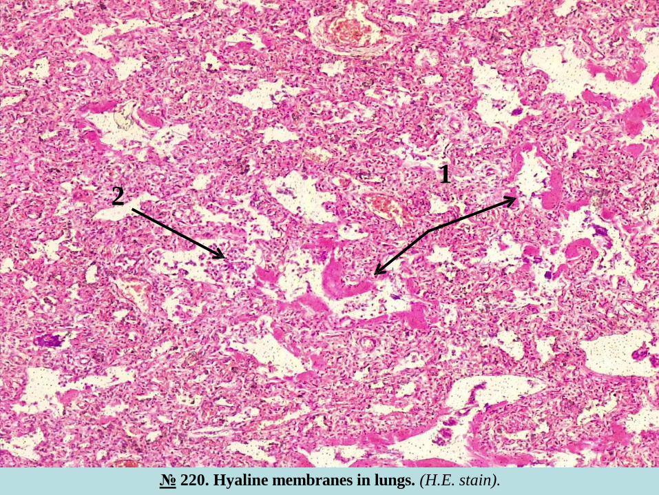
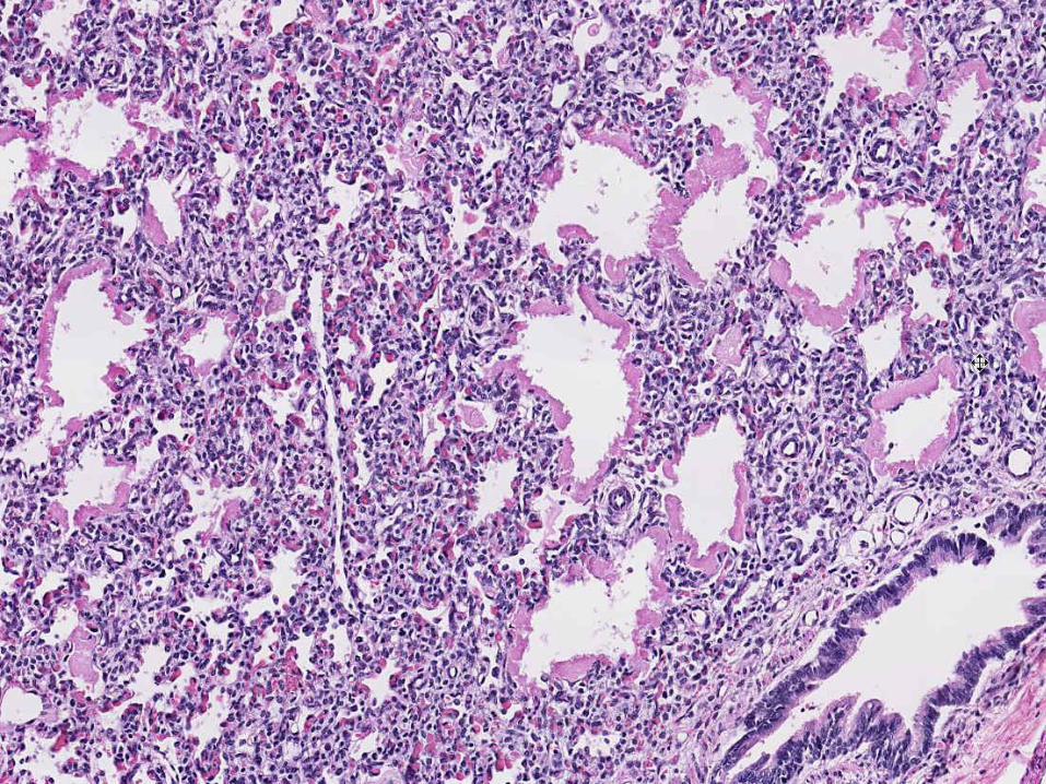
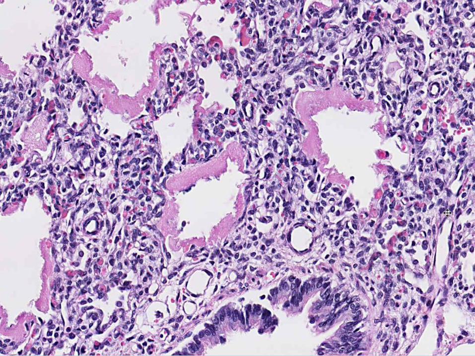
№ 220. Hyaline membranes in lungs. (H.E. stain).
Indications:
1. Densified proteic masses in shape of rings that adhere to the walls of the alveoli.
2. Inflammatory exudate in the lumen alveoli and interalveolar septa.
In most alveoli, alveolar ducts and respiratory bronchioles with deposits of protein masses in the form of
continuous or fragmented rings are observed. Thickness of alveolar walls is increased, of dense consistency
with variable intensely homogeneous, eosinophilic color deposists which lining the walls, called "hyaline
membranes“. Some alveoli are dilated, others collapsed (atelectasis), the alveolar septa are thickened,
congested, in their thickness and in the lumen of some alveoli weakly pronounced inflammatory exudate is
determined.
Hyaline membranes are the most characteristic morphological substrate of Newborn Respiratory Distress
Syndrome (NRDS), which is also called Hyaline Membrane Disease and is the most common cause of death
among newborns. It is usually found in premature babies. Occurs in ~ 60% of children born at gestational
age under 28 weeks and less than 5% of those born after 34 weeks. It is significant that hyaline membranes
are never seen in stillborn babies or those who die in the first 5 days after birth. The main pathogenetic
mechanism is the inability of the immature lung to produce enough surfactant, which leads to alveolar
collapse, atelectasis, hypoxemia and extravasation of plasma proteins. Lesions of the endothelium and
alveolar epithelium are observed. As a result of these lesions, hyaline membranes are formed, consisting of
plasma proteins rich in fibrin with necrotic and scaly alveolocytes.

Hyaline membranes are a barrier to gas exchange and cause acute respiratory failure. The mortality in
RDS of the newborn reaches 20-30%.
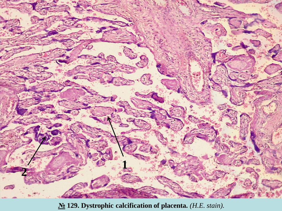
№ 129. Dystrophic calcification of placenta. (H.E. stain).
Indications:
1. Chorionic villi.
2. Calcium deposits into stroma of chorionic villi.
In the microspecimen are small, scattered deposits of calcium salts of basophilic color, located in the
stroma of chorionic villi are seen. The villi are atrophied, sclerosed, the blood vessels are equally sclerosed,
in the intervilositary spaces deposits of eosinophilic, homogeneous fibrin is observed. Some villi are necrotic,
surrounded by fibrinoid masses. On the surface of the villi in some places there are small foci of proliferation
of the syncytiotrophoblastic epithelium (syncytial buds), formed by giant polynuclear cells, with intensely
basophilic nuclei.
Macroscopically on the maternal surface of the placenta there are yellowish-whitish foci of calcinosis on
the red background of the placental tissue. Calcinosis of the chorionic villi and other dystrophic lesions of the
placenta appear towards the end of pregnancy and are particularly characteristic for prolonged (post-term)
pregnancies ≥ 42 weeks / gestation. Calcinosis also develops as a result of various pathological processes,
which can occur in the placenta during pregnancy, eg., calcium deposits in fibrinoid foci, foci of necrosis in
the stroma of villi, sclerosed blood vessels, vascular thrombi. An important role is played by extragenital
diseases, which are found in pregnant women, such as diabetes, hypertension, gestosis, preeclampsia.

II. Macrospecimens:
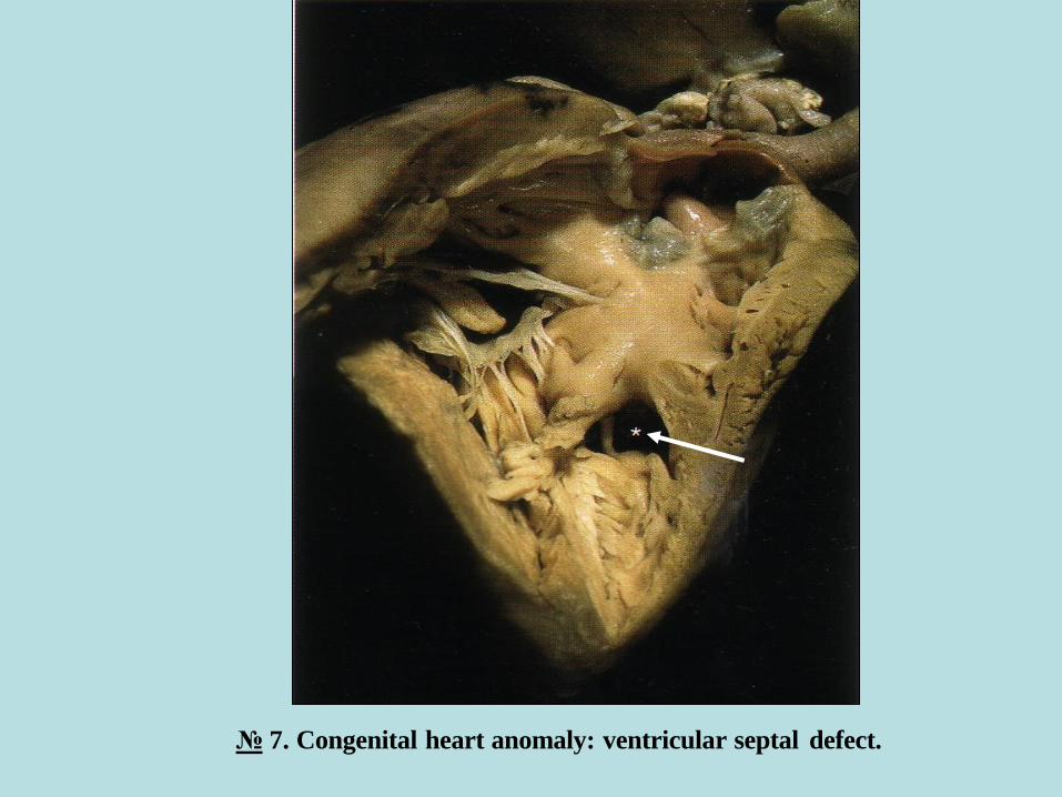
№ 7. Congenital heart anomaly: ventricular septal defect.
In the interventricular septum there is a defect with a diameter of 1.5-2 cm, located in the basal,
membranous region, the wall of the left ventricle has a normal thickness. Due to this defect, abnormal
communication takes place between the left ventricle and the right ventricle - "left to right shunt". In such
abnormalities the pulmonary blood flow increases and no cyanosis and hypoxia (cardiac malformation of the
cyanotic or white type) are observed.
Ventricular septal defects are the most common congenital malformation of the heart (~ 30% of the total
number), the usual location being at the level of the membranous, fibroconnective tissue part of the septum.
In most cases, the defect closes spontaneously in childhood. Small defects are asymptomatic but may
progress clinically. Large defects require early surgical correction to prevent the progression of the "left to
right" shunt, which can lead to congestive heart failure. In approximately 70% of cases, ventricular septal
defects are associated with other congenital heart malformations.
№ 77. Polycystic liver disease.
In the liver on cut section are present multiple cystic cavities of varying sizes and shapes, the liver
parenchyma between cysts is with signs of steatosis. In most cases it is associated with cystic fibrosis of the
pancreas, respiratory tract, salivary and sweating glands, being one of the manifestations of "fibrocystic
disease" (microspecimen no. 190).
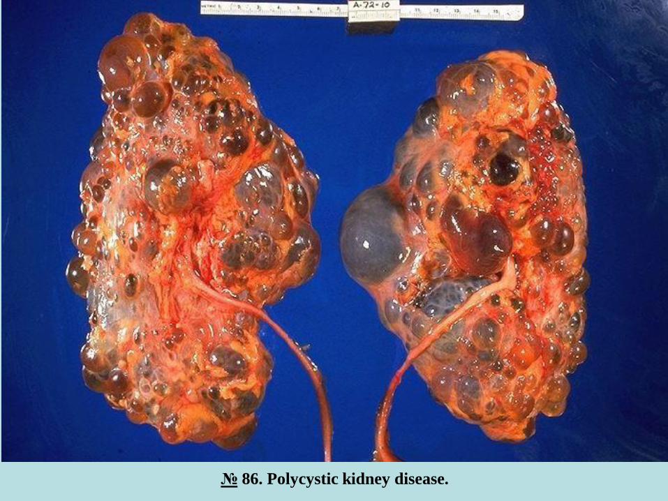
№ 86. Polycystic kidney disease.
The kidney has a voluminous mass, consisting of round and oval cysts, with sizes ranging from 0.5 cm to 3-
4 cm with thin walls, smooth inner surface, clear contents, between cysts there is atrophied renal parenchyma
or even absent.
It is the morphological substrate of adult polycystic kidney disease - a condition with autosomal dominant
transmission. It has an incidence of 1 in 500-1000 people and is ~ 10% of cases of chronic kidney disease.
Cysts can form at any level of the nephron. In some cases it is associated with hepatic and pancreatic cysts.
Complications: chronic renal failure, urinary tract infections (pyelonephritis), hypertension (cerebral
hemorrhage).
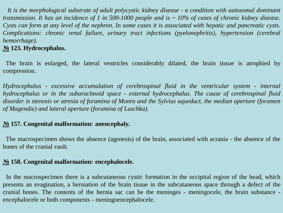
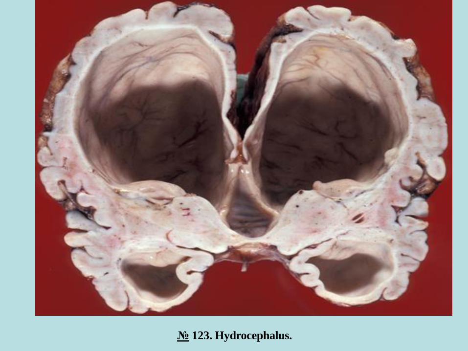
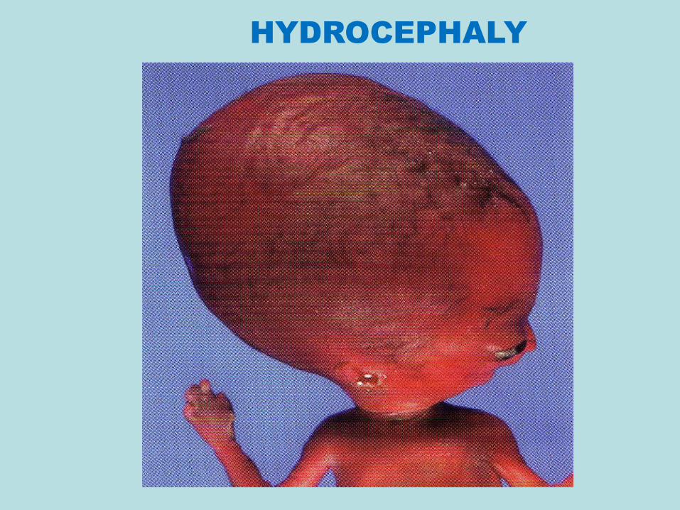
№ 123. Hydrocephalus.
The brain is enlarged, the lateral ventricles considerably dilated, the brain tissue is atrophied by
compression.
Hydrocephalus - excessive accumulation of cerebrospinal fluid in the ventricular system - internal
hydrocephalus or in the subarachnoid space - external hydrocephalus. The cause of cerebrospinal fluid
disorder is stenosis or atresia of foramina of Monro and the Sylvius aqueduct, the median aperture (foramen
of Magendie) and lateral aperture (foramina of Luschka).
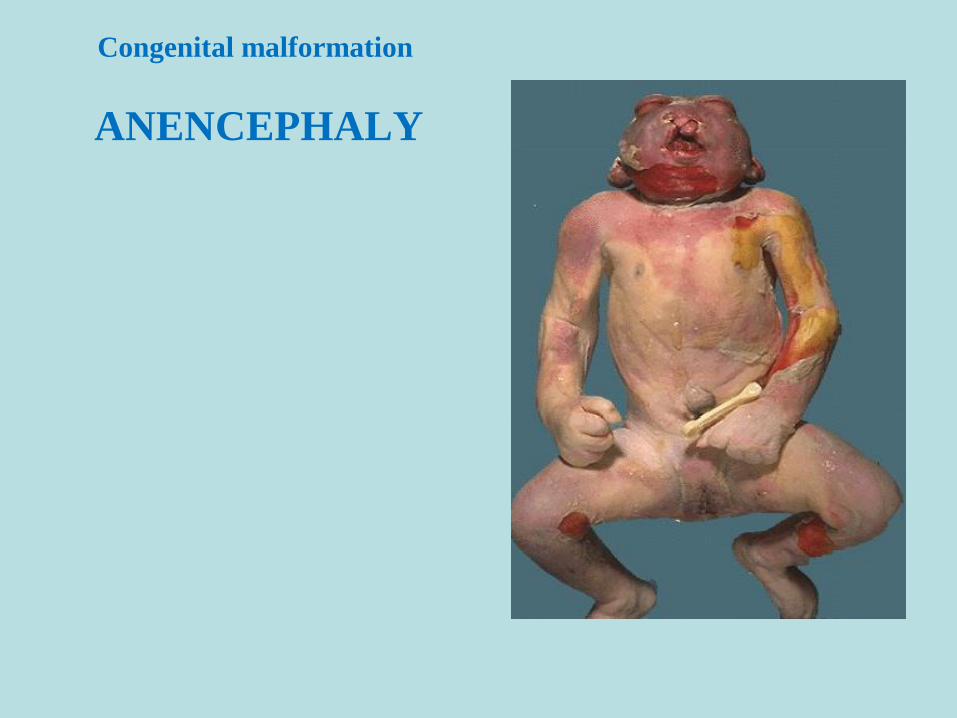
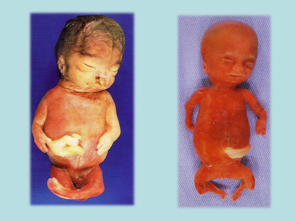
№ 157. Congenital malformation: anencephaly.
The macrospecimen shows the absence (agenesis) of the brain, associated with acrania - the absence of the
bones of the cranial vault.
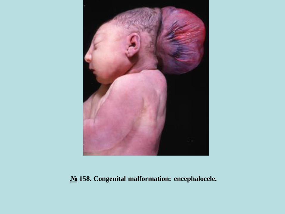
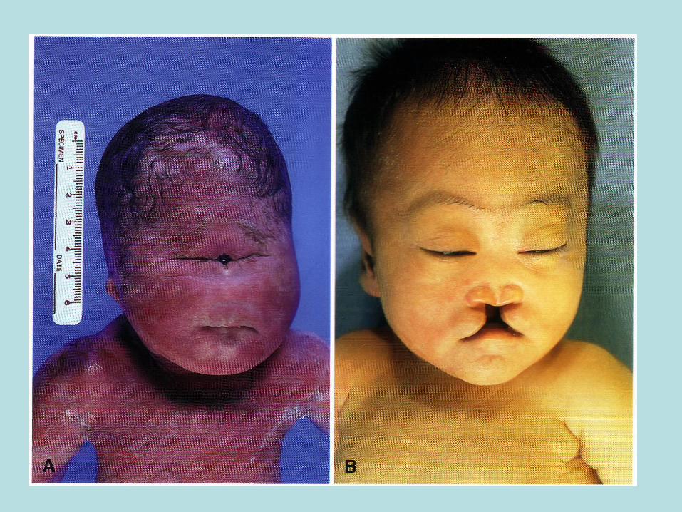
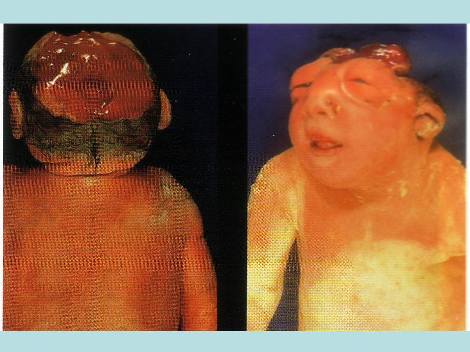
№ 158. Congenital malformation: encephalocele.
In the macrospecimen there is a subcutaneous cystic formation in the occipital region of the head, which
presents an evagination, a herniation of the brain tissue in the subcutaneous space through a defect of the
cranial bones. The contents of the hernia sac can be the meninges - meningocele, the brain substance -
encephalocele or both components - meningoencephalocele.
№ 69. Convolute renal tube metamorphosis in cytomegaloviral infection. (H.E. stain).
2
1
№ 190. Cystic fibrosis of pancreas. (H.E. stain).
3
1,2
№ 191. Cerebral tissue changes in toxoplasmosis. (H.E. stain).
2
1
№ 220. Hyaline membranes in lungs. (H.E. stain).
21
№ 129. Dystrophic calcification of placenta. (H.E. stain).
21
№ 7. Congenital heart anomaly: ventricular septal defect.

№ 77. Polycystic liver disease.
№ 86. Polycystic kidney disease.
№ 123. Hydrocephalus.

№ 157. Congenital malformation: anencephaly.
№ 158. Congenital malformation: encephalocele.
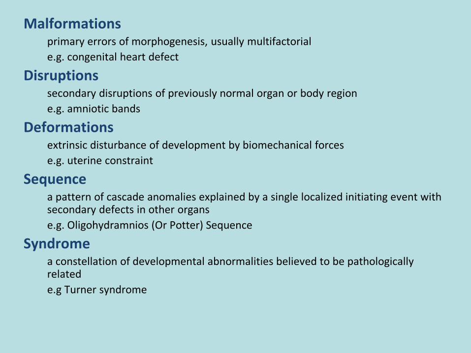
Malformationsprimary errors of morphogenesis, usually multifactorial
e.g. congenital heart defect
Disruptionssecondary disruptions of previously normal organ or body region
e.g. amniotic bands
Deformationsextrinsic disturbance of development by biomechanical forces
e.g. uterine constraint
Sequencea pattern of cascade anomalies explained by a single localized initiating event with secondary defects in other organs
e.g. Oligohydramnios (Or Potter) Sequence
Syndromea constellation of developmental abnormalities believed to be pathologically related
e.g Turner syndrome
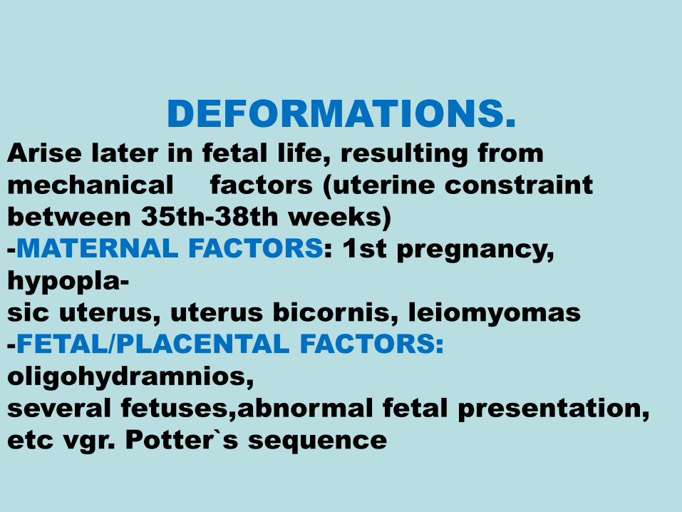
DEFORMATIONS.Arise later in fetal life, resulting from
mechanical factors (uterine constraint
between 35th-38th weeks)
-MATERNAL FACTORS: 1st pregnancy,
hypopla-
sic uterus, uterus bicornis, leiomyomas
-FETAL/PLACENTAL FACTORS:
oligohydramnios,
several fetuses,abnormal fetal presentation,
etc vgr. Potter`s sequence
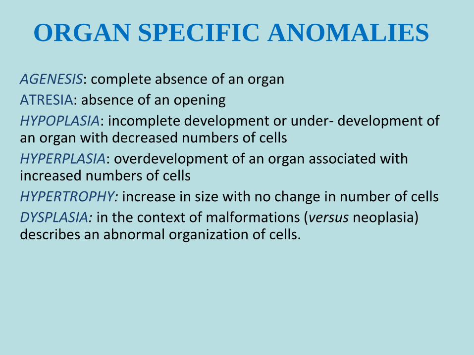
ORGAN SPECIFIC ANOMALIES
AGENESIS: complete absence of an organ
ATRESIA: absence of an opening
HYPOPLASIA: incomplete development or under- development of an organ with decreased numbers of cells
HYPERPLASIA: overdevelopment of an organ associated with increased numbers of cells
HYPERTROPHY: increase in size with no change in number of cells
DYSPLASIA: in the context of malformations (versus neoplasia) describes an abnormal organization of cells.

IMPLANTATION
AND THE
SURVIVAL OF
EARLY
PREGNANCY
Only 50-60% of all conceptions advance beyond 20 weeks
Implantation occurs at day 6-7
75% of loses are implantation failures and are not recognized
Pregnancy loss after implantation is 25-40%

EMBRYONIC
DEVELOPMEN
T
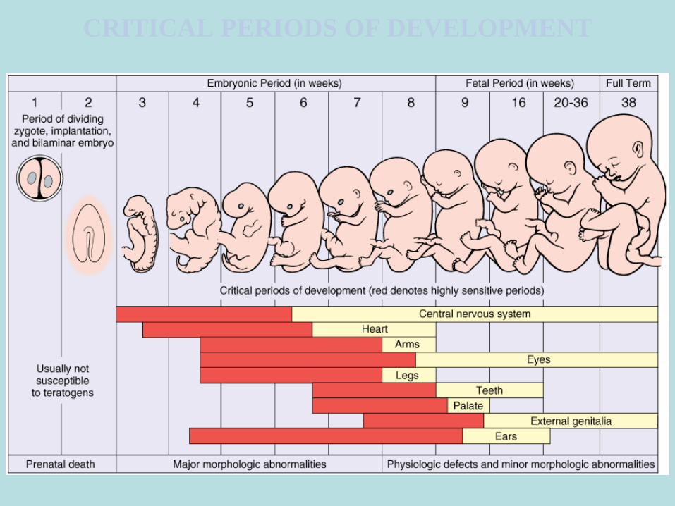
Embryonic period
weeks 1- 8 of pregnancy
organogenesis occurs in this period
Fetal period
weeks 9 to 38
marked by further growth and maturation
CRITICAL PERIODS OF DEVELOPMENT
BLASOPATHY
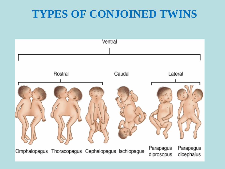
TYPES OF CONJOINED TWINS
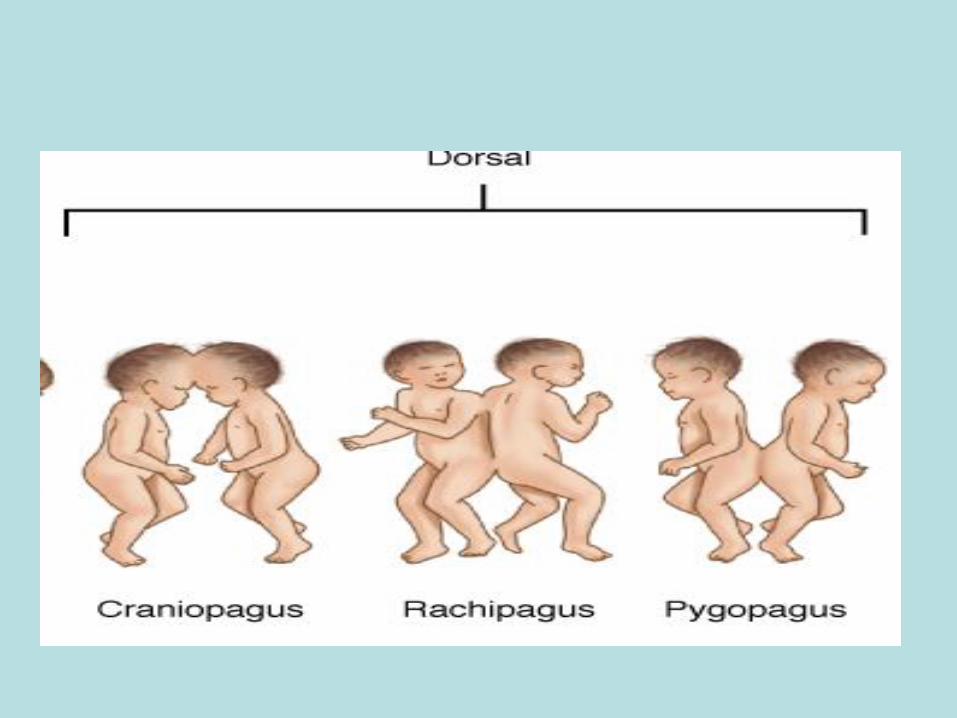
TYPES OF
CONJOINED
TWINS

CEPHALOTHORACOPAGUS
MALFORMATIONSPolydactyly &
syndactyly Cleft Lip
Severe Lethal Malformation
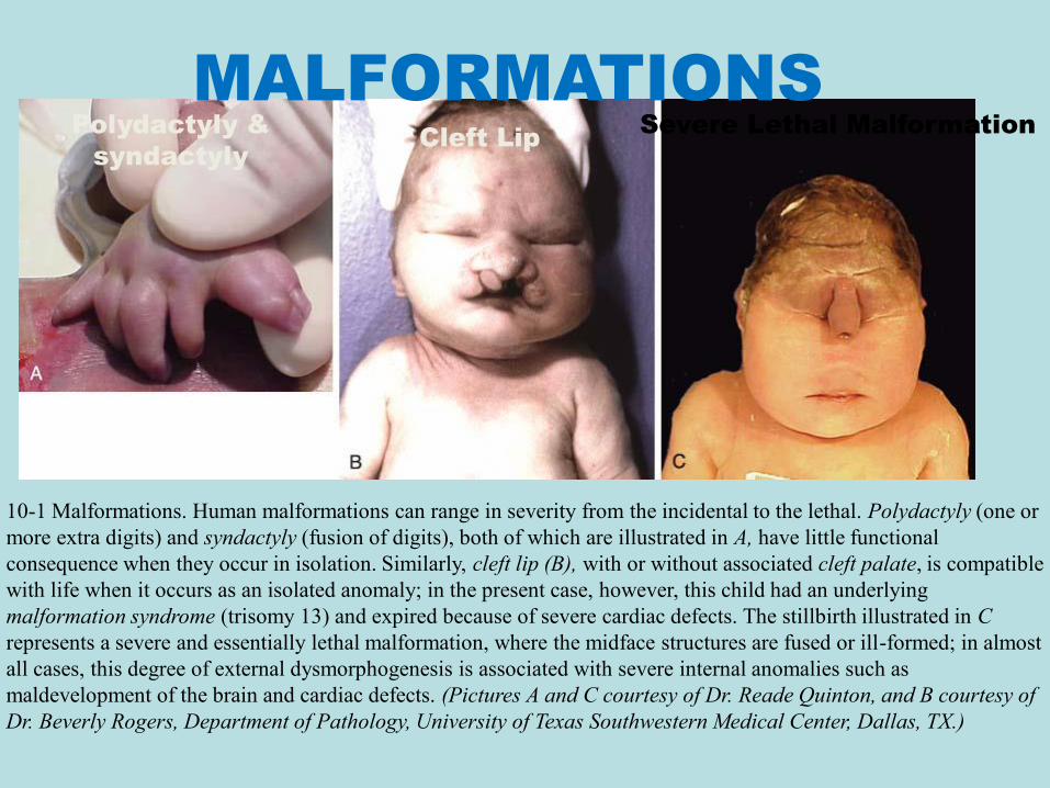
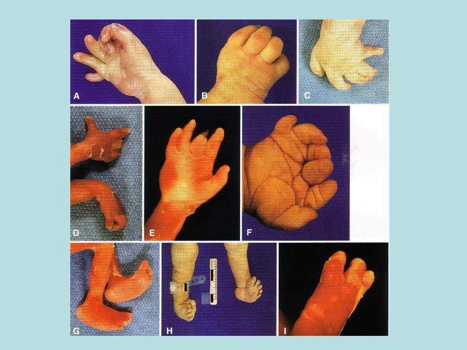
10-1 Malformations. Human malformations can range in severity from the incidental to the lethal. Polydactyly (one or
more extra digits) and syndactyly (fusion of digits), both of which are illustrated in A, have little functional
consequence when they occur in isolation. Similarly, cleft lip (B), with or without associated cleft palate, is compatible
with life when it occurs as an isolated anomaly; in the present case, however, this child had an underlying
malformation syndrome (trisomy 13) and expired because of severe cardiac defects. The stillbirth illustrated in C
represents a severe and essentially lethal malformation, where the midface structures are fused or ill-formed; in almost
all cases, this degree of external dysmorphogenesis is associated with severe internal anomalies such as
maldevelopment of the brain and cardiac defects. (Pictures A and C courtesy of Dr. Reade Quinton, and B courtesy of
Dr. Beverly Rogers, Department of Pathology, University of Texas Southwestern Medical Center, Dallas, TX.)
Congenital malformation
ANENCEPHALY
HYDROCEPHALY
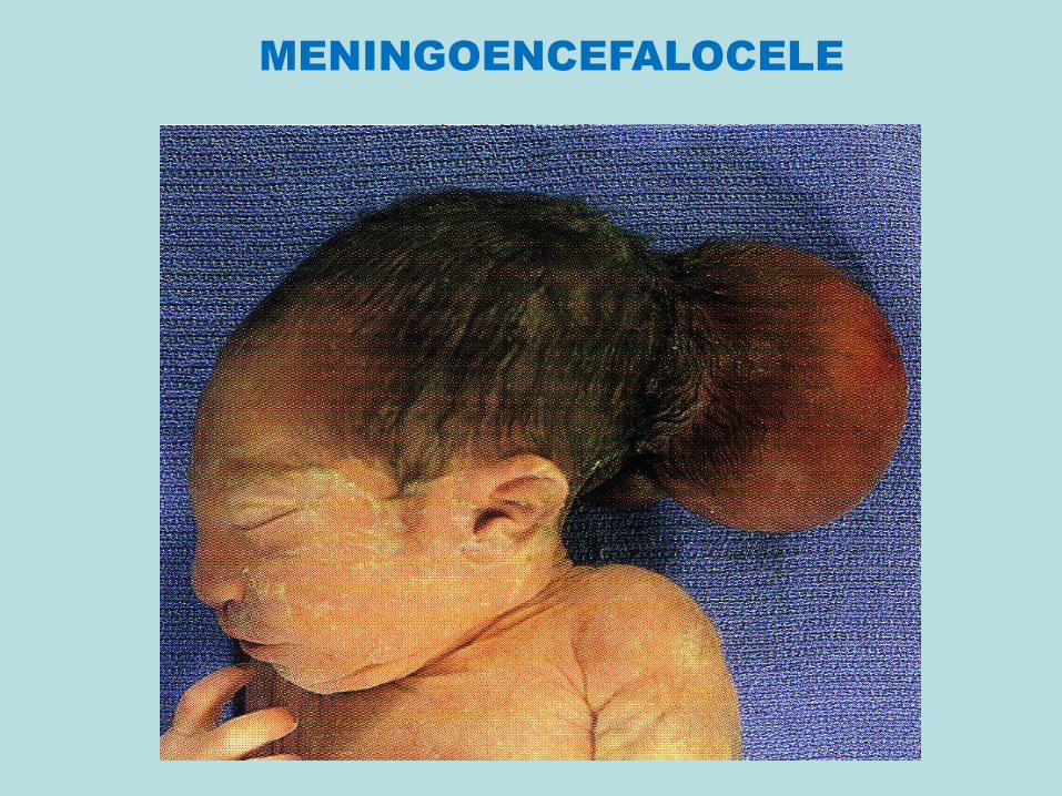
MENINGOENCEFALOCELE

IHTIOSIS
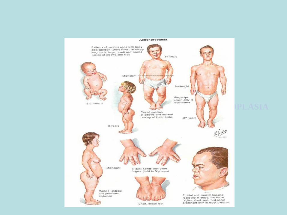
ACHONDROPLASIA
CAUSES OF CONGENITAL
MALFORMATIONS
Genetic
karyotypic aberrations
single gene mutations
Environmental
infection
maternal disease
drugs and chemicals
irradiation
Multifactorial
Unknown
A. Chromosomal aberrations are present in
about 10-50% of livebirth infants w/some
malformation:
1. Down syndrome(trisomy 21-1/1000 n.b)
2. Klinefelter syndrome(47-XXY)
3. Turner syndrome(45-X0)
4. Patau syndrome(trisomy 13)
GENETIC CAUSES OF
CONGENITAL
MALFORMATIONS

ENVIRONMENTAL
A. Viruses.
CMV intrauterine infection(highest risk in
second trimester of pregnancy)
Rubella syndrome(greater risk in 1st eight
wks of gestation)→cataracts, persistent
ductus arteriosus, tetralogy of Fallot, etc.
B. Drugs/chemicals: alcohol, androgens,
anticonvulsivants, etc
C. Radiation
MULTIFACTORIAL

HYDROPS FETALIS
Chromosomal abnormalities
Turner syndrome with cystic hygromas
other
Cardiovascular with heart failure
anemia with high output failure
immune hemolytic anemia
hereditary hemolytic anemia (α-thalassemia)
parvovirus B19 infection
twin to twin in utero transfusion
congenital heart defects
HYDROPS FETALIS
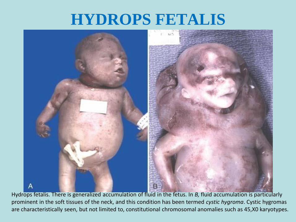
Hydrops fetalis. There is generalized accumulation of fluid in the fetus. In B, fluid accumulation is particularly prominent in the soft tissues of the neck, and this condition has been termed cystic hygroma. Cystic hygromas are characteristically seen, but not limited to, constitutional chromosomal anomalies such as 45,X0 karyotypes.

Timming of prenatal teratogenic
insult has an important impact on
the occurrence and type of
malformation produced.
Intrauterine development in
humans are divided in 2 phases:
A. Embryonic period(first 9 wks)
B. Fetal period(following wks)
MECHANISMS OF MALFORMATIONS
THE CENTRAL
NERVOUS
SYSTEM
THROUGHOUT
LIFE
Congenital malformations
Hydrocephalus
Neural tube defects
Anencephaly – cerebrum and cerebellum are absent
Spina bifida – absence of vertebral lamina
Cerebral palsy – voluntary muscles are poorly controlled
Results from damage to the motor cortex
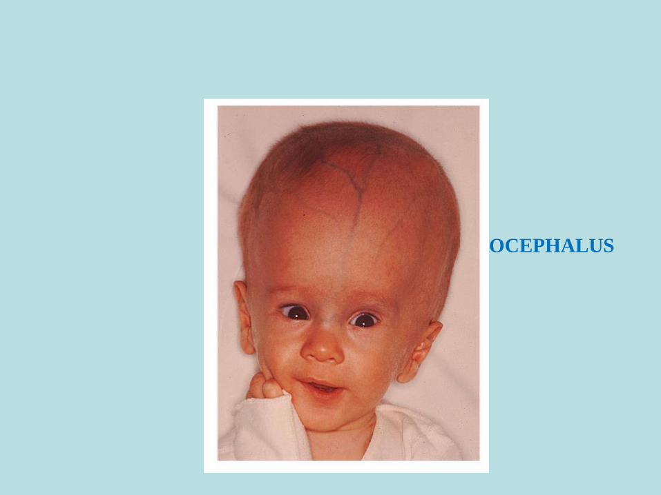
HYDROCEPHALUS
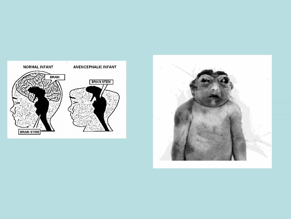
ANENCEPHALY

SPINA BIFIDA
CARDIAC
MALFORMATIONS
Present in 0.8% of North American and European children
Most common category of congenital structural malformation
Commonly divided into noncyanotic (L → R) and cyanotic (R → L) categories based on direction of shunting
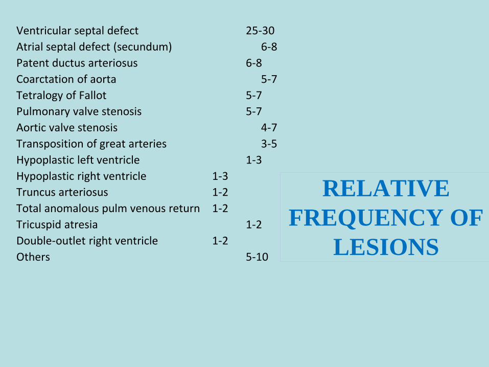
RELATIVE
FREQUENCY OF
LESIONS
Ventricular septal defect 25-30
Atrial septal defect (secundum) 6-8
Patent ductus arteriosus 6-8
Coarctation of aorta 5-7
Tetralogy of Fallot 5-7
Pulmonary valve stenosis 5-7
Aortic valve stenosis 4-7
Transposition of great arteries 3-5
Hypoplastic left ventricle 1-3
Hypoplastic right ventricle 1-3
Truncus arteriosus 1-2
Total anomalous pulm venous return 1-2
Tricuspid atresia 1-2
Double-outlet right ventricle 1-2
Others 5-10
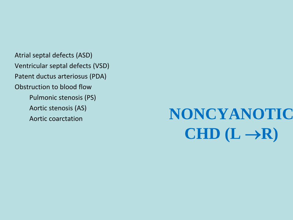
NONCYANOTIC
CHD (L →R)
Atrial septal defects (ASD)
Ventricular septal defects (VSD)
Patent ductus arteriosus (PDA)
Obstruction to blood flow
Pulmonic stenosis (PS)
Aortic stenosis (AS)
Aortic coarctation
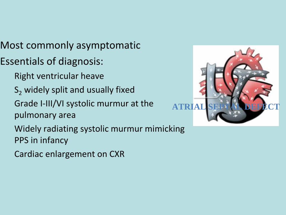
ATRIAL SEPTAL DEFECT
Most commonly asymptomatic
Essentials of diagnosis:
Right ventricular heave
S2 widely split and usually fixed
Grade I-III/VI systolic murmur at the pulmonary area
Widely radiating systolic murmur mimicking PPS in infancy
Cardiac enlargement on CXR
54
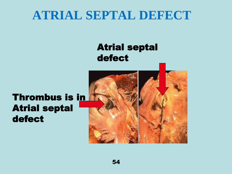
ATRIAL SEPTAL DEFECT
Thrombus is in
Atrial septal
defect
Atrial septal
defect

Atrial Septal Defect
VENTRICULAR
SEPTAL DEFECT
Single most common congenital heart malformation, accounting for almost 30% of all CHD
Defects can occur in both the membranous portion of the septum (most common) and the muscular portion
57
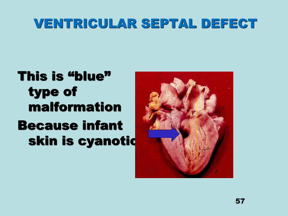
This is “blue”
type of
malformation
Because infant
skin is cyanotic
VENTRICULAR SEPTAL DEFECT
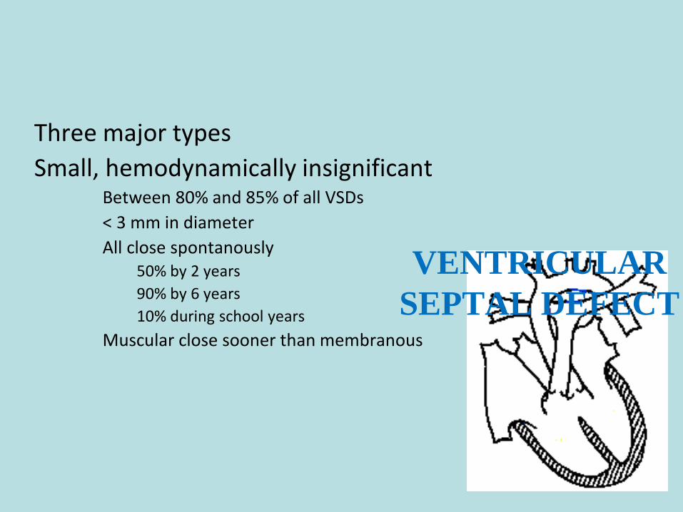
VENTRICULAR
SEPTAL DEFECT
Three major types
Small, hemodynamically insignificantBetween 80% and 85% of all VSDs
< 3 mm in diameter
All close spontanously50% by 2 years
90% by 6 years
10% during school years
Muscular close sooner than membranous

PATENT
DUCTUS
ARTERIOSUS
Persistence of normal fetal vessel joining the pulmonary artery to the aorta
Closes spontaneously in normal term infants at 3-5 days of age
CYANOTIC CHD (R →L)
Tetralogy of Fallot (TOF)
Tricuspid atresia (TA)
Total anomalous pulmonary venous return (TAPVR)
Truncus arteriosus
Transposition of the great vessels
Hypoplastic left heart syndrome (HLH)
Pulmonary atresia (PA) / critical PS
Double outlet right ventricle (DORV)
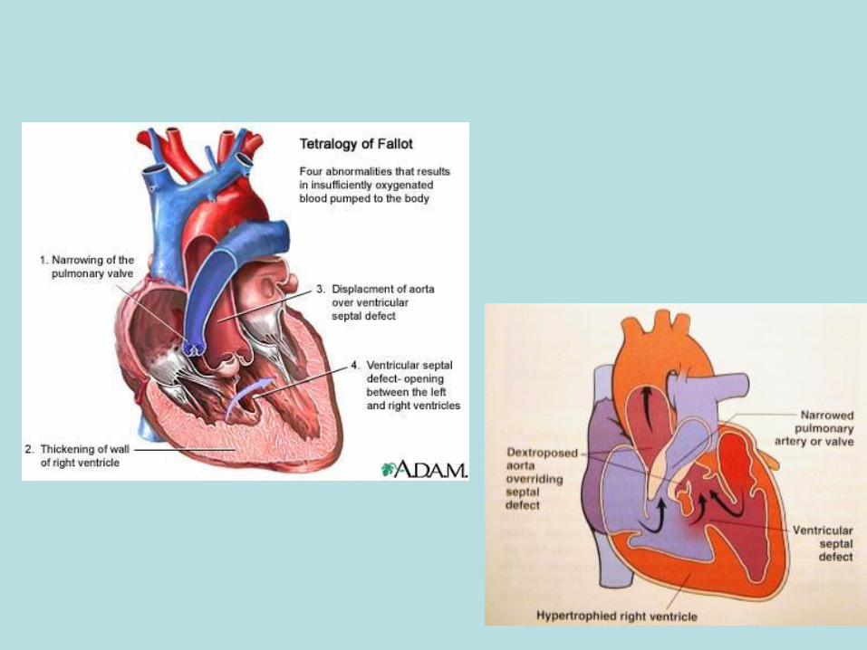
TETRALOGY OF FALLOT
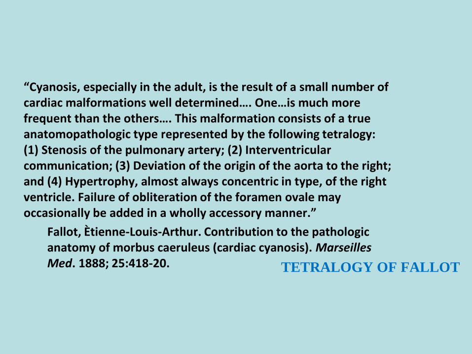
“Cyanosis, especially in the adult, is the result of a small number of cardiac malformations well determined…. One…is much more frequent than the others…. This malformation consists of a true anatomopathologic type represented by the following tetralogy: (1) Stenosis of the pulmonary artery; (2) Interventricular communication; (3) Deviation of the origin of the aorta to the right; and (4) Hypertrophy, almost always concentric in type, of the right ventricle. Failure of obliteration of the foramen ovale may occasionally be added in a wholly accessory manner.”
Fallot, Ètienne-Louis-Arthur. Contribution to the pathologic anatomy of morbus caeruleus (cardiac cyanosis). Marseilles Med. 1888; 25:418-20.
TETRALOGY OF FALLOT
TETRALOGY OF FALLOT
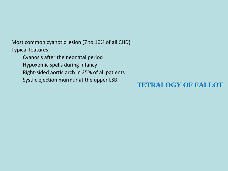
Most common cyanotic lesion (7 to 10% of all CHD)
Typical features
Cyanosis after the neonatal period
Hypoxemic spells during infancy
Right-sided aortic arch in 25% of all patients
Systlic ejection murmur at the upper LSB
TETRALOGY OF FALLOT

KIDNEY
CONGENITA
L
PATHOLOG
Y
AGENESIS
HYPOPLASIA
ECTOPIC
HORSESHOE
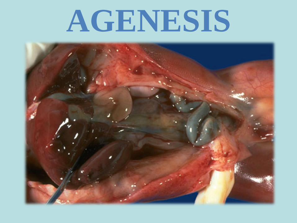
AGENESIS

HYPOPLASIA
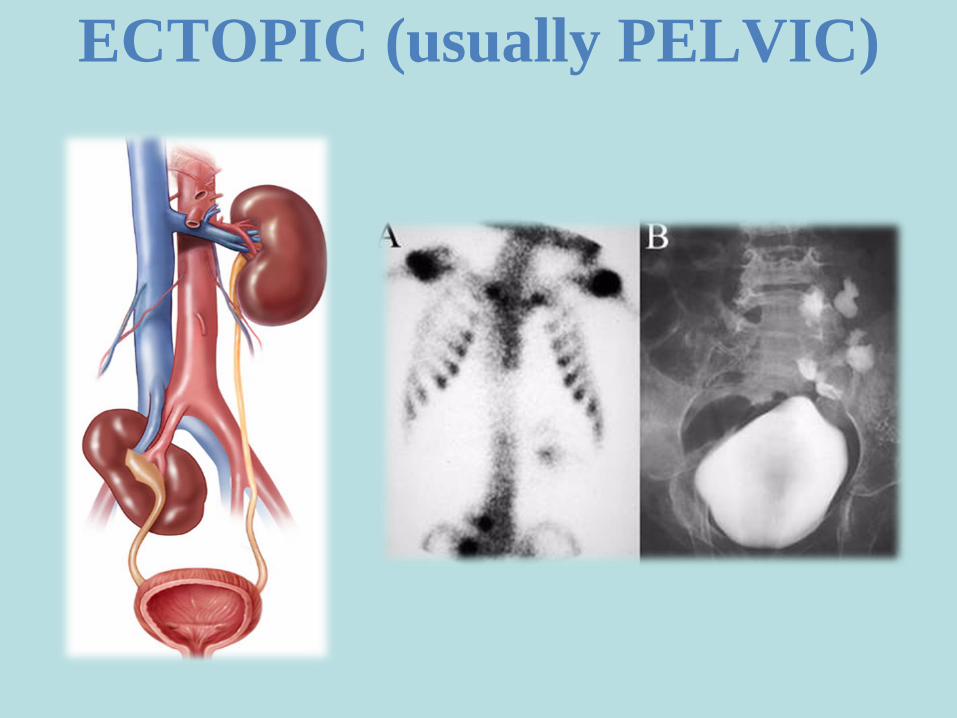
ECTOPIC (usually PELVIC)
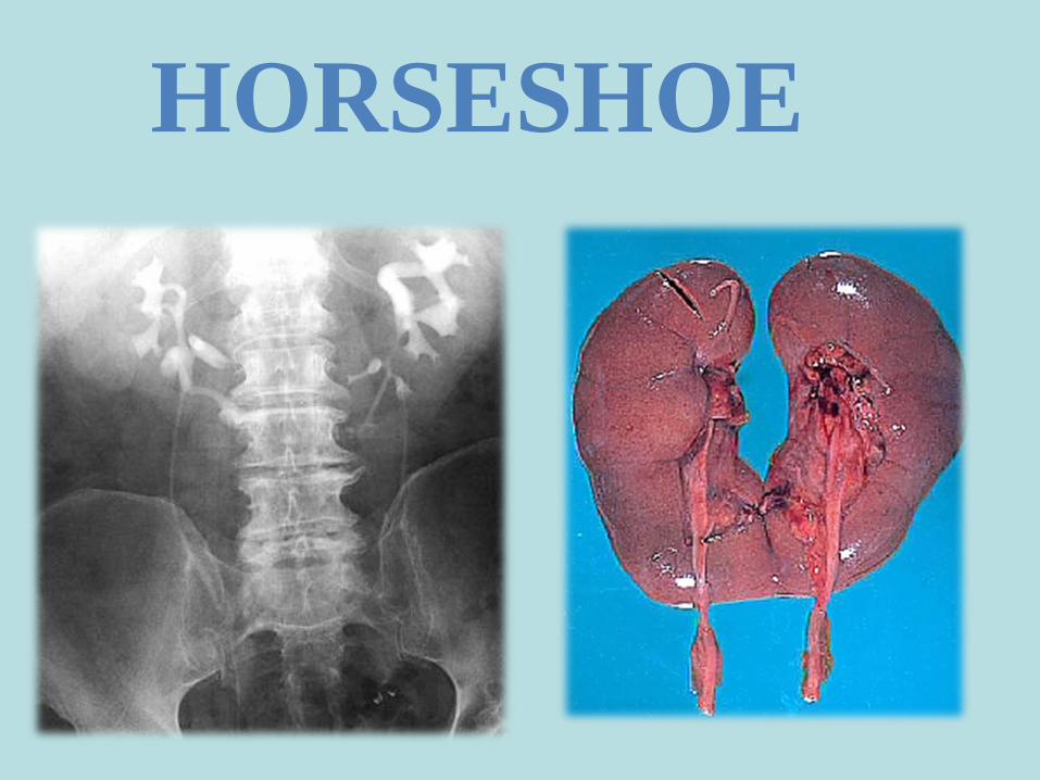
HORSESHOE
BIRTH RELATED STRESSORS
Newborn at risk due to asphyxia
Newborn with respiratory distress/Transient Tachypnea
Newborn with meconium aspiration syndrome
Newborn with persistent pulmonary hypertension
Newborn with complications due to respiratory therapy
Newborn with cold stress
Newborn with hypoglycemia
Newborn with jaundice
Newborn with polycythemia
Newborn with infection
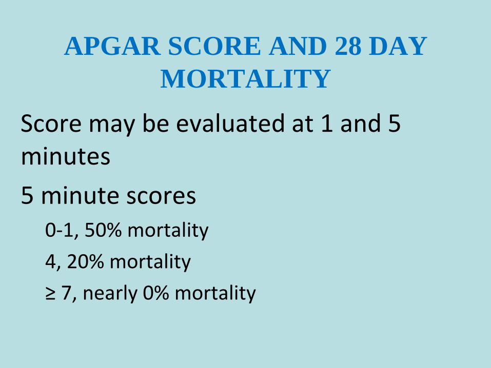
APGAR SCORE AND 28 DAY
MORTALITY
Score may be evaluated at 1 and 5 minutes
5 minute scores0-1, 50% mortality
4, 20% mortality
≥ 7, nearly 0% mortality

PERINATAL PATHOLOGY OF THE FETUS AND NEWBORN
Group of diseases that arise in newborns due to trauma, hypoxia, toxic-metabolic and infectious injury of organs and tissues, as a result of adverse pregnancy or childbirth
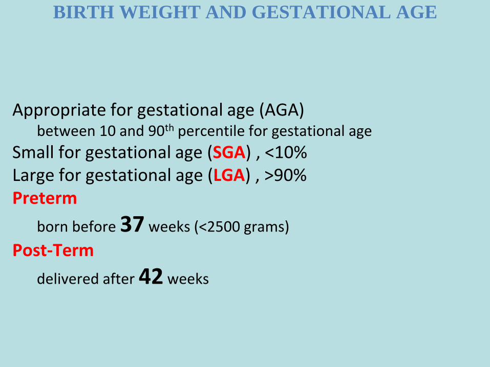
BIRTH WEIGHT AND GESTATIONAL AGE
Appropriate for gestational age (AGA)between 10 and 90th percentile for gestational age
Small for gestational age (SGA) , <10%Large for gestational age (LGA) , >90%Preterm
born before 37 weeks (<2500 grams)
Post-Term
delivered after 42 weeks
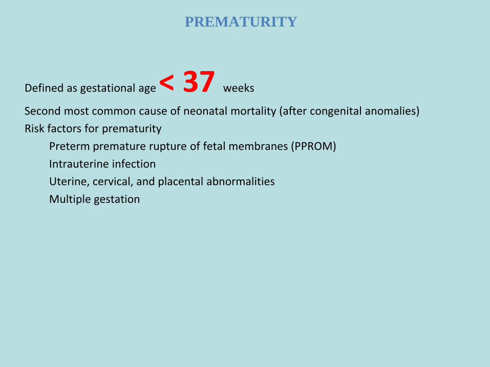
PREMATURITY
Defined as gestational age < 37 weeks
Second most common cause of neonatal mortality (after congenital anomalies)
Risk factors for prematurity
Preterm premature rupture of fetal membranes (PPROM)
Intrauterine infection
Uterine, cervical, and placental abnormalities
Multiple gestation
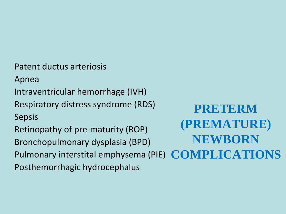
PRETERM
(PREMATURE)
NEWBORN
COMPLICATIONS
Patent ductus arteriosis
Apnea
Intraventricular hemorrhage (IVH)
Respiratory distress syndrome (RDS)
Sepsis
Retinopathy of pre-maturity (ROP)
Bronchopulmonary dysplasia (BPD)
Pulmonary interstital emphysema (PIE)
Posthemorrhagic hydrocephalus

POST TERM
INFANT
Gestation > 42 weeks
Must determine if EDC is truly post term
After 42 weeks placenta loses ability to nourish the fetus
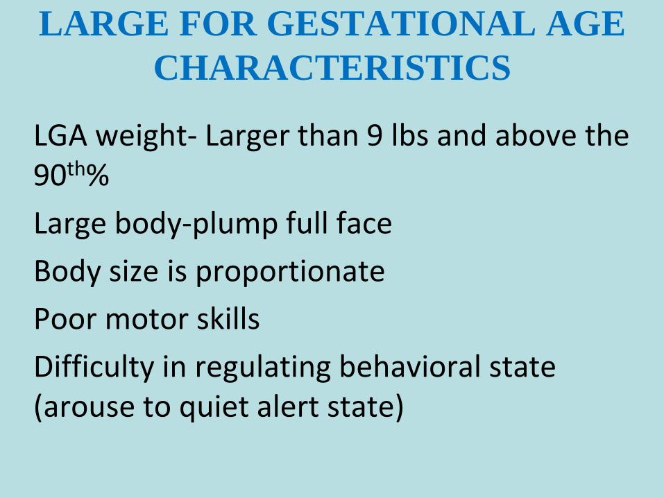
LARGE FOR GESTATIONAL AGE
CHARACTERISTICS
LGA weight- Larger than 9 lbs and above the 90th%
Large body-plump full face
Body size is proportionate
Poor motor skills
Difficulty in regulating behavioral state (arouse to quiet alert state)
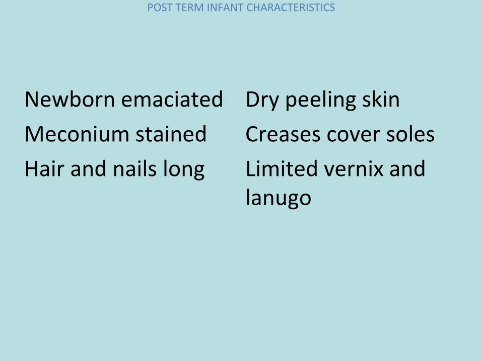
POST TERM INFANT CHARACTERISTICS
Newborn emaciated
Meconium stained
Hair and nails long
Dry peeling skin
Creases cover soles
Limited vernix and lanugo
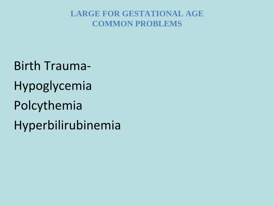
LARGE FOR GESTATIONAL AGE
COMMON PROBLEMS
Birth Trauma-
Hypoglycemia
Polcythemia
Hyperbilirubinemia
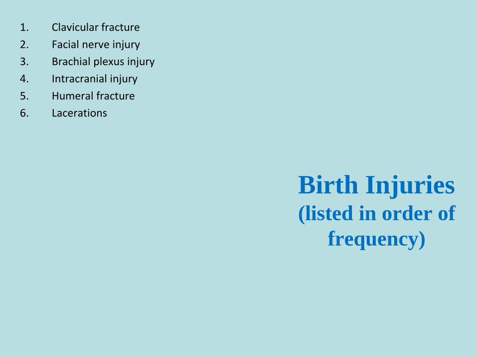
Birth Injuries(listed in order of
frequency)
1. Clavicular fracture
2. Facial nerve injury
3. Brachial plexus injury
4. Intracranial injury
5. Humeral fracture
6. Lacerations
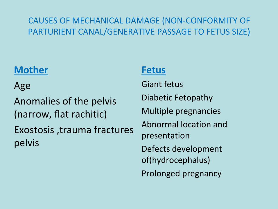
CAUSES OF MECHANICAL DAMAGE (NON-CONFORMITY OF PARTURIENT CANAL/GENERATIVE PASSAGE TO FETUS SIZE)
Mother
Age
Anomalies of the pelvis (narrow, flat rachitic)
Exostosis ,trauma fractures pelvis
Fetus
Giant fetus
Diabetic Fetopathy
Multiple pregnancies
Abnormal location and presentation
Defects development of(hydrocephalus)
Prolonged pregnancy
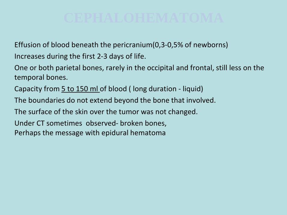
CEPHALOHEMATOMA
Effusion of blood beneath the pericranium(0,3-0,5% of newborns)
Increases during the first 2-3 days of life.
One or both parietal bones, rarely in the occipital and frontal, still less on the temporal bones.
Capacity from 5 to 150 ml of blood ( long duration - liquid)
The boundaries do not extend beyond the bone that involved.
The surface of the skin over the tumor was not changed.
Under CT sometimes observed- broken bones, Perhaps the message with epidural hematoma

CEPHALOHEMA
TOMA
From 7-10 days - reduced in size
Usually disappear in 3-8 weeks.
With significant hemorrhages of compacted periosteum, hematoma ossified, which leads to distortion or asymmetry of the skull.
Diff.diagnoz-labor tumor; hemorrhage beneath the aponeurosis; cerebral hernia.
Complications: anemia, due to considerable blood loss; jaundice , due to progress of hemorrhage resolution, suppuration.
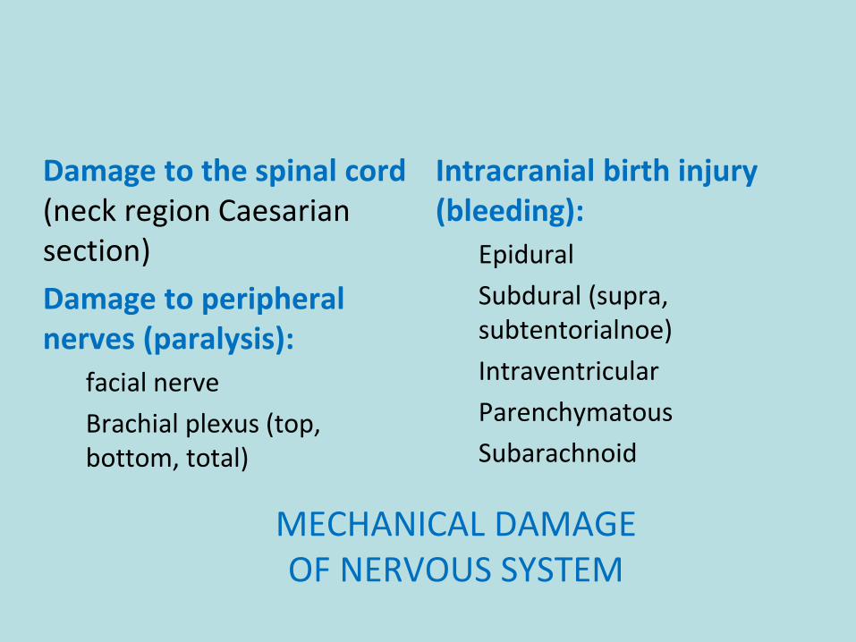
MECHANICAL DAMAGE OF NERVOUS SYSTEM
Damage to the spinal cord(neck region Caesarian section)
Damage to peripheral nerves (paralysis):
facial nerve
Brachial plexus (top, bottom, total)
Intracranial birth injury (bleeding):
Epidural
Subdural (supra, subtentorialnoe)
Intraventricular
Parenchymatous
Subarachnoid
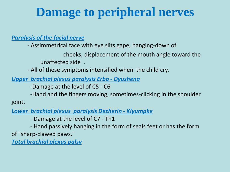
Damage to peripheral nerves
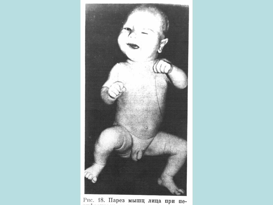
Paralysis of the facial nerve - Assimmetrical face with eye slits gape, hanging-down of
cheeks, displacement of the mouth angle toward the unaffected side .
- All of these symptoms intensified when the child cry.
Upper brachial plexus paralysis Erba - Dyushena -Damage at the level of C5 - C6 -Hand and the fingers moving, sometimes-clicking in the shoulder
joint.
Lower brachial plexus paralysis Dezherin - Klyumpke - Damage at the level of C7 - Th1 - Hand passively hanging in the form of seals feet or has the form
of "sharp-clawed paws." Total brachial plexus palsy

HYPOXIC РТ
Asphyxia (suffocation) -
Asphyxia - fetal (center.) and post-natal
Hypoxia - the prolonged repeated limitations of constant O2 supplyleads to excess accumulation in the organism of CO2 and other incompletely oxidized products (80% of all damages to CNS).
Hypoxia -chronic intrauterine
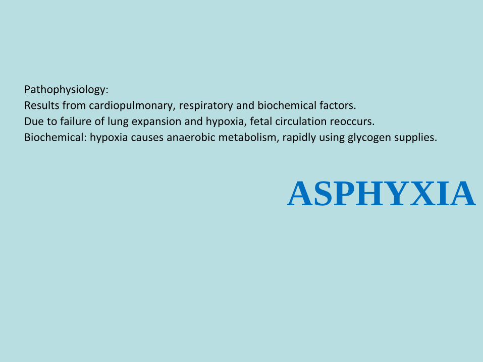
ASPHYXIA
Pathophysiology:
Results from cardiopulmonary, respiratory and biochemical factors.
Due to failure of lung expansion and hypoxia, fetal circulation reoccurs.
Biochemical: hypoxia causes anaerobic metabolism, rapidly using glycogen supplies.
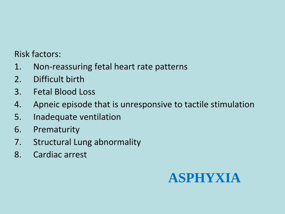
ASPHYXIA
Risk factors:
1. Non-reassuring fetal heart rate patterns
2. Difficult birth
3. Fetal Blood Loss
4. Apneic episode that is unresponsive to tactile stimulation
5. Inadequate ventilation
6. Prematurity
7. Structural Lung abnormality
8. Cardiac arrest
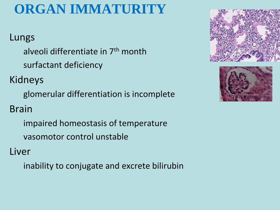
ORGAN IMMATURITY
Lungs
alveoli differentiate in 7th month
surfactant deficiency
Kidneys
glomerular differentiation is incomplete
Brain
impaired homeostasis of temperature
vasomotor control unstable
Liver
inability to conjugate and excrete bilirubin
NEONATAL RESPIRATORY
DISTRESS SYNDROME (RDS)60,000 cases / year in USA with 5000 deaths
Incidence is inversely proportional to gestational age
The cause is lung immaturity with decreased alveolar surfactant
surfactant decreases surface tension
first breath is the hardest since lungs must be expanded
without surfactant, lungs collapse with each breath
RDS RISK FACTORS
1) Prematurity
by far the greatest risk factor
affected infants are nearly always premature
2) Maternal diabetes mellitus
insulin suppresses surfactant secretion
3) Cesarean delivery
normal delivery process stimulates surfactant secretion
RDS PATHOLOGY
Grosssolid and airless (no crepitance)
sink in water
appearance is similar to liver tissue*
Microscopicatelectasis and dilation of alveoli
hyaline membranes composed of fibrin and cell debris line alveoli (HMD former name)
minimal inflammation
SUDDEN INFANT DEATH
SYNDROMENIH Definition
sudden death of an infant under 1 year of age which remains unexplained after a thorough case investigation, including performance of a complete autopsy, examination of the death scene, and review of the clinical history
Crib death
another name based on the fact that most die in their sleep
RISK FACTORS FOR SIDSParental
Young maternal age (age <20 years)Maternal smoking during pregnancyDrug abuse in either parent, specifically paternal marijuana and maternal opiate, cocaine useShort intergestational intervalsLate or no prenatal careLow socioeconomic groupAfrican American and American Indian ethnicity (? socioeconomic factors)
InfantBrain stem abnormalities, associated defective arousal, and cardiorespiratory controlPrematurity and/or low birth weightMale sexProduct of a multiple birthSIDS in a prior siblingAntecedent respiratory infections
EnvironmentProne sleep positionSleeping on a soft surfaceHyperthermiaPostnatal passive smoking
MORPHOLOGY OF SIDS
SIDS is a diagnosis of exclusionNon-specific autopsy findings
Multiple petechiae
Pulmonary congestion ± pulmonary edema
These may simply be agonal changes as they are found in non-SIDS deaths also
Subtle changes in brain stem neuronsAutopsy typically reveals no clear cause of death
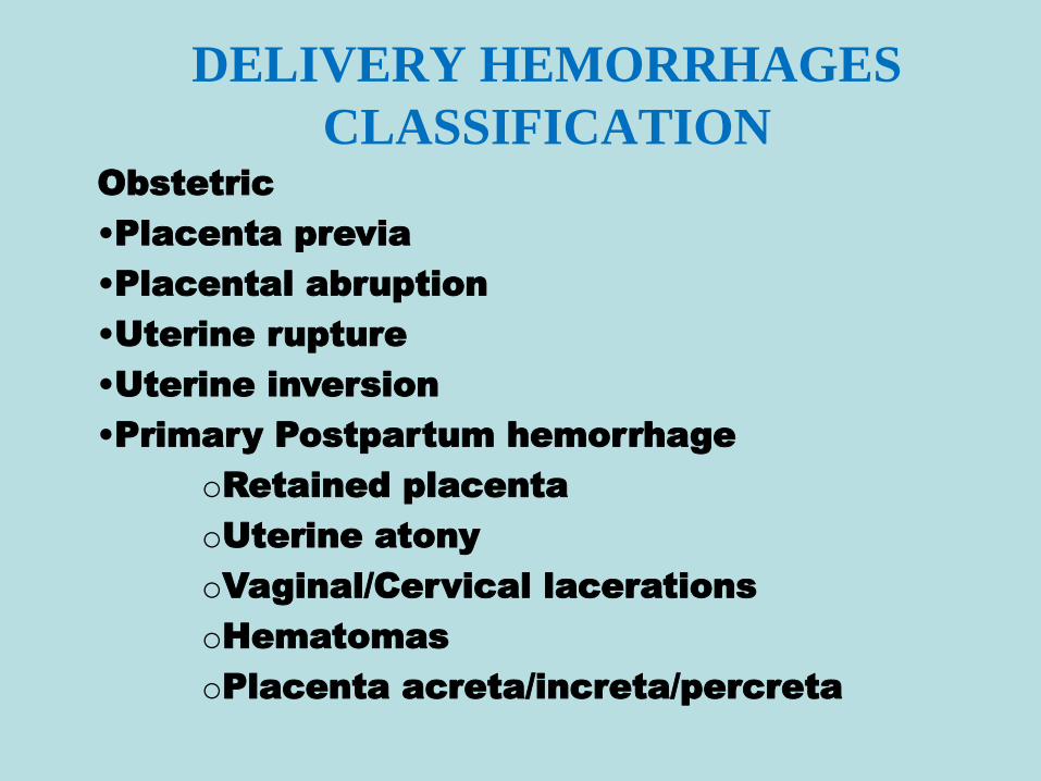
Obstetric
•Placenta previa
•Placental abruption
•Uterine rupture
•Uterine inversion
•Primary Postpartum hemorrhage
oRetained placenta
oUterine atony
oVaginal/Cervical lacerations
oHematomas
oPlacenta acreta/increta/percreta
DELIVERY HEMORRHAGES
CLASSIFICATION
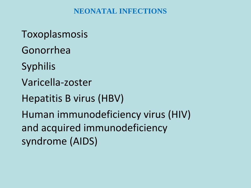
NEONATAL INFECTIONS
Toxoplasmosis
Gonorrhea
Syphilis
Varicella-zoster
Hepatitis B virus (HBV)
Human immunodeficiency virus (HIV) and acquired immunodeficiency syndrome (AIDS)
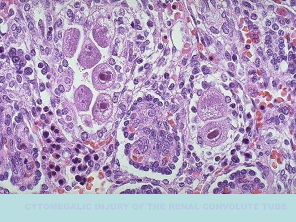
CYTOMEGALIC INJURY OF THE RENAL CONVOLUTE TUBE
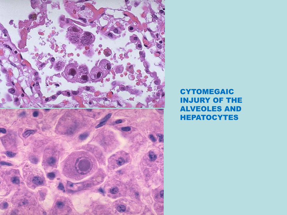
CYTOMEGAIC
INJURY OF THE
ALVEOLES AND
HEPATOCYTES

TOXOPLASIS,
MACRO – CEREBRAL
ABSCESS,
MICRO –MYCROGLIAL
GRANULOMMA
AND PSEUDOCYST
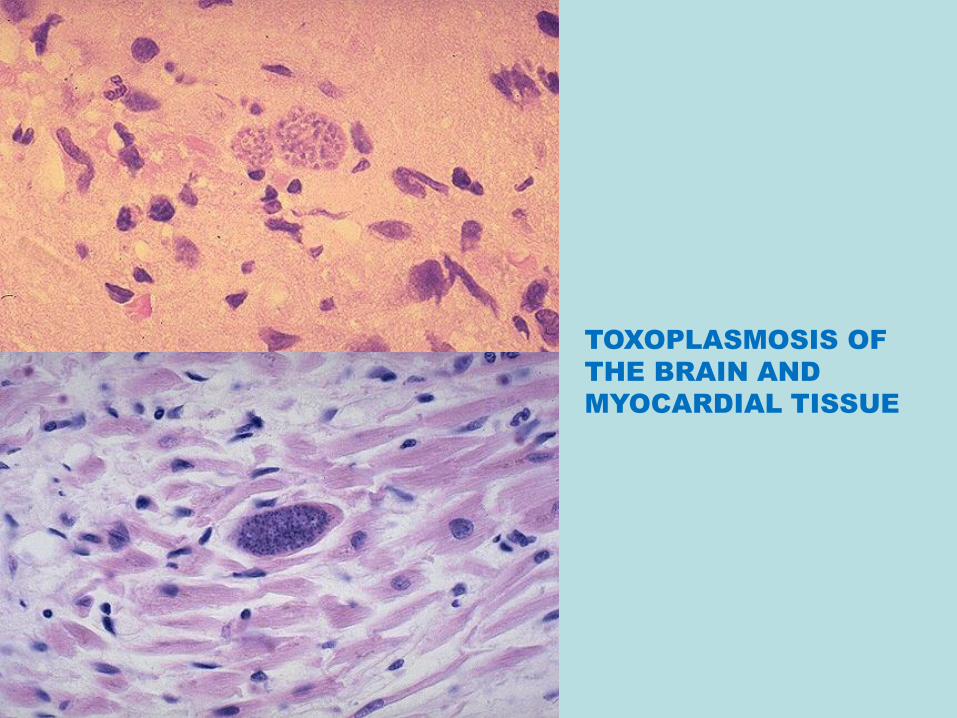
TOXOPLASMOSIS OF
THE BRAIN AND
MYOCARDIAL TISSUE

CYSTIC FIBROSIS OF THE
PANCREAS
MUCOVISCIDOSIS
OF THE LUNG
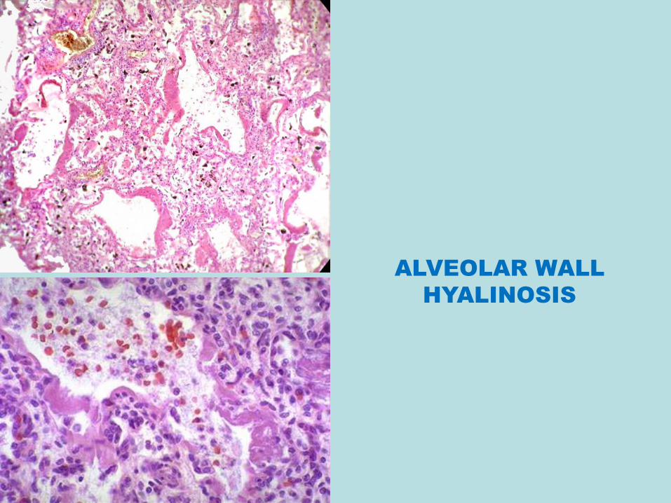
ALVEOLAR WALL
HYALINOSIS

TUMORSBenign
Malignant

CONGENITAL
CAPILLARY
HEMANGIOMA
At birth At 2 years
After spontaneous
regression
Congenital capillary hemangioma at birth (A) and at age 2 years (B) after spontaneous regression.
TERATOMAS
Composed of cells derived from more than one germ layer, usually all three
Sacrococcygeal teratomas
most common childhood teratoma
frequency 1:20,000 to 1:40,000 live births
4 times more common in boys than girls
Aproximately 12% are malignant
often composed of immature tissue
occur in older children
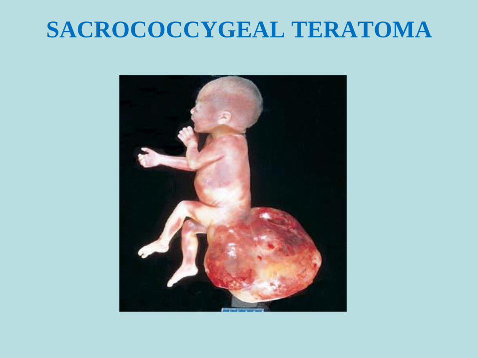
SACROCOCCYGEAL TERATOMA
Recommended