
Postmenopause• Issues for primary care physicians and gynecologists:
– Hormonal changes
– Cardiovascular disease
– Osteoporosis
– Post menopausal bleeding (PMB)
– Abnormal PAP’s
– Sexual Function
– Depression
– Cancer screening and prevention
– Lifestyle and habits

Causes of Mortality Men and WomenCauses of Mortality Men and Women
Cardiovascular Disease 2199.9/100,000Cardiovascular Disease 2199.9/100,000 Heart disease 1772.2/100,000 Heart disease 1772.2/100,000 Cerebrovascular diseases Cerebrovascular diseases 427.7/100,000427.7/100,000
Malignant Neoplasms (all) 1316.6/100,000Malignant Neoplasms (all) 1316.6/100,000
Influenza and pneumonia 166.4/100,000Influenza and pneumonia 166.4/100,000

Postmenopause• Cardiovascular Disease
– Leading cause of death in men and women
– Due to lack of protective effect of estrogen
– Preventive measures• Exercise
• Diet
• Quit smoking
• Moderate alcohol intake
• Low dose aspirin ??

Postmenopause• Hot flushs
– Very disturbing
– Every few min to hours. Mostly first year
– Less in obese women
– treatment
• HRT
• Tibolone
• SSRIs may reduce by 60%
• Gabapentine (neurontoin)
• Clonidine (captopril)


Clinical Definitions• Osteopenia- reduction in
bone mass below the theoretical fracture threshold.
• Osteoporosis- reduction in the bone mass to a level where there is increased risk of fracture in the absence of trauma. Or a fracture has already occurred.

“I’ve lost six inches in height and none of my clothes fit me anymore. Plus, it’s hard to get clothes that look nice when my back is so hunched over.”
Typical comments from people with osteoporosis

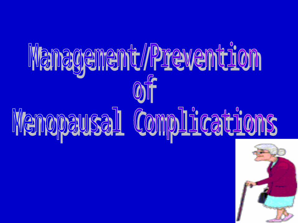
OverviewOverview
Bone withOsteoporosis
NormalBone
Osteoporosis causes weak bones. In this common disease, bones lose minerals like calcium. They become fragile and break easily.

A woman’s hip fracture risk equals her combined risk of breast, uterine and ovarian cancer.

The most common breaks in weak bones are in the wrist, spine and hip.

Osteoporotic bone
Osteoporotic fracture

Classification:• Primary
– Type 1- post menopausal
– Type 2- senile
• Secondary
– Medications
– Steroid induced
– Medical conditions
• Spinal cord injury

Type 1 (postmenopausal)• Women > Men (6:1 ratio)
• Related to estrogen
• Affects trabecular bone
• Associated with vertebral fractures
• Unrelated to calcium intake
• High bone turnover
Vertebral compression fractures

Type 2 (Senile Osteoporosis)
• Elderly Age >75yrs
• Women:Men 2:1
• Calcium deficiency is major factor
• Affects trabecular and cortical bone
• Vertebral compression, hip, and distal radius fxs.
• Slow bone turnover
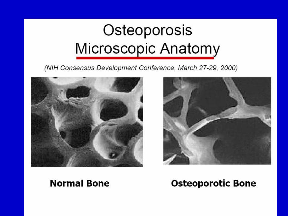
Common Locations of Osteoporotic Fractures in
General Population
• Thoracic (vertebral) compression fractures
• Hip fractures
• Wrist/extremity fractures.
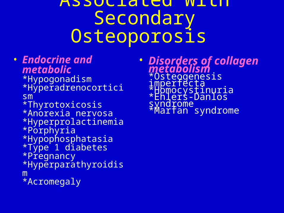
Medical Conditions Associated With Secondary Osteoporosis
• Endocrine and metabolic*Hypogonadism*Hyperadrenocorticism*Thyrotoxicosis*Anorexia nervosa*Hyperprolactinemia*Porphyria*Hypophosphatasia*Type 1 diabetes*Pregnancy*Hyperparathyroidism*Acromegaly
• Disorders of collagen metabolism*Osteogenesis imperfecta*Homocystinuria*Ehlers-Danlos syndrome*Marfan syndrome

Medical Conditions Associated With Secondary Osteoporosis
• NutritionalMalabsorption syndromes, malnutritionChronic liver diseaseGastric operationsVitamin D deficiencyAlcoholism
• OtherRheumatoid arthritisMyelomaCertain cancersImmobilizationRenal tubular acidosisHypercalciuriaChronic obstructive pulmonary diseaseOrgan transplantationCholestatic liver diseaseMastocytosisThalassemia
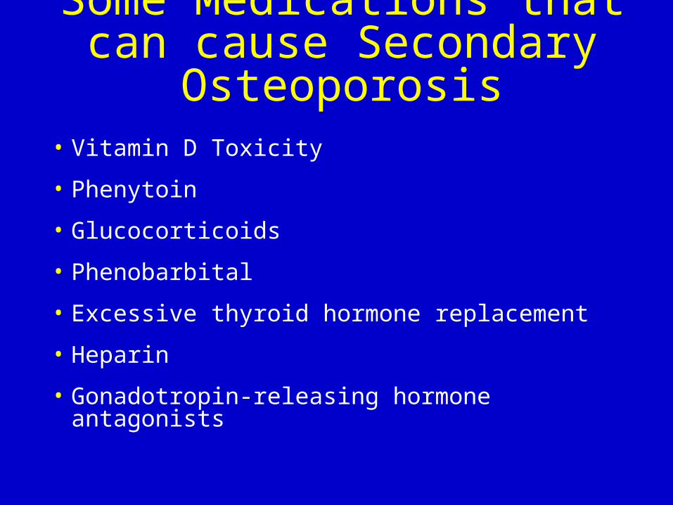
Some Medications that can cause Secondary Osteoporosis
• Vitamin D Toxicity
• Phenytoin
• Glucocorticoids
• Phenobarbital
• Excessive thyroid hormone replacement
• Heparin
• Gonadotropin-releasing hormone antagonists

How does this obvious bone loss happen?
• Bone is a living, growing, tissue
• Healthy bones are not quiescent. They are constantly destruction, but imbalance between the formation being remodeled.
• This is not simply a problem of bony and destruction of bone.
• Two types of bone
– Cortical
– Trabecular

Cortical (Compact) Bone• 80% of the skeletal mass
• Provides a protective outer shell around every bone in the body
• Slower turnover
• Provides strength and resists bending or torsion
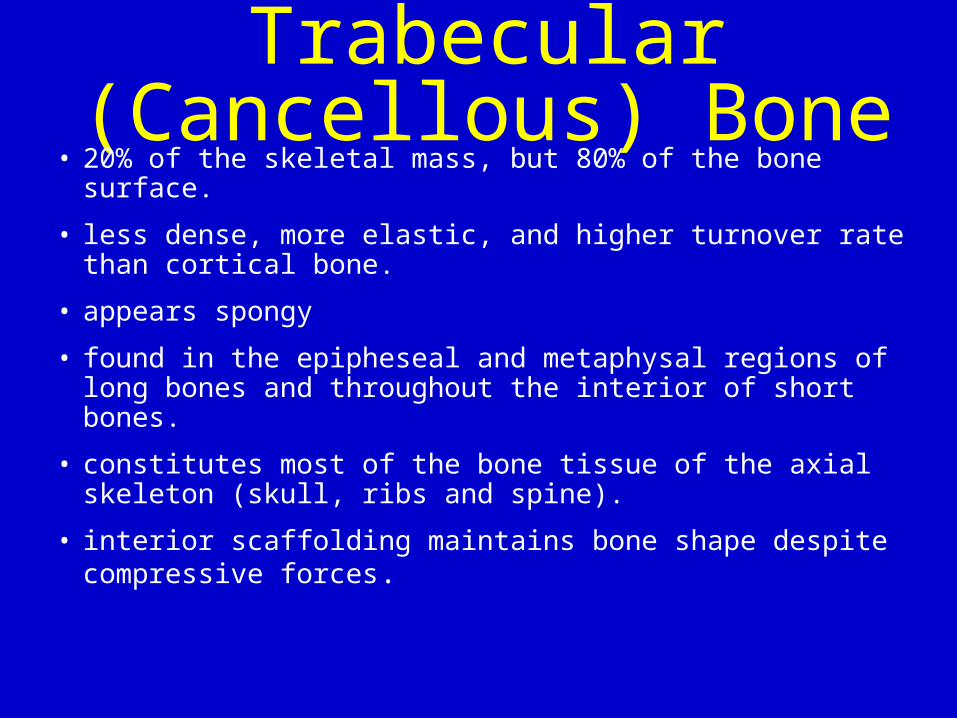
Trabecular (Cancellous) Bone
• 20% of the skeletal mass, but 80% of the bone surface.
• less dense, more elastic, and higher turnover rate than cortical bone.
• appears spongy
• found in the epipheseal and metaphysal regions of long bones and throughout the interior of short bones.
• constitutes most of the bone tissue of the axial skeleton (skull, ribs and spine).
• interior scaffolding maintains bone shape despite compressive forces.

Types of Cells in Bone Tissue

Bone Remodeling
• OsteoBlasts Build Bone.
• OsteoClasts Create Cavities in bone.

Bone mass declines with age.
• Remodeling occurs at discrete foci called bone remodeling units (BRUs).
• Number of active BRUs increase with age, resulting in increased bone turn over.
• Osteoblasts are not able to completely fill cavities created by osteoclasts and less mineralized bone is formed.
• Endosteal bone loss is partially compensated by periosteal bone formation. This leads to trabecular thinning.

Bone Remodeling Unit
Osteoclast
Osteoblast
New bone
Lining cells

• Scanning electron micrograph of slice of osteoporotic cancellous bone from the fourth lumbar vertebra of an elderly woman.
• www.grad.ucl.ac.uk


Bones are living organs
• Calcium is deposited and withdrawn from bones daily.
• Bones build to about age 30.
• We need to build up a healthy bone account while young and continue to make deposits with age.

• After mid-30’s, you begin to slowly lose bone mass. Women lose bone mass faster after menopause, but it happens to men too.
• Bones can weaken early in life without a healthy diet and the right kinds of physical activity.
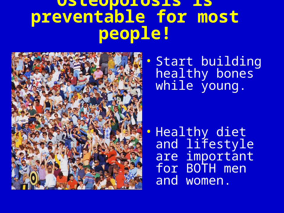
The good news: Osteoporosis is preventable for most people!
• Start building healthy bones while young.
• Healthy diet and lifestyle are important for BOTH men and women.

The National OsteoporosisFoundation (NOF) recommends FIVE simple steps to bone health and osteoporosis prevention …
Simple Prevention Simple Prevention StepsSteps

Step 1
Get your daily recommended amounts of calcium and vitamin D.

Calcium requirements vary by age
If this is your ageThen you need
this much calcium each day (mg)
0 to 6 months 210
7 to 12 months 270
1 to 3 years 500
4 to 8 years 800
9 to 18 years 1,300
19 to 50 years 1,000
Over 50 years 1,200
Growthspurt

You need more vitamin D as you age
Age
Daily vitamin D needs in International Units (IU)
600 IU
200 IU
400 IU
0
100
200
300
400
500
600
up to 50 51-70 over 70

What about Vitamin D? Main dietary sources of vitamin D are:
• Fortified milk (400 IU per quart)
• Some fortified cereals
• Cold saltwater fish (Example: salmon, halibut, herring, tuna, oysters and shrimp)
• Some calcium and vitamin/mineral supplements

Vitamin D from sunlight exposure
• Vitamin D is manufactured in the skin following direct exposure to sun.
• Amount varies with time of day, season, latitude and skin pigmentation.
• 10–15 minutes exposure of hands, arms and face 2–3 times/week may be sufficient (depending on skin sensitivity).
• Clothing, sunscreen, window glass and pollution reduce amount produced.
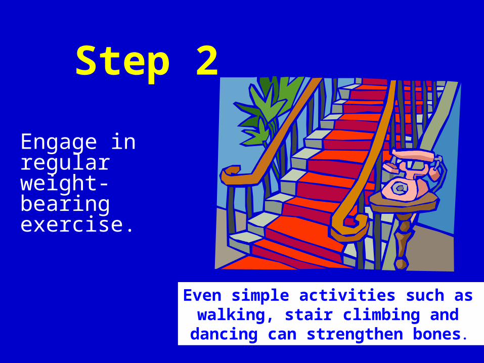
Step 2
Engage in regular weight-bearing exercise.
Even simple activities such as walking, stair climbing and
dancing can strengthen bones.

Step 3Avoid smoking and excessive alcohol & soft drinks
12 oz. 5 oz.
1.5 oz.
MyPyramid.gov recommends no more than 1 drink per day
for women and 2 for men.

Step 4
Talk to your doctor about bone health.

Step 5
Have a bone density test and take medication when appropriate.
Sou
rce
of p
hoto
: US
DA
AR
S P
hoto
Uni
t Pho
to b
y P
eggy
Gre
b
Testing is a simple, painless procedure.

Diagnostic Outline• What is bone density testing?
• Why is it done?
• Who should be tested?
• When should it be repeated?
• How is it interpreted?
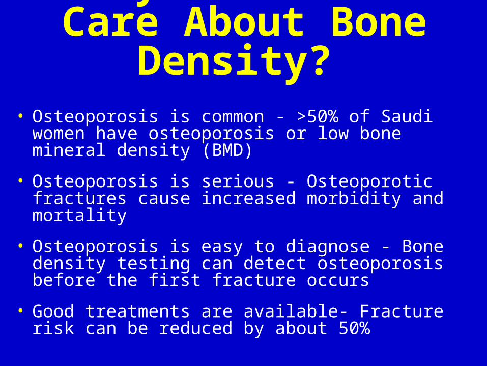
Why Should We Care About Bone Density?
• Osteoporosis is common - >50% of Saudi women have osteoporosis or low bone mineral density (BMD)
• Osteoporosis is serious - Osteoporotic fractures cause increased morbidity and mortality
• Osteoporosis is easy to diagnose - Bone density testing can detect osteoporosis before the first fracture occurs
• Good treatments are available- Fracture risk can be reduced by about 50%

Bone Densitometry• Non-invasive test for measurement of BMD
• Major technologies
– Dual-energy X-ray Absorptiometry (DXA)
– Quantitative Ultrasound (QUS)
– Quantitative Computerized Tomography (QCT)
• Many manufacturers
• Numerous devices
• Different skeletal sites

4848
Indications For Bone Density Testing
• All women age 65 and older ( 50 in Saudi)
• All men age 70 and older
• Adults with a fragility fracture
• Adults with a disease or condition associated with low bone density
• Adults taking medication associated with low bone density
• Anyone being treated for low bone density to monitor treatment effect
• Anyone not receiving therapy, in whom evidence of bone loss would lead to treatment
Women discontinuing treatment should be considered for bone density testing according to the indications listed above.
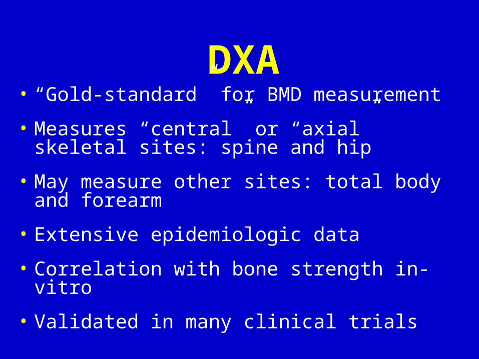
DXA• “Gold-standard” for BMD measurement
• Measures “central” or “axial” skeletal sites: spine and hip
• May measure other sites: total body and forearm
• Extensive epidemiologic data
• Correlation with bone strength in-vitro
• Validated in many clinical trials

DXA Technology
X-ray Source (produces 2 photon energies with different attenuation profiles)
Photons Collimator (pinhole for pencil beam, slit for fan beam)
Patient
Detector (detects 2 tissue types - bone and soft tissue)
Very low radiation to patient.
Very little scatter radiation to technologist

DXA Imaging of the Spine For Detection of Vertebral
FracturesHologic: IVA or RVA
Normal Fracture
GE: DVA or LVA
Normal Fracture

Bone Density Testing Is Done For Three
Reasons
• To diagnose osteoporosis
• To predict fracture risk
• To monitor therapy

What DXA Really Measures:
Bone Mineral Content (BMC) In Grams and Area In cm2
• “Areal” BMD is calculated in g/cm2
• “T-score” compares the patient’s BMD with the young-normal mean BMD and expresses the difference as a standard deviation (SD) score
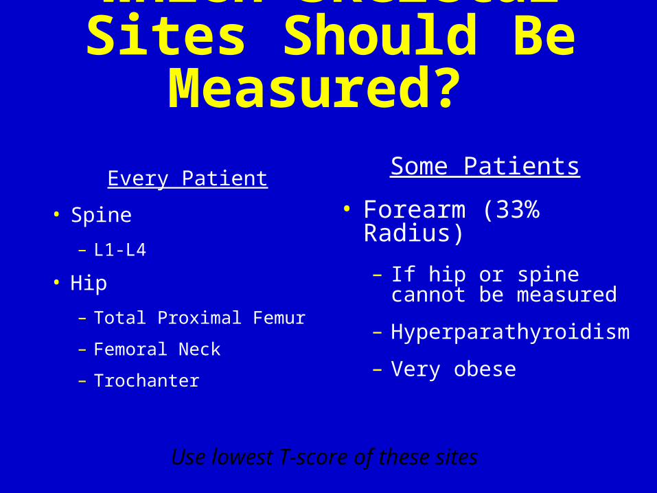
Which Skeletal Sites Should Be Measured?
Every Patient
• Spine
– L1-L4
• Hip
– Total Proximal Femur
– Femoral Neck
– Trochanter
Some Patients
• Forearm (33% Radius)
– If hip or spine cannot be measured
– Hyperparathyroidism
– Very obese
Use lowest T-score of these sites

T-score
Example:
T-score = 0.7 g/cm2 - 1.0 g/cm2
0.1 g/cm2= - 3.0
Patient’s BMD – Young-Adult Mean BMD
1 SD of Young-Adult Mean BMD

WHO Study Group. 1994.WHO Study Group. 1994. 5656
Diagnostic Classification
Classification T-score
Normal -1 or greater
Osteopenia Between -1 and -2.5
Osteoporosis -2.5 or less
Severe Osteoporosis -2.5 or less and fragility fracture
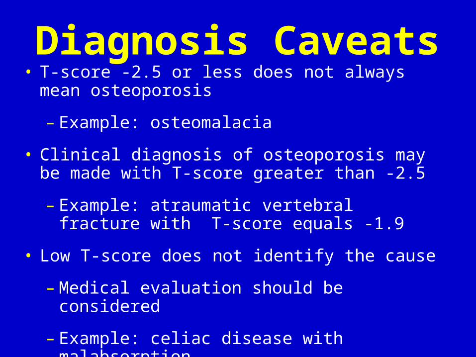
Diagnosis Caveats• T-score -2.5 or less does not always mean
osteoporosis
– Example: osteomalacia
• Clinical diagnosis of osteoporosis may be made with T-score greater than -2.5
– Example: atraumatic vertebral fracture with T-score equals -1.9
• Low T-score does not identify the cause
– Medical evaluation should be considered
– Example: celiac disease with malabsorption

Why -2.5?Professor John Kanis says:
“Such a cutoff value identifies approximately 30% of postmenopausal women as having osteoporosis using measurements made at the spine, hip or forearm. This is approximately equivalent to the lifetime risk of fracture at these sites.”

Fracture Risk Doubles With Every SD
Decrease in BMD
0
5
10
15
20
25
30
35
-5.0 -4.0 -3.0 -2.0 -1.0 0.0 1.0
Bone Density (T-score)
Relative Risk
for Fracture

Bone Density & Age vs. Fracture Risk
0
10
20
30
40
50
1.0 0.5 0.0 -0.5 -1.0 -1.5 -2.0 -2.5 -3.0 -3.5 -4.0
Femoral Neck T-score
Ten Year Fracture
Probability (%)
Age
80
70
60
50
Probability of first fracture of hip, distal forearm, proximal humerus, and symptomatic vertebral fracture in women of Malmö, Sweden.

Why Do Serial BMD Testing?
• To monitor response to therapy by finding an increase or stability of bone density
• To evaluate for non-response by finding loss of bone density - suggesting the need for reevaluation of treatment and evaluation for secondary causes of osteoporosis
• To follow patients not being treated who are at risk of bone loss, in order to determine if treatment is needed

When Should Repeat BMD Testing Be Done?
• When expected change in BMD equals or exceeds the “Least Significant Change” (LSC)
• Intervals between BMD testing should be determined according to each patient’s clinical status
– Consider one year after initiation or change of therapy
– Longer intervals once therapeutic effect is established
– Shorter intervals when rapid bone loss is expected

Always Compare BMD
Never Compare T-scores

The Osteoporosis Challenge
• To educate all patients on measures to maintain good bone health
• To identify patients at high risk for osteoporosis
• To use bone densitometry to detect low bone density BEFORE a fracture occurs
• To use pharmacologic therapy appropriately

Postmenopause• Osteoporosis Treatment
– HRT/ERT
– Bisphosphonates (Alendronate, Risedronate, Zoledronic acid)
– SERM’s (Raloxifene, Tamoxifen)
– Calcitonin
– Parathyroid hormone and fluoride
– Plus weight-bearing exercise, 1200 mg Calcium, and 800 u Vitamin D

*Hormone therapy (HRT).Estrogen + progesterone HRT
Estrogen HRT
The treatment of menopause symptoms (eg, vasomotor and
urogenital) remains the primary indication for EPT and ET
#Benefits
#Risks

*Significant risk for coronary heart disease (CHD) and venous thromboembolism observed during first year of therapy. CHD risk not significantly elevated in the following years
•breast cancer risk directly related to duration of therapy. Increased after 10 years of therapy
•the WHI study, fewer than 10 additional cases each of heart attack, stroke and breast cancer occurred each year among 10,000 women taking combination hormone therapy compared with 10,000 women taking placebo

• WHI 2002 -- HRT vs Placebo: Outcomes, ave follow up 5.2 yrs (Hazard Ratio)
– Invasive Breast Cancer = 38 vs 30/10,000 person yrs (1.26)
– CHD = 37 vs 30/10,000 (1.29)
– Stroke = 29 vs 21/10,000 (1.41)
– Venous Thromboembolic disease = 34 vs 16/10,000 (2.11)
– Colorectal cancer = 10 vs 16/10,000 (0.63)
– Hip fracture = 10 vs 15/10,000 (0.66)
– Vertebral fracture = 9 vs 15/10,000 (0.66)
– No change in endometrial and lung CA

Bisphosphonates Proven to Prevent Bone Loss.
• Cyclic etidronate is shown to prevent BMD loss in acute SCI when given 800mg qd for 2 wks. Pearson et al, APMR, 1997.
• Pamidronate (iv) 30mg iv x 3 days given with vitamin D and calicum inhibits bone resorption and reverses inhibition of PTH. Chen and Stein, JSCM, 2001.
• 30mg pamidronate monthly for 6 months with calcium inhibits bone hyperresorption and significantly reduces bone loss. Nance et al, APMR, 1999.
• Aledronate (fusamax) 10 mg/d or 70 mg/wk and tiludronate have also been found to be effective.
• Several different bisphosphonates have been shown to be effective. No one drug is clearly better than the others.
• The long term effects and risks of treatment with bisphosphonates is unknown.

Mechanism of Action Bisphosphonates
1. Inhibit osteoclast mediated bone resorption
2. Inhibit osteoclast
1. Activity
2. Recruitment
3. Adhesion
3. Osteoclast apoptosis

N2 (nitrogen)
Ca (calcium)

Eastell, R. N Engl J Med 1998;338:736-746
Effect of Therapy on Lumbar-Spine Bone Mineral Density in Postmenopausal Women with Osteoporosis

Postmenopause• Sexual Function
– At least 50% of postmenopausal women are sexually active
– Ask them
– H&P, ?Medications
– Advise: OTC lubricants (Astroglide, KY, Replens), Vaginal estrogen (Vagifem, Premarin, Estrace), Topical testosterone
Postmenopause• Depression
– 10-20% of people will have at least one episode of depression in their lifetime
– Affects twice as many women as men
– Risk of recurrence
• 50% with one episode
• 75% with two episodes
• 85% with >2 episodes

Postmenopause• Depression
– Medications:
• SSRI’s (Prozac, Zoloft, Celexa, Lexapro etc)
• Wellbutrin XR
– Other treatments:
• Psychotherapy
• Physical activity
• Sleep
• Get involved
Postmenopause• Cancer Screening and Prevention
– Mammograms annually
– Colonoscopy q 5 years
– Annual hemoccult testing
– Quit smoking
– Regular physicals

Postmenopause• Lifestyle and Habits
– Physical Activity
– Calcium 1200 mg/d
– Vitamin D 800 units/d
– Quit smoking
– Low dietary fat intake, high fiber intake
– Moderate Alcohol consumption
– Sleep
Recommended