Plasmodium (Malarial Parasite)
• Object– To study morphological structures of Plasmodia,
to identify morphological structures of developing stages of erythrocytic schizogony and gametocytes of P. vivax, and to differentiate the ring-form and gametocytes of P. vivax from P. falciparum.
– To study laboratory diagnostic methods of malarial parasites.

CHROMATIN
CYTOPLASM
RED BLOOD CELL
CCCHROMATIN
CYTOPLASM
SCHUFFNER’S
STIPPLING
CHROMATIN
CYTOPLASM
SCHSCHUFFNER’S
STIPPLING
MEROZOITES
MALARIAL PIGMENT
CYTOPLASM
CHROMATIN
SCHUFFNER’S
STIPPLING
The asexual phase of the malarial life cycle occurs in humans. Following inoculation of the human host with infectious sporozoites from a mosquito, an initial proliferative phase (exoerythrocytic schizogony) occurs in the liver. Merozoites are then released into the bloodstream and invade red blood cells, initiating erythrocytic schizogony. The parasite grows within the red cell from a early trophozoite (ring forms) to a late schizont, feeding on hemoglobin and producing malarial pigment as a waste product. When the schizont ruptures, merozoites are released into the bloodstream initiating the next schizogonic cycle. The patient's tertian or quartan fever profile approximates the time of release of merozoites into the bloodstream. After several cycles, some merozoites develop into male and female gametocytes which, when ingested by a mosquito, complete the sexual phase of the life cycle.
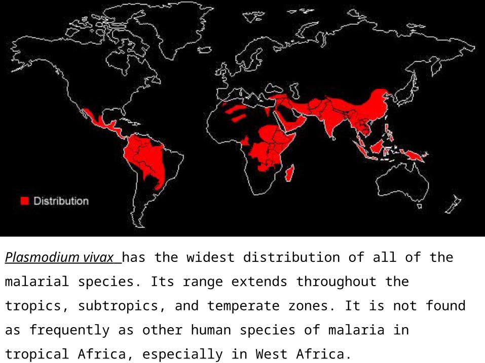
Plasmodium vivax has the widest distribution of all of the malarial species. Its
range extends throughout the tropics, subtropics, and temperate zones. It is not
found as frequently as other human species of malaria in tropical Africa,
especially in West Africa.

Plasmodium vivax
• Early Trophozoite (ring form, R.) (Pv. R.): – Ring form one-third the
diameter of a red blood cell;– Heavy chromatin dot;
Cytoplasma circle around vacuole.

Plasmodium vivax
• Late Trophozoite (Pv. T.):– Irregular ameboid mass that
almost fills the entire red blood cell.
– Fine, golden-brown pigment may be present.
– One or more small vacuoles retained until schizont stage.
– Schüffner's stirpling may be seen on Giemsa-stained smears.

Plasmodium vivax
• Early Schizon (Pv. S.):– Division of chromatin
apparent. – Cytoplasmic bands may
contain clumps of brown pigment.
Plasmodium vivax
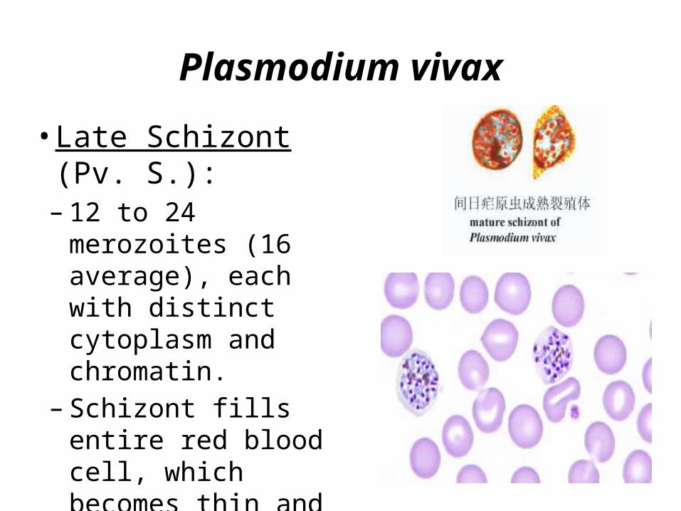
• Late Schizont (Pv. S.):– 12 to 24 merozoites (16
average), each with distinct cytoplasm and chromatin.
– Schizont fills entire red blood cell, which becomes thin and transparent.

Plasmodium vivax
• female gametocytes ( G.)♀ : Rounded or oval with
homogeneous cytoplasm. Diffuse delicate golden to light
brown malarial pigment throughout parasite.
The macrogametocyte displays a compact, deep red staining chromatin mass arranged near the cell periphery and cytoplasm which stains bright blue.

Plasmodium vivax
• male gametocytes ( G.)♂ : Rounded or oval with
homogeneous cytoplasm. Diffuse delicate golden to light
brown malarial pigment throughout parasite.
The microgametocyte displays a large chromatin mass which stains pink to purple and cytoplasm which stains pale blue.
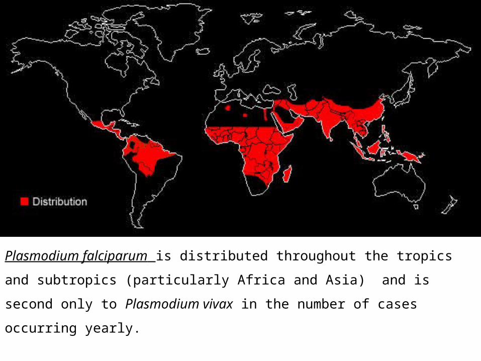
Plasmodium falciparum is distributed throughout the tropics and subtropics
(particularly Africa and Asia) and is second only to Plasmodium vivax in the
number of cases occurring yearly.
Plasmodium falciparum
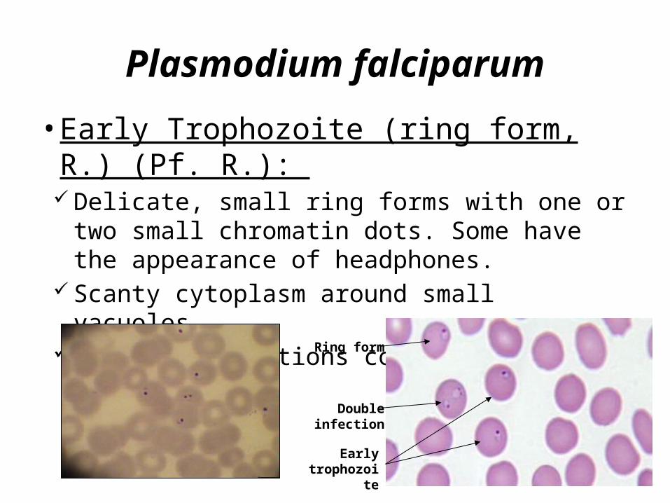
• Early Trophozoite (ring form, R.) (Pf. R.): Delicate, small ring forms with one or two small
chromatin dots. Some have the appearance of headphones.
Scanty cytoplasm around small vacuoles. Multiple infections common.
Early trophozoite
Ring form
Double infection
Plasmodium falciparum
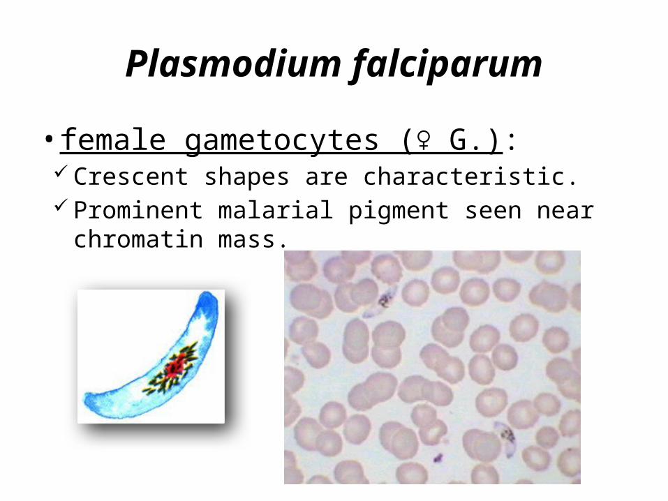
• female gametocytes ( G.)♀ : Crescent shapes are characteristic. Prominent malarial pigment seen near chromatin mass.

Plasmodium falciparum
• male gametocytes ( G.)♂ : sausage shapes are characteristic. Prominent malarial pigment seen near chromatin mass.

The microscopic examination of Giemsa-stained thick and thin blood films is recommended for the detection and identification of all blood parasites. Wright's stain is also acceptable and usually more readily available.
thick and thin blood films
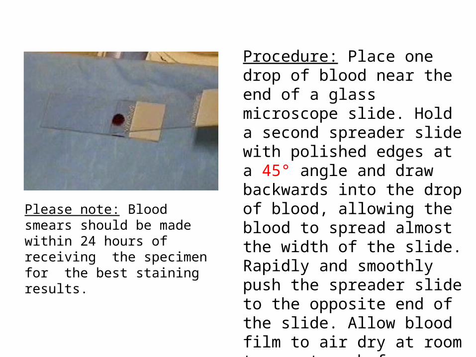
Please note: Blood smears should be made within 24 hours of receiving the specimen for the best staining results.
Procedure: Place one drop of blood near the end of a glass microscope slide. Hold a second spreader slide with polished edges at a 45° angle and draw backwards into the drop of blood, allowing the blood to spread almost the width of the slide. Rapidly and smoothly push the spreader slide to the opposite end of the slide. Allow blood film to air dry at room temperature before staining.
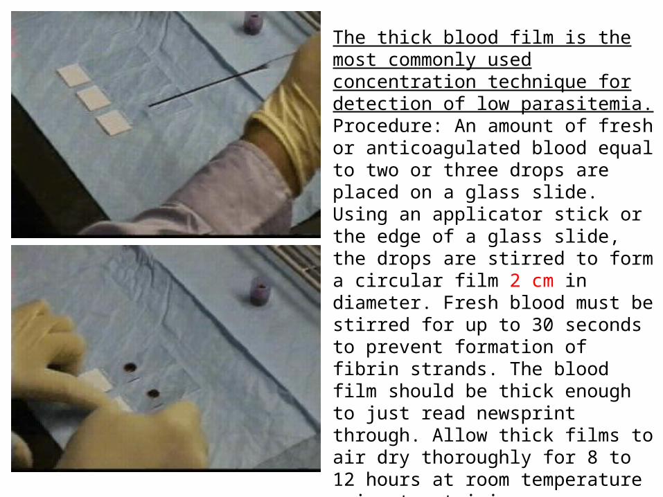
The thick blood film is the most commonly used concentration technique for detection of low parasitemia. Procedure: An amount of fresh or anticoagulated blood equal to two or three drops are placed on a glass slide. Using an applicator stick or the edge of a glass slide, the drops are stirred to form a circular film 2 cm in diameter. Fresh blood must be stirred for up to 30 seconds to prevent formation of fibrin strands. The blood film should be thick enough to just read newsprint through. Allow thick films to air dry thoroughly for 8 to 12 hours at room temperature prior to staining. Please note: Thick smears should have most of the RBC’S lysed to help the detection of very low numbers of blood parasites.

Classroom work
•Drawing the pictures of P.v. R, T, S, G (any
of two stages) & P.f. R or G
Recommended


![Studies on Porous and Morphological Structures of · PDF fileResearchers have developed biaxial stretching technology to produce expanded PTFE membranes [6,7,8,9,10,11,12]; however,](https://img.dokumen.tips/doc/110x75/5a708ea17f8b9ab1538c120f/studies-on-porous-and-morphological-structures-of-wwwjeffjournalorginjinj052p31-38pdfpdf.jpg)


