Bulgarian Journal of Veterinary Medicine, 2014, 17, No 2, 7987 ISSN 1311-1477; online at http://tru.uni-sz.bg/bjvm/bjvm.htm
Original article
DISTRIBUTION OF NICOTINAMIDE ADENINE DINUCLEOTIDE PHOSPHATE DIAPHORASE POSITIVE MAST CELLS IN THE
NORMAL PORCINE GALL BLADDER
I. S. STEFANOV1, A. P. VODENICHAROV1, M. V. GULUBOVA2 & J. R. ANANIEV2
1Department of Veterinary Anatomy, Histology and Embryology, Faculty of Veterinary Medicine, 2Department of General and Clinical Pathology, Faculty
of Medicine; Trakia University, 6000 Stara Zagora, Bulgaria.
Summary
Stefanov, I. S., A. P. Vodenicharov, M. V. Gulubova & J. R. Ananiev, 2014. Distribution of nicotinamide adenine dinucleotide phosphate diaphorase positive mast cells in the normal porcine gall bladder. Bulg. J. Vet. Med., 17, No. 2, 79–87. The aim of the current study was to determine the localisation and number of NADPH-d mast cells in the normal domestic pig gall bladder. The gall bladders of 6 male and 6 female pigs were examined by using light microscopic enzymohistochemical and immunohistochemical techniques. Enzymohis-tochemistry was performed to display NADPH diaphorase activity. Then, the avidin-biotin-peroxi-dase complex technique for detection of mast cell tryptase expression was performed in order to con-firm that NADPH-d positive cells, observed in the studied organ, are actually mast cells. Light mi-croscopy showed strong to medium NADPH-d reactivity in the mast cells granules. Mast cells num-ber was the highest in the propria of gall bladder fundus, body and neck as well as in the same layer of the cystic duct Ductus cysticus, followed by fibromuscular layer. The smallest density of mast cells was observed in the subserosal layer. However, statistically significant difference between the number of these cells in the muscular layer and the tela subserosa of gall bladder fundus was not detected. The density and localisation of observed tryptase positive mast cells were the same. In conclusion, the results of this study gave us a reason to presume that NADPH-d positive cells observed in the wall of porcine gall bladder and its excretory duct possess metabolic pathway for nitric oxide synthesis. Based on obtained data it is suggested that NADPH-d positive mast cells are most probably involved in the regulation of the function of epithelium, smooth muscle layer and blood vessels of the organ.
Key words: gall bladder, mast cells, NADPH-diaphorase, pig, tryptase
INTRODUCTION
Nitric oxide (NO) is produced from the oxidation of the amino acid L-arginine by the enzyme nitric oxide synthase (NOS). This family of enzymes is generally di-
vided into constitutive, calcium dependent (neuronal NOS, nNOS, NOS1 and endo-thelial NOS, eNOS, NOS3) or inducible, calcium independent (inducible NOS,
Distribution of nicotinamide adenine dinucleotide phosphate diaphorase positive mast cells in…
BJVM, 17, No 2 80
iNOS, NOS2) (Alderton et al., 2001). NO is a potent mediator with diverse roles in regulating cellular functions, including nitrosylation of proteins, involved in sig-nalling pathways (Alderton et al., 2001; Stamler et al., 2001). The role of NO in smooth muscle relaxation is also acknow-ledged (Cals-Grierson & Ormerod, 2004).
The ability of mast cells (MC) to pro-duce NO is well known. MC arise from bone marrow haematopoietic precursor cells and mature and reside in tissues (Kulka & Befus, 2003). Gilchrist et al. (2004) demonstrated the expression of NOS isoforms and production of NO by various MC populations including rat peritoneal MC and human skin MC. The presence of enzymes such as NADPH-d and NOS involved in NO metabolism within the mast cells in the pigs kidney and canine paranal sinus wall suggests a possible metabolic pathway for NO syn-thesis in porcine and canine mast cells (Vodenicharov & Bozhilova-Pastirova, 2010; Stefanov & Vodenicharov, 2012; Stefanov et al., 2012). Studies of human mast cells have been focused on the local-isation of these cells that accumulate near and around intrahepatic large bile ducts and intrahepatic peribiliary glands (Koda et al., 2000) and in the wall of common bile duct (Gulubova & Vlaykova, 2004). There are also many studies on NADPH-d expression in neuronal structures of gall bladder in different species (Sutherland, 1967) but no data concerning the NADPH-d positive mast cells in that or-gan in swine are available. At the same time, the physiological similarities be-tween porcine and human extrahepatic bile ducts support the use of pigs in ex-perimental models (Sand et al., 1997).
These facts motivated us to undertake the current study to determine the distribu-tion of NADPH-d mast cells in domestic
pig’s gall bladder. The presence of NADPH-d positive mast cells in the organ was demonstrated to prove that these cells possessed a metabolic pathway for NO synthesis. The obtained data could be used as background for further investiga-tions to elucidate the role of NADPH-d positive mast cells in the regulation of the epithelium function and smooth muscle relaxation in the organ.
MATERIALS AND METHODS
Animals
The material was obtained from the wall of gall bladder fundus, body and neck and as well as from the wall of middle part of the cystic duct of 6 male and 6 female pigs (Landrace×Bulgarian White), 6 months of age, weighing 92–98 kg, slaughtered for meat consumption in a slaughterhouse in accordance with Bulga-rian law.
Enzymohistochemical reaction for detec-tion of NADPH diaphorase
Part of the samples was immediately im-mersed in 4% paraformaldehyde (Sigma Aldrich Chemie, Switzerland) in phos-phate-buffered saline (PBS), pH 7.2, for 8 h at 4 °C. Then the samples were removed and soaked in solution of 30% sucrose in PBS overnight. Sections of 15 µm thick-ness were prepared by means of a freezing microtome (Slee, Mainz, Germany). The free-floating sections were further proc-essed according to the protocol of Sherer-Singler et al. (1983) by incubation in a solution containing nitro blue tetrazolium (0.2 mg/mL, Sigma Aldrich Chemie GmbH, Germany), β-NADPH (Santa Cruz Biotech, Santa Cruz, CA, USA) and Tri-ton X-100 (0.5%) (Merck Belgalabo, Overijse, Belgium) in PBS (0.1 M, pH
I. S. Stefanov, A. P. Vodenicharov, M. V. Gulubova & J. R. Ananiev
BJVM, 17, No 2 81
7.4) for 1–2 h at 37 °C. Microscopic as-sessment of the reaction was scored as absent (0), weak (+), medium (++) and strong (+++).
Light microscopic immunohistochemistry
Another part of the same samples was used for immunohistochemical staining to detect mast cell tryptase. The avidin-biotin-peroxidase complex technique was performed on formalin-fixed and paraffin-embedded tissues. Then the tissues were cut to 5–6 µm thick serial sections, which were mounted on glass slides. The sec-tions were deparaffinised twice in xylene, followed by absolute ethanol: 96 °C, 70 °C and 50 °C series, each for 10 min. The further immunohistochemical procedure was described earlier by Gulubova & Vlaykova (2006). In brief, the sections were rinsed in phosphate buffer saline (PBS), pH 7.4 and internal peroxidase was blocked by incubation in 1.2% hy-drogen peroxide (Peroxidase block K0673, DAKO A/S, Glostrup, Denmark) in methanol for 30 min and rinsed in PBS with pH 7.4 for 15 min. Immunohisto-chemical reaction was carried out using primary monoclonal mouse anti-human mast cell tryptase antibody (N M7052, DAKO clone AA1) with dilution 1:100. The sections were washed in PBS, pH 7.4, incubated with Dako REAL™ EnVi-sion™ Detection System, Peroxidase/ DAB+, Rabbit/Mouse, K5007 for 60 min, then visualised with 3,3′ diaminoben-zidine and counterstained with Mayer’s haematoxylin. Sections incubated with non-immune sera instead of the primary antibodies were used as negative controls.
Microscopic assessment of the immu-nocytochemical reaction was scored as absent (0), weak (+), medium (+ +) and strong (+ + +).
Statistical analysis
Cross sections of the wall of gall bladder fundus, body and neck and of its excretory duct of each animal were used. On serial sections from each part of the gall blad-der, ten 1 mm2 fields were investigated. Data for mast cells density (number/mm2) are given as mean ± SD. Light microscope (ZEISS Primo Star, Germany), camera (Progres, Capture 2.6 – JENOPTIK) and software analysis programme (Soft Imag-ing System GmbH) were used. Statistical data processing was done by using Stu-dent’s t-test. The difference was consid-ered as significant at P<0.05.
RESULTS
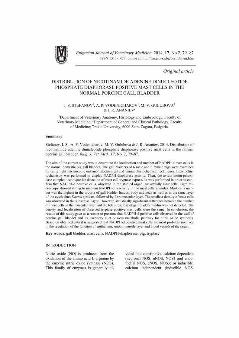
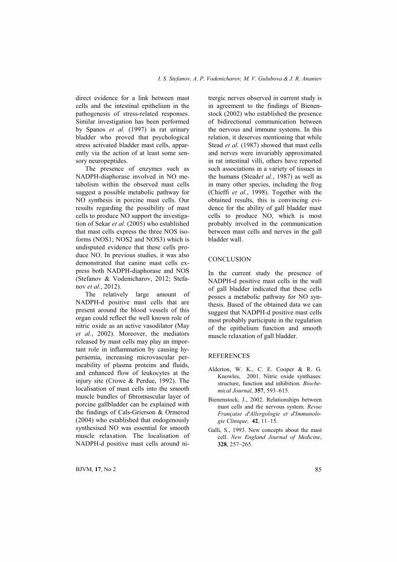
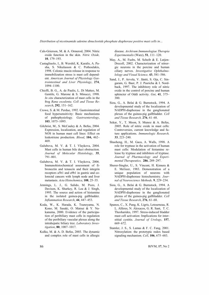
The distribution of NADPH-d positive mast cells in the three layers – the lamina propria of the mucosa, the muscular layer and tela subserosa of the porcine gall bladder wall and the cystic duct was in-vestigated. Light microscopic observation showed strong to medium enzyme reacti-vity in cytoplasmic positive granules of NADPH-d cells (Fig. 1). Nuclei of posi-tive cells exhibited a negative reaction. In the propria of this organ, most NADPH-d mast cells were localised close to vessels of microcirculatory bed and near the basal membrane of biliary lining epithelium (Fig. 1). In the gall bladder neck mast cells were also found around the glands. Some of them were detected around the nerves in all layers of the organ (Fig. 2).
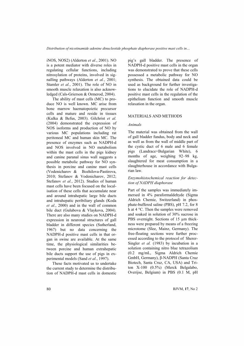
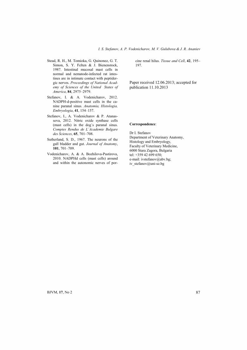
In the fibromuscular and subserosal layers the same cells were observed mainly in the vicinity of blood vessels (Fig. 3). NADPH-d positive mast cells were also located between smooth muscle bundles of the fibromuscular layer. In the wall of arteries and veins NADPH-d mast cells were localized mainly in the adventi-tial layer, followed by the tunica media.

Distribution of nicotinamide adenine dinucleotide phosphate diaphorase positive mast cells in…
BJVM, 17, No 2 82
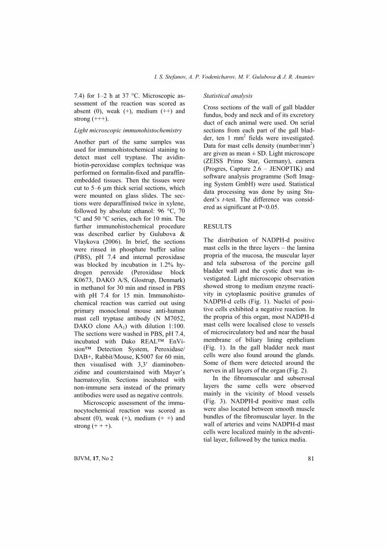
It is important to note that NADPH-d positive mast cells showed the same local-isation as tryptase positive MC (Fig. 4).
The number of NADPH-d positive mast cells in the lamina propria of the mucosa of gall bladder fundus, body and
neck and as well as in the same layer of the cystic duct was significantly higher than in the muscular layer and tela subse-rosa in both genders (Table 1).
In the different gall bladder parts and in the cystic duct, the number of mast cells
EPR
Fig. 1. Mast cells (arrows) containing NADPH-d positive granules and unstained nucleus, localised in the propria (PR) of gall bladder body. E – lamina epithelialis mucosae. Bar = 20 µm.
G
G
Fig. 2. NADPH-d positive mast cells (arrowheads) near the glands (G) and around the autonomic nitrergic nerve fibres (arrows) in the wall of gall bladder neck. Bar = 20 µm.

I. S. Stefanov, A. P. Vodenicharov, M. V. Gulubova & J. R. Ananiev
BJVM, 17, No 2 83
in the muscle layer was higher than in the subserosal layer. However, no statistically significant difference was observed bet-
ween MC number in the muscular layer and tela subserosa of gall bladder fundus.
A
Fig. 3. Localisation of NADPH-d mast cells (arrowheads) near the media of small arteriae (A) and around smooth muscle bundles (arrows) in the fibromuscular layer of gall bladder neck.
Bar = 20 µm.
E
PR
TM
Fig. 4. Tryptase positive mast cells (arrows) localisation in the propria of the mucosa (PR) and near the smooth muscle cells (arrowheads) of the muscular layer (tunica muscularis, T) of gall bladder
body. These cells showed the same localisation as NADPH-d positive mast cells. E – lamina epithelialis mucosae. Counterstaining with haematoxylin. Bar = 20 µm.
Distribution of nicotinamide adenine dinucleotide phosphate diaphorase positive mast cells in…
BJVM, 17, No 2 84
The density and localisation of NADPH-d positive mast cells were identi-cal to those of mast cell tryptase positive cells in males and females, but a statistical significant difference was not established (Table 1).
DISCUSSION
NADPH-d positive mast cells localization and density in the different parts and lay-ers of porcine gall bladder as well as its excretory duct were established for the first time. Our results showed that in the wall of porcine gall bladder NADPH-d positive cells had the same morphology and localisation like tryptase positive mast cells. According to Shaoheng et al. (1998) tryptase has been used as a marker for human mast cells. Based on this fact we
proved that the NADPH-d positive cells in the studied organ are most probably mast cells. Other authors such as Jennings et al. (1995) established the distribution of his-tamine-containing mast cells in the mu-cosa and muscular/serosa layers of guinea pig gall bladder but there was no data about the presence of NADPH-d positive mast cells. We assumed that the localisa-tion of mast cells near the basal membrane of the mucous epithelium and around the glands of gall bladder neck was connected with their role in regulation of epithelial function. Our findings correlate with the results of Castagliuolo et al. (1998) who directly demonstrated the participation of mast cells in colonic goblet cell discharge and prostaglandin E2 secretion caused by restraint of mice. Their findings provide
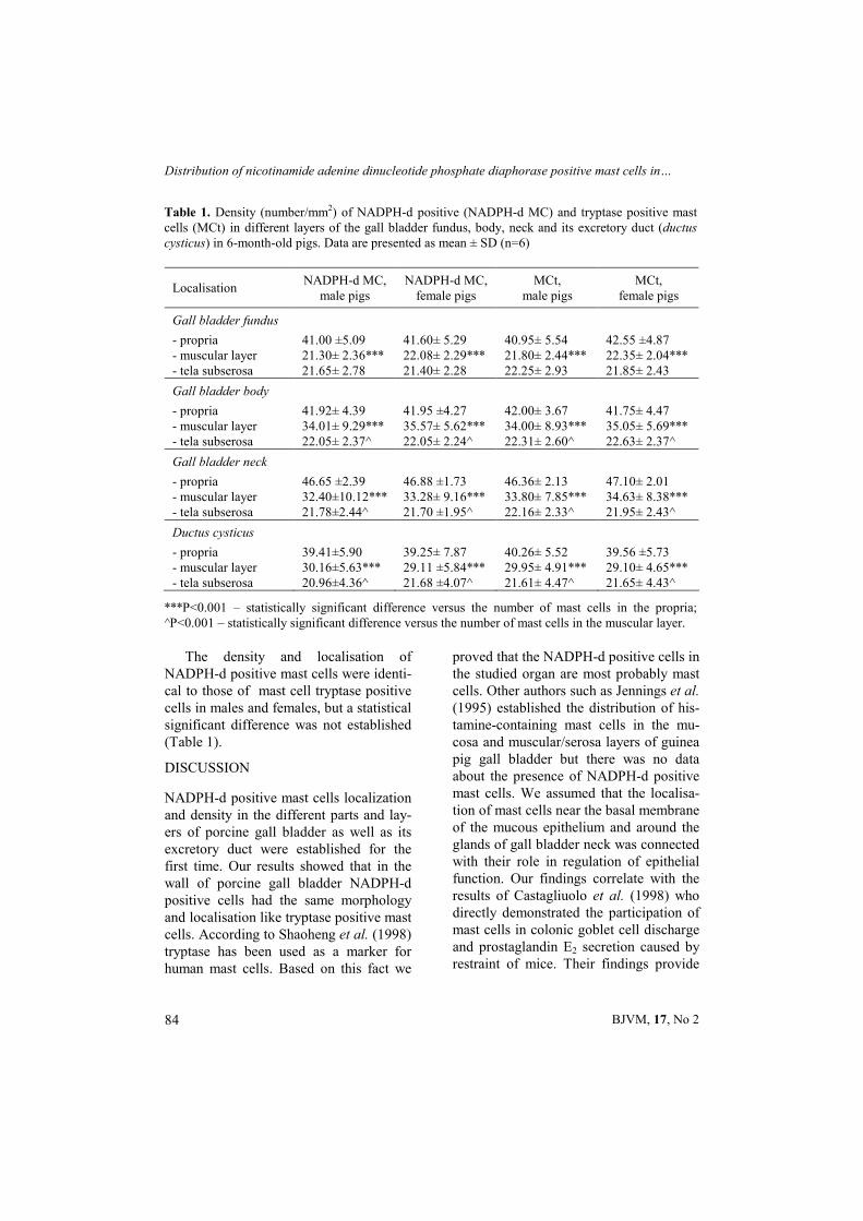
Table 1. Density (number/mm2) of NADPH-d positive (NADPH-d MC) and tryptase positive mast cells (MCt) in different layers of the gall bladder fundus, body, neck and its excretory duct (ductus cysticus) in 6-month-old pigs. Data are presented as mean ± SD (n=6)
Localisation NADPH-d MC,
male pigs NADPH-d MC,
female pigs MCt,
male pigs MCt,
female pigs
Gall bladder fundus
- propria 41.00 ±5.09 41.60± 5.29 40.95± 5.54 42.55 ±4.87 - muscular layer 21.30± 2.36*** 22.08± 2.29*** 21.80± 2.44*** 22.35± 2.04*** - tela subserosa 21.65± 2.78 21.40± 2.28 22.25± 2.93 21.85± 2.43
Gall bladder body
- propria 41.92± 4.39 41.95 ±4.27 42.00± 3.67 41.75± 4.47 - muscular layer 34.01± 9.29*** 35.57± 5.62*** 34.00± 8.93*** 35.05± 5.69*** - tela subserosa 22.05± 2.37^ 22.05± 2.24^ 22.31± 2.60^ 22.63± 2.37^
Gall bladder neck
- propria 46.65 ±2.39 46.88 ±1.73 46.36± 2.13 47.10± 2.01 - muscular layer 32.40±10.12*** 33.28± 9.16*** 33.80± 7.85*** 34.63± 8.38*** - tela subserosa 21.78±2.44^ 21.70 ±1.95^ 22.16± 2.33^ 21.95± 2.43^
Ductus cysticus
- propria 39.41±5.90 39.25± 7.87 40.26± 5.52 39.56 ±5.73 - muscular layer 30.16±5.63*** 29.11 ±5.84*** 29.95± 4.91*** 29.10± 4.65*** - tela subserosa 20.96±4.36^ 21.68 ±4.07^ 21.61± 4.47^ 21.65± 4.43^
***P<0.001 – statistically significant difference versus the number of mast cells in the propria; ^P<0.001 – statistically significant difference versus the number of mast cells in the muscular layer.
I. S. Stefanov, A. P. Vodenicharov, M. V. Gulubova & J. R. Ananiev
BJVM, 17, No 2 85
direct evidence for a link between mast cells and the intestinal epithelium in the pathogenesis of stress-related responses. Similar investigation has been performed by Spanos et al. (1997) in rat urinary bladder who proved that psychological stress activated bladder mast cells, appar-ently via the action of at least some sen-sory neuropeptides.
The presence of enzymes such as NADPH-diaphorase involved in NO me-tabolism within the observed mast cells suggest a possible metabolic pathway for NO synthesis in porcine mast cells. Our results regarding the possibility of mast cells to produce NO support the investiga-tion of Sekar et al. (2005) who established that mast cells express the three NOS iso-forms (NOS1; NOS2 and NOS3) which is undisputed evidence that these cells pro-duce NO. In previous studies, it was also demonstrated that canine mast cells ex-press both NADPH-diaphorase and NOS (Stefanov & Vodenicharov, 2012; Stefa-nov et al., 2012).
The relatively large amount of NADPH-d positive mast cells that are present around the blood vessels of this organ could reflect the well known role of nitric oxide as an active vasodilator (May et al., 2002). Moreover, the mediators released by mast cells may play an impor-tant role in inflammation by causing hy-peraemia, increasing microvascular per-meability of plasma proteins and fluids, and enhanced flow of leukocytes at the injury site (Crowe & Perdue, 1992). The localisation of mast cells into the smooth muscle bundles of fibromuscular layer of porcine gallbladder can be explained with the findings of Cals-Grierson & Ormerod (2004) who established that endogenously synthesised NO was essential for smooth muscle relaxation. The localisation of NADPH-d positive mast cells around ni-
trergic nerves observed in current study is in agreement to the findings of Bienen-stock (2002) who established the presence of bidirectional communication between the nervous and immune systems. In this relation, it deserves mentioning that while Stead et al. (1987) showed that mast cells and nerves were invariably approximated in rat intestinal villi, others have reported such associations in a variety of tissues in the humans (Steadet al., 1987) as well as in many other species, including the frog (Chieffi et al., 1998). Together with the obtained results, this is convincing evi-dence for the ability of gall bladder mast cells to produce NO, which is most probably involved in the communication between mast cells and nerves in the gall bladder wall.
CONCLUSION
In the current study the presence of NADPH-d positive mast cells in the wall of gall bladder indicated that these cells posses a metabolic pathway for NO syn-thesis. Based of the obtained data we can suggest that NADPH-d positive mast cells most probably participate in the regulation of the epithelium function and smooth muscle relaxation of gall bladder.
REFERENCES
Alderton, W. K., C. E. Cooper & R. G. Knowles, 2001. Nitric oxide synthases: structure, function and inhibition. Bioche-mical Journal, 357, 593–615.
Bienenstock, J., 2002. Relationships between mast cells and the nervous system. Revue Française d'Allergologie et d'Immunolo-gie Clinique, 42, 11–15.
Galli, S., 1993. New concepts about the mast cell. New England Journal of Medicine, 328, 257–265.
Distribution of nicotinamide adenine dinucleotide phosphate diaphorase positive mast cells in…
BJVM, 17, No 2 86
Cals-Grierson, M. & A. Ormerod, 2004. Nitric oxide function in the skin. Nitric Oxide, 10, 179–193.
Castagliuolo, I., B. Wershil, K. Karalis, A. Pa-sha, S. Nikulasson & C. Pothoulakis, 1998. Colonic mucin release in response to immobilization stress is mast cell depend-ent. American Journal of Physiology Gas-trointestinal and Liver Physiology, 274, 1094–1100.
Chieffi, B. G., A. de Paulis, L. Di Matteo, M. Gentile, G. Marone & S. Minucci, 1998. In situ characterization of mast cells in the frog Rana esculenta. Cell and Tissue Re-search, 292, 151–162.
Crowe, S. & M. Perdue, 1992. Gastrointestinal food hypersensitivity: Basic mechanisms of pathophysiology. Gastroenterology, 103, 1075–1095.
Gilchrist, M., S. McCauley & A. Befus, 2004. Expression, localization, and regulation of NOS in human mast cell lines: Effect on leukotriene production. Blood, 104, 462–469.
Gulubova, M. V. & T. I. Vlaykova, 2004. Mast cells in human bile duct obstruction. Journal of Molecular Histolology, 35, 791–801.
Gulubova, M. V. & T. I. Vlaykova, 2006. Immunohistochemical assessment of fi-bronectin and tenascin and their integrin receptors a5b1 and a9b1 in gastric and co-lorectal cancers with lymph node and liver metastasis. Acta Histochemica, 108, 25–35.
Jennings, L. J., G. Salido, M. Pozo, J. Davison, K. Sharkey, R. Lea & J. Singh, 1995. The source and action of histamine in the isolated guinea-pig gallbladder. Inflammation Research, 44, 447–453.
Koda, W., K. Harada, K. Tsuneyama, N. Kono, M. Sasaki, O. Matsui & Y. Na-kanuma, 2000. Evidence of the participa-tion of peribiliary mast cells in regulation of the peribiliary vascular plexus along the intrahepatic biliary tree. Laboratory Inves-tigation, 80, 1007–1017.
Kulka, M. & A. D. Befus, 2003. The dynamic and complex role of mast cells in allergic
disease. Archivum Immunologiae Therapia Experimentalis (Warz), 51, 111–120.
May, A., M. Fusbs, M. Scheib & E. Lutjen-Drecoll, 2002. Characterization of nitrer-gic neurons in the porcine and human ciliary nerves. Investigative Ophthalmo-lology and Visual Science, 43, 581–586.
Sand, J., P. Arvola, V. Jäntti, S. Oja, C. Sin-garam, G. Baer, P. J. Pasricha & I. Nord-back, 1997. The inhibitory role of nitric oxide in the control of porcine and human sphincter of Oddi activity. Gut, 41, 375–380.
Siou, G., A. Belai & G. Burnstock, 1994. A developmental study of the localization of NADPH-diaphorase in the ganglionated plexus of the guinea-pig gallbladder. Cell and Tissue Research, 276, 61–68.
Sekar, Y., T. Moon, S. Munoz & A. Befus, 2005. Role of nitric oxide in mast cells: Controversies, current knowledge and fu-ture applications. Immunologic Research, 33, 223–240.
Shaoheng, H., M. Gaca, A. Walls, 1998. A role for tryptase in the activation of human mast cells: Modulation of histamine re-lease by tryptase and inhibitors of tryptase. Journal of Pharmacology and Experi-mental Therapeutics, 286, 289–297.
Sherer-Singler, U., S. Vincent, H. Kimura & E. McGeer, 1983. Demonstration of a unique population of neurons with NADPH-diaphorase histochemistry. Jour-nal of Neuroscience Methods, 9, 229–234.
Siou, G., A. Belai & G. Burnstock, 1994. A developmental study of the localization of NADPH-diaphorase in the ganglionated plexus of the guinea-pig gallbladder. Cell and Tissue Research, 276, 61–68.
Spanos, C., X. Pang, K. Ligris, Letourneau, R. L. Alferes, N. Alexacos, G. R. Sant, T. C. Theoharides, 1997. Stress-induced bladder mast cell activation: Implications for inter-stitial cystitis. Journal of Urology, 157, 669–672.
Stamler, J. S., S. Lamas & F. C. Fang, 2001. Nitrosylation: the prototypic redox based signaling mechanism. Cell, 106, 675–683.
I. S. Stefanov, A. P. Vodenicharov, M. V. Gulubova & J. R. Ananiev
BJVM, 17, No 2 87
Stead, R. H., M. Tomioka, G. Quinonez, G. T. Simon, S. Y. Felten & J. Bienenstock, 1987. Intestinal mucosal mast cells in normal and nematode-infected rat intes-tines are in intimate contact with peptider-gic nerves. Proceedings of National Acad-emy of Sciences of the United States of America, 84, 2975–2979.
Stefanov, I. & A. Vodenicharov, 2012. NADPH-d-positive mast cells in the ca-nine paranal sinus. Anatomia, Histologia, Embryologia, 41, 154–157.
Stefanov, I., A. Vodenicharov & P. Atanas-sova, 2012. Nitric oxide synthase cells (mast cells) in the dog`s paranal sinus. Comptes Rendus de L`Academie Bulgare des Sciences, 65, 701–708.
Sutherland, S. D., 1967. The neurons of the gall bladder and gut. Journal of Anatomy, 101, 701–709.
Vodenicharov, A. & A. Bozhilova-Pastirova, 2010. NADPHd cells (mast cells) around and within the autonomic nerves of por-
cine renal hilus. Tissue and Cell, 42, 195–197.
Paper received 12.06.2013; accepted for publication 11.10.2013
Correspondence: Dr I. Stefanov Department of Veterinary Anatomy, Histology and Embryology, Faculty of Veterinary Medicine, 6000 Stara Zagora, Bulgaria tel: +359 42 699 650; e-mail: [email protected]; [email protected]
Recommended