REVIEW
Morphing in nature and beyond: a review of natural
and synthetic shape-changing materials
and mechanisms
Kate Oliver1, Annela Seddon1,2,* , and Richard S. Trask3
1Bristol Centre for Functional Nanomaterials, HH Wills Physics Laboratory, University of Bristol, Tyndall Avenue, Bristol BS8 1TL, UK2HH Wills Physics Laboratory, School of Physics, University of Bristol, Tyndall Avenue, Bristol BS8 1TL, UK3Department of Mechanical Engineering, University of Bath, North Rd, Bath BA2 7AY, UK
Received: 20 May 2016
Accepted: 10 August 2016
Published online:
19 August 2016
� The Author(s) 2016. This
article is published with open
access at Springerlink.com
ABSTRACT
Shape-changing materials open an entirely new solution space for a wide range
of disciplines: from architecture that responds to the environment and medical
devices that unpack inside the body, to passive sensors and novel robotic
actuators. While synthetic shape-changing materials are still in their infancy,
studies of biological morphing materials have revealed key paradigms and
features which underlie efficient natural shape-change. Here, we review some of
these insights and how they have been, or may be, translated to artificial solu-
tions. We focus on soft matter due to its prevalence in nature, compatibility with
users and potential for novel design. Initially, we review examples of natural
shape-changing materials—skeletal muscle, tendons and plant tissues—and
compare with synthetic examples with similar methods of operation. Stimuli to
motion are outlined in general principle, with examples of their use and
potential in manufactured systems. Anisotropy is identified as a crucial element
in directing shape-change to fulfil designed tasks, and some manufacturing
routes to its achievement are highlighted. We conclude with potential directions
for future work, including the simultaneous development of materials and
manufacturing techniques and the hierarchical combination of effects at multi-
ple length scales.
Introduction
Complex multifunctional materials are common
within the natural world across all length scales and
taxa: from the high tensile strength of spider silk [1]
and the compressive properties of bone [2], to the
iridescence of the butterfly wing [3] and the fast
actuation of cuttlefish skin [4]. These materials dis-
play intricate architectures across the nano-, micro-
and mesoscales, allowing for an impressive array of
tailored functional materials properties. Natural
Address correspondence to E-mail: [email protected]
DOI 10.1007/s10853-016-0295-8
J Mater Sci (2016) 51:10663–10689
Review
materials have long been an inspiration to the mate-
rials chemist, physicist and engineer, and using the
combined toolkits of synthetic chemistry and
advanced fabrication techniques, many of these
materials have been successfully replicated and, in
some cases, improved upon in the synthetic world
(e.g. a gecko-inspired adhesive which, unlike the
original lizards, also adheres underwater) [5].
However, an area where nature still outperforms
our current synthetic capabilities is that of active, self-
shaping and stimuli-responsive materials. Material
structures found in nature must be more than just a
static object, framework or skin. Natural structures
need to be triggered under certain conditions, or by
external stimuli, for basic biological functions to
occur, and where possible exhibit multiple capabili-
ties. What is remarkable, however, is that the variety
of ways that this can be achieved across the plant and
animal kingdom takes place with relatively limited
chemical diversity and with large structural changes
triggered under mild, ambient conditions. This forms
a severe contrast to the variety of synthetic molecules
and extreme environments available to human
designers and suggests the existence of a wide solu-
tion space once key biological principles are extracted
and understood.
Naturally, the properties of actuating materials
inform the potential for further development and
refinement of response, and ultimately applications.
Many studies of shape memory alloys and piezo-
electric materials, some of the most prominent shape-
changing materials, exist—–see Mohd Jani [6] and
Irschik [7]—but these differ radically from the natural
solutions seen around us. Therefore, in keeping with
the biomimetic theme of this review, the materials
focused on here will be ‘soft’ actuators— soft mate-
rials capable of changing their shape in response to
stimuli. Soft materials are usefully characterised by
Doi as having fundamental ‘building blocks’ on the
order of 10–10,000 A and thus having relatively slow
response times between 1 and 104 s , a high response
to stimuli that necessitates non-linear description and
being easily driven out of equilibrium [8]. The sub-
section of soft materials that behave as actuators,
capable of moving themselves or their surroundings,
forms the topic of this review. As examples, this
includes materials such as muscle and unfolding
petals.
While soft materials depart from the traditional
materials of engineering and may seem to lack
robustness, they also offer novel advantages, as
detailed by Quake and Scherer [9]. They are efficient
to manufacture at small scale and they can be easily
integrated with hard elements as required, for
instance as a valve seal. In devices, soft materials give
increased compatibility with human and animal
users, and greater adaptability which confers the
ability to operate in unpredictable, extreme environ-
ments [10, 11]. Such behaviour has many prospective
applications, from biomedical devices [12] and pas-
sive building materials [13], to microfluidics [14],
product design [15] and soft robotics [10, 16].
In this article, the paradigm of self-shaping and
actuating materials found in nature will be intro-
duced, to lay the groundwork for a review of the
current literature on soft synthetic analogues. Can-
didate materials for biomimicry of response will be
identified, along with their mechanisms, limitations
and triggers. It will be seen that control of structure,
and therefore available manufacturing possibilities,
will be crucial to realise the goal of synthetic mor-
phing materials.
Benchmarking natural materials againstsynthetics
There are many examples of actuation response of
natural materials to changes in environmental
humidity. This can range from the simple curling of a
wet leaf as it dries, to more complex processes such
as self-burial of seeds [17]. The passive opening and
closing of the scales of a pinecone is an elegant
example of hygroscopic actuation for seed dispersal,
accompanied by a simple synthetic model. The
reversible movement of the pine cone scales is driven
by differences in structural orientation of the cellu-
lose microfibrils within cell walls across the structure.
The microfibrils have a greater resistance to extension
along their axis of alignment, and so by varying this
angle relative to the body of the cell, deformation of
the volume can be channelled preferentially in one
direction. Cells making up the outside of the scale are
orientated to elongate on exposure to humidity,
whereas the inner layer are more resistant to elon-
gation [18].
This leads to a behaviour which has been com-
pared to that of a thermally responsive bimetallic
strip, which undergoes bending due to the differing
thermal responses of the two constituent metals
10664 J Mater Sci (2016) 51:10663–10689
bound together [18, 19]. This is a familiar and
tractable model in engineering and physics with a
long history. First modelled by Timoshenko in 1925
[20], bimetallic strips were a common control
switching element before being replaced with mod-
ern relays. The form has, from the very early work on
shape-changing hydrogels by Hu [21], been a simple
test-case for soft shape-changing materials. Since then
research in the area has blossomed, using techniques
including chemical variation [21], variation in cross-
linking density [22], and alignment of restrictive
elements [23] to achieve controlled movement, as
discussed in more detail later in this manuscript.
Cellulose catapults: a natural example
In an effort to overcome limitations in chemical
diversity and operating temperature, nature pro-
grammes the sequential arrangement and orienta-
tions of individual structural units, while
simultaneously tailoring their function and attach-
ment strategies to maximise the design for survival
and longevity. A key element is the transmission and
amplification of small degrees of strain across the
length scales. As will be described below, this can
translate a relatively simple and modest response to
external stimuli into large-scale movements such as
twisting, bending and opening.
Many plants exploit the principle of cellulose ori-
entation directing moisture driven deployment, from
the opening of eudicot seedpods [24] to the unfurling
of the desert resurrection plant [19]. More complex
behaviour is produced with more elaborate cellulose
arrangements, and closer examination of this illumi-
nates principles that may be helpful for reproducing
their properties.
Erodium circodia, commonly known as redstem
filaree (among many other names), is a small
Mediterranean plant related to the geranium. Filarees
disperse by firing their seeds around half a metre,
powered only by the arrangement and drying of
cellulose and lignin. The awn, or seed delivery
mechanism, of E. circodia is a thin support which
attaches the seeds to the main body of the plant, and
as it desiccates curls into a spring, storing elastic
energy. At a critical point, this material cracks and
the elastic energy is released. This behaviour is
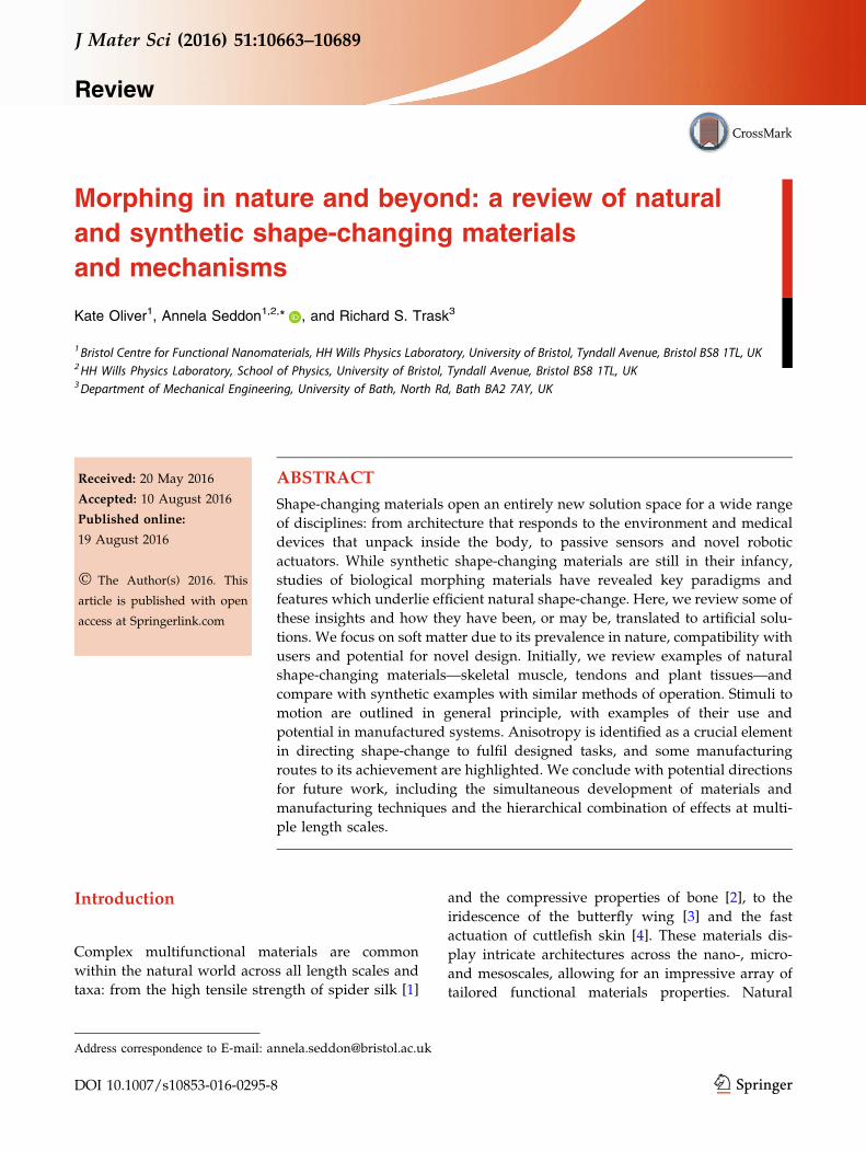
illustrated in Fig. 1, as recorded and analysed by
Evangelista et al. [17].
Within the awn, each constituent cell is encircled
by cellulose in a helical arrangement. Importantly,
the axis of the helix is not aligned with the axis of the
cell, causing the cell itself to bend such that it packs in
a helical form. Thus, the off-axis alignment of the
cellulose nanostructure instigates a similar direc-
tionality in the cellular alignment, which results in
the macroscopic coiling effect seen [25]. As the
material dehydrates, the coil becomes tighter, storing
more and more tension until the material eventually
fractures. This has been successfully mathematically
modelled by Aharoni et al. [26] and in a simplified
version reproduced in a physical model by Abraham
et al. [25]. This latter work demonstrates that one
does not need to reproduce all the elegant details of
the natural solution to draw on the concept.
This is all achieved with cellulose microfibrils
embedded in a matrix of polysaccharides, aromatics
and structural proteins—materials far removed from
the inorganic chemical palette of most modern engi-
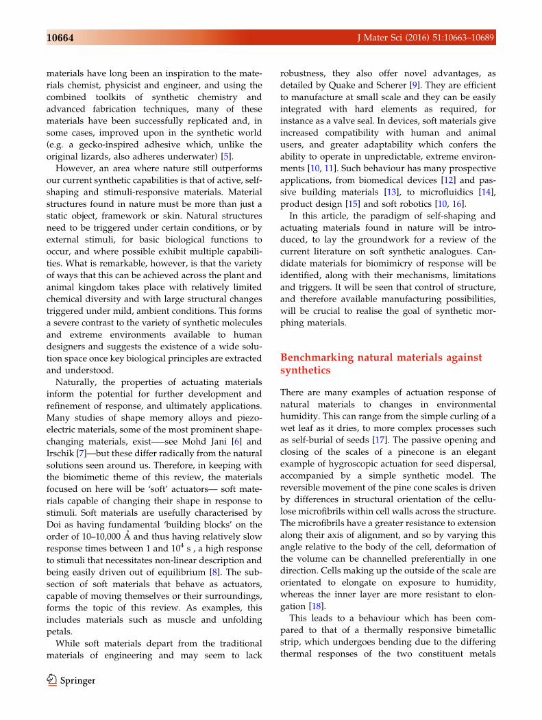
neers. For comparison purposes, we compared the
range of an equivalent volume to the filaree seed and
awn (Fig. 1), composed of a variety of natural and
synthetic shape-changing materials, if this were to be
launched under the propulsion of its own storage
energy. This provides a way to parametrise the
energy stored in a fixed unit volume, while building
Figure 1 The capability of natural shape-change. a Representative
launch trajectory of the filaree awn, as recorded by Evangelista
et al., shown by multiple exposures at 4-ms intervals. Movement is
from left to right. Scale bar 10 cm. b Trajectory predicted by
modelling, matching the initial launch trajectory [inset box, seed
(green) and tip (red) positions shown at 2-ms intervals] and final
distance thrown (blue star). Reproduced from Evangelista et al.
with permission from the Company of Biologists. [17] Copyright
2011.
J Mater Sci (2016) 51:10663–10689 10665

in a penalty for greater weight. Thus, the energy
density per unit weight is converted into an easy-to-
visualise range of lengths, highlighting the capability
of natural, low-density materials. Comparative val-
ues are shown in Fig. 2.
Our figures are derived from some simple
assumptions, and the application of conservation of
energy and Newtonian mechanics. We assume a
cylindrical volume of which undergoes uniform
contraction. Values for the stored energy contained
within such a volume are drawn from a variety of
sources (see supplementary information) and are
drawn from both active contributions (for materials
such as muscle and shape memory polymer) and
passively stored elastic energy (for materials such as
gels and spider silk, for which the highest strain
value which remains in the Hookean region is used).
These are, where necessary, converted into energy
per unit mass using available measures of density
(sources in supplementary information). We then
assume an equivalent percentage of stored strain
energy as in the filaree is converted into rectilinear
motion—2 % as calculated by Evangelista et al.
Finally, we take a constant launch angle and,
neglecting drag, use standard calculations of projec-
tile motion to find the range of a synthetic awn and
seed.
This is not presented as a detailed analysis or a
design strategy. But nevertheless it provides a simple
comparison between actuating materials of different
classes in terms of energy available for self-motion
and highlights some features of interest. The poten-
tial of soft biological materials such as filaree and
tendon is clear in this context. Their advantage
derives from their low density—a crucial point for
self-shaping materials that are required to move their
own weight, as in many realistic applications. In
synthetic competitors, both shape memory polymer
and hydrogel are comparable to the best plant and
animal material energy storages shown, with only
dragline spider silk—a highly anomalous material
which has resisted many attempts to analyse and
reproduce—exceeding the capacity of our most suc-
cessful synthetic by this (admittedly artificial) metric,
Kevlar. Both Kevlar and spider silk are liquid crys-
talline in solution and achieve alignment during the
process of spinning extrusion, from which their
properties derive [27].
These back-of-the-envelope considerations suggest
some initial areas of interest for a researcher looking
into self-shaping materials through a biological lens.
Gels suggest the potential of low-density, soft mate-
rials; memory polymers show a promising high-
Figure 2 Comparing the capabilities of various natural and
synthetic shape-changing materials, using the example of ballistic
seed dispersal, which incorporates both the capacity for elastic
energy storage and an allowance for density. They are compared to
the natural example of the filaree, which has an energy density of
approximately 750 kJ/kg and a mass of 5 9 10-3g, enabling
projection 2.62 m (without drag). Literature values and
calculations of energy density within the Hookean region are used
to extrapolate range for plant (solid green), animal (dashed coral),
soft and hard synthetic (dotted light and dark grey, respectively)
materials. Derivation is based on the calculations of Evangelista
et al., simplified to neglect drag and assume a constant volume.
[17] Full details of sources and values are given in Supplementary
Information.
10666 J Mater Sci (2016) 51:10663–10689
energy density; and Kevlar and spider silk suggest
the importance of molecular alignment.
It can be seen that of the animal tissues that exist
inside the organism (thus exempting spider silk),
pure elastin has the best performance. However, in
nature, this must be used as a composite, due to the
need for biological tissues to meet multiple perfor-
mance requirements. This reduces their energy stor-
age capacity somewhat. Analogously, the high
theoretical performance of carbon nanotubes is unli-
kely to translate wholly into a practical material.
However, performance can reasonably be expected to
match or exceed the performance of CNT yarn, which
again compares well with elastin. The ability of these
efficient molecules to improve performance, even at
low proportions in the material, hints at the potential
of nanoinclusions and other composites.
We therefore see that to emulate the complex
actuation observed in the stork’s bill awn and other
natural morphing systems, we require knowledge of
the inherent properties of the material (the swelling
matrix), the stimuli (the conditions or trigger
required, for example the magnitude of dehydration
the plant will experience), the reinforcement archi-
tecture (the direction of the fibrils and their hierar-
chical assembly), and the cost in energy for actuation.
Without waiting for 4.5 billion years for evolution to
produce protein machines to conduct this assembly,
we should also consider methods of manufacturing
these structural features.
These topics will inform the rest of the review,
which will address some prototypical natural mor-
phing materials and related synthetics, run through
triggering stimuli including hygroscopic, chemical,
heat, light and electrical and magnetic inputs, and
finally discuss methods of modifying such materials
in order to direct the response in a programmed
direction. We conclude with some promising future
directions.
Natural morphing materials and syntheticsubstitutes
A vast amount of actuating soft materials exist,
including shape memory polymers (SMPs), hydro-
gels, elastomers and liquid crystals. This library only
expands when the potential for composites or alter-
native processing is considered. The addition of sec-
ondary materials may enhance properties such as
tensile strength, compressive strength, toughness and
elasticity, or confer response to external stimuli.
Therefore, this section aims instead to focus on a few
key natural actuating materials, discuss the principles
at both the micro- and macroscale by which each
operates, sketch an option for artificial emulation and
its mechanism, and provide an overview of their
potential uses and limitations. We will highlight bio-
inspired and otherwise notable uses of the material,
alone or as a composite, and where relevant
applications.
Numerous reviews are available for a deeper look
at the topics united in this review. Broad sketches of
actuating polymers and polymer gels are provided
by Behl, Ahn and Geryak [28–30], and composites by
Meng; [31] more specific reviews will be highlighted
as topics are discussed. We are indebted to Ionov for
his informative division of polymeric actuators
according to their working principles [32], from
which we draw inspiration for our comparison of
natural actuators with similar artificial systems.
Skeletal muscle—active contraction
The most obvious example of a natural actuator is
muscle, a tissue specialised to decrease in length
when free to move or increase in tension when con-
strained. This is achieved using hierarchical princi-
ples, triggered by the presence of calcium ions, and
produces macroscale contraction from staggered
fibrous structures.
Within each muscle cell are filaments known as
myofibrils, formed from small cylindrical units, sar-
comeres, interspersed with Z-discs (Fig. 3). Hun-
dreds of thin filaments of the protein actin are
attached to the capping Z-discs at each end of the
sarcomere, and thick filaments of myosin float freely,
interdigitated with the actin filaments at both ends.
The release of Ca2? ions causes the myosin heads at
both ends of the thick filament to pull on the
anchored actin filaments, reducing the distance
between Z-units and thus the length of the sarcom-
eres by around 70 % [33].
This effect is magnified to the microscopic level by
multiple, aligned myofibrils, formed from sarcomeres
packaged within stabilizing proteins, notably the
stretchy, massive protein titin, which gives the mus-
cle elastic properties and keeps the arrangement in
place [34]. Thousands of these sub-units make up a
single myofibril, which are aligned inside the muscle
J Mater Sci (2016) 51:10663–10689 10667

cells. Muscle cells are then bunched into fascicles,
which define the direction of contraction of the
muscle tissue, and inform the complex motions
enabled by muscles. These range from the 486 ms-2
acceleration achieved by the chameleon’s tongue [35]
to the exquisite shape control of cuttlefish papillae
[4]. The degree of macroscale muscle contraction
varies between approximately 20–40 % [36].
Artificial systems in which molecular-scale struc-
tures change position or orientation upon the appli-
cation of an energy source, causing a change in
dimensions while retaining approximately constant
volume, include liquid crystals [37] and ionic-poly-
mer metal composites, [38] among other electroactive
polymers (EAPs) [39]. This latter is a very large cat-
egory containing multiple methods of actuations,
from the piezoelectric behaviour of some organic
polymers to the high-performance behaviour of car-
bon nanotube supercapacitors. Due to limitations of
space and their potential for anisotropic structuring,
we focus on liquid crystal elastomers (LCEs) as a
synthetic analogue for muscle. However, the reader is
directed to further reviews for more information on
EAPs and related systems [40–42].
Shape-change of these materials is an active pro-
cess, necessitating constant stimulation, and thus
power use, to retain deformation. This permits con-
trol, both remote and local, but limits efficiency and
independence of operation. However, this is also a
feature of well-established synthetic actuation meth-
ods such as hydraulic or piezoelectric control and so
should not be necessarily be regarded as a
disqualification.
Comparisons between liquid crystals and biologi-
cal muscles have a long history, beginning with the
original proposal and theoretical analysis by De
Gennes in 1975 and continuing to the present day
[43–46]. This well-understood state of matter exhibits
a shift in stiffness [47], volume and optical properties
as a result of changes in the internal order of rod-like
Figure 3 The hierarchical structure of muscle, an active contract-
ing material. A skeletal muscle fibre is surrounded by a plasma
membrane called the sarcolemma. Inside, the muscle fibre is
composed of many fibrils, where each fibril is composed of
sarcomeres, the individual units of contraction. A ‘Z-disc’ at either
end is attached to thin actin filaments (shown here in green), with
thick myosin filaments in between (shown as thick purple lines).
Motion is achieved by the grasping of actin by myosin, which
draws the filaments together. Reproduced from Anatomy and
Physiology under Creative Commons Attribution License 4.0 from
OpenStax. Copyright 2016.
10668 J Mater Sci (2016) 51:10663–10689

sub-unit mesogens. LCs are found throughout nat-
ure, from spider silk to retinal proteins [27], although
as yet the reason for the prominence of this state is
not clear. However, studies of insect wing muscles
have yielded crystalline forms under X-ray [48],
demonstrating that at least some kinds of muscle
structures benefit from high levels of order. It is also
known that cell membranes are composed of liquid
crystalline structures with various phases. Their
function is the subject of ongoing research, but it is
thought they may play a role in cell division and
response to external influences [49]. As synthetic
actuators, liquid crystals are bound to polymers,
which are cross-linked to form a structural role
analogous to titin in muscle; [34] the backbone pro-
vides a flexible anchor for the mesogenic groups as
they reorientate, allowing the recovery of some
strain. As the sub-units are aligned the polymer
network is stretched: as the external stimulus is
removed, it recovers a more entropically favourable
coiled configuration. The combined system is known
as a LCE and was initially developed by Kupfer and
Finkelmann [50]. Figure 4a shows a schematic of a
liquid crystal elastomer reminiscent of a muscle (i),
and the polymer sub-unit which would make it up
(ii).
Mesogenic units may be connected via an element
on the short side (‘end-on’), in the middle of the
longer side (‘side-on’), or within the backbone itself
(shown in Fig. 4b) and may be joined to an existing
backbone, polymerised, or cross-linked and poly-
merised in one step [51]. Commonly used mesogenic
units include azo-containing groups, which under
photostimulus shift from a rod-like transisomer to a
kinked cis-version. This change in shape results in a
photo-induced change in packing, and therefore
volume. This was demonstrated by Tsutsumi and
compared to theory by Hogan et al. [52, 53]. Meso-
gens with acrylate or methacrylate moieties, which
are readily polymerised, may be varied to adapt the
material properties. ABA triblock copolymers allow
the construction of alternative structures, such as
dilute gels [54]. Options for components and syn-
thetic routes are discussed by Ohm [51], and other
useful reviews may be found in Jiang et al. [55],
Chambers et al. [56], and White et al. [37]. Existing
issues and open questions are highlighted by
Urayama [57].
The direction of contraction depends on the axis of
alignment for the mesogenic units, known as the
director, and analogous to the myofibril direction.
This may be constant throughout a material, creating
simple uniaxial deformation of up to 300 %, or vary
in orientation creating twists [58] and out of plane
effects [59]. Programmable orientation of the director
in discrete volume elements across an LCE was
demonstrated in 2015, unlocking arbitrary deforma-
tion through ‘voxelation’ [60]. This enables the exer-
tion of greater forces, as the 55 % contraction
attainable in a single element of the material is
amplified by local forces to give a 3000 % stroke.
Additional larger scale structure may be imparted by
soft lithography, such as the work by Buguin et al. in
using soft lithography to create forest of thermo-re-
sponsive LCE micropillars, suggested as tiny artificial
pumps or switchable surfaces [61].
Tendons—release of stored energy
A second large-scale division of actuation materials is
those that are capable of storing energy and, when
triggered, causing deformation. This may result from
(a)i)
ii)
(b) i) ii)
iii)
R
R R
R R R RNNNN
N
Figure 4 Sketches of the structure of LCEs and their analogies to
muscles. a A striated artificial muscle after de Gennes (1997)
(i) based on a triblock copolymer RNR (ii). Shown here in a
lamellar phase with the elastomer part R cross-linked. Similarities
to the structure of muscle fibrils (shown in Fig. 3) are clear, with
the domain R replacing the Z-disc and the aligned mesogenic units
performing similarly to the interpenetrating fibrils, although
changing shape via extension rather than contraction. b Options
for attachment of mesogenic liquid crystalline (LC) groups
(denoted by shaded ovals) to elastomeric polymer backbone:
(i) main-chain LC polymer; (ii) side-on side-chain LC polymer;
and (iii) end-on side-chain LC polymer. Any of the configurations
illustrated in b may be used in block N of a, demonstrating the
diversity of LCE chemistry possible. Adapted from Li et al., with
permission from John Wiley and Sons [205]. Copyright 2004.
J Mater Sci (2016) 51:10663–10689 10669

the simple removal of the initial stimulus (in the case
of elastomers), or require an additional trigger to
escape a local energy minimum and release the
internal stored energy. The first case is analogous to
many elastic tissues in animals, for which tendons
will be our example. The second is rarely seen in
nature, but forms a large class of interesting engi-
neering materials known as SMPs.
Tendons are viscoelastic tissues composed of col-
lagen, which connect muscle to bone and in some
cases, depending on their anatomical role, may also
exhibit energy-conserving elastic properties [62]. Like
muscles, they too show hierarchical structures with
liquid crystalline order (Fig. 5). The smallest element
of collagen, the tropocollagen molecule, consists of
three intertwined helices which are then cross-linked
into fibres, wound into fibrils, and grouped into fas-
cicles to form a tendons [63]. It is notable that the
strain of the fibre is exceeded by that of the fibril and
subsequently of the fascicle: in other words, the
hierarchical structure ensures the properties of the
whole are greater than the sum of the parts [64].
Healthy tendons have been suggested to be aux-
etic, showing an absence of Poisson’s contraction
when subjected to uniaxial stress. This unusual
property is thought to derive from the varying angle
of collagen fibrils throughout the fascicle, which was
observed to vary depending on the biological role of
the tendon, and affects its elastic response [65]. This is
notably efficient: under stresses of 20 MPa and fre-
quencies of 1–2 Hz, tendon tissues were shown to
return up to 93 % of strain energy [66].
Shape memory polymers are somewhat analogous
in that they derive their ability to reshape from an
external applied deformation, which is translated
into stored energy in the material and upon release
enables a return to the original shape (see Fig. 6).
However, in the case of SMPs, they may be ‘set’ into
an intermediary shape and remain there indefinitely,
until the input of sufficient additional energy.
At a microscale, the behaviour arises from a system
of netpoints, which may be physical or chemical
bonds, and connecting chains which switch their
flexibility under different conditions, the most com-
mon of which is a change in heat [67]. In a typical
application, the system is heated above the glass
temperature of the chain sections, when they become
flexible. It is then deformed by an external force while
cooling occurs to below Tg, ‘locking’ the chains in
place in a high-energy state. When the temperature is
raised above Tg, the chains regain mobility and
release strain to gain entropy, and return to their
Figure 5 The hierarchical structure of tendon, an energy storing
material. a Simplified tendon structure. Tendon is made of a
number of parallel fascicles containing collagen fibrils (marked F),
which are themselves assemblies of parallel molecules (marked
M). b The tendon fascicle can be viewed as a composite of
collagen fibrils (having a thickness of several hundred nanometres
and a length in the order of 10 lm) in a proteoglycan-rich matrix,
subjected to a strain eT. c Some of the strain will be taken up by a
deformation of the proteoglycans (pg) matrix. The remaining
strain, eF, is transmitted to the fibrils (F). d Triple-helical collagen
molecules (M) are packed within fibrils in a staggered way.
Reprinted from Fratzl, with permission from Elsevier. [64]
Copyright 2003.
10670 J Mater Sci (2016) 51:10663–10689

original maximum entropy state, corresponding to
the relaxed macroscopic shape.
Recent experimental work on feathers and hair
have identified shape memory behaviour in keratin-
based natural materials [68, 69]. As seen in human
hair styling, deformation can be set in by, for
instance, wrapping around a curler while wet, but the
original form will be recovered when wetted again.
The authors hypothesise that in these materials, a
combination of crystalline regions, chemical bonds,
and hydrogen bonds act as netpoints to hold the
molecular shape. Hydrogen bonds are removed from
their original position when water penetrates the
hair, leaving just chemical and crystalline netpoints
to hold the polymers in place. These may be distorted
away from their lowest energy configuration during
drying and form temporary hydrogen bonds to lock
in the temporary shape, but upon the addition of
more water, the netpoints relax again into the original
shape. Xiao and Hu suggest that different ratios
between chemical and crystalline netpoints derive
from the protein makeup of the hair and cause dif-
ferent fixity ratios of hairs from different individuals
[68]. However, there is still much work to be done
before the body of research on natural SMPs is as rich
as that existing on tendon.
Synthetic SMPs have been demonstrated that show
multiple shapes, cycling between three or more
intermediary stages with varying stimulus intensity
(multi-way SMP), and that can reversibly cross
between two programmed states, rather than just
returning to the lowest energy state (2 W-SME) [70].
In addition to heat, SMPs have been developed which
respond to light [71], magnetism [72] and hydration
with cooling [73]. A major advantage of SMPs over
other shape-changing materials mentioned so far is
their programmability, which enables them to take
multiple forms and functions. Existing limitations for
SMPs have been given as a low-energy storage
modulus, limiting their force and stroke, a slower
response time than hard materials, and a deteriora-
tion in response to repeated cycling [74].
The family of SMPs is large; as a start, any
copolymer combination with different glass temper-
atures for each section fulfils this requirement, with
the polymer of highest Tg acting as the netpoint.
Beyond the possibilities of chemical diversity, the
thermal, structural and shape memory properties for
a given combination may be modified by adjusting
the molecular weight of each polymer component
[75], the molecular architecture and temperature
history [76]. Post-synthesis manufacturing techniques
also affect response, as shown by Zhuo et al. when
varying the voltage and solution concentration of
electrospun polyurethane nanofibers [77].
Further possibilities are opened by the addition of
composite reinforcement materials, such as nanocel-
lulose [78] or CNTs [79, 80] although it should be
noted that composite reinforcement is far from a
magic bullet and may in fact degrade SMP perfor-
mance [81]. The reader is referred to a number of
excellent general reviews of SMPs and their com-
posites [82–85] for more exhaustive assessments of
previously studied polymers and their properties. A
Figure 6 The molecular
mechanism of a dual shape
memory polymer throughout a
thermal cycle. Black dots
netpoints; blue lines molecular
chains of low mobility below
transition temperature; red
lines molecular chains of high
mobility above transition
temperature. Reproduced from
Zhao et al. with permission
from Elsevier. [70] Copyright
2015.
J Mater Sci (2016) 51:10663–10689 10671
focus on electrically active SMPs may be found in Liu
et al. [86].
Plant tissues—volume-change
As seen in the earlier example, plants achieve large,
complex actuation as a result of relatively modest
changes in volume. Examples abound, from the
opening of leaves and tracking of the sun, to the
projectile dispersion of seeds and spores and the
clasping of climbing tendrils.
Swelling-based actuation occurs over a timescale of
milliseconds to hours and crosses length scales from
stomata on the leaf surface, composed of just two
cells, to the entire plant structure [87]. The pulvinus,
an organ found at the base of leaves and inside the
stem, consists of internal cells that swell and contract
[88]. This enables plants to track the sun with leaves
or flowers, and modulate their temperatures and
rates of photosynthesis. A wide number of angios-
perms have been studied, including alpine flowers
[89] and the Cornish mallow [90].
Volume-changes may be combined with other
structural features to achieve faster responses
through the sudden release of energy stored in other
formats. Fracture-based launching was discussed in
the context of the filaree awn and may also be seen in
flowers [91]. The Sporangium fern exploits cavitation
effects caused by a drop in pressure as volume is
redistributed to catapult its spores onwards [92, 93].
The Venus flytrap exhibits a bistable structure which,
when stored elastic energy from turgor pressure
crosses a threshold, triggers rapid curvature in a
perpendicular direction on the plane [94]. This
enhances the response rate of a turgor-based system
sufficiently to capture highly mobile prey.
Approaches to plant-based actuation and emula-
tion are thoroughly covered by Fratzl and Burgert
[95]. A common starting point has been selective
reinforcement of materials that swell in response to
hydration, often hydrogels—three-dimensional
polymer networks, as little as 0.5 % w/w, swollen in
solvent. Interestingly, responsive hydrogels made of
pectin are found in the vasculature tissue of plants,
which react to increasing ion concentration with
corresponding changes in volume that modify the
uptake of liquids [96]. Hydrogels as a class are typi-
cally brittle, slow to reach maximum deformation,
and exert a limited force. However, their ease of
processing and modification, as well as their
demonstrated efficacy in natural systems, makes
them a well-used model system with potential uses in
biomedical fields [12, 97, 98].
Hydrogels may change volume by as much as 1100
times [99] in response to chemical, hygroscopic, heat,
or light triggers, and may also exhibit shape memory
characteristics [70, 100]. Properties depend on cross-
link density, choice of polymer, copolymer or blend,
nature of cross-link bonding [101], and solvent. In
addition, structured or unstructured reinforcement
may be added. Well-studied hydrogel chemistries
include synthetics such as acrylamide, PEG and PVA,
and biopolymers such as cellulose [102] and gelatin
[103]. A general review of the theory used to under-
stand stimuli-responsive hydrogels, and the multi-
tude of work on the subject, can be found in Koetting
[104].
While they are easy to work with and share simi-
larities with some biological materials, hydrogels
hold two major drawbacks; their slow response time
and their lack of mechanical strength. Their need for
hydration is also a challenge. However, the recent
success of dye-sensitised solar cells, which are
achieving commercial success despite currently
relying on a corrosive liquid electrolyte, suggests this
does not necessarily disqualify a technology [105]. As
Calvert has commented, fruits such as oranges
require hydration and yet may be transported across
the world without issue [106].
Swelling response is diffusion-based and shows a
power law relationship between time and length
scale [107]. Response rate can be increased with the
addition of microchannels [108], mimicking the
operation of vasculature and allowing integration
into microfluidic devices. Addition of graft chains,
creating a comb-type hydrogel, has also been
demonstrated to improve the response speed of some
nanocomposites by around a factor of three [109].
Hope for strong hydrogel-based devices is inspired
by the properties of gels seen in nature. Synthetic
hydrogels typically have fracture energies of about 10
Jm-2; cartilage, meanwhile, withstands an additional
two orders of magnitude, fracturing at around 1000
Jm-2 [106]. Many strategies are currently being
employed to strengthen hydrogels, including double
networks, composite addition and structural modifi-
cation, but there is a long way to go before their
potential can be fully realised [110].
Possible solutions are improvements in gel nanos-
tructure or microstructure. For example, the 20 % of
10672 J Mater Sci (2016) 51:10663–10689
20 nm collagen fibres found in the cornea are theo-
rised to be the source of its 4 MPa tensile strength
[106]. A similar concept was demonstrated with fibre
reinforcement of an epoxy-based hydrogel, increas-
ing its breaking stress by a factor of 20 [111]—despite
to retain other properties simultaneously, the fibres
would need to be significantly smaller, as seen in the
cornea.
Modification of nanostructure has been able to
produce hydrogels with a large swelling ratio, rapid
response rate and elastic properties that permit
extensive deformation without fracture [112]. In a
two-step synthesis, functionalised nanogels of less
than 100 nm in diameter are created, which are then
subsequently joined together. This approach gener-
ates heterogeneous mesh-like structures relatively
easy, suggesting biomimetic hierarchical nanostruc-
tures need not require extensive manufacturing.
Beyond the modification of structure, the addition
of nanocomposites to hydrogels may confer still more
properties [113]. Nanoparticles (NP) such as silicates
and metal oxides can promote cross-linking, increase
strength and modulate shear response. In particular,
metal oxide NPs can add an orthogonal degree of
responsiveness [114], improve conductivity [115] and
give additional properties such as antimicrobial
activity [115]. Cellulosic polymers have been used to
sensitize hydrogels to temperature, pH and redox
potential [116]. More information on hydrogels can
be found in Ionov [110, 117].
Remote control: stimuli to shape-change
A major influence on the potential applications and
development of responsive materials is, of course,
how they can be triggered into movement. Again,
natural systems operate with a limited palette of
stimuli, with the vast majority of actuation being
achieved by hygroscopic and chemical gradients.
These are familiar to us in everyday life: the wilting
and blooming of flowers and leaves arises from
hydroscopic triggers, while muscle contraction is
mediated by Ca?? concentration within the
myocytes.
In the artificial world, the prospects for remote
control and integration with existing systems make
light and electrical and magnetic fields attractive
candidates for controlling response, and raise the
possibility of moving beyond natural capabilities.
Hygroscopic
Hydrogels are the prototypical material for hygro-
scopic response, changing in size over 1100-fold
when solvent particles have fully infiltrated their
polymer networks and caused expansion through
hydrophilic effects [99]. However, other polymers
and LCEs [118] can also show this property as the
networks hydrate. Overall global swelling of polymer
networks may be controlled by the use of different
solvents [119], while local swelling may be modu-
lated via modulation of vasculature [120], chemical
composition [121] or altering of cross-link density
[122], for example via ionoprinting [123]. In the case
of LCEs, deformation is anisotropic and determined
by the direction of mesogenic alignment [118]. Vari-
ation in swelling across a hydrogel sample may be
used to move the hydrogel itself [124], surroundings
[125] or inclusions [126].
In shape memory networks with polyurethane, the
shape memory effect may be triggered by percolation
of water or other low molecular weight solvents
through the network. The presence of the water
lowers the glass temperature of the cross-linking
polymer to below ambient, whereupon the material
returns to its original conformation [127, 128]. The
example system of pyridine with polyurethanes
attains shape recovery of over 90 % following this
stimulation [129].
Cellulose has been used as a reversible support to
confer responsive reinforcement to polymers, in an
example of tunicate biomimicry. Surface hydroxyl
groups on cellulose nanofibers have strong tendency
to associate with others, which is reversible with
hydration. Capadona et al. demonstrated a switch-
able 40-fold change in the tensile modulus of their
system and suggested applications in electrodes and
medical devices [130]. Reversible hydrogen bonding
between cellulose fibres provides another way to
modulate the shape memory effect in polyurethanes,
independent of thermal contributions [131].
The ability of the pine cone, wheat awn and other
plants to undergo humidity-responsive actuation has
provided inspiration for a simple biomimetic model
system, consisting of paper (the active, cellulose
containing component) glued onto a flexible polymer
[18]. This mimic undergoes more complex actuation
behaviour than the natural systems but is sufficiently
robust to create a ‘‘flower’’ which can open and close
in response to wetting over timescales of around an
J Mater Sci (2016) 51:10663–10689 10673
hour. Many related systems have been developed, for
applications ranging from walls which open to pro-
vide venting in warm weather [13] to a synthetic leaf
that wilts when tea is brewed [15], and a hygroscopic
generator [132].
Chemical
Presence of chemical species is a ubiquitous natural
trigger, whether this be ion concentration, pH
change, or presence of a specific antigen. Volume-
changes resulting from chemical triggers to hydro-
gels may reach 350-fold [133]. Polymeric thin films
such as SU-8, a commonly used photoresist, may also
be treated to show bending responses to solvent
stimuli [134].
To create a polymer hydrogel that is sensitive to
pH, acidic or basic functional groups must be added
to the polymer backbone or side chains. As the pH of
the aqueous medium contained within the hydrogel
is altered, the polymer will release or accept protons
which will trigger a response within the gel. For
example, as the pH of an acidic gel is raised, the
degree of ionisation of the polymer increases, which
leads to electrostatic repulsion and swelling of the
gel. A reduction in pH would lead to swelling in a
basic polymer-based hydrogel. This swelling
response of pH-sensitive hydrogels has been rigor-
ously analysed by Brannon-Peppas in terms of Flory–
Huggins and rubber elasticity theory, with added
contributions from ionic interaction [135]. pH-re-
sponsive hydrogels are considered strong candidates
for drug delivery due to their potential in sensing and
targeting [12].
DNA reactive materials could be an interesting
addition to the current growth in synthetic DNA
construction and design. Hydrogels have been
developed that selectively contract when exposed to
specific antigens [136] and single strands of DNA
[137], the latter with measurable difference in
response to only one base change. These are sug-
gested for use as DNA-sensing devices or DNA-
triggered actuators. Double network shape memory
hydrogels have been created with two artificial DNA
sequences, one pH-sensitive and one double-stran-
ded, conjoined to acrylamide. These gels turn quasi-
liquid when exposed to selected pH levels which
remove one network, but can recover their pre-pro-
grammed shapes using the secondary duplex DNA
network [138, 139]. Microscale DNA-responsive
hydrogel devices have been developed that are cap-
able of bending in response to their chemical envi-
ronment, and even selectively detecting, trapping
and releasing bacterial cells [140].
Solvent-responsive polymers may be combined
with flexible electrodes such as graphene to create
self-rolling actuators and sensors for uses such as
operation in confined spaces, as wearables and tools
[141].
Applications shown for chemically responsive
hydrogels include liquid microlenses composed of a
ring of hydrogel enclosing water. This swells in
response to pH, raising the meniscus within it, and
has been formed into an insect-inspired compound
eye, suggested for diagnostic or lab-on-chip applica-
tion [142]. Eddington el al. demonstrated an inter-
nally self-regulating valve which maintained a
constant pH due to the swelling and contracting of a
pH-sensitive hydrogel blocking the inlet [120].
Dicker et al. propose a synthetic analogue to the
sun-avoiding Cornish Mallow leaf, using a chemi-
cally responsive hydrogel rendered light-sensitive
with the addition of photobase, with particular
applications for passive control of solar panels [143].
Finally, cyclic motions are also possible: a Belousov–
Zhabotinsky oscillating chemical reaction has been
demonstrated in a hydrogel, creating a cyclic ‘walk-
ing’ motion reminiscent of self-sustaining responses
seen in biology [144].
Heat
Thermal response is perhaps the best-known trigger
for passive motion in the artificial world. Varying
thermal coefficients are easy to observe, and
bimetallic strip-based control systems have adopted
this approach since the eighteenth century. Many
commercial plastics such as polyesters and poly-
urethane are thermoplastics exhibiting the shape
memory effect, due to their ease of processing.
However, their use in post-manufacture shape-
changing applications is currently novel.
A feature of soft matter is that its characteristic
energy levels are on the order of the thermal fluctu-
ations in the environment, and therefore, many
examples of thermo-responsive self-shaping soft
matter exist. The solvent-polymer interactions which
mediate the hydrogel volume-change are tempera-
ture dependent, as is the mobility of SMP netpoints.
10674 J Mater Sci (2016) 51:10663–10689

LCE orientation changes and cis/trans switches also
occur around room temperature thermal energy.
The balance between the entropic and enthalpic
energy of polymers in hydrogels is necessarily tem-
perature dependent, and for some hydrogels, there
exists a point such that their polymers switch from a
hydrophilic to a hydrophobic state. To minimise
energy, they undergo a coil-globule transition,
shielding hydrophobic sites and gaining in entropy
from collapsing into a less stretched configuration.
This decreases the space between them, ejecting sol-
vent and losing volume. The temperature at which
this occurs may be adjusted by the temperature of
processing [145], blending [146] and microstructure
[147], in addition to the inclusion of composites
which may adjust temperature response through
density changes [148], or Joule or surface plasmon
resonance heating (see later sections on light, elec-
tricity and magnetism for these mechanisms).
The bending of a hydrogel bilayer rod with one
thermo-responsive face was demonstrated by Hu and
co-workers in 1995 [21] and modelled as a bimetallic
strip. More modern work has explored different
approaches to the creation of gradients or conjoined
materials with different properties, as detailed in the
final section.
There are many suggested applications for thermo-
responsive materials, including power generation
through body heat, self-opening curtains [149] and
thermal control valves [150]. Kim et al. demonstrated
a responsive hydrogel surface which could expose or
reveal arbitrarily shaped functionalised regions in
response to temperature, suggested for use in lab-on-
chip diagnoses [151].
Light
Use of electromagnetic or radiation sensitive material
opens up opportunities for remote activation and
gradated stimulation compatible with existing con-
trol systems. Liquid crystal-based systems are well-
known for their light response, triggering the transi-
somer switch referred to earlier. For LCEs, polymer
systems and hydrogels, the addition of nanoparticle
composites with tuned plasmon resonances have
been shown to add photo-response by triggering
heating [109, 152, 153].
The degree of response can be modulated by
varying composite proportions. Zhang et al. [154]
noted that the percentage of carbon nanotubes in a
thermosensitive hydrogel material affects hydrogel
response time to both optical (near IR) and thermal
stimuli. They use these materials as heat-activated
hinges attached to solid plates, and by varying the
weight percentage of additives are able to adjust the
response speed of petals opening on a model flower
as a function of distance. Similar effects have been
achieved by others using gold NP [155] and mag-
netite particles [109].
Materials with two modes of response to optical
stimuli open up interesting possibilities for feedback
loops. Camacho-Lopez et al. demonstrated a dye-
doped LCE capable of spontaneously ‘swimming’ in a
variety of surfactants: as the material is struck by light
and expands, it dips below the surface, and indarkness
shrinks and rises oncemore to complete the cycle [156].
The swimming action is directed away from the light
source, which therefore is a potential source of direc-
tional control over a self-maintaining feedback loop,
similar to those seen in many natural systems.
A similar bi-directional response from UV-re-
sponsive thin polydomain liquid crystal network
films can be controlled by varying the polarisation
direction of the light source. Oscillating the source
polarisation direction therefore gives a method of
optically controlling a cantilever [157]. A hydrogel
micro-cantilever has also been shown to respond to
UV illumination via expansion of the illuminated
side and therefore bending, but this is so far irre-
versible, suggesting applications for light-triggered
final assembly of microdevices rather than control
[158]. The use of LCE sensitive to different wave-
lengths of light within the same system demonstrated
the potential for complex behaviour achieved in situ
with optical triggers [159].
It is also possible to use light to control optical
systems, which suggests passive sensor applications.
For example, soft liquid hydrogel microlenses can be
remotely focused with IR light, and this is suggested
for applications in endoscopy [160]—in contrast to
the earlier-mentioned use of chemically responsive
hydrogels which require direct stimuli but combine
the functions of sensing and actuation. Koerner pro-
vides more information on light-triggered polymeric
systems [161].
Electrical and magnetic
The electrical pulse of the action potential is a key
stimulus to actuation and shaping in the natural
J Mater Sci (2016) 51:10663–10689 10675
world, where muscle contraction results from ion
channels opened by voltages on the order of 10 mV
[162]. Synthetically, multiple electroactive shape-
changing polymers are known, of which none exhi-
bits the high gain of the muscle interaction: indeed,
many require kilovolts to match the modest 20 %
contraction of muscle [163].
A broad range of electroactive polymers are
described in Bar-Cohen’s canonical book on the
subject [39] and have now been an active area of
research for 30 years—however, no clear leader
appears to be emerging in the field and we would
refer an interested reader to the aforementioned
book, reviews by Mirfakhai [163], Otero [164] and
Baughman [165] for a deeper look at the subject. Early
studies by Baughman showed the potential of carbon
nanotube actuators [166], which have the advantages
of low mass density, high mechanical strength and
two directions of response depending on current
direction. More recently, novel forms of carbon have
been combined with conductive polymers, to
improve the performance of said polymers while
retaining their low cost and ease of processability
[167, 168]. Flexible carbon-based materials may also
be used as electrodes and in electrolytes, enabling the
creation of multipurpose actuators that are also
supercapacitors [169].
The addition of conductive elements to trigger
heating, and therefore thermoresponse, can be
applied to hydrogels, LCEs and SMPs. This essen-
tially reduces down into the earlier section on
thermal triggers, with additional potential for con-
trol and integration [86, 170]. Felton et al. demon-
strated this approaching using Joule heating to
trigger a sequence of plates connected by SMP
hinges to fold into both an origami crane [171] and a
walking robot [172].
Magnetic NP, commonly ferrites, may be added to
hydrogels or SMPs to confer magnetoresponse upon
these materials. This may act directly via the orien-
tation of the magnetic domains in an external field, or
through induced heating effects that then trigger a
thermoresponse [173]. This methodology permits
control of the shape memory effect by varying mag-
netic field strength and offers potential for ‘topping
up’ the internal heat via induction effects, thus
reducing the external temperature required to trigger
[72]. Magnetic NPs may be blended into LCEs [173],
SMPs [174] or hydrogels [170], or cross-linked into
the polymer backbone [175].
The inclusion of magnetic NP enables magnetic
fields to be used to guide microdevices including
surgical microgrippers [176] and pollution-sensing
fish [177].
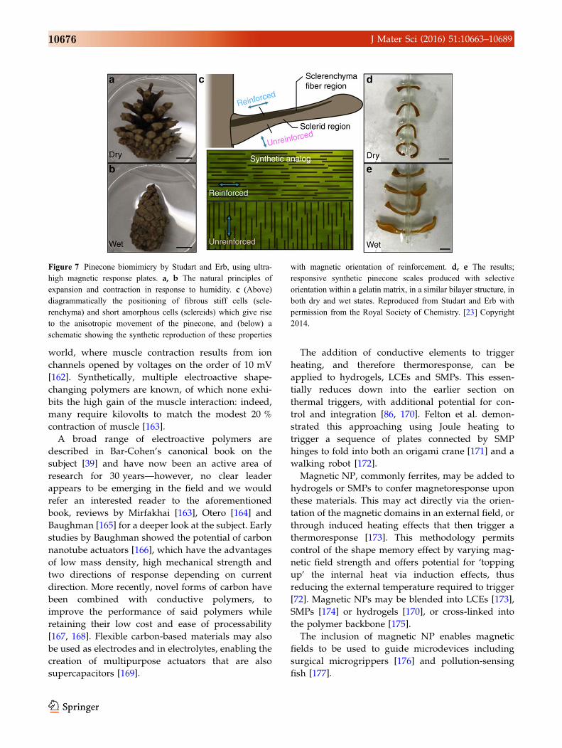
Figure 7 Pinecone biomimicry by Studart and Erb, using ultra-
high magnetic response plates. a, b The natural principles of
expansion and contraction in response to humidity. c (Above)
diagrammatically the positioning of fibrous stiff cells (scle-
renchyma) and short amorphous cells (sclereids) which give rise
to the anisotropic movement of the pinecone, and (below) a
schematic showing the synthetic reproduction of these properties
with magnetic orientation of reinforcement. d, e The results;
responsive synthetic pinecone scales produced with selective
orientation within a gelatin matrix, in a similar bilayer structure, in
both dry and wet states. Reproduced from Studart and Erb with
permission from the Royal Society of Chemistry. [23] Copyright
2014.
10676 J Mater Sci (2016) 51:10663–10689
Beyond isotropy: modification of structure
In order to achieve meaningful and complex
material response, it is necessary to control the
degree of sensitivity to stimuli, and also its direc-
tion. Non-trivial responses require some form of
anisotropy in either the stimuli or the material. The
former approach is easy in the case of electromag-
netic fields or radiation, but less so for diffuse
hygroscopic or chemical stimuli. Since these are the
two most commonly found triggers in biological
situations, evolved solutions tend to focus on
manipulating the response through material struc-
ture, as seen in the earlier example of the wheat
awn constrained in its swelling by fibre direction-
ality. This also has the advantage of being inde-
pendent and self-contained, although it carries the
disadvantage of not being reconfigurable—often
referred to as ‘programmable’ in the literature.
Shape memory materials offer the prospect of
resetting target and intermediate shapes, but
require intervention to achieve this—holding a
material curled, or stretching it, while it cools.
There are multiple ways to analyse and learn
from natural material features, which have merit in
different situations. For the purposes of this dis-
cussion, we have found it helpful to consider the
distinction between how material properties are
achieved, and the role they play for the organism
overall. Using the filaree awn as an example, the
variation in contraction direction is caused by fibre
alignment. However, this varying contraction is
used to create a spiralling structure which winds
itself more tightly as it dries before breaking. So, a
researcher looking to learn from the awn may
choose to emulate orientation and alignment of
constrictive elements, or the behaviour of a tight-
ening spiral approaching breakpoint.
As seen with mimicry of the filaree awn [25],
shape-change may be reproduced without using the
same method seen in the natural example. Con-
versely, we will draw attention to some ways of
reproducing the aligned or gradated structures that
give rise to shape-changing properties in nature
which have not yet been used to create synthetic
shape-change. Together, we hope that this palette of
options will spark ideas for how biological features
may be emulated, and encourage researchers to keep
an open mind regarding routes to the desired
outcome.
Reproducing positioning and orientation
One-dimensional elements such as fibres and rods
may restrict the movement of their surrounding
matrix in a given direction, as noted above for plant
cells. Alternatively, aligned one-dimensional ele-
ments may create one-dimension contraction, as the
myofibrils do in muscle tissue [178], or expansion.
We see that placement and orientational control at
nano- and micro-length scales would be necessary to
emulate these features.
Mesogenic elements in liquid crystals determine
the direction of actuation [179]. Various methods of
alignment are used to obtain single-crystal mon-
odomains, which ensure these elements act together.
Shearing surface force [44], electrical [180] and mag-
netic fields [61] are all well-known methods for
aligning mesogens before initiating polymerisation to
preserve this orientation. Photoalignment can create
spatially varying responses in two dimensions,
demonstrated both continuously [181, 182] and using
a discrete voxel approach [60]. The development of
local feature manipulation in LCEs is reviewed
comprehensively by White et al. [37].
Techniques for positioning constraining fibres and
plates have a long history in manufacture, primarily
for reinforcing composites. Two-dimensional orien-
tation dominates, although ultimately, as in nature,
three-dimensional structures are required to respond
to forces from all directions and so this is a natural
final goal. Embedding of rigid units into polymers
has been demonstrated with high-aspect ratio silicon
nanocolumns (HARNS) around which a hydrogel
was formed. The result was HARNS embedded in the
hydrogel in specific orientations, which altered with
respect to each other in accordance with the degree of
hydration of hydrogel [126], suggested for use in
microfluidics. Magnetic field effects have been used
to align micrometre rods and platelets with super-
paramagnetic coating in various polymers [183],
which has enabled twisting and bending motions via
the application of local constraint (Fig. 7). There is
potential for combination of this method with slip
casting to fill moulded structures [184], but evidently
magnetic field manipulation is restricted to strongly
paramagnetic materials.
A more general approach has been demonstrated
to position reinforcement fibres within a polymer
matrix using ultrasound; [185] this allows arbitrary
orientation in two dimensions and some degree of
J Mater Sci (2016) 51:10663–10689 10677
positional control, and is suitable for any material as
long as it differs in density to the matrix. Future
challenges involve extending this to three dimensions
and accessing orientations which cross between
layers.
The alignment of fibres in the direction of
extrusion shows great potential when combined
with 3D printing technologies. This has hitherto
been a subject of research for stiff materials; for
example, carbon nanofibers added to ABS printing
were shown to align with the direction of extrusion.
The bar samples produced showed decreased
swelling and greater tensile strength, although at
the cost of increased brittleness [186]. More com-
plex extruded honeycomb structures rendered in
resin with aligned fibres delivered a superior
Young’s modulus for the lightweight samples,
compared with other printing materials and natural
balsa wood [187].
In softer materials, the addition of woven fibres has
been successful in reinforcing hydrogels for strength
[111] and directional confinement [188]. The align-
ment of constricting nanofibres within responsive
electrospun polymer threads is a rapid way to gen-
erate hydrogels with ply angles analogous to those of
classical composite design [189]. However, recent
work by the Lewis group at Harvard’s Wyss Institute
(Fig. 8) has taken this work into another elegant
dimension, deriving mathematical models that allow
the construction of arbitrary-curved shapes through
the precise control of cellulose fibre direction, given
by the path of an extrusion-based 3D printer [190].
Extension of these orientating techniques to three
dimensions would enable the creation of combined
radial, longitudinal and helical fibres and structures;
all key elements underlying the varied achievements
of natural muscle, as exemplified by the hydrostatic
skeleton [191].
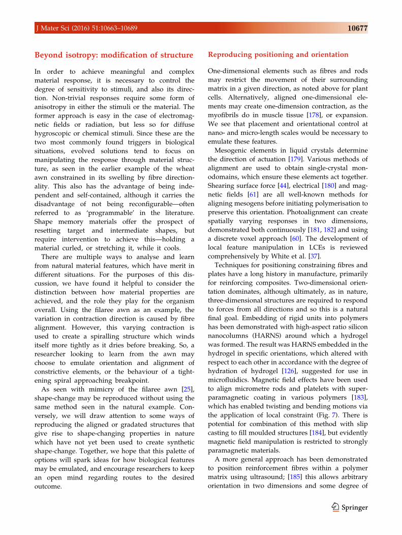
Figure 8 Directional fibre orientation via extrusion, by Lewis
et al., used to replicate complex orchid curvature and shape. a
alignment during printer extrusion and the mesostructure which
translates this into programmed curvature. b, c The cellulose
alignment achieved during printing (scale bar 200 lm). d Multiple
different shape-changes when immersed in water (scale bar 5 mm),
in emulation of e, the dendrobium helix orchid. Adapted from
Nature Materials with permission from Macmillan Publishers Ltd.
[190] Copyright 2015.
10678 J Mater Sci (2016) 51:10663–10689
Creating bending and gradated response
Considering the system from a different perspective,
one can abstract the concept of simply joining mate-
rials with differing responses to the same stimulus.
The bilayer motif is found in nature (for example, the
pine cone and Venus fly trap), in the classic bimetallic
strip, and from the very beginning of work in
hydrogel actuators [21]. This is a prototypical exam-
ple in many papers demonstrating newly discovered
actuating materials. Stoychev et al. have explored the
shapes formed by bilayers with varying dimensions
as a result of diffusion and surface interactions, using
a combination of finite element modelling and
experimental work [192].
Lithographically patterning a bilayer structure
introduces an extra dimension to bilayer fabrication,
as shown by Bassik et al. [193]. Only those areas
exposed to polymerising UV remain attached to the
bottom layer, thus creating a surface with some
bilayer bending regions and some monolayer flat
regions. This is used to create an all-hydrogel version
of a Venus flytrap. In another example of two-di-
mensional patterning, Andres et al. localise inkjet
deposition of carbon nanotube composites within a
polymer, creating folding regions with reduced
hygroscopic swelling (as hinge points) [194].
More complicated curvature from a two-material
system was demonstrated by Wu in 2013 [121], where
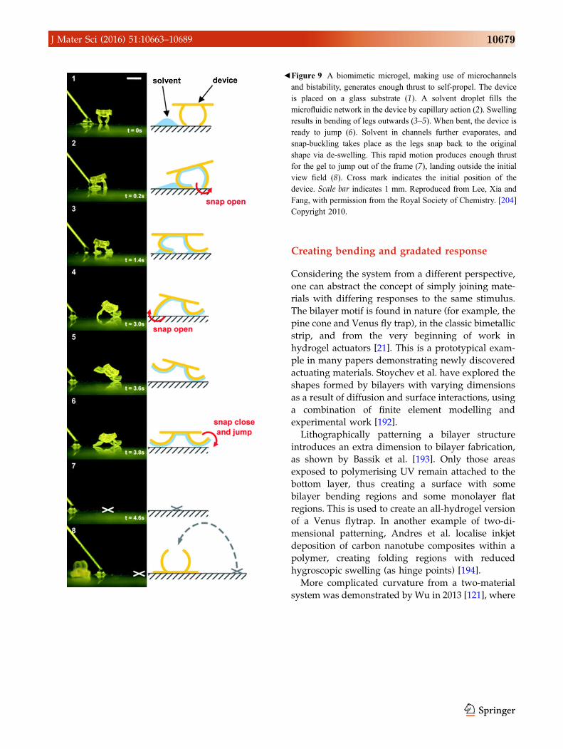
bFigure 9 A biomimetic microgel, making use of microchannels
and bistability, generates enough thrust to self-propel. The device
is placed on a glass substrate (1). A solvent droplet fills the
microfluidic network in the device by capillary action (2). Swelling
results in bending of legs outwards (3–5). When bent, the device is
ready to jump (6). Solvent in channels further evaporates, and
snap-buckling takes place as the legs snap back to the original
shape via de-swelling. This rapid motion produces enough thrust
for the gel to jump out of the frame (7), landing outside the initial
view field (8). Cross mark indicates the initial position of the
device. Scale bar indicates 1 mm. Reproduced from Lee, Xia and
Fang, with permission from the Royal Society of Chemistry. [204]
Copyright 2010.
J Mater Sci (2016) 51:10663–10689 10679
hydrogels with thin, directed stripes of alternating
chemical composition and coefficient of expansion
were used to create complex deforming surfaces. The
relatively small lateral modulation involved is
directly reminiscent of plant motion, where, as in
nature, small effects add up to create an overall larger
movement.
A natural development from narrowly spaced
alternating stripes is the transition to a continuously
varying spatial response. This may be a gradient in
composition; a natural example would be the com-
bination of Type I and Type II muscles, whose dif-
ferent metabolic processes and response rates enable
the many different behaviours required of skeletal
muscle by changing the ratio of just two components
[195]. Alternatively, it may be derived from structural
variation or local amplification of a global stimulus.
The first case, of varying composition, has been
realised in many different synthetic systems (see for
example, Yu et al. with SMPs [196] and Maeda et al.
with polymer gels [144]). A simple case is the varia-
tion of monomer concentration: gradients in mono-
mer concentration throughout the injection forming
of NIPAM sheets may be used to program the
Gaussian curvature in a circular sample, allowing the
spontaneous formation of domes and hyperbolic frills
when heated [197]. Another approach is to introduce
a second molecular species; for example, limited
diffusion of a second polyacrylamide monomer into a
pre-existing NIPAM gel is used to construct a bilayer
strip and a flexible ‘hand’ gripping unit [21]. The
gradient may also be created via post-treatment. For
example, Zhao et al. expose a porous polymer net-
work to a deprotonating acetone diffusion gradient,
which causes variation in the degree of electrostatic
complexation across the hydrogel and therefore its
swelling response [119].
Structural modifications may result from altering
the density of cross-links [22, 123] and the size of pores
in hydrogels [198]. A more complex approach post-
cured an SMP on a spatially varying thermal gradient,
grading the glass temperature spatially and thus
modulating the temperature atwhichdifferent regions
of the material regain their original shape [199]. Inert
surface features such as a layer of micropillars can also
direct the direction of deformation [46].
Finally, local amplification may derive from simple
changes in colouration [200], or increased energy
transmission induced by particles with a tailored
surface plasmon resonance [155].
Form: a final boost to shape-change
Multifunctional materials exist to overcome the lim-
itations of form. The sea cucumber not only needs a
hard, defensive skin to resist ocean currents and
predators, but also a soft, compliant dermis to take
up shelter through narrow gaps in corals and to
perform defensive evisceration [201]. Evidently, it
can only have one skin, so the solution is to vary its
stiffness, shifting between a tensile modulus of 5 and
50 MPa by modulating the interactions of collagen
fibrils within the material [201]. This has been suc-
cessfully emulated by Shanmuganathan et al. by the
hydration-moderated interactions of cellulose nano-
whiskers in low-density polymers [202].
Here, therefore, we see an example of material
overcoming limitations of form. But the opposite
situation can also be true: form may overcome the
limitations of materials. Structural features, such as
bistable shells [94], flexing keels [203] or collapsible
chambers [92], expand the properties of materials
through the structures they are formed into.
An example is an excellent final illustration of our
journey through the materials, triggers and forms that
can enable shape-change in soft materials. As dis-
cussed earlier, the variety of hydrogels available,
combined with the many techniques for introducing
anisotropy into their bulk, makes them an attractive
material for volume-change-based actuation. How-
ever, the material experiences slow deployment times,
as expansion is a diffusion-limited process [107], and
the force exerted is typically small.
A solution to a similar problem is seen in the Venus
flytrap, where the relatively weak forces exerted by
water swelling release built-in strain, triggering a
switch between two local minimal energy confor-
mations [94]. Lee et al. emulated this with a combi-
nation of vasculature and pre-stress in the material to
create the ‘jumping hydrogel’ (Fig. 9) [204]. Mesos-
cale channels created lithographically within a small
hydrogel sample localise solvent exposure and
therefore nanoscale swelling. The smaller scale also
reduces diffusion length and therefore actuation
time. Expansion and extension in a targeted small
region were sufficient to flip the macroscale object
into a second stable position, attain a maximum
angular velocity an order of magnitude larger than
the biological system, and release sufficient energy to
propel it into the air. As noted by the authors, this
design overcomes two known issues with hydrogels,
10680 J Mater Sci (2016) 51:10663–10689

namely their mechanical weakness and slow actua-
tion, through incorporation in a wider system.
Conclusion
A primary aim of this review was to examine the
functionality and activation mechanisms of synthetic
shape-changing materials used by the scientific com-
munity and benchmark our current knowledge of self-
shapingmaterials against the strategies found in nature.
As one might imagine, the conclusion is unavoidable;
there will be no single material and manufacturing
methodwhich achieves the self-shaping the engineering
and scientific community desires. Instead, we need to
combine techniques to overcome shortcomings.
Key elements that have emerged in this review are the
potential of nanoscopic features such as contracting
fibres (as seen inmuscle) and restrictive plates (as seen in
plant tissues) to instigate and direct motion. These
nanoelements must be united in a structured network,
through embedding in amatrix or otherwise, potentially
also including secondary mesoscale features. With cur-
rent technology and materials, creating nanostructured
materials in complex shapes requires either multiple
steps (e.g. alignment followed by assembly), or dual
development of materials and manufacturing (for
example, alignment of fibre constructs during additive
manufacture) [138, 140, 143].
From the top-down, we should look to create struc-
tures that minimise the force required to self-shape,
and where possible target and combine stimuli to
maximise the magnitude of the trigger delivered. The
complementary use of different stimuli, such as the
application of magnetic fields to reduce the tempera-
ture required for SMP relaxation [114], is a promising
area, as is the potential of electrical or optical stimuli
for remote, directional and modulated control.
Two themes are discernible from these examples:
the potential for synergy between materials and form,
and the need for features crossing multiple length
scales—which require collaboration between manu-
facture and materials development. We therefore
suggest simultaneous development of techniques for
structuring materials, and the materials they will be
used upon, as a fruitful direction. Diversification
through the use of multiple techniques to create
structures across complementary length scales, and
responding to multiple stimuli, will combinatorially
expand what is possible in this field.
Development of novel self-shaping materials can
be expected to unlock applications and designs not
yet considered, with corresponding changes in qual-
ity of life, material sustainability and environmental
benefits. We consider that hierarchical material fea-
tures, from nano- to macroscale, developed side-by-
side with minimal-force structural designs, have the
highest chance of blending nature’s responsive
design space with the constructed world.
Acknowledgements
KO and AS are funded through an Engineering and
Physical Sciences Centre for Doctoral Training award to
the Bristol Centre for Functional Nanomaterials, EPSRC
Grant code EP/G036780/1. RST is an Engineering and
Physical Sciences Research Council (EPSRC) Fellow
funded under EPSRC ‘Engineering Fellowships for
Growth’, Grant Number EP/M002489/1.
Compliance with ethical standards
Conflict of interest The authors declare that they
have no conflict of interest.
Availability of research data There is no underly-
ing data associated with this paper.
Electronic supplementary material The online
version of this article (doi:10.1007/s10853-016-0295-
8) contains supplementary material, which is avail-
able to authorised users.
Open Access This article is distributed under the
terms of the Creative Commons Attribution 4.0
International License (http://creativecommons.org/
licenses/by/4.0/), which permits unrestricted use,
distribution, and reproduction in any medium, pro-
vided you give appropriate credit to the original
author(s) and the source, provide a link to the Crea-
tive Commons license, and indicate if changes were
made.
References
[1] Nova A, Keten S, Pugno NM, Redaelli A, Buehler MJ
(2010) Molecular and nanostructural mechanisms of
deformation, strength and toughness of spider silk fibrils.
Nano Lett 10:2626–2634. doi:10.1021/nl101341w
J Mater Sci (2016) 51:10663–10689 10681
[2] Rho J-Y, Kuhn-Spearing L, Zioupos P (1998) Mechanical
properties and the hierarchical structure of bone. Med Eng
Phys 20:92–102. doi:10.1016/S1350-4533(98)00007-1
[3] Saranathan V, Osuji CO,Mochrie SGJ, Noh H, Narayanan S,
Sandy A, Dufresne ER, PrumRO (2010) Structure, function,
and self-assembly of single network gyroid (I4132) photonic
crystals in butterfly wing scales. Proc Natl Acad Sci USA
107:11676–11681. doi:10.1073/pnas.0909616107
[4] Allen JJ, Bell GRR, Kuzirian AM, Hanlon RT (2013)
Cuttlefish skin papilla morphology suggests a muscular
hydrostatic function for rapid changeability. J Morphol
274:645–656. doi:10.1002/jmor.20121
[5] Lee H, Lee BP, Messersmith PB (2007) A reversible wet/
dry adhesive inspired by mussels and geckos. Nature
448:338–341. doi:10.1038/nature05968
[6] Mohd Jani J, Leary M, Subic A, Gibson MA (2014) A
review of shape memory alloy research, applications and
opportunities. Mater Des 56:1078–1113. doi:10.1016/j.
matdes.2013.11.084
[7] Irschik H (2002) A review on static and dynamic shape
control of structures by piezoelectric actuation. Eng Struct
24:5–11. doi:10.1016/S0141-0296(01)00081-5
[8] Doi M (2013) Soft matter physics. OUP, Oxford
[9] Quake SR (2000) From micro- to nanofabrication with soft
materials. Science 290:1536–1540. doi:10.1126/science.
290.5496.1536
[10] Bauer S, Bauer-Gogonea S, Graz I, Kaltenbrunner M, Keplin-
ger C, Schwodiauer R (2014) 25th anniversary article: a soft
future: from robots and sensor skin to energy harvesters. Adv
Mater 26:149–161. doi:10.1002/adma.201303349
[11] Rus D, Tolley MT (2015) Design, fabrication and control of
soft robots. Nature 521:467–475. doi:10.1038/nature14543
[12] Deligkaris K, Tadele TS, Olthuis W, van den Berg A (2010)
Hydrogel-based devices for biomedical applications. Sens
Actuators B 147:765–774. doi:10.1016/j.snb.2010.03.083
[13] Holstov A, Bridgens B, Farmer G (2015) Hygromorphic
materials for sustainable responsive architecture. Constr
Build Mater 98:570–582. doi:10.1016/j.conbuildmat.2015.
08.136
[14] Zarzar LD, Kim P, Aizenberg J (2011) Bio-inspired design
of submerged hydrogel-actuated polymer microstructures
operating in response to pH. Adv Mater 23:1442–1446.
doi:10.1002/adma.201004231
[15] Yao L, Ou J, Cheng C-Y, Steiner H, Wang W, Wang G, Ishii
H (2015) BioLogic. In: Proceedings of the 33rd annual
ACM conference on human factors computing systems—
CHI’15. ACM Press, New York, pp 1–10
[16] Kim S, Laschi C, Trimmer B (2013) Soft robotics: a
bioinspired evolution in robotics. Trends Biotechnol
31:287–294. doi:10.1016/j.tibtech.2013.03.002
[17] Evangelista D, Hotton S, Dumais J (2011) The mechanics
of explosive dispersal and self-burial in the seeds of the
filaree, Erodium cicutarium (Geraniaceae). J Exp Biol
214:521–529. doi:10.1242/jeb.050567
[18] Reyssat E, Mahadevan L (2009) Hygromorphs: from pine
cones to biomimetic bilayers. J R Soc Interface 6:951–957.
doi:10.1098/rsif.2009.0184
[19] Rafsanjani A, Brule V, Western TL, Pasini D (2015) Hydro-
responsive curling of the resurrection plant Selaginella
lepidophylla. Sci Rep 5:8064. doi:10.1038/srep08064
[20] Timoshenko S (1925) Analysis of bi-metal thermostats.
J Opt Soc Am 11:233–255. doi:10.1364/JOSA.11.000233
[21] Hu Z, Zhang X, Li Y (1995) Synthesis and application of
modulated polymer gels. Science 269:525–527. doi:10.
1126/science.269.5223.525
[22] Guvendiren M, Yang S, Burdick JA (2009) Swelling-in-
duced surface patterns in hydrogels with gradient
crosslinking density. Adv Funct Mater 19:3038–3045.
doi:10.1002/adfm.200900622
[23] Studart AR, Erb RM (2014) Bioinspired materials that self-
shape through programmed microstructures. Soft Matter
10:1284–1294. doi:10.1039/c3sm51883c
[24] Bar-On B, Sui X, Livanov K, Achrai B, Kalfon-Cohen E,
Wiesel E, Wagner HD (2014) Structural origins of mor-
phing in plant tissues. Appl Phys Lett 105:033703. doi:10.
1063/1.4891191
[25] Abraham Y, Tamburu C, Klein E, Dunlop JWC, Fratzl P,
Raviv U, Elbaum R (2012) Tilted cellulose arrangement as
a novel mechanism for hygroscopic coiling in the stork’s
bill awn. J R Soc Interface 9:640–647. doi:10.1098/rsif.
2011.0395
[26] Aharoni H, Abraham Y, Elbaum R, Sharon E, Kupferman R
(2012) Emergence of spontaneous twist and curvature in
non-euclidean rods: application to erodium plant cells. Phys
Rev Lett 108:238106. doi:10.1103/PhysRevLett.108.
238106
[27] Knight DP, Vollrath F (2002) Biological liquid crystal
elastomers. Philos Trans R Soc Lond B 357:155–163.
doi:10.1098/rstb.2001.1030
[28] Behl M, Lendlein A (2007) Shape-memory polymers. Mater
Today 10:20–28. doi:10.1016/S1369-7021(07)70047-0
[29] Ahn S, Kasi RM, Kim S-C, Sharma N, Zhou Y (2008)
Stimuli-responsive polymer gels. Soft Matter 4:1151–1157.
doi:10.1039/b714376a
[30] Geryak R, Tsukruk VV (2014) Reconfigurable and actuat-
ing structures from soft materials. Soft Matter 10:1246–
1263. doi:10.1039/c3sm51768c
[31] Meng H, Li G (2013) A review of stimuli-responsive shape
memory polymer composites. Polymer (Guildf) 54:2199–
2221. doi:10.1016/j.polymer.2013.02.023
10682 J Mater Sci (2016) 51:10663–10689

[32] Ionov L (2014) Polymeric actuators. Langmuir 31:5015–
5024. doi:10.1021/la503407z
[33] Lodish H, Kaiser C, Bretscher A, Amon A, Berk A, Krieger
M, Polegh H, Scott M (2013) Myosin thick filaments and
actin thin filaments in skeletal muscle slide past one another
during contraction. In: Molecular cell biology, 7th edn.
W. H. Freeman and Co, New York, p 801–802
[34] Meyers MA, Chen P-Y, Lin AY-M, Seki Y (2008) Bio-
logical materials: structure and mechanical properties. Prog
Mater Sci 53:1–206. doi:10.1016/j.pmatsci.2007.05.002
[35] Wainwright PC, Kraklau DM, Bennett AF (1991) Kine-
matics of tongue projection in Chamaeleo oustaleti. J Exp
Biol 159:109–133
[36] Vogel S (2003) Achieving motility. In: Vogel S (ed)
Comparative biomechanics—life’s physical world. Prince-
ton University Press, Princeton, pp 453–582
[37] White TJ, Broer DJ (2015) Programmable and adaptive
mechanics with liquid crystal polymer networks and elas-
tomers. Nat Mater 14:1087–1098. doi:10.1038/nmat4433
[38] Shahinpoor M, Bar-Cohen Y, Simpson JO, Smith J (1998)
Ionic polymer-metal composites (IPMCs) as biomimetic
sensors, actuators and artificial muscles—a review. Smart
Mater Struct 7:R15–R30. doi:10.1088/0964-1726/7/6/001
[39] Bar-Cohen Y (2004) Electroactive polymer (EAP) actuators
as artificial muscles: reality, potential, and challenges.
Electroact Polym. doi:10.1117/3.547465
[40] Pugal D, Jung K, Aabloo A, Kim KJ (2010) Ionic polymer-
metal composite mechanoelectrical transduction: review and
perspectives. Polym Int 59:279–289. doi:10.1002/pi.2759
[41] Bahramzadeh Y, Shahinpoor M (2014) A review of ionic
polymeric soft actuators and sensors. Soft Robot 1:38–52.
doi:10.1089/soro.2013.0006
[42] Brochu P, Pei Q (2010) Advances in dielectric elastomers
for actuators and artificial muscles. Macromol Rapid
Commun 31:10–36. doi:10.1002/marc.200900425
[43] de Gennes P-G (1997) Un muscle artificiel semi-rapide.
Comptes Rendus l’Academie des Sci—Ser IIB—Mech
324:343–348. doi:10.1016/S1251-8069(99)80044-X
[44] Li M-H, Keller P (2006) Artificial muscles based on liquid
crystal elastomers. Philos Trans A 364:2763–2777. doi:10.
1098/rsta.2006.1853
[45] Thomsen DL, Keller P, Naciri J, Pink R, Jeon H, Shenoy D,
Ratna BR (2001) Liquid crystal elastomers with mechanical
properties of a muscle. Macromolecules 34:5868–5875.
doi:10.1021/ma001639q
[46] Shahsavan H, Salili SM, Jakli A, Zhao B (2015) Smart
muscle-driven self-cleaning of biomimetic microstructures
from liquid crystal elastomers. Adv Mater 27:6828–6833.
doi:10.1002/adma.201503203
[47] Agrawal A, Chipara AC, Shamoo Y, Patra PK, Carey BJ,
Ajayan PM, Chapman WG, Verduzco R (2013) Dynamic
self-stiffening in liquid crystal elastomers. Nat Commun
4:1739. doi:10.1038/ncomms2772
[48] Iwamoto H, Inoue K, Yagi N (2006) Evolution of long-
range myofibrillar crystallinity in insect flight muscle as
examined by X-ray cryomicrodiffraction. Proc Biol Sci
273:677–685. doi:10.1098/rspb.2005.3389
[49] Seddon JM, Squires AM, Conn CE, Ces O, Heron AJ,
Mulet X, Shearman GC, Templer RH (2006) Pressure-
jump X-ray studies of liquid crystal transitions in lipids.
Philos Trans A 364:2635–2655. doi:10.1098/rsta.2006.
1844
[50] Kupfer J, Finkelmann H (1991) Nematic liquid single
crystal elastomers. Die Makromol Chemie Rapid Com-
mun 12:717–726. doi:10.1002/marc.1991.030121211
[51] Ohm C, Brehmer M, Zentel R (2010) Liquid crystalline
elastomers as actuators and sensors. Adv Mater 22:3366–
3387. doi:10.1002/adma.200904059
[52] Hogan PM, Tajbakhsh AR, Terentjev EM (2002) UV
manipulation of order and macroscopic shape in nematic
elastomers. Phys Rev E 65:1–8. doi:10.1103/PhysRevE.65.
041720
[53] Tsutsumi O, Shiono T, Tomiki I, Galli G (1997) Photo-
chemical phase transition behavior of nematic liquid crys-
tals with azobenzene moieties as both mesogens and
photosensitive chromophores. J Phys Chem B 101:1332–
1337. doi:10.1021/jp961565d
[54] Kempe MD, Scruggs NR, Verduzco R, Lal J, Kornfield JA
(2004) Self-assembled liquid-crystalline gels designed from
the bottom up. Nat Mater 3:177–182. doi:10.1038/nmat
1074
[55] Jiang H, Li C, Huang X (2013) Actuators based on liquid
crystalline elastomer materials. Nanoscale 5:5225–5240.
doi:10.1039/c3nr00037k
[56] Chambers M, Finkelmann H, Remskar M, Sanchez-Ferrer
A, Zalar B, Zumer S (2009) Liquid crystal elastomer–
nanoparticle systems for actuation. J Mater Chem 19:1524–
1531. doi:10.1039/B812423J
[57] Urayama K (2007) Selected issues in liquid crystal elas-
tomers and gels. Macromolecules 40:2277–2288. doi:10.
1021/ma0623688
[58] Sawa Y, Ye F, Urayama K, Takigawa T, Gimenez-Pinto V,
Selinger RLB, Selinger JV (2011) Shape selection of twist-
nematic-elastomer ribbons. Proc Natl Acad Sci USA
108:6364–6368. doi:10.1073/pnas.1017658108
[59] de Haan LT, Schenning APHJ, Broer DJ (2014) Programmed
morphing of liquid crystal networks. Polymer (Guildf) 55:
5885–5896. doi:10.1016/j.polymer.2014.08.023
J Mater Sci (2016) 51:10663–10689 10683
[60] Ware TH, McConney ME, Wie JJ, Tondiglia VP, White TJ
(2015) Actuating materials. Voxelated liquid crystal elas-
tomers. Science 347:982–984. doi:10.1126/science.1261019
[61] Buguin A, Li M-H, Silberzan P, Ladoux B, Keller P (2006)
Micro-actuators: when artificial muscles made of nematic
liquid crystal elastomers meet soft lithography. J Am Chem
Soc 128:1088–1089. doi:10.1021/ja0575070
[62] Alexander R (1991) Energy-saving mechanisms in walking
and running. J Exp Biol 160:55–69
[63] Kastelic J, Galeski A, Baer E (1978) The multicomposite
structure of tendon. Connect Tissue Res 6:11–23. doi:10.
3109/03008207809152283
[64] Fratzl P (2003) Cellulose and collagen: from fibres to tis-
sues. Curr Opin Colloid Interface Sci 8:32–39. doi:10.1016/
S1359-0294(03)00011-6
[65] Gatt R, Vella Wood M, Gatt A, Zarb F, Formosa C,
Azzopardi KM, Casha A, Agius TP, Schembri-Wismayer P,
Attard L, Chockalingam N, Grima JN (2015) Negative
Poisson’s ratios in tendons: an unexpected mechanical
response. Acta Biomater 24:201–208. doi:10.1016/j.actbio.
2015.06.018
[66] Bennett MB, Ker RF, Imery NJ, Alexander RM (1986)
Mechanical properties of various mammalian tendons. J Zool
209:537–548. doi:10.1111/j.1469-7998.1986.tb03609.x
[67] Leng J, Lan X, Liu Y, Du S (2011) Shape-memory poly-
mers and their composites: stimulus methods and applica-
tions. Prog Mater Sci 56:1077–1135. doi:10.1016/j.
pmatsci.2011.03.001
[68] Xiao X, Hu J, Hu J, Zhu Y, Huang H, Lv J, Meng H, Li G,
Anis A, Meng H, Zhou X, Mendez J, Zhu Y, Li J, Ma X,
Tian H, Xue L, Dai S, Li Z, Jung YC, So HH, Cho JW,
Chen S, Hu J, Yuen CW, Chen L, Huang WM, Yang B, An
L, Li C, Chan YS, Lu H, Liu Y, Leng J, Du S, Dagnon KL,
Huang H, Hu J, Zhu Y, Hu J, Zhu Y, Hu J, Dong ZE, Liu Y,
Zimmermann EA, Fratzl P, Barth FG, Dawson C, Vincent
JFV, Rocca AM, Elbaum R, Zaltzman L, Burgert I, Fratzl P,
Meyers MA, McKittrick J, Chen PY, Fratzl P, Weinkamer
R, Liu ZQ, Jiao D, Zhang ZF, Xu W, Ke G, Wu J, Wang X,
Barba C, Ackbarow T, Chen X, Keten S, Buehler MJ,
Speakman JB, Feughelman M, Feughelman M, Feughel-
man M, Fraser RDB, MacRae TP, Astbury WT, Street A,
Astbury WT, Pauling L, Wortmann FJ, Rigby BJ, Phillips
DG, Eaves JD, Schiessel H, Metzler R, Blumen A, Non-
nenmacher TF, Williams G, Watts DC, Kumar B, Hu JL,
Pan N, Behl M, Razzaq MY, Lendlein A, Lv H, Liu Y,
Zhang D, Leng J, Du S, Du H, Zhang J, Liu Y, Kilpelainen
I, Akhtar W, Richard-Lacroix M, Pellerin C, Yao J, Liu Y,
Yang S, Liu J, Ishida Y, Chabanne L, Antonietti M, Shalom
M, Yan X, Ma M, Guo L, Anderson DG, Langer R, Nagia
FA, EL-Mohamedy RSR, Liu C, Qin H, Mather PT (2016)
Animal hairs as water-stimulated shape memory materials:
mechanism and structural networks in molecular assem-
blies. Sci Rep 6:26393. doi:10.1038/srep26393
[69] Liu ZQ, Jiao D, Zhang ZF (2015) Remarkable shape
memory effect of a natural biopolymer in aqueous envi-
ronment. Biomaterials 65:13–21. doi:10.1016/j.biomater
ials.2015.06.032
[70] Zhao Q, Qi HJ, Xie T (2015) Recent progress in shape
memory polymer: new behavior, enabling materials, and
mechanistic understanding. Prog Polym Sci 49–50:79–120.
doi:10.1016/j.progpolymsci.2015.04.001
[71] Lendlein A, Jiang H, Junger O, Langer R (2005) Light-
induced shape-memory polymers. Nature 434:879–882.
doi:10.1038/nature03496
[72] Kumar UN, Kratz K, Heuchel M, Behl M, Lendlein A
(2011) Shape-memory nanocomposites with magnetically
adjustable apparent switching temperatures. Adv Mater
23:4157–4162. doi:10.1002/adma.201102251
[73] Wang CC, Huang WM, Ding Z, Zhao Y, Purnawali H
(2012) Cooling-/water-responsive shape memory hybrids.
Compos Sci Technol 72:1178–1182. doi:10.1016/j.comps
citech.2012.03.027
[74] Rousseau IA (2008) Challenges of shape memory poly-
mers: a review of the progress toward overcoming SMP’s
limitations. Polym Eng Sci 48:2075–2089. doi:10.1002/
pen.21213
[75] Momtaz M, Razavi-Nouri M, Barikani M (2014) Effect of
block ratio and strain amplitude on thermal, structural, and
shape memory properties of segmented polycaprolactone-
based polyurethanes. J Mater Sci 49:7575–7584. doi:10.
1007/s10853-014-8466-y
[76] Messori M, Degli Esposti M, Paderni K, Pandini S, Passera
S, Ricco T, Toselli M (2012) Chemical and thermome-
chanical tailoring of the shape memory effect in poly(e-
caprolactone)-based systems. J Mater Sci 48:424–440.
doi:10.1007/s10853-012-6757-8
[77] Zhuo H, Hu J, Chen S (2011) Study of the thermal prop-
erties of shape memory polyurethane nanofibrous nonwo-
ven. J Mater Sci 46:3464–3469. doi:10.1007/s10853-011-
5251-z
[78] Auad ML, Contos VS, Nutt S, Aranguren MI, Marcovich
NE (2008) Characterization of nanocellulose- reinforced
shape memory polyurethanes. Polym Int 57:651–659.
doi:10.1002/pi.2394
[79] Miaudet P, Derre A, Maugey M, Zakri C, Piccione PM,
Inoubli R, Poulin P (2007) Shape and temperature memory
of nanocomposites with broadened glass transition. Science
318:1294–1296. doi:10.1126/science.1145593
[80] Koerner H, Price G, Pearce NA, Alexander M, Vaia RA
(2004) Remotely actuated polymer nanocomposites—
10684 J Mater Sci (2016) 51:10663–10689
stress-recovery of carbon-nanotube-filled thermoplastic
elastomers. Nat Mater 3:115–120. doi:10.1038/nmat1059
[81] Nishikawa M, Wakatsuki K, Takeda N (2010) Thermo-
mechanical experiment and analysis on shape recovery
properties of shape memory polymer influenced by fiber
reinforcement. J Mater Sci 45:3957–3960. doi:10.1007/
s10853-010-4545-x
[82] Sun J-Y, Zhao X, Illeperuma WRK, Chaudhuri O, Oh KH,
Mooney DJ, Vlassak JJ, Suo Z (2012) Highly stretchable
and tough hydrogels. Nature 489:133–136. doi:10.1038/
nature11409
[83] Ratna D, Karger-Kocsis J (2007) Recent advances in shape
memory polymers and composites: a review. J Mater Sci
43:254–269. doi:10.1007/s10853-007-2176-7
[84] Hager MD, Bode S, Weber C, Schubert US (2015) Shape
memory polymers: past, present and future developments.
Prog Polym Sci 49:3–33. doi:10.1016/j.progpolymsci.
2015.04.002
[85] Yan X, Wang F, Zheng B, Huang F (2012) Stimuli-re-
sponsive supramolecular polymeric materials. Chem Soc
Rev 41:6042–6065. doi:10.1039/c2cs35091b
[86] Liu Y, Lv H, Lan X, Leng J, Du S (2009) Review of
electro-active shape-memory polymer composite. Compos
Sci Technol 69:2064–2068. doi:10.1016/j.compscitech.
2008.08.016
[87] Stahlberg R (2009) The phytomimetic potential of three
types of hydration motors that drive nastic plant move-
ments. Mech Mater 41:1162–1171. doi:10.1016/j.mechmat.
2009.05.003
[88] KollerD (1990)Light-driven leafmovements.PlantCellEnviron
13:615–632. doi:10.1111/j.1365-3040.1990.tb01079.x
[89] Luzar N, Gottsberger G (2001) Flower heliotropism and
floral heating of five alpine plant species and the effect on
flower visiting in Ranunculus montanus in the Austrian
Alps. Arct Antarct Alp Res 33:93–99. doi:10.2307/1552282
[90] Schwartz A, Koller D (1978) Phototropic response to
vectorial light in leaves of Lavatera cretica L. Plant Physiol
61:924–928. doi:10.1104/pp.61.6.924
[91] Whitaker DL, Webster LA, Edwards J (2007) The biome-
chanics of Cornus canadensis stamens are ideal for cata-
pulting pollen vertically. Funct Ecol 21:219–225. doi:10.
1111/j.1365-2435.2007.01249.x
[92] Noblin X, Rojas NO, Westbrook J, Llorens C, Argentina M,
Dumais J (2012) The fern sporangium: a unique catapult.
Science 335:1322–1322. doi:10.1126/science.1215985
[93] Llorens C, Argentina M, Rojas N, Westbrook J, Dumais J,
Noblin X, Skotheim J, Mahadevan L, Dumais J, Forterre Y,
Noblin X, Rojas N, Westbrook J, Llorens C, Argentina M,
Dumais J, Borno R, Steinmeyer J, Maharbiz M, Martone P,
Prantl K, Renner O, Ursprung A, King A, Ritman K,
Milburn J, Haider K, Steudle E, Caupin F, Herbert E,
Skotheim J, Mahadevan L, Epstein P, Plesset M (2016) The
fern cavitation catapult: mechanism and design principles.
J R Soc Interface 13:20150930. doi:10.1098/rsif.2015.0930
[94] Forterre Y, Skotheim JM, Dumais J, Mahadevan L (2005)
How the venus flytrap snaps. Nature 433:421–425. doi:10.
1038/nature03185
[95] Burgert I, Fratzl P (2009) Actuation systems in plants as
prototypes for bioinspired devices. Philos Trans A 367:
1541–1557. doi:10.1098/rsta.2009.0003
[96] Zwieniecki MA, Melcher PJ, Michele Holbrook NM (2001)
Hydrogel control of xylem hydraulic resistance in plants.
Science 291:1059–1062. doi:10.1126/science.1057175
[97] Guan Y, Zhang Y (2011) PNIPAM microgels for biomed-
ical applications: from dispersed particles to 3D assemblies.
Soft Matter 7:6375–6384. doi:10.1039/c0sm01541e
[98] Guvendiren M, Lu HD, Burdick JA (2012) Shear-thinning
hydrogels for biomedical applications. Soft Matter 8:260–
272. doi:10.1039/C1SM06513K
[99] Martin BD, Linhardt RJ, Dordick JS (1998) Highly swelling
hydrogels from ordered galactose-based polyacrylates. Bio-
materials 19:69–76. doi:10.1016/S0142-9612(97)00184-1
[100] Osada Y, Matsuda A (1995) Shape memory in hydrogels.
Nature 376:219. doi:10.1038/376219a0
[101] Hennink WE, van Nostrum CF (2012) Novel crosslinking
methods to design hydrogels. Adv Drug Deliv Rev 64:223–
236. doi:10.1016/j.addr.2012.09.009
[102] Chang C, Zhang L (2011) Cellulose-based hydrogels: pre-
sent status and application prospects. Carbohydr Polym
84:40–53. doi:10.1016/j.carbpol.2010.12.023
[103] Lam CX, Mo X, Teoh S, Hutmacher D (2002) Scaffold
development using 3D printing with a starch-based poly-
mer. Mater Sci Eng C 20:49–56. doi:10.1016/S0928-
4931(02)00012-7
[104] Koetting MC, Peters JT, Steichen SD, Peppas NA (2015)
Stimulus-responsive hydrogels: theory, modern advances,
and applications. Mater Sci Eng R Rep 93:1–49. doi:10.
1016/j.mser.2015.04.001
[105] Hardin BE, Snaith HJ, McGehee MD (2012) The renais-
sance of dye-sensitized solar cells. Nat Photonics
6:162–169. doi:10.1038/nphoton.2012.22
[106] Calvert P (2009) Hydrogels for soft machines. Adv Mater
21:743–756. doi:10.1002/adma.200800534
[107] Sato Matsuo E, Tanaka T (1988) Kinetics of discontinuous
volume-phase transition of gels. J Chem Phys 89:1695.
doi:10.1063/1.455115
[108] Beebe D, Moore J, Bauer J, Yu Q, Liu R, Devadoss C, Jo B
(2000) Functional hydrogel structures for autonomous flow
control inside microfluidic channels. Nature 404:588–590.
doi:10.1038/35007047
J Mater Sci (2016) 51:10663–10689 10685

[109] Lee E, Kim D, Kim H, Yoon J (2015) Photothermally
driven fast responding photo-actuators fabricated with
comb-type hydrogels and magnetite nanoparticles. Sci Rep
5:15124. doi:10.1038/srep15124
[110] Ionov L (2014) Hydrogel-based actuators: possibilities and
limitations. Mater Today 17:494–503. doi:10.1016/j.mat
tod.2014.07.002
[111] Agrawal A, Rahbar N, Calvert PD (2013) Strong fiber-
reinforced hydrogel. Acta Biomater 9:5313–5318. doi:10.
1016/j.actbio.2012.10.011
[112] Xia L-W, Xie R, Ju X-J, Wang W, Chen Q, Chu L-Y (2013)
Nano-structured smart hydrogels with rapid response and
high elasticity. Nat Commun 4:2226. doi:10.1038/
ncomms3226
[113] Schexnailder P, Schmidt G (2008) Nanocomposite polymer
hydrogels. Colloid Polym Sci 287:1–11. doi:10.1007/
s00396-008-1949-0
[114] Mohr R, Kratz K, Weigel T, Lucka-Gabor M, Moneke M,
Lendlein A (2006) Initiation of shape-memory effect by
inductive heating of magnetic nanoparticles in thermo-
plastic polymers. Proc Natl Acad Sci USA 103:3540–3545.
doi:10.1073/pnas.0600079103
[115] Lee W-F, Tsao K-T (2009) Effect of silver nanoparticles
content on the various properties of nanocomposite
hydrogels by in situ polymerization. J Mater Sci 45:89–97.
doi:10.1007/s10853-009-3896-7
[116] Hebeish A, Farag S, Sharaf S, Shaheen TI (2014) Thermal
responsive hydrogels based on semi interpenetrating network
of poly(NIPAm) and cellulose nanowhiskers. Carbohydr
Polym 102:159–166. doi:10.1016/j.carbpol.2013.10.054
[117] Ionov L (2013) Biomimetic hydrogel-based actuating sys-
tems. Adv Funct Mater 23:4555–4570. doi:10.1002/adfm.
201203692
[118] Harris KD, Bastiaansen CWM, Broer DJ (2006) A glassy
bending-mode polymeric actuator which deforms in
response to solvent polarity. Macromol Rapid Commun
27:1323–1329. doi:10.1002/marc.200600342
[119] Zhao Q, Dunlop JWC, Qiu X, Huang F, Zhang Z, Heyda J,
Dzubiella J, Antonietti M, Yuan J (2014) An instant multi-
responsive porous polymer actuator driven by solvent
molecule sorption. Nat Commun 5:4293. doi:10.1038/
ncomms5293
[120] Eddington DT, Liu RH, Moore JS, Beebe DJ (2001) An
organic self-regulating microfluidic system. Lab Chip
1:96–99. doi:10.1039/b108078d
[121] Wu ZL, Moshe M, Greener J, Therien-Aubin H, Nie Z,
Sharon E, Kumacheva E (2013) Three-dimensional shape
transformations of hydrogel sheets induced by small-scale
modulation of internal stresses. Nat Commun 4:1586.
doi:10.1038/ncomms2549
[122] Senff H, Richtering W (2000) Influence of cross-link den-
sity on rheological properties of temperature-sensitive
microgel suspensions. Colloid Polym Sci 278:830–840.
doi:10.1007/s003960000329
[123] Palleau E, Morales D, Dickey MD, Velev OD (2013)
Reversible patterning and actuation of hydrogels by elec-
trically assisted ionoprinting. Nat Commun 4:2257. doi:10.
1038/ncomms3257
[124] Islam MR, Li X, Smyth K, Serpe MJ (2013) Polymer-based
muscle expansion and contraction. Angew Chem Int Ed
Engl 52:10330–10333. doi:10.1002/anie.201303475
[125] Teramoto N, Shigehiro O, Ogawa Y, Maruyama Y, Shi-
masaki T, Shibata M (2014) Polymer foam-reinforced
hydrogels inspired by plant body frameworks as high-per-
formance soft matter. Polym J 46:592–597. doi:10.1038/pj.
2014.41
[126] Sidorenko A, Krupenkin T, Taylor A, Fratzl P, Aizenberg J
(2007) Reversible switching of hydrogel-actuated nanostruc-
tures into complex micropatterns. Science 315:487–490.
doi:10.1126/science.1135516
[127] Huang WM, Yang B, An L, Li C, Chan YS (2005) Water-
driven programmable polyurethane shape memory poly-
mer: demonstration and mechanism. Appl Phys Lett
86:114105. doi:10.1063/1.1880448
[128] Yang B, Huang WM, Li C, Lee CM, Li L (2004) On the
effects of moisture in a polyurethane shape memory poly-
mer. Smart Mater Struct 13:191–195. doi:10.1088/0964-
1726/13/1/022
[129] Chen S, Hu J, Zhuo H (2011) Study on the moisture
absorption of pyridine containing polyurethane for mois-
ture-responsive shape memory effects. J Mater Sci
46:6581–6588. doi:10.1007/s10853-011-5606-5
[130] Capadona JR, Shanmuganathan K, Tyler DJ, Rowan SJ,
Weder C (2008) Stimuli-responsive polymer nanocompos-
ites inspired by the sea cucumber dermis. Science
319:1370–1374. doi:10.1126/science.1153307
[131] Zhu Y, Hu J, Luo H, Young RJ, Deng L, Zhang S, Fan Y,
Ye G (2012) Rapidly switchable water-sensitive shape-
memory cellulose/elastomer nano-composites. Soft Matter
8:2509–2517. doi:10.1039/c2sm07035a
[132] Ma M, Guo L, Anderson DG, Langer R (2013) Bio-in-
spired polymer composite actuator and generator driven by
water gradients. Science 339:186–189. doi:10.1126/sci
ence.1230262
[133] Tanaka T, Fillmore D, Sun S-T, Nishio I, Swislow G, Shah
A (1980) Phase transitions in ionic gels. Phys Rev Lett
45:1636–1639. doi:10.1103/PhysRevLett.45.1636
[134] Jamal M, Zarafshar AM, Gracias DH (2011) Differentially
photo-crosslinked polymers enable self-assembling microflu-
idics. Nat Commun 2:527. doi:10.1038/ncomms1531
10686 J Mater Sci (2016) 51:10663–10689
[135] Brannon-Peppas L, Peppas NA (1991) Equilibrium swel-
ling behavior of pH-sensitive hydrogels. Chem Eng Sci
46:715–722. doi:10.1016/0009-2509(91)80177-Z
[136] Miyata T, Asami N, Uragami T (1999) A reversibly antigen-
responsive hydrogel. Nature 399:766–769. doi:10.1038/21619
[137] Gu H, Chao J, Xiao S-J, Seeman NC (2010) A proximity-
based programmable DNA nanoscale assembly line. Nature
465:202–205. doi:10.1038/nature09026
[138] Cheng E, Xing Y, Chen P, Yang Y, Sun Y, Zhou D, Xu L,
Fan Q, Liu D (2009) A pH-triggered, fast-responding DNA
hydrogel. Angew Chem Int Ed Engl 48:7660–7663. doi:10.
1002/anie.200902538
[139] Hu Y, Lu C-H, Guo W, Aleman-Garcia MA, Ren J, Willner
I (2015) A shape memory acrylamide/DNA hydrogel
exhibiting switchable dual pH-responsiveness. Adv Funct
Mater 25:6867–6874. doi:10.1002/adfm.201503134
[140] Kaehr B, Shear JB (2008) Multiphoton fabrication of
chemically responsive protein hydrogels for microactua-
tion. Proc Natl Acad Sci USA 105:8850–8854. doi:10.
1073/pnas.0709571105
[141] Deng T, Yoon C, Jin Q, Li M, Liu Z, Gracias DH (2015)
Self-folding graphene-polymer bilayers. Appl Phys Lett
106:203108. doi:10.1063/1.4921530
[142] Dong L, Agarwal AK, Beebe DJ, Jiang H (2006) Adaptive
liquid microlenses activated by stimuli-responsive hydro-
gels. Nature 442:551–554. doi:10.1038/nature05024
[143] Dicker MPM, Rossiter JM, Bond IP, Weaver PM (2014)
Biomimetic photo-actuation: sensing, control and actuation
in sun-tracking plants. Bioinspir Biomim 9:036015. doi:10.
1088/1748-3182/9/3/036015
[144] Maeda S, Hara Y, Yoshida R, Hashimoto S (2010) Active
polymer gel actuators. Int J Mol Sci 11:52–66. doi:10.3390/
ijms11010052
[145] Rathjen CM, Park C-H, Goodrich PR, Walgenbach DD
(1995) The effect of preparation temperature on some
properties of a temperature-sensitive hydrogel. Polym Gels
Netw 3:101–115. doi:10.1016/0966-7822(94)00030-B
[146] Snowden MJ, Chowdhry BZ, Vincent B, Morris GE (1996)
Colloidal copolymer microgels of N-isopropylacrylamide
and acrylic acid: pH, ionic strength and temperature effects.
J Chem Soc Faraday Trans 92:5013–5016. doi:10.1039/
ft9969205013
[147] Nguyen HH, Payre B, Fitremann J, Lauth-de Viguerie N,
Marty J-D (2015) Thermoresponsive properties of PNI-
PAM-based hydrogels: effect of molecular architecture and
embedded gold nanoparticles. Langmuir 31:4761–4768.
doi:10.1021/acs.langmuir.5b00008
[148] Kim H, Lee JA, Sim HJ, Lima MD, Baughman RH, Kim SJ
(2016) Temperature-responsive tensile actuator based on
multi-walled carbon nanotube yarn. Nano Micro Lett
8:254–259. doi:10.1007/s40820-016-0084-6
[149] Yamamoto Y, Kanao K, Arie T, Akita S, Takei K (2015) Air
ambient-operated pNIPAM-based flexible actuators stimu-
lated by human body temperature and sunlight. ACS Appl
Mater Interfaces 7:11002–11006. doi:10.1021/acsami.
5b02544
[150] Bakarich SE, Gorkin R, Panhuis M, Het IN, Spinks GM
(2015) 4D printing with mechanically robust, thermally
actuating hydrogels. Macromol Rapid Commun 36:1211–
1217. doi:10.1002/marc.201500079
[151] Kim J, Yoon J, Hayward RC (2010) Dynamic display of
biomolecular patterns through an elastic creasing instability
of stimuli-responsive hydrogels. Nat Mater 9:159–164.
doi:10.1038/nmat2606
[152] Yang L, Setyowati K, Li A, Gong S, Chen J (2008) Rev-
ersible infrared actuation of carbon nanotube-liquid crys-
talline elastomer nanocomposites. Adv Mater 20:2271–
2275. doi:10.1002/adma.200702953
[153] Zhang H, Xia H, Zhao Y (2012) Optically triggered and
spatially controllable shape-memory polymer–gold nanopar-
ticle composite materials. J Mater Chem 22:845–849. doi:10.
1039/C1JM14615G
[154] Zhang X, Pint CL, Lee MH, Schubert BE, Jamshidi A, Takei
K, Ko H, Gillies A, Bardhan R, Urban JJ, Wu M, Fearing R,
Javey A (2011) Optically- and thermally-responsive pro-
grammable materials based on carbon nanotube-hydrogel
polymer composites. Nano Lett 11:3239–3244. doi:10.1021/
nl201503e
[155] Hribar KC, Metter RB, Ifkovits JL, Troxler T, Burdick JA
(2009) Light-induced temperature transitions in biodegrad-
able polymer and nanorod composites. Small 5:1830–1834.
doi:10.1002/smll.200900395
[156] Camacho-Lopez M, Finkelmann H, Palffy-Muhoray P,
Shelley M (2004) Fast liquid-crystal elastomer swims into
the dark. Nat Mater 3:307–310. doi:10.1038/nmat1118
[157] Serak S, Tabiryan N, Vergara R, White TJ, Vaia RA,
Bunning TJ (2010) Liquid crystalline polymer cantilever
oscillators fueled by light. Soft Matter 6:779–783. doi:10.
1039/B916831A
[158] Xiong Z, Zheng M-L, Dong X-Z, Chen W-Q, Jin F, Zhao
Z-S, Duan X-M (2011) Asymmetric microstructure of
hydrogel: two-photon microfabrication and stimuli-re-
sponsive behavior. Soft Matter 7:10353–10359. doi:10.
1039/c1sm06137b
[159] van Oosten CL, Bastiaansen CWM, Broer DJ (2009)
Printed artificial cilia from liquid-crystal network actuators
modularly driven by light. Nat Mater 8:677–682. doi:10.
1038/nmat2487
J Mater Sci (2016) 51:10663–10689 10687

[160] Zeng X, Jiang H (2008) Tunable liquid microlens actuated
by infrared light-responsive hydrogel. Appl Phys Lett
93:151101. doi:10.1063/1.2996271
[161] Koerner H, White TJ, Tabiryan NV, Bunning TJ, Vaia RA
(2008) Photogenerating work from polymers. Mater Today
11:34–42. doi:10.1016/S1369-7021(08)70147-0
[162] Lodish H, Berk A, Zipursky SL, Matsudaira P, Baltimore
D, Darnell J (2000) Intracellular ion environment and
membrane electric potential. In: Freeman WH (ed)
Molecular cell biology, 4th edn. Scientific American
Books, New York
[163] Mirfakhrai T, Madden JDW, Baughman RH (2007) Poly-
mer artificial muscles. Mater Today 10:30–38. doi:10.1016/
S1369-7021(07)70048-2
[164] Otero TF, Martinez JG, Arias-Pardilla J (2012) Biomimetic
electrochemistry from conducting polymers. A review:
artificial muscles, smart membranes, smart drug delivery
and computer/neuron interfaces. Electrochim Acta 84:112–
128. doi:10.1016/j.electacta.2012.03.097
[165] Baughman RH (1996) Conducting polymer artificial muscles.
Synth Met 78:339–353. doi:10.1016/0379-6779(96)80158-5
[166] Baughman RH, Cui C, Zakhidov AA, Iqbal Z, Barisci JN,
Spinks GM, Wallace GG, Mazzoldi A, De Rossi DD, De
Rinzler AG, Jaschinski O, Roth S, Kertesz M, Baughman
RH, Smela E, Inganas O, Lundstrom I, Otero TF, Sansinena
JM, Della Santa A, De Rossi D, Mazzoldi A, Gandhi MR,
Murray P, Spinks GM, Wallace GG, Kaneto K, Kaneko M,
Min Y, MacDiarmid AG, Liu J, Rinzler AG, Pietronero L,
Strassler S, Kertesz M, Chan CT, Kamitakahara WA, Ho
KM, Eklund PC, Baughman RH, Murthy NS, Eckhardt H,
Kertesz M, Nixon DE, Perry GS, Murakami Y, Kishimoto
T, Suematsu H, Fisher JE, Kim HJ, Cajipe VB, Kami-
takahara WA, Zaresky JL, Eklund PC, Baron F, Flandrois S,
Hauw C, Gaultier J, Flandrois S, Hauw C, Mathur RB,
Spudich JA, Wong EW, Sheehan PE, Leber CM, Falvo
MR, Thess A, Ye Y, Radin J-P, Yeager E, Gerischer H,
McIntyre R, Scherson D, Storck W, Oren Y, Glatt I, Livnat
A, Kafri O, Soffer A, Rao AM, Eklund PC, Bandow S,
Thess A, Smalley RE, Gao G, Cagin T, Goddard WA,
Treacy MMJ, Ebbesen TW, Gibson JM, Wong EW, Shee-
han PE, Lieber CM, Zhang QM, Bharti V, Zhao X, Forster
RJ (1999) Carbon nanotube actuators. Science 284:1340–
1344. doi:10.1126/science.284.5418.1340
[167] Liang J, Huang L, Li N, Huang Y, Wu Y, Fang S, Oh J,
Kozlov M (2012) Electromechanical actuator with con-
trollable motion, fast response rate, and high-frequency
resonance based on graphene and polydiacetylene. ACS
Nano 6:4508–4519. doi:10.1021/nn3006812
[168] Zheng W, Razal JM, Whitten PG, Ovalle-Robles R, Wal-
lace GG, Baughman RH, Spinks GM (2011) Artificial
muscles based on polypyrrole/carbon nanotube laminates.
Adv Mater 23:2966–2970. doi:10.1002/adma.201100512
[169] Torop J, Palmre V, Arulepp M, Sugino T, Asaka K, Aabloo
A (2011) Flexible supercapacitor-like actuator with carbide-
derived carbon electrodes. Carbon N Y 49:3113–3119.
doi:10.1016/j.carbon.2011.03.034
[170] Satarkar NS, Biswal D, Hilt JZ (2010) Hydrogel
nanocomposites: a review of applications as remote con-
trolled biomaterials. Soft Matter 6:2364–2371. doi:10.
1039/b925218p
[171] Felton SM, Tolley MT, Shin B, Onal CD, Demaine ED, Rus
D, Wood RJ (2013) Self-folding with shape memory com-
posites. Soft Matter 9:7688–7694. doi:10.1039/c3sm51003d
[172] Felton S, Tolley M, Demaine E, Rus D, Wood R (2014)
Applied origami. A method for building self-folding machines.
Science 345:644–646. doi:10.1126/science.1252610
[173] Kaiser A, Winkler M, Krause S, Finkelmann H, Schmidt
AM (2009) Magnetoactive liquid crystal elastomer
nanocomposites. J Mater Chem 19:538–543. doi:10.1039/
B813120C
[174] Razzaq MY, Behl M, Lendlein A (2012) Memory-effects of
magnetic nanocomposites. Nanoscale 4:6181–6195. doi:10.
1039/c2nr31332d
[175] Fuhrer R, Athanassiou EK, Luechinger NA, Stark WJ
(2009) Crosslinking metal nanoparticles into the polymer
backbone of hydrogels enables preparation of soft, mag-
netic field-driven actuators with muscle-like flexibility.
Small 5:383–388. doi:10.1002/smll.200801091
[176] Breger JC, Yoon C, Xiao R, Kwag HR, Wang MO, Fisher
JP, Nguyen TD, Gracias DH (2015) Self-folding thermo-
magnetically responsive soft microgrippers. ACS Appl
Mater Interfaces 7:3398–3405. doi:10.1021/am508621s
[177] Zhu W, Li J, Leong YJ, Rozen I, Qu X, Dong R, Wu Z,
Gao W, Chung PH, Wang J, Chen S (2015) 3D-printed
artificial microfish. Adv Mater 27:4411–4417. doi:10.1002/
adma.201501372
[178] Wang Z, Hang G, Wang Y, Li J, Du W (2008) Embedded
SMA wire actuated biomimetic fin: a module for biomi-
metic underwater propulsion. Smart Mater Struct 17:0250
39. doi:10.1088/0964-1726/17/2/025039
[179] Kondo M, Yu Y, Ikeda T (2006) How does the initial
alignment of mesogens affect the photoinduced bending
behavior of liquid-crystalline elastomers? Angew Chemie
Int Ed 45:1378–1382. doi:10.1002/anie.200503684
[180] Brehmer M, Zentel R, Wagenblast G, Siemensmeyer K
(1994) Ferroelectric liquid-crystalline elastomers. Macro-
mol Chem Phys 195:1891–1904. doi:10.1002/macp.1994.
021950601
[181] Ware TH, Perry ZP, Middleton CM, Iacono ST, White TJ
(2015) Programmable liquid crystal elastomers prepared by
10688 J Mater Sci (2016) 51:10663–10689
thiol-ene photopolymerization. ACS Macro Lett 4:942–
946. doi:10.1021/acsmacrolett.5b00511
[182] de Haan LT, Sanchez-Somolinos C, Bastiaansen CMW,
Schenning APHJ, Broer DJ (2012) Engineering of complex
order and the macroscopic deformation of liquid crystal
polymer networks. Angew Chemie 124:12637–12640.
doi:10.1002/ange.201205964
[183] Erb RM, Sander JS, Grisch R, Studart AR (2013) Self-shap-
ing composites with programmable bioinspired microstruc-
tures. Nat Commun 4:1712. doi:10.1038/ncomms2666
[184] Le Ferrand H, Bouville F, Niebel TP, Studart AR (2015)
Magnetically assisted slip casting of bioinspired heteroge-
neous composites. Nat Mater 14:1172–1179. doi:10.1038/
nmat4419
[185] Llewellyn-Jones TM, Drinkwater BW, Trask RS (2016) 3D
printed components with ultrasonically arranged microscale
structure. Smart Mater Struct 25:02LT01. doi:10.1088/
0964-1726/25/2/02LT01
[186] Shofner ML, Lozano K, Rodrıguez-Macıas FJ, Barrera EV
(2003) Nanofiber-reinforced polymers prepared by fused
deposition modeling. J Appl Polym Sci 89:3081–3090.
doi:10.1002/app.12496
[187] Compton BG, Lewis JA (2014) 3D-printing of lightweight
cellular composites. Adv Mater 26:6043–6043. doi:10.
1002/adma.201470235
[188] Etches JA, Bond IP (2011) Development of a self-actuating
fibre reinforced ionic epoxy gel polymer composite. Smart
Mater Struct 20:045020. doi:10.1088/0964-1726/20/4/
045020
[189] Liu L, Jiang S, Sun Y, Agarwal S (2016) Giving direction
to motion and surface with ultra-fast speed using oriented
hydrogel fibers. Adv Funct Mater 26:1021–1027. doi:10.
1002/adfm.201503612
[190] Sydney Gladman A, Matsumoto EA, Nuzzo RG, Mahade-
van L, Lewis JA (2016) Biomimetic 4D printing. Nat Mater
15:413–418. doi:10.1038/nmat4544
[191] Kier WM (2012) The diversity of hydrostatic skeletons.
J Exp Biol 215:1247–1257. doi:10.1242/jeb.056549
[192] Stoychev G, Zakharchenko S, Turcaud S, Dunlop JWC,
Ionov L (2012) Shape-programmed folding of stimuli-re-
sponsive polymer bilayers. ACS Nano 6:3925–3934.
doi:10.1021/nn300079f
[193] Bassik N, Abebe BT, Laflin KE, Gracias DH (2010) Pho-
tolithographically patterned smart hydrogel based bilayer
actuators. Polymer (Guildf) 51:6093–6098. doi:10.1016/j.
polymer.2010.10.035
[194] Andres CM, Zhu J, Shyu T, Flynn C, Kotov NA (2014)
Shape-morphing nanocomposite origami. Langmuir 30:5378–
5385. doi:10.1021/la404955s
[195] Damron TA (2008) Skeletal muscle anatomy, physicology
and mechanics. In: Tornetta P III, Einhorn TA (eds)
Oncology and basic science, 2nd edn. Lippincott Williams
& Wilkins, Philadelphia
[196] Yu K, Ritchie A, Mao Y, Dunn ML, Qi HJ (2015) Con-
trolled sequential shape changing components by 3D
printing of shape memory polymer multimaterials. Procedia
IUTAM 12:193–203. doi:10.1016/j.piutam.2014.12.021
[197] Klein Y, Efrati E, Sharon E (2007) Shaping of elastic sheets
by prescription of non-Euclidean metrics. Science 315:1116–
1120. doi:10.1126/science.1135994
[198] Luo R, Wu J, Dinh N-D, Chen C-H (2015) Gradient porous
elastic hydrogels with shape-memory property and aniso-
tropic responses for programmable locomotion. Adv Funct
Mater 25:7272–7279. doi:10.1002/adfm.201503434
[199] DiOrio AM, Luo X, Lee KM, Mather PT (2011) A func-
tionally graded shape memory polymer. Soft Matter
7:68–74. doi:10.1039/C0SM00487A
[200] Liu Y, Boyles JK, Genzer J, Dickey MD (2012) Self-
folding of polymer sheets using local light absorption. Soft
Matter 8:1764–1769. doi:10.1039/C1SM06564E
[201] Motokawa T (1981) The stiffness change of the holothurian
dermis caused by chemical and electrical stimulation.
Comp Biochem Physiol Part C 70:41–48. doi:10.1016/
0306-4492(81)90076-9
[202] Shanmuganathan K, Capadona JR, Rowan SJ, Weder C
(2010) Biomimetic mechanically adaptive nanocomposites.
Prog Polym Sci 35:212–222. doi:10.1016/j.progpolymsci.
2009.10.005
[203] Harrington MJ, Razghandi K, Ditsch F, Guiducci L,
Rueggeberg M, Dunlop JWC, Fratzl P, Neinhuis C, Burgert
I (2011) Origami-like unfolding of hydro-actuated ice plant
seed capsules. Nat Commun 2:337. doi:10.1038/ncom
ms1336
[204] Lee H, Xia C, Fang NX (2010) First jump of microgel;
actuation speed enhancement by elastic instability. Soft
Matter 6:4342–4345. doi:10.1039/c0sm00092b
[205] Li M-H, Keller P, Yang J, Albouy P-A (2004) An artificial
muscle with lamellar structure based on a nematic triblock
copolymer. Adv Mater 16:1922–1925. doi:10.1002/adma.
200400658
J Mater Sci (2016) 51:10663–10689 10689
Recommended


![Structure of Cellulose Microfibrils in Primary Cell Walls · Structure of Cellulose Microfibrils in Primary Cell Walls from Collenchyma1[C][W][OA] Lynne H. Thomas, V. Trevor Forsyth,](https://img.dokumen.tips/doc/110x75/5ac6323c7f8b9ae06c8e342d/structure-of-cellulose-microbrils-in-primary-cell-of-cellulose-microbrils.jpg)