Bacterial classification – basic principles. Bacterial morphology.
The lecture prepared by Bożena Dera-Tomaszewska, Ph.D.
Medical University of Gdańsk Microbiology Division
Department of Molecular Microbiology and Serology, National Salmonella Centre
2009/2010

‘Taxonomy is written by taxonomists for taxonomists; in this form this subject is so dull that few, if any, non-taxonomists are tempted to read it and presumably even fewer try their hand at it. It is the most subjective branch of any biological discipline, and in many ways is more of an art than a science’.
S. T. Cowan (Sense and nonsense in bacterial taxonomy. J. Gen. Microbiol., 1971, 67:1-8) Taxonomy is generally considered a synonym of
systematics and consists of three separate but interrelateded areas. Taxonomy is traditionally divided into three parts.

Bacteria are classified and identified to distinguish one organism from another and to group similar organisms by criteria of interest to microbiologists or other scientists.
classification identification nomenclature
Taxonomyis the science of
D e f i n i t i o n s CLASSIFICATION is the orderly arrangement of organisms into taxonomic groups (known as taxa*) on the basis of their similarities and relationships. Closely related organisms (i.e., organisms having similar characteristics) are placed into the same taxon.
IDENTIFICATION is the process of determining whether an isolate belongs to one of the established, named taxa or represents a previously unidentified species.
Identification is the practical use of classification criteria to:
1) distinguish certain organisms from others,
2) verify the authenticity or utility of strain or a particular reaction,
3) isolate and identify the organism that causes a disease.
NOMENCLATURE (naming) is the assignment of names to the various taxa according to international rules.
__________________________________
* sing. taxon, pl. taxa

phenotypic characteristics (microscopic and macroscopic morphologies, growth and biochemical characteristics, and other bacteria features obtained as the results of using phenotypic methods such as biotyping, serotyping, antibiogram patterns analysis, phage typing)
analytic characteristics (cell wall fatty acids, whole cell lipids, whole cell proteins, whole cell enzymes)
genotypic characteristics (genome size, guanine plus cytosine ratio, and other characteristics obtained as the results of using such genotypic techniques as DNA hybridization, nucleic acid sequences analysis, plasmid analysis, ribotyping, chromosomal DNA fragment analysis)
THE GOAL IS TO COLLECT AS MUCH INFORMATION AS POSSIBLE AND TO EVALUATE ALL RESULTS IN RELATION TO EACH OTHER IN ORDER TO DRAW USEFUL CONCLUSIONS.
Characteristics used to identify and classify the BACTERIA
Phenotypic classification of bacteria (1)
microscopic morphology
Bacteria can be classified by : - their ability to retain the Gram stain (Gram - positive or
Gram - negative) - the size (1-1.3 µm x 3-10 µm; 0.1-0.2 µm x 0.2-0.7 µm) and
shape (coccus, bacillus, spiral) of the individual organisms - their arrangements based on planes of division (e.g.,
diplococcus, streptococcus, tetrad, sarcina, staphylococcus) - their motility - the arrangement of their flagella - the presence of spores, capsules and inclusion bodies
Phenotypic classification of bacteria (2)
macroscopic morphology
Different bacteria will frequently produce colonies which differ in their morphological appearance.
The macroscopic appearance of bacteria colonies can be used to identify bacteria, e.g.:
- size and shape of the colonies - elevation, and the appearance of the edge (or margin) of the colonies - optical characteristics (opaque, translucent, dull, mucoid, dry, e.t.c.) - pigmentation of the colonies - haemolytic properties on agar containing blood - swarm properties on appropriate media - autofluoresce under long-wavelength ultraviolet light
A colony is a collection of bacterial (or fungal) cells of a given species growing in a single site on a solid medium, visible with a naked eye.
It is accepted that a single colony corresponds to progeny of one bacterial (or fungal) cell [colony forming unit, cfu ].

bacterial colony

After a single bacterial cell lands on the surface of a solid culture medium, it divides over and over again, ultimately producing a ‘mountain’ of bacteria, known as bacterial colony.
A colony contains millions of organisms. Size of colonies is determined by the organism’s rate of growth (generation time = i.e., the time it takes for one procaryotic cell to become two cells).
Formation of bacterial colony on solid growth medium. In this illustration, the generation time is assumed to be 30 min.

IIlustration of bacteria colony form, elevation and margin. http://www.sciencebuddies.org/mentoring/project_ideas/MicroBio_Interpreting_Plates.shtml
Phenotypic classification of bacteria (3)
growth characteristics
- A primarily distinguishing characteristic is whether an organism grows aerobically, anaerobically or facultatively (i.e., in either the presence or absence of oxygen). The proper atmospheric conditions are essential for isolating and identifying bacteria.
- Other important growth assessments include the incubation temperature, pH, nutrients required and resistance to antibiotics.
Phenotypic classification of bacteria (4)
biochemical characteristics
The most common methods that are still used to identify bacteria consists of measuring the presence or absence of specific biochemical markers.
Most bacteria are identified and classified largely on the basic of their reactions in a series biochemical tests.
Some tests are used routinely for many groups of bacteria (oxidase, nitrate reduction, amino acid degrading enzymes, fermentation or utilization of carbohydrates); others are restricted to a single family, genus or species (coagulase test for staphylococci, pyrrolidonyl arylamidase test for Gram-positive cocci).

Phenotypic classification of bacteria (5)
biotyping
- the term biotyping can be used to describe members of a group of living entities which resemble one another but which can be distinguished from one another on the basis of one or more biological parameters
- biotyping has been applied in many instances to improve our understanding of the spread of epidemic strains (for epidemiologic purposes) and also to phylogenetic studies which aim to determine how groups of bacteria may be related.
Phenotypic classification of bacteria (6)
serotyping
Serotyping is a powerful tool for bacteria identification.
Many bacteria possess antigens that are unique. Cell wall (O), flagellar (H) and capsular (K) antigens are used to aid in classifying at certain organisms.
Serotyping is a definitive typing method which can be used to identify organisms that are :
- inert in biochemical testing - difficult or impossible to grow - associated with specific disease syndromes - or need to be identified rapidly.
Serotyping is also used to subdivide bacteria below the species level for epidemiological purposes.
Phenotypic classification of bacteria (7)
antibiogram patterns analysis
Analysis of antibiogram patterns (patterns of susceptibility to
different antibiotics or other antimicrobial agents) is commonly performed but has limited discriminatory power.
Most of the studies of bacteria phenotypic resistance determination are rather performed for the epidemiologic purposes.

Phenotypic classification of bacteria (8)
phage typing
Phage typing is a method applied to obtain discrimination of commonly isolated serological types of some bacteria.
Phage typing evaluates the susceptibility or resistance of bacteria isolates to set of selected bacteriophages (viruses that infect bacteria).
Phage typing has been used primarily as an aid in epidemiologic surveillance of diseases caused by Staphylococcus aureus, mycobacteria, Pseudomonas aeruginosa, Vibrio cholerae and Salmonella.
Analytic classification of bacteria
Analysis of the analytic characteristics of bacteria has also been used to classify bacteria.
cell wall fatty acid analysis whole cell lipid analysis whole cell protein analysis cellular enzymes analysis (multilocus enzyme electrophoresis)
Although, these analytical methods are accurate and reproducible, they are labour intensive, and the instrumentation is expensive; for these reasons the analyses are used primarily in reference laboratories.
The most precise method for bacteria classifying is analysis of their genetic material.
Genotypic classification of bacteria
Genome size Guanine + cytosine ratio DNA hybridization Nucleic acid sequence analysis Plasmid analysis Ribotyping Chromosomal DNA fragment analysis
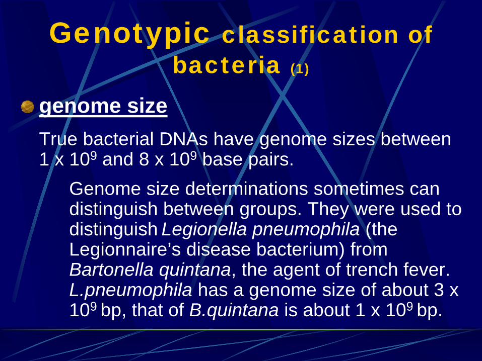
Genotypic classification of bacteria (1)
genome size
True bacterial DNAs have genome sizes between 1 x 10⁹ and 8 x 10⁹ base pairs.
Genome size determinations sometimes can distinguish between groups. They were used to distinguish Legionella pneumophila (the Legionnaire’s disease bacterium) from Bartonella quintana, the agent of trench fever. L.pneumophila has a genome size of about 3 x 10⁹ bp, that of B.quintana is about 1 x 10⁹ bp.
Genotypic classification of bacteria (2)
guanine* (G) plus cytosine (C) ratio The G + C content in bacterial DNA ranges from
25 to 75 percent. This percentage is specific, but not exclusive, for a species; two strains with similar G + C content may not belong to the same species.
If the G + C contents are very different, however, the strains cannot be members of the same species.
*(guanine, cytosine = DNA nucleoside bases)

Genotypic classification of bacteria (3)
DNA hybridization
The ideal means of bacteria identifying and classifying would be to compare each gene sequence in a given strain with the gene sequence for every known species. This cannot be done, but the total DNA of one organism can be compared with that of any other organism by a method called nucleid acid hybridization or DNA hybridization.
This method can be used to measure the number of DNA sequences that any two organisms have in common and estimate the percentage of divergence within DNA sequences that are related but not identical.
•DNA hybridization (3a)
Bacterial species → is composed of strains that are 70-100% DNA related
Different species → DNA relatedness is O% to about 65%
It is important to emphasize that the term ‘related’ does not mean ‘ identical’ or ‘homologus’. Similar but nonidentical nucleic acid sequences can reassociate.
DNA relatedness is determined by allowing single-stranded
DNA from one strain to reassociate with single-stranded DNA from a second strain, to form a double-stranded DNA molecule. This is a specific, temperature-dependent reaction. The optimal temperature for DNA reassociation is 25 to 30 °C below the temperature at which native double-stranded DNA denatures into single strands.

•DNA hybridization (3b)
Diagram of DNA reassociation. (Top)- Perfectly reassociated DNA base sequence in which all nucleotides
are paired by hydrogen bonds. (Middle)- Perfectly paired DNA base sequence in the center with unpaired, single-strand ends on each strand. (Bottom)- None of the bases in the sequence (left to right) GCTACGTCAGT on the top strand are complementary to the sequence TACGATGCAGT in the bottom strand.
Double-stranded DNA is formed through hydrogen bounds that can occur only between the complementary bases A and T or G and C.
Genotypic classification of bacteria (4)
nucleic acid sequence analysis
The nucleic acid sequence analysis is an extension of the hybridization method.
The specific molecular probes are used to localize specific nucleic acid sequences that are unique to a genus, species or subspecies. These sequences are amplified and the obtained genetic material is sequenced to define the precise identity of the isolate.
This method primarily analyzes sequences of ribosomal DNA. It has been used to define the evolutionary relationship among organisms.
Genotypic classification of bacteria (5)
Plasmid analysis Ribotyping Chromosomal DNA fragment analysis
All those methods
have been used, primarily to classify organisms at the subspecies level for epidemiologic investigations.
All living organisms are categorized into larger groups based on their similarities and differences.
In 1969, Robert H. Whittaker proposed a Five-Kingdom System of Classification in which all living organisms are placed into five Kingdoms: Monera, Protista, Plantae, Fungi, Animalia.
Bacteria and archaeans (= „ancient” bacteria) are in the Kingdom Monera (since 1974 – named Procaryotae)
In the late 1970s, a molecular biologist Carl R. Woese et al., devised a Three-Domain System of Classification that was based on the sequences of nucleotide bases in organisms’ ribosomal RNA molecules (which has become most favoured by microbiologists!!!).
In this System, there are two domains of procaryotes (Archaea and Bacteria) and one domain (Eucarya or Eukarya), which includes all eucaryotic organisms.
The taxonomic units in bacterial classification
Domain Phylum Class Order Family Genus Species Subspecies
________________________________________
sing.: phylum; genus; species; subspecies pl.: phyla; genera; species; subspecies
The taxonomy of bacteria is not very definitively worked out yet, especially the higher levels of classification.
For classification purposes, organisms are usually organized into subspecies, species, genera, families, and higher orders. The most important level of classification is the species level.
In addition to species and subspecies designations, clinical microbiologists must be familiar with genera and families. A genus is a group of related species, and a family is a group of related genera.

S P E C I E S A bacterial species is a distinct organism with certain characteristic features or a group of organisms that resemble one another closely in the most important features of their organization.
Species, groups of similar organisms within a genus, are designated by biochemical and other phenotypic criteria and by DNA relatedness, which groups strains* on the basis of their overall genetic similarity.
Within one species, strains and subgroups can be differ by the disease they produce, their environmental habitat and many other characteristics
* strain = a population of cells derived from a single cell
The Domain Bacteria contains 25 phyla, 34 classes, 78 orders, 230 families, 1227 genera, and 6,740 species.
New species will continue to be described.
(Bergey’s Manual of Systematic Bacteriology, 2nd ed. , Vol. 2, 2005)

Phyla (7) and medically important genera (50) within the Domain Bacteria
Phylum Some medically important genera B12. Proteobacteria (Gram-negative bacteria)
Bordetella, Brucella, Campylobacter, Citrobacter, Ehrlichia, Enterobacter, Escherichia, Francisella, Haemophilus, Helicobacter, Klebsiella, Legionella, Neisseria, Pasteurella, Proteus, Pseudomonas, Rickettsia, Salmonella, Serratia, Shigella, Spirillum, Vibrio, Yersinia 23
B13. Firmicutes (the low G+C Gram-positive bacteria)
Bacillus, Clostridium, Enterococcus, Lactobacillus, Listeria, Mycoplasma, Peptococcus, Peptostreptococcus, Staphylococcus, Streptococcus, Ureaplasma, Veillonella 12
B14. Actinobacteria (the high G+C Gram-positive bac.)
Actinomyces, Corynebacterium, Mobiluncus, Mycobacterium, Nocardia, Propionibacterium 6
B16. Chlamydiae Chlamydia, Chlamydophila 2
B17. Spirochaetes Borrelia, Leptospira, Treponema 3
B20. Bacteroides Bacteroides, Porphyromonas, Prevotella 3
B21. Fusobacteria Fusobacterium 1

Identification is practical division of taxonomy. Identification determines whether an unknown organism belongs to a previously defined group or forms a new taxon.
„To identify an organism” means to learn the organism’s species name – i.e., to speciate it.
1. The first step of identification is phenotypic grouping of strains by morphological, biochemical and any other characteristics of interests. 2. The phenotypic groups are then tested for DNA relatedness. 3. The third and the most important step is reexamination of the biochemical characteristics of the DNA relatedness group.
BACTERIAL IDENTIFICATION (1)
When attempting to identify an organism that has been isolated from a clinical specimen, laboratory workers are very much like detectives. They gather „clues” (characteristics, attributes, properties, and traits) about the organism until they have sufficient clues to identify (speciate) the organism. In most cases, the clues that have been gathered will match the characteristics of an established species.
BACTERIAL IDENTIFICATION (2)
BACTERIA NOMENCLATURE (1)
Bacteria are named so that investigators can define and discuss them without the necessity of listing their characteristics.
Bacteria are given scientific names based on the binominal (= two names) system of nomenclature.
The first name is referred to as the genus and the second name is termed the species (e.g., Escherichia coli).
The names usually come from Latin or Greek and describe some characteristics of the organism. Organisms are named for: who discovered them, where they were found and what they look like. A species name should mean the same thing to everyone.

BACTERIA NOMENCLATURE (2)
To correctly write the scientific name of a bacterium, the first letter of the genus should be capitalized while the species name should be in lower case letters.
Both the genus and species names are always italicized.
Several examples:
Bacillus subtilis Escherichia coli Staphylococcus aureus Vibrio cholerae Clostridium perfringens Pseudomonas aeruginosa Klebsiella pneumoniae Neisseria gonorrhoeae

BACTERIA NOMENCLATURE (3)
When employing binominal nomenclature, the following additional conventions are employed:
- the species name is never used without the genus name (e.g., coli standing alone)
- the genus name may be used without the species name (e.g., Escherichia may stand alone, though no longer actually describes a species)
- the first time a binominal nomenclature is used, the genus name is spelled out (e.g., Escherichia coli), thereafter it can be abbreviated (e.g., E. coli ); the species name is never abbreviated
- the abbreviation ‘sp.’ is used to designate a single species, whereas the abbreviation ‘spp.’ is used to designate more than one species

BACTERIA NOMENCLATURE (4)
In addition to the proper scientific names for bacteria acceptable terms like:
staphylococci (for Staphylococcus spp.) streptococci (for Streptococcus spp.) clostridia (for Clostridium spp.) pseudomonads (for Pseudomonas spp.) mycoplasmas (for Mycoplasma spp.) rickettsias (for Rickettsia spp.) chlamydias (for Chlamydia spp.)
are commonly used.
Nicknames and slang terms frequently used within the hospitals are: GC and gonococci (for Neisseria gonorrhoeae), meningococci (for N. meningitidis), pneumococci (for Streptococcus pneumoniae), staph (for Staphylococcus or staphylococcal), and strep (for Streptococcus or streptococcal).

Bergey’s Manual of Systematic Bacteriology
The possibility that one might draw conclusions about phylogenetic relationships among bacteria is reflected in the five-volume set of books entitled Bergey’s Manual of Systematic Bacteriology (sometimes referred to as microbiologist’s „bible”). The Manual is an effort to classify known bacteria and to make this information accessible in the form of a key.
This bacteriologist’s most important reference is currently being rewritten. When all five volumes have been completed, they will contain descriptions of more than 6,700 validly named species of bacteria.
A companion volume, Bergey’s Manual of Determinative Bacteriology, serves as an aid in the identification of those bacteria that have been described and cultured.

SIZES, SHAPES and ARRANGEMENTS of bacteria (1)
Most bacteria come in one of three basic shapes:
THE COCCUS (an average coccus is about 0.5-1.0 µm in diameter)
THE ROD (or BACILLUS) (an average bacillus is 0.5-1.0 µm wide by 1.0- 4.0 µm long)
THE SPIRAL (spirals range in size from 1 µm to over 100 µm in length)
SIZES, SHAPES and ARRANGEMENTS of bacteria (2)
THE COCCUS (sing. coccus; pl. cocci)
The cocci are generally spherical bacteria (although some appear oval, elongated or flattened on one side) having one of several distinct arrangements based on their planes of division (and tendency to remain attached after replication).
D I V I S I O N: in one plane produces either a DIPLOCOCCUS (pair of cocci) or STREPTOCOCCUS (chain of cocci) arrangement.
in two planes produces a TETRAD (square of 4 cocci).
in three planes produces a SARCINA (cube of 8 cocci) arrangement.
in random planes produces a STAPHYLOCOCCUS (cocci in irregular, often grape-like clusters) arrangement.
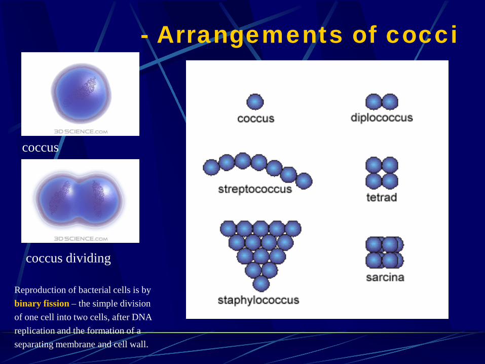
- Arrangements of cocci
coccus dividing
coccus
Reproduction of bacterial cells is by binary fission – the simple division of one cell into two cells, after DNA replication and the formation of a separating membrane and cell wall.

http://commons.wikimedia.org/wiki/Image:Cocci_arrangement.png
After binary fission, the daughter cells may separate completely from each other or may remain connected, forming various morphologic arrangement:
a. streptococcus b. diplococcus c. tetrad d. sarcina e. staphylococcus
SIZES, SHAPES and ARRANGEMENTS of bacteria (3)
THE ROD (or BACILLUS) (sing. bacillus; pl. bacilli)
Bacilli are rod-shaped bacteria. Bacilli all divide in one plane (and only across their short axis) producing one of the following arrangement:
- bacillus (a single bacillus)
- diplobacillus (a pair of bacilli) - streptobacillus (a chain of bacilli)
- coccobacillus (short bacillus that looks like a coccus)

- Arrangements of bacilli
SIZES, SHAPES and ARRANGEMENTS of bacteria (4)
THE SPIRAL
Spirals come in one of three forms:
vibrio – an incomplete spiral or coma-shaped rod
spirillum* – a thick, rigid spiral
spirochete – a thin, flexible spiral; they are the thinnest of the bacteria, often having a width of only 0.25 – 0.5 µm.
* sing. spirillum; pl. spirilla
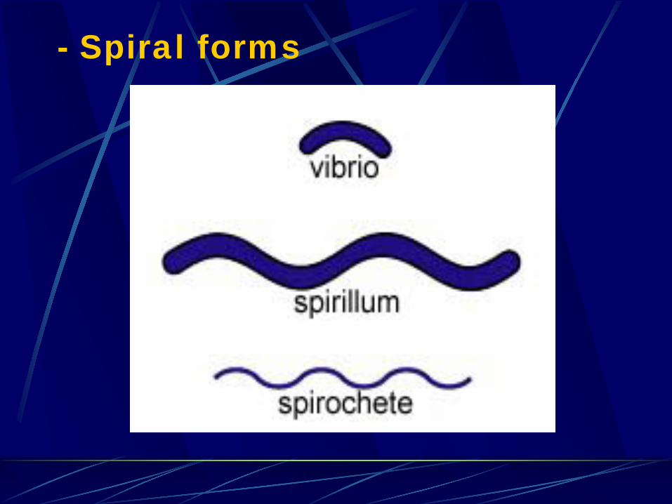
- Spiral forms
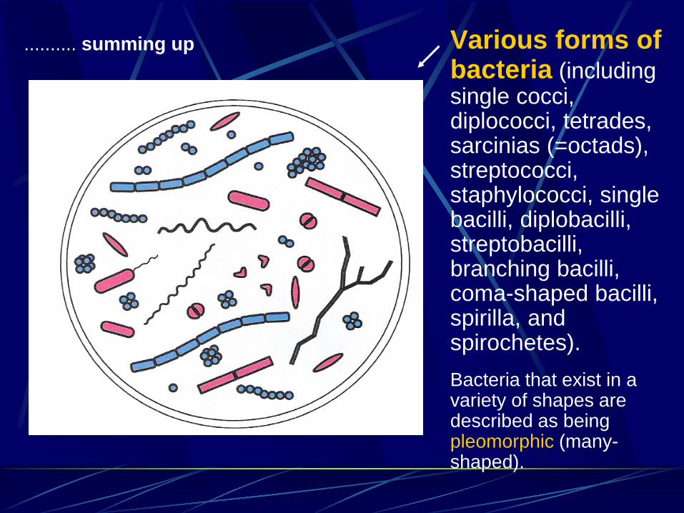
Various forms of bacteria (including single cocci, diplococci, tetrades, sarcinias (=octads), streptococci, staphylococci, single bacilli, diplobacilli, streptobacilli, branching bacilli, coma-shaped bacilli, spirilla, and spirochetes).
Bacteria that exist in a variety of shapes are described as being pleomorphic (many-shaped).
.......... summing up

Descriptive terms are used to broadly categorize clinical bacteria in several useful ways.
A useful system of clinical bacteria categorization would give an indication of the probability of infection by a particular organism.

are categorized:
according to their Gram stain characteristics

are classified:
taxonomically as to families, genera and species

are categorized:
according to their growth condition requirements (anaerobic, facultative anaerobic and aerobic bacteria)

are categorized:
according to which region of the body they inhabit as part of normal flora (indigenous flora) or from which part they are frequently isolated or cause disease

are categorized:
according to how dangerous they are (pathogens and opportunistic bacteria)

are categorized:
according to the time needed for causing a health problem (some organisms can gain entry into the bloodstream and causing a problem only after a long period as cumulative effect; some pathogens proactively create portals of entry and these are called invasive)

An example of a useful classification scheme for clinical
bacteria organizing
The many bacteria that are presented according to that scheme will be discussed in detail in subsequent lectures.
It should be noted, that the list of presented
organisms is not complete. Many genera that are identified in clinical samples are omitted to simplify that presentation.
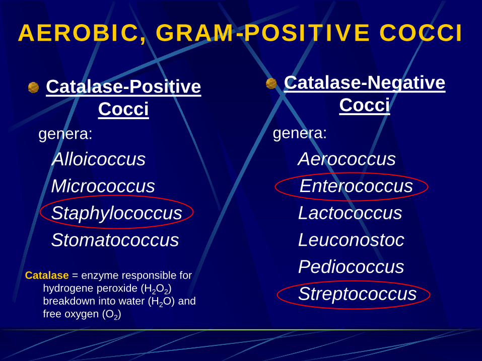
AEROBIC, GRAM-POSITIVE COCCI
Catalase-Positive Cocci
genera: Alloicoccus
Micrococcus Staphylococcus Stomatococcus Catalase = enzyme responsible for
hydrogene peroxide (H2O2) breakdown into water (H2O) and free oxygen (O2)
Catalase-Negative Cocci
genera: Aerococcus Enterococcus Lactococcus Leuconostoc Pediococcus Streptococcus
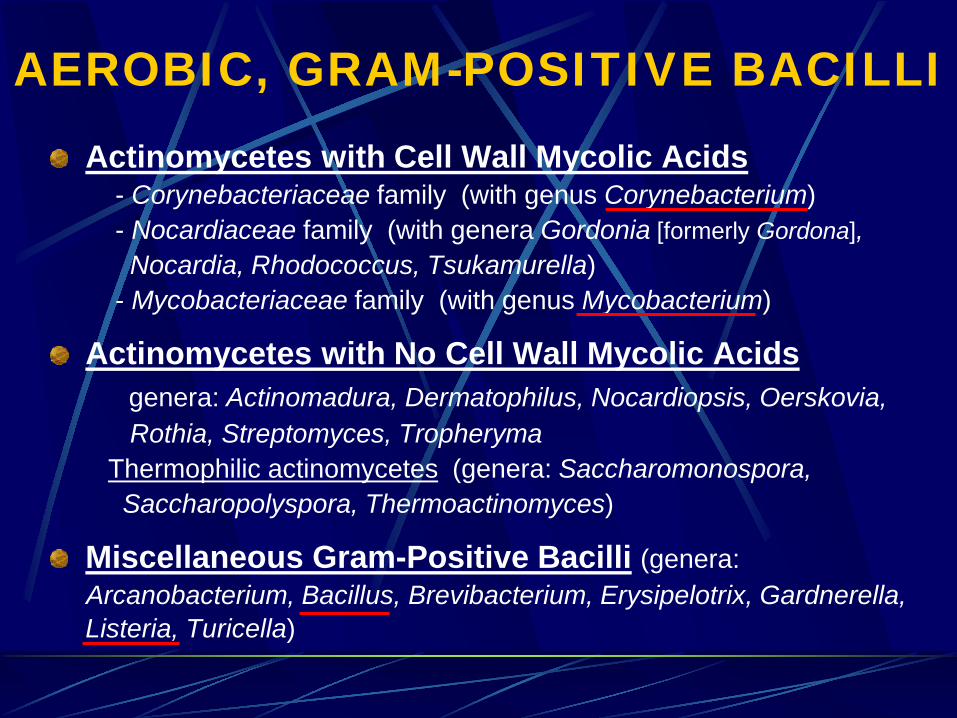
AEROBIC, GRAM-POSITIVE BACILLI Actinomycetes with Cell Wall Mycolic Acids
- Corynebacteriaceae family (with genus Corynebacterium) - Nocardiaceae family (with genera Gordonia [formerly Gordona], Nocardia, Rhodococcus, Tsukamurella) - Mycobacteriaceae family (with genus Mycobacterium)
Actinomycetes with No Cell Wall Mycolic Acids genera: Actinomadura, Dermatophilus, Nocardiopsis, Oerskovia, Rothia, Streptomyces, Tropheryma Thermophilic actinomycetes (genera: Saccharomonospora, Saccharopolyspora, Thermoactinomyces)
Miscellaneous Gram-Positive Bacilli (genera: Arcanobacterium, Bacillus, Brevibacterium, Erysipelotrix, Gardnerella,
Listeria, Turicella)

AEROBIC, GRAM-NEGATIVE COCCI, COCCOBACILLI and BACILLI
Cocci and Coccobacilli (genera: Branhamella, Moraxella, Neisseria)
Bacilli (families: Enterobaceriaceae, Vibrionaceae, Aeromonadaceae, Plesiomonadaceae, Campylobacteriaceae, Helicobacteriaceae, Pseudomonadaceae, Pasteurellaceae)
Miscellaneous genera (Acinetobacter, Bartonella, Bordetella, Brucella, Burkholderia, Capnocytophaga, Cardiobacterium, Eikenella, Francisiella, Kingella, Legionella, Spirillum, Stenotrophomonas, Streptobacillus)
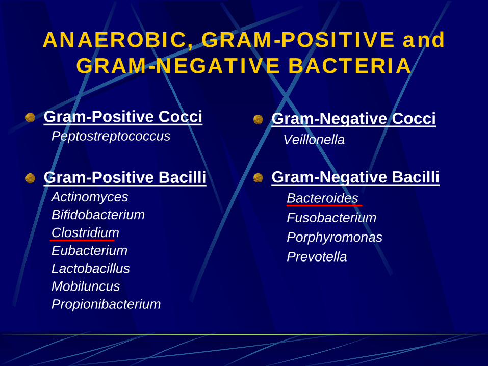
ANAEROBIC, GRAM-POSITIVE and GRAM-NEGATIVE BACTERIA
Gram-Positive Cocci Peptostreptococcus
Gram-Positive Bacilli Actinomyces Bifidobacterium Clostridium Eubacterium Lactobacillus Mobiluncus Propionibacterium
Gram-Negative Cocci Veillonella
Gram-Negative Bacilli Bacteroides Fusobacterium Porphyromonas Prevotella
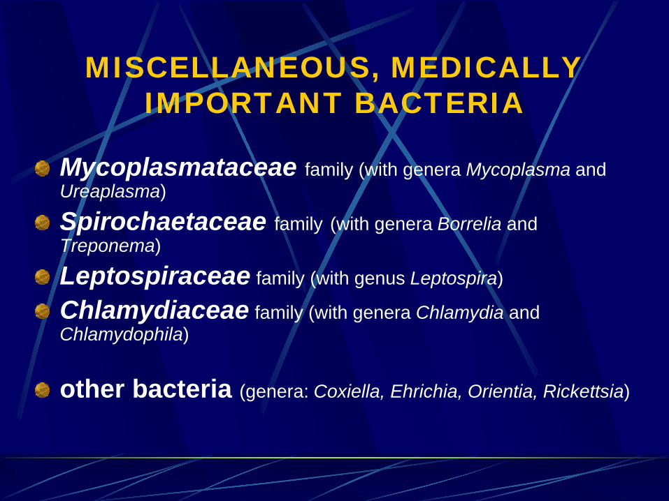
MISCELLANEOUS, MEDICALLY IMPORTANT BACTERIA
Mycoplasmataceae family (with genera Mycoplasma and Ureaplasma)
Spirochaetaceae family (with genera Borrelia and Treponema)
Leptospiraceae family (with genus Leptospira)
Chlamydiaceae family (with genera Chlamydia and Chlamydophila)
other bacteria (genera: Coxiella, Ehrichia, Orientia, Rickettsia)

Representations of metric units of measure and numbers.

Images accompanying that lecture have been acquired from numerous sources. My thanks to everyone who has shared images with me for teaching purposes.
The lecture cannot be put on any WWW site without a written permission from the author.

References 1. Patric R. Murray et al., Bacterial Classification. In: Medical Microbiology -
4th ed., Chapter 2, Mosby, Inc., U.S., 2002, pp. 7-10 2. Ellen Jo Baron, Classification. In: Medical Microbiology, Chapter 3, Section I,
Bacteriology. http://www.gsbs.utmb.edu/microbook/ch003.htm 3. Gary E. Kaiser, The prokaryotic cell: Bacteria, A. Sizes, shapes, and
arrangements of bacteria, Unit 1: BIOL 230 Lectures, http://student.ccbcmd.edu/courses/bio141/lecguide/unit1/index.html
4. Bacteria Cell Shapes and Arrangements, http://www.mansfield.ohio-state.edu/~sabedon/biol12010.htm
5. Bacterial colonies, http://helios.bto.ed.ac.uk/bto/microbes/shape.htm 6. Clinical Bacteria, http://www.buddycom.com/bacteria/bacteria.html 7. Stephen T. Abedon, Introduction to Microbiology (supplemental Lecture),
http://www.mansfield.ohio-state.edu/~sabedon/biol12005.htm 8. Howard Gest, Bacterial clasification and taxonomy: a ‘primer’ for the new
millennium, Microbiology Today, Vol26/May99, pp.70-72 9. Biological Diversity: Bacteria and Archaeans, Biologigal Diversity 2,
http://www.emc.maricopa.edu/faculty/farabee/BIOBK/BioBookDiversity_2.html 10. Paul G. Engelkirk, Gwendolyn R. W. Burton, Cell structure and taxonomy.
Diversity of microorganisms – Part 1. Acellular and Procaryotic microbes. In: Burton’s Microbiology for the health sciences - 8th ed., Chapter 3 & 4, Lippincott Williams & Wilkins, U.S., 2004, pp. 27-44, 45-71

References 11. Geo F. Brooks, Janet S. Butel, Classification of Bacteria. In: Jawetz,
Melnik, & Adelberg’s Medical Microbiology, twenty-third ed., Chapter 3, Lange Medical Books/McGraw-Hill, Medical Publishing Division, 2004, pp. 42-50
12. Warren Levinson, Ernest Jawetz, Structure of bacterial cells. In: Medical Microbiology & Immunology. Examination & Board review, seventh ed., Chapter 2, Lange Medical Books/McGraw-Hill, Medical Publishing Division, 2002, pp. 4-13.
13. Peter A. R. Vandamme, Taxonomy and Classification of Bacteria. In: Mannual of Clinical Microbiology, Volume 1, 8th ed., Chapter 19, ASM Press, Washington, USA, 2003, pp. 271-285.
Recommended