Computer Engineering and Intelligent Systems www.iiste.org
ISSN 2222-1719 (Paper) ISSN 2222-2863 (Online)
Vol 2, No.4, 2011
124
Iris Recognition with Fake Identification
Pradeep Kumar
ECE Deptt. , Vidya Vihar Institute Of Technology
Maranga, Purnea, Bihar-854301, India
Tel: +917870248311, Email: [email protected]
Abstract
Iris recognition, the ability to recognize and distinguish individuals by their pattern, is the most
reliable biometric in terms of recognition and identification performance. However, performance
of these systems is affected by the heterogeneous images (regarding focus, contrast, or
brightness) and with several noise factors (iris obstruction and reflection) when the cooperation
is not expectable from the subject. Current Iris recognition system does not deal with the noise
data and substantially increase their error rates in these conditions. An Iris classification method
is proposed on the segmented and normalized iris image that divides the image into six regions,
followed by independent feature extraction in each region. This will provide the iris signature in
terms of binary values, then that are compared with each region for the identification. In addition
to this Fake identification is also done in this paper. Fake, the original image is forged by fixing
lenses over the iris portion. This can be identified by using fast Fourier transform.
Keywords: Noncooperative Iris Recognition, Iris Classification, Feature Extraction, Biometrics,
Fake Identification.
1. Introduction
The use of biometric systems has been increasingly encouraged by both governments and private
entities in order to replace or increase traditional security systems. Biometric is based on a
physiological or behavioural characteristic of the person. A biometric system provides automatic
recognition of an individual based on some sort of unique feature or characteristic possessed by
the individual. Biometric systems have been developed based on fingerprints, facial features,
voice, hand geometry (Muron.A 2001), handwriting, the retina, and the iris. Biometric systems
work by first capturing a sample of the feature, such as recording a digital sound signal for voice
recognition, or taking a digital colour image for face recognition. The sample is then transformed
using some sort of mathematical function into a biometric template. The biometric template will
provide a normalized, efficient and highly discriminating representation of the feature, which can
then be objectively compared with other templates in order to determine identity. Most biometric
systems (Gabor.G 1946) allow two modes of operation. An enrolment mode for adding templates

Computer Engineering and Intelligent Systems www.iiste.org
ISSN 2222-1719 (Paper) ISSN 2222-2863 (Online)
Vol 2, No.4, 2011
125
to a database, and an identification mode, where a template is created for an individual and then
a match is searched for in the database of pre-enrolled templates.
Assuming that, in spite of noise, the iris was accurately segmented, we propose a classification
strategy more robust to noise factors. I observed that, in most cases, the noisy data is localized
(Proenca.H 2006) in some of the iris subparts. My method is based on the division of the
segmented iris into six regions, followed by the independent feature extraction in each one.
Further, through the comparison between signatures extracted from correspondent iris regions,
we obtain six dissimilarity values that are fused through a classification rule. The hope is that
most of the iris regions are noise-free and that accurate recognition can be achieved, even in
highly noisy images. In section 2 basics about iris recognition is discussed. In section 3 proposed
methodology is explained and section 4 and 5 deals about result implementation and fake
identification respectively.
2. Iris Recognition
Iris is commonly recognized as one of the most reliable biometric measures: it has a random
morphogenesis and no genetic penetrance. The iris is a protected internal organ of the eye,
located behind the cornea and the aqueous humour. It is the only internal organ of the body that
is normally visible externally. Images of the iris adequate for personal identification (Proenca .H
2007) with very high confidence can be acquired from distances of up to about 3 feet (1 meter).
The human iris begins to form during the third month of gestation. The structures creating its
distinctive pattern are complete by the eighth month of gestation. In fact, the iris patterns are
characterized by high level of stability and distinctiveness. Each individual has a unique iris; the
difference even exists between identical twins (Daugman.J.G 2004) and between the left and
right eye of the same person.
The overall iris recognition system can be given by Fig.1. In 1987 L. Flom and A. Safir studied
the problem (Daugman.J.G 1993) and concluded that iris morphology remains stable through all
human life, as well estimated the probability for two similar irises on distinct persons at 1 in
1072.
The cooperative behaviour demanded to the users and the highly constrained imaging conditions
clearly restrict the range of domains where iris recognition can be applied. It is highly probable
that image capturing on less constrained conditions (either at-a-distance, on-the-move, with
minor users' cooperation and within dynamic imaging environments) lead to the appearance of
extremely heterogeneous (Ma.L 2007) images and with several other types of data in the
captured iris regions (e.g., iris obstructions due to eyelids or eyelashes, reflections, off-angle or
motion blurred images).

Computer Engineering and Intelligent Systems www.iiste.org
ISSN 2222-1719 (Paper) ISSN 2222-2863 (Online)
Vol 2, No.4, 2011
126
Fig-1: Stages of Iris Recognition
The emerging needs for a safer and quicker access (buildings, weapons, and restricted areas)
requires non-cooperative techniques. In this paper, I consider a non-cooperative technique where
the user has no active participation in the image-capture process and is not even aware of the
recognition process.
As an example, we can think of a building access where users do not need to look through a
small hole to have their irises recognized (MA.L 2004), but instead, an image-capture system
captures the necessary information from their irises as they approach the door. This is much less
invasive and will enable the dissemination of iris recognition systems to everyday applications.
Obviously, these image-capture conditions tend to acquire images with more heterogeneous
characteristics with respect to reflection areas, brightness and contrast or focus conditions.
Human iris recognition process is basically divided into five steps,
Iris Acquisition
Localization
Normalization
Feature Extraction
Matching

Computer Engineering and Intelligent Systems www.iiste.org
ISSN 2222-1719 (Paper) ISSN 2222-2863 (Online)
Vol 2, No.4, 2011
127
The first stage of the iris recognition is the iris Acquisition. The eye image can be obtained by
using CCD Cameras (Flom.L 1987). For the academic purpose, The CASIS, MMU and the
UBIRIS (Chinese academic) provided about thousands of iris images in free of cost. We use both
of these CASIA, MMU and UBIRIS databases.
After the image acquisition, (Vatsa.M 2005) the next stage of the iris recognition deals with iris
segmentation. This consists of localizing the iris inner (pupillary) and outer (scleric) borders. In
1993, Daugman proposed an integro-differential operator to find both the iris inner and outer
borders. Similarly, T.Camus et. al., proposed integro- differential (Kalka .N 2006) operators that
search over the IN3 space, with the goal of maximizing the equations that identify the iris
borders. Wildes achieved iris segmentation through a gradient-based binary edge map
construction followed by circular Hough transform. H.Proenca et. al., proposed a method based
on Wildes‟ method, which, together with a clustering process, achieved robustness for non
cooperative environments.
It is possible to varying the pupil’s size depending upon various images and in the imaging
distance. In order to compensate these variations, it is usual to translate the segmented iris region
into a fixed length and dimensionless polar coordinate system. This stage is usually
accomplished through the method proposed by Daugman. The method is termed as Daugman‟s
Rubber Sheet Model. From the view point of feature extraction, previous iris recognition method
can be roughly divided into three major characteristics: Phase-based method, Zero- crossing
representation method and texture-analysis based method. Daugman used multiscale quadrature
wavelet to extract texture phase structure information of the iris to generate a 2,048-bit iriscode
and compared the difference between a pair of iris representation by computing their Hamming
distance.
Finally, the obtained iris signature is compared with the database (Wildes.R.P 1997), producing a
numeric dissimilarity values. If this value is higher than a threshold, the system outputs a non-
match, meaning that each signature belongs to different irises. Otherwise, the system outputs a
match, meaning that both signatures were extracted from the same iris. In this stage, it is
common to apply different distance metrics (Hamming, Euclidean, Weighted Euclidean), or
methods based on signal correlation.
3. Proposed Methodology
3.1. Noncooperative Iris Recognition
Computer Engineering and Intelligent Systems www.iiste.org
ISSN 2222-1719 (Paper) ISSN 2222-2863 (Online)
Vol 2, No.4, 2011
128
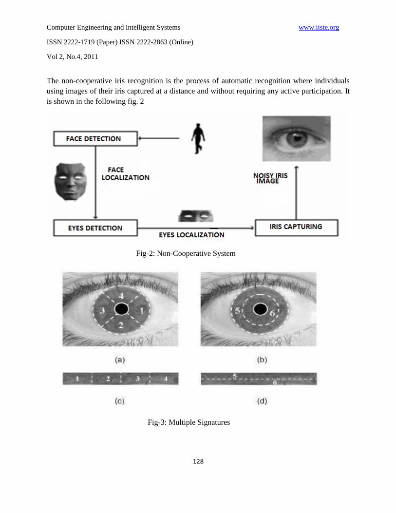
The non-cooperative iris recognition is the process of automatic recognition where individuals
using images of their iris captured at a distance and without requiring any active participation. It
is shown in the following fig. 2
Fig-2: Non-Cooperative System
Fig-3: Multiple Signatures

Computer Engineering and Intelligent Systems www.iiste.org
ISSN 2222-1719 (Paper) ISSN 2222-2863 (Online)
Vol 2, No.4, 2011
129
The main drawback of real time implementation of Iris recognition is lies in the segmented
image. Where most cases the eye image contains noise such that eyelids, eyelash. These noisy
patterns spread across iris and gives less active components of iris patterns. So that the
segmentation part of iris is not possibly acquired and it not at all useful for further stages of iris
recognition system. The non-cooperative system is possible by applying a new algorithm
namely, multiple signature. i.e., the human iris is going to be divided into six regions.
In terms of non cooperative, this increasing the probability of capturing very heterogeneous
factor with several noise factors. In most cases, the noisy data is localized in some of the iris
subparts. Our method is based on the division of the segmented iris into six regions, followed by
the independent feature extraction in each one. Further, through the comparison between
signatures extracted from correspondent iris regions, we obtain six dissimilarity values that are
fused through a classification rule. The hope is that most of the iris regions are noise-free and
that accurate recognition can be achieved, even in highly noisy images. To maintain a good
effectiveness of iris recognition, the multiple signature algorithms is applied in the segmentation
module.
In the context of non-cooperative recognition, the most relevant value is the accuracy
degradation as function of the images‟ quality. We observed that our method is clearly less
dependent of the image characteristics, since it presented the smallest accuracy degradation
(Tuceryan 1994) between both sessions - just about 0.14%. This is in contrast with all the
remaining methods, especially those proposed by Martin-Roche et al., Daugman and Camus and
Wildes. It must be stressed that our method is the one that presented the highest accuracy on
images from the second session, indicating that it is well adapted to deal with noisy images. The
multiple signature algorithm based on segmentation is shown below:
In fig 3, (a) Division of the iris in four different parts. (b) Division of the iris in “outer” and
“inner” parts. (c) Correspondent regions of (a) in the normalized iris image. (d) Correspondent
regions of (b) in the normalized iris image.
By comparing with other systems, Wildes‟ method achieved the best results in absolute terms,
having 98.74% accuracy on the first session images. However, as the image quality decreases, its
accuracy degraded more than 2%. This fact may indicate that, if we incorporate other noise
factors, its accuracy will be strongly affected, which discourages its use in the non-cooperative
setting. The implemented variants of this method, both the preprocessing methods and the
alternative edge detection algorithms, didn‟t get significant improvements when compared to the
original method.

Computer Engineering and Intelligent Systems www.iiste.org
ISSN 2222-1719 (Paper) ISSN 2222-2863 (Online)
Vol 2, No.4, 2011
130
My proposal’s computation time is about 17% higher than that of Wildes‟ algorithm; these 17%
are used in the feature extraction and clustering process. We consider that with proper algorithm
optimization this computation time gap about 0.3 seconds) will become irrelevant.
Division of the whole iris into six regions is the main concept behind multiple signatures. Here
Regions 1 to 4 correspond to successive quadrants of the iris. Regions 5 and 6 correspond,
respectively, to the outer and inner parts of the iris.
The main motivation for this division was the observation that the most common types of noise
(iris obstructions and reflections) are usual, respectively, in the upper/lower and left/ right
portions of the iris. Also, reflections resultants from natural and artificial lighting environments
are predominantly localized, respectively, in the outer and inner iris regions. The proposed
division strategy minimizes the number of regions simultaneously affected by each type of noise.
Common feature extraction proposals usually focus on the lower and middle-low frequency
components of the signal. This implies that small portions of non-detected noise can corrupt the
whole biometric signature and decrease the recognition accuracy.
Based on this, we proposed a new iris classification strategy that divides the segmented and
normalized iris into six regions and makes an independent feature extraction and comparison for
each of these regions. Iris classification is achieved through a fusion rule that uses a threshold set
to combine the dissimilarity values resultant from the comparison between correspondent iris
regions.
This indicates that the proposed method is adequate for less constrained image capturing
environments, such as in a non cooperative setting, and broadens the range of domains where iris
recognition can be applied. However, we stress that these results are dependent on the previous
accurate iris segmentation, which is highly challenging, given the dynamics of non cooperative
environments. The requirement of optical frameworks that are able to capture iris images with
enough quality and of real-time face and eye localization methods is assumed too.
4. Implementation Result
4.1 Iris Databases
There are presently seven public and freely available iris image databases for biometric
purposes: CASIA, MMU (Camus .T 2002), BATH (Tuceryan 1994), UPOL (Dorairaj.V 2005),

Computer Engineering and Intelligent Systems www.iiste.org
ISSN 2222-1719 (Paper) ISSN 2222-2863 (Online)
Vol 2, No.4, 2011
131
ICE, and UBIRIS. The CASIA database is by far the most widely used for iris biometric
purposes. However, its images incorporate few types of noise, almost exclusively related with
eyelid and eyelash obstruction, similarly to the images from MMU and BATH databases. UPOL
images (Camus .T 2002) were captured with an optometric framework, obtaining optimal images
with extremely similar characteristics. Although ICE and WVU databases contain images with
more noise factors, their lack of images with significant reflections within the iris rings
constitutes a weak point regarding the simulation of Noncooperative imaging conditions.
Oppositely, images of the UBIRIS database were captured under natural lighting and
heterogenous imaging conditions, which explains their higher heterogeneity. Based on the
manual verification of the iris segmentation accuracy in each of the images, we selected 800
images from 80 subjects of the UBIRIS database.
4.2 Description of Experiments
We implemented the recognition method described by Daugman (Flom.L 1987), (Vatsa.M 2005)
and compared the obtained results when following the method as described by the author and
using the proposed iris division and classification strategies. Initially, we made the feature
extraction and comparison using the whole segmented iris, extracting a total of 2,048 bits.
Further, according to Fig. 3, we divided the iris into six regions and, through feature extraction,
obtained 512 and 1,024 bits, respectively, for the signatures extracted from the iris regions 1 to 4
and 5 to 6.
The Iris recognition method is divided into the following stages:
Segmentation: The segmentation is the first phase of the Iris recognition. This phase can extract
only the iris part from the human eye. We implement the circular edge detection method by
using canny edge detector for segmentation.

Computer Engineering and Intelligent Systems www.iiste.org
ISSN 2222-1719 (Paper) ISSN 2222-2863 (Online)
Vol 2, No.4, 2011
132
(a) (b)
Fig-4: (a) Required Segmented Result. (b) Poor Performance Result
Fig-5: Required Normalization Result with Size of 240 × 20.
Initially the segmented result is obtained by removing the pupil and the eyelash. These are
removed by using the threshold values, so the performance of the segmentation is not satisfied.
Then the segmentation is done without removing eyelash and pupil.
Normalization: After the segmentation of both iris borders, to compensate for the varying size of
the pupil and capturing distance, we translated the images into a dimensionless polar coordinate
system, according to the process known as the Daugman Rubber Sheet (Flom.L 1987), (Vatsa.M
2005). Generally the segmentation phase will remove the pupil and other than the iris. In order to
reduce the complexity of normalization process the segmentation phase itself extracts the
required part (as shown in figure 4) from the iris. The output of the normalization will show as
below.
Multiple Signature: When we talk about noncooperation, the captured iris images are normally
with the noisy one. That is, most of the images are with obstruction and reflection. So the
introduction of multiple signature is necessary here. Generally the size of the normalized image
is 240 × 20. This is going to be divided into six regions as four 60 20 patterns and two 240 10
patterns. The concept of multiple signatures is given as below.

Computer Engineering and Intelligent Systems www.iiste.org
ISSN 2222-1719 (Paper) ISSN 2222-2863 (Online)
Vol 2, No.4, 2011
133
Fig-6: Multiple Signature 1/6, 2/6, 3/6, 4/6 are Fig-7: The Input Image Data\img_005_1_2.jpg is
60 × 20 Patterns and Multiple Signature 5/6 Matced with the Database Image data\img_005_1_3.jpg.
and 6/6 are 240 × 10 patterns Here 005 Indicate the 5th Person.
Feature Extraction: This iris data encoding was accomplished through the use of two-
dimensional Gabor filter.
Feature Comparison: The binary feature comparison allowed the use of the Hamming distance as
the similarity measure between two iris signatures. The output of the final iris recognition is
given as above fig.
5. Fake Identification
The fake identification module enables the user to find weather the query image is an original or
forged one. If the given image is finding to be as a fake one, there is no need for iris recognition
for that particular image. This can be identified as given in Figure below. That is the difference
between the original image and the fake image is shown here. In order to identify the fake image,
the FFT (Fast Fourier Transform) (Dorairaj.V 2005) is applied on the given image. When the
lenses are fixed over the iris portion the quality of the real image is going to be affected. This
added advance can be used for fake identification.

Computer Engineering and Intelligent Systems www.iiste.org
ISSN 2222-1719 (Paper) ISSN 2222-2863 (Online)
Vol 2, No.4, 2011
134
Fig-8: Comparison of Original Image with Fake Image After Applying FFT
6. Conclusion
In this paper, I addressed the problems motivated by the existence of noise in the captured iris
images and the correspondent increase of the error rates, with particular relevance to the false
rejections, in the context of non cooperative iris recognition. Also fake identification is
introduced for the lens images fixing over the iris portion.
Acknowledgment
The author would like to thank Miss K.Jayanthi and Dr.Abhay Kumar for their insightful advice
and guidance, and unknown reviewers for their useful remarks and suggestions.
Reference
Camus.T and Wildes.R, “Reliable and Fast Eye Finding in Close-Up Images,” Proc. IEEE 16th
Int‟l Conf. Pattern Recognition, pp. 389-394, Aug. 2002.
Daugman.J.G, “How Iris Recognition Works”, IEEE Trans. on Circuit and System for Video
Technology, vol.14, no.1, pp21-30, January 2004.
Daugman.J.G, “High Confidence Visual Recognition of Persons by a Test of Statistical
Independence,” IEEE Trans. on Pattern Analysis and Machine Intelligence, vol. 25, no. 11, pp.
1148-1161, Nov. 1993.

Computer Engineering and Intelligent Systems www.iiste.org
ISSN 2222-1719 (Paper) ISSN 2222-2863 (Online)
Vol 2, No.4, 2011
135
Dorairaj.V, Schmid.N, and Fahmy.G, “Performance evaluation of Nonideal Iris Based
Recognition System Implementing Global ICA
Encoding”, Proc. IEEE Int‟l Conf. Image Processing, pp. 285-288, Sept. 2005.
Flom.L and Safir.A, “Iris Recognition System,” US Patent 4 641 394, 1987.
Gabor.D, “Theory of Communication”, J. Inst. Elect. Eng., vol. 93, pp. 429-459, 1946.
Kalka.N, Zuo.J, Schmid.N, and Cukic.B, “Image Quality Accessment for Iris Biometric,” Proc.
SPIE Conf. Biometric Technology for Human Identification III, vol. 6202, pp. 263-273, Apr.
2006.
Ma.L, Wang.Y, and Tan.T, “Iris Recognition Using Circular Symmetric Filters,” Proc. 25th Int‟l
Conf. Pattern Recognition, vol. 2, pp. 414-417, Aug.2002.
Ma.L, Tan.T, Zhang.D, and Wang.Y, “ Local Intensity Variation Analysis for Iris Recognition
“, pattern Recognition, vol. 37, no.6, pp. 1287-1298, 2004.
Muron.A, Petr.K. And Jaroslav.P, “Identification of persons by means of the Fourier Spectra of
the Optical Transmission Binary Models of the Human Irises”, optics Communication, vol. 192,
2001, pp. 161-167.
Proenca.H and Alexandre.L.A, “Iris Segmentation Methodology for Noncooperative Iris
Recognition,” IEE Proc. Vision, Image, and Signal Processing, vol. 153, no. 2, pp. 199-205,
April 2006.
Proenca.H and Alexandra.L.A, “Towards Noncooperative Iris Recognition: A Classification
Approach using Multiple Signatures”, IEEE Trans. On Pattern Analysis and Machine
Intelligence, vol. 29, no. 4, pp. 607-612, April 2007.
Tuceryan, M.: „Moment based texture segmentation‟, pattern recognit. Lett., 1994, 15, pp. 659-
668.
Vatsa.M, Singh.R, and Noore.A, “Reducing the False Rejection Rate of Iris Recognition Using
Textural and Topological Features,” Int‟l J. Signal Processing, vol. 2, no. 1, pp. 66-72, 2005.
Wildes.R.P, “Iris Recognition: An Emerging Biometric Technology,” Proc. IEEE, vol. 85, no. 9,
pp. 1348-1363, Sept. 1997.

This academic article was published by The International Institute for Science,
Technology and Education (IISTE). The IISTE is a pioneer in the Open Access
Publishing service based in the U.S. and Europe. The aim of the institute is
Accelerating Global Knowledge Sharing.
More information about the publisher can be found in the IISTE’s homepage:
http://www.iiste.org
The IISTE is currently hosting more than 30 peer-reviewed academic journals and
collaborating with academic institutions around the world. Prospective authors of
IISTE journals can find the submission instruction on the following page:
http://www.iiste.org/Journals/
The IISTE editorial team promises to the review and publish all the qualified
submissions in a fast manner. All the journals articles are available online to the
readers all over the world without financial, legal, or technical barriers other than
those inseparable from gaining access to the internet itself. Printed version of the
journals is also available upon request of readers and authors.
IISTE Knowledge Sharing Partners
EBSCO, Index Copernicus, Ulrich's Periodicals Directory, JournalTOCS, PKP Open
Archives Harvester, Bielefeld Academic Search Engine, Elektronische
Zeitschriftenbibliothek EZB, Open J-Gate, OCLC WorldCat, Universe Digtial
Library , NewJour, Google Scholar
Recommended