Department of Pathology
Inflammatory Bowel Disease Neoplasia
Mary P. Bronner, MD
Division Chief of Anatomic Pathology University of Utah

Department of Pathology
Neoplastic Progression in Chronic Inflammatory GI Dz
Inflammation
Dysplasia
Carcinoma
Department of Pathology
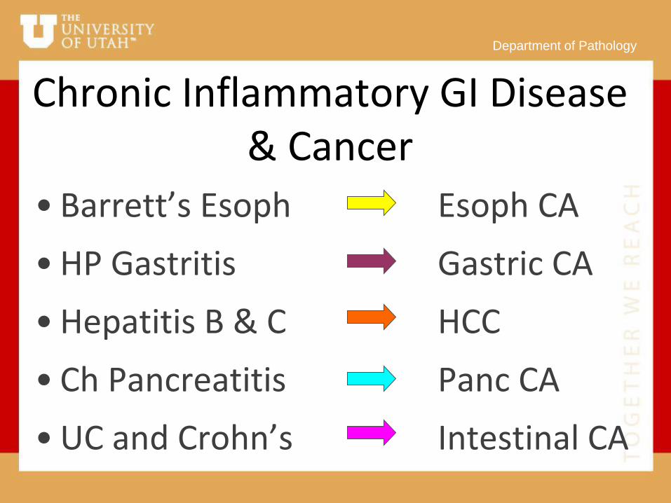
Chronic Inflammatory GI Disease & Cancer
• Barrett’s Esoph Esoph CA
• HP Gastritis Gastric CA
• Hepatitis B & C HCC
• Ch Pancreatitis Panc CA
• UC and Crohn’s Intestinal CA

Department of Pathology
Ulcerative Colitis: A Paradigm

Department of Pathology

Department of Pathology
Managing Cancer Risk in UC
•Ignore it
•“Prophylactic” colectomy
•Colonoscopic surveillance for dysplasia / early carcinoma

Department of Pathology
Optimal Colonic Biomarker
• Pancolonic distribution
• Predate incurable cancer
• Objective
• Sensitive, Specific, PPV, NPV


Gold Standard Biomarker: Dysplasia

Department of Pathology
Dysplasia: Problems • Sampling
• Distinction from reactive change
• Observer variation
• Natural history incompletely understood

Department of Pathology
Adequate Bx Sampling
Histology
From: Rubin CE, et al. Gastroenterology 1992;103:1611
Dysplasia Cancer No. Bx’s for 90% confidence 33 34 No. Bx’s for 95% confidence 56 64

Department of Pathology
UC Surveillance Protocol
10 cm
5 cm
X X
X X
X X
X X
X
X X
X
X X X
X X X
X X X
X X X
X X X
X X
X
X X
X X
X X X X
X X X
X X
X

Department of Pathology
Rectosigmoid Predominance of Ulcerative Colitis Cancer
Location of Colorectal Carcinoma
RS D T A/C
52% 12% 21% 15%
Choi PM. Gastroenterology 1993;104:666 Summary of 5 Studies

Department of Pathology
Dysplasia: Problems • Sampling
• Distinction from reactive change
• Observer variation
• Natural history incompletely understood

Department of Pathology

Department of Pathology

Department of Pathology
Dysplasia: Problems • Sampling
• Distinction from reactive change
• Observer variation
• Natural history incompletely understood

Department of Pathology
Outcome of 40 UC LGD Patients
• 78% no progression, avg f/u 5y (1-13 y)
• 22% HGD, avg f/u 1.5 y (1-3 y)
• ≥3 LGD biopsies: 9x progression risk
• 2 non-compliant patients developed Dukes’ A cancer
Brentnall, Bronner, et al. Prospective study of progression of LGD in UC.
Inflamm Bowel Dis 18:2240-6, 2012.

Dysplastic Field:
Limited

Department of Pathology
Better Biomarkers of
Cancer Risk Greatly
Needed!

Department of Pathology

Department of Pathology
Chromosomal Instability? • FCM Aneuploidy - Detects gross
chromosomal instability
• CGH - Detects clonal gains and losses of chromosomal regions
• FISH - Detects clonal and non-clonal chromosomal abnormalities

Department of Pathology
Biopsy Sampling: Flow Cytometry
Rubin CE, et al. Gastroenterology 1992;103:1611
Dysplasia Cancer
No. Bx for 90% confidence 20 8
No. Bx for 95% confidence 30 14

Morphologic
+ DNA Ploidy
Neoplastic Field:
Larger

Department of Pathology
Metaphase Comparative Genomic Hybridization in UC 39% (15/38) of diploid bx’s near dysplasia or cancer showed CGH detectable alterations Performed in collaboration with
F. Waldman, UCSF

Department of Pathology
Array-based Comparative
Genomic Hybridization (CGH) Chromosomes replaced
by ordered array of
targets
Karyotyping of
metaphase spreads not
necessary
Greatly increased
resolution

Department of Pathology
Array CGH in UC
• 100% (9/9) UC-progressors
extensive chromosomal gains and
losses
• FISH and PCR targets identified
Bronner MP, Mod Pathol 2010;23:1624-33

Department of Pathology
Ulcerative Colitis A-CGH
PROGRESSORS
NON-PROGRESSORS
Gain
Loss
Bronner MP, Mod Pathol 2010;23:1624-33.

Department of Pathology
BAC CGH Whole Genome Log2-Ratio Plots of All Chromosomes
Normal Non-UC Control UC Non-progressor
UC Progressor UC Progressor
Bronner MP, Mod Pathol 2010;23:1624-33.

Morphology
+ DNA Ploidy
+ CGH
Neoplastic Field:
Larger Still

Department of Pathology
Non-Clonal Change in UC: Wider Field?
• DNA Flow & CGH detect clonally expanded abnormalities only
• Larger fields of non-clonal instability? Detectable in negative biopsies, even from rectum?
• Assessed by Fluorescence In Situ Hybridization (FISH)?

Department of Pathology
UC FISH Hypothesis:
UC progressors differ from UC non-progressors using non-clonal
genomic instability biomarkers on single negative rectal biopsies

Department of Pathology
FISH • Interphase nuclear suspensions placed
on glass slide
• Locus specific probes (Chrom 8, 11, 17, 18) & centromeres (green and red)
• Red and green FISH spots counted per 100 nuclei

Normal Cells Abnormal Cells

Department of Pathology
Control Normal Colon FISH Chrom11 Probe Set
Red and Green Signal Counts
% o
f N
ucle
i
0r2g 1r1g 1r2g 1r3g 2r1g 2r2g 2r3g 2r4g 3r2g 3r3g
1 0 0 0 1 2 2 2 2 0
20
40
60
80
100 90

Department of Pathology
Diploid Neg Rectal Bx UC Progressor
Chrom11 Probe Set
Red and Green Signal Counts
% o
f N
ucle
i
0r2g 1r1g 1r2g 1r3g 2r1g 2r2g 2r3g 2r4g 3r2g 3r3g
4 2 1 0 1
8 11
0 0
74
0
20
40
60
80
100
Department of Pathology
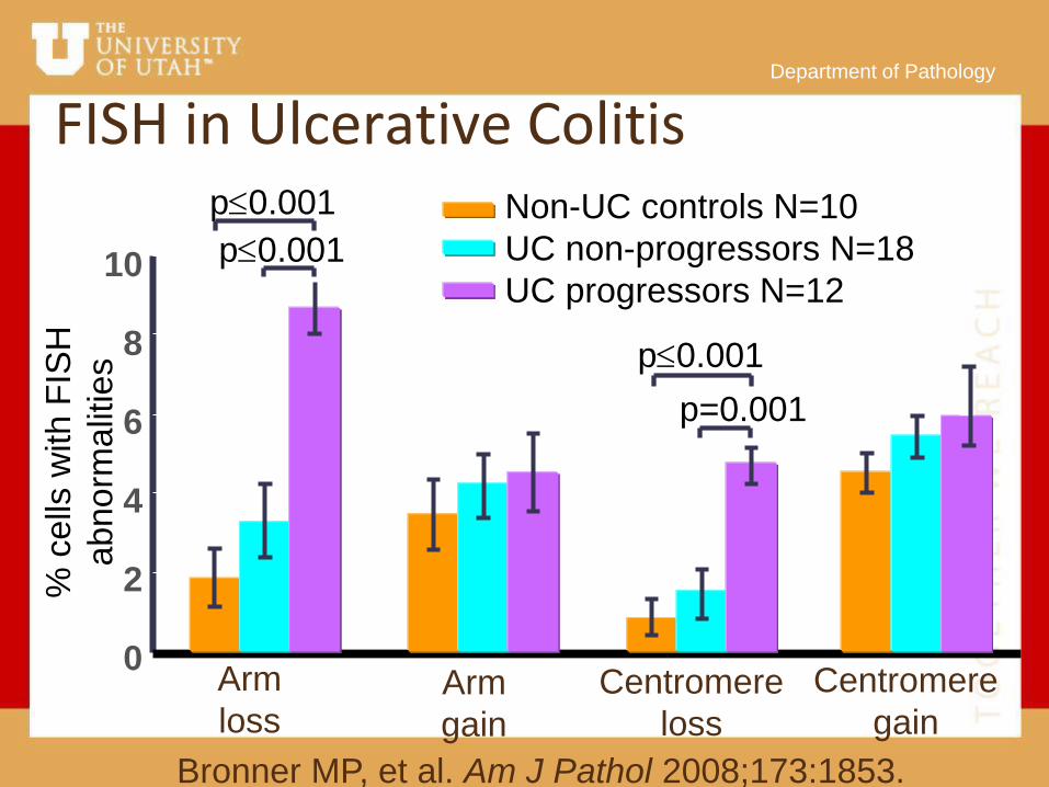
FISH in Ulcerative Colitis %
ce
lls w
ith
FIS
H
abnorm
alit
ies
Arm
loss
0
2
4
6
8
10
Arm
gain
Centromere
loss
Centromere
gain
p0.001
p0.001
p=0.001
p0.001
Non-UC controls N=10
UC non-progressors N=18
UC progressors N=12
Bronner MP, et al. Am J Pathol 2008;173:1853.

Department of Pathology
8q: c-myc
Specificity
Sen
sit
ivit
y
0.0 0.2 0.4 0.6 0.8 1.0
0.0
0
.2
0.4
0
.6
0.8
1
.0
11q: CyclinD1
0.0 0.2 0.4 0.6 0.8 1.0
0.0
0
.2
0.4
0
.6
0.8
1
.0
17p: p53
Specificity
Sen
sit
ivit
y
0.0 0.2 0.4 0.6 0.8 1.0
0.0
0.2
0
.4 0
.6
0.8
1
.0
1-Specificity 1-Specificity
Sen
sit
ivit
y
18q: DCC
Sen
sit
ivty
0.0 0.2 0.4 0.6 0.8 1.0
0.0
0
.2 0
.4 0
.6 0
.8 1
.0
1-Specificiy
ROC Analysis of FISH Biomarkers
Bronner MP, et al. Am J Pathol 173:1853-1860, 2008 1-Specificity

Department of Pathology
All 4 chromosomes combined
1-Specificity
Se
ns
itiv
ity
0.0 0.2 0.4 0.6 0.8 1.0
0.0
0
.2
0.4
0
.6
0.8
1
.0
Bronner MP, et al. Am J Pathol 2008;173:1853.
ROC Analysis of FISH Biomarkers

Department of Pathology
Consequences of Shortened Telomeres
• Sticky chromosomal ends
• Bridge-breakage-fusion cycles
• Chromosomal arm losses/gains and dicentrics
Studied by peptide nucleic acid (PNA)
probe ISH or RT PCR

Department of Pathology

Department of Pathology
Telomere Shortening in UC E
pith
elia
l:
Str
om
al T
elo
me
re R
atio
0
1.4
Non-UC
control
1.2
1.0
0.8
0.6
0.4
0.2
Non-
progressors
Progressors
p=0.08
p=0.001
p=0.02
O’Sullivan J, et al. Nat Genet 2002;32:280-284.

Anaphase Bridges in UC %
Ana
ph
ase
Brid
ge
s
0
0.03
Non-UC control
0.025
Non-progressors
Progressors
p=0.011
p=0.0002
0.02
0.015
0.01
0.005
Bronner MP, et al. Am J Pathol 173:1853-1860, 2008

Department of Pathology
NGS miRNA bioclassifier of UC patients at increased risk of colon cancer
• Why miRNAs?
– Small size (~21nt) more stable, less
ribonuclease degradation
– Readily detectable in FFPE and stained
slides
– Important roles in immune regulation

Department of Pathology miRNAs misregulation in UC-P, UC-NP
• Linear discriminant analysis to predict UC-P vs. UC-NP
• Robust candidate panel selected for RT-PCR & additional cohort validation
UC-NP vs. nl (26 miRNAs)
UC-P vs. nl (29 miRNAs)
11 18 15
No
rmal
ized
Rea
d c
ou
nt
110
100
1000
1000
0
UC_NP+UC_P UC_NP+UC_P UC_NP+UC_P UC_NP+UC_P UC_NP+UC_P
UC_NP
UC_P
110
100
1000
1000
0
UC_NP+UC_P UC_NP+UC_P UC_NP+UC_P UC_NP+UC_P UC_NP+UC_P
UC_NP
UC_P
mir
110
100
1000
1000
0
UC_NP+UC_P UC_NP+UC_P UC_NP+UC_P UC_NP+UC_P UC_NP+UC_P
UC_NP
UC_P
UC-NP 9/10
UC-P 10/10
miRNA
Panel

Histology
+ DNA Ploidy
+ CGH
+FISH
+Telomeres
+Ana Bridges
+miRNA
Neoplastic Field:
Entire Colon

Department of Pathology

Department of Pathology
UC Polypoid Dysplasia
You’re dalmed if you do, and dalmed if
you don’t
Teri Brentnall,MD

Department of Pathology

Department of Pathology
Dysplasia in UC vs Adenoma
•No clinical features
•No endoscopic features
•No pathologic features
•No molecular tests

Department of Pathology
HOWEVER • If the lesion can be demonstrably
completely removed endoscopically
• Has only Low-Grade Dysplasia
• There is no other dysplasia on adequate sampling
• Then, careful follow-up may be considered

Department of Pathology
UC Dysplasia Management Continue Surveillance with
adequate sampling:
–Single site LGD while in surveillance
–Indefinite of negative for dysplasia

Department of Pathology
UC Dysplasia Management Consider Colectomy:
–Multiple LGD sites
–LGD on more than one endoscopy
–LGD at initial colonoscopy
–Excessive inflammatory polyps

Department of Pathology
Inflammatory Polyps

Department of Pathology
UC Dysplasia Management
Colectomy Indicated:
–HGD
–Endoscopically unresectable dysplastic lesion

Department of Pathology
Conclusions • Molecular alterations are widespread in
UC, CD, CP, HP, HCV
• Single non-dysplastic bx alterations show
promise for reducing sampling error
• Paradigm for cancer in chronic
inflammatory disease

Department of Pathology
Further Work: • Reproducibility
• Longitudinal analyses
• Prospective validation
• High throughput
• Reduced numbers of markers
• Mechanism: why progressors?

Department of Pathology
Thanks To My Colleagues: Bonnie Shadrach
Teri Brentnall
Peter Rabinovitch
Ru Chen
David Crispin
Rosana Risques
Jacintha O’Sullivan
Noah Welker
Keith Lai
Danielle Elsberry
Ryan O’Connell
June Round
John Valentine

Department of Pathology
Recommended