Methods
29
I. AGROBACTERIUM TUMEFACIENS TRANSFORMATION
The LBA4404 strain of Argrobacterium tumefaciens (LBA 4404) carrying either of
the genes constructs plasmids were used to transform tobacco (Nicotiana.tabacum cv.
xanthi) and tomato (Lycopersicon estculentum cv. Money maker, Cheery tomato)
following the standard procedure described by Horsch et al., (1985).
Surface-sterilized leaf discs or other axenic explants were infected with the A.
tumefaciens carrying the vector of choice and co-cultured on regeneration medium for
2 to 3 days without any selection pressure. After the co-cultivation, the explants were
transferred to regeneration/selection medium. This contained 500 mg/L carbenicillin
(or 250 mg/l cefotaxime) to kill the bacteria and the appropriate antibiotic
(hygromycin / kanamycin depending on the construct used) to inhibit untransformed
plant cells. During the next 3 weeks, the transformed cells grew into callus or
differentiated into shoots via organogenesis. Within 3 to 6 weeks, the shoots
developed enough to remove them from the explants and induce rooting in
preparation for transfer to soil.
For tomato, seeds were sterilized by soaking in bleach solution for 20-30 minutes
followed by at least three rinses in sterile water, germinated on RM medium and grew
under moderate (room) light and temperature until cotyledons were fully expanded.
Cotyledons were removed by cutting at their petiole, and then cut in half to increase
the wound edge. Explants were precultured for 1 or 2 days upside down on RMOP
medium to allow initial growth and to eliminate those that were damaged during
sterilization or handling. Agrobacterium tumefaciens culture was grown overnight in
LB with appropriate antibiotics to select for the vector. The culture was prepared for
inoculation of explants by taking an overnight culture and diluting 1 to 10 times with
liquid RM medium to get O.D around 0.6. Explants were inoculated by immersion in
the culture of Agrobacterium tumefaciens and blotted dry gently as soon as all
wounded edges have contact the inoculums. The explants were placed upside down
Methods
30
on culture plates and incubated for two to three days. Transferred explants to RMOP
selection medium and incubated under light for 4 to 6 weeks. After 2-3 weeks, cut
explants to separate clearly independent sites of transformation and transfer to fresh
selection medium. Transferred entire explants to RM rooting medium as shoots
appeared, even if not suitable for removal of individual shoots from the explants.
When rooted, agar from base of plantlets was washed off and planted in sterile soil in
pots. Placed pots in transparent boxes and closed tightly to retain humidity. After 7 to
10 days, slowly crack opened to reduce the humidity gradually until plants are
acclimatised to the ambient humidity. Fertilized and grow under standard plant
growth conditions.
II. PARTICLE GUN MEDIATED TRANSFORMATION
Preparation of gold particles for shooting
Taken 50 mg of tungsten powder in a microcentrifuge tube, added 1 ml of freshly-
opened absolute ethanol, set water bath at 950C and kept the tungsten containing
microcentrifuge tube for 2 hours with intermittent inversion and tapping. Centrifuged
for 10 seconds at high speed in a microcentrifuge, discarded the supernatant.
Resuspended the tungsten particles in 1 ml of fresh ethanol and transferred into 15 ml
round bottom falcon plastic tube. Sonicated 3 times for 5 minutes each while keeping
on ice, transferred the tungsten suspension to an eppendorf tube and centrifuged 10
seconds at high speed. Discarded the supernatant and washed tungsten with sterile
double distilled water three times and resuspended the tungsten finally in 1 ml water.
Sample preparation Mixed the below components maintaining the order with slow vortexing before
adding the next component
1. 50µl tungsten solution
2. 10µl DNA in TE buffer (1µg/µl)
Methods
31
3. 50µl of CaCl2 (2.5M)
4. 20µl spermidine free base (0.1M)
Vortexed for 30 minutes in cold room and proceeded further with the sample
preparation (Keept the sample on ice throughout the preparation period). Added 200
µl ethanol, centrifuged 10 secs at 10,000 rpm and removed supernatant. Added 200µl
ethanol and resuspended very well by pipetting or mild vortexing, centrifuged 10 secs
and replaced the ethanol and repeated the above process 3 times. Resuspended the
final DNA coated tungsten in 30µl of ethanol. Used 5µl sample for shooting each leaf
or embryogenic calli
Coating DNA to microcarriers Aliquoted out 5 µl of DNA coated tungsten suspension onto macrocarrier disc and
allowed alcohol to evaporate in a sterile environment (a laminar flow hood).
Preparing rice embryogenic calli for bombardment
Dehulled the mature seeds and sterilized in 70% ethyl alcohol for 1 min and then in
45% chlorox (5-25% Na-hypochlorite solution) with 1-2 drops tween-20 for 30
minutes. Rinsed seeds with sterile distilled water 4-5 times and inoculated sterile
seeds in callus introduction medium RM with 2 mg 2,4 D/l. After 6-7 days, separated
the callus from scutellar tissue from the endosperm and inoculated them on callus
introduction medium. After 1 month collected callus and incubated in a gyratory
shaker at 90-120 rpm, subcutured the callus every week for first month and every two
weeks for next 5-6 months. Collected embryogenic callus and arranged these in a 1-
inch diameter circle at the centre of a Petri dish containing bombardment medium and
2 sheet of Whatman #4 circles paper (sterilized), then incubated the samples overnight
in dark.
Methods
32
Shooting rice embryogenic calli
Biorad shooting kit was used through out the study (500 Standard pressure kit cat No.
165-2283). Before shooting, sterilized the number of discs and stopping screens that
were need in a Petri dish containing absolute ethanol (100%) for 10 – 15 min.. The
discs and screens are dried by standing them up along the side of a sterile Petri dish
and allowed them to air-dry under sterile conditions in the laminar flow hood.
Bombarded the calli as per the procedure described in the Biorad PDS-1000 He
manual.
Transferred bombarded explants to RM2D (RM medium with 2,4-D 2 mg /l)
containing 50 mg hygromycin/l. Incubated in dark at 25oC for 3 weeks. Transferred
surviving embryogenic calli to RM2D containing the same concentration of antibiotic,
selected 3 times at 2 weeks interval. Transferred only the embryogenic calli that
showed fresh growth to the next step. Transferred developing pro-embryos to
generation medium with or without selection. Transferred the plantlets to rooting
medium (without hormones and antibiotic).
III. CONSTRUCTION OF PLASMID VECTORS AND DNA PURIFICATION
1. Plasmid DNA isolation (alkali method)
This method was adopted from Sambrook et al. (1989). Picked single colonies of
bacteria harboring the plasmid DNA of interest into a Flask containing of LB media
supplemented with the appropriate antibiotic and incubated at 37oC for overnight with
shaking at 250 rpm. Harvested the cells by centrifugation at 5000 rpm for 5 minutes
and decanted the supernatant. The pellet was suspended in 1.5 ml of solusion I
(25mM Tris-HCL, pH 8.0, 10mM EDTA, 50 mM Glucose) mixed, and incubated for
5 minutes on ice. Added 2 ml of freshly prepared solution II (0.2 N NaOH, 1% SDS),
mixed by inverting and incubated at room temperature for 5 minutes. Added 1.5 ml of
Methods
33
solution III (3M potassium Acetate, pH 4.8), mixed by inverting and incubated on ice
for 20 minutes. Centrifuged at 12,000 x g for 10 minutes at 4oC, the supernatant was
collected and treated with RNase A (10 mg/ml) at 37oC for 30 minutes. The
supernatant containing plasmid was extracted twice with phenol: chloroform: isoamyl
alcohol (24:24:1). Precipitated the DNA by adding 2 volumes of 95% ethanol or by
0.7 volume isopropanol and resuspended the dried DNA pellet in TE buffer.
2. Plasmid DNA purification by Qiagen column
After the cells were harvested by centrifugation at 5000 rpm for 5 minutes, resuspend
the bacterial pellet in 4 ml of buffer P1 premixed with RNase A. Added 4 ml of buffer
P2, mixed gently by inverting 4-6 times and incubated at room temperature for 5
minutes. Added 4 ml of buffer P3, mixed immediately but gently and incubated on ice
for 15 minutes. Centrifuged 12,000 rpm for 30 minutes at 4oC. Equilibrated a
QIAGEN-tip 100 by applying 4 ml of buffer QBT and allowed the column to empty
by gravity flow. Applied the supernatant from centrifuge to the QIAGEN-tip and
entered the resin by gravity flow. Washed the QIAGEN-tip with 2 x10 ml of buffer
QC. Eluted DNA with 5 ml of buffer QF and precipitated DNA with 0.7 volumes of
room temperature isopropanol. Centrifuged immediately at 12,000-15,000 x g for 30
minutes at 4oC and carefully removed the supernatant. Washed DNA with 2 ml of 70
% ethanol, air dried for 5 minutes and redissolved in a suitable volume of TE buffer.
3. Restriction digestions
Restriction enzyme digestions are performed by incubating double-stranded DNA
molecules with an appropriate amount of restriction enzyme, in its respective buffer
as recommended by the supplier, and at the optimal temperature for that specific
enzyme. Typical digestions included a unit of enzyme per microgram of starting DNA
for 1 hour to overnight depend on enzyme to insure complete digestion. The volume
of the reaction depended on the amount and size of the DNA being digested. Larger
Methods
34
DNA amounts were digested in larger volumes (between 50-100 µl). For double
digestion, a suitable buffer was used based on suppliers instructions.
4. Agarose gel electrophoresis
Agarose gel electrophoresis is employed to check the progression of a restriction
enzyme digestion, to quickly determine the yield and purity of a DNA isolation or
PCR reaction, and to size fractionate DNA molecules, which then could be eluted
from the gel or used for membrane transfer. Prior to gel casting, dried agarose is
dissolved in buffer by heating and the warm gel solution then is poured into a mold,
which is fitted with a well-forming comb. The percentage of agarose in the gel varied.
Although 0.7% agarose gels (in 1X TEA or TEB buffer) typically are used, in cases
where the accurate size fractionation of DNA molecules smaller than 1 kb is required,
a 1, 1.5, or 2% agarose gel is prepared, depending on the expected size(s) of the
fragment(s). Ethidium bromide is included in the gel matrix to enable fluorescent
visualization of the DNA fragments under UV light. Agarose gels are submerged in
electrophoresis buffer in a horizontal electrophoresis apparatus. The DNA samples are
mixed with gel tracking dye and loaded into the sample wells. Electrophoresis usually
is at 70 - 100 volts for 0.5-1 hour at room temperature, depending on the desired
separation. Size markers were co-electrophoresed with DNA samples, often λ DNA
digested with HindIII, was used when appropriate for fragment size determination.
After electrophoresis, the gel was placed on a UV light box and a picture of the
fluorescent ethidium bromide-stained DNA separation pattern was photographed with
a camera (gel documentation system).
5. Elution of DNA fragments using Qiaquick gel extraction columns
The agarose gel piece containing DNA fragment was taken in a microcentrifuge tube,
added 3 volumes of QG buffer as supplied with QIAQUICK gel extraction kit and
dissolved by heating 60oC for 15 minutes. The mixture was loaded on to QIAQUICK
spin column and spun briefly. The flow through was discarded and the column was
Methods
35
washed twice with buffer PE. The purified DNA fragment was eluted with 50 µl of
TE buffer.
6. DNA ligation
DNA ligations were performed by incubating DNA fragments with appropriate
linearized cloning vector in the presence of a buffer and T4 DNA ligase. For blunt end
ligations or single digestion, dephosphorylated double-stranded DNA linearized
vector with Alkaline Phosphatase (AP) was used. These usually included parallel
ligations in the absence of insert DNA to determine the background clones arising
from self-ligation of inefficiently phosphatased vector. Parallel ligations also were
performed with a known blunt-ended insert to insure that the blunt-ended ligation
reaction would yield sufficient insert containing clones, independent of the repair
process. Combined the following reagents in a microcentrifuge tube and incubated
overnight at 4oC to 16oC. A typical reaction included the following:
DNA fragments 100-1000 ng
Cloning vector 2 µl (10 ng/µl)
10X ligation buffer 1 l (or 2 µl)
T4 DNA ligase (NEB 202L) 1 µl (400 U/µl)
sterile ddH2O to 10 µl (or 20 µl)
Depend on concentration of vector DNA and fragment, volume was adjusted for
ligation reaction.
7. E.coli competent cell preparation and transfer of plasmid DNA
For the calcium chloride method, a glycerol cell culture stock of the respective E. coli
strain DH5α was thawed and added to 5 ml of liquid media. This culture then was
preincubated at 37oC for overnight, transferred to flask containing 50 ml LB and
incubated at 37oC on incubator-shaker for 2-3 hours. The cells were pelleted by
centrifugation, resuspended in calcium chloride solution, and incubated in an ice-
Methods
36
water bath. After another centrifugation step, the resulting cell pellet again was
resuspended in calcium chloride with 12.5 % glycerol to yield the final competent cell
suspension. Competent cells are stored at -80oC.
In detail, thawed a frozen glycerol stock of the appropriate strain of E. coli, added it to
a falcon containing 5 ml of LB media, and pre-incubated at 37oC overnight with
shaking. Inoculated 5ml of overnight culture in Erlenmeyer flask containing 50 ml LB
for 2-4 hours incubated at 37oC with shaking at 250 rpm. Transferred 40 ml of the
cells to a sterile 50 ml polypropylene centrifuge tube, and collected the cells by
centrifugation at 3000 rpm for 10 minutes at 4oC. After centrifugation, decanted the
supernatant and resuspended the cell pellet in 20 ml of cold, sterile 50 mM calcium
chloride, incubated in an ice-water bath for 20 minutes, and centrifuged at 3000 rpm
for 10 minutes at 4oC. Decanted the supernatant and gently resuspended the cell pellet
in 4 ml of cold, sterile 50 mM calcium chloride (with 12.5% glycerol) to yield the
final competent cell suspension. The competent cells then should be placed at -70oC
and can be stored indefinitely
To use competent cells for transformation, removed from freezer and thawed for a
few minutes at 37oC, placed on ice, added plasmid DNA or ligation reaction and
incubated for 15-20 minutes. Heat shock was given at 42oC for 2 minutes and cooled
briefly, added 0.7 ml of LB and incubated for 1 hour at 37oC before spreading on
selection LB plates with supplement of appropriate antibiotic.
8. Agrobacterium competent cell preparation and transfer of plasmid DNA
Inoculated colony in 2 ml YEM with streptomycin (25mg/l) and incubated overnight
at 30oC shaker. Transferred culture to 200 ml YEM in a sterile 500ml flask and kept
at 30oC with shaking at 250 rpm until the OD reached 0.3 (4-5 hours). Spun in sterile
cap tubes 3000 rpm at 4oC for 10 minutes, checked to make sure cells are pelleted, if
not repeated at higher speed. Aspirated supernatant, resuspended pellet in 20ml ice
cold 0.1M calcium chloride solution (filter sterile), respun and resuspended pellet in
Methods
37
2ml ice cold sterile 0.1M calcium chloride (with 12.5% glycerol). Dispensed in 50µl
aliquots in pre-chilled, sterile microcentrifuge tubes, frozen in liquid nitrogen and
stored at -70oC.
To use competent cells for transformation, removed from freezer and thawed for a
few minutes at 37oC, placed on ice, added plasmid DNA and incubated was given for
15-20 minutes as in the standard transformation procedure. Cool shock was given in
liquid nitrogen for 2 minutes, 5 minutes at 37oC and cooled briefly on ice, added 0.7
ml of LB and incubated for 1 hour at 30oC before spreading on selection LB plates
with supplement of appropriate antibiotic.
IV. GUS ASSAY – HISTOCHEMICAL
Solutions
1. 0.5 M MES (2-[N-Morpholino] ethanesulfonic acid) pH 5.6
For 100 ml: dissolve 9.76 g of MES (Sigma) in 80 ml double H2O. Adjusted
pH to 5.6 with NaOH and made up the volume. Stored at room temperature.
2. 50 mM Sodium phosphate buffer (Na PO4) pH 7.0
57.7 ml of Na2HPO4 (1 M stock)
42.3 ml of NaH2PO4 (1 M stock)
Distilled water to 2000 ml
3. Histochemical reagent [2 mM 5-bromo-4-chloro-3-indolyl glucuronide (X-
Gluc) in 50 mM NaPO4, pH 7.0]
For 10 ml: Dissolved 10 mg of X-Gluc in 100 µl of dimethyl formamide in a
pyrex tube. Made up to 10 ml with 50 mM NaPO4 pH 7.0. Made up fresh
when required. Stored solid X-Gluc-desiccated at -20oC or -70oC.
4. 70% Ethanol
Placed the tissue in fixation solution (formaldehyde 0.2% in 50 mM NaPO4 pH 7.0)
and vacuum infiltrated tissue briefly (2 min) and incubated for 4-5 min at room
Methods
38
temperature followed by several washes (3 times) in 50 mM NaPO4 pH 7.0. Added
100 µl of X-gluc and incubated for 2 to 4 hours (or overnight) at 37oC. After staining,
rinsed sections in 70% ethanol for 5 minutes and then mounted on microscope slides
or visualized directly in a Petri plate under miscropse.
V. GUS ASSAY-SPECTROFLUOMETER
GUS extraction buffer
50 mM Na2HPO4, pH 7.0
10 mM beta-mercaptoethanol
10 mM EDTA
Assay buffer (MUG)
1mM MUG in extraction buffer (Dissolve 22 mg 4-methyl-umbelliferyl-beta-D-
glucuronide in 50 ml GUS extraction buffer).
Stop Buffer
0.2 M Na2CO3. Dissolve 21.2 gm Na2CO3 in 1L of distilled water.
Calibration standards
1 mM methylumbelliferone (MU). Dissolved 19.8 mg methylumbelliferone in 100 ml
distilled water and wraped bottle in aluminium foil and stored at 4oC.
+ 100 µM MU: add 100 µl of 1 mM stock solution to 900 µl stop buffer, mix well.
+ 10 µM MU: add 100 µl of 100µM to 900µl stop buffer, mix well.
+ 1µM MU: add 100 µl of 10 µM MU to 900µl stop buffer, mix well.
Extraction and assay
Homogenized tissue (100 mg) in 300 µl of GUS extraction buffer and centrifuged at
12,000 rpm. Quantified the protein. Added 1-5 µg of protein to 1 ml assay buffer.
Mixed thoroughly with pipet or vortexed. At defined time intervals (0, 15, 30, 60, 90
Methods
39
min), removed 200 µl aliquots into Eppendorf tubes containing 800 ml stop buffer.
Read fluorescence with excitation at 365 nm emission at 455 nm in DNA
FLUOROMETER (model TKO100).
Calibration
Took 1 ml stop buffer and set the reading to `0’ (zero). Added 1 ml of 1000 nM MU
into cuvette and set the reading to 1000. Read the sample. The value get was
expressed as nM of MU due to GUS activity in the sample.
Quantitation of proteins (Bradford method)
Materials
1. 0.5 mg/ml bovine serum albumin (BSA)
2. 0.15 M NaCl
3. Bradford reagent from BioRad
Aliquoted 0, 5, 10, 15 and 20 µl of 0.5 mg/ml BSA and added 0.15 M NaCl to bring
the volume to 100 µl in each tube. Added 1 ml Bradford reagent (five times diluted)
and vortexed, waited 2 min at room temperature. Determined the OD at 595 nm using
spectrophotometer and made a standard curve by plotting absorbance versus protein
concentration. Determined the absorbance for the unknown and used the standard
curve to determine the concentration of protein in the unknown sample.
VI. PCR ANALYSIS
Standard procedures were followed for PCR amplification of DNA fragments. The
general procedure followed is given below:
A. Template DNA 2 µl (contained about 0.5 µg of DNA)
B. Taq Buffer (10 X) 5 µl
C. dNTPs (1mM) 5 µl
D. Primer GUS 5’ 1 µl (10-25 pmoles)
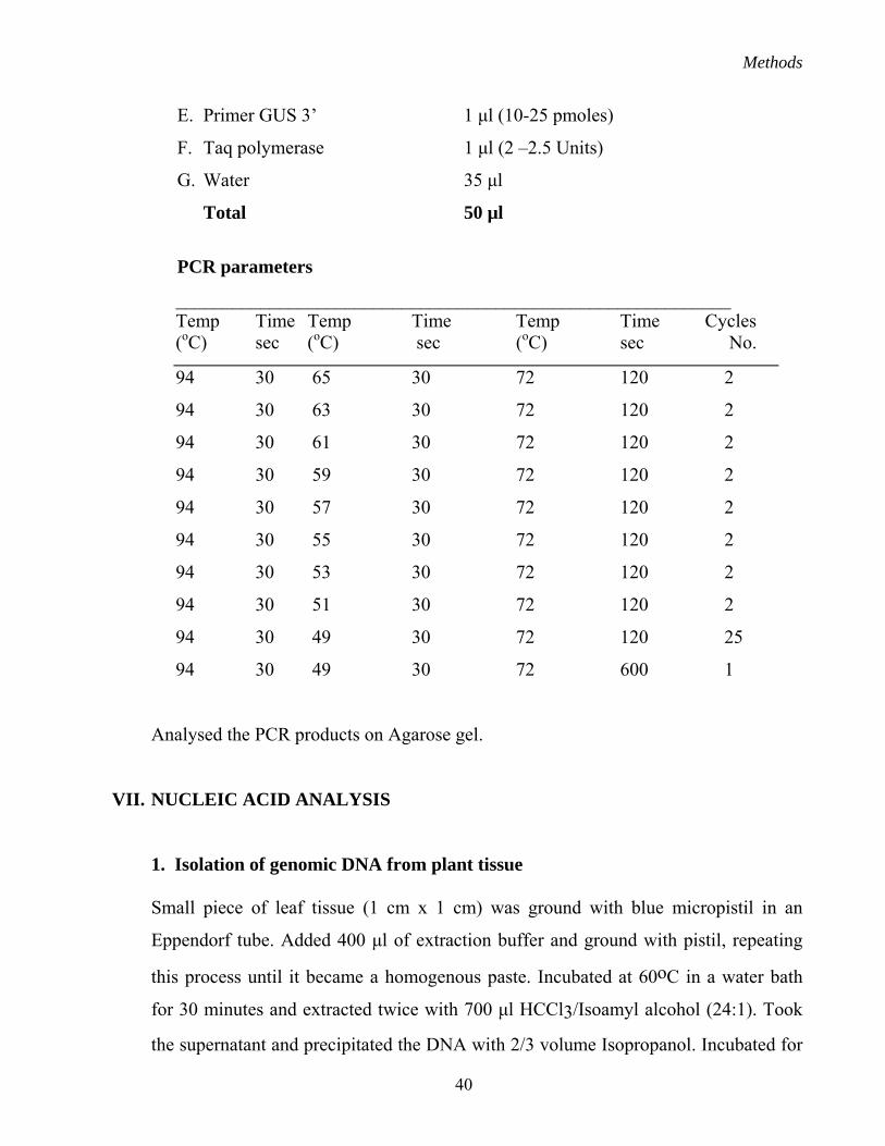
Methods
40
E. Primer GUS 3’ 1 µl (10-25 pmoles)
F. Taq polymerase 1 µl (2 –2.5 Units)
G. Water 35 µl
Total 50 µl
PCR parameters
___________________________________________________________ Temp Time Temp Time Temp Time Cycles (oC) sec (oC) sec (oC) sec No.
94 30 65 30 72 120 2
94 30 63 30 72 120 2
94 30 61 30 72 120 2
94 30 59 30 72 120 2
94 30 57 30 72 120 2
94 30 55 30 72 120 2
94 30 53 30 72 120 2
94 30 51 30 72 120 2
94 30 49 30 72 120 25
94 30 49 30 72 600 1
Analysed the PCR products on Agarose gel.
VII. NUCLEIC ACID ANALYSIS
1. Isolation of genomic DNA from plant tissue
Small piece of leaf tissue (1 cm x 1 cm) was ground with blue micropistil in an
Eppendorf tube. Added 400 µl of extraction buffer and ground with pistil, repeating
this process until it became a homogenous paste. Incubated at 60oC in a water bath
for 30 minutes and extracted twice with 700 µl HCCl3/Isoamyl alcohol (24:1). Took
the supernatant and precipitated the DNA with 2/3 volume Isopropanol. Incubated for
Methods
41
30 minutes at -20oC, spun for 3 minutes with full speed in microcentrifuge at room
temperature. Washed the pellet with 70% ethanol 3 times. Resuspended the DNA in
30-50 µl of T.E buffer or H2O.
Extraction Buffer 100 ml
2% CTAB 2 grams
1.4 M NaCl (28 ml of 5M)
20 mM EDTA (pH8) (4 ml of 0.5M)
100 mM Tris/HCl (pH8) (10 ml of 1M)
100 mM bME (fresh) 1ml neat
2. Isolation of total RNA from plant tissue
All the plastic and glass ware was treated with DEPC and autoclaved to eliminate
possible contamination of RNAse. Powdered 0.25g of leaf tissue in a mortar pestle in
liquid nitrogen, added 0.5 ml extraction buffer and allowed frozen tissue to thaw and
transferred into an eppendorf tube. Added 0.5ml phenol preheated to 70oC to the
powdered tissue, vortexed for 30 seconds. Centrifuged for 5 min at 14,000 rpm to
separate phenol. Transfer the supernatant to a clean eppendorf tube. Re-extracted
phenol with 0.5 ml extraction buffer. Centrifuge for 5 minutes at 14,000 rpm to
separate phenol from extraction buffer. Combined supernatants in a clean tube from
the two extraction steps. Added 0.5 ml of phenol-chloroform to the supernatant.
Vortexed 20 seconds and separated phases by centrifugation for 5 minutes at 4oC.
Transferred the supernatant to a fresh eppendorf tube and added 1/3 volume of 10M
LiCl (Ratios of supernatant: 10M LiCl 600 µl: 200 µl; 700 µl: 233 µl; 800 µl: 267 µl),
Mixed and precipitated overnight at 4oC. Centrifuged at 14,000 rpm for 10 min at
4oC, washed the pellet with 2.5M LiCl solution at 4oC. Washed the pellet with 70%
ethanol (made in DEPC treated water) at 4oC. Vacuum dried the RNA and dissolved
in sterile DEPC treated water (0.1 ml).
Methods
42
- RNA Extraction Buffer (Final Concentrations) 0.2M Sodium Acetate (pH 5.2) 1% SDS
0.01M EDTA (pH 8.0)
- Composition of RNA extraction Buffer (500 ml)
40.67 ml Sterile Water
3.33 ml 3M Sodium Acetate (pH 5.2)
5.0 ml 10% SDS
1.0 ml 0.5M EDTA (pH 8.0)
- Phenol-saturated with Tris.Cl (pH 8.0)
- Phenol-chloroform
- 10M LiCl sterilized by autoclaving
- 2.5M LiCl sterilized by autoclaving
Using TRIzol Reagent method (Gibco BRL)
Homogenized tissue samples in 1ml of TRIzol Reagent per 50-100 mg of tissue using
glass homogenizer. Following homogenization, insoluble material from the
homogenate was removed by centrifugation at 12,000 x g for minutes at 2 to 8oC. The
supernatant contained RNA. Incubated the homogenized samples for 5 minutes at 15
to 30oC to permit the complete dissociation of nucleoprotein complexes. Added 0.2
ml of chloroform per 1ml of TRIzol Regeant. Shaken tubes vigorously by hand for 15
seconds and incubated them at 15 to 30oC for 2-3 minutes. Centrifuged the samples at
no more than 12,000 x g for 15 minutes at 2-8oC. Transferred the aqueous phase to the
fresh tube, precipitated the RNA from the aqueous phase by mixing with isopropyl
alcohol (0.5 ml for 1ml TRIzol Regeant). Removed the supernatant and washed the
RNA pellet once with 75% ethanol, mixed the sample by vortexing and centrifuged at
7,500 x g for 5 minutes at 2 to 8 oC.
Methods
43
3. Spectrophotometric estimation of nucleic acids
The quantity and quality of DNA and RNA were determined by measure the
absorbance at 260 nm and 280 nm. It was calculated OD=1 for 50 µg/ml for DNA and
40 µg/ml for RNA (A260). The purity of DNA and RNA were determined by
calculating the ratio of A260/A280.
4. Southern hybridization
The genomic DNA (30 µg) was digested overnight with restriction enzymes. The
digested products were run on a 0.8 % agarose gel in 1X TBE, pH 8.3 containing
ethidium bromide (0.5 µg/l) at 40 volts for approximately 10-15 hours. The gel was
visualized and photographed. For breakage of large DNA in gel by depurination,
placed the gel in a tray and added 250 ml of 0.2 N HCl at room temperature. Rocked
occasionally for 15 minutes, decanted the acid, and repeated in two times. Rinsed with
water briefly and proceeded immediately to the next step of alkaline denaturation:
Added 250 ml of 0.5 M NaOH and 1.5 M NaCl and gently agitated for 15 minutes.
Decanted alkali and repeated one more time. Neutralized gel with 250 ml of 1 M
ammonium acetate for 10 minutes, repeated one more time and transferred to
membrane.
5. Northern blotting
The agarose is melted by boiling in 10 mM sodium phosphate buffer, pH 6.8
containing 1 µl of 10 mg/ml ethidium bromide per 100 ml of buffer, then cooled to
60o C and poured in the casting tray. In a 1.5 ml sterile Eppendorf tube 30 µl of the
RNA dissolved in sterile H2O was mixed with 2 µl of sterile 6X loading buffer.
6X loading buffer
0.25% (w/v) bromophenol blue
0.25% (w/v) xylene cyanol
30% (w/v) glycerol
Methods
44
1.2% SDS
60 mM sodium phosphate (pH 6.8)
The mixture was incubated at 75oC for 5 minutes followed by immediate loading
of the sample to a submarine gel. When analyzing many samples, the denatured
RNA can be placed on ice before loading on a gel. The gel was electrophoresed at
3 to 7 V/cm in 10 mM sodium phosphate buffer, pH 6.8, containing 1 µl of 10
mg/ml ethidium bromide per 100 ml of buffer. Because the buffering capacity of
the electrophoretic buffer was relatively weak due to its low ionic strength,
constant recirculation of the buffer was maintained to prevent the formation of an
undesirable pH gradient which can lead to degradation of the RNA during
electrophoresis. The migrating RNA in the gel was visualised with medium-wave
UV light to verify the migration and integrity of the RNA.
Pre hybridization
Placed DNA bound gene screen plus paper in a heat sealable plastic bag
(hybridization bag). Added approximately 20 ml (~70 µl/cm2) of hybridization
buffer.
Prehybridization buffer (1000 ml)
20 X SSC 250 ml
50% Dextran sulphate 100 ml
1.0 M sodium phosphate pH 7.2 50 ml
50 X Denhardts solution 100 ml
0.5 M EDTA 5 ml
20% SDS 20 ml
Distilled water 475 ml
1000 ml
Divided into 100 ml aliquots and store at –20oC
Methods
45
Added 200 µl (5 mg/ml) heat denatured salmon sperm DNA to the hybridization
bag, mixed well and sealed the bag. Removed all air bubbles before sealing the
bag and prehybridized at 65o C for 3-8 hrs.
Hybridization
Added ~108 dpm denatured DNA probe (nick-translated 32p following the Amersham
DNA labelling kit) heat sealed the bag. Placed sealed bag into a second bag and heat
sealed. Wet paper towel placed in the second bag helped prevent drying of the
membrane.
Washing of membrane
Hybridized for 8-24 hours at 65o C in a water bath shaker. Cut open bag and removed
membrane. Washed 2 times for 10 minutes in 250 ml of 2X SSC + 0.1% SDS at room
temperature. Washed 2 times for 15 minutes in 250 ml of 0.5X SSC + 0.1% SDS at
65oC in a water bath shaker. Wash 2 times for 8 minutes in 250 ml of 2X SSC at room
temperature.
20X SSC (1000 ml)
NaCl 175.3 g
Sodium citrate 88.2 g
Dissolved in 800 ml of water. Adjusted pH to 7 with NaOH or HCl and made up the
volume to 1000 ml.
Autoradiography of 32P on membrane
Covered the membrane in Saranwrap and exposed to X-ray film over night by placing
in side a cassette at –70 o C. To develop the film, brought the cassette to room
temperature before developing. This was to prevent condensation on film and damage
to intensifying screen. Developed the X-ray film: 1 minute in Kodak GBX 20%
Methods
46
developer solution, rinse in water and 3 minute in Kodak GBX 20% fixer solution.
Hung firm to dry.
6. cDNA for primer extension and Real time PCR (Invitrogen kit)
A 20 µl reaction volume can be used for 1ng to 5µg of total RNA or 1ng-500 ng of
RNA. Added the following components to a nuclease-free microcentrifuge tube: 1µl
Oligo (dT)12-18 or gene specific primer, 1 ng to 5 µg total RNA, 1µl 10 mM dNTP
Mix (10 mM each dATP, dGTP, dCTP and dTTP at neutral pH) and distilled water to
make up the volume to 12µl. Heated mixture to 65oC for 5 minutes and quick chilled
on ice. Collected the contents of the tube by brief centrifugation and added: 4µl 5X
First-Strand Buffer, 2µl 0.1 M DTT and 1 µl RNAseOUT Recombinant Ribonuclease
Inhibitor (40 Units/µl). Mixed contents of the tube gently and incubated at 42 oC for 2
minutes. Added 1 µl (200 Units) of SuperScript II (Reverse transcriptase), mixed by
pipetting gently up and down. Incubated 50 minutes at 42oC. Inactivated the reaction
by heating at 70oC for 15 minutes. The cDNA can now be used as a template for
amplification in PCR. However, amplification of some PCR targets (>1kb) could
require the removal of RNA complementary to the cDNA. To remove complementary
RNA, added 1 µl (2 units) of E.coli RNase H and incubated at 37oC for 20 minutes.
7. Transcription initiation analysis
Primer extension was performed using preamplification kit (Invitrogen). Reaction was
carried out with 10 µg of total RNA using the GUS internal primer. Primer was
labeled with (gamma 32P) ATP using T4 polynucleotide kinase (Promega). The size of
the extension product was determined by comparison with the DNA sequence
generated using the same primer and pITB450 DNA (Sequenase II kit, USB).
Methods
47
8. Induction and analysis of GUS expression
Tetracycline (Tc,1 mg/L) was used for the induction of GUS expression in single
leaves and in in vitro grown plants. Kinetics of induction was followed by real time
PCR and by quantifying the GUS activity in the Tc-treated and untreated leaf samples
at defined time periods.
9. Real time PCR
The RNA isolated from various explants were converted into cDNA using
Preamplification Superscript kit (Invitrogen) and poly dT oligonucleotide. GUS
specific beacon primers and cDNA as a template was used to detect the GUS
expression. Amplification of actin as a control for cDNA amounts was also included
in the same PCR reactions.
10. DNA sequencing.
Sequencing of recombinant clones was done either by sequenase version 2.0 kit
(USB-Amersham, Life Sciences), which was based on Sanger’s dideoxy chain
termination methods. Plasmid DNA was denatured with 0.2 M NaOH and 0.2 mM
EDTA at room temperature for 10 minutes. The denatured plasmid was purified by
passing it through TE-equilibrated G-50 column made in a 500 µl effendorf tube and
centrifuged at 3000 rpm for 3 minutes at room temperature. Annealing of the primers
to the denatured DNA was done at 37 oC for 15 minutes. This was followed by a
labelling reaction, which was performed essentially as described by the supplier
(USB-Amersham, USA) in the presence of S35-dATP and incubated at 37oC for 10
minutes. Termination of the reaction was done by the addition of respective
dideoxynuclotides and further incubation at 37oC for 5 minutes. Stop buffer was
added and the reaction mix was boiled, cooled and loaded onto 6% urea-acrylamide
TBE gel pre-warmed to 55oC. Electrophoresis was carried out at constant power (70
Methods
48
watts) to maintain the gel temperature at 55oC. Three loading were done to completely
resolve the nucleotide sequence. After completion of the run, the gel was transferred
to a Whatman sheet and dried in vacuum heating drier. The dried gel was exposed to
autoradiography film at room temperature.
VIII. WESTERN BLOTTING AND SDS-POLYACRYLAMIDE GEL
ELECTROPHORESIS (PDS-PAGE)
Homogenized leaf tissue (100 mg) in 300 µl of GUS extraction buffer as described in
GUS assay protocol. Quantified the protein and loaded 20-50 µg of protein per
sample.
Preparation of separating gel
In a 25-ml sidearm flask, mixed 30% acrylamide/0.8% bisacrylamide solution,
4xTris-Cl/SDS, pH 8.8 (see reagents, below), and 3.05 ml H2O. Degassed under
vacuum 10 to 15 min. Added 0.025 ml of 10% ammonium persulfate and 0.005 ml
TEMED. Swirled gently to mix and use immediately.
Reagents used in gels
30% acrylamide/0.8% bisacrylamide
Mixed 30.0 g acrylamide and 0.8 N,N’-methylene-bisacrylamide in a total volume of
100 ml H2O. Filtered the solution through a 0.45-mm filter and stored at 4oC in the
dark.
4xTris-Cl/SDS, pH 6.8 (0.5 Tris-Cl containing 0.4% SDS)
Dissolved 6.05 g Tris base and 0.4 g SDS in 40 ml H2O. Adjusted to pH 6.8 with 1 N
HCl, added H2O to 100 ml total volume and filtered the solution through a 0.45-mm
filter and stored at 4oC.
Methods
49
4xTris-Cl/SDS, pH 8.8 (1.5 M Tris-Cl containing 0.4% SDS)
Dissolved (1 g Tris base and 2 g SDS in 300 ml H2O. Adjusted to pH 8.8 with 1 N
HCl. Added H2O to 500 ml total volume. Filter the solution through a 0.45-mm filter
and store at 4oC.
Bromphenol blue solution
20 ml glycerol
4 mg Bromphenol blue
Add H2O to 100 ml
5xSDS/electrophoresis buffer
15.1 g Tris base
72.0 g glycine
5.0 g SDS
Add H2O to 1000 ml
Do not adjust the pH of the stock solution, since the pH is 8.3 when the solution is
diluted to 1x for use in the protocol.
4X Sample Loading Buffer
To 20 ml H2O add:
1.52 g Tris Base
20 mL Glycerol
2.0 SDS
2.0 mL β-mercaptoethanol
adjusted pH 6.8 with HCl and add H2O to 50 ml
The gel was made and then run until the Bromphenol Blue tracking dye has reached
the bottom of the separating gel.
Methods
50
Transfer of proteins to hybond C+ membrane
Cut two pieces of Whatman 3MM filter paper, cut to the same size as the gel and
prewetted with electroblotting buffer. Placed the prewetted filter, gel, nitrocellulose
membrane and again filters sequentially from the cathode. Care was taken to remove
air bubbles after each step by gently rolling a glass tube on the set-up. Placed this
sandwich into a plastic support. Placed the support containing the sandwich into the
electroblotting apparatus in the correct orientation. Filled the tank with electroblotting
buffer. Connected the leads of the power supply to the corresponding and cathode
sides of the electroblotting apparatus. Electrophoretically transferred the proteins from
the gel to the nitrocellulose at 100 mA constant current 1 hour.
Western blotting analysis Following transfer of proteins (Western) onto nitrocellulose or nylon membrane,
washed the membrane in 40 ml of TSW buffer for 1 hour. Repeated the washing for
30 minutes. To the 40 ml buffer, added 50 µl of rabbit anti-β-glucoronidase serum.
Incubated with gentle shaking for 1 hour. Decanted solution and wash with 20 ml of
TSW buffer for 30 minutes. To 50 ml of TSW buffer, add 5 µl goat anti-rabbit
alkaline phosphatase conjugate as a secondary antibody (concentration: 1 mg/ml).
Incubate with gentle shaking for 30 minutes. Decanted solution and washed with 20
ml of TSW buffer for 30 minutes. Replaced TSW buffer with the following buffer;
100 mM Tris-HCl, pH 9.5, 100 mM NaCl and 5 mM MgCl2. Added 33 µl NBT (Nitro
blue tetrazolium, 50 mg/ml in 70% Dimethylformamide) and 16.5 µl BCIP (5-bromo-
4-chloro-3-indolyl-phosphate, 50 mg/ml in Dimethylformamide) in 5 ml of the TSW
buffer. Full colour development should occur within 30 minutes.
Composition of TSW buffer: 10 mM Tris-HCl, pH 7.4 0.9% NaCl 0.25% Gelatin 0.1% Triton X-100 0.02% SDS
Recommended