
Purdue UniversityPurdue e-Pubs
Department of Entomology Faculty Publications Department of Entomology
8-16-2011
Hessian Fly-Associated Bacteria: Transmission,Essentiality, and Composition.Raman BansalKansas State University
Scot HulbertWashington State University
Brandi SchemerhornPurdue University
John C. ReeseKansas State University
R Jeff WhitworthKansas State University
See next page for additional authors
Follow this and additional works at: http://docs.lib.purdue.edu/entmpubs
This document has been made available through Purdue e-Pubs, a service of the Purdue University Libraries. Please contact [email protected] foradditional information.
Recommended CitationBansal, Raman; Hulbert, Scot; Schemerhorn, Brandi; Reese, John C.; Whitworth, R Jeff; Stuart, Jeffrey J.; and Chen, Ming-Shun,"Hessian Fly-Associated Bacteria: Transmission, Essentiality, and Composition." (2011). Department of Entomology FacultyPublications. Paper 5.http://dx.doi.org/10.1371/journal.pone.0023170

AuthorsRaman Bansal, Scot Hulbert, Brandi Schemerhorn, John C. Reese, R Jeff Whitworth, Jeffrey J. Stuart, andMing-Shun Chen
This article is available at Purdue e-Pubs: http://docs.lib.purdue.edu/entmpubs/5

Hessian Fly-Associated Bacteria: Transmission,Essentiality, and CompositionRaman Bansal1, Scot Hulbert2, Brandi Schemerhorn3, John C. Reese1, R. Jeff Whitworth1, Jeffrey J.
Stuart4, Ming-Shun Chen1,5*
1 Department of Entomology, Kansas State University, Manhattan, Kansas, United States of America, 2 Department of Plant Pathology, Washington State University,
Pullman, Washington, United States of America, 3 United States Department of Agriculture-Agricultural Research Service and Department of Entomology, Purdue
University, West Lafayette, Indiana, United States of America, 4 Department of Entomology, Purdue University, West Lafayette, Indiana, United States of America, 5 Hard
Winter Wheat Genetics Research Unit, United States Department of Agriculture-Agricultural Research Service, Kansas State University, Manhattan, Kansas, United States of
America
Abstract
Plant-feeding insects have been recently found to use microbes to manipulate host plant physiology and morphology. Gallmidges are one of the largest groups of insects that manipulate host plants extensively. Hessian fly (HF, Mayetiola destructor) is animportant pest of wheat and a model system for studying gall midges. To examine the role of bacteria in parasitism, a systematicanalysis of bacteria associated with HF was performed for the first time. Diverse bacteria were found in different developmentalHF stages. Fluorescent in situ hybridization detected a bacteriocyte-like structure in developing eggs. Bacterial DNA was alsodetected in eggs by PCR using primers targeted to different bacterial groups. These results indicated that HF hosted differenttypes of bacteria that were maternally transmitted to the next generation. Eliminating bacteria from the insect with antibioticsresulted in high mortality of HF larvae, indicating that symbiotic bacteria are essential for the insect to survive on wheat seedlings.A preliminary survey identified various types of bacteria associated with different HF stages, including the genera Enterobacter,Pantoea, Stenotrophomonas, Pseudomonas, Bacillus, Ochrobactrum, Acinetobacter, Alcaligenes, Nitrosomonas, Arcanobacterium,Microbacterium, Paenibacillus, and Klebsiella. Similar bacteria were also found specifically in HF-infested susceptible wheat,suggesting that HF larvae had either transmitted bacteria into plant tissue or brought secondary infection of bacteria to thewheat host. The bacteria associated with wheat seedlings may play an essential role in the wheat-HF interaction.
Citation: Bansal R, Hulbert S, Schemerhorn B, Reese JC, Whitworth RJ, et al. (2011) Hessian Fly-Associated Bacteria: Transmission, Essentiality, andComposition. PLoS ONE 6(8): e23170. doi:10.1371/journal.pone.0023170
Editor: Paulo Lee Ho, Instituto Butantan, Brazil
Received March 18, 2011; Accepted July 8, 2011; Published August 16, 2011
This is an open-access article, free of all copyright, and may be freely reproduced, distributed, transmitted, modified, built upon, or otherwise used by anyone forany lawful purpose. The work is made available under the Creative Commons CC0 public domain dedication.
Funding: USDA-ARS. The funders had no role in study design, data collection and analysis, decision to publish, or preparation of the manuscript.
Competing Interests: The authors have declared that no competing interests exist.
* E-mail: [email protected]
Introduction
Higher eukaryotic organisms including insects host diverse
beneficial bacteria [1]–[4]. These bacteria can perform a range of
functions that benefit their hosts, including synthesis of essential
amino acids [5], digestion of food ingredients that are otherwise
inaccessible to their hosts [6], [7], and facilitating host reproduc-
tion [7], [8] and stress tolerance [2], [9]–[12]. Bacteria and related
microbes also play an important role in inter-species interactions
[13], [14]. In insect-plant interactions, phytoplasmas, a group of
specialized biotrophic bacteria hosted by the leafhopper Macrosteles
quadrilineatus, secrete effectors into host plants of the insect that
alter the phenotypic appearance of the plant to make it more
attractive to the leafhopper [15], [16]. Gall midges are also insects
that alter plant function, including the formation of nutritive tissue
at the feeding site [17], and in many cases, the formation of out-
growth galls [18]. It is not known whether bacteria associated with
gall midges play any role in nutritive cell and gall formation.
The Hessian fly (HF, Mayetiola destructor) is a gall midge and a
serious pest of wheat [19], [20]. HF is also becoming a model
organism for studying interactions between galling insects and host
plants [21]. The insect has an extraordinary ability to manipulate
wheat development. A single larva is sufficient to induce formation
of nutritive cells at the feeding site, to inhibit wheat growth [22],
and to reprogram physiological pathways of infested plants [23],
[24]. Much of the host manipulation by HF is likely achieved
through salivary secretions [25]–[27] but a role of HF-associated
bacteria in plant manipulation cannot be excluded. Bacteria have
been found both in HF and HF-infested wheat [28], [29], but no
systematic survey has been conducted. The objectives of this study
were to determine if HF-associated bacteria were transmitted
maternally, if bacteria were essential for the insect to survive on
wheat, and to survey the composition of bacteria associated with
HF and HF-infested wheat.
Materials and Methods
InsectsThe insect population was derived from a field infestation
collected from Ellis County, Kansas [30], [31].
Culturing bacteria from HF and HF-infested wheatInsects were surface-sterilized as described by Howard et al.
[32]. Surface-sterilized insects were placed into an Eppendorf tube
with sterile water and homogenized. The homogenate was spread
onto NA plates and incubated aerobically at either 20uC or 37uC
PLoS ONE | www.plosone.org 1 August 2011 | Volume 6 | Issue 8 | e23170

for 24 to 120 h to isolate bacterial colonies with different growth
rates. Colonies obtained were then individually streaked onto fresh
NA plates for re-purification. Liquid cultures of pure colonies were
stored in 30% glycerol solutions at 280uC. To access unculturable
bacteria, PCR was performed using a pair of primers that are
universal for the bacterial 16 S rRNA gene and HF total larval DNA
as template. The resulting PCR products were sequenced directly.
To isolate bacterial colonies from HF-infested wheat, wheat
tissues at the feeding site eight days post infestation (DPI) were
collected after the removal of HF larvae. The collected wheat tissues
were homogenized, and individual bacterial colonies were isolated
as described in isolation of bacterial colonies from HF individuals.
Determination of colony forming unitsFor colony forming units (CFUs) determination, surface-
sterilized insects were individually homogenized in 300 ml of
sterile water. A five ml aliquot from each homogenate was diluted
10-fold with a total of four serial dilutions. A 50 ml sample of each
diluted homogenate was spread on separate NA plates. Bacterial
growth was examined 24–36 h later. An average count of bacterial
colonies was taken from three replicates.
DNA extraction, amplification, and sequencingIndividual bacterial colonies were grown at 37uC in separate
tubes containing liquid Luria broth (LB) media overnight. DNA
was extracted using a cetyl trimethylammonium bromide (CTAB)
method [33]. The 16S DNA from individual bacteria was PCR-
amplified using the universal primer pair 27F and 1492R (Table
S1). PCR reactions were performed in 25 ml of a solution
containing 10 ng of bacterial DNA, 16 PCR mix (Promega,
Madison, WI) and 0.32 mM of each primer. PCR was performed
with an initial 5-min denaturation at 95uC, followed by 30 cycles
of 30 sec at 94uC, 30 sec at 55uC, and 30 sec at 72uC, and a final
5-min extension at 72uC. PCR products were purified using a
QIAquick Kit (Qiagen, Valencia, CA) and sequenced using a
single PCR primer at the KSU sequencing center.
Identification of bacteria through culture-independentapproach
To identify bacteria without culturing, the 16S DNA was
amplified through PCR using the universal primers 27F and
1492R (Table S1) with total DNA from whole insects as template.
PCR products were run on a 1.2% agarose gel. The expected
DNA band was cut out from the gel and purified using the
QIAquick Kit. PCR fragments were cloned into pGEMH-T
(Promega). Individual plasmid DNA samples were extracted and
were sequenced. DNA sequencing was carried our using M13
forward and reverse universal primers on an ABI Prism 3700
DNA Analyzer at Kansas State University sequencing facility.
Visualization of bacteria in eggs through florescent in situhybridization
To determine if bacteria are transmitted transovarially, we used
fluorescent in situ hybridization (FISH). Fresh eggs were fixed in
4% paraformaldehyde (PFA, pH 7) in phosphate buffered saline
(PBS, 137 mM NaCl, 2.7 mM KCl, 4.3 mM Na2HPO4, 1.47 mM
KH2PO4, pH 7.4) for 3 h at room temperature (RT). To increase
permeability, eggs were dehydrated in an ethanol series
(2630 min in each 70% and 96% ethanol then 2620 min in
100%) and then washed with PBS. Eggs were then treated with
proteinase K (50 mg/ml in PBS) for 15 min, followed by a PBS
wash. To quench any auto-fluorescence, eggs were treated with
6% H2O2 in ethanol overnight [34]. Oligo EUB338 (Table S1)
was labeled with Alexa Fluor-488 and used as probe. Hybridiza-
tion was performed at 46uC for 3 h in a solution containing
900 mM NaCl, 20 mM Tris/HCl, 35% formamide, 0.01% SDS,
and 5 ng/ml of the probe. Eggs treated with RNAase served as
negative controls. After hybridization, eggs were washed twice
with a buffer containing 80 mM NaCl, 20 mM Tris/HCl
(pH 8.0), 5 mM EDTA, and 0.01% SDS at 46uC for 30 min.
The eggs were then counterstained with propidium iodide and
mounted in a glass slide, and were imaged on a Zeiss LSM 5
PASCAL (laser scanning confocal microscope) at KSU Microsco-
py Facility. This instrument provided three dimensional recon-
structions from egg different sections.
Detection of specific bacterial groups in eggs throughPCR
PCR was used to detect the presence of specific bacterial genera
in HF eggs. PCR primers were designed to be genera-specific
based on published sequences (Table S1) or sequences obtained in
the present study. PCR reactions were performed using 100 ng of
HF egg DNA as template as described above.
Antibiotic treatments of plantsSince there is no HF artificial diet, antibiotics were applied
directly to wheat seedlings. Ten to 15 wheat seedlings (Karl92)
were grown in individual pots. The plants in a mesh cage were
infested at the 1.5-leaf stage with one HF female per plant.
Antibiotics used for treatments were kanamycin (10 mg/ml),
penicillin (5 mg/ml), rifampicin (1 mg/ml), ampicillin (5 mg/ml),
streptomycin (5 mg/ml), gentamicin (1 mg/ml), and a mixture of
kanamycin and streptomycin (10 and 5 mg/ml). The antibiotics
(20 ml solution per pot per day) were sprayed onto wheat seedlings
once a day starting from four DPI for a total of four consecutive
days. Control plants were sprayed with water. Each treatment was
repeated twice and each replication had 15 to 20 plants. Live
insects were counted in infested plants 23 DPI.
To determine if larval hatch and migration to the feeding site
were affected, the kanamycin/streptomycin mixture was applied
when eggs had just been laid. The antibiotics were again applied
once per day for four consecutive days. Before spraying, eggs on a
plant were counted and the plant was tagged. After another four
days, larvae at the feeding site were counted, and the successful
hatch/migration rate was calculated.
Antibiotic treatments of HF larvaeTo examine if there was any direct, acute toxicity of antibiotics on
HF larvae, fresh HF larvae were soaked directly in a kanamycin/
streptomycin solution for 24, 48 and 72 hrs. Larvae soaked in water
were taken as controls. Individual larva was then placed onto the leaf
axel of wheat seedlings with one insect per plant. These larvae were
able to enter into the plant and establish a feeding site. Live larvae
were recorded 18 days after the larval placement on wheat leaves.
Quantitative real-time PCRQuantitative real-time PCR (qPCR) were carried out with iQ
SYBR green super mix using primers that targeted specific
bacterial groups (Table S1) as described previously [35]. Each
reaction was carried out with one mg total DNA from whole insects
as template, 0.5 mM each primer and 12.5 ml iQ SYBR green
super mix in 25 ml total volume. Three biological replications were
performed. Quantification of 16S DNA in HF was calculated by
subtracting cycle threshold (Ct) values from the corresponding
actin Ct values. The relative abundance of 16S DNA was
determined by the expression 22DCt.
Hessian Fly-Associated Symbiotic Bacteria
PLoS ONE | www.plosone.org 2 August 2011 | Volume 6 | Issue 8 | e23170
Statistical analysisSignificance of differences between treatments and controls was
analyzed using an F-test in SAS. Differences between two
treatments were analyzed using Tukey’s HSD. Insect survival
and mortality data with different antibiotic treatments were
analyzed using ANOVA.
Results
Adult female hind gut is full of bacteria and ends at theovarioles
The internal morphology of the HF female abdomen clearly
indicated that anatomical modifications have evolved to both
propagate bacteria in the hindgut and transmit those bacteria to
HF ovarioles. The alimentary system of non-feeding third-instar
female larvae is composed of a short proctodeum, a relatively large
midgut (mesenteron), two yellow malpighian tubules, and a
relatively short and narrow hindgut (proctodeum, Fig. 1a). During
pupal development the contents of the midgut change in color,
from green to red, and the hindgut lengthens and develops. By the
time the adult emerges, the hindgut is clearly composed of an
ileum, a colon, and a rectum (Fig. 1). In adult males, the
alimentary system is very different from that of a female.
Specifically, the colon is a narrow, yellow, transparent tube that
leads to a small white bulb that demarcates the end of the ileum
and the beginning of the colon. The colon is a narrow white tube
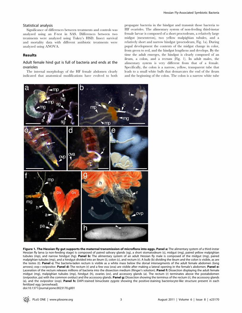
Figure 1. The Hessian fly gut supports the maternal transmission of microflora into eggs. Panel a: The alimentary system of a third-instarHessian fly larva (a non-feeding stage) is composed of paired salivary glands (sg), a short stomatodeum (s), midgut (mg), paired yellow malpighiantubules (mp), and narrow hindgut (hg). Panel b: The alimentary system of an adult Hessian fly male is composed of the midgut (mg), pairedmalpighian tubules (mp), and a hindgut divided into an ileum (i), colon (c), and rectum (r). A bulb (b) dividing the ileum and the colon is visible, as arethe testes (t). Panel c: The bacteria-laden rectum is visible as a white mass below the dorsal intersegments of the adult female abdomen (longarrows); ovp = ovipositor. Panel d: The rectum (r) and a few ova (ova) are visible after making a lateral opening in the female’s abdomen. Panel e:Laceration of the rectum releases millions of bacteria into the dissection medium (Ringer’s solution). Panel f: Dissection displaying the adult femalemidgut (mg), malpighian tubules (mp), hindgut (h), ovaries (ov), and accessory glands (a). The rectum (r) terminates above the postabdomen(ovipositor, pa) with the common oviduct and the accessory glands. Panel g: Dissection showing the terminus of the rectum (r), the accessory glands(a), and the ovipositor (ovp). Panel h: DAPI-stained binucleate zygote showing the positive-staining bacteriocyte-like structure present in eachfertilized egg (arrowhead).doi:10.1371/journal.pone.0023170.g001
Hessian Fly-Associated Symbiotic Bacteria
PLoS ONE | www.plosone.org 3 August 2011 | Volume 6 | Issue 8 | e23170

about twice the length of the ileum that leads to the rectum
(Fig. 1b). In adult females, the hindgut is visible below the posterior
dorsal intersegments of the abdomen (Fig. 1c), and filled with
bacteria (Fig. 1c–g). In comparison, the relatively underdeveloped
male hindgut is not visible and contains no bacteria (Fig. 1b and
data not shown). The posterior of the hindgut gradually narrows to
form a long thin rectal tube that terminates against the internal
dorsal surface of the eighth abdominal segment, above the
postabdomen (Fig. 1g). It appears that the gut of the adult female
allows bacterial fermentation and then delivers bacteria to the ova
as they enter the ovipositor. The female hindgut could also serve as
a nutrient delivery system.
Bacteria in different developmental stages of HFTo estimate the number of culturable bacteria in different
developmental stages, a CFU assay was conducted (Figure 2) on
whole insects. HF pupae (1.56105 per insect) and the third, non-
feeding instar larvae (1.16105 per insect) had the highest CFUs,
followed by adults (2.36104 per insect). First (6.86102 per insect)
and second (6.76102 per insect) instars exhibited much lower
CFUs.
Maternal transmission of HF-associated bacteriaThe bacterial locale specificity in the female hindgut suggested
that some of the HF-associated bacteria were likely transmitted
maternally to the next generation. To determine if that is the case,
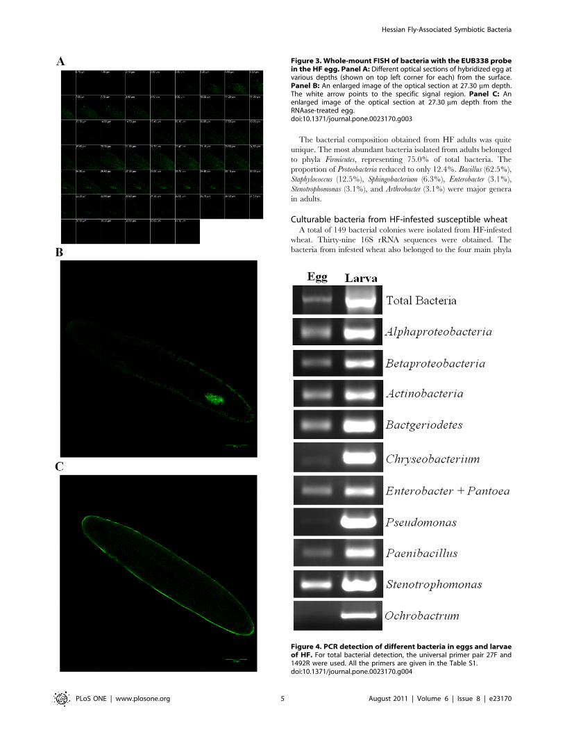
FISH with a universal 16S rRNA probe was performed in HF
eggs. Under a laser scanning confocal microscope, we were able to
see egg sections through optical sectioning (Figure 3A). A
hybridization signal was detected near the apical portion of an
egg at 25.90 mm below the surface. The specific signal was visible
up to 30.10 mm depth, but not thereafter. A closer observation at
the 27.30 mm section revealed that several irregular signal spots
were located next to each other and formed a rough oval shape
(Figure 3B), which was likely a bacteriocyte [36]. No specific signal
observed in control RNAase-treated egg preparations (Figure 3C).
To further identify different types of bacteria that were present
in HF eggs, PCR reactions with primers specific to different
bacterial classes or genera were carried out. Primers specific to
bacterial classes Alphaproteobacteria, Betaproteobacteria, Actinobacteria,
and Bacteriodetes all detected signals (Figure 4). Primers specific to
bacterial genera Enterobacter/Pantoea, Paenibacillus, and Stenotropho-
monas detected relatively strong signals. Primers specific to
bacterial genera Chryseobacterium and Pseudomonas detected weak
signals. Primers specific to bacterial genus Ochrobactrum detected no
signal.
Impact of antibiotics on HF larval survivalTo determine the impact of bacteria on HF larval survival, we
used different antibiotics to deprive bacteria of the insect. In
general, antibiotic treatments had a negative impact on HF larval
survival (Figure 5A). The survival rates of HF larvae were 33%,
70%, 64%, 48%, 23%, 69%, and 25% after treatment with
kanamycin, penicillin, rifampicin, ampicillin, streptomycin, gen-
tamicin, and a kanamycin-streptomycin mixture respectively.
Since kanamycin and streptomycin were the most effective
antibiotics, a more detailed study was carried out with a mixture of
these two antibiotics. To determine the effective time for antibiotic
treatments, the kanamycin/streptomycin mixture was applied to
wheat seedlings at three different time intervals: 3–7, 5–9, and 10–
14 days after HF adult infestation. The numbers of larvae that
survived on plants treated at the 3–7, and 5–9 day intervals was 3–
5 times less than that on control plants (F1,78 = 238.37 and
P,0.0001 for the 3 to 7 interval, and F1,78 = 85.84 and P,0.0001
for the 5 to 9 interval) (Figure 5B). No difference was found in
insect survival between plants treated at the 10–14 day interval
and control plants (F1,78 = 0.41, P = 0.5241).
To exclude other possible ways that could result in reduction of
insects in antibiotic-treated plants, we examined the effect of
antibiotic treatments on the rates of egg hatch and successful larval
migration into the feeding site (Figure 5C). No difference was
observed between antibiotic-treated plants and water-treated
controls in terms of egg hatch and larval migration (F1,22 = 1.62,
P = 0.216). On the other hand, numerous larvae were found dead
at the feeding site of antibiotic-treated seedlings (Figure 5D). Dead
larvae were rarely found at the feeding site of control plants [37].
The impact of the kanamycin/streptomycin mixture on larval
survival was also examined by directly soaking larvae in the
antibiotic solution, and then individually placing the larvae back
onto wheat seedlings. There was a small decrease (P.0.05) in
survival after the antibiotic soak for 24 and 48 hrs, but a dramatic
decrease (P,0.0001) after soak for 72 hrs (Figure 5E).
Culturable bacteria from different developmental HFstages
To identify major culturable bacteria associated with HF, a total
of 482 bacterial colonies were isolated from different stages of the
insect. Of these colonies, 284 were randomly chosen to sequence
their 16S rRNA genes. A total of 270 high quality sequences were
obtained, with 87, 65, 37, 49, and 32 derived from bacterial
colonies isolated from first, second, and third instars, pupae, and
adults, respectively.
Bacteria from four phyla were identified, including Proteobacteria,
Firmicutes, Actinobacteria, and Bacteroidetes (Table S2). The composi-
tion of bacteria associated with the first and second instars was
very similar at the levels of phyla and class. At the level of genus,
however, there were differences in bacterial compositions between
first and second instars. Proteobacteria was still predominant in third
instar larvae and pupae, but its proportion reduced. The decrease
in relative proportion of Proteobacteria was due to an increase in
Firmicutes and Bacteriodetes in third instar larvae; and an increase in
Actinobacteria and Bacteriodetes in pupae.
Figure 2. Colony forming units (CFUs) of bacteria in HF first(Hf1), second (Hf2), and third (Hf3) instars, pupae (Hfp), andadults (Hfa). Black bars represent CFUs per larva while gray barsrepresent CFUs per mg of insects.doi:10.1371/journal.pone.0023170.g002
Hessian Fly-Associated Symbiotic Bacteria
PLoS ONE | www.plosone.org 4 August 2011 | Volume 6 | Issue 8 | e23170
The bacterial composition obtained from HF adults was quite
unique. The most abundant bacteria isolated from adults belonged
to phyla Firmicutes, representing 75.0% of total bacteria. The
proportion of Proteobacteria reduced to only 12.4%. Bacillus (62.5%),
Staphylococcus (12.5%), Sphingobacterium (6.3%), Enterobacter (3.1%),
Stenotrophomonas (3.1%), and Arthrobacter (3.1%) were major genera
in adults.
Culturable bacteria from HF-infested susceptible wheatA total of 149 bacterial colonies were isolated from HF-infested
wheat. Thirty-nine 16S rRNA sequences were obtained. The
bacteria from infested wheat also belonged to the four main phyla
Figure 4. PCR detection of different bacteria in eggs and larvaeof HF. For total bacterial detection, the universal primer pair 27F and1492R were used. All the primers are given in the Table S1.doi:10.1371/journal.pone.0023170.g004
Figure 3. Whole-mount FISH of bacteria with the EUB338 probein the HF egg. Panel A: Different optical sections of hybridized egg atvarious depths (shown on top left corner for each) from the surface.Panel B: An enlarged image of the optical section at 27.30 mm depth.The white arrow points to the specific signal region. Panel C: Anenlarged image of the optical section at 27.30 mm depth from theRNAase-treated egg.doi:10.1371/journal.pone.0023170.g003
Hessian Fly-Associated Symbiotic Bacteria
PLoS ONE | www.plosone.org 5 August 2011 | Volume 6 | Issue 8 | e23170

cultured from HF, namely 51.3% Proteobacteria, 33.3% Firmicutes,
7.7% Bacteroidetes, and 5.1% Actinobacteria (Table S2). For Proteobac-
teria, the Gammaproteobacteria were again the most abundant (35.9%),
followed by Betaproteobacteria (12.8%) and Alphaproteobacteria (2.6%).
Enterobacter (23.1%), Bacillus (23.1%), Paenibacillus (7.7%), Chryseobac-
terium (7.7%), Achromobacter (6.1%), Arthrobacter (5.1%), Pantoea (2.6%),
Pseudomonas (2.6%), Staphylococcus (2.6%), and Microbacterium (2.6%)
were the major genera in HF-infested susceptible wheat. No
bacterial colonies could be obtained from non-infested wheat.
Major bacteria in HF identified through a culture-independent approach
Since culturing could only identify bacteria culturable under our
conditions, a culture-independent, PCR-based method was also
used for a more comprehensive analysis. For this approach, we
analyzed bacterial compositions of first instars, pupae, and adults.
A total of 233 high quality 16 rRNA sequences were obtained
from 316 clones. Among them, 154, 59, and 20 sequences were
derived from first instars, pupae, and adults, respectively. The
bacteria identified from the culture-independent analysis included
the four phyla identified from the culturing analysis described
previously. In addition, a new phylum, Aquificae, was also detected
from the culture-independent analysis. The relative abundance of
Proteobacteria, Firmicutes, Actinobacteria, Bacteroidetes, and Aquificae was
75.8, 4.6, 14.4, 2.6, and 2.6%, respectively, in first instars; 91.5,
3.4, 3.4, 0, and 0% in pupae; and 65.0, 0, 20, 15.0, and 0% in
adults (Table S2).
At the genus level, Acinetobacter (53.6%), Ochrobactrum (6.5%),
Alcaligenes (5.2%), Kocuria (5.2%), Nitrosomonas (3.9%), Bacillus
(3.9%), Arthrobacter (3.9%), Arcanobacterium (3.3%), and Sphingobacter-
ium (2.6%) were the major genera in first instars. Pseudomonas
(25.4%), Acinetobacter (18.6%), Klebsiella (18.6%), Enterobacter
(15.3%), Stenotrophomonas (3.4%), Paenibacillus (3.4%), and Microbac-
terium (3.4%), were major genera in pupae. Ochrobactrum (55.5%),
Alcaligenes (10%), Arthrobacter (10.0%), Microbacterium (5.0%), and
Sphingobacterium (5.0%) were major genera in adults.
Impact of antibiotics on bacterial abundance in HFThe kanamycin-streptomycin mixture reduced the population
size of total bacteria associated with HF larvae significantly
Figure 5. Impact of antibiotics on HF larval survival. The numbers of insects that survived on wheat seedlings after an antibiotic treatmentwere determined 24 DPI. The numbers of larvae that survived and passed into the pupal stage were expressed as mean6S.E. per plant with a 95%confidence interval. In panels B, C, and E, black bars represent antibiotic treatment whereas gray bars represent water-treated controls. An asterisk (*)represents a statistically significant (P#0.5) difference between treatment and control. Panel A: Average number of HF larvae that survived on wheatseedlings treated with different antibiotics. The numbers 1 to 8 represent treatment with kanamycin, penicillin, rifampicin, ampicillin, streptomycin,gentamicin, a mixture of kanamycin and streptomycin, and water, respectively. Panel B: Effective time for antibiotic treatment. Wheat seedlings wereinitially treated with the mixture of kanamycin and streptomycin on day 3, 5, and 10 DPI, respectively. After the initial treatment, the antibioticmixture was applied once per day for four consecutive days. Panel C: Lack of impact of the kanamycin/streptomycin mixture on larval hatching andmigration into the feeding site. The hatching and migration rate was calculated from a total of 587 eggs in antibiotic-sprayed plants and 384 eggs inwater sprayed plants. Eggs were counted 48 hrs after infestation. Larvae that successfully migrated to the base of the plants were counted 7 DPI. Thedifferences in percent hatching and migration rate were compared by fisher’s probability test (P = 0.216). Panel D: Dead larvae on wheat seedlingstreated with the kanamycin/streptomycin mixture. Each black arrow points to a dead first instar larva. The white arrow points to a dead, deformedsecond instar larva. The white triangle points to a live, second instar larva. Panel E: Impact of the kanamycin/streptomycin mixture on larval survivalby direct larval soaking. Total numbers of larvae tested were 117, 115, and 218 for 24, 48, and 72 hrs antibiotic exposure, respectively; and 113, 119,and 220 for 24, 48, and 72 hrs water exposure. The numbers of live insects were counted at 24 days after the treatments.doi:10.1371/journal.pone.0023170.g005
Hessian Fly-Associated Symbiotic Bacteria
PLoS ONE | www.plosone.org 6 August 2011 | Volume 6 | Issue 8 | e23170

(Figure 6). In antibiotic-treated plants, total bacterial 16S DNA
was 36% (t = 3.024, df = 4, P,0.05) and 76% (t = 3.428, df = 4,
P,0.05) lower in one and three day-old larvae, respectively. For
different bacterial groups, Alphaproteobacteria was 87% (t = 1.244,
df = 4, P,0.05) and 99% (t = 3.918, df = 4, P,0.05) lower in one
and three day-old larvae. The overall trend suggested a reduction
in the 16S DNA contents corresponding to bacterial groups
Betaproteobacteria, Enterobacter, Pseudomonas, Paenibacillus, and Steno-
trophomonas. However, the degree of reduction was not as dramatic
as Alphaproteobacteria and variations among replicates were larger.
Irrespective of antibiotic treatments, a small proportion of
larvae survived and became pupae. The relative abundances of the
16S DNA from total bacteria and from specific bacterial groups in
control and antibiotic-surviving pupae were compared (Figure 6).
Relative abundance of 16S DNA from total bacteria was 84.0%
less in antibiotic-treated pupae. The relative abundance of 16S
DNA from specific bacterial groups was significantly reduced for
all groups, but the percentage of reduction varied among them.
Due to the different impact of antibiotics on different bacterial
groups, the bacterial compositions in HF larvae and pupae were
shifted following the antibiotic treatment (Figure 7). Under control
conditions, the relative proportion of Alphaproteobacteria to Betapro-
teobacteria was 87% to 13% in three-day old larvae (Figure 7A).
After the antibiotic treatment, the relative proportion of
Alphaproteobacteria to Betaproteobacteria was 36% to 64%, indicating
that Alphaproteobacteria were more sensitive to antibiotics than
Betaproteobacteria. On the other hand, in the larvae that survived the
antibiotic treatment and became pupae, the proportion of
Figure 6. Relative abundance of the 16S DNA of different bacteria associated with HF following antibiotic- (black bars) and water(gray bars) - treatments. A mixture of kanamycin and streptomycin was used for antibiotic treatment. The relative abundance of total and specificgroups of bacteria was determined by qPCR using universal and group-specific primer pairs listed in Table S1. The mean abundance (6 S.E.) wasderived from three biological replicates. An asterisk (*) represents a statistically significant (P#0.5) difference between treatment and control. Alpha,Beta, Enter, Pseu, Paeni, Steno, and Total represent bacterial groups of classes Alphaproteobacteria and Betaproteobacteria; and genera Enterobacter,Pseudomonas, Paenibacillus, and Stenotrophomonas; and total bacteria; respectively. The numbers ‘‘1’’ and ‘‘3’’ under the abscissae of the first sevenpanels represent 1- and 3-day old larvae. The last panel represents the relative abundance of different bacterial groups as well as total bacteria in HFpupae. The pupae for the antibiotic treatment were from a small portion of larvae survived the antibiotic treatment. The relative abundance ofbacterial groups was shown in log scale on the left ordinate whereas the relative abundance of total bacteria was shown in normal scale on the rightordinate.doi:10.1371/journal.pone.0023170.g006
Hessian Fly-Associated Symbiotic Bacteria
PLoS ONE | www.plosone.org 7 August 2011 | Volume 6 | Issue 8 | e23170

Alphaproteobacteria was 69%; significantly higher than the 46% in
control insects. Changes in bacterial composition after the
antibiotic treatment were also apparent at the genus level
(Figure 7B). Primers specific to four genera, three from
Gammaproteobacteria (Enterobacter, Pseudomonas, and Stenotrophomonas)
and one from Firmicutes (Paenibacillus), again detected dramatic
differences in the relative abundance of their DNA between
antibiotic-treated and control insects. The percentages for
Enterobacter, Pseudomonas, and Stenotrophomonas, and Paenibacillus were
57, 7, 4, and 32 in control three-day old larvae; and the
percentages changed to 14, 4, 14, and 68 in the same age larvae
after the antibiotic treatment. The percentages for Enterobacter,
Pseudomonas, and Stenotrophomonas, and Paenibacillus were 17, 13, 60,
and 10 in control pupae; and the percentages changed to 18, 10, 5,
and 67 in the pupae derived from larvae that survived the
antibiotic treatment.
Discussion
In this study, we conducted an analysis on the possible
transmission mechanism, essentiality, and composition of bacteria
associated with HF. Our results revealed that most HF-associated
bacteria were likely to be transmitted maternally through eggs. A
bacteriocyte-like structure was observed in developing eggs by
FISH (Figure 3B). PCR with primers specific to individual
bacterial groups revealed that most of bacteria detected in other
stages of HF were also present in eggs (Figure 4). The presence of
huge numbers of bacteria in the hindgut of HF female adults
specifically (not present in male adults), and the fact that the
hindgut ends in ovarioles may provide an unique way among
insects to allow bacteria somehow entered into developing eggs
from the hindgut. However, the exact mechanism for the physical
transfer of HF-associated bacteria remains to be delineated. In
aphids, Buchnera spp. are liberated from bacteriocytes in adult
insects, circulated in body fluids, and then enter fertilized eggs
through an opening on the egg surface [35]. In whiteflies, psyllids,
mealybugs, and cockroaches, bacteria are transferred into eggs by
transferring whole bacteriocytes from female adults directly into
eggs [38]. In tsetse flies, bacterial symbionts Wigglesworthia
glossinidia and Sodalis glossinidius are transmitted into next
generation through milk glands [39].
The maternal transmission of HF-associated bacteria suggests a
close relationship between bacterial symbionts and the insect [40].
In general, the essentiality of symbiotic bacteria for insect survival
is often examined by depriving insects of bacteria with diets
containing antibiotics. In this study, HF-associated bacteria were
targeted with antibiotics, and the resulting effect on HF survival, if
any, was determined. Because of the lack of a HF artificial diet, we
conducted antibiotic treatments by applying antibiotic solutions
onto wheat leaves, assuming that the antibiotics could penetrate
into wheat tissues, and eventually entering HF larvae along with
other food gradients. As shown in Figure 5A, the application of
different antibiotics resulted in 30–77% decrease in HF larval
survival rates. The reduction in HF survival was correlated with
the deprivation of bacteria from the host insect. Therefore, the loss
of bacteria was likely the reason for the reduction in HF larval
survival. However, antibiotics, due to their inherent toxicity, could
have been responsible for lowering the survival of the insect.
Accordingly, we conducted further experiments to see if there was
toxicity of antibiotics directly to the insect, and our results
suggested that the high mortality of HF larvae was not due to
toxicity of antibiotics directly to the insect. First, the application of
antibiotics when HF larvae were five days old (during the
transition from first to second instar) did not increase larval
Figure 7. Alternation in bacterial composition after antibiotictreatments. A mixture of kanamycin and streptomycin was used forantibiotic treatment. Panel A: Proportions of Alphaproteobacteria(Alpha) and Betaproteobacteria (Beta) in antibiotic-treated (Antibiotic)and control 3-day old larvae (3-d larvae) and pupae. Panel B:Proportions of bacterial groups Enterobacter (En.), Pseudomonas (Ps.),Paenibacillus (Pa.), and Stenotrophomonas (St.) in antibiotic-treated andcontrol larvae and pupae.doi:10.1371/journal.pone.0023170.g007
Hessian Fly-Associated Symbiotic Bacteria
PLoS ONE | www.plosone.org 8 August 2011 | Volume 6 | Issue 8 | e23170

mortality (Figure 5B, 10 DPI), indicating that the antibiotics did
not have a toxic effect on larval metabolism. Second, directly
soaking HF larvae in an antibiotic solutions for 24 and 48 hrs
resulted in only a small increase in larval mortality (Figure 5E),
indicating that the antibiotics did not have an acute, direct toxic
effect on HF larvae. The increase in mortality at a later time point
was likely due to the depletion of bacteria. In addition, antibiotic
treatments have not shown severe direct toxicity to other insects
[41]–[44]. Taken together, our evidence suggested that depriva-
tion of bacteria from the insect by antibiotics was the cause for
larval mortality and that symbiotic bacteria were essential for
larval survival.
The exact roles of bacteria in HF remain to be determined.
Symbiotic bacteria perform a range of functions in other insects,
including synthesizing essential amino acids [45]–[49], synthesiz-
ing vitamins [50], digesting nutrients [47], [51], increasing
tolerance of host insects to biotic and abiotic stresses [11], [12],
[52]–[54], performing nitrogen fixation [55], [56] and playing a
role in insect – plant interactions [13], [15], [16], [57]–[59]. A
large number of bacterial species reside in the HF larval gut. These
gut bacteria could play a role in synthesis of necessary nutrients for
HF larvae, or digestion of nutrients otherwise inaccessible to the
insect. Bacteria could also play a critical role in HF – wheat
interaction. Approximately 70% of bacterial genera detected in
HF larvae through culturing were also found in HF infested wheat
(Table S2). Considering the extraordinary ability of HF larvae to
manipulate host plants [17], [22] and reprogram metabolic
pathways of wheat seedlings [23], [24], it was possible that
bacteria were secreted into host plants as part of oral secretions
and participated in changing wheat metabolism once they were in
wheat seedlings. Further investigation would be needed to
examine these possibilities.
It would be interesting to know what types of bacteria were
essential for HF larvae to survive. Initial evidence suggested that
Alphaproteobacteria might be important for HF larval survivability
according to their response to antibiotic treatments. Alphaproteo-
bacteria was 5.6 time more abundant than Betaproteobacteria in 3-day
old larvae under control conditions (Figure 6B). After an antibiotic
treatment, Alphaproteobacteria decreased to almost one third of
Betaproteobacteria in the same stage of larvae. Associated with the
decrease in abundance of Alphaproteobacteria and other bacteria was
the high death rate of HF larvae. In pupae derived from larvae
that survived the antibiotic treatment, Alphaproteobacteria were again
predominant. These observations suggested that the loss of
Alphaproteobacteria might be responsible for larval death. Again,
further tests are needed to confirm this postulation.
A preliminary survey of the bacterial composition through
culturing and culture-independent approaches revealed that
diverse bacteria existed in HF and that the bacterial composition
changed in different developmental stages of the insect (Table S2).
Among the most abundant bacteria detected through culturing
were Enterobacter, Pantoea, Klebsiella, Pseudomonas, Stenotrophomonas,
Staphylococcus, and Achromobacter. Enterobacter was the most dominant
among cultured bacteria recovered from the three HF larval
instars and pupae, with relative abundance ranging from 32–38%.
The recovery of Enterobacter from all stages of HF indicated a stable
relationship between the two partners. Bacterial genera found
abundant through the culture-independent method included
Acinetobacter, Alcaligenes, Nitrosomonas, and Ochrobactrum. Acinetobacter
was the most dominant, representing 54% of total 16S DNA in the
first HF instar. Some bacteria detected through culturing were not
detected through the culture-independent approach, suggesting
that a more comprehensive survey is needed to identify more HF-
associated bacteria.
In summary, diverse bacteria were associated with different
developmental stages of HF. Most of these bacteria were
transmitted maternally to the next generation indicating an close
relationship between the insect and its associated bacteria.
Depletion of microbes from HF larvae with antibiotics caused a
high mortality of the insect in wheat seedlings, indicating that
antibiotic-sensitive microbes were essential for the insect to survive
on wheat seedlings. This research is the first systematic analysis of
bacteria associated with HF and provides a foundation for future
research to elucidate the complex tritrophic interactions among
wheat, HF, and their symbiotic bacteria.
Supporting Information
Table S1 Primers used in this study.
(DOC)
Table S2 Relative frequency (%) of different bacteria derived
from different stages of Hessian fly and infested wheat by culture
dependent and culture-independent methods.
(DOC)
Acknowledgments
The authors thank Drs. Anna Whitfield and John Fellers from the
Department of Plant Pathology, KSU, Manhattan, KS, for reviewing an
earlier version of the manuscript. This paper is contribution 11-126-J from
the Kansas Agricultural Experiment Station. Mention of a trademark of a
proprietary product does not constitute a guarantee of warranty of the
product by the United States Department of Agriculture, and does not
imply its approval to the exclusion of other products that may also be
suitable.
Author Contributions
Conceived and designed the experiments: M-SC RB SH JJS. Performed
the experiments: RB SH JJS M-SC. Analyzed the data: RB SH BS JJS M-
SC. Contributed reagents/materials/analysis tools: BS JCR RJW JJS M-
SC. Wrote the paper: RB SH BS JCR RJW JJS M-SC.
References
1. Moran NA (2001) Bacterial menageries inside insects. (Commentary). Proc Natl
Acad Sci USA 292: 1096–1098.
2. Moran NA (2006) Symbiosis (A primer). Curr Biol 16: R866–871.
3. Wu D, Daugherty SC, Van Aken SE, Pai GH, Watkins KL, et al. (2006)
Metabolic complementarity and genomics of the dual symbiosis of sharpshoot-
ers. PloS-Biol 4: e188.
4. Janson EM, Stireman JO, Singer MS, Abbot P (2008) Phytophagous insect-
microbe mutualisms and adaptive evolutionary diversification. Evolution 62:
997–1012.
5. Douglas AE (1998) Nutritional interactions in insect-microbial symbioses:
Aphids and their symbiotic bacteria Buchnera. Annu Rev Entomol 43: 17–37.
6. Brune A (2003) Symbionts aiding digestion. 1102–1107. In Carde RT, Resh VH,
eds. Encyclopedia of insects Academic Press, New York.
7. Pais R, Lohs C, Wu Y, Wang J, Aksoy S (2008) The obligate mutualist
Wigglesworthia glossinidia influences reproduction, digestion, and immunity
processes of its host, the tsetse fly. Appl Env Microbiol 74: 5965–5974.
8. Nogge G (1976) Sterility in tsetse flies (Glossina morsitans Westwood) caused by
loss of symbionts. Experientia 32: 995–996.
9. Oliver K, Russell J, Moran N, Hunter M (2003) Facultative bacterial symbionts
in aphids confer resistance to parasitic wasps. Proc Natl Acad Sci USA 100:
1803–1807.
10. Oliver KM, Moran NA, Hunter MS (2005) Variation in resistance to parasitism
in aphids is due to symbionts and not host genotype. Proc Natl Acad Sci USA
102: 12795–12800.
11. Scarborough CL, Ferrari J, Godfray HC (2005) Aphid protected from pathogen
by endosymbiont. Science 310: 1781.
Hessian Fly-Associated Symbiotic Bacteria
PLoS ONE | www.plosone.org 9 August 2011 | Volume 6 | Issue 8 | e23170

12. Vorburger C, Gehrer L, Rodriguez P (2009) A strain of the bacterial symbiont
Regiella insecticola protects aphids against parasitoids. Biol Lett rsbl2009.0642v1-rsbl20090642.
13. Tsuchida T, Koga R, Fukatsu T (2004) Host plant specialization governed by
facultative symbiont. Science 303: 1989–1989.14. Hosokawa T, Kikuchi Y, Shimada M, Fukatsu T (2007) Obligate symbiont
involved in pest status of host insect. Proc Royal Soc London B 274: 1979–1984.15. Hogenhout SA, Oshima K, Ammar E-D, Kakizawa S, Kingdom HN, et al.
(2008) Phytoplasmas: bacteria that manipulate plants and insects. Mol Plant
Pathol 9: 403–423.16. Bai S, Correa VR, Toruno TY, Ammar E-D, Kamoun S, et al. (2009) AY-WB
phytoplasma secretes a protein that targets plant cell nuclei. Mol Plant MicrobeInteract 22: 18–30.
17. Harris MO, Freeman TP, Rohfritsch O, Anderson KG, Payne SA, et al. (2006)Virulent HF (Diptera: Cecidomyiidae) larvae induce a nutritive tissue during
compatible interactions with wheat. Ann Entomol Soc Am 99: 305–316.
18. Ananthakrishnan TN, ed. Biology of Gall Insects Oxford and IBH PublishingCo. New Delhi, India.
19. Hatchett JH, Starks KJ, Webster JA (1987) Insect and mite pests of wheat. In:Heyne EG, ed. Wheat and Wheat Improvement. Agronomy monograph no. 13
(2nd edn) ASA-CSSA-SSSA, Madison. pp 625–675.
20. Pauly PJ (2002) Fighting the HF. Environ Hist 7: 385–507.21. Stuart JJ, Chen MS, Harris M (2008) Hessian fly. In: W. Hunter, C. Kole, eds.
Genome Mapping and Genomics in Animals, vol 1 Genome Mapping andGenomics in Arthropods, Springer, Berlin, Heidelberg, New York. pp 93–100.
22. Byers RA, Gallun RL (1971) Ability of the HF to stunt winter wheat. I. Effect oflarval feeding on elongation of leaves. J Econ Entomol 65: 955–958.
23. Liu XL, Bai J, Huang L, Zhu L, Liu X, et al. (2007) Gene expression of different
wheat genotypes during attack by virulent and avirulent HF (Mayetioladestructor) larvae. J Chem Ecol 33: 2171–2194.
24. Zhu L, Liu XM, Liu X, Jeannotte R, Reese JC, et al. (2008) HF (MayetiolaDestructor) attack causes dramatic shift in carbon/nitrogen metabolism in
wheat. Mol Plant-Microbe Interact 21: 70–78.
25. Chen MS, Fellers JP, Stuart JJ, Reese JC, Liu XM (2004) A group of relatedcDNAs encoding secreted proteins from HF [Mayetiola destructor (Say)] salivary
glands. Insect Mol Biol 13: 101–108.26. Chen MS, Zhao HX, Zhu YC, Scheffler B, Liu XM, et al. (2008) Analysis of
Transcripts and Proteins Expressed in the Salivary Glands of HF (Mayetioladestructor) Larvae. J Insect Physiol 54: 1–16.
27. Chen MS, Liu X-M, Yang Z, Zhao H, Shukle RH, et al. (2010) Unconventional
conservation among genes encoding small secreted salivary gland proteins froma gall midge. BMC Evol Biol 10: 296.
28. Boosalis GM (1954) HF in relation to the development of crown and basal stemrot of wheat. Phytopathol 44: 224–229.
29. Mittapalli O, Shukle RH, Sardesai N, Giovanini MP, Williams CE (2006)
Expression patterns of antibacterial genes in the HF. J Insect Physiol 52:1143–1152.
30. Gagne RJ, Hatchett JH (1989) Instars of the Hessian fly (Diptera: Cecidomyii-dae). Ann Entomol Soc Am 82: 73–79.
31. Harris MO, Rose S (1989) Temporal changes in egglaying behavior of the HF.Entomol Exp Appl 53: 17–29.
32. Howard DJ, Bush GL, Breznak JA (1985) The evolutionary significance of
bacteria associated with Rhagoletis. Evolution 39: 405–417.33. Doyle JJ, Doyle JL (1987) A rapid DNA isolation procedure from small
quantities of fresh leaf tissues. Phytochem Bull 19: 11–15.34. Koga R, Tsuchida T, Fukatsu T (2009) Quenching autofluorescence of insect
tissues for in situ detection of endosymbionts. Appl Entomol zool 44: 281–291.
35. Buchner P (1965) Endosymbiosis of Animals with Plant MicroorganismsInterscience Publishers, New York.
36. Giovanini MP, Putoff DP, Nemacheck JA, Mittapalli O, Saltzmann KD, et al.(2006) Gene-for-gene defense of wheat against the Hessian fly lacks a classical
oxidative burst. Mol Plant-Microbe Interact 10: 1023–1033.
37. Chen MS, Liu XM, Wang H, El Bouhssini M (2009) HF (Mayetiola destructor)
interactions with barley, rice, and wheat seedlings. J Econ Entomol 102:1663–1672.
38. Costa HS, Toscano NC, Henneberry TJ (1996) Mycetocyte inclusion in the
oocytes of Bemisia argentifolii (Homoptera: Aleyodidae). Ann Entomol Soc Am89: 694–699.
39. Denlinger DL, Ma WC (1975) Maternal nutritive secretions as possible channelsfor vertical transmission of microorganisms in insects: the tsetse fly example.
Annals NY Acad Sci 266: 162–165.
40. Gil R, Latorre A, Moya A (2004) Bacterial endosymbionts of insects: insightsfrom comparative genomics. Environ Microbiol 6: 1109–1122.
41. Wilkinson TL (1998) The elimination of intracellular microorganisms frominsects: an analysis of antibiotic-treatment in the pea aphid (Acyrthosiphon
pisum). Comp Biochem Physiol Ser A 119: 871–881.42. Wilkinson TL, Ashford DA, Pritchard J, Douglas AE (1997) Honeydew sugars
and osmoregulation in the pea aphid Acyrthosiphon pisum. J Experimen Biol
200: 2137–2143.43. Wilkinson TL, Douglas AE (1995) Aphid feeding, as influenced by the disruption
of the symbiotic bacteria. J Insect Physiol 41: 635–640.44. Wilkinson TL, Douglas AE (1996) The impact of aposymbiosis on amino acid
metabolism of pea aphids. Entomologia Experimentalis et Applicata 80:
279–282.45. Baumann P, Baumann L, Lai CY, Rouhbakhsh D, Moran NA, et al. (1995)
Genetics, physiology, and evolutionary relationships of the genus Buchnera:intracellular symbionts of aphids. Annu Rev Microbiol 49: 55–94.
46. Febvay G, Liadouze I, Guillaud J, Bonnot G (1995) Analysis of energetic aminoacid metabolism in Acyrthosiphon pisum: a multidimensional approach to
amino acid metabolism in aphids. Arch Insect Biochem Physiol 29: 45–69.
47. Douglas AE (2009) The microbial dimension in insect nutritional ecology. FuncEcol 23: 38–47.
48. Sasaki T, Hayashi H, Ishikawa H (1991) Growth and reproduction of thesymbiotic and aposymbiotic pea aphids, Acyrthosiphon pisum maintained on
artificial diets. J Insect Physiol 37: 749–56.
49. Shigenobu S, Watanabe H, Hattori M, Sasaki Y, Ishikawa H (2000) Genomesequence of the endocellular bacterial symbiont of aphids Buchnera sp. APS.
Nature 407: 81–86.50. Nogge G (1981) Significance of symbionts for the maintenance of an optimal
nutritional state for successful reproduction in haematophagous arthropods.Parasitology 82: 101–104.
51. Lundgren JG, Lehman RM (2010) Bacterial gut symbionts contribute to seed
digestion in an omnivorous beetle. PLoS one 5: e10831.52. Dunbar HE, Wilson ACC, Ferguson NR, Moran NA (2007) Aphid thermal
tolerance is governed by a point mutation in bacterial symbionts. PLoS Biology5: e96.
53. Russell JA, Moran NA (2006) Costs and benefits of symbiont infection in aphids:
variation among symbionts and across temperatures. Proc Royal Soc B 273:603–10.
54. Oliver KM, Hunter MS, Degnan PH, Moran NA (2009) Bacteriophages encodefactors required for protection in a symbiotic mutualism. Science 325: 992–994.
55. Lilburn TC, Kim KS, Ostrom NE, Byzek KR, Leadbetter JR, et al. (2001)Nitrogen fixation by symbiotic and free-living spirochetes. Science 292:
2495–2498.
56. Kneip C, Lockhart P, Vob C, Maier U-G (2007) Nitrogen fixation in eukaryotes– New models for symbiosis. BMC Evol Biol 7: 55.
57. Schaefer CW, Panizzi AR (2000) Heteroptera of economic importance CRCPress 2000 Boca Raton, FL.
58. Fukatsu T, Hosokawa T (2002) Capsule-transmitted gut symbiotic bacterium of
the Japanese common plataspid stinkbug, Megacopta punctatissima. ApplEnviron Microbiol 68: 389–396.
59. Hosokawa T, Kikuchi Y, Nikoh N, Shimada M, Fukatsu T (2006) Strict host-symbiont cospeciation and reductive genome evolution in insect gut bacteria.
PLoS Biol 4: e377. doi:10.1371/journal.pbio.0040337.
Hessian Fly-Associated Symbiotic Bacteria
PLoS ONE | www.plosone.org 10 August 2011 | Volume 6 | Issue 8 | e23170
Recommended


