GENETIC ANALYSIS OF SERRATIA MARCESCENS,
THE CAUSAL AGENT OF CUCURBIT
YELLOW VINE DISEASE
By
QUAN ZHANG
Bachelor of Science Central China Agricultural University
Wuhan, Hubei, P. R. China 1997
Master of Science China Agricultural University
Beijing, P. R. China 2000
Submitted to the Faculty of the Graduate College of the
Oklahoma State University in partial fulfillment of
the Degree of DOCTOR OF PHILOSOPHY
December, 2004

GENETIC ANALYSIS OF SERRATIA MARCESCENS,
THE CAUSAL AGENT OF CUCURBIT
YELLOW VINE DISEASE
Thesis Approved:
Jacqueline Fletcher Thesis Advisor
Ulrich Melcher Carol Bender Moses Vijayakumar Gordon Emslie
Dean of the Graduate College
ii

ACKNOWLEDGEMENTS
I would like to express my sincere gratitude to my advisor, Dr. Jacqueline Fletcher,
for her support, patience, and intelligent guidance throughout my graduate studies. Her
editorial advice was essential to the completion of this dissertation. I would also like to
thank Dr. Ulrich Melcher, who gave me much invaluable advice. My thanks extend to my
other committee members, Dr. Carol Bender and Dr. Moses Vijayakumar, for offering
me priceless assistance and suggestions.
I am very grateful to Astri Wayandande, Michael Berg, Kikin Mutaqin and
Guanpingsheng, Luo, for suggestions and joys brought to me. I will also give a special
thanks to Gerry Smith and other secretaries at the Entomology and Plant Pathology
Department for providing me a good working atmosphere.
Last, but not least, I would like to thank my wife Kang for her understanding and love
during the past few years. Her support and encouragement was in the end what made this
dissertation possible.
iii
TABLE OF CONTENTS Chapter Page I. Literature Review ............................................................................................................ 1
Introduction to Cucurbit Yellow Vine Disease.......................................................... 1 Niche Versatility of S. marcescens ............................................................................ 2 S. marcescens Pathogenicity and Virulence .............................................................. 5 Epidemiological Typing............................................................................................. 7 Genome Plasticity in Enterobacteriaceae ................................................................. 9 Genome-wide Comparison Strategies...................................................................... 14 Chromosomal Mutation Strategies .......................................................................... 15 Literature Cited ........................................................................................................ 19
II. Introduction .................................................................................................................. 31
Literature Cited ........................................................................................................ 35 III. Genotyping of Serratia marcescens Strains Associated with Cucurbit Yellow Vine
Disease ..................................................................................................................... 37 Abstract .................................................................................................................... 37 Introduction.............................................................................................................. 38 Materials and Methods............................................................................................. 40 Results...................................................................................................................... 43 Discussion................................................................................................................ 45 Acknowledgments.................................................................................................... 49 Literature Cited ........................................................................................................ 50
IV. Genomic Comparison of Plant Pathogenic and Non-pathogenic Serratia marcescens
Strains Using Suppressive Subtractive Hybridization ............................................. 62 Abstract .................................................................................................................... 62 Introduction.............................................................................................................. 63 Materials and Methods............................................................................................. 64 Results...................................................................................................................... 68 Discussion................................................................................................................ 73 Literature Cited ........................................................................................................ 77
V. Transposon Mutagenesis of Serratia marcescens, the Causal Agent of Cucurbit
Yellow Vine Disease, and Virulence Screening on Caenorhabditis elegans .......... 90 Abstract .................................................................................................................... 90 Introduction.............................................................................................................. 90 Materials and Methods............................................................................................. 93
iv
Chapter Page
Results and Discussion ............................................................................................ 98 Literature Cited ...................................................................................................... 101
VI. Appendix I. Summary of FOSU1 ............................................................................ 108 VII. Appendix II. Summary of FOSU2 .......................................................................... 110
v
LIST OF TABLES
Table Page
Chapter III
III-1. Strains of cucurbit yellow vine-associated and non-associated Serratia marcescens, and other Serratia species, used in this study.…..........................
.....54
III-2. DNA relatedness disclosed by DNA-DNA hybridization, with respect to strains of CYVD-associated and non-associated strains of Serratia marcescens, and other Serratia species.............................................................
.....57
Chapter IV
IV-1. Bacterial strains tested in this study and their relative relatedness to CYVD representative strain C01-A...............................................................................
.....82
IV-2. Sequence analysis of Z01-A specific clones..................................................... .....83
IV-3. Conservation of tester specific sequences among Serratia marcescens strains.....85
Chapter V
V-1. Test bacteria and nematodes used in this study.....................................................104
V-2. Minimum inhibitory concentration of antibiotics for experimental bacterial strains.................................................................................................................
....104
V-3. Caenorhabditis elegans feeding inhibition....................................................... ....104
vi
LIST OF FIGURES
Figure Page
Chapter III
III-1. Agarose gel electrophoresis gels of rep-PCR products generated from CYVD strains, Serratia marcescens from various niches, and other Serratia types strains......................................................................................
.....60
III-2. Consensus phylogenetic tree of the relationships among Serratia strains and species, compiled from binary data based on the banding patterns of three different rep-PCR reaction sets..............................................................
.....61
Chapter IV
IV-1. Sequence analysis of FOSU1 and its fimbrial gene cluster............................ .....88
IV-2. Agarose gel electrophoresis of 16S rDNA and sequence a43 amplified from a dilution series of genomic DNA and plasmid DNA preparations.....................................................................................................
.....88
IV-3. CYVD pathogenic and non-pathogenic strains detected by multiplex PCR employing primers YV1/4 and a79F/R...........................................................
.....89
Chapter V
V-1. Z01-A transformants, Z01-A, JM109 and DH5α tested by two independent PCRs...............................................................................................................
...106
V-2. Southern hybridization analysis of EcoRI-digested Z01-RR1 transformants and Escherichia coli JM109 probed with a DIG-labeled PCR product of the kanamycin resistance gene.......................................................................
...106
V-3. Survival of Caenorhabditis elegans N2 fed on Escherichia coli OP50, Serratia marcescens Db11 and CYVD strains...............................................
...107
vii

CHAPTER I
LITERATURE REVIEW
Introduction to Cucurbit Yellow Vine Disease
Vine declines of cucurbits have increased in number and severity in the past 20 years
and are yield-limiting factors in many intensive production areas around the world (13).
In 1988, a new disease called cucurbit yellow vine disease (CYVD) was observed in
central Texas and Oklahoma. CYVD is characterized by rapid and general yellowing of
leaves appearing over a 3-4 day period, followed by gradual or rapid decline and death of
the vine in several cucurbit crops (13). Symptoms resembled those reported in diseases
caused by phloem-associated bacteria in other crops. Avila et al. (6) found that the
CYVD organism was detectable by polymerase chain reaction (PCR) using nonspecific
primers designed from prokaryotic 16S rDNA. The deduced nucleotide sequence of the
amplified 16S ribosomal DNA placed this organism within the gamma-3 proteobacteria.
In 1998, pure bacterial cultures isolated from diseased zucchini and watermelon,
designated Z01-A and W01-A, respectively, were considered to be candidate causal
agents. Koch’s postulates were completed using isolate Z01-A by mechanical
inoculations and by transmission via an insect vector, the squash bug, Anasa tristis (14).
Rascoe et al. (70), after extensive analysis of the 16S rDNA and groE sequences of Z01-
A, showed that the cucurbit isolates Z01-A and W01-A were Serratia marcescens.
Bacteria of this species occupy a variety of ecological niches. However, the degree of
genetic homogeneity of the CYVD strains of S. marcescens compared to other isolates
1
remains unknown. In addition, the genetic factors that are responsible for the plant
pathogenicity in these strains of S. marcescens have not been determined.
Niche Versatility of S. marcescens
S. marcescens, a gram-negative bacillus classified as a member of the
Enterobacteriaceae, was the first species identified in the genus Serratia. Most S.
marcescens isolates produce a red pigment, prodigiosin, which in early times was often
mistaken for fresh blood (26). In the past few decades this pigmented bacterium has been
identified in various ecological niches including soil, water, air, plants, insects, animals
and even human beings.
When the name S. marcescens was assigned by Bizio (8) to this bacterium it was
generally considered as a saprophytic microorganism living in soil. S. marcescens might
play a role in the biological cycle of metals by metabolizing organic iron and dissolving
gold and copper (64). On the other hand, it is one of the most frequent contaminants of
laboratory cultures of bacteria. It is also found in foods, particularly in starchy variants,
which provide an excellent growth environment (34).
S. marcescens is also an insect pathogen (15). Red strains of S. marcescens have been
isolated from healthy, diseased, or dead field-collected insects, but the species is most
frequently reported as a pathogen of insectary-reared insects (77), in which it causes a
lethal septicemia after invasion into the hemocoel. Nonchromogenic strains are also
pathogenic and exhibit no significant biochemical differences from classical red strains.
Inglis et al. (36) reported that egg production from adult Heliothis virescens inoculated as
2
larvae was reduced by 30%, and egg hatch was reduced by 12% over a 10 day-period due
to infection by S. marcescens.
The first description of nosocomial human infection caused by S. marcescens was
Wheat’s (85) report of 11 cases over a 6-month period at Stanford University Hospital.
S. marcescens was isolated from preterm infants (gestational age 25-30 weeks) cared for
in the intensive care unit. Later S. marcescens was reported to be the most common
Gram-negative bacterium associated with contact lens related keratitis (65). S.
marcescens may also be responsible for appreciable morbidity among patients with
acquired immunodeficiency syndrome (AIDS), especially when the disease is
accompanied by a low CD4+ cell count, neutropaenia, and hospitalization (46).
Biochemical analysis showed that almost all clinical strains of S. marcescens secrete a
cytotoxin, Sh1A, that causes hemolysis of human and animal erythrocytes and the release
of inflammatory mediators from leukocytes (9).
Mention of plants as a source of Serratia species, predominantly S. marcescens and S.
liquefaciens, can be found in several papers and culture collection catalogs. Plant-
associated S. marcescens isolates have been investigated for a possible role in the
contamination of humans (88). However, it is still unclear if the biotypes found in human
patients are the same as those found in plants. S. marcescens may be harmful to host
plants in some cases, but beneficial in other cases. For example, S. marcescens 90-166
was described as a plant growth promoting rhizobacterium (PGPR), which could induce
systemic resistance in cucumber against different pathogenic agents including bacteria,
fungi and even viruses (84). Shinde and Lukezic (76) reported that two groups of Gram-
negative bacteria were consistently associated with a root and crown disease of alfalfa.
3
Based on biochemical characterization Lukezic et al. (44) concluded that one group was
S. marcescens, but no further characterization of this particular strain has been reported.
Only a few bacteria have been identified to be pathogens of both plants and humans.
Among them, Burkholderia cepacia is much better characterized than S. marcescens.
Like S. marcescens, B. cepacia is resistant to many antibiotics and utilizes a wide variety
of substrates. Genotypic and phenotypic relationships within B. cepacia have been
studied (39, 86). The B. cepacia genome is comprised of multiple chromosomes and is
rich in insertion sequences. These two features play a key role in the evolution of novel
degradative functions and the unusual adaptability of B. cepacia. The same mechanism
may also apply to S. marcescens, but whether this is true or not still needs to be verified.
The potential of S. marcescens to utilize a wide range of nutrients is reflected clearly
by its ability to survive and grow under extreme conditions, including in disinfectants,
antiseptics and double distilled water. Szewzyk et al. (81) studied the survival and growth
of S. marcescens strain K202 at different oxygen concentrations in de-ionized water
containing materials derived from blood bags. The rate of bacterial survival and growth
was highest under anaerobic conditions.
S. marcescens is unique among enteric bacteria in many respects. It secretes
extracellular chitinases, several proteases, a nuclease and a lipase, and produces a wetting
agent or surfactant called “serrawettin”. In keeping with its varied habitats, S. marcescens
produces alternate forms of differentially flagellated cells. These cells display different
types of motility depending on whether the growth medium is solid or liquid (1).
Infections caused by S. marcescens may be difficult to treat because of its resistance
to a variety of antibiotics, including ampicillin and cephalosporins. The resistance of S.
4
marcescens to β-lactam antibiotics may arise from two mechanisms: first, high level
production of chromosomal AmpC cephalosporinases combined with decreased
membrane permeability; second, synthesis of β-lactamase, which hydrolyses
carbapenems (42). S. marcescens resistance to aminoglycosides results from its ability to
prevent the drugs from reaching the target site on the ribosome in one of two ways: first,
alterations in the cell envelope can prevent uptake of the drug; and second, the drug itself
may be modified by an “inactivating enzyme” that adenylates, acetylates, or
phosphorylates the aminoglycoside hydroxyl or amino groups.
S. marcescens Pathogenicity and Virulence
Adherence and hydrophobicity. Piliation has been shown to be a determinant of
microbial adherence to host epithelial surfaces. A nosocomial isolate of S. marcescens
possesses pili and adheres to uroepithelial cells (89). Two classes of adhesins have been
suggested for S. marcescens (71). The pili of one class, designated mannose-resistance
(MR), agglutinate chicken erythrocytes in the presence of D-mannose; those of the other
class, mannose-sensitive (MS), exhibit mannose sensitive haemagglutination of guinea-
pig and chicken erythrocytes. Mizunoe et al. (53) assessed the effect of bacterial piliation
on S. marcescens interaction with human leucocytes and found that the MS-piliated strain
was more susceptible to phagocytosis than was the MR- or nonpiliated strain.
S. marcescens possesses hydrophobic surfaces (55). Cell surface hydrophobicity has
been linked to partitioning of S. marcescens at air: water and oil: water interfaces, as well
as adhesion to solid surfaces including catheters and other plastics (5). When Ness-
Greenstein et al. (62) tested the feasibility of increasing the cell surface hydrophobicity of
5

commonly used strains of E. coli by transformation with DNA from S. marcescens, the
transformed E. coli became more hydrophobic. Mallick (45) compared the
hydrophobicity of two S. marcescens mutant strains, of which one is non-pigmented and
the other overproduces red pigment. An extra protein of the outer membrane
(approximately 40 K Da) might be responsible for higher surface hydrophobicity in the
non-pigmented mutant of S. marcescens.
Lipopolysaccharide. Lipopolysaccharide (LPS; the biologically active constituent of
endotoxin) is comprised of three regions, lipid A, the O-antigen and the core. The O-
antigen, a repetitive saccharide chain, is the most important immunogenic component
determining the O-serotype of bacteria. Palomar et al. (63) showed that bactericidal
activity of phages against S. marcescens depends upon the O-side chain length. Two S.
marcescens genes involved in core LPS biosynthesis were cloned and characterized.
These two genes conferred resistance to bacteriocin 28b when introduced into E. coli
NM554 (23).
LPS from Gram-negative bacteria is well recognized to be a potent microbial toxin. It
has been postulated to play a critical role in the initiation of the proinflammatory events
that contribute to the pathogenesis of human sepsis (69). However, there is a poor
correlation between serum LPS levels and mortality in septic patients. Luchi and
Morrison (43) compared the chemical, structural, and biological differences among LPS
from S. marcescens and other clinical bacterial isolates. The relatively minor differences
in LPS activity seemed unlikely to explain the discrepancy between serum endotoxin
levels and mortality in patients with Gram-negative sepsis.
6

Extracellular products. S. marcescens produces several extracellular enzymes and is
one of the most efficient organisms for the biological degradation of chitin. Sundheim et
al. (79) cloned two chromosomal fragments encoding chitinolytic activity from S.
marcescens strain BJL200. Brurberg et al. (12) analyzed one of the fragments and
determined the nucleotide sequence of a chitinase gene. Several different chitinase genes
are present in S. marcescens. Suzuki et al. (80) found a novel chitinase gene, named chiC,
in S. marcescens 2170. The protein product of this gene has a fibronectin type III-like
domain.
Letoffe et al.(41) identified a S. marcescens extracellular protein, HasA (for heme
acquisition system), which is able to bind heme and is required for iron acquisition from
heme and hemoglobin by the bacterium. HasA does not have a signal peptide and has no
sequence similarity to other proteins. When HasA secretion was reproduced in E. coli, it
was shown that, like many proteins lacking a signal peptide, HasA has a C-terminal
targeting sequence and is secreted by a specific ATP-binding cassette (ABC) transporter
(74).
Epidemiological Typing
Typing methods for S. marcescens involve either phenotypic or genotypic
characterization and are based on the assumption that closely related organisms will
possess a unique characteristic that distinguishes them from unrelated isolates.
Phenotypic approaches based on metabolic or biological characteristics have included
serotyping, biotyping, bacteriocin typing, phage typing and whole cell fingerprinting
7

(47). Biotyping has the advantage of using bacteriological techniques that can be
employed on a routine basis. However, the method is time consuming and laborious, and
biotyping cannot always distinguish between different epidemiological types (30).
Serotyping is another commonly used method for typing S. marcescens, but has
limitations associated with the tedious determination of both O and K antigens, and the
presence of a few untypable mutants (40). Bacteriocin typing is a powerful method, but it
also may not distinguish between different epidemiological types, while phage typing
may be of value only in subdividing strains of the same O group from the same incident
of infection (68).
Genotyping methods include pulsed field gel electrophoresis (18), plasmid profiling
(60), restriction fragment length polymorphism (7), ribotyping (47) and various PCR
approaches. Ribotyping was reported to be more discriminatory than biotyping,
serotyping and bacteriocin typing (3). However, because of technical difficulties and the
prolonged time needed for Southern blot analysis, its clinical use has been limited.
Esterase electrophoretic typing of S. marcescens is reliable, but it is also technically
difficult compared to other methods. The popularity of PCR-based typing methods is
rapidly increasing due to the speed at which results are obtained. Patton et al. (66)
employed different PCR-based methods to type clinical S. marcescens isolates.
Randomly amplified polymorphic DNA (RAPD) and repetitive element (RE) based PCR
were used to amplify total DNA prepared from each of 62 clinical S. marcescens isolates.
Three different random primers designated 1060, 1254 and 1283 were used individually
in RAPD-PCR. Primers representing enterobacterial repetitive intergenic consensus
(ERIC) sequences, extragenic palindromic (REP) elements, and polymorphic GC-rich
8
repetitive sequences (PGRS) constituted the REP-PCR. The data indicated that all of
these PCR-based approaches are a valid means of discriminating among isolates of S.
marcescens, and the amount of differentiation depends on the primer used.
Genome Plasticity in Enterobacteriaceae
At present (August 2004) 177 bacterial genomes, including those of 18
enterobacteria, have been completely sequenced
(http://www.ncbi.nlm.nih.gov/genomes/MICROBES/Complete.html). Many other
bacterial sequencing projects are underway. In particular, shotgun sequencing of S.
marcescens db11, a spontaneous mutant of insect pathogen S. marcescens db10 that is
resistant to streptomycin, has begun. At this writings there were 80,227 reads totaling
51.619 Mb and giving a theoretical coverage of 99.99% of the 4.6 Mb genome
(http://www.sanger.ac.uk/Projects/S_marcescens/). Comparative analyses of the known
bacterial genome sequences are leading us away from the view that bacterial genomes are
static, monolithic structures, and towards the view that they are relatively fluid or plastic
structures. Genome plasticity is manifested not only in the acquisition or loss of genetic
information but also in large-scale rearrangements affecting genomic organization.
Chromosomal rearrangement. Recombination between homologous DNA regions
may cause inversions, deletions, and duplications, and generate variation across large
molecular distances. Chromosomal rearrangement has occurred frequently within the
family Enterobacteriaceae. Spontaneous chromosomal duplications are common in
populations of E. coli and Salmonella typhimurium. The chromosomal maps of S. typhi,
9
S. paratyphi, S. gallinarum and S. pullorum show rearrangements due to homologous
recombination between rRNA genes (73).
Transposable elements. Transposable elements such as insertion sequences (IS) and
composite transposons are another source of DNA variability. Naas et al. (59) observed a
high degree of genetic diversity in bacterial subclones of a stab culture of E. coli K-12
that had been stored for 30 years. The authors suggested that the genetic diversity was
caused mainly by transposition of IS elements. In addition, the degree of diversity was
dependent upon the type of IS. Inactivation of metabolic, regulatory, or virulence genes
of E. coli following insertion of IS has been described (11). Transposon rearrangements
also exist for the aerobactin gene cluster in the pathogenicity island SHI-2 in Shigella
flexnerii strain SA100, which is flanked by two copies of IS2 (83).
Conjugative transposons. Conjugative transposons are able to directly mediate
horizontal gene transfer. Hochhut et al. (35) provided evidence that a self-transmissible
element in Salmonella strains is in fact a conjugative transposon. This element, termed
CTnscr94 and carrying genes for a sucrose metabolic pathway, integrates at two specific
attachment sites into the E. coli chromosome, independent of RecA. R391 is another
putative conjugative transposon found in Proteus rettgeri (56).
Plasmids. Pathogenic strains of Enterobacteriaceae often harbor plasmids mediating
special pathogenicity traits. Examples are the EAF-plasmid of E. coli (82), and plasmid
p0157 of enterohemorrhagic E. coli O157:H7 (31).
10
Glare (28) reported that the ability of Serratia entomophila and S. proteamaculans to
cause amber disease, which causes the death of the New Zealand grass grub, Costleytra
zealandica, is dependant on the presence of a large plasmid. The transfer of this plasmid
alone to several other Enterobacteriaceae resulted in the ability of the transformed strains
to cause the disease. Al-Harithy et al. (2) analyzed conjugative R plasmids derived from
74 clinical isolates of S. marcescens. They found that the phenotypes of different isolates
correlated with the genetic pattern of R plasmids, and 27 percent of resistant strains
transferred 32 R plasmids to E. coli or Klebsiella by mixed culture.
The integration of plasmids into the chromosome has been observed in S. flexneri.
Insertion of a 220 kb plasmid alters the plasmid-encoded virulence factors (90). The Y.
pestis plasmid pMT1 also integrates into the bacterial chromosome, but the virulence
factors are not destroyed. All the known sequences of enterobacterial virulence plasmids
point to a mosaic structure, in which virulence genes are often flanked by IS elements
(11).
Bacteriophages. There are many examples of phage-mediated gene transfer between
different bacterial species and even genera, e.g. between Salmonella and E. coli by phage
P22 (61). The transferred genes may be virulence factors. It was observed that Shiga-
toxin-producing E. coli is able to spread the toxin among E. coli strains. Figueroa-Bossi
et al. (25) described two S. typhimurium phages, Gifsy-1 and Gifsy-2, which influence
the virulence of Salmonella strains in mice. Another example is phage SopΦ, which
encodes an effector protein translocated by a type III secretion system. SopΦ is able to
infect a wide range of Gram-negative bacteria including Shigella, S. marcescens, S.
11

typhymurium, Klebsiella pneumoniae, and Yersinia. The horizontal transfer of the SopE
gene into other Enterobacteriaceae by this phage supplies the recipient bacteria with
effector proteins for interaction with host cells (52). Prophages can constitute as much as
10-20% of a bacterium's genome and are major contributors to differences between
individuals within species. Many of these prophages appear to be defective and are in a
state of mutational decay. Prophages, including defective ones, can contribute important
biological properties to their bacterial hosts (16). Besides fully functional prophages, four
additional types of prophage-related entities have been characterized: defective and
satellite prophages, bacteriocins and gene transfer agents.
Pathogenicity islands (PAIs). The concept of a PAI was first established by Jorg
Hacker and his colleges (32). When investigating the genetic basis of virulence of
uropathogenic E. coli (UPEC) strains 536 and J96, this research group observed a genetic
linkage of determinants encoding P fimbriae, P-related fimbriae and hemolysins. This
linked group of genes was later called a PAI.
Schmidt and Hensel (75) summarized the structure of PAIs as follows: (i) PAIs carry
one or more virulence genes; (ii) PAIs are present in the genomes of pathogenic bacteria
but absent from the genomes of nonpathogenic representatives of the same species; (iii)
PAIs occupy a large genomic region; (iv) PAIs often differ from the core genome in their
base composition and may also prefer different codons for translation; (v) PAIs are often
located adjacent to tRNA genes; (vi) PAIs are frequently associated with mobile genetic
elements; (vii) PAIs are unstable and delete with high frequencies; (viii) PAIs often show
12

a mosaic structure rather than comprising homogeneous segments of horizontally
acquired DNA.
Gain or loss of PAIs is thought to be a major step in the evolution of pathogenic
Enterobacteriaceae. Incorporation of a PAI could transform a normally benign organism
into a pathogen in a single step. The locus of enterocyte effacement (LEE) was initially
described in enteropathogenic E. coli (EPEC) strains, the causal agents of infant diarrhea
(49). It was demonstrated that all the genes necessary for the pathogenicity are located on
LEE. Horizontal transfer of the LEE island in vitro confers the whole pathogenicity
phenotype to benign laboratory E. coli strains (50). PAIs themselves are subject to
variation. The aerobactin-encoding island of Shigella, SHI-2, was recently sequenced
from two different S. flexneri strains belonging to serotypes 2a and 5 (54). Whereas the
region between selC and the aerobactin operon was nearly identical in the two strains, the
region 3’ to the aerobactin operon was markedly different.
With the acquisition of an increasing amount of genomic sequence information, it
became clear that the genomes of prokaryotes are highly diverse mosaic structures. A
more ubiquitous occurrence of entities with properties similar to PAI was discovered.
The designation PAI has been extended to include “genomic islands”, which encode a
wide range of functions. Hacker and Carniel (33) proposed a model for the development
of specialized genomic islands. In this model, a bacterial cell first acquires blocks of
genes. Selection processes may favor the maintenance and development of genomic
islands that increase fitness. The “fitness islands” may then specialize as saprophytic,
symbiotic or pathogenic islands.
13
Genome-wide Comparison Strategies
Physical and integrated maps. Pulsed-field gel electrophoresis of macro-restriction
fragments, the first method enabling whole-genome comparisons, has been used as a tool
for molecular epidemiological and population-genetic studies of S. marcescens (37). It
has also been used to dissect the Salmonella typhimurium genome by production of
physical maps and restriction fragment catalogs (87). The physical maps were useful for
estimating genome size and structure, and provided a scaffold for establishing integrated
maps of ordered cosmid and bacterial artificial chromosome (BAC) libraries, which are
essential for the rapid completion of genome sequences.
Microarrays. DNA microarrays could be used to compare genomes between target
bacteria and closely related, but sequenced, strains. Ge et al. (27) constructed the first
Rickettsia prowazekii microarray and used it to compare genomic variation between the
virulent Breinl and avirulent Madrid E strains, whose genome has been sequenced. An
oligonucleotide-based Affymetrix GeneChip, in conjunction with powerful informatics,
can accurately identify deletions in a region as small as 300 bp (72). But because of
cross-hybridization, it is difficult to use microarrays to detect deletions in multi-gene
families, insertions, inversions and duplications (10).
Subtractive hybridization. Genes that are present in certain isolates of a given
bacterial species and absent from or significantly different in others can be of great
interest biologically. It is impractical and expensive to sequence the entire genomes of
multiple strains of a species. Subtractive hybridization allows strain specific DNAs to be
14
selected directly and is attractive because it eliminates the need to score any particular
phenotype or to do extensive mapping or sequencing (78). A sensitive PCR-based version
of this technique, suppressive subtractive hybridization (SSH), has been widely used to
compare genomes of closely related bacteria (4 22). SSH allows researchers to rapidly
sequence and identify unique regions in the target bacterial chromosomes or plasmids. It
is especially useful to derive information on differences in life style and metabolism of
related bacterial strains when one strain’s genome has been sequenced.
Computational analysis. The alignment of complete genome sequences is the
ultimate DNA-based comparative strategy. With the growing number of completed
genome sequences, and advances in bioinformatics, highly refined comparisons of
sequence variation between two strains are possible using genome alignment tools such
as GLIMMER (20). This is by far the most informative approach and its power is
increasing as more sequences become available. At this time, the genome-sequencing
project of S. marcescens db11 is at the final stage of completion
(http://www.sanger.ac.uk/Projects/S_marcescens/).
Chromosomal Mutation Strategies
Forward and reverse genetics are the two primary ways to link the sequence and
function of a specific gene. In reverse genetics, typically scientists start with the selection
of a specific sequence and try to gain insight into the phenotypic change caused by
targeted gene disruption. Other approaches, such as antisense, cosuppression and RNA
interference strategies also may be used (48). These approaches rely on sequence
15

information as retrieved from the genome and expressed sequence tag (EST) sequencing
or transcription profiling projects. On the other hand, forward genetics is used to identify
the sequence change that underlies a specific mutant phenotype. The starting point is an
already available or a predicted phenotypic mutant of interest. In bacterial gene function
analysis, both reverse and forward genetics depend heavily on mutagenesis.
Two methods are frequently used to generate bacterial genetic mutants. One involves
rec-independent transposons and the other achieves gene disruption by rec-dependent
recombination of cloned genomic DNA with a homologous chromosomal locus.
Mutation can be random or targeted. Whether targeted or random, the successes of
mutation are impacted by the general ability to deliver DNA into the specific bacterial
cell.
Transposon-based gene disruption. Transposon mutagenesis is especially useful for
bacterial species with poorly described genetic systems or when existing molecular tools
are inadequate. Historically, there were often difficulties with this strategy. Problems
include high degrees of insertion specificity, limited host range, instability after
transposition. These problems were largely resolved with the development of
minitransposons (38) and self-cloning transposons (51). Minitransposons are specialized
transposons that arrange the cognate transposase outside of the transposon's inverted
repeats. A self-cloning transposon has a conditional origin of replication within the basic
transposon, so that the disrupted gene could be easily isolated from the genomic library
of the mutant. Dennis (21) developed a modular self-cloning minitransposon, called a
plasposon, for rapid genetic analysis of gram-negative bacterial genomes. This new
16

synthetic transposon has been successully used to engineer Burkholderia (17) and
Yersinia (67).
In vitro transposition reactions have been used to generate genome-wide insertion
mutations in a diverse group of bacteria (29). Typically, purified genomic DNA of the
target organism is subjected to in vitro transposition, followed by transformation of the
mutated DNA into the host with selection for a marker on the transposon. A DNA
transposition complex, called a transpososome, could be constructed in vitro in the
absence of divalent metal ions that are essential for progression of the transposition
reaction to completion. The preassembled transpososomes are readily transformable and,
once in the metal ion-rich intracellular environment, produce normal chromosomal
transposon insertions (24). Commercial kits for performing in vitro mutagenesis using
Tn5 (Epicentre Technologies, Madison, WI), Tn7 (New England Biolabs, Beverly, MA)
and Mu (Invitrogen, Carlsbad, CA) transposons are now available and can be easily
adapted to deliver the antibiotic resistance marker of choice. A limiting factor in
transposome mutagenesis is the efficiency of transfer of the transposome particle into the
cell. Mutagenesis frequencies in E. coli are approximately 100-fold less than
electroporation frequencies of standard plasmid vectors (29).
Homologous recombination-based mutagenesis. Gene disruption by homologous
recombination relies on crossover events between cloned genomic DNA and a cognate
chromosomal locus. Target genes are disrupted by a single crossover or by exchange of
the target locus with the plasmid-borne allele (via a double crossover). Both methods
require extensive vector constructions.
17
A PCR mediated gene replacement method was established to simplify the bacterial
genetic modification (19 57 58). This method utilizes the Red recombination system,
encoded by bacteriophage lambda genes gam, bet and exo, that operates on linear DNA.
Electroporation is used to introduce a linear DNA fragment carrying the synthesized
mutation directly into the cell, where the three lambda genes are induced to assist
recombination. Incorporation of the mutation into the chromosome occurs where two
crossovers flank the mutant site. The PCR-mediated gene replacement is limited by the
efficiency of electroporation.
18

Literature Cited
1. Alberti, L., and R. M. Harshey. 1990. Differentiation of Serratia marcescens 274
into swimmer and swarmer cells. J Bacteriol 172:4322-8.
2. al-Harithy, R. N., and R. al-Ssum. 1993. Non-transmissible and self-transmissible
plasmids conferring drug resistance in clinical isolates of Serratia marcescens
from hospitals in Riyadh, Saudi Arabia. New Microbiol 16:63-71.
3. Alonso, R., H. M. Aucken, J. C. Perez-Diaz, B. D. Cookson, F. Baquero, and T.
L. Pitt. 1993. Comparison of serotype, biotype and bacteriocin type with rDNA
RFLP patterns for the type identification of Serratia marcescens. Epidemiol
Infect 111:99-107.
4. Antonopoulos, D. A., K. E. Nelson, M. Morrison, and B. A. White. 2004. Strain-
specific genomic regions of Ruminococcus flavefaciens FD-1 as revealed by
combinatorial random-phase genome sequencing and suppressive subtractive
hybridization. Environ Microbiol 6:335-46.
5. Ashkenazi, S., E. Weiss, and M. M. Drucker. 1986. Bacterial adherence to
intravenous catheters and needles and its influence by cannula type and bacterial
surface hydrophobicity. J Lab Clin Med 107:136-40.
6. Avila, F. J., B. D. Bruton, J. Fletcher, J. L. Sherwood, S. D. Pair, and U. Melcher.
1998. Polymerase chain reaction detection and phylogenetic characterization of an
agent associated with yellow vine disease of cucurbits. Phytopathology 88:428-
36.
7. Bingen, E., E. Denamur, N. Lambert-Zechovsky, N. Braimi, M. el Lakany, and J.
Elion. 1992. DNA restriction fragment length polymorphism differentiates
19

recurrence from relapse in treatment failures of Streptococcus pyogenes
pharyngitis. J Med Microbiol 37:162-4.
8. Bizio, B. 1823. Lettera di Bartolomero Bizio al chiarissimo canonico Angelo
sopra il fenomeno della polenta porporina. Biblioteca Italiana o sia Giorbale di
Letteratura Scienze a Arti 30:275-95.
9. Braun, V., R. Ondraczek, and S. Hobbie. 1993. Activation and secretion of
Serratia hemolysin. Zentralbl Bakteriol 278:306-15.
10. Brosch, R., A. S. Pym, S. V. Gordon, and S. T. Cole. 2001. The evolution of
mycobacterial pathogenicity: clues from comparative genomics. Trends Microbiol
9:452-8.
11. Brunder, W., H. Schmidt, M. Frosch, and H. Karch. 1999. The large plasmids of
Shiga-toxin-producing Escherichia coli (STEC) are highly variable genetic
elements. Microbiology 145:1005-14.
12. Brurberg, M. B., V. G. Eijsink, and I. F. Nes. 1994. Characterization of a
chitinase gene (chiA) from Serratia marcescens BJL200 and one-step purification
of the gene product. FEMS Microbiol Lett 124:399-404.
13. Bruton, B. D., J. Fletcher, S. D. Pair, M. Shaw, and H. Sittertz-Bhatkar. 1998.
Association of a phloem-limited bacterium with yellow vine disease in cucurbits.
Plant dis 82:512-20.
14. Bruton, B. D., F. Mitchell, J. Fletcher, S. D. Pair, A. Wayadande, U. Melcher, J.
Brady, B. Bextine, and T. W. Popham. 2003. Serratia marcescens, a phloem-
colonizing, squash bug-transmitted bacterium: causal agent of cucurbit yellow
vine disease. Plant Dis 87:937-44.
20

15. Bucher, G. E. 1960. Potential bacterial pathogens of insects and their
characteristics. Journal of Insect Pathol 2:172-95.
16. Casjens, S. 2003. Prophages and bacterial genomics: what have we learned so far?
Mol Microbiol 49:277-300.
17. Chang, H. K., P. Mohseni, and G. J. Zylstra. 2003. Characterization and
regulation of the genes for a novel anthranilate 1,2-dioxygenase from
Burkholderia cepacia DBO1. J Bacteriol 185:5871-81.
18. Chetoui, H., E. Delhalle, P. Melin, M. J. Struelens, R. De Ryck, P. Osterrieth, and
P. De Mol. 1998. Typing of nosocomial strains of Serratia marcescens:
comparison of pulsed-field gel electrophoresis of macrorestriction fragments with
biotyping, esterase typing and ribotyping. Res Microbiol 149:137-43.
19. Datsenko, K. A., and B. L. Wanner. 2000. One-step inactivation of chromosomal
genes in Escherichia coli K-12 using PCR products. Proc Natl Acad Sci U S A
97:6640-5.
20. Delcher, A. L., D. Harmon, S. Kasif, O. White, and S. L. Salzberg. 1999.
Improved microbial gene identification with GLIMMER. Nucleic Acids Res
27:4636-41.
21. Dennis, J. J., and G. J. Zylstra. 1998. Plasposons: modular self-cloning
minitransposon derivatives for rapid genetic analysis of gram-negative bacterial
genomes. Appl Environ Microbiol 64:2710-5.
22. Dwyer, K. G., J. M. Lamonica, J. A. Schumacher, L. E. Williams, J. Bishara, A.
Lewandowski, R. Redkar, G. Patra, and V. G. DelVecchio. 2004. Identification of
Bacillus anthracis specific chromosomal sequences by suppressive subtractive
21
hybridization. BMC Genomics 5:15.
23. Enfedaque, J., S. Ferrer, J. F. Guasch, J. Tomas, and M. Regue. 1996. Bacteriocin
28b from Serratia marcescens N28b: identification of Escherichia coli surface
components involved in bacteriocin binding and translocation. Can J Microbiol
42:19-26.
24. Fernandes, P. J., J. A. Powell, and J. A. Archer. 2001. Construction of
Rhodococcus random mutagenesis libraries using Tn5 transposition complexes.
Microbiology 147:2529-36.
25. Figueroa-Bossi, N., S. Uzzau, D. Maloriol, and L. Bossi. 2001. Variable
assortment of prophages provides a transferable repertoire of pathogenic
determinants in Salmonella. Mol Microbiol 39:260-71.
26. Gaughran, E. R. 1969. From superstition to science: the history of a bacterium.
Trans N Y Acad Sci 31:3-24.
27. Ge, H., Y. Y. Chuang, S. Zhao, M. Tong, M. H. Tsai, J. J. Temenak, A. L.
Richards, and W. M. Ching. 2004. Comparative genomics of Rickettsia
prowazekii Madrid E and Breinl strains. J Bacteriol 186:556-65.
28. Glare, T. R., M. R. H. Hurst, and S. Grkovic. 1996. Plasmid transfer among
several members of the family Enterobacteriaceae increases the number of
species capable of causing experimental amber disease in grass grub. FEMS
Microbiol Lett 139:117-20.
29. Goryshin, I. Y., J. Jendrisak, L. M. Hoffman, R. Meis, and W. S. Reznikoff. 2000.
Insertional transposon mutagenesis by electroporation of released Tn5
transposition complexes. Nature Biotechnol 18:97-100.
22
30. Grimont, F., and P. A. Grimont. 1986. Ribosomal ribonucleic acid gene restriction
patterns as potential taxonomic tools. Ann Inst Pasteur Microbiol 137B:165-75.
31. Haarmann, C., H. Karch, M. Frosch, and H. Schmidt. 1998. A 3.3-kb plasmid of
enterohemorrhagic Escherichia coli O157:H7 is closely related to the core region
of the Salmonella typhimurium antibiotic resistance plasmid NTP16. Plasmid
39:134-40.
32. Hacker, J., L. Bender, M. Ott, J. Wingender, B. Lund, R. Marre, and W. Goebel.
1990. Deletions of chromosomal regions coding for fimbriae and hemolysins
occur in vitro and in vivo in various extraintestinal Escherichia coli isolates.
Microb Pathog 8:213-25.
33. Hacker, J., and E. Carniel. 2001. Ecological fitness, genomic islands and bacterial
pathogenicity. A Darwinian view of the evolution of microbes. EMBO Rep
2:376-81.
34. Hejazi, A., and F. R. Falkiner. 1997. Serratia marcescens. J Med Microbiol
46:903-12.
35. Hochhut, B., K. Jahreis, J. W. Lengeler, and K. Schmid. 1997. CTnscr94, a
conjugative transposon found in enterobacteria. J Bacteriol 179:2097-102.
36. Inglis, G. D., and A. M. Lawrence. 2001. Effects of Serratia marcescens on the
F1 generation of laboratory- reared Heliothis virescens (Lepidoptera: Noctuidae).
J Econ Entomol 94:362-6.
37. Ishii, Y., J. Alba, S. Kimura, K. Nakashima, Y. Abe, and K. Yamaguchi. 2002.
Rapid pulsed-field gel electrophoresis technique for determination of genetic
diversity of Serratia marcescens. J Infect Chemother 8:368-70.
23

38. Kahrs, A. F., S. Odenbreit, W. Schmitt, D. Heuermann, T. F. Meyer, and R. Haas.
1995. An improved TnMax mini-transposon system suitable for sequencing,
shuttle mutagenesis and gene fusions. Gene 167:53-7.
39. Kang, Y., R. Carlson, W. Tharpe, and M. A. Schell. 1998. Characterization of
genes involved in biosynthesis of a novel antibiotic from Burkholderia cepacia
BC11 and their role in biological control of Rhizoctonia solani. Appl Environ
Microbiol 64:3939-47.
40. Larose, P., B. Picard, M. Thibault, F. Grimont, and P. Goullet. 1990. Nosocomial
Serratia marcescens individualized by five typing methods in a regional hospital.
J Hosp Infect 15:167-72.
41. Letoffe, S., F. Nato, M. E. Goldberg, and C. Wandersman. 1999. Interactions of
HasA, a bacterial haemophore, with haemoglobin and with its outer membrane
receptor HasR. Mol Microbiol 33:546-55.
42. Livermore, D. M. 1992. Carbapenemases. J Antimicrob Chemother 29:609-13.
43. Luchi, M., and D. C. Morrison. 2000. Comparable endotoxic properties of
lipopolysaccharides are manifest in diverse clinical isolates of gram-negative
bacteria. Infect Immun 68:1899-904.
44. Lukezic, F. L., D. C. Hildebrand, M. N. Schroth, and P. A. Shinde. 1982.
Association of Serratia marcescens with crown rot of alfalfa in Pennsylvania
Medicago sativa. Phytopathology 72:714-18.
45. Mallick, S. A. 1996. Cell surface hydrophobicity and its relation to outer
membrane proteins of Serratia marcescens. Indian J Exp Biol 34:107-10.
46. Manfredi, R., A. Nanetti, M. Ferri, and F. Chiodo. 2000. Clinical and
24
microbiological survey of Serratia marcescens infection during HIV disease. Eur
J Clin Microbiol Infect Dis 19:248-53.
47. Maslow, J., and M. E. Mulligan. 1996. Epidemiologic typing systems. Infect
Control Hosp Epidemiol 17:595-604.
48. Matzke, M., A. J. Matzke, and J. M. Kooter. 2001. RNA: guiding gene silencing.
Science 293:1080-3.
49. McDaniel, T. K., K. G. Jarvis, M. S. Donnenberg, and J. B. Kaper. 1995. A
genetic locus of enterocyte effacement conserved among diverse enterobacterial
pathogens. Proc Natl Acad Sci U S A 92:1664-8.
50. McDaniel, T. K., and J. B. Kaper. 1997. A cloned pathogenicity island from
enteropathogenic Escherichia coli confers the attaching and effacing phenotype
on E. coli K-12. Mol Microbiol 23:399-407.
51. Merriman, T. R., and I. L. Lamont. 1993. Construction and use of a self-cloning
promoter probe vector for gram-negative bacteria. Gene 126:17-23.
52. Mirold, S., W. Rabsch, M. Rohde, S. Stender, H. Tschape, H. Russmann, E. Igwe,
and W. D. Hardt. 1999. Isolation of a temperate bacteriophage encoding the type
III effector protein SopE from an epidemic Salmonella typhimurium strain. Proc
Natl Acad Sci U S A 96:9845-50.
53. Mizunoe, Y., T. Matsumoto, M. Haraoka, M. Sakumoto, S. Kubo, and J.
Kumazawa. 1995. Effect of pili of Serratia marcescens on superoxide production
and phagocytosis of human polymorphonuclear leukocytes. J Urol 154:1227-30.
54. Moss, J. E., T. J. Cardozo, A. Zychlinsky, and E. A. Groisman. 1999. The selC-
associated SHI-2 pathogenicity island of Shigella flexneri. Mol Microbiol 33:74-
25
83.
55. Mudd, S., and E. Mudd. 1924. The penetration of bacteria through capillary
spaces. IV. A kinetic mechanism in interfaces. J Exp Med 40:635-45.
56. Murphy, D. B., and J. T. Pembroke. 1995. Transfer of the IncJ plasmid R391 to
recombination deficient Escherichia coli K12: evidence that R391 behaves as a
conjugal transposon. FEMS Microbiol Lett 134:153-8.
57. Murphy, K. C. 1998. Use of bacteriophage lambda recombination functions to
promote gene replacement in Escherichia coli. J Bacteriol 180:2063-71.
58. Murphy, K. C., K. G. Campellone, and A. R. Poteete. 2000. PCR-mediated gene
replacement in Escherichia coli. Gene 246:321-30.
59. Naas, T., M. Blot, W. M. Fitch, and W. Arber. 1994. Insertion sequence-related
genetic variation in resting Escherichia coli K-12. Genetics 136:721-30.
60. Nasu, M., J. Goto, and I. Goto. 1982. An epidemiological study of Serratia
marcescens infections by bacteriocin typing. Microbiol Immunol 26:795-801.
61. Neal, B. L., P. K. Brown, and P. R. Reeves. 1993. Use of Salmonella phage P22
for transduction in Escherichia coli. J Bacteriol 175:7115-8.
62. Ness-Greenstein, R. B., M. Rosenberg, R. J. Doyle, and N. Kaplan. 1995. DNA
from Serratia marcescens confers a hydrophobic character in Escherichia coli.
FEMS Microbiol Lett 125:71-5.
63. Palomar, J., R. Montilla, M. C. Fuste, and M. Vinas. 1993. The role of O-antigen
in susceptibility of Serratia marcescens to non- immune serum. Microbios
76:189-96.
64. Pares. 1964. Action de Serratia marcescens dans le cycle biologique des metaux.
26

Annales de Institut Pasteur 107:136-141.
65. Parment, P. A. 1997. The role of Serratia marcescens in soft contact lens
associated ocular infections. A review. Acta Ophthalmol Scand 75:67-71.
66. Patton, T. G., S. Katz, R. J. Sobieski, and S. S. Crupper. 2001. Genotyping of
clinical Serratia marcescens isolates: a comparison of PCR-based methods.
FEMS Microbiol Lett 194:19-25.
67. Petersen, S., and G. M. Young. 2002. Essential role for cyclic AMP and its
receptor protein in Yersinia enterocolitica virulence. Infect Immun 70:3665-72.
68. Pitt, T. L. 1988. Epidemiological typing of Pseudomonas aeruginosa. Eur J Clin
Microbiol Infect Dis 7:238-47.
69. Rangel-Frausto, M. S. 1999. The epidemiology of bacterial sepsis. Infect Dis Clin
North Am 13:299-312, vii.
70. Rascoe, J., M. Berg, U. Melcher, F. Mitchell, B. D. Bruton, S. D. Pair, and J.
Fletcher. 2003. Identfication, phylogenetic analysis and biological
characterization of Serratia marcescens causing cucurbit yellow vine disease.
Phytopathology 93:1233-39.
71. Reid, G., and J. D. Sobel. 1987. Bacterial adherence in the pathogenesis of urinary
tract infection: a review. Rev Infect Dis 9:470-87.
72. Salamon, H., M. Kato-Maeda, P. M. Small, J. Drenkow, and T. R. Gingeras.
2000. Detection of deleted genomic DNA using a semiautomated computational
analysis of GeneChip data. Genome Res 10:2044-54.
73. Sanderson, K. E., and S. L. Liu. 1998. Chromosomal rearrangements in enteric
bacteria. Electrophoresis 19:569-72.
27
74. Sapriel, G., C. Wandersman, and P. Delepelaire. 2002. The N terminus of the
HasA protein and the SecB chaperone cooperate in the efficient targeting and
secretion of HasA via the ATP-binding cassette transporter. J Biol Chem
277:6726-32. Epub 2001 Nov 6.
75. Schmidt, H., and M. Hensel. 2004. Pathogenicity islands in bacterial
pathogenesis. Clin Microbiol Rev 17:14-56.
76. Shinde, P. A., and F. L. Lukezic. 1974. Isolation pathogenicity and
characterization of fluprescent pseudomonads associated with discolored alfalfa
roots. Phytopathology 64:865-871.
77. Steinhaus, E. A. 1959. Serratia marcescens Bizio as an insect pathogen. Hilgardia
28:351-380.
78. Straus, D., and F. M. Ausubel. 1990. Genomic subtraction for cloning DNA
corresponding to deletion mutations. Proc Natl Acad Sci U S A 87:1889-93.
79. Sundheim, L., A. R. Poplawsky, and A. H. Ellingboe. 1988. Molecular cloning of
two chitinase genes from Serratia marcescens and their expression in
Pseudomonas species. Physiol Molec Plant Pathol 33:483-91.
80. Suzuki, K., M. Taiyoji, N. Sugawara, N. Nikaidou, B. Henrissat, and T.
Watanabe. 1999. The third chitinase gene (chiC) of Serratia marcescens 2170 and
the relationship of its product to other bacterial chitinases. Biochem J 343:587-96.
81. Szewzyk, U., R. Szewzyk, and T. A. Stenstrom. 1993. Growth and survival of
Serratia marcescens under aerobic and anaerobic conditions in the presence of
materials from blood bags. J Clin Microbiol 31:1826-30.
82. Tobe, T., T. Hayashi, C. G. Han, G. K. Schoolnik, E. Ohtsubo, and C. Sasakawa.
28
1999. Complete DNA sequence and structural analysis of the enteropathogenic
Escherichia coli adherence factor plasmid. Infect Immun 67:5455-62.
83. Vokes, S. A., S. A. Reeves, A. G. Torres, and S. M. Payne. 1999. The aerobactin
iron transport system genes in Shigella flexneri are present within a pathogenicity
island. Mol Microbiol 33:63-73.
84. Wei, G., J. W. Kloepper, and S. Tuzun. 1996. Induced systemic resistance to
cucumber diseases and increased plant growth by plant growth-promoting
rhizobacteria under field conditions. Phytopathology 86:221-4.
85. Wheat, R. P., A. Zuckerman, and L. A. Rantz. 1951. Infection due to
chromobacteria; report of 11 cases. AMA Arch Intern Med 88:461-6.
86. Wigley, P., and N. F. Burton. 1999. Genotypic and phenotypic relationships in
Burkholderia cepacia isolated from cystic fibrosis patients and the environment. J
Appl Microbiol 86:460-8.
87. Wong, K. K., and M. McClelland. 1992. Dissection of the Salmonella
typhimurium genome by use of a Tn5 derivative carrying rare restriction sites. J
Bacteriol 174:3807-11.
88. Wright, C., S. D. Kominos, and R. B. Yee. 1976. Enterobacteriaceae and
Pseudomonas aeruginosa recovered from vegetable salads. Appl Environ
Microbiol 31:453-4.
89. Yamamoto, T., A. Ariyoshi, and K. Amako. 1985. Fimbria-mediated adherence of
Serratia marcescens strain US5 to human urinary bladder surface. Microbiol
Immunol 29:677-81.
90. Zagaglia, C., M. Casalino, B. Colonna, C. Conti, A. Calconi, and M. Nicoletti.
29

1991. Virulence plasmids of enteroinvasive Escherichia coli and Shigella flexneri
integrate into a specific site on the host chromosome: integration greatly reduces
expression of plasmid-carried virulence genes. Infect Immun 59:792-9.
30

CHAPTER II
INTRODUCTION
A vine decline of melons, squashes and pumpkins known as cucurbit yellow vine
disease (CYVD) has been consistently associated with rod-shaped bacteria in plant
phloem elements (2). A bacterium was cultured from diseased plants, and Koch’s
postulates were completed by mechanical inoculations and by transmission via an insect
vector, the squash bug, Anasa tristis, confirming that this bacterium is the causal agent of
CYVD (3). Avila et al. (1) found that the CYVD organism was detectable by polymerase
chain reaction (PCR) using nonspecific primers designed from prokaryotic 16S r DNA.
The deduced nucleotide sequence for 16S ribosomal DNA placed this organism within
the gamma-3 proteobacteria, with the nearest apparent relative being Serratia
marcescens. Sequence analysis showed that there were limited differences between the
16S rDNA sequence of this organism and that of the S. marcescens type strain. Based on
these differences Avila et al. (1) were able to design more specific primers, designated
YV1, YV2 and YV3, for detecting CYVD. These primer sets worked in most cases for
detecting the yellow vine causal agent, but these primers could not differentiate some
nonphytopathogenic S. marcescens strains from CYVD strains. Thus one of my major
objectives was to develop a more discriminatory method to detect CYVD strains. Based
on a genetic marker identified by suppressive subtractive hybridization we designed a
CYVD strain specific primer set. A multiplex PCR employing this primer set and a
species-specific primer set were able to discriminate between the CYVD causal agent and
bacteria from other ecological niches (Chapter IV).
31

Two CYVD strains, W01-A and Z01-A, were isolated from diseased watermelon and
zucchini, respectively. To substantiate the placement of the CYVD strains within the
genus Serratia and to ascertain its phylogenetic position, Rascoe et al. (4) sequenced two
highly conserved genomic regions, 16S rDNA and groE, for the two CYVD isolates as
well as for eight S. marcescens isolates from different environmental niches. The data
were used to establish phylogenetic trees reflecting the relationships among these isolates
as well as their relationships with other members of the Enterobacteriacae. All the trees
confirmed that W01-A and Z01-A were almost identical and were indeed S. marcescens
strains. Of the strains used in his study, human clinical isolates H01-A and H02-A and S.
marcescens type strain 14880 showed the closest relationship with these two CYVD
isolates.
Phenotypic typing of W01-A and Z01-A using other methods, such as Biolog, Vitek,
API-20 and fatty acid analysis, also has been done (4). These two isolates were
unidentifiable by Vitek. W01-A was identified as Alcaligenes xylosoxydans by three
separate fatty acid analyses (FAME, performed by E. Dickstein, University of Florida),
and as Aeromonas veroni using Biolog (Biolog Inc., Hayward, CA, also performed by E.
Dickstein). Isolate Z01-A was identified as Pantoea agglomerans using API-20, as A.
xylosoxydans, Edwardsiella ictaluri, and Proteus vulgaris in three separate fatty acid
analyses, and as V. cholerae using Biolog. The results indicate that the metabolic
capabilities and fatty acid profiles of these cucurbit bacteria are different from those of
the type strain S. marcescens, although the microbes are clearly identified as S.
marcescens by 16S r DNA and groE sequence data. Surprisingly, W01-A and Z01-A
were unable to metabolize some substrates usually considered diagnostic for S.
32
marcescens. For example, neither CYVD isolate tested positive for the presence of
DNase and oxidase.
There are at least two possible explanations for this apparent disparity: i) the
preservation of 16S rDNA and groE sequences may not represent the overall genome
similarity and the phylogenetic status; ii) CYVD strains are descendants of S. marcescens
but their genomes may have changed significantly overall, and the gain of virulence to
cucurbit plants was due to a genomic insertion. In this study we used both rep-PCR and
DNA-DNA hybridization (The latter experiment was done by Dr. Robert Weyant’s
research group at the Centers for Disease Control and Prevention, Atlanta, GA, as a part
of a collaboration) to confirm the identity of CYVD strains and to examine the taxonomic
position of the CYVD bacteria. In addition, genetic relationships among CYVD strains
were also evaluated by these two methods to seek for the clues as to their site of origin,
pattern of spread, and disease epidemiology. Our results demonstrated that CYVD
associated strains clustered together as a S. marcescens subgroup significantly different
from other strains of the species (Chapter III, already published in Phytopathology (6)).
The ultimate goal is to identify the pathogenicity determinants of CYVD strains. S.
marcescens DNA variability may arise from plasmids, bacteriophages, transposable
elements and pathogenecity islands (PAIs), through which the trait of pathogenicity was
obtained. Suppressive subtractive hybridization is a powerful method for identifying
genetic differences between closely related organisms. In this study, we subtracted the
genome of a S. marcescens rice endophytic strain, IRBG502, from that of a CYVD strain,
Z01-A. A library of DNA sequences specific to CYVD pathogenic strains was obtained.
Sequence analysis showed that the majority of the sequences resembled genes involved in
33
the synthesis of bacterial surface molecules. By fosmid library construction and dot
hybridization we identified two fosmid clones, FOSU1 and FOSU2, which contain
multiple Z01-A specific sequences. Both fosmid clones were sequenced by Bruce Roe’s
laboratory at the University of Oklahoma. Interestingly, a phage gene cluster and a
genome island containing a type 1 fimbrial (pilus) gene cluster were identified on FOSU2
and FOSU1, respectively. These gene clusters and other CYVD strain specific genes are
good candidates for further functional analysis aimed at deciphering the CYVD
phytopathogenicity (Chapter IV).
A different strategy of analyzing S. marcescens pathogenicity is to randomly knock
out bacterial genes and screen for virulence defective or attenuated mutants. In this work,
insertion mutants of Z01-A were obtained by conjugational transposon mutagenesis
(Chapter V). Bacterial mutant phytopathogenicity screening, however, is extremely
difficult because of the low percentage of successful plant inoculations and the inability
to obtain full symptom expression following mechanical inoculation. For that reason we
explored an alternative screening method. For certain pathogens capable of infecting a
broad range of organisms, there exist universal virulence factors necessary for full
pathogenicity regardless of the host (5). We thus evaluated the utility of Caenorhabditis
elegans, an economic free-living worm, as an in vivo model for the study of S.
marcescens virulence factors (Chapter V). Both killing assays and feeding inhibition
assays showed that CYVD strains were only moderately toxic to C. elegans. In the future,
experiments with more replications and optimized assay conditions may provide further
information for evaluating this screening model.
34
Literature Cited
1. Avila, F. J., B. D. Bruton, J. Fletcher, J. L. Sherwood, S. D. Pair, and U. Melcher.
1998. Polymerase chain reaction detection and phylogenetic characterization of an
agent associated with yellow vine disease of cucurbits. Phytopathology 88:428-
36.
2. Bruton, B. D., J. Fletcher, S. D. Pair, M. Shaw, and H. Sittertz-Bhatkar. 1998.
Association of a phloem-limited bacterium with yellow vine disease in cucurbits.
Plant dis 82:512-20.
3. Bruton, B. D., F. Mitchell, F. Fletcher, S. D. Pair, A. Wayadande, U. Melcher, J.
Brady, B. Bextine, and T. W. Popham. 2003. Serratia marcescens, a phloem-
colonizing, squash bug-transmitted bacterium: causal agent of cucurbit yellow
vine disease. Plant Dis 87:937-44.
4. Rascoe, J., M. Berg, U. Melcher, F. Mitchell, B. D. Bruton, S. D. Pair, and J.
Fletcher. 2003. Identfication, phylogenetic analysis and biological
characterization of Serratia marcescens causing cucurbit yellow vine disease.
Phytopathology 93:1233-39.
5. Tan, M. W., and F. M. Ausubel. 2000. Caenorhabditis elegans: a model genetic
host to study Pseudomonas aeruginosa pathogenesis. Curr Opin Microbiol 3:29-
34.
6. Zhang, Q., R. Weyant, A. G. Steigerwalt, L. A. White, U. Melcher, B. D. Bruton,
S. D. Pair, F. L. Mitchell, and J. Fletcher. 2003. Genotyping of Serratia
marcescens strains associated with cucurbit yellow vine disease by repetitive
35
elements-based polymerase chain reaction and DNA-DNA hybridization.
Phytopathology 93:1240-6
36

CHAPTER III
GENOTYPING OF SERRATIA MARCESCENS STRAINS ASSOCIATED WITH
CUCURBIT YELLOW VINE DISEASE
Abstract
The bacterium that causes cucurbit yellow vine disease (CYVD) has been placed in
the species Serratia marcescens based on 16S rDNA and groE sequence analysis.
However, phenotypic comparison of the organism with S. marcescens strains isolated
from a variety of ecological niches showed significant differences. In this study, we
compared the genomic DNA of S. marcescens strains from different niches as well as
type strains of other Serratia species through rep-PCR. CYVD strains showed identical
banding patterns despite the fact that they were from different cucurbit hosts, geographic
locations, and years of isolation. In the phylogenetic trees generated from rep-PCR
banding patterns, CYVD strains were clearly differentiated from other strains but formed
a loosely related group with S. marcescens strains from other niches. The homogeneity of
CYVD strains was further supported by a DNA relatedness study (done by Dr. R.
Weyant’s research group as a part of the collaboration) in that labeled DNA from the
cantaloupe isolate, C01-A, showed an average relative binding ratio (RBR) of 99% and
0.33 percent divergence to other CYVD strains. Used as a representative strain of CYVD,
the labeled C01-A had a RBR of 76% and a 4.5 percent divergence to the S. marcescens
type strain. These data confirm the previous placement of CYVD strains in species
Serratia marcescens. Our investigations, including rep-PCR, DNA-DNA hybridization
37
and previous phenotyping experiments, have demonstrated that CYVD associated strains
of S. marcescens cluster together in a group significantly different from other strains of
the species.
Introduction
Cucurbit yellow vine disease (CYVD) was first observed in squash (Cucurbita
maxima) and pumpkin (C. pepo) in 1988 in Oklahoma and Texas (7), but is now known
to affect other cucurbits including watermelon and cantaloupe. The disease has been
confirmed in Arkansas (J. C. Correll, personal communication), Tennessee (4),
Massachusetts (26), Kansas, Colorado and Nebraska (diagnosis of CYVD in the latter
three states is unpublished). Affected plants exhibit characteristic symptoms of
yellowing, stunting, gradual decline and phloem discoloration. Losses can range from
less than 5% to 100% in affected fields.
Disease symptoms are consistently associated with the presence in the phloem of a
rod-shaped, Gram-negative bacterium, detected using transmission electron microscopy
(7). The bacterium was cultured, and Koch’s postulates were completed by mechanical
inoculations and by transmission via an insect vector, the squash bug, Anasa tristis,
confirming that this bacterium is the causal agent of CYVD (6).
Two CYVD pathogenic strains, W01-A and Z01-A, were originally isolated from
diseased watermelon and zucchini, respectively. Sequence analysis of 16S rDNA and
groE gene fragments of these two strains indicated that they shared more than 97%
sequence similarity with the type strain of Serratia marcescens (19). The possible
identity of the CYVD bacterium as S. marcescens was unexpected. Different strains of
38

this species can assume roles as soil saprophytes (17), plant endophytes (24), insect
pathogens (8), and even opportunistic human pathogens (25), but the only previous
reports of plant pathogenicity for this species, in sainfoin (21) and alfalfa (13), were made
when the tools of bacterial identification were less definitive than those of today.
Biological characterization of Z01-A and W01-A using BIOLOG and fatty acid
profiling showed that, despite the strong similarity of their 16S rDNA and groE genes to
those of other S. marcescens, CYVD strains were quite unique in some other respects,
lacking a number of metabolic functions and possessing different lipid complements than
those present in S. marcescens strains from other niches (19).
This apparent disparity led us to seek a more definitive confirmation of the identity of
CYVD strains and to examine the taxonomic position of the CYVD bacteria based on
techniques that take into consideration the sequence and organization of the entire
chromosome. In addition, the broad geographic distribution and potential devastation of
this disease make it important to better understand the genetic relationships among
CYVD strains, as these may provide clues as to their site of origin, pattern of spread, and
disease epidemiology.
Repetitive elements-based polymerase chain reaction (rep-PCR) generates DNA
fingerprints by amplifying different-sized DNA fragments lying between the repetitive
elements in a genome. The amount of differentiation among the tested strains depends on
the primer used, but overall the rep-PCR banding patterns of strains from the same
bacterial species can be quite diverse and often reveal relationships not seen by other
methods. This method has been used successfully to fingerprint clinical isolates of S.
marcescens (18). DNA-DNA hybridization, on the other hand, has the advantage of
39

comparing organisms at the whole chromosome level. The goals of this work were to
type CYVD strains, based on repetitive elements, and to determine the DNA relatedness
among CYVD strains as well as between plant pathogenic strains and those from other
niches.
Materials and Methods
Bacterial isolates and growth conditions. Tested bacteria included type strains of
several Serratia species, several S. marcescens strains from different ecological niches,
and several CYVD isolates from different cucurbit hosts, geographical locations and
years of isolation (Table 1). Bacteria were stored, -80ºC, in aliquots in 1.5 ml LB broth
containing 15% glycerol. For use in experiments, bacteria were streaked onto Luria-
Bertani (LB) agar (20) and incubated at 28ºC for 24 h.
DNA isolation. Single colonies were transferred to 5 ml of LB broth and incubated at
28ºC, shaking at 220 rpm, for 18 hr. DNA was extracted using a modified version of the
hexadecyl trimethyl ammonium bromide (CTAB) method (2). Briefly, 5 ml of bacterial
culture were centrifuged 5 min at 10,000 g, 4ºC, the supernatant was removed and the
pellet was resuspended in 200 µl TE buffer (10mM Tris-HCl, 1 mM EDTA, pH 8.0). The
mixture was incubated 30 min at 60ºC, then 100 µl of 5 M NaCl and 80 µl CTAB
solution (10% CTAB in 0.7 M NaCl) were added and mixed, and the suspension was
incubated at 60ºC for 10 min. The solution was extracted with an equal volume of
phenol/chloroform/isoamyl alcohol (25:24:1). The aqueous phase was mixed with 0.6 vol
isopropanol, and DNA precipitated at -20ºC for at least 15 min. DNA was collected by
40
centrifugation (20,000 x g, 15 min, 4ºC) and washed with 70% ethanol. The DNA pellet
was air dried at room temperature for 15 min and was then dissolved in 100 ul TE buffer
(pH 8.0). The DNA solution was stored at -20ºC until use.
Rep-PCR. The BOX primer (14), PGRS primer (18) and ERIC primers (11) were
synthesized by the Oklahoma State University Recombinant DNA/ Protein Resource
Facility. Amplification was performed with each of three primer sets: BOX/BOX,
PGRS/PGRS, and ERIC1R/ERIC2. Each reaction consisted of 5 µl 1x Gitshier buffer
(16.6 mM [NH4]2SO4; 67 mM Tris-HCl, pH 8.8; 6.7 mM MgCl2; 6.7 mM EDTA; 30 mM
β-mercaptoethanol), 10% bovine serum albumin, 10% dimethyl sulfoxide (v/v) (Fluka
Chemical Corp., Ronkonkoma, NY), 50 pmoles of primer, 125mM of each dNTP, 2 units
of Taq polymerase (Promega, Madison, WI), and 1 µl of the bacterial DNA solution. The
total volume per reaction was 25 µl. Amplification was carried out in a DNA thermal
cycler 480 (Perkin-Elmer Cetus, Norwalk, CT). Reaction conditions were slightly
modified from previous studies (18). With all three primer sets, each round of
amplification began with initial incubation at 94ºC (3 sec) and 92ºC (30 sec). This was
followed by 52ºC (1 min) and 65ºC (8 min) for the BOX primer, 50ºC (1 min) and 65ºC
(8 min) for the ERIC primers, or 54ºC (1 min) and 72ºC (8 min) for the PGRS primers.
A total of 35 cycles was used. An 8 µl aliquot of the PCR product was analyzed on a
1.5% agarose gel, electrophoresed at 70 V (4 V/cm) for 5 h in 1x TAE buffer (pH 8.3).
Gels were stained with 0.5 µg/ml ethidium bromide and differences in DNA fingerprint
patterns were assessed visually.
Comparisons of DNA fingerprint patterns were performed by measurement of band
positions of PCR products. The presence or absence of each band in each bacterial strain
41
was converted into binary data (1 for presence and 0 for absence). These data were then
used as an input file for the MIX parsimony program of PHYLIP (10) to generate a
phylogenetic tree. In cases of multiple data sets the sets were combined to generate a
consensus tree using CONSENSE (9). Finally all the trees were visualized using the
TREEVIEW program (16). The reliability of the dendrogram was assessed by a bootstrap
analysis with the SEQBOOT program (9). One thousand repeated samplings with
replacement were made in each analysis. The frequencies with which each group was
formed in repeated cycles of dendrogram construction were used as a measure of the
relative reliability of the clusters of the strains.
DNA-DNA hybridization. Purification of genomic DNA was performed using the
method of Brenner et al. (5). Briefly, the bacterial cells were lysed with 0.05 M TE buffer
containing 0.5% SDS and 50 µg/ml pronase, followed by phenol extraction. DNA from
the aqueous phase was then precipitated with 95% ethanol and washed with 70% ethanol.
The resulting crude DNA solution was then incubated overnight at 37ºC with 50 µg/ml
RNase containing SDS (0.5% final concentration) and pronase (50 µg/ml final
concentration). The DNA was extracted once with phenol and twice with chloroform,
followed by three precipitations with ethoxyethanol. DNAs were sheared by sonication at
4ºC (9).
The procedures used for the determination of DNA relatedness by hybridization in
free solution by the hydroxyapatite method have been described previously (5). The
DNAs were labeled enzymatically in vitro with [32P]dCTP by nick translation. DNA-
42

DNA hybridization experiments were performed at 60ºC for optimal DNA reassociation
and also at the stringent temperature of 75ºC. The percent divergence of hybrids was
calculated as decrease of 1ºC in thermal stability of a heterologous DNA duplex, which
correlates to approximately 1% unpaired bases within related DNA. The guidelines of
Wayne et al. (23) were used in defining hybridization groups. These guidelines indicate
that strains within the same species should have a DNA relatedness, as measured by
relative binding ratio (RBR), of greater than or equal to 70% and a divergence less than
or equal to 5%. The experiments of DNA-DNA hybridization were performed twice.
Results
Rep-PCR. A total of 29 CYVD associated S. marcescens strains, 16 CYVD non-
associated S. marcescens strains and five other Serratia type strains were tested by Rep-
PCR. Each reaction was repeated at least three times and only the repeatable banding
patterns were used for phylogenetic analysis. The PCR products of all 29 CYVD
associated S. marcescens strains, regardless of the geographic location, plant host species
or collection date, showed a homogeneous pattern (data not shown). Five of these strains
were finally selected to compare with other strains (Figure 1.A, 1.B, 1.C, lanes 1-5) and
to generate phylogenetic trees (Figure 2). S. marcescens isolates from other ecological
niches, as well as the type strains of different Serratia species, showed a variety of
patterns, all of which were different from those of CYVD isolates (Figure 1.A, 1.B, 1.C,
lanes 6-24).
Distinguishable banding patterns were generated in all PCR primer combinations. For
reactions using PGRS and BOX primers, the size of the amplicons ranged from 0.5 kb to
43
6 kb. Two rice endophyte strains, R02-A (IRBG 502) and R03-A (IRBG 505), showed
identical patterns using both PGRS and BOX primers. Two insect pathogenic strains, I06-
A and I09-A, were also identical with each other. Two bands generated from PGRS PCR,
with sizes of 1.2 kb and 1.4 kb, respectively, were shared by all the S. marcescens strains.
In BOX-primed PCR one band of 0.6 kb was shared by all the S. marcescens strains.
Twenty-seven bands were amplified using ERIC primers, and the size of amplified DNA
ranged from 0.3 kb to 5 kb. Although fewer bands were generated from ERIC primers,
the banding patterns of S. marcescens appeared to be as diverse as those from BOX and
PGRS reactions.
The banding patterns of BOX, PGRS and ERIC PCR products were each scored
separately as binary data. Phylogenetic trees were generated for each of the three rep-
PCR reactions. The three trees were similar in that the CYVD strains grouped together
within a large branch containing the S. marcescens type strain (data not shown). When
the data from all three PCR reactions were combined, the consensus phylogenetic tree
was similar to the trees generated from individual reactions except that branches with low
bootstrap value were collapsed. All the S. marcescens strains tested fell into two groups.
DNA-DNA hybridization. DNA-relatedness studies were conducted using labeled
reference DNA from C01-A, the S. marcescens type strain, G03-A (JM-983), the S.
proteomaculans type strain and G02-A (JM-965), respectively (Table 2). As a
representative CYVD strain, C01-A, from cantaloupe, was hybridized to all the strains
listed in Table 2 at both 60ºC and 75ºC, while the other labeled reference strains were
hybridized only at 60ºC to a limited number of strains as shown. At the optimal
44

reassociation temperature (60ºC), labeled C01-A DNA showed an average RBR of 99%
(range from 94 to 100%) and 0.33 percent divergence (from 0.0 to 1.0) to other CYVD
strains. At the same temperature, C01-A hybridized with the S. marcescens type strain
with a 76% RBR and 4.5 percent divergence. In addition, C01-A DNA had an RBR of
73 to 90% relative to all the other S. marcescens reference strains, with an average
percent divergence of 2.1 (range from 1.0 to 3.5). In contrast, the relatedness values
between C01-A and type strains of other Serratia species were well below 70%, and the
percent divergences were 10.5 to 13.5. Labeled DNA from the S. marcescens type strain
hybridized to unlabeled C01-A DNA with a 69% RBR, slightly lower than the 76% RBR
obtained when labeled C01-A DNA reacted with the type strain. However, the percent
divergences obtained from both reactions were lower than 5.0. According to the criteria
of species definition (23), our data clearly illustrate that C01-A and other CYVD strains
group together and belong to species S. marcescens. In more stringent hybridization
reactions carried out at 75ºC, labeled C01-A DNA showed an average RBR of 72%
(range from 62 to 88%) to S. marcescens DNA from different niches, which reflects the
reliability of results obtained at 60ºC.
Discussion
As a PCR based fingerprinting method, rep-PCR has the advantages of convenience
and sensitivity. In our study 29 CYVD strains showed 100% similarity by rep-PCR
despite the fact that they were collected from different plant species, in different years
and at different geographic locations. This method clearly discriminated CYVD-
associated strains from strains of S. marcescens not associated with CYVD.
45

Rep-PCR, based on repetitive elements such as ERIC, BOX and PGRS, has been used
to study the epidemiology of a wide array of microorganisms. These three families of
unrelated repetitive DNA sequences vary in location throughout the chromosome and
show different discriminatory ability depending on the species examined. In this study
ERIC PCR generated fewer bands than did BOX and PGRS, similar to the observation of
Patton et al. (18) for S. marcescens but in contrast to a previous study by Liu et al.(12)
using the same species. Nevertheless, the phylogenetic trees generated in our study from
PCR patterns amplified by all three primer sets were very similar. The final consensus
phylogenetic tree generated from rep-PCR banding patterns was also similar to the ones
generated from 16S rDNA and groE sequence analysis (19). All the CYVD strains were
identical to each other and fell into a group containing CYVD non-associated S.
marcescens strains.
Results from the DNA-DNA hybridization study also illustrate that CYVD strains
form a closely related group. As a reference strain of this group, C01-A was more than
70% related to the S. marcescens type strain, with a percent divergence lower than 5.0.
This result supported the placement of CYVD strains in the species S. marcescens. In a
previous study (19) the biological tests BIOLOG, Vitek, API-20E and FAME indicated
that the metabolic capabilities and fatty acid composition of the CYVD strains were quite
different from those of CYVD non-associated strains, including the S. marcescens type
strain. The approximately 25-30% difference at the genome level might be responsible
for these phenotypic differences.
The DNA relatedness between C01-A and other CYVD strains ranged from 94% to
100%, as shown by DNA-DNA hybridization. This result is in contrast to the unanimous
46
identity revealed in rep-PCR analysis. Considering the facts that in rep-PCR the
amplicons may only represent a small portion of the genome, and bands with the same
electrophoretic mobility were not compared for DNA sequence similarity, we believe that
DNA-DNA hybridization is likely a more accurate measure than is rep-PCR of the
genetic complementarities. Even the lowest relatedness measured among CYVD strains
(94% RBR between C01-A and P01-A) reflects the overall similarity among pathogenic
strains, and the percent divergence of 0.0 suggests that the C01-A and P01-A similarity is
actually even higher than suggested by the 94% RBR. This is in contrast to the results for
S. marcescens clinical isolates, in which the rep-PCR banding patterns of different
clinical isolates were quite diverse. This result suggests to us that CYVD strains of S.
marcescens may have diverged from an ancestor much more recently than the human
isolates have.
Rascoe et al. (19) found that strain G02-A (JM965), which had been tentatively
identified as S. marcescens, is significantly different from the S. marcescens type strain
by 16S rDNA and groE analysis. In this study, the 15% RBR and 13.0% divergence
clearly demonstrated that G02-A belongs to a species other than S. marcescens. Strain
G03-A (JM983), which had been tentatively designated as a strain of S. plymuthica, has a
RBR greater than 70% with S. proteomaculans, suggesting that it belongs to this species.
Both rep-PCR and DNA-DNA hybridization results showed that, among the CYVD
non-associated bacteria tested, the rice endophytic strains R02-A and R01-A were those
most closely related to CYVD strains. Rice strain R03-A, which was not tested by DNA-
DNA hybridization, was indistinguishable from R02-A based on BOX and PGRS PCR, a
result in good agreement with data of Tan et al. (22), who characterized the rice
47

endophytic strains more fully. Like the CYVD strains, all rice strains were non-
pigmented and differed from the S. marcescens type strain in several key nutrient
utilization characteristics. Preliminary results (Bruton et al., unpublished) showed that
none of the three rice strains were pathogenic to cucurbit plants. Comparing the rice
endophytes with the phytopathogenic CYVD strains on the genetic level may be a
starting point to the identification of virulence factors of CYVD strains.
In a separate study, Bextine et al. (3) demonstrated that the squash bug, Anasa tristis,
is a vector of CYVD strains of S. marcescens. Insects of two other taxa (leafhoppers and
mirids) also were able to transmit the CYVD pathogen under artificial conditions (1).
However, in the work reported here, all strains isolated from field collected insects were
much more distantly related to CYVD strains than were the rice endophytes. These
findings suggest the possibility that S. marcescens strains associated with plants may
have followed two routes of evolutionary adaptation, one as a plant pathogen and the
other as a harmless and possibly even beneficial endophyte. They also indicate that
different strains of S. marcescens have different types of relationships with insects that
harbor them, some as entomo-pathogens and others in a vector-host relationship.
Compared to the S. marcescens endophyte 90-166 and clinical isolate H01-A, CYVD
strains lack 30 of the 95 metabolic capabilities in the BIOLOG test (19). Whether CYVD
strains fail to produce those enzymes as a result of regulatory control of gene expression
or by loss of genes due to genomic rearrangements during evolution, or both, is still
unclear. Ochman (15) recently suggested that changes in genome repertoire, occurring
through gene acquisition and deletion, are the major events underlying the emergence and
evolution of bacterial pathogens and symbionts. In the future, analysis of S. marcescens
48
from other niches that are more closely related to CYVD strains of S. marcescens might
provide clues as to the origin and the evolutionary path of plant pathogenic strains.
Acknowledgments
We thank J. K. Ladha, J. Kloepper, A. H. Purcell, D. Adamson, S. P. Deng and
American Type Culture Collection (ATCC) for providing bacterial cultures and M. Berg
and P. V. Alejandro for thoughtful review of the manuscript. The work was funded by the
Oklahoma State University Targeted Research Initiative Program, the Oklahoma
Agricultural Experiment Station, Project #2092, and the USDA Southern Regional IPM
Program.
The DNA-DNA hybridization experiment was done by Dr. Robert Weyant’s research
group at the Centers for Disease Control and Prevention, Atlanta, GA, as a part of a
collaboration. This paper was originally published in Phytopathology (27). The complete
paper is presented here for integrity.
49
Literature Cited
1. Al-Zadjali, T. 2002. Experimental vectors of Serratia marcescens, causal agent of
cucurbit yellow vine disease. M.S. Thesis. Oklahoma State University, Stillwater,
OK, 74078.
2. Ausubel, F. M., Brent R., Kingston, R. E., Moore, D. D., Seidman, J. G., Smith, J.
A. and Struhl K. 1987. Current Protocols in Molecular Biology. John Wiley &
Sons, New York, N. Y.
3. Bextine, B., A. Wayadande, B. D. Bruton, S. D. Pair, F. Mitchell, and J. Fletcher.
2001. Effect of insect exclusion on the incidence of yellow vine disease and of the
associated bacterium in squash. Plant Dis 85:875-8.
4. Bost, S. C., F. L. Mitchell, U. Melcher, S. D. Pair, J. Fletcher, A. Wayandande,
and B. D. Bruton. 1999. Yellow vine of watermelon and pumpkin in Tennessee.
Plant Dis 83:587.
5. Brenner, D. J., A. C. McWhorter, J. K. Knutson, and A. G. Steigerwalt. 1982.
Escherichia vulneris: a new species of Enterobacteriaceae associated with human
wounds. J Clin Microbiol 15:1133-40.
6. Bruton, B. D., F. Mitchell, J. Fletcher, S. D. Pair, A. Wayadande, U. Melcher, J.
Brady, B. Bextine, and T. W. Popham. 2003. Serratia marcescens, a phloem-
colonizing, squash bug-transmitted bacterium: causal agent of cucurbit yellow
vine disease. Plant Dis 87:937-44.
7. Bruton, B. D., S. D. Pair, T. W. Popham, and B. O. Cartwright. 1995. Occurrence
of yellow vine, a new disease of squash and pumpkin, in relation to insect pests,
mulches, and soil fumigation. Subtrop Plant Sci 47:53-8.
50
8. Bucher, G. E. 1960. Potential bacterial pathogens of insects and their
characteristics. Journal of Insect Pathol 2:172-95.
9. Falkow, S., and R. V. Citarella. 1965. Molecular homology of F-merogenote
DNA. J Molec Biol 12:138-51.
10. Felsenstein, J. 1989. Phylogeny inference package. Cladistics 5:164-6.
11. Gillings, M., and M. Holley. 1997. Repetitive element PCR fingerprinting (rep-
PCR) using enterobacterial repetitive intergenic consensus (ERIC) primers is not
necessarily directed at ERIC elements. Lett Appl Microbiol 25:17-21.
12. Liu, P. Y., Y. J. Lau, B. S. Hu, J. M. Shir, M. H. Cheung, Z. Y. Shi, and W. S.
Tsai. 1994. Use of PCR to study epidemiology of Serratia marcescens isolates in
nosocomial infection. J Clin Microbiol 32:1935-8.
13. Lukezic, F. L., D. C. Hildebrand, M. N. Schroth, and P. A. Shinde. 1982.
Association of Serratia marcescens with crown rot of alfalfa in Pennsylvania
Medicago sativa. Phytopathology 72:714-8.
14. Lupski, J. R., and G. M. Weinstock. 1992. Short, interspersed repetitive DNA
sequences in prokaryotic genomes. J Bacteriol 174:4525-9.
15. Ochman, H., and N. A. Moran. 2001. Genes lost and genes found: evolution of
bacterial pathogenesis and symbiosis. Science 292:1096-9.
16. Page, R. D. 1996. Treeview: an application to display phylogenetic trees on
personal computers. Comp Applic Biosci 12:357-8.
17. Pares, M. 1964. Action de Serratia marcescens dans le cycle biologique des
metaux. Annales de Institut Pasteur 107:136-41.
51
18. Patton, T. G., S. Katz, R. J. Sobieski, and S. S. Crupper. 2001. Genotyping of
clinical Serratia marcescens isolates: a comparison of PCR-based methods.
FEMS Microbiol Lett 194:19-25.
19. Rascoe, J., M. Berg, U. Melcher, F. Mitchell, B. D. Bruton, S. D. Pair, and J.
Fletcher. 2003. Identfication, phylogenetic analysis and biological
characterization of Serratia marcescens causing cucurbit yellow vine disease.
Phytopathology 93:1233-39.
20. Sambrook, J. E., E. F. Fritsch, and T. Maniatis. 1989. Molecular cloning: a
laboratory manual. 2nd Edition. Cold Spring Harbor Laboratory, Cold Spring
Harbor, N.Y.
21. Sears, R. G., R. L. Ditterline, and D. E. Mathre. 1975. Crown and root rotting
organisms affecting sainfoin (Onobrychis vicifolia) in Montana. Plant Dis Rep
59:423-6.
22. Tan, Z., T. Hurek, P. Gyaneshwar, J. K. Ladha, and B. Reinhold-Hurek. 2001.
Novel endophytes of rice form a taxonomically distinct subgroup of Serratia
marcescens. Syst Appl Microbiol 24:245-51.
23. Wayne, L. G., D. J. Brenner, R. R. Colwell, P. A. D. Grimont, O. Kandler, M. I.
Krichevsky, and L. H. Moore. 1987. Report of the ad hoc committee on
reconciliation of approaches to bacterial systematics. Int J Syst Bacteriol 37:463-
4.
24. Wei, G., J. W. Kloepper, and S. Tuzun. 1996. Induced systemic resistance to
cucumber diseases and increased plant growth by plant growth-promoting
rhizobacteria under field conditions. Phytopathology 86:221-4.
52

25. Whalen, T. A. 1970. Serratia marcescens: a pathogen. Science 168:64.
26. Wick, R. L., J. Lernal, J. Fletcher, F. Mitchell, and B. D. Bruton. 2001. Detection
of cucurbit yellow vine disease in squash and pumpkin in Massachusetts. Plant
Dis 85:1031.
27. Zhang, Q., R. Weyant, A. G. Steigerwalt, L. A. White, U. Melcher, B. D. Bruton,
S. D. Pair, F. L. Mitchell, and J. Fletcher. 2003. Genotyping of Serratia
marcescens strains associated with cucurbit yellow vine disease by repetitive
elements-based polymerase chain reaction and DNA-DNA hybridization.
Phytopathology 93:1240-6.
53
Table 1. Strains of cucurbit yellow vine-associated and non-associated Serratia
marcescens, and other Serratia species, used in this study.
Strain
Strain designation
used in this study
Alternative
designation, if
any
Host Location Collection
date
Rep-
PCR h
DNA-
DNA
hybridiza
-tion h
CYVD strains
W01-A--W01-J a Watermelon Texas 1999 Y Y
W04-A Watermelon Arkansas 2000 Y ND
W05-A Watermelon Arkansas 2001 Y Y
W08-A Watermelon Oklahoma 1997 Y Y
W09-A Watermelon Oklahoma 1997 Y Y
Z01-A Zucchini Oklahoma 1999 Y Y
Z01-B Zucchini Oklahoma 1999 Y ND
Z02-A Zucchini Oklahoma 1999 Y ND
Z02-B Zucchini Oklahoma 1999 Y Y
P01-A Pumpkin Oklahoma 1999 Y Y
P01-B Pumpkin Oklahoma 1999 Y ND
P02-A Pumpkin Oklahoma 1999 Y Y
P02-B Pumpkin Oklahoma 1999 Y ND
P03-A 99-2515 Pumpkin Oklahoma 2000 Y ND
P07-A 00B058 Pumpkin Massachusetts 2001 Y ND
P08-A 00B060 Pumpkin Massachusetts 2001 Y Y
C01-A Cantaloupe Oklahoma 1999 Y Y
C01-B Cantaloupe Oklahoma 1999 Y ND
C02-A Cantaloupe Oklahoma 1999 Y Y
C02-B Cantaloupe Oklahoma 1999 Y ND
Endophytic strains
R01-A b IRBG 501 Rice Philippines Unknown Y Y
R02-A b IRBG 502 Rice Philippines Unknown Y Y
54
R03-A b IRBG 505 Rice Philippines Unknown Y ND
G01 c 90-166 Cotton Alabama Unknown Y Y
G02 c JM-965 Cotton Unknown Unknown Y Y
G03 c JM-983 Cotton Unknown Unknown Y Y
Animal pathogenic strains
I02-A d 0-8-12 Insect California 1961 Y ND
I06-A d 56-1-1 Insect California 1960 Y ND
I09-A d 73-1-1 Insect California 1961 Y ND
H01-A e Human Oklahoma 1999 Y Y
H02-A e Human Oklahoma 1999 Y Y
V01-A e 94100675 Feline Oklahoma Unknown Y ND
Saprophytic
S01-A c CP01(4)CU Rhizosphere Unknown Unknown Y Y
S02-A c 98A-742 Soil Florida Unknown Y Y
S03-A f ROX6 Soil Oklahoma 2001 Y ND
Serratia species
S. fonticola g spring water ATCC
#29844
Unknown Y Y
S. grimesii g unknown ATCC
#14460
Unknown Y Y
S. liquefaciens g milk ATCC
#27592
Unknown Y Y
S. marcescens g water ATCC
#13880
Unknown Y Y
S. plymuthica g water ATCC #183 Unknown Y Y
S. rubidaea g unknown ATCC
#27593
Unknown Y Y
S. ficaria g unknown ATCC
#33105
Unknown ND Y
55

S. proteamaculans g unknown ATCC
#19323
Unknown ND Y
S. odorifera g unknown ATCC
#33077
Unknown ND Y
a For the cucurbit isolates, the strains were named as follows: W01-A, W=type of host plant; 01=number
assigned to the plant from which the strain was isolated; A=number assigned to one particular strain
isolated from that plant. W01-A-W01-J represents 10 isolates, and all other lines list only single isolates.
Strains isolated from the same host, state and year were from the same field.
b Provided by J. K. Ladha, International Rice Reasearch Institute, Los Banos, Philippines.
c Provided by J. Kloepper, University of Alabama, Auburn, AL.
d Provided by A. H. Purcell, University of California, Berkeley, CA.
e Provided by D. Adamson, Medical Arts Laboratory, Oklahoma City, OK.
f Provided by S. P. Deng, Oklahoma State University, Stillwater, OK
g Provided by the American Type Culture Collection, Manassas, VA. All the strains listed are the type
strain of the respective species.
h Y=test performed; ND=not done.
56
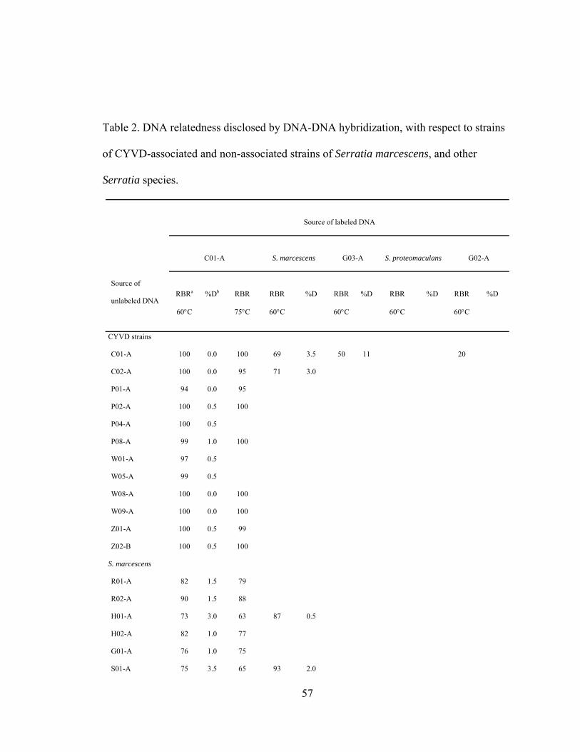
Table 2. DNA relatedness disclosed by DNA-DNA hybridization, with respect to strains
of CYVD-associated and non-associated strains of Serratia marcescens, and other
Serratia species.
Source of labeled DNA
C01-A
S. marcescens
G03-A
S. proteomaculans
G02-A
Source of
unlabeled DNA
RBRa
60°C
%Db
RBR
75°C
RBR
60°C
%D
RBR
60°C
%D
RBR
60°C
%D
RBR
60°C
%D
CYVD strains
C01-A 100 0.0 100 69 3.5 50 11 20
C02-A 100 0.0 95 71 3.0
P01-A 94 0.0 95
P02-A 100 0.5 100
P04-A 100 0.5
P08-A 99 1.0 100
W01-A 97 0.5
W05-A 99 0.5
W08-A 100 0.0 100
W09-A 100 0.0 100
Z01-A 100 0.5 99
Z02-B 100 0.5 100
S. marcescens
R01-A 82 1.5 79
R02-A 90 1.5 88
H01-A 73 3.0 63 87 0.5
H02-A 82 1.0 77
G01-A 76 1.0 75
S01-A 75 3.5 65 93 2.0
57
S02-A 73 3.5 62 91 1.0
Strains to be
identified
G02-A 15 13.0 22 100 0.0
G03-A 46 9.5 100 74 6.1 22
Serratia type
strain
S. marscescens 76 4.5 66 100 0.0 44 18
S. ficaria 66 10.5 52 29
S. plymuthica 51 11 61 64 11.2 22
S. liquefaciens 49 12.5 74 8.5 65 9.1 21
S.
proteomaculans
48 13.5 79 6.5 100 0.0 21
S. odorifera 42 13.0 41 22
S. grimsii 41 12.0 58 65 9.7 16
S. rubidaea 40 12.0 42
S. fonticola 39 12.5 40
a RBR, or relative binding ratio, is determined by the equation: (percent bound to
hydroxylapatite in heterologous reactions) / (percent DNA bound in homologous
reactions) x 1000.
b %D, percent divergence, assumed that a 1ºC decrease in thermal stability of a
heterologous DNA duplex compared with that of the homologous duplex was caused by
1% unpaired bases.
58
Figure 1. Agarose electrophoresis gels of rep-PCR products generated from CYVD
strains, Serratia marcescens from various niches, and other Serratia type strains.
A=PGRS primer, B=ERIC primers, and C=BOX primer. Lanes labeled M were run with
the DNA ladder (DirectLoadTM, Sigma, St. Louis, MO), sizes of fragments are listed on
the right. Lane 1, C01-A; Lane 2, P01-A; Lane 3, W01-A; Lane 4, Z01-A; Lane 5, P03-
A; Lane 6, R01-A; Lane 7, R02-A; Lane 8, R03-A; Lane 9, S01-A; Lane10, S02-A; Lane
11, H01-A; Lane 12, H02-A; Lane 13, G01-A; Lane 14, V01-A, Lane 15, I06-A; Lane
16, I09-A; Lane 17, S. marcescens; Lane 18, G02-A; Lane 19, G03-A; Lane 20, S.
grimesii; Lane 21, S. plymuthica; Lane 22, S. rubideae; Lane 23, S. liquefaciens; Lane 24,
S. fonticola.
Figure 2. Consensus phylogenetic tree of the relationships among Serratia strains and
species, compiled from binary data based on the banding patterns of three different rep-
PCR reaction sets. Branches with bootstrap values less than 500 were collapsed.
59
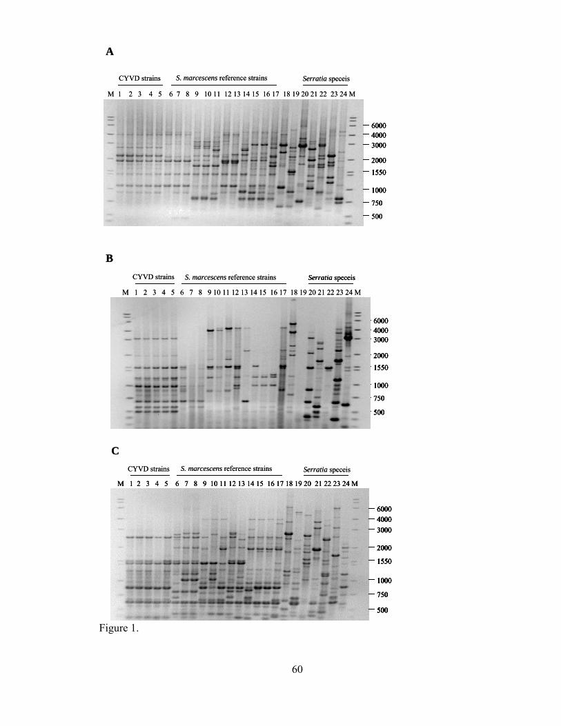
A
M 1 2 3 4 5 6 7 8 9 10 11 12 13 14 15 16 17 18 19 20 21 22 23 24 M
60004000
1550
750
1000
2000
3000
500
CYVD strains S. marcescens reference strains Serratia speceis
B
60004000
1550
750
1000
2000
3000
500
M 1 2 3 4 5 6 7 8 9 10 11 12 13 14 15 16 17 18 19 20 21 22 23 24 M
CYVD strains S. marcescens reference strains Serratia speceis
C
M 1 2 3 4 5 6 7 8 9 10 11 12 13 14 15 16 17 18 19 20 21 22 23 24 M
60004000
1550
750
1000
2000
3000
500
CYVD strains S. marcescens reference strains Serratia speceis
A
M 1 2 3 4 5 6 7 8 9 10 11 12 13 14 15 16 17 18 19 20 21 22 23 24 M
60004000
1550
750
1000
2000
3000
6000600040004000
15501550
750750
10001000
20002000
30003000
500500
CYVD strains S. marcescens reference strains Serratia speceis
B
60004000
1550
750
1000
2000
3000
500
6000600040004000
15501550
750750
10001000
20002000
30003000
500500
M 1 2 3 4 5 6 7 8 9 10 11 12 13 14 15 16 17 18 19 20 21 22 23 24 M
CYVD strains S. marcescens reference strains Serratia speceisSerratia speceis
C
M 1 2 3 4 5 6 7 8 9 10 11 12 13 14 15 16 17 18 19 20 21 22 23 24 M
60004000
1550
750
1000
2000
3000
500
M 1 2 3 4 5 6 7 8 9 10 11 12 13 14 15 16 17 18 19 20 21 22 23 24 M
60004000
1550
750
1000
2000
3000
500
6000600040004000
15501550
750750
10001000
20002000
30003000
500500
CYVD strains S. marcescens reference strains Serratia speceis
Figure 1.
60

G03-A
S. rubideae
G02-A
S. plymuthica
S. liquefaciens
S. grimesii
S. fonticola
S. marcescens type strain
V01-A
I09-A
I06-A990
591
H01-A
S02-A
S01-A824536
612
G01-A
R01-A
H02-A
R02-A
R03-A958
743
P01-A
C01-A
W01-A
Z01-A
P03-A
996
500
Figure 2.
61

CHAPTER IV
GENOMIC COMPARISON OF PLANT PATHOGENIC AND NON-
PATHOGENIC SERRATIA MARCESCENS STRAINS USING SUPPRESSIVE
SUBTRACTIVE HYBRIDIZATION
Abstract
The cucurbit yellow vine disease (CYVD) is caused by a cosmopolitan bacterium
Serratia marcescens. CYVD-associated S. marcescens strains, however, are significantly
different from non-phytopathogenic strains as shown by the chemical utilization
phenotypes. To identify the genetic differences responsible for pathogenicity related
phenotypes, we used a suppressive subtractive hybridization strategy (SSH). S.
marcescens strain Z01-A, isolated from CYVD-affected zucchini, was used as the tester,
whereas S. marcescens rice endophytic strain R02-A (IRBG 502) was used as the driver.
SSH revealed 48 sequences that were present in Z01-A, but absent in R02-A. Sequence
analysis showed that a large proportion of these sequences resembled genes involved in
synthesis of surface structures. By construction of a fosmid library and colony
hybridization, a phage gene cluster and a genome island containing a fimbrial gene
cluster were identified. A multiplex PCR employing primers designed from a strain
specific marker and a species specific marker was able to discriminate between S.
marcescens strains causing CYVD and those from other ecological niches.
62

Introduction
Cucurbit yellow vine disease (CYVD) was first observed in squash (Cucurbita
maxima) and pumpkin (C. pepo) in 1988 in Oklahoma and Texas (6). Since then the
disease has been confirmed in nine other states (4) (29) and Fletcher, J., personal
communication). CYVD is characterized by rapid and general yellowing of leaves
appearing over a 3-4 day period, followed by gradual or rapid decline and death of the
vine in several cucurbit crops (5).
The causal agent of CYVD was identified as the cosmopolitan bacterium S.
marcescens (18). Non-CYVD strains of this species can assume roles as soil or water
resident saprophytes, plant endophytes, insect pathogens, and even opportunistic human
pathogens. None of these other strains is able to cause CYVD and phenotypic differences
between them and CYVD-pathogenic strains include substrate utilization and fatty acid
profiles (18). Rep-PCR fingerprinting and DNA-DNA hybridization also revealed
significant differentiation between CYVD pathogenic strains and S. marcescens strains
from other niches (31). Much remains to be learned, however, about the nature of the
genetic materials that are responsible for the differences.
Suppressive subtractive hybridization is a powerful method for identifying DNA
fragments that are present in one organism (tester) but absent from the other (driver),
especially if these two organisms are closely related (1). It has been widely used for
bacterial genome analysis to discover new epidemiological markers, virulence factors or
host specificity determinants (30). In this work, my goal was to identify the genetic
differences responsible for pathogenicity in cucurbits. We compared the genomes of a
CYVD strain, Z01-A, and a rice endophytic strain, R02-A, in an effort to identify genes
63
or genetic markers present in the phytopathogenic strain but absent from a closely related
nonpathogenic strain. We report the identification of a pool of DNA sequences specific to
CYVD pathogenic strains. In addition, we identified a phage gene cluster and a genome
island containing a type 1 fimbrial (pilus) gene cluster. Furthermore, we designed a
primer set specific for CYVD strains of S. marcescens based on an identified genetic
marker.
Materials and Methods
Strains. Test bacteria included several S. marcescens strains from different ecological
niches, and several CYVD strains from different cucurbit hosts (Table 1). Bacteria were
stored, -80ºC, in aliquots of 1.5 ml Luria-Bertani (LB) broth (22) containing 15%
glycerol. For use in experiments, bacteria were streaked onto LB agar and incubated at
28ºC for 24 hr. E. coli was grown at 37ºC on LB agar or in LB broth.
DNA manipulation. Genomic DNA of CYVD and non-phytopathogenic S.
marcescens strains was isolated using a bacterial genomic DNA miniprep protocol (2).
Plasmid DNA of subtracted library colonies was isolated by the alkaline lysis mini-prep
protocol (22).
Suppressive subtractive hybridization and differential screening. CYVD strain
Z01-A was subtracted from a rice endophytic S. marcescens strain, R02-A, using
CLONTECH PCR-selectTM Bacterial Genome Subtraction Kit (Clontech, Palo Alto, CA,
USA). The hybridization temperature was set at 68ºC to accommodate the high G+C
64
content of the S. marcescens species. The numbers of cycles for primary and secondary
PCR were 26 and 12, respectively. Subtracted sequences were inserted into the TA vector
using the TOPO TA Cloning® kit (Invitrogen, Carlsbad, CA, USA) and electroporated
into One Shot® TOP10 ElectrocompTM E. coli according to the manufacturer’s protocol.
Subtracted library clones were randomly selected as template for PCR amplification
using nested primers 1 and 2R provided by the genome subtraction kit. PCR products
were then transferred to duplicate nylon membranes with a dot blot method (13). Each
membrane was hybridized (2) to an individual probe made from a CYVD or a non-
phytopathogenic strain. The digoxigenin-labeled probe was detected with CSPD (Roche,
Switzerland), and exposures were from 30 min to 3 h.
DNA sequencing and analysis. Insert sequences in the subtracted library were
sequenced by the Oklahoma State University (OSU) Recombinant DNA/ Protein
Resource Facility. Sequencing reactions were performed as recommended by the supplier
(Applied Biosystems, Inc.) and analyzed on an ABI 3100 automated DNA sequencer.
Probable biological functions of the products encoded by genes in the raw sequences
were identified using MyPipeOnline 2.00b, a program designed by the OSU
bioinformatics group (3). Based on the analysis, redundant sequences were identified and
contigs were formed. Individual sequences were also analyzed by BLASTX and/or
BLASTN searches against the GenBank database.
Fosmid cloning and hybridization. A Z01-A fosmid library was constructed using a
CopyControlTM Fosmid Library Production Kit (Epicentre, Madison, WI, USA). Library
construction employed recommended protocols, with slight modifications. Briefly, gel
65
slices containing fractionated DNA were washed twice with 1X GELase Digestion Buffer
before being incubated with GELase for digestion. To induce fosmid clones to high copy
number, the induction solution was added just before the bacterial culture reached log
phase. The incubation period was held to three hours or less to avoid toxicity.
E. coli colonies containing fosmid clones were transferred and fixed to Hybond N+
nylon membranes (Amersham Biosciences, Piscataway, NJ, USA) following the
manufacturer’s instructions. PCR amplicons of subtracted Z01-A unique sequences,
amplified using nested primers 1 and 2R provided by the genome subtraction kit, were
equally pooled and labeled using a DIG DNA Labeling Kit (Roche Applied Science,
Mannheim, Germany). The probe was then hybridized to the blot as described for colony
hybridization.
Once a few fosmid clones were identified to contain phytopathogenic-strain-unique
sequences, reverse hybridizations were conducted to confirm the actual number of unique
sequences located in each fosmid clone. PCR amplicons of subtracted Z01-A unique
sequences were arrayed on a nylon membrane (as described above) and hybridized to a
Dig probe made from one fosmid clone. Similar arrays were made and hybridized to
other individual fosmid clones as well.
Plasmid or chromosome origin of subtracted sequences determination.
Subtracted clone a43, which is clustered with a putative plasmid partition gene in FOSU1
locus, was selected as a target for quantitative PCR to determine whether the subtracted
sequences are located on the chromosome or on a plasmid. A pair of primers, designated
a43F (5’- CGCAGAACATCAACATATCTTAGCC -3’) and a43R (5’-
66
TACCGTAGTAGTGCTGCATGAG -3’), were designed based on a43 using Primer3
software (20). Amplification of a segment of 16S rDNA, the genetic marker on the
chromosome, was carried out as a control. Genomic DNA and plasmid DNA of Z01-A
were prepared from 1.5 ml and 150 ml of log-phase bacterial culture, respectively, and
suspended in 50µl distilled sterile water. Each DNA preparation was diluted 50x, 100x,
200x, 400x and 800x and 1 µl of each solution was used in each PCR reaction. PCR
conditions for amplification of 16S rDNA were as follows: one initial denaturation cycle
at 95°C for 5 min, followed by 26 cycles of 94°C for 30 s, 55°C for 30 s, and 72°C for 1
min 30 s and one final extension cycle of 72°C for 10 min. The amplification of a43 was
carried out under the same conditions except that the annealing temperature was 60°C.
Multiplex polymerase chain reaction (PCR) development. Primers were designed
based on CYVD pathogenic strain specific sequences using Primer3 software (20).
Primers YV1 and YV4, which were designed from the 16S rDNA region of the S.
marcescens genome and shown to be specific to this species (15), were used together
with the new primers in a multiplex PCR to obtain both species and strain specificity.
Bacteria grown in broth culture were washed once with 0.5M sodium chloride and
resuspended in distilled water, and 1 µl suspension was used as template. Multiplex PCR,
performed with the GeneAmp PCR System 9600 (Perkin–Elmer Cetus, Überlingen,
Germany), was carried out in 25-µl volumes including 5µl of 5X Git buffer, 0.5 mM of
dNTP, 0.1 µM of each primer and 2U of Taq DNA polymerase. PCR conditions were as
follows: one initial denaturation cycle at 95°C for 5 min, followed by 34 cycles of 94°C
67

for 40 s, 60°C for 1min, and 72°C for 1 min 30 s and one final extension cycle of 72°C
for 7 min.
Results
Suppressive subtractive hybridization of R02-A from Z01-A. A Z01-A subtracted
library of approximately 400 clones was produced by SSH. Clones from which no
amplicon or multiple amplicons were amplified using nested primers 1 and 2R were
discarded. Selection of clones for further characterization was based on their sizes to
avoid repeated sampling of clones containing the same insert sequences. A total of 183
clones were end sequenced, among which 94 different insert sequences were identified.
Forty nine percent of the 94 inserts were represented at least twice. PCR products of the
clones representing the 94 were then arrayed as described (Materials and Methods) on
duplicate nylon membranes and hybridized to Z01-A and R02-A genomic DNA probes,
respectively. As expected, all the sequences showed hybridization signals with strain
Z01-A (data not shown). Forty-six sequences were shown to be false positives for SSH,
as they hybridized to tester strain R02-A. The remaining 48 DNA fragments were present
in Z01-A but not in R02-A.
A large proportion of the tester specific sequences resembling genes involved in
synthesis of surface molecules. Analysis of the 48 subtracted sequences by BLASTX
search showed that eight of them resembled hypothetical bacterial genes of unknown
function, while nine other sequences had no significant matches with known sequences
(Table 2). Among the clones that had significant similarity to sequences with known
68
function, a large proportion was judged to be involved in the synthesis of bacterial
surface molecules. Clones A40, C19 and C34 resembled genes rmlC, wbbL and wbbA,
respectively, which encode proteins of the rhamnose synthesis pathway and have been
previously identified in the S. marcescens N28 wb O antigen gene cluster (21). The O
antigen is the most external component of lipopolysaccharides (LPS), and its structure
consists of a polymer of oligosaccharide repeating units. The sequence of the clone B1
insert resembled that of a surA isomerase gene, whose product participates in the
assembly of outer membrane proteins (10). A defined surA deletion mutant of
Salmonella enterica serovar Typhimurium C5 was defective in the ability to adhere to
and invade eukaryotic cells (26). Phosphatase amsI, the predicted product of clone A3,
may participate in changes of phosphorylation required for the biosynthesis of
extracellular polysaccharide (EPS) (7).
In addition to the clones mentioned above, nine clones contained sequences
resembling fimbrial genes and six others had ORFs resembling phage proteins. Although
only one gene for a transposase, namely IstA of insertion element IS21 (19), was
identified, it was represented in 38 subtracted clones, indicating its high copy numbers in
the genome.
Identification of two gene clusters. To study the linkage of multiple fimbrial genes
as well as other subtracted sequences, a fosmid library of strain Z01-A containing 410
clones was constructed and maintained on a LB plate. The average size of each clone
insert was 40kb. Using pooled Z01-A specific sequences as the probe (Materials and
Methods), 19 fosmid clones were positive in colony hybridization. The ten clones having
69

the strongest hybridization signals were chosen to study the linkage of individual Z01-A
specific sequences in the genome. Clones were individually labeled and hybridized to 34
Z01-A specific sequences arrayed as described (Materials and Methods) on duplicate
nylon membranes. Hybridization results suggested three categories of clones: 1) two
clones, one of which was designated FOSU1, overlapped each other as shown by
hybridization to 13 sequences, including genes that may be involved in type 1 fimbriae
(pili) synthesis; 2) four other clones, one of which was designated FOSU2, were from the
another locus as they all hybridized to seven Z01-A specific sequences, including five
encoding putative phage proteins; 3) four clones duplicated each other in hybridizing to
two Z01-A specific sequences. Because of their content of multiple Z01-A sequences,
FOSU1 and FOSU2 were selected for shot-gun sequencing. The partial sequence of
FOSU1 (accession number: AC148074) and the full sequence of FOSU2 (accession
number: AC148075) were annotated and one gene cluster was identified in each clone.
Sequence analysis showed that a total of 13 insertion sequences occurred in multiple
loci of the FOSU1 clone (Appendix I) (Figure 1). The average G+C content for FOSU1 is
51.67%, a value much lower than the approximately 59% G+C content calculated for S.
marcescens db11, for which shot-gun sequences are available in the Sanger Institute
database (http://www.sanger.ac.uk/Projects/S_marcescens/). These two features together
indicate that the FOSU1 locus is likely a genome island obtained by lateral gene transfer.
A fimbrial gene cluster, which may be involved in synthesis of filamentous surface
adhesive organelles called type1 pili, was identified at the end of the genome island
(Figure 1). Putative proteins encoded by genes within this cluster include: the fimbrins
fimA and fimI, the adaptor proteins, fimG and fimF, the usher proteins, fimD and fimC,
70

the adhesin, fimH, and the regulatory proteins, fimB and fimE (23). The gene cluster has
the same gene organization as those of other fimbrial gene clusters from related
organisms, except that the regulatory gene fimE is truncated and fimB is located 21kb
upstream. Interestingly, the G+C content of this gene cluster is only 49.94%, even lower
than that of the FOSU1 genome island.
FOSU2 also contained a gene cluster, in which each ORF had a corresponding
homolog in the genome of Fels-1, a phage of Salmonella typhimurium LT2 (Appendix II)
(14). The highest match for each ORF, however, was from various phages, including
Fels-1, Gifsy-2 (14), N15 (ref|NP_046907.1|), CP933U (17) , CP933K (8) and HK97 (9).
Interestingly, all these phages have similar genomic architecture at the gene cluster
region. The Z01-A prophage, however, lacks a virulence factor, superoxide dismutase
SodCIII, that is present in Fels-1. The G+C content of the Z01-A prophage gene cluster is
57.26%, which is close to the 56.46% G+C content for Fels-1 in Samonella typhimurium
LT2 (14).
The location of FOSU1 island on the chromosome. The identification of an ORF
putatively encoding a plasmid partitioning protein within the FOSU1 locus raises the
question of whether flanking sequences are from a plasmid or from the chromosome.
Theoretically, it is possible that a trace amount of plasmid DNA could have remained in
the genomic DNA preparations, and vice versa. However, plasmid DNA would be
enriched in the plasmid preparation and chromosomal DNA would be enriched in the
genomic DNA preparation. In this study, the intensity of a43 sequence amplicon
amplified from the genomic DNA preparation was much greater than amplified from the
71
plasmid preparation, clearly showing that a43, together with the flanking sequences in
FOSU1, are located on the bacterial chromosome (Figure 2).
The conservation of subtracted sequences among S. marcescens strains. The
extent of polymorphism among S. marcescens strains for the subtracted sequences was
assessed by dot hybridization. Sequences were arrayed on duplicate nylon membranes
and hybridized to different CYVD strains or non-phytopathogenic strains. All the 94
sequences hybridized to probes made from genomic DNA of CYVD strains P01-A, C01-
A, W01-A and W09-A, respectively. However, the intensity of hybridization signal
varied among different sequences (data not shown). Forty nine, 39 and 41 of the total 94
sequences hybridized to nonpathogenic strains R01-A, 90-166 and db11, respectively.
The conservation of sequences among S. marcescens strains varied (Table 3). Thirty-four
sequences were present only in strain Z01-A. To confirm the reliability of the
hybridization result, all the sequences were subjected to BLASTN search against the
genome sequences of S. marcescens db11 (Table 3). A homolog with identity greater
than 87% was found in the S. marcescens db11 genome database for every Z01-A
subtracted sequence that hybridized to the db11 genomic DNA probe. On the other hand,
no homolog was found for any of the sequences that were negative for hybridization to
db11. Although clone c98 had a match in the db11 database with an expect value of e-19,
the identity was only 62%.
Multiplex PCR diagnosis. Neither BLASTN nor BLASTX searches of GenBank
database revealed sequence similarity to subtracted clone a79. In addition, the DNA
sequence of a79 was negative for hybridization to all tested CYVD non-phytopathogenic
72
strains (Table 3). A new pair of primers, designated a79F (5’-
CCAGGATACATCCCATGATGAC-3’) and a79R (5’-
CATATTACCTGCTGATGCTCCTC-3’), were designed based on a79. The multiplex
PCR was carried out as described (Materials and Methods). A PCR amplicon of 338 bp
was amplified from CYVD strains, but not from non-phytopathogenic S. marcescens
strains. As a comparison, a 452bp amplicon was amplified from all S. marcescens strains
using primers YV1 and YV4. No fragments were amplified from non- S. marcescens
strains (Figure 3).
Discussion
DNA-DNA hybridization results from a previous study (31) showed that S.
marcescens endophytic strain R02-A, of all the strains tested, was most closely related to
CYVD strains. With a relative binding ratio of 90% and a 1.5 percent divergence from
CYVD representative strain C01-A, and confirmed to be non-pathogenic to cucurbits,
R02-A is an ideal driver strain to subtract from the well-studied CYVD pathogenic strain
Z01-A.
This is the first report of using SSH to identify the genetic differences between S.
marcescens strains, which generally have high G+C content. About 51% of the subtracted
differential sequences were confirmed by dot hybridization to be present in Z01-A, but
absent in R02-A. The reliability of dot hybridization was further validated by
hybridization of subtracted Z01-A sequences with S. marcescens db11, in which a
hybridization signal always corresponded to greater than 87% sequence identity. In
addition, except for IstA of IS21, which had a number of copies on the Z01-A genome, all
73
other sequences were represented no more than 4 times, indicating that the library was
not significantly biased (data not shown). The fact that about half of the differential
sequences were represented only once suggests that the potential library of Z01-A
specific DNAs was not exhausted in our experiments.
A large number of subtracted clones identified in this study resembled known genes
involved in synthesis of O antigen and type 1 pili, both of which are important bacterial
surface structures. O antigen is the outmost component of LPS, the major structural and
immunodominant molecules of the outer membrane. Interestingly, there was evidence
that lateral gene transfer of O antigen genes occurred among subspecies of
Salmonella enterica (11). A large variation in genes encoding surface structures has also
been revealed in previous genome sequencing of pathogenic bacteria, such as Escherichia
coli O157:H7 (27) and Streptococcus pneumoniae (28). It has been suggested that the
variation is obtained by lateral gene transfer as a result of diversifying selection pressure
to evade host immune defenses. A similar phenomenon was reported by Nesbo et al. (16),
who compared different strains of Thermotoga using SSH. It was reasoned, however, that
variation in surface structures might be a general feature of closely related bacterial
strains. In this study, clone A40 and B1 were detected by hybridization in non-
phytpathogenic strains, suggesting that at least some of the genetic variation was a result
of divergence instead of lateral gene transfer. The CYVD pathogen investigated in this
study lives predominantly in the phloem sieve tubes, a tissue that is rich in nutrients and
high in osmotic pressure. In addition, it is transmitted by an insect vector, the squash bug
(Anasa trisits). Although it is not known whether CYVD strains of S. marcescens are able
74

to colonize other ecological niches, it is certainly possible. Adaptation to such widely
diverse niches might be the driving force for the genetic variation of surface molecules.
The most striking result in this study is the identification of two gene clusters present
in CYVD strains of S. marcescens, but absent in closely related, but nonphytopathogenic
strains. Several lines of evidence suggest that the fimbrial gene cluster was likely part of
a genome island acquired from other species: i) the prevalence of putative insertion
sequences at the flanking region; ii) low G+C content; iii) gene organization similar to
that of related bacterial species; iv) the presence of the fimbrial gene cluster only in
CYVD strains, and not in any CYVD non-phytopathogenic strains, as shown by the DNA
hybridization. In other bacteria the whole gene cluster is responsible for the production
and control of type 1 fimbriae (25). The adhesin fimH, which is located at the distal tip of
the pilus, mediates not only bacterial adherence, but also invasion of human bladder
epithelial cells (12). The expression of pilins, however, could be turned on and off by
site-specific DNA inversion of a 312 bp fragment containing the promoter region of fimA
(24). The switch process is catalyzed by two recombinases, fimB and fimE. Our work
showed that Z01-A had only a truncated version of fimE, and that fimB is located 21kb
further upstream of the gene cluster in Z01-A than it is in other bacteria. These features
would impact the production of type one pili. In the future, electron microscopic
observation, fimbrial gene cluster knockout and complementation experiments would
provide evidence whether this cluster of genes contributes to CYVD strain pathogenicity.
We also studied the distribution of the subtracted sequences, including the two gene
clusters, among CYVD strains isolated from different cucurbit species and among non-
phytopathogenic strains from various niches. All CYVD strains tested were positive by
75
hybridizations for the subtracted sequences with various degrees of intensity, consistent
with their average 99% relatedness determined in a DNA-DNA hybridization study (31).
The three CYVD non-associated S. marcescens strains, on the other hand, each
hybridized to a distinct portion of the subtracted Z01-A sequences. Notably, R01-A and
R02-A shared the greatest number of common sequences among the CYVD
nonpathogenic strains (data not shown), confirming the close relatedness of these strains
that was predicted earlier from DNA-DNA hybridization and rep-PCR experiments (31).
Testing the hybridization of the subtracted sequences to a more extensive set of S.
marcescens strains might reveal more genetic variations that underlie the evolutionary
process.
SSH proved to be a useful method for identifying molecular markers for
epidemiological studies. When choosing the markers we purposely avoided the two gene
cluster regions as the occurrence of gene transfer among related bacterial species could
complicate marker identification. Using a primer pair designed based on a subtracted
Z01-A specific sequence, a79, we were able to differentiate CYVD strains from non-
phytopathogenic strains of S. marcescens. The selected marker is likely to be specific as
it has no sequence similarity, at either the DNA or the translated protein level, with any
sequence in GenBank. The multiplex PCR technique, which includes a primer pair to
detect the bacterium at the species level, added further reliability to the new diagnostic
tool. In the future it can be used in vector identification, field disease diagnoses, and
contamination-free examination in laboratory.
76
Literature Cited
1. Agron, P. G., M. Macht, L. Radnedge, E. W. Skowronski, W. Miller, and G. L.
Andersen. 2002. Use of subtractive hybridization for comprehensive surveys of
prokaryotic genome differences. FEMS Microbiol Lett 211:175-82.
2. Ausubel, F. M., R. Brent, R. E. Kingston, D. D. Moore, J. G. Seidman, J. A. Smith,
and S. K. 1987. Current Protocols in Molecular Biology. John Wiley & Sons, New
York, N. Y.
3. Ayoubi, P., X. Jin, S. Leite, X. Liu, J. Martajaja, A. Abduraham, Q. Wan, W. Yan, E.
Misawa, and R. A. Prade. 2002. PipeOnline 2.0: automated EST processing and
functional data sorting. Nucleic Acids Res 30:4761-9.
4. Bost, S. C., F. L. Mitchell, U. Melcher, S. D. Pair, J. Fletcher, A. Wayandande, and
B. D. Bruton. 1999. Yellow vine of watermelon and pumpkin in Tennessee. Plant
Dis 83:587.
5. Bruton, B. D., J. Fletcher, S. D. Pair, M. Shaw, and H. Sittertz-Bhatkar. 1998.
Association of a phloem-limited bacterium with yellow vine disease in cucurbits.
Plant dis 82:512-20.
6. Bruton, B. D., S. D. Pair, T. W. Popham, and B. O. Cartwright. 1995. Occurrence of
yellow vine, a new disease of squash and pumpkin, in relation to insect pests,
mulches, and soil fumigation. Subtrop Plant Sci 47:53-8.
7. Bugert, P., and K. Geider. 1997. Characterization of the amsI gene product as a low
molecular weight acid phosphatase controlling exopolysaccharide synthesis of
Erwinia amylovora. FEBS Lett 400:252-6.
77

8. Jin, Q., Z. Yuan, J. Xu, Y. Wang, Y. Shen, W. Lu, J. Wang, H. Liu, J. Yang, F. Yang,
X. Zhang, J. Zhang, G. Yang, H. Wu, D. Qu, J. Dong, L. Sun, Y. Xue, A. Zhao, Y.
Gao, J. Zhu, B. Kan, K. Ding, S. Chen, H. Cheng, Z. Yao, B. He, R. Chen, D. Ma, B.
Qiang, Y. Wen, Y. Hou, and J. Yu. 2002. Genome sequence of Shigella flexneri 2a:
insights into pathogenicity through comparison with genomes of Escherichia coli
K12 and O157. Nucleic Acids Res 30:4432-41.
9. Juhala, R. J., M. E. Ford, R. L. Duda, A. Youlton, G. F. Hatfull, and R. W. Hendrix.
2000. Genomic sequences of bacteriophages HK97 and HK022: pervasive genetic
mosaicism in the lambdoid bacteriophages. J Mol Biol 299:27-51.
10. Lazar, S. W., and R. Kolter. 1996. SurA assists the folding of Escherichia coli outer
membrane proteins. J Bacteriol 178:1770-3.
11. Li, Q., and P. R. Reeves. 2000. Genetic variation of dTDP-L-rhamnose pathway
genes in Salmonella enterica. Microbiology 146:2291-307.
12. Martinez, J. J., M. A. Mulvey, J. D. Schilling, J. S. Pinkner, and S. J. Hultgren. 2000.
Type 1 pilus-mediated bacterial invasion of bladder epithelial cells. EMBO J
19:2803-12.
13. Mavrodi, D. V., O. V. Mavrodi, B. B. McSpadden-Gardener, B. B. Landa, D. M.
Weller, and L. S. Thomashow. 2002. Identification of differences in genome content
among phlD-positive Pseudomonas fluorescens strains by using PCR-based
subtractive hybridization. Appl Environ Microbiol 68:5170-6.
14. McClelland, M., K. E. Sanderson, J. Spieth, S. W. Clifton, P. Latreille, L. Courtney,
S. Porwollik, J. Ali, M. Dante, F. Du, S. Hou, D. Layman, S. Leonard, C. Nguyen, K.
Scott, A. Holmes, N. Grewal, E. Mulvaney, E. Ryan, H. Sun, L. Florea, W. Miller, T.
78
Stoneking, M. Nhan, R. Waterston, and R. K. Wilson. 2001. Complete genome
sequence of Salmonella enterica serovar Typhimurium LT2. Nature 413:852-6.
15. Melcher, U., F. Mitchell, J. Fletcher, and B. Bruton. 1999. New primer sets
distinguish the cucurbit yellow vine bacterium from an insect endosymbiont.
Phytopathology 89.
16. Nesbo, C. L., K. E. Nelson, and W. F. Doolittle. 2002. Suppressive subtractive
hybridization detects extensive genomic diversity in Thermotoga maritima. J
Bacteriol 184:4475-88.
17. Perna, N. T., G. Plunkett, 3rd, V. Burland, B. Mau, J. D. Glasner, D. J. Rose, G. F.
Mayhew, P. S. Evans, J. Gregor, H. A. Kirkpatrick, G. Posfai, J. Hackett, S. Klink, A.
Boutin, Y. Shao, L. Miller, E. J. Grotbeck, N. W. Davis, A. Lim, E. T. Dimalanta, K.
D. Potamousis, J. Apodaca, T. S. Anantharaman, J. Lin, G. Yen, D. C. Schwartz, R.
A. Welch, and F. R. Blattner. 2001. Genome sequence of enterohaemorrhagic
Escherichia coli O157:H7. Nature 409:529-33.
18. Rascoe, J., M. Berg, U. Melcher, F. Mitchell, B. D. Bruton, S. D. Pair, and J.
Fletcher. 2003. Identfication, phylogenetic analysis and biological characterization of
Serratia marcescens causing cucurbit yellow vine disease. Phytopathology 93:1233-
9.
19. Reimmann, C., R. Moore, S. Little, A. Savioz, N. S. Willetts, and D. Haas. 1989.
Genetic structure, function and regulation of the transposable element IS21. Mol Gen
Genet 215:416-24.
20. Rozen, S., and H. Skaletsky. 2000. Primer3 on the WWW for general users and for
biologist programmers. Methods Mol Biol 132:365-86.
79

21. Saigi, F., N. Climent, N. Pique, C. Sanchez, S. Merino, X. Rubires, A. Aguilar, J. M.
Tomas, and M. Regue. 1999. Genetic analysis of the Serratia marcescens N28b O4
antigen gene cluster. J Bacteriol 181:1883-91.
22. Sambrook, J. E., E. F. Fritsch, and T. Maniatis. 1989. Molecular cloning: a
laboratory manual. 2nd Edition. Cold Spring Harbor Laboratory, Cold Spring Harbor,
N.Y.
23. Schilling, J. D., M. A. Mulvey, and S. J. Hultgren. 2001. Structure and function of
Escherichia coli type 1 pili: new insight into the pathogenesis of urinary tract
infections. J Infect Dis 183 Suppl 1:S36-40.
24. Sohanpal, B. K., H. D. Kulasekara, A. Bonnen, and I. C. Blomfield. 2001.
Orientational control of fimE expression in Escherichia coli. Mol Microbiol 42:483-
94.
25. Soto, G. E., and S. J. Hultgren. 1999. Bacterial adhesins: common themes and
variations in architecture and assembly. J Bacteriol 181:1059-71.
26. Sydenham, M., G. Douce, F. Bowe, S. Ahmed, S. Chatfield, and G. Dougan. 2000.
Salmonella enterica serovar typhimurium surA mutants are attenuated and effective
live oral vaccines. Infect Immun 68:1109-15.
27. Tarr, P. I., L. M. Schoening, Y. L. Yea, T. R. Ward, S. Jelacic, and T. S. Whittam.
2000. Acquisition of the rfb-gnd cluster in evolution of Escherichia coli O55 and
O157. J Bacteriol 182:6183-91.
28. Tettelin, H., K. E. Nelson, I. T. Paulsen, J. A. Eisen, T. D. Read, S. Peterson, J.
Heidelberg, R. T. DeBoy, D. H. Haft, R. J. Dodson, A. S. Durkin, M. Gwinn, J. F.
Kolonay, W. C. Nelson, J. D. Peterson, L. A. Umayam, O. White, S. L. Salzberg, M.
80
R. Lewis, D. Radune, E. Holtzapple, H. Khouri, A. M. Wolf, T. R. Utterback, C. L.
Hansen, L. A. McDonald, T. V. Feldblyum, S. Angiuoli, T. Dickinson, E. K. Hickey,
I. E. Holt, B. J. Loftus, F. Yang, H. O. Smith, J. C. Venter, B. A. Dougherty, D. A.
Morrison, S. K. Hollingshead, and C. M. Fraser. 2001. Complete genome sequence
of a virulent isolate of Streptococcus pneumoniae. Science 293:498-506.
29. Wick, R. L., J. Lernal, J. Fletcher, F. Mitchell, and B. D. Bruton. 2001. Detection of
cucurbit yellow vine disease in squash and pumpkin in Massachusetts. Plant Dis
85:1031.
30. Winstanley, C. 2002. Spot the difference: applications of subtractive hybridisation to
the study of bacterial pathogens. J Med Microbiol 51:459-67.
31. Zhang, Q., R. Weyant, A. G. Steigerwalt, L. A. White, U. Melcher, B. D. Bruton, S.
D. Pair, F. L. Mitchell, and J. Fletcher. 2003. Genotyping of Serratia marcescens
strains associated with cucurbit yellow vine disease by repetitive elements-based
polymerase chain reaction and DNA-DNA hybridization. Phytopathology 93:1240-6.
81
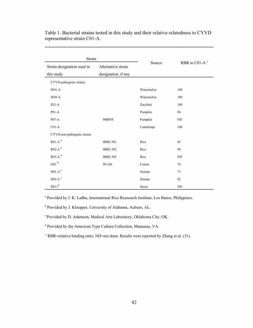
Table 1. Bacterial strains tested in this study and their relative relatedness to CYVD representative strain C01-A.
Strain
Strain designation used in
this study
Alternative strain
designation, if any
Source RBR to C01-A e
CYVD-pathogenic strains
W01-A Watermelon 100
W09-A Watermelon 100
Z01-A Zucchini 100
P01-A Pumpkin 94
P07-A 00B058 Pumpkin ND
C01-A Cantaloupe 100
CYVD-non-pathogenic strains
R01-A a IRBG 501 Rice 82
R02-A a IRBG 502 Rice 90
R03-A a IRBG 505 Rice ND
G01 b 90-166 Cotton 76
H01-A c Human 73
H02-A c Human 82
Db11d Insect ND
a Provided by J. K. Ladha, International Rice Reasearch Institute, Los Banos, Philippines.
b Provided by J. Kloepper, University of Alabama, Auburn, AL.
c Provided by D. Adamson, Medical Arts Laboratory, Oklahoma City, OK.
d Provided by the American Type Culture Collection, Manassas, VA.
e RBR=relative binding ratio; ND=not done. Results were reported by Zhang et al. (31).
82
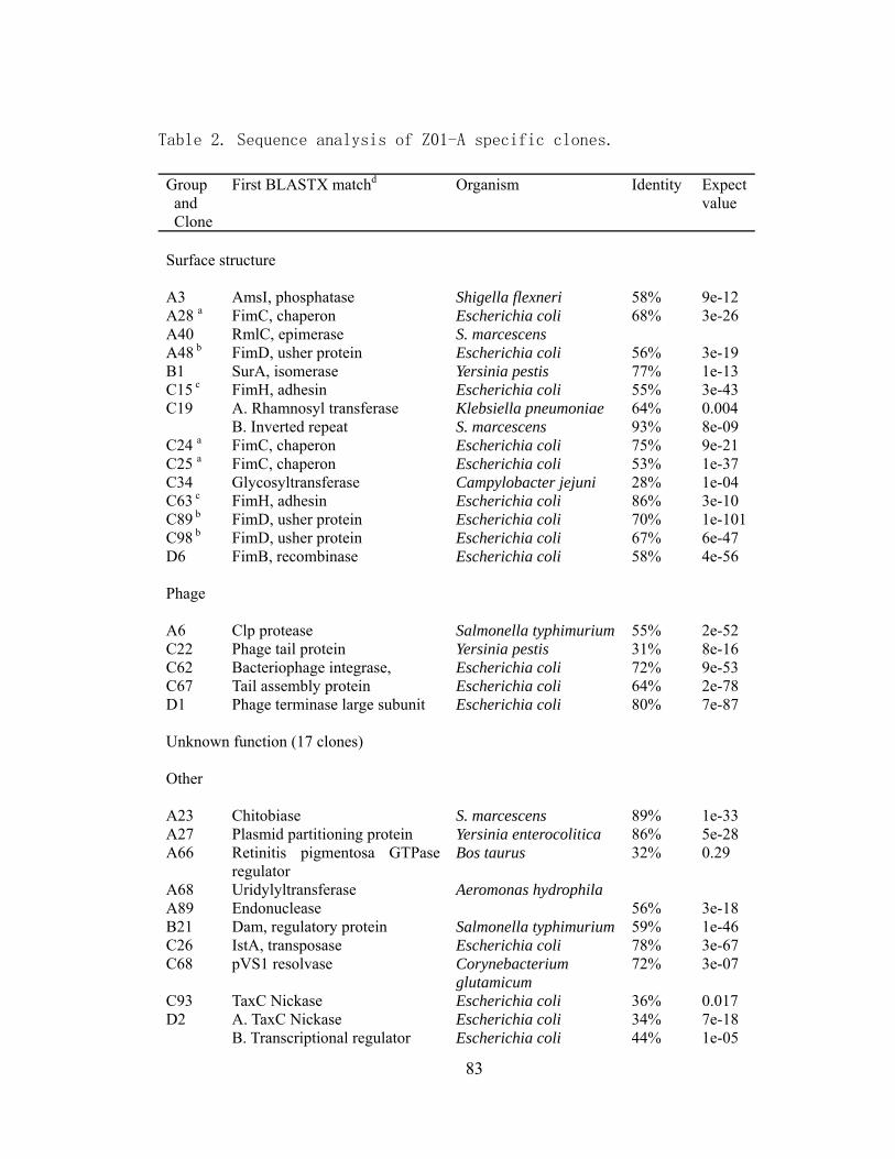
Table 2. Sequence analysis of Z01-A specific clones.
Group
and Clone
First BLASTX matchd Organism Identity Expect value
Surface structure A3 AmsI, phosphatase Shigella flexneri 58% 9e-12 A28 a FimC, chaperon Escherichia coli 68% 3e-26 A40 RmlC, epimerase S. marcescens A48 b FimD, usher protein Escherichia coli 56% 3e-19 B1 SurA, isomerase Yersinia pestis 77% 1e-13 C15 c FimH, adhesin Escherichia coli 55% 3e-43 C19 A. Rhamnosyl transferase
B. Inverted repeat Klebsiella pneumoniae S. marcescens
64% 93%
0.004 8e-09
C24 a FimC, chaperon Escherichia coli 75% 9e-21 C25 a FimC, chaperon Escherichia coli 53% 1e-37 C34 Glycosyltransferase Campylobacter jejuni 28% 1e-04 C63 c FimH, adhesin Escherichia coli 86% 3e-10 C89 b FimD, usher protein Escherichia coli 70% 1e-101 C98 b FimD, usher protein Escherichia coli 67% 6e-47 D6 FimB, recombinase Escherichia coli 58% 4e-56 Phage A6 Clp protease Salmonella typhimurium 55% 2e-52 C22 Phage tail protein Yersinia pestis 31% 8e-16 C62 Bacteriophage integrase, Escherichia coli 72% 9e-53 C67 Tail assembly protein Escherichia coli 64% 2e-78 D1 Phage terminase large subunit Escherichia coli 80% 7e-87 Unknown function (17 clones) Other A23 Chitobiase S. marcescens 89% 1e-33 A27 Plasmid partitioning protein Yersinia enterocolitica 86% 5e-28 A66 Retinitis pigmentosa GTPase
regulator Bos taurus 32% 0.29
A68 Uridylyltransferase Aeromonas hydrophila A89 Endonuclease 56% 3e-18 B21 Dam, regulatory protein Salmonella typhimurium 59% 1e-46 C26 IstA, transposase Escherichia coli 78% 3e-67 C68 pVS1 resolvase Corynebacterium
glutamicum 72% 3e-07
C93 TaxC Nickase Escherichia coli 36% 0.017 D2 A. TaxC Nickase
B. Transcriptional regulator Escherichia coli Escherichia coli
34% 44%
7e-18 1e-05
83
D26 A. Putative exported protein B. Inverted repeat
Yersinia pestis S. marcescens
67% 95%
8e-28 6e-11
a Clones A28, C24 and C25 matched with different parts of fimC.
b Clones A48, C89 and C98 matched with different parts of fimD.
c Clones C15 and C63 matched with different parts of fimH.
d If more than one gene was covered by the clone, they are given as follows: A, gene X; B, gene
Y.
84
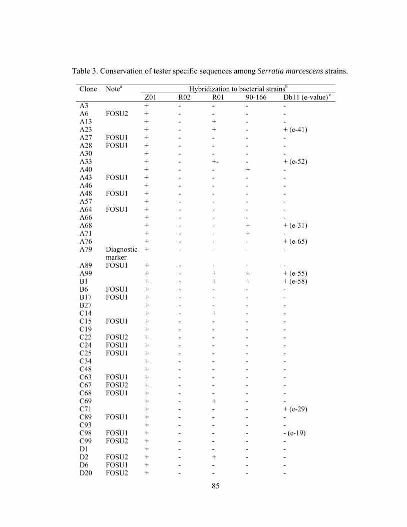
Table 3. Conservation of tester specific sequences among Serratia marcescens strains.
Hybridization to bacterial strainsbClone Notea
Z01 R02 R01 90-166 Db11 (e-value) c
A3 + - - - - A6 FOSU2 + - - - - A13 + - + - - A23 + - + - + (e-41) A27 FOSU1 + - - - - A28 FOSU1 + - - - - A30 + - - - - A33 + - +- - + (e-52) A40 + - - + - A43 FOSU1 + - - - - A46 + - - - - A48 FOSU1 + - - - - A57 + - - - - A64 FOSU1 + - - - - A66 + - - - - A68 + - - + + (e-31) A71 + - - + - A76 + - - - + (e-65) A79 Diagnostic
marker + - - - -
A89 FOSU1 + - - - - A99 + - + + + (e-55) B1 + - + + + (e-58) B6 FOSU1 + - - - - B17 FOSU1 + - - - - B27 + - - - - C14 + - + - - C15 FOSU1 + - - - - C19 + - - - - C22 FOSU2 + - - - - C24 FOSU1 + - - - - C25 FOSU1 + - - - - C34 + - - - - C48 + - - - - C63 FOSU1 + - - - - C67 FOSU2 + - - - - C68 FOSU1 + - - - - C69 + - + - - C71 + - - - + (e-29) C89 FOSU1 + - - - - C93 + - - - - C98 FOSU1 + - - - - (e-19) C99 FOSU2 + - - - - D1 + - - - - D2 FOSU2 + - + - - D6 FOSU1 + - - - - D20 FOSU2 + - - - -
85
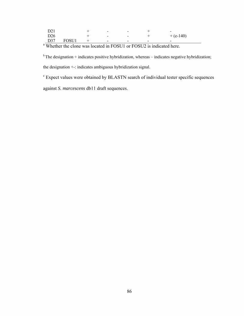
D21 + - - + - D26 + - - + + (e-140) D37 FOSU1 + - - - -
a Whether the clone was located in FOSU1 or FOSU2 is indicated here.
b The designation + indicates positive hybridization, whereas – indicates negative hybridization;
the designation +-: indicates ambiguous hybridization signal.
c Expect values were obtained by BLASTN search of individual tester specific sequences
against S. marcescens db11 draft sequences.
86

Figure 1. Sequence analysis of FOSU1 and its fimbrial gene cluster
A. Diagrammatic representation of FOSU1. Spotted rectangles represent different
insertion sequences. Plain grey rectangles represent ORFs of unknown function or
structural proteins.
B. Putative ORFs in the fimbrial gene cluster identified in this study.
C. Putative ORFs in the fimbrial gene cluster from E.coli O157:H7 EDL933 (upper) and
Shigella flexneri 2a str 2457T (lower).
D. G+C content of the fimbrial cluster.
Figure 2. Agarose gel electrophoresis of 16S rDNA (Lanes 1-5) and sequence a43 (Lanes
6-10) amplified from a dilution series of genomic DNA (Upper set, 50x, 100x, 200x,
400x, 800x dilution of original isolated DNA solution, respectively) and plasmid
preparations (Lower set, 50x, 100x, 200x, 400x, 800x dilutionof original isolated DNA
solution, respectively).
Figure 3. CYVD pathogenic (lanes 2-5) and non-pathogenic strains (lanes 6-12) detected
by multiplex PCR employing primers YV1/4 and a79F/R.
A. 452 bp band amplified by YV1/4
B. 338 bp band amplified by a79F/R
87

fimH fimG fimF fimD fimC fimI fimA fimE ISSM1ISSM1
1kb
fimH fimG fimF fimD fimC fimI fimA fimE fimB
fimH fimG fimF fimD fimC fimI fimA fimE fimBgntP
gntP fimDIS1
yjhA
yjhA
A.
B.
C.
Fimbrial clusterFimB
61.66
49.94
25.0
D. 61.66
49.94
25.0
D.
Figure 1.
Figure 2.
88
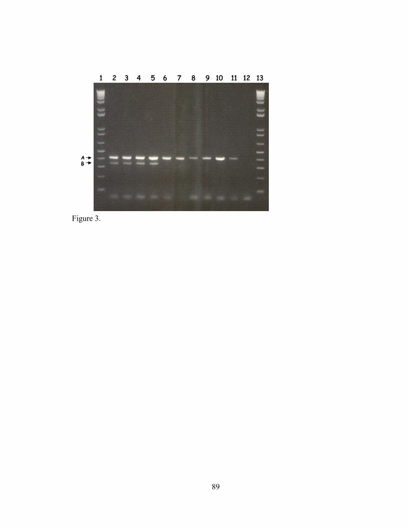
1 2 3 4 5 6 7 8 9 10 11 12 13
AB
1 2 3 4 5 6 7 8 9 10 11 12 13
AB
Figure 3.
89

CHAPTER V
TRANSPOSON MUTAGENESIS OF SERRATIA MARCESCENS, THE CAUSAL
AGENT OF CUCURBIT YELLOW VINE DISEASE, AND VIRULENCE
SCREENING ON CAENORHABDITIS ELEGANS
Abstract
Conjugational mutagenesis was conducted on Serratia marcescens, the causal agent
of cucurbit yellow vine disease, to create insertion mutants. Transformants were
identified by antibiotic selection using kanamycin and confirmed by amplification of the
kanamycin resistance gene using the polymerase chain reaction. Southern hybridization
analysis, however, indicated that the insertion of transposons into the S. marcescens
chromosome was site-specific rather than random. In this study, we also evaluated the
utility of Caenorhabditis elegans as an in vivo model for the study of S. marcescens
virulence factors. Both killing assays and feeding inhibition assays showed that S.
marcescens was only moderately toxic to C. elegans. This model, therefore, is not
appropriate for large-scale virulence screening.
Introduction
Serratia marcescens is a Gram-negative bacterium that has adapted to various
environmental niches, including soil, water, insects and human bodies. This bacterium is
also associated with an epidemic plant disease, namely cucurbit yellow vine disease
90
(CYVD) (3). Symptomatic cucurbits typically show yellowing, wilting and phloem
discoloration. Initially identified in central Oklahoma and central Texas, this disease has
now been detected in at least ten U.S. states.
Phenotypic and genotypic comparisons of CYVD-associated S. marcescens strains
with non-phytopathogenic strains from various niches showed significant differences (13)
(17). With the complete sequence of the S. marcescens insect pathogenic strain db11
available (http://www.sanger.ac.uk/Projects/S_marcescens), I would expect that
comparative genomics would greatly facilitate our understanding of the evolution of this
bacterium. However, to fully understand the pathogenicity mechanisms that lead to
CYVD, it is necessary to conduct functional genetic analysis experimentally. Bacterial
mutagenesis is an essential tool for this purpose.
Transposon mutagenesis has provided an important tool for analyzing gene structure,
function and regulation. Among different types of transposons, Tn5 has been used
widely for Gram-negative bacterial mutagenesis because of its high insertion frequency
and low specificity (Mobile Element II, 2002). Estepa and Harshey (7) reported the first
transposon mutagenesis of a strain of S. marcescens, Sm 274, using a broad-host-range
conjugative suicide plasmid vector. In their study, multiple transposition events were
obtained after elimination of the transferred plasmid. It was speculated that the presence
of multiple plasmids was responsible for this phenomenon.
De Lorenzo et al. (5) reported the construction of minitransposons, containing various
selectable antibiotic resistance markers, for engineering bacteria. An important feature of
minitransposons is that the transposase gene is cognately arranged outside of the
transposon’s cut-end sequence. As a result, a single transposition of the mintransposon
91

into the bacterial chromosome occurs, and the insert is stably inherited. Dennis and
Zylstra (6) added another feature, self-cloning ability, into minitransposons, and called
the resulting constructs plasposons. Together these two features greatly facilitate the
rapid genetic analysis of Gram-negative bacterial genomes.
Bruton et al. (4) provided evidence showing that the CYVD bacterium is transmitted
by the squash bug, Anasa tristis. In that study, cucurbit-derived strains of S. marcescens
were introduced into greenhouse-grown squash plants by mechanical puncture
inoculation and into field-grown squash plants by enclosure with S. marcescens-fed
squash bugs. Up to 60 percent of the mechanically inoculated plants and up to 17 percent
of field plants caged with inoculative squash bugs developed phloem discoloration and
tested positive for S. marcescens by CYVD-specific PCR. However, the symptoms of
plant decline and collapse, normally part of the CYVD syndrome, were not observed in
mechanically inoculated plants in the greenhouse. The low percentage of successful
inoculations and the inability to obtain full symptom expression following mechanical
inoculation makes bacterial mutant pathogenicity screening extremely difficult,
especially since thousands of mutants may need to be analyzed in the genetic study.
There is a need for development of screening methods that are convenient and reliable.
Tan and Ausubel (16) first used Caenorhabditis elegans, a free-living nematode well
known for its genetically tractable traits, convenience and economical maintenance, to
investigate host-pathogen interactions. C. elegans died within 24 h when fed on a lawn of
Pseudomonus aeruginosa, a bacterium capable of causing diseases in both plants and
animals. Another cosmopolitan bacterium, Burkholderia pseudomallei, also causes the
92
mortality of C. elegans (10). More recently, Kurz et al. (9) extended the utility of C.
elegans as an in vivo model for the study of S. marcescens virulence factors.
In early studies, killing assays relied on the exposure of C. elegans to bacterial
pathogens grown on solid media and the visual scoring of nematode mortality. This test
was especially useful in testing the mechanisms of toxin-based paralysis. Smith et al. (15)
developed a liquid-based feeding inhibition assay for the assessment of bacterial
virulence, which allowed the effects of bacterial pathogens on C. elegans, including food
evading, to be quantified more precisely. In this study we evaluated the capability of S.
marcescens to cause disease in C. elegans by both a killing assay and a feeding inhibition
test.
Materials and Methods
Bacterial strains, nematodes and culture conditions. Bacteria and nematodes used
in this study are presented in Table 1. E. coli JM109 (pTnMod-KmO) and E. coli DH5α
(pRK2013) were obtained from Carol L. Bender (Oklahoma State University, OK). S.
marcescens Z01-A, isolated originally from zucchini squash with CYVD, has been
described (3). Bacteria were stored, -80ºC, in 1.5 ml Luria-Bertani (LB) (14) broth
aliquots containing 15% glycerol. For use in experiments, bacteria were streaked onto LB
agar or inoculated into LB broth and incubated at 28ºC for 24 h. E. coli was grown at
37ºC in LB broth.
C. elegans wild type strain N2 was used for killing assay, and C. elegans phm-2, a
mutant strain with aberrant food grinding apparatus (2), was used for feeding inhibition
93
assay. The nematode culture methods, nematode growth medium (NGM) and K-medium
have been described (8).
Selection of Z01-A rifampcin resistant spontaneous mutants. CYVD strain Z01-A
was grown overnight at 28ºC in LB broth. A quantity of 100 µl of the bacterial
suspension was added to 5 ml LB broth containing 15 µg /ml rifampicin, and allowed to
grow for 48 h at 28ºC. One hundred µl was subsequently transferred to 30 µg /ml
rifampicin and grown as before. A quantity of 100 µl of these cells was plated onto LB
agar containing 30 µg/ml rifampicin. One colony, designated Z01-RR1, was selected and
transferred to LB broth, grown to log phase and stored frozen in aliquots as described
above (Rascoe, unpublished data).
To determine whether the rifampicin resistance phenotype of Z01-RR1 is stably
maintained a loopful of Z01-RR1 stock solution from a frozen aliquot was streaked onto
LB agar without rifampicin. Each day, an additional streak was made from the previous
bacterial culture. After 20 generations of streaks, a loopful of bacterial cells was streaked
onto LB agar containing 30 µg/ml rifampicin to determine whether antibiotic resistance
had been retained.
Determination of minimum inhibitory concentration (MIC) of antibiotics.
Antibiotics used for selection in these experiments included kanamycin and rifampicin. A
series of two-fold dilutions of the antibiotic stock solutions were prepared in LB broth.
Around 100 bacterial cells were inoculated into each dilution of antibiotic broth culture
94
and shaken for 24 hours at 28ºC (S. marcescens Z01-A) or 48 h at 37ºC (E. coli JM109,
DH5α). Growth was assessed by the turbidity of the culture medium at 550nm.
Conjugational mutagenesis. S. marcescens Z01-RR1, E. coli JM109 (pTnMod) and
E. coli DH5α (pRK2013) were individually grown in LB broth to mid-log phase. Three
hundred and thirty microliters of each culture were added to a single tube, vortexed and
centrifuged at 5000x g for 1 min. The supernatant was discarded and the pellet
resuspended in 150 µl LB broth and plated on a non-selective LB plate. The mating plate
was incubated for 14 h at 28ºC. The bacterial cells were then scraped from the agar
surface and resuspended in 1 ml 10% sterile glycerol. The resultant bacterial cell culture
was plated on LB plates containing 75µg/ml kanamycin and 100µg/ml rifampicin. Ten
colonies were selected in each mating experiment.
PCR amplification. 16S rDNA sequence was amplified by polymerase chain
reaction (PCR) from S. marcescens Z01-RR1using primers YV1 (5’-
GGGAGCTTGCTCCCCGG-3’) and YV4 (5’-
AACGTCAATTGATGAACGTATTAAGT-3’). PCR amplification was performed in
25 µl reaction mixtures containing a bacterial colony, 1x Gitshier buffer (16.6 mM
[NH4]2SO4; 67 mM Tris-HCl, pH 8.8; 6.7 mM MgCl2; 6.7 mM EDTA; 30 mM β-
mercaptoethanol), 100 µM of each dNTP, 0.5 µM each of primers YV1 and YV4, and 2
U Taq DNA polymerase (Promega, Madison, WI, USA). This mixture was placed in a
GeneAmp PCR system 9600 (Perkin-Elmer Cetus) thermal cycler and subjected to a
5 min denaturation at 95 °C followed by 30 cycles at 95 °C for 30 s, 55 °C for 30 s and
95
72 °C for 90 s. The reaction mixture was held at 72 °C for 10 min and then stored at 4 °C
until analyzed on a 1% agarose gel. The kanamycin resistance gene marker was amplified
from a JM109 or DH5α colony using primers km1 (5’-
CCGAGGCAGTTCCATAGGATG-3’) and km2 (5’-
TTTATATGGGTATAAATGGGCTCGC-3’). The program for amplification of the
kanamycin resistance gene was the same as that described above except that the
annealing temperature was raised to 64 °C. Z01-A transformants were subjected to two
independent PCR amplifications using primer pairs YV1/4 and Km1/2, respectively.
Southern hybridization. Genomic DNA was extracted using a hexadecyl trimethyl
ammonium bromide (CTAB) method (1). The sample of each strain was prepared for
electrophoresis by digesting 2 µg of genomic DNA with EcoRI restriction enzymes in a
total of 20 µl, and the DNA fragments were separated in a 0.8% agarose gel. The gels
were blotted onto nylon membranes (Amersham Biosciences, Piscataway, NJ, USA)
following the manufacturer’s instructions. The probe was made from PCR product
amplified from the kanamycin resistance gene and labeled with the DIG High Prime
DNA labeling kit (Roche, Switzerland). Prehybridization and hybridization were done
with DIG Easy Hyb (Roche, Switzerland) at 42°C in a hybridization bag incubated in a
water bath. The digoxigenin-labeled probe was detected with CSPD (Roche,
Switzerland), and exposures were from 30 min to 3 h.
Killing assay of C. elegans. Nematode growth medium (NGM) was poured into 5.5
cm petri plates, covered and allowed to dry at room temperature for 2 days. Ten
96
microliters of an overnight culture of a test bacterial strain was spread on a NGM plate
and incubated at 37°C for 24 hours. Optimum growth of the bacterial culture was then
obtained by incubating the plates at room temperature for another 8-24 hours depending
on the growth rate of different bacteria. Each plate was seeded with 20 L4-stage C.
elegans worms (post-embryonic development of C. elegans involves growth through four
larvae stages, L1 to L4, before the final molt to the adult stage). Plates were scored for
nematode survival every 8 hours, and three replicates per trial were performed. A worm
was considered dead when it no longer moved in response to stimulation. Worms that
dried out on the sidewall of the plate were excluded from the analysis.
C. elegans feeding inhibition assay. The feeding inhibition assay was carried out
using the method by Smith et al. (15), except for minor changes. Briefly, a bacterial pellet
was resuspended in K-medium to an OD550 of approximately 1.2. C. elegans mutant phm-
2 worms grown on an E. coli lawn were washed with K-medium (2) and brought to a
concentration of 10 worms/µl by removing the supernatant after 20 min incubation.
Experiments were conducted in 24 well polystyrene cell culture trays (Costar, Corning,
NY, USA). Aliquots of 1.7 ml of bacterial suspension, 200 µl of evenly mixed worms
and 100 µl of K-medium were added to each well. For the feeding inhibition control, the
K-medium was replaced with 100 µl 2 M sodium salicylate solution, which inhibits the
feeding of C. elegans. To measure the natural degradation of the bacteria in K-medium, a
well without nematodes was also prepared. Every 24 h during the incubation, 1 ml
samples were transferred to sterile disposable cuvettes and the OD550s were measured
after the worms settled to the bottom.
97

Results and Discussion
To further the genetic analysis of S. marcescens, the causal agent of CYVD,
mutagenesis was conducted on this bacterium to create a pool of mutants. S. marcescens
is an enterobacterium that exhibits very low efficiency of transformation (11), but
successful transfer of a plasmid into S. marcescens by conjugation has been reported
(12). In the current study we used E. coli JM109 (pTnMod-OKm) as the donor strain and
E. coli DH5α (pRK2013) as the helper strain for transconjugation. To facilitate selecting
conjugational transformants from triparents, a phenotypic marker was obtained by
selecting a spontaneous rifampicin resistant Z01-A mutant. The mutant showed resistance
even after 20 passages of selection on LB without rifampicin, demonstrating that the
antibiotic resistance phenotype was stably inherited. The spontaneous mutant, designated
Z01-RR1, was used for conjugational mutagenesis.
The MICs of kanamycin and rifampicin for each of the triparental strains were
determined before conjugational mutagenesis (Table 2). The final kanamycin
concentration used for selecting conjugational transformants was 75µg/ml, which is
slightly lower than the MIC for JM109 and DH5α. The final concentration of rifampicin,
100 µg/ml, on the other hand, was higher than the MIC of JM109 and DH5α but lower
than that of Z01-RR1.
Selected transformants were confirmed by two independent PCRs. The first PCR,
using primer YV1 and YV4, amplified a 452 bp segment of 16S rDNA sequence that is
specific to recipient, Z01-RR1. The second PCR, using primers km1 and km2, amplified
a 750 bp segment of the kanamycin resistance gene, which is absent in the recipient
strain. The resulting products from both reactions were mixed and electrophoresed. Two
98
bands, 452 bp and 750 bp, were detected in all ten randomly selected conjugational
transformants (Figure 1). As a comparison, a single band of 452 bp was amplified from
the recipient strain Z01-A and a band of 750 bp was amplified from the donor strain
JM109 and the helper strain DH5α. These results confirmed that all selected colonies
were true transformants.
The Southern hybridization experiment result again showed that a single band was
recognized in all six randomly selected transformants by a kanamycin resistance gene
specific probe (Figure 2). However, all the bands were of approximately the same size,
which indicates that the insertion of transposons into the Z01-A chromosome was site
specific rather than random. An alternative explanation is that the transposon preferably
inserted into a Z01-A plasmid that has no EcoRI specific restriction site. In either case the
transformants obtained in this study would not be appropriate for use in large scale
virulence screening.
Because screening for loss of pathogenicity of CYVD strains on the host plant is so
difficult, other screening methods were investigated to study pathogenicity defective
mutants. Preliminary experiments by others (Luo and Fletcher, personal communication)
have demonstrated the unsuitability of using other plant species, such as onion, carrot,
and tobacco, for virulence screening. In this study we investigated the capability of
CYVD wild type strains and their derivatives to cause mortality of C. elegans N2. In the
positive control, 90% of the worms fed on S. marcescens db11 died within 4 days (Figure
3), whereas half of the worms fed on E. coli OP50 were still alive after 6 days. Overall, a
slightly lower percentage of C. elegans survived when fed on CYVD wild type strains
W01-A, Z01-A and the rifampicin resistant derivative Z01-RR, as compared to worms
99

fed on E. coli OP50. These results illustrate that CYVD of S. marcescens strains are only
moderately toxic to C. elegans. Similar results were obtained in the feeding inhibition
assay, in which a C. elegans mutant was used. The mutant C. elegans phm-2 has a
malfunctional grinder. Due to the aberrant grinder, increased numbers of bacteria can
pass through the nematode pharynx into the gut (2). In the control treatment, the optical
density of the bacterial suspensions from which the worms had settled down fell over
time as the nematodes fed on E. coli OP50. In the first 24 h of incubation the rate of
decrease in optical density was similar for all treatments (Table 3). However, in the
incubation period from 24 h to 48 h there were differences among treatments. When C.
elegans phm-2 was incubated with S. marcescens Z01-RR1 or db11, the rate of reduction
of OD550 was higher than when the salicylic acid inhibitor was added to the worms
incubated with E. coli OP50, but lower than when worms were incubated with only E.
coli, indicating that Z01-RR1 was only moderately toxic to the C.elegans worms. This
data, however, is not statistically significant as the experiment was conducted only once.
In a separate experiment, Ewbank et al. (unpublished data) found that all the worms fed
on W01-A died within four days. This is clearly in contrast to the findings in this study.
In the future, replicate experiments with more sampling time intervals should be
conducted to assess the virulence of CYVD strains on C. elegans.
100
Literature Cited
1. Ausubel, F. M., Brent R., Kingston, R. E., Moore, D. D., Seidman, J. G., Smith, J.
A. and Struhl K. 1987. Current Protocols in Molecular Biology. John Wiley &
Sons, New York, N. Y.
2. Avery, L. 1993. The genetics of feeding in Caenorhabditis elegans. Genetics
133:897-917.
3. Bruton, B. D., J. Fletcher, S. D. Pair, M. Shaw, and H. Sittertz-Bhatkar. 1998.
Association of a phloem-limited bacterium with yellow vine disease in cucurbits.
Plant dis 82:512-20.
4. Bruton, B. D., F. Mitchell, J. Fletcher, S. D. Pair, A. Wayadande, U. Melcher, J.
Brady, B. Bextine, and T. W. Popham. 2003. Serratia marcescens, a phloem-
colonizing, squash bug-transmitted bacterium: causal agent of cucurbit yellow
vine disease. Plant Dis 87:937-44.
5. de Lorenzo, V., M. Herrero, U. Jakubzik, and K. N. Timmis. 1990. Mini-Tn5
transposon derivatives for insertion mutagenesis, promoter probing, and
chromosomal insertion of cloned DNA in gram-negative eubacteria. J Bacteriol
172:6568-72.
6. Dennis, J. J., and G. J. Zylstra. 1998. Plasposons: modular self-cloning
minitransposon derivatives for rapid genetic analysis of gram-negative bacterial
genomes. Appl Environ Microbiol 64:2710-5.
7. Estepa, G. R., and R. M. Harshey. 1989. Multiple high-frequency transpositions
of Tn5 in Serratia marcescens 274 from a thermosensitive replication mutant of
plasmid R388. Gene 81:355-9.
101
8. Hope, I. A. 1999. C. elegans: A practical approach. Oxford University Press, New
York.
9. Kurz, C. L., S. Chauvet, E. Andres, M. Aurouze, I. Vallet, G. P. Michel, M. Uh, J.
Celli, A. Filloux, S. De Bentzmann, I. Steinmetz, J. A. Hoffmann, B. B. Finlay, J.
P. Gorvel, D. Ferrandon, and J. J. Ewbank. 2003. Virulence factors of the human
opportunistic pathogen Serratia marcescens identified by in vivo screening.
EMBO J 22:1451-60.
10. O'Quinn, A. L., E. M. Wiegand, and J. A. Jeddeloh. 2001. Burkholderia
pseudomallei kills the nematode Caenorhabditis elegans using an endotoxin-
mediated paralysis. Cell Microbiol 3:381-93.
11. Palomar, J., J. F. Guasch, M. Regue, and M. Vinas. 1990. The effect of nuclease
on transformation efficiency in Serratia marcescens. FEMS Microbiol Lett
57:255-8.
12. Park, J. E., K. E. Young, H. G. Schlegel, H. G. Rhie, and H. S. Lee. 2003.
Conjugative plasmid mediated inducible nickel resistance in Hafnia alvei 5-5. Int
Microbiol 6:57-64.
13. Rascoe, J., M. Berg, U. Melcher, F. Mitchell, B. D. Bruton, S. D. Pair, and J.
Fletcher. 2003. Identfication, phylogenetic analysis and biological
characterization of Serratia marcescens causing cucurbit yellow vine disease.
Phytopathology 93:1233-39.
14. Sambrook, J. E., Fritsch, E. F., and Maniatis, T. 1989. Molecular cloning: a
laboratory manual. 2nd Edition. Cold Spring Harbor Laboratory, Cold Spring
Harbor, N.Y.
102
15. Smith, M. P., T. R. Laws, T. P. Atkins, P. C. Oyston, D. I. de Pomerai, and R. W.
Titball. 2002. A liquid-based method for the assessment of bacterial pathogenicity
using the nematode Caenorhabditis elegans. FEMS Microbiol Lett 210:181-5.
16. Tan, M. W., and F. M. Ausubel. 2000. Caenorhabditis elegans: a model genetic
host to study Pseudomonas aeruginosa pathogenesis. Curr Opin Microbiol 3:29-
34.
17. Zhang, Q., R. Weyant, A. G. Steigerwalt, L. A. White, U. Melcher, B. D. Bruton,
S. D. Pair, F. L. Mitchell, and J. Fletcher. 2003. Genotyping of Serratia
marcescens strains associated with cucurbit yellow vine disease by repetitive
elements-based polymerase chain reaction and DNA-DNA hybridization.
Phytopathology 93:1240-1246.
103
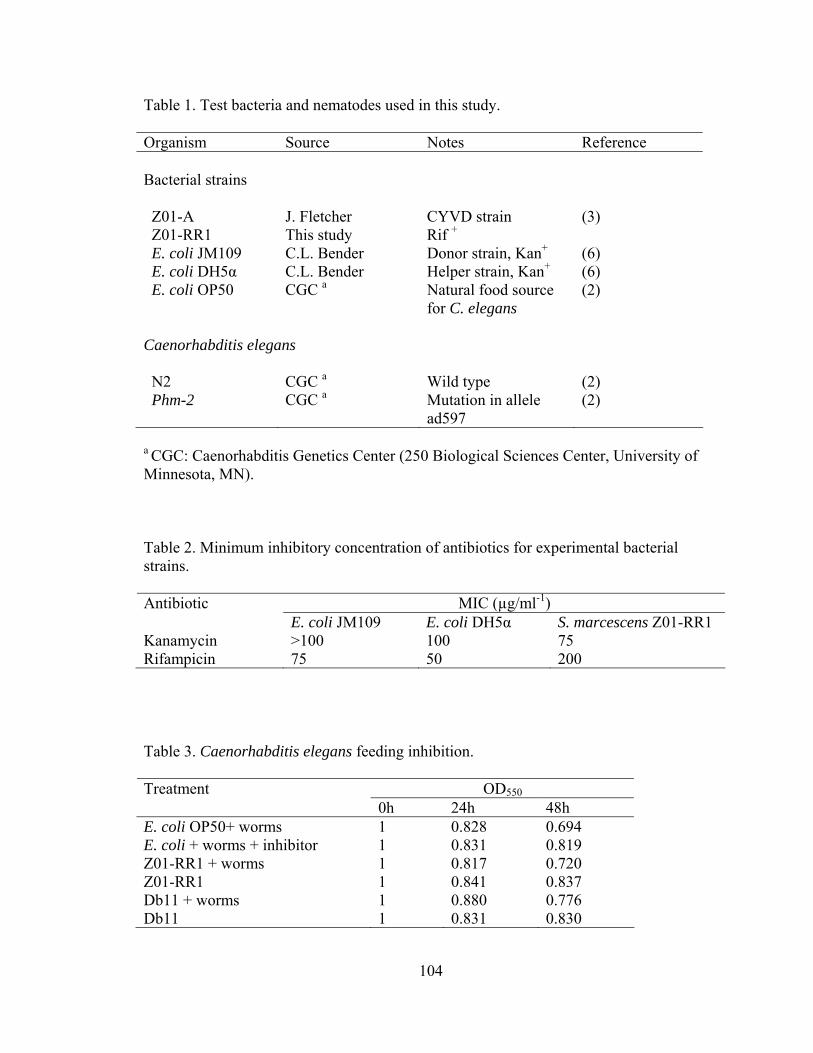
Table 1. Test bacteria and nematodes used in this study.
Organism Source Notes Reference Bacterial strains Z01-A J. Fletcher CYVD strain (3) Z01-RR1 This study Rif + E. coli JM109 C.L. Bender Donor strain, Kan+ (6) E. coli DH5α C.L. Bender Helper strain, Kan+ (6) E. coli OP50 CGC a Natural food source
for C. elegans (2)
Caenorhabditis elegans N2 CGC a Wild type (2) Phm-2 CGC a Mutation in allele
ad597 (2)
a CGC: Caenorhabditis Genetics Center (250 Biological Sciences Center, University of Minnesota, MN). Table 2. Minimum inhibitory concentration of antibiotics for experimental bacterial strains.
MIC (µg/ml-1) Antibiotic E. coli JM109 E. coli DH5α S. marcescens Z01-RR1
Kanamycin >100 100 75 Rifampicin 75 50 200 Table 3. Caenorhabditis elegans feeding inhibition.
OD550Treatment 0h 24h 48h
E. coli OP50+ worms 1 0.828 0.694 E. coli + worms + inhibitor 1 0.831 0.819 Z01-RR1 + worms 1 0.817 0.720 Z01-RR1 1 0.841 0.837 Db11 + worms 1 0.880 0.776 Db11 1 0.831 0.830
104

Figure 1. Z01-A transformants (lanes 1-10), Z01-A (lane 11), JM109 (lane12) and DH5α
(lane 13) tested by two independent PCRs. A: 750 bp band amplified by Km1/Km2; B:
452 bp band amplified by YV1/YV4.
Figure 2. Southern hybridization analysis of EcoRI-digested Z01-RR1 transformants
(Lanes 1-3, 5-7) and Escherichia coli JM109 (lane 4) probed with a DIG-labeled PCR
product of the kanamycin resistance gene. The PCR product of the kanamycin resistance
gene was loaded as control (lane 8).
Figure 3. Survival of Caenorhabditis elegans N2 fed on Escherichia coli OP50, Serratia marcescens Db11 and CYVD strains.
105
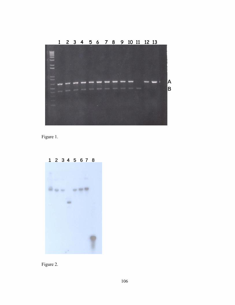
AB
1 2 3 4 5 6 7 8 9 10 11 12 13
AB
1 2 3 4 5 6 7 8 9 10 11 12 13
Figure 1.
1 2 3 4 5 6 7 8 1 2 3 4 5 6 7 8
Figure 2.
106
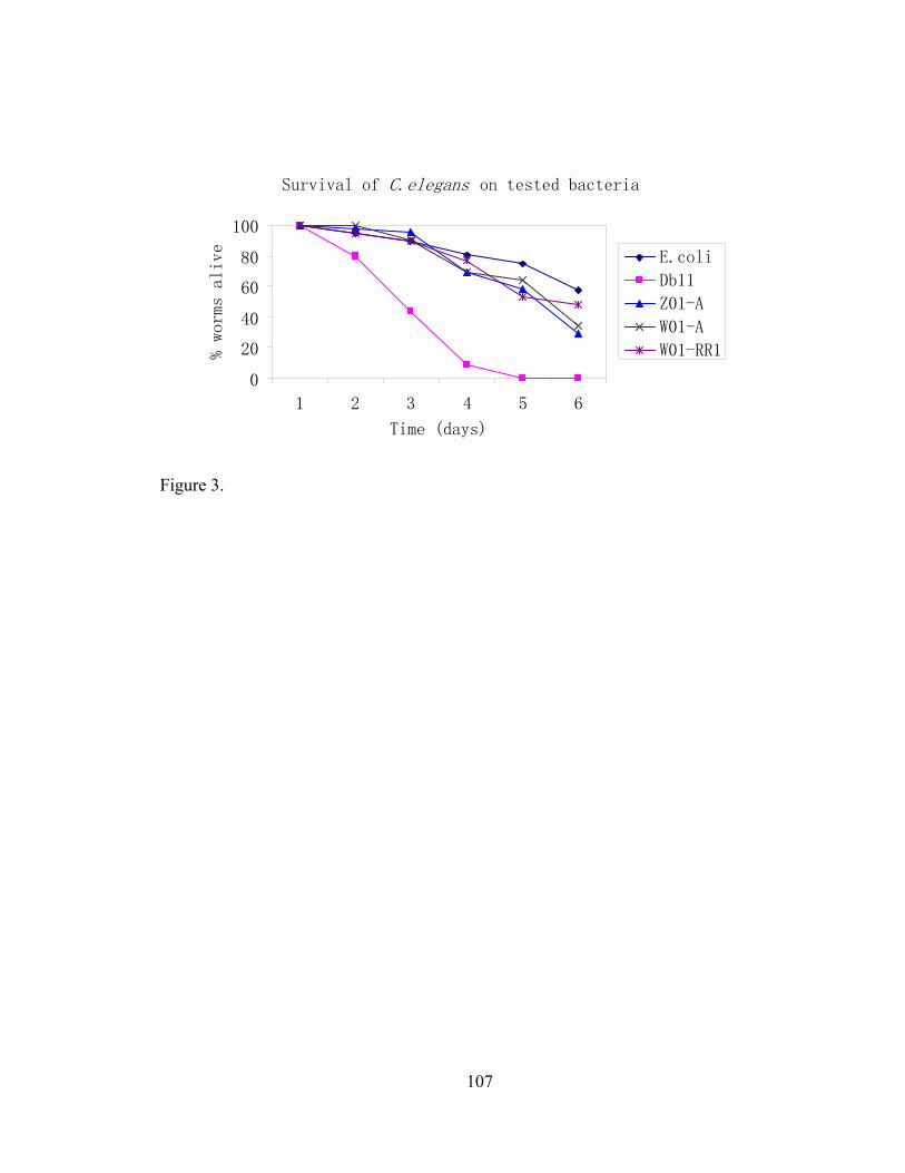
Survival of C.elegans on tested bacteria
0
20
40
60
80
100
1 2 3 4 5 6
Time (days)
% worms alive
E.coli
Db11
Z01-A
W01-A
W01-RR1
Figure 3.
107
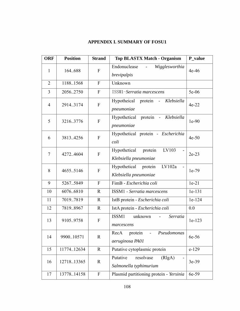
APPENDIX I. SUMMARY OF FOSU1
ORF Position Strand Top BLASTX Match - Organism P_value
1 164..688 F Endonuclease - Wigglesworthia
brevipalpis 4e-46
2 1188..1568 F Unknown
3 2056..2750 F ISSM1-Serratia marcescens 5e-06
4 2914..3174 F Hypotheical protein - Klebsiella
pneumoniae 4e-22
5 3216..3776 F Hypothetical protein - Klebsiella
pneumoniae 1e-90
6 3813..4256 F Hypothetical protein - Escherichia
coli 4e-50
7 4272..4604 F Hypothetical protein LV103 -
Klebsiella pneumoniae 2e-23
8 4655..5146 F Hypothetical protein LV102a -
Klebsiella pneumoniae 1e-79
9 5267..5849 F FimB - Escherichia coli 1e-21
10 6076..6810 R ISSM1 - Serratia marcescens 1e-131
11 7019..7819 R IstB protein - Escherichia coli 1e-124
12 7819..8967 R IstA protein - Escherichia coli 0.0
13 9105..9758 F ISSM1 unknown - Serratia
marcescens 1e-123
14 9900..10571 R RecA protein - Pseudomonas
aeruginosa PA01 6e-56
15 11774..12634 R Putative cytoplasmic protein e-129
16 12718..13365 R Putative resolvase (RlgA) -
Salmonella typhimurium 3e-39
17 13778..14158 F Plasmid partitioning protein - Yersinia 6e-59
108
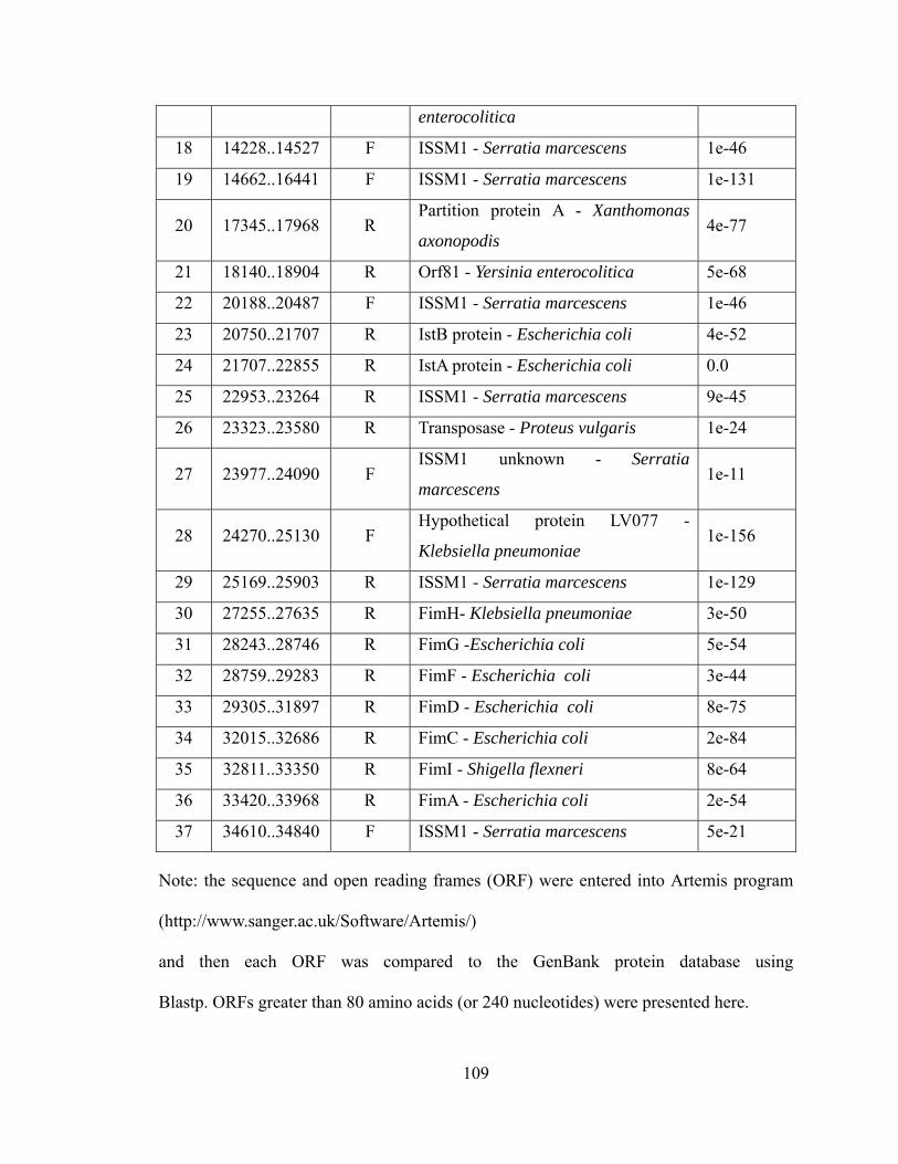
enterocolitica
18 14228..14527 F ISSM1 - Serratia marcescens 1e-46
19 14662..16441 F ISSM1 - Serratia marcescens 1e-131
20 17345..17968 R Partition protein A - Xanthomonas
axonopodis 4e-77
21 18140..18904 R Orf81 - Yersinia enterocolitica 5e-68
22 20188..20487 F ISSM1 - Serratia marcescens 1e-46
23 20750..21707 R IstB protein - Escherichia coli 4e-52
24 21707..22855 R IstA protein - Escherichia coli 0.0
25 22953..23264 R ISSM1 - Serratia marcescens 9e-45
26 23323..23580 R Transposase - Proteus vulgaris 1e-24
27 23977..24090 F ISSM1 unknown - Serratia
marcescens 1e-11
28 24270..25130 F Hypothetical protein LV077 -
Klebsiella pneumoniae 1e-156
29 25169..25903 R ISSM1 - Serratia marcescens 1e-129
30 27255..27635 R FimH- Klebsiella pneumoniae 3e-50
31 28243..28746 R FimG -Escherichia coli 5e-54
32 28759..29283 R FimF - Escherichia coli 3e-44
33 29305..31897 R FimD - Escherichia coli 8e-75
34 32015..32686 R FimC - Escherichia coli 2e-84
35 32811..33350 R FimI - Shigella flexneri 8e-64
36 33420..33968 R FimA - Escherichia coli 2e-54
37 34610..34840 F ISSM1 - Serratia marcescens 5e-21
Note: the sequence and open reading frames (ORF) were entered into Artemis program
(http://www.sanger.ac.uk/Software/Artemis/)
and then each ORF was compared to the GenBank protein database using
Blastp. ORFs greater than 80 amino acids (or 240 nucleotides) were presented here.
109

APPENDIX II. SUMMARY OF FOSU2
ORF Position Strand Top BLASTX Match - Organism P_value
1 350..841 F Hypothetical protein STM0909 - Phage
Fels-1 2e-66
2 834..2927 F Hypothetical protein - Phage Gifsy-2 0.0
3 3133..4653 F Hypothetical protein STM0911 - Phage
Fels-1 0.0
4 4586..6631 F Fels-1 prophage protease subunits -
Phage Fels-1 0.0
5 6712..7089 F Hypothetical prtein in prophage CP-
933U - Escherichia coli 8e-14
6 7086..7403 F Hypothetical protein STM0913.2N -
Phage Fels-1 1e-17
7 7369..7929 F Phage tail component - Phage Fels-1 1e-46
8 8037..8327 F Gp12 - Bacteriophage N15 8e-33
9 8338..8841 F Tail fiber component V of prophage CP-
933U - Escherichia coli 2e-65
10 8857..9246 F Putative tail component of prophage
CP-933K - Shigella flexneri 1e-18
11 9556..12531 F Phage tail component H- like protein -
Salmonella typhimurium 0.0
12 12566..12862 F Phage tail component M - like protein-
Salmonella typhimurium 2e-26
13 12903..13601 F Fels-1 prophage minor tail protein -
Phage Fels-1 5e-72
14 13617..14345 F Putative tail component of prophage -
Escherichia coli 6e-92
15 14249..14896 F Tail assembly protein-Phage Gifsy-2 4e-51
16 14915..18988 F Putative tail component of prophage - 0.0
110

Escherichia coli
17 18988..19365 F Unknown
18 20588..21505 F Tail fiber - Bacteriophage HK97 3e-36
19 21558..22106 F Tail fiber protein of prophage CP-933X
- Escherichia coli 2e-29
20 22120..23277 F Tail fiber protein - Yersinia pestis 2e-34
21 23602..23925 R Putative cytoplasmic protein -
Salmonella typhimurium 1e-15
22 26199..26453 F Putative phage integrase - Yersinia
pestis 1e-36
23 28117..28812 F Unknown
24 30142..30378 F Unkown
25 30950..31693 F Ant2 - Enterobacteria phage 6e-21
26 31690..31995 F Unknown
27 32084..32452 F Unknown
28 32452..33084 F Hypothetical protein - Bacteriophage
P27 7e-18
29 33094..33450 F Unknown
Note: the sequence and open reading frames (ORF) were entered into Artemis program
(http://www.sanger.ac.uk/Software/Artemis/)
and then each ORF was compared to the GenBank NR protein database using
Blastp. ORFs greater than 80 amino acids (or 240 nucleotides) were presented here.
111
VITA
Quan Zhang
Candidate for the Degree of
Doctor of Philosophy
Thesis: GENETIC ANALYSIS OF SERRTIA MARCESCENS: THE CAUSAL AGENT
OF CUCURBIT YELLOW VINE DISEASE Major Field: Plant Pathology Biographical:
Personal Data: Born in Xishui, HuBei, China on August 28, 1976, the son of Chunfang Zhang and Baoyin Sun.
Education: Graduated from Huang Gang High School, Huangzhou, Huibei in July
1993; received Bachelor of Science degree in Plant Pathology from Central China Agricultural University, Wuhan, China in July 1997; received Master of Science degree in Plant Pathology from China Agricultural University, Beijing in July 2000. Completed the requirements for the Doctor of Philosophy degree with a major in Plant Pathology at Oklahoma State University in December 2004.
Experience: Employed by Oklahoma State University as a Graduate Research
Assistant from 2000 to 2004. Professional Membership: Sigma Xi, American Phytopathological Society.
Recommended