DOCTORAL PROGRAM OF SCIENCE AND MATERIALS ENGINEERING
DOCTORAL PROGRAM IN MECHANICAL ENGINEERING
DOCTORAL THESIS
Study of porcelain-zirconia composites for dental applications
RAFAELA LUIZ PEREIRA SANTOS
UFRN Advisor: Prof. Dr. Rubens Maribondo do Nascimento
UMINHO Advisor: Prof. Dr. Filipe Samuel Silva
UMINHO Co-Advisor: Prof. Dr. Bruno Henriques
Natal – RN
2015
Federal University of Rio Grande do Norte
University of Minho
UNIVERSIDADE FEDERAL DO RIO GRANDE DO NORTE
PROGRAMA DE PÓS-GRADUAÇÃO EM CIÊNCIA E ENGENHARIA
DE MATERIAIS
UNIVERSIDADE DO MINHO
PROGRAMA DOUTORAL EM ENGENHARIA MECÂNICA
Study of porcelain-zirconia composites for dental applications
Tese submetida à
UNIVERSIDADE FEDERAL DO RIO GRANDE DO NORTE E
UNIVERSIDADE DO MINHO
Como parte dos requisitos para obtenção do grau de
DOUTOR EM CIÊNCIA E ENGENHARIA DE MATERIAIS
DOUTOR EM ENGENHARIA MECÂNICA
RAFAELA LUIZ PEREIRA SANTOS
Tese N° 169
Orientadores: Prof. Dr. Rubens Maribondo do Nascimento
Prof. Dr. Bruno Henriques
Prof. Dr. Filipe Samuel Silva
Natal-RN
2015
UNIVERSIDADE FEDERAL DO RIO GRANDE DO NORTE
PROGRAMA DE PÓS-GRADUAÇÃO EM CIÊNCIA E ENGENHARIA DE
MATERIAIS
UNIVERSIDADE DO MINHO
PROGRAMA DOUTORAL EM ENGENHARIA MECÂNICA
Study of porcelain-zirconia composites for dental applications
RAFAELA LUIZ PEREIRA SANTOS
Esta tese de doutorado foi julgada adequada como parte dos requisitos para a obtenção
do título de DOUTOR EM CIÊNCIA E ENGENHARIA DE MATERIAS/MECÂNICA
BANCA EXAMINADORA
_________________________________________________
Prof. Dr. Antonio Eduardo Martinelli (UFRN)
_________________________________________________
Prof. Dr. Daniel Araújo de Macedo (UFRN)
__________________________________________________
Prof. Dr. Mario Godinho Junior (UFG)
____________________________________________________
Prof. Dr. Romualdo Rodrigues Menezes (UFCG)
i Study of porcelain-zirconia composites for dental applications
Dedication
This thesis is dedicated to my parents...

ii Study of porcelain-zirconia composites for dental applications
iii Study of porcelain-zirconia composites for dental applications
Acknowledgements
I would like to thank God for blessing me abundantly and fulfill their promises in my life.
I would like to thank my thesis advisors Dr. Rubens Maribondo (UFRN), Dr. Bruno
Henriques and Dr. Filipe Silva (UMinho) for taking me under his wing and teaching me
everything I know about research. Thank you so much for give me the opportunity to
work with you and for you always support, encouraging and helping me see the true value
in research and inspiring me to be a better researcher.
To my parents, Roberto, Socorro and family Raquel, Rayane, Ruthe, Hermano and Felipe,
I would like to first and foremost thank you for the amazing love and support you have
given me throughout the years. Thank you for encouraging me endlessly to pursue higher
education and explaining to me how for ever giving me the opportunity to further my
professional training. Thank you for encouraging me endlessly to pursue higher education
and explaining to me how through education and knowledge comes enlightenment and
humility. As well, in my difficult moments you were both endlessly comforting and even
more supporting.
To my friends, colleagues and teachers of the Chemical Synthesis of the Materials Lab
(LSQM), in special to (Fabiana, Raquel, Raimison, Laura, Yara, Mayara, Mara, Patrícia,
Viviane and Camyla).
To my friends and colleagues of the Microfabrication and Systems Integration Lab
(MSFILab) – University of Minho – Portugal.
To my dear Pedro by support, friendship, love, affection, encouragement, attention and
understanding in the last stage of this thesis.
To my dear friends, Valéria, Micheline and Alana by encouraging and companionship
even the distance.
iv Study of porcelain-zirconia composites for dental applications
I would like to thank to members of my thesis committee for guidance and direction in
the design and completion of this project.
To Doctoral Program in Science and Materials Engineering (UFRN) and Doctoral
Program in Mechanical Engineering (UMINHO) by the opportunity to conduct my
research in these institutions.
To CAPES-Brazil and Portuguese Foundation of Science and Technology through the
projects: EXCL/EMS-TEC/0460/2012 and UID/EEA/04436/2013 by the financial
support.
v Study of porcelain-zirconia composites for dental applications
Abstract
Study of porcelain-zirconia composites for dental applications
All-ceramic restorations have been widely used as aesthetic standard in nowadays dentistry,
to replace the tooth structure (appearance and function). However, failures associated with
chipping and delamination of the veneering porcelain from zirconia substructure still high when
compared to metal-ceramic systems. This study aimed address these problems by improving the
mechanical strength of veneering porcelain and by improving its adhesion to zirconia substrate.
First, the addition of a stronger ceramic second phase (zirconia particles) to feldspathic porcelain
was studied. This study began with the preliminary characterization of zirconia synthesized by
Complex Polymerization Method (CPM) and commercial zirconia. It was found that the
synthesized zirconia did not show improved properties when compared to commercial zirconia.
Commercial zirconia powders were thus used thereafter. The influence of the type of powder (pre-
sintered or agglomerated) in the mechanical properties and wear of porcelain composites
(20vol.%) was evaluated. The pre-sintered powders increased the mechanical strength and wear
of the composites compared to agglomerated powders. Then, it was determined the amount of
second phase (0-50vol.%) that maximizes the mechanical properties of the porcelain-zirconia
composites. It was concluded that the composites with 30vol.% of zirconia showed the best results
for wear and flexural strength (198.5MPa). Finally, it was evaluated the influence of the type of
surface treatment (sandblasting or holes) and the deposition of a composite interlayer (porcelain
reinforced with 30vol.% of zirconia), on the zirconia surface, on the bond strength between the
veneering porcelain and the zirconia substructure. The highest bond strength values were found
for specimens having simultaneously holes on the surface of zirconia and the composite interlayer
between the zirconia substructure and the veneering porcelain (138 ± 19MPa). It was
demonstrated that the correct configuration of the interface between the zirconia substructure and
the veneering porcelain can lead to all-ceramic restorations with enhanced mechanical strength
and improved clinical performance.
Keywords: Feldspathic porcelain, Zirconia, CPM, Hot pressing, Mechanical Properties,
Composites.
.
vi Study of porcelain-zirconia composites for dental applications
Resumo
Estudo dos compósitos de porcelana-zircônia para aplicações odontológicas
Restaurações totalmente cerâmicas tem sido largamente empregada como padrão em
odontologia, na substituição da estrutura dentária (aparência e função). Contudo, as falhas por
lascamento e delaminação da porcelana e da subestrutura de zircônia continuam se apresentando
elevadas face às verificadas em sistemas metalo-cerâmicos. Assim, este estudo visou melhorar
quer a resistência mecânica da porcelana de revestimento quer a sua adesão ao substrato de
zircônia em sistemas cerâmicos utilizados em restaurações dentárias. Primeiramente estudou-se a
inserção de uma segunda fase cerâmica mais resistente (zircônia) na porcelana feldspática de
revestimento. Esta fase iniciou com uma prévia caracterização da zircônia sintetizada via Método
de Complexação Polimérica (CPM) e zircônia comercial. Concluiu-se que a zircônia sintetizada
não apresentava propriedades melhoradas face à zircônia comercial, sendo utilizada a zircônia
comercial nos estudos subsequentes. A influência do tipo de pó (aglomerado ou pré-sinterizado)
nas propriedades mecânicas e de desgaste dos compósitos de porcelana (20vol.%) foi avaliada.
Os pós pré-sinterizados aumentaram a resistência mecânica e de desgaste do compósito em
comparação aos pós aglomerados. Posteriormente, realizou-se a determinação da quantidade de
segunda fase (0-50vol.%) que maximizasse as propriedades mecânicas dos compósitos porcelana-
zircônia. Concluiu-se que os compósitos com (30vol.%) de zircônia apresentaram os melhores
resultados de desgaste e resistência a flexão (198,5MPa). Finalmente, foi avaliada a influência do
tratamento superficial da subestrutura de zircônia (jateamento e furos) e a inserção de uma camada
intermediária compósita (porcelana reforçada com 30vol.% de zircônia) na resistência de adesão
porcelana/subestrutura. Os maiores valores de adesão foram registrados aquando da utilização
simultânea de furos e da camada intermediária compósita (138±19MPa). Com isso, verifica-se
que a correta configuração da interface zircônia-porcelana pode produzir restaurações cerâmicas
com propriedades acrescidas de resistência mecânica, as quais devem melhorar
significativamente o seu desempenho clínico.
Palavras-chave: Porcelana feldspática, Zircônia, CPM, Prensagem à quente, Propriedades
Mecânicas, Compósitos.

vii Study of porcelain-zirconia composites for dental applications
Table of contents
Acknowledgments .......................................................................................................... iii
Abstract ............................................................................................................................ v
Resumo ........................................................................................................................... vi
Table of contents ........................................................................................................... vii
List of abbreviations ..................................................................................................... xii
List of Figures .............................................................................................................. xiii
List of Tables .............................................................................................................. xviii
Scope and Structure of the thesis ............................................................................... xix
Motivation and Aim of this thesis ............................................................................. xxii
CHAPTER I – Introduction
1.Introduction .................................................................................................................... 1
1.1 Teeth (Function and Structure) ................................................................................. 1
1.2 Dental Materials ....................................................................................................... 4
1.3 Dental Ceramics ....................................................................................................... 6
1.3.1 Dental porcelain Classification ........................................................................... 9
1.3.2 Porcelain composition ..................................................................................... 10
1.3.3 Processing ........................................................................................................ 13
1.3.4 Sintering Cycles .............................................................................................. 15
1.4 All-ceramic restorations .......................................................................................... 17
1.4.1 Zirconia .......................................................................................................... 20
1.4.2 Synthesis of Zirconia ...................................................................................... 25
1.5 Particulate composites ............................................................................................. 27
1.6 Ceramic-ceramic composites interaction/Adhesion ................................................ 29
1.6.1 Bond strength tests ......................................................................................... 32
1.7 Application of dental Ceramic ................................................................................ 34
References ....................................................................................................................... 36
viii Study of porcelain-zirconia composites for dental applications
CHAPTER II - Comparative study between commercial zirconia and zirconia
synthesized with different Yttria content and calcination temperatures by Complex
Polymerization Method (CPM).
Abstract ............................................................................................................................ 56
1. Introduction .......................................................................................................... 57
2. Experimental Procedure ....................................................................................... 58
2.1 Materials ........................................................................................................... 58
2.2 Methods ............................................................................................................ 58
2.2.1 Characterizations ......................................................................................... 60
3. Results and Discussion ....................................................................................... 60
4. Conclusions ........................................................................................................ 69
Acknowledgements ................................................................................................ 70
References .............................................................................................................. 70
CHAPTER III - Strenghtening and toughening of dental porcelain by the inclusion of an
yttria-stabilized zirconia reinforcing phase
Abstract ............................................................................................................................ 75
1. Introduction ......................................................................................................... 76
2. Materials and Methods ......................................................................................... 77
3. Results ................................................................................................................. 78
3.1 Microstructural analysis ................................................................................... 78
3.2 Mechanical tests ............................................................................................... 80
4. Discussion .......................................................................................................... 82
5. Conclusion ......................................................................................................... 83
Acknowledgements ................................................................................................ 83
References ....................................................................................................................... 84
CHAPTER IV – On the mechanical properties and microstructure of zirconia
reinforced dental porcelain composites
Abstract ............................................................................................................................ 86
1. Introduction .......................................................................................................... 87
2. Materials and Methods ......................................................................................... 89
2.1 Base Materials .................................................................................................. 89
ix Study of porcelain-zirconia composites for dental applications
2.2 Composites Processing ..................................................................................... 90
2.3 Microstructural Characterizations .................................................................... 90
2.4 Mechanical Tests .............................................................................................. 91
3. Results ................................................................................................................ 92
4. Discussion .......................................................................................................... 96
5. Conclusions ........................................................................................................ 99
Acknowledgements ................................................................................................ 99
References .............................................................................................................. 100
CHAPTER V– Wear performance of feldspathic porcelain reinforced by zirconia
agglomerates and by zirconia particles
Abstract .......................................................................................................................... 105
1. Introduction ........................................................................................................ 106
2. Experimental Procedure ..................................................................................... 107
2.1 Materials ....................................................................................................... 107
2.2 Fabrication of zirconia reinforced feldspathic porcelain composites ........... 108
2.3 Microstructural and Chemical analysis ........................................................ 109
2.4 Hardness Measurements ............................................................................... 109
2.5 Wear Tests .................................................................................................... 109
3. Results and Discussion ....................................................................................... 110
3.1 Microstructural and Chemical Characterization .......................................... 110
3.2 Hardness Values .......................................................................................... 112
3.3 Specific wear rate ........................................................................................ 112
3.4 Coefficient of friction .................................................................................. 113
3.5 Microscopic analyses on worn surfaces ...................................................... 114
4. Conclusions ...................................................................................................... 119
Acknowledgements .............................................................................................. 119
References ............................................................................................................ 120

x Study of porcelain-zirconia composites for dental applications
CHAPTER VI – Tribological behavior of Dental Porcelain composites reinforced by
Yttria-Stabilized Zirconia
Abstract .......................................................................................................................... 125
1. Introduction ............................................................................................................ 126
2. Experimental Procedure ......................................................................................... 127
2.1 Materials ........................................................................................................... 127
2.2 Fabrication of zirconia reinforced feldspathic porcelain composites ............ 128
2.3 Microstructural and Chemical Analysis ........................................................ 129
2.4 Hardness Measurements ................................................................................ 129
2.5 Wear Tests ..................................................................................................... 129
3. Results and Discussion ....................................................................................... 130
3.1 Microstructural Characterization .................................................................. 130
3.2 Hardness Values ............................................................................................ 132
3.3 Specific wear rate .......................................................................................... 132
3.4 Coefficient of friction .................................................................................... 133
3.5 Microscopic analysis on worn surfaces......................................................... 135
4. Conclusions ........................................................................................................ 140
Acknowledgements .............................................................................................. 141
References ............................................................................................................ 141
CHAPTER VII – Shear bond strength of veneering porcelain to zirconia: effect of
surface treatment by CNC-milling and composite layer deposition on zirconia
Abstract .......................................................................................................................... 148
1. Introduction ........................................................................................................ 149
2. Materials and Methods ....................................................................................... 150
2.1 Materials ........................................................................................................ 150
2.2 Specimens preparation .................................................................................. 151
2.3 Mechanical Tests ........................................................................................... 153
2.4 Analysis of the zirconia-porcelain interface and failure mode ...................... 153
2.5 Statistical analysis ......................................................................................... 154
2.6 Zirconia surface characterization .................................................................. 154
3. Results ................................................................................................................. 154

xi Study of porcelain-zirconia composites for dental applications
4. Discussion ............................................................................................................ 161
5. Conclusion ........................................................................................................... 161
Acknowledgements ................................................................................................. 162
References ............................................................................................................... 162
CHAPTER VIII – General Discussion
8.2 Commercial zirconia vs. synthesized zirconia ....................................................... 167
8.3 Zirconia particle effect in feldspathic porcelain matrix .......................................... 168
8.4 Zirconia content effect in mechanical performance of the composites feldspathic
porcelain-zirconia .......................................................................................................... 170
8.5 Wear effect according the type of zirconia particles applied ................................. 171
8.6 Wear effect according the zirconia content in feldspathic porcelain matrix ........... 172
8.7 Surface treatment and adhesion ............................................................................... 173
Summarizing .................................................................................................................. 175
References ..................................................................................................................... 176
Main conclusions and future perspectives ..................................................................... 181
Future perspectives ........................................................................................................ 183
xii Study of porcelain-zirconia composites for dental applications
List of Abbreviations
CPM – Complex Polymerization Method
XRD – X –Ray diffraction
Y-TZP – Yttria – Tetragonal Zirconia Polycrystals
TGA/DTA – Thermogravimetric analysis
SEM – Scanning Electron Microscopy
FEG-SEM – Field Emission Guns Electron Microscopy
EDS – Energy Dispersion Spectroscopy
KIC – Fracture Toughness
HV – Hardness
E – Young’s Modulus
PMMA – polymethylmethacrylate
CTE – Coefficient of Thermal Expansion
COF – Coefficient friction
CNC – Milling Machine
CAD – Computer Aided Design
Ra – surface roughness
UFRN – Federal University of Rio Grande do Norte
UMINHO – University of MINHO
xiii Study of porcelain-zirconia composites for dental applications
List of Figures
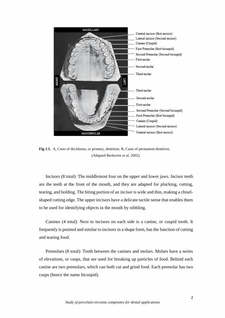
Fig 1.1 A, casts of deciduous, or primary dentition, B, casts of permanent dentition ...... 2
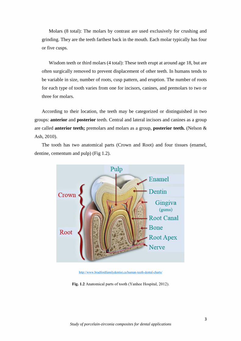
Fig 1.2 Anatomical parts of tooth ...................................................................................... 3
Fig 1.3 Example of restorative materials (Metal and Ceramic) ........................................ 5
Fig 1.4 Classification of dental ceramics .......................................................................... 8
Fig. 1.5 Classification of dental porcelains according the firing temperatures ................. 9
Fig. 1.6 Ternary diagram of K2O Al2O3 SiO2 ................................................................. 11
Fig 1.7 (a) Metal ceramic and (b) all-ceramic prostheses ............................................... 12
Fig 1.8 Hot pressing process ........................................................................................... 15
Fig 1.9 Example of sintering cycles for dental porcelain ................................................ 15
Fig 1.10 A, stress induced in a three-unit bridge by a flexural force (P). B, stresses induced
in a two-unit cantilever bridge ......................................................................................... 20
Fig. 1.11 Transformation toughening of zirconia............................................................ 21
Fig. 1.12 Phase diagram of ZrO2/Y2O3 ........................................................................... 22
Fig. 1.13 Vacancy transport ............................................................................................ 23
Fig. 1.14 Complexation reaction andpolymerization in the CPM .................................. 26
Fig 1.15 Particulate reinforced composite ....................................................................... 29
Fig 1.16 Classification of bond mechanism .................................................................... 30
Fig 1.17 Three point bending of a ceramic specimen according ISO 6872 .................... 32
Fig 1.18 Shear bond test for restorative materials ........................................................... 33
Fig 1.19 Porcelain crown................................................................................................. 34
Fig 2.1 Flowchart of the zirconia synthesis by CPM ...................................................... 59
Fig 2.2 XRD for specimens with varied Yttria content in zirconia (A, B and C) and
comparative XRD between zirconia with Yttria content (3, 4 and 5% at 1000ºC) and
commercial zirconia – CZ with 3% of Yttria (D) ............................................................ 61
Fig 2.3 Crystallite size of zirconia doped with Yttria in different contents (ZY0,3, ZY0,4 e
ZY0,5) obtaining by Pechini Method and commercial zirconia (CZY0,3) ....................... 62
Fig 2.4 The lattice parameters of Yttria doped zirconia in different contents (ZY0,3, ZY0,4
e ZY0,5) obtaining by CPM and commercial zirconia (CZY0,3) ...................................... 64
Fig 2.5 Unit cell parameters of Yttria doped zirconia in different contents (ZY0,3, ZY0,4 e
ZY0,5) obtaining by Pechini Method and commercial zirconia (CZY0,3) ........................ 65

xiv Study of porcelain-zirconia composites for dental applications
Fig 2.6 TGA/DTA curves of specimens with varied of Yttria content in Zirconia (A, B,
C) and commercial zirconia (D) ..................................................................................... 66
Fig 2.7 Raman spectrometry of Yttria doped zirconia in different contents and calcination
temperature ..................................................................................................................... 68
Fig 3.1 Difractogram of the (A) Feldspathic Porcelain – Z0, (B) Feldspathic porcelain
reinforced composites with zirconia agglomerates - ZA and (C) zirconia particles -ZP 78
Fig 3.2 Micrographs of the (A) Feldspathic porcelain composites reinforced zirconia
agglomerates - ZA (A) and (B) zirconia particles –ZP. Zirconia appears as white phase
and porcelain as dark phase ............................................................................................. 79
Fig 3.3 Detail of interface between the zirconia particles ZP (white phase) and the
porcelain (dark phase) ..................................................................................................... 80
Fig 3.4 Micrographs of the fractured surface of three composites. (A) Feldspathic
porcelain – Z0, (B) Feldspathic porcelain reinforced composites with zirconia
agglomerates - ZA and (C) zirconia particles -ZP ........................................................... 80
Fig 3.5 Flexural strength of the (A) Feldspathic porcelain – Z0, (B) porcelain reinforced
composites with zirconia agglomerates - ZA and (C) zirconia particles -ZP ................. 81
Fig 3.6 Vickers Hardness of the (A) Feldspathic porcelain – Z0, (B) Feldspathic porcelain
reinforced composites with zirconia agglomerates - ZA and (C) zirconia particles -ZP 82
Fig 4.1 SEM micrograph of porcelain powders (A) and zirconia sintered particles (B).
......................................................................................................................................... 90
Fig 4.2 Microstructures obtained by scanning electron microscopy of (A) Feldspathic
porcelain and feldspathic porcelain-zirconia composites (B) Z10, (C) Z20, (D) Z30, (E)
Z40 and (F) Z50. White arrows indicate residual porosity. Dashed lines signalize zirconia
agglomerates .................................................................................................................... 92
Fig 4.3 Detail of the interface between the porcelain matrix (dark phase) and the zirconia
reinforcing particles (white phase) .................................................................................. 93
Fig 4.4 Flexural strength of feldspathic porcelain matrix composites with different
volume contents of zirconia phase (Z0) 0%, (Z10) 10%, (Z20) 20%, (Z30) 30%, (Z40)
40% and (Z50) 50% ......................................................................................................... 94
Fig 4.5 SEM micrograph of the fracture surface of a Z10 specimen showing in detail the
interface between the feldspathic porcelain matrix and a big zirconia particle (pre-sintered
agglomerate). ................................................................................................................... 94
xv Study of porcelain-zirconia composites for dental applications
Fig 4.6 Vickers indentation with induced crack propagation in (A) Feldspathic porcelain
and (B) Z30 composite ................................................................................................... 95
Fig 4.7 X-Ray diffraction pattern of the feldspathic porcelain-zirconia composite, Z30.
......................................................................................................................................... 96
Fig 4.8 Plot of the flexural strength of feldspathic porcelain-zirconia composites as
compared to the rule of mixtures models, Voigt and Reuss ............................................ 96
Fig 5.1 FEGSEM images showing: A) Feldspathic porcelain, B) zirconia agglomerates
and C) pre-sintered zirconia powders ............................................................................ 108
Fig 5.2 FEGSEM images of the microstructure of A) Feldspathic porcelain (Z0); B)
Feldspathic porcelain reinforced by zirconia agglomerates (ZA) and C) Feldspathic
porcelain reinforced by zirconia particles (ZP) ............................................................. 110
Fig 5.3 Detailed view of the FEGSEM interface between the zirconia particles (white
phase) and the porcelain matrix (dark phase) ................................................................ 111
Fig 5.4 Hardness of the feldspathic porcelain and zirconia reinforced feldspathic
porcelain composites ..................................................................................................... 112
Fig 5.5 Specific wear rate of the feldspathic porcelain and zirconia reinforced feldspathic
porcelain composites ..................................................................................................... 113
Fig 5.6 Evolution of COF along the time for: A) Feldspathic porcelain, B) Feldspathic
porcelain composite reinforced with zirconia agglomerates (ZA) and C) Feldspathic
porcelain composites reinforced with zirconia particles (ZP) ....................................... 114
Fig 5.7 FEG-SEM images of the midle area of wear track for: A) Feldspathic porcelain,
B) Feldspathic porcelain composite reinforced with zirconia agglomerates (ZA) and C)
Feldspathic porcelain composites reinforced with zirconia particles (ZP) ................... 115
Fig 5.8 FEG-SEM images of the wear track: A) Feldspathic porcelain, B) Feldspathic
porcelain composite reinforced with zirconia agglomerates (ZA) and C) Feldspathic
porcelain composites reinforced with zirconia particles (ZP) ....................................... 116
Fig 5.9 FEG-SEM images of the Al2O3 ball after sliding against feldspathic porcelain
composite reinforced by zirconia particles (ZP) samples. ............................................. 117
Fig 6.1 FEGSEM images showing: A) Dental feldspathic porcelain and B) zirconia
powders .......................................................................................................................... 128

xvi Study of porcelain-zirconia composites for dental applications
Fig 6.2 Microstructure of the feldspathic porcelain and zirconia reinforced feldspathic
porcelain composites samples A) Z0; B) Z10; C) Z20; D) Z30; E) Z40 and F) Z50 .... 131
Fig 6.3 Hardness of the zirconia reinforced feldspathic porcelain composites as a function
of the zirconia particles content ..................................................................................... 132
Fig 6.4 Specific wear rate of the feldspathic porcelain and zirconia reinforced feldspathic
porcelain composites as a function of the zirconia particles content ............................ 133
Fig 6.5 Evolution of COF with sliding distance for samples: A) Z0; B). Z10; C) Z20; D)
Z30; E) Z40 and F) Z50 ................................................................................................. 134
Fig 6.6 COF of the feldspathic porcelain and zirconia reinforced feldspathic porcelain as
a function of the zirconia content ................................................................................. 135
Fig 6.7 FEG-SEM images of the midle area of the wear track A) Z0; B) Z10; C) Z20; D).
Z30; E) Z40 and F) Z50 ................................................................................................. 136
Fig 6.8 FEG-SEM images of the wear track (2000x magnification):A) Z0; B) Z10; C)
Z20; D) Z30; E) Z40 and F) Z50 ................................................................................... 137
Fig 6.9 FEG-SEM images of the wear track (5000x magnification): A) Z0; B) Z10; C)
Z20; D) Z30; E) Z40 and F) Z50 ................................................................................... 137
Fig 6.10 SEM images of Al2O3 ball after sliding against dental feldspathic porcelain
composites reinforced by 30 vol. % of zirconia alloy and EDS spectrum ................... 138
Fig. 7.1 Schematic of the cilindrical zirconia substrates and ilustration of the two types
of surface treatments performed: A) CNC-created surface holes; B) rough surfaces by
grit-blasting .................................................................................................................... 152
Fig. 7.2. Shear bond strength results for each type of zirconia-porcelain interface design:
RZ- porcelain bonded to rough zirconia substrate; PZ - porcelain bonded to zirconia
substrate with surface holes; RZI - application of a composite interlayer between the
porcelain and the rough zirconia substrate; PZI - application of a composite interlayer
between the porcelain and the zirconia substrate with surface holes. Same superscript
letters indicate homogeneous groups (Tukey’s test, α=0.05) ........................................ 155
Fig. 7.3. FEG-SEM images of the veneering porcelain (dark phase) to zirconia (white
phase) for RZ specimens ............................................................................................... 156
Fig. 7.4. FEG-SEM images showing the zirconia-interlayer-porcelain region of the RZI
specimens ...................................................................................................................... 156
Fig. 7.5.FEG-SEM images of the veneering porcelain to zirconia inside and outside the
holes region (PZ) ........................................................................................................... 157
xvii Study of porcelain-zirconia composites for dental applications
Fig 7.6 FEG-SEM images of the zirconia-composite interlayer interface inside and
outside the holes region (PZI) ....................................................................................... 157
Fig 7.7 FEG-SEM images of the fracture surface of the zirconia substrates with surface
holes (PZ) ...................................................................................................................... 158
Fig 7.8 FEG-SEM images of the fracture surface of the zirconia specimens with
composite interlayer (PZI) ............................................................................................. 158
Fig 7.9 FEG-SEM images of the fracture surface of the sandblasted zirconia specimens
(RZ) ............................................................................................................................... 158

xviii Study of porcelain-zirconia composites for dental applications
List of Tables
Table 1.1. Techniques of processing and thesis characteristics ...................................... 14
Table 2.1. Reagents used for obtaining the zirconia powder by CPM ........................... 58
Table 4.1 Chemical composition, material and manufacturer of feldspathic porcelain and
Y-TZP used in this study. ................................................................................................ 89
Table 4.2 Hardness (GPa) and Fracture toughness (KIC) of feldspar-based porcelain (Z0)
and porcelain-zirconia composites (Z10, Z20, Z30, Z40, Z50) ...................................... 95
Table 5.1 Chemical composition of Dental feldspathic porcelain powder (wt%) ........ 107
Table 5.2 Chemical composition of 3Y-TZP powder (wt%) ........................................ 108
Table 5.3 Composition of the stock Fusayama artificial saliva ................................... 110
Table 6.1 Chemical composition of Dental feldspathic porcelain powder (wt%) ....... 128
Table 6.2 Chemical composition of 3Y-TZP powder (wt%) ....................................... 128
Table 6.3 Composition of the stock Fusayama artificial saliva ................................... 130
Table 7.1 Chemical composition and manufacturer of the materials used in this study
....................................................................................................................................... 151
Table 7.2 Mechanical properties of the base materials used in this study .................... 155
xix Study of porcelain-zirconia composites for dental applications
Scope and structure of the thesis
This thesis is elaborated in 8 chapters. Chapter 1 gave a general introduction
including a literature overview on the aspects related to the dental applications
specifically to ceramic-ceramic materials. The topics dealt with are: 1.1 Teeth (Function
and Structure), 1.2 Dental materials, 1.3 Dental ceramics, 1.3.1 Dental porcelain
classification, 1.3.2 Porcelain Composition, 1.3.3 Processing, 1.3.4 Sintering Cycles, 1.4
All ceramic restorations, 1.4.1 Zirconia, 1.4.2 Synthesis of Zirconia, 1.5 Particulate
Composites, 1.6 Ceramic-Ceramic composites interaction/Adhesion, 1.6.1 Bond Strength
Tests and 1.7 Applications of dental Ceramic.
The following chapters contain 6 research reports that receive Introduction,
Experimental Procedure (Materials and Methods), Results, Discussion and Conclusions
of the experimental work, performed during this study.
In Chapter 2, details about the previous characterization and comparative study
of commercial zirconia (Y-TZP) and zirconia powders obtained by CPM (Complex
Polymerization Method). This comparison is based in a different technique of
manufacture applied and the use of Yttria content (3, 4 and 5 mol.%) in zirconia
synthesized as also theirs implications arising during the processing. This paper is
submitted for publication in Quimica Nova.
Chapter 3, is devoted to the study of the mechanical behavior of different types
of zirconia powders incorporated on feldspathic porcelain matrix. The first tests were
performed using two types of particles (yttria-stabilized zirconia (ZrO2-3%Y2O3)
agglomerates and pre-sintered yttria-stabilized zirconia (ZrO23%Y2O3)) in a 20% vol.%
of proportion. These results were compared to feldspathic porcelain on strengthening and
toughening behavior. This paper is published in International of Medical, Health,
Biomedical and Pharmaceutical Engineering (Vol. 9 No.5 2015).
Chapter 4, is based in Chapter 3 from the superior mechanical properties of the
zirconia particles in porcelain matrix. Thus, the reinforcements are incorporated in
feldspathic porcelain matrix in different volume contents (0-50% vol.%) and are
evaluated the microstructural characteristics by Scanning Electron Microscopy (SEM),
Energy Dispersion Spectroscopy (EDS) and XRD Diffraction (XRD) and mechanical
performance by the transverse rupture strength test. This paper is submitted to the journal
Dental Materials - Elsevier.

xx Study of porcelain-zirconia composites for dental applications
Chapter 5, examines the wear behavior of dental composites reinforced by 20%
vol.% (tetragonal zirconia 3Y-TZP). The results are compared between two types of
particles (agglomerates and pre-sintered particles) and monolithic porcelain. This paper
is submitted for publication in Journal of Dentistry.
Chapter 6, the wear characterization of dental feldspathic porcelain reinforced by
Yttria-Stabilized Zirconia in different volume contents (0-50% vol.%) are performed. The
results are compared as monolithic porcelain in wear resistance, coefficient of friction
and hardness. This paper is submitted to the journal Dental Materials.
Chapter 7 is devoted to the experimental evaluation of the bond strength between
zirconia substructure and porcelain through a composite ceramic-ceramic graded
transition interlayer. Several ceramic-ceramic were produced and tested as interlayers
aiming at the improvement of ceramic-ceramic adhesion. The bond strength results are
compared as substructures with/without interlayer and different surface treatments. This
paper is submitted to the journal Dental Materials.
Finally, Chapter 8 addresses a general discussion and the main conclusions are
drawn. In this chapter also presented a perspective for future works.
In this thesis, all chapters can be read independently because they have been
written in a form which suited for publication in international scientific journals,
according to the rules of the University of Minho and UFRN.

xxi Study of porcelain-zirconia composites for dental applications
Motivation and aim of this thesis
The tooth structure and teeth loss cause problems related to the health (teeth
function) and discomfort in social environment. The development of the materials and
processing methods in terms of mechanical properties, wear resistance, chemical attack
resistance have gained the researcher’s attention in the last four decades, by improving of
the suitable replacement of the natural teeth. Such characteristics cannot be reached only
to monolithic materials or layers processing, whereas the real tooth structure it is more
complex. Based on the previously stated, various materials and composites have been
used to minimize these undesirable effects, e.g., metal-ceramic restorations, related to
reestablish function and aesthetics. However, these materials required considerable
reduction of the tooth structure.
In this context, ceramic materials, e.g., zirconia-based materials has been widely
used in dental studies due to good mechanical properties in addition to aesthetics
appearance. In another hand, the success of all-ceramic restorations depends of a strong
bond between ceramic feldsphatic and zirconia substructure. These materials had to be
adapted, e.g., in the thermal expansion coefficient and firing temperature to achieve
optimal bonding.
The idea of this thesis is based on previous studies (Huang et al 2014, Henriques
et al 2012; Zhang et al, 2004) which uses different contents of reinforcement (metals and
ceramics) in porcelain matrix. Based on these ideas, the studies were outlined in evaluate
of the mechanical performance, wear, substructure surface and interaction veneer-core,
and core itself may contribute to the performance of the restorations. Thus, the influence
of processing, type of powders and zirconia volume content in porcelain matrix was
assessed.
The above factors emphasize, the aim of this thesis for further investigations on
porcelain-zirconia composites, the improvement of all-ceramic restorations, and
especially the interaction of zirconia used as reinforcement and porcelain (veneering) and
its influence on the performance of the whole restoration. Thus, the general and specific
objectives can be drawn:
xxii Study of porcelain-zirconia composites for dental applications
General Objective
The aim of this study was to evaluate the mechanical properties and
microstructural characteristics of a dental ceramic feldspathic reinforced zirconia in
different aspects (Obtaining method, type and volume content of reinforcement) on
mechanical, wear and adhesion/interaction (bond strength) behavior.
Specific Objectives
Comparative study between commercial zirconia and zirconia synthesized with
Yttria content and calcination temperatures by Complex Polymerization Method
(CPM). (Chapeter 2);
Strengthening and toughening of dental porcelain by the inclusion of the different
types of yttria-stabilized zirconia as reinforcing phase (Chapter 3);
Mechanical and microstructural study of zirconia in varied volume contents
reinforced dental porcelain composites (Chapter 4);
Wear performance of dental porcelain composites reinforced by different types of
yttria-stabilized zirconia (Chapter 5).
Tribological behavior of dental porcelain composites reinforced by different
volume contents (0-50%) of yttria-stabilized zirconia (Chapter 6).
Effect of CNC-created surface holes and of a composite interlayer on the shear
bond strength of porcelain to zirconia substructures (Chapter 7).
1 Study of porcelain-zirconia composites for dental applications
CHAPTER I
1. Introduction
The technological evolution of dental ceramics, have been remarkable over the past four
decades to replace the natural tooth. From feldspathic porcelains to zirconia-based all-
ceramics, tremendous progress have been made in terms of mechanical performance. In
this context, this chapter present a literature overview on the aspects related to the dental
applications and specifically in ceramic-ceramic systems.
1.1 Teeth (Function and Structure)
Tooth is a hard structure, protruding whitish composed of pulp, dentin and enamel
which is implanted in the jaw and mandible (or dental records in human) of many
vertebrates (Stuart, 2004). It is used to cut, hold and grind food, starting the digestion
process from the swallowing. Teeth are also importance in helping correct sound
pronunciation, speech articulation and facial expression.
The first step in understanding dental anatomy is to learn the nomenclature used
to describe or classify the material included in the subject. The dental anatomy may
be divided in two terms: mandibular (lower jaw or mandible) and maxillary (upper
jaw or maxila). When, more than one name is used in literature to describe something,
the two most commonly used names will be used initially. After that, they may be
combined or used separately as consistent with the literature of a particular specialty
of dentistry, for example, primary or deciduous dentition, permanent or succedaneous
dentition (Nelson & Ash, 2010).
In a human adult, the permanent dentition consisting of 32 teeth is completed from
18 to 25 years of age if the third molar included and can be divided: 16 in the upper
maxillary arch and 16 in the lower mandibular arch (Fig 1.1). The teeth can be
classified in incisors, canines, premolars, molars and wisdom teeth or third molar.
2 Study of porcelain-zirconia composites for dental applications
Fig 1.1. A, Casts of deciduous, or primary, dentition. B, Casts of permanent dentition.
(Adapted Berkovitz et al, 2002).
Incisors (8 total): The middlemost four on the upper and lower jaws. Incisor teeth
are the teeth at the front of the mouth, and they are adapted for plucking, cutting,
tearing, and holding. The biting portion of an incisor is wide and thin, making a chisel-
shaped cutting edge. The upper incisors have a delicate tactile sense that enables them
to be used for identifying objects in the mouth by nibbling.
Canines (4 total): Next to incisors on each side is a canine, or cuspid tooth. It
frequently is pointed and similar to incisors in a shape form, has the function of cutting
and tearing food.
Premolars (8 total): Teeth between the canines and molars. Molars have a series
of elevations, or cusps, that are used for breaking up particles of food. Behind each
canine are two premolars, which can both cut and grind food. Each premolar has two
cusps (hence the name bicuspid).
3 Study of porcelain-zirconia composites for dental applications
Molars (8 total): The molars by contrast are used exclusively for crushing and
grinding. They are the teeth farthest back in the mouth. Each molar typically has four
or five cusps.
Wisdom teeth or third molars (4 total): These teeth erupt at around age 18, but are
often surgically removed to prevent displacement of other teeth. In humans tends to
be variable in size, number of roots, cusp pattern, and eruption. The number of roots
for each type of tooth varies from one for incisors, canines, and premolars to two or
three for molars.
According to their location, the teeth may be categorized or distinguished in two
groups: anterior and posterior teeth. Central and lateral incisors and canines as a group
are called anterior teeth; premolars and molars as a group, posterior teeth. (Nelson &
Ash, 2010).
The tooth has two anatomical parts (Crown and Root) and four tissues (enamel,
dentine, cementum and pulp) (Fig 1.2).
http://www.bradfordfamilydentist.ca/human-teeth-dental-charts/
Fig. 1.2 Anatomical parts of tooth (Yanhee Hospital, 2012).
4 Study of porcelain-zirconia composites for dental applications
The crown of a tooth is that part of the tooth which is covered with enamel and this the
part usually visible in the mouth. The root is the part embedded in the jaw. It anchors the
tooth in its bony socket and is normally not visible.
Enamel – The hardest substance in the body, white outer art of the tooth. Enamel is
mostly made of calcium phosphate, a rock-hard mineral.
Dentine - not as hard as enamel, forms the bulk of the tooth and can be sensitive if the
protection of the enamel is lost. Is made of living cells, which secrete a hard mineral
substance.
Pulp – is a connective tissue organ containing a number of structures, among which
are arteries, veins, a lymphatic system, blood and nerves. Its primary function is to form
the dentin of the tooth.
Cementum – A layer of connective tissue covering the roots of the teeth firmly to the
gums and jawbone. It is not as hard as enamel.
Periodontal ligament – Made up of thousands of fibers which fasten the cementum to
the bony socket. These fibers anchor the tooth to the jaw bone and act as shock absorbs
for the tooth which is subjected to heavy forces during chewing.
Oral mucosa – This is the term used to describe the moist tissue that lines the mouth.
Gingiva (gums) – Soft tissue that immediately surrounds the teeth and bone. It
protects the bone and the roots of the teeth and provides an easily lubricated surface.
Bone – Provides a socket to surround and support the roots of the teeth.
Nerves and blood supply – Each tooth and periodontal ligament has a nerve supply
and the teeth are sensitive to a wide variety of stimulus. The blood supply is necessary to
maintain the vitality of the tooth.
1.2 Dental Materials
Dental materials, are materials generally used due the need to replace tooth structure
loss, because the presence of dental caries (dental cavities), but also tooth wear and dental
trauma. These materials may to maintain or improve the quality of life of the dental
patient, preventing disease, relieving pain, improving mastication efficiency and
enhancing speech appearance (cosmetics). (O’Brien, 2002, Anusavice, 2006)
The choice of these materials it depends on features such as biocompatibility, bond
permanently to tooth structure or bone, match the natural appearance of tooth enamel,

5 Study of porcelain-zirconia composites for dental applications
dentin and other tissues and capacity of initiate tissue repair or regeneration of missing or
damaged tissues. (O´Brien, 2002). Historically, various materials have been used as tooth
crown and root replacements (animal teeth, bone, human teeth, ivory, seashells, ceramics
and metals). Actually, dental materials used in dentistry are divided in four groups:
metals, ceramics, polymers and composites (Anusavice, 2006).
According (Anusavice, 2006) dental materials may be classified in three categories:
preventive materials, restorative materials or auxiliary materials.
Preventive dental materials – include pit and fissure sealants, sealing agents that
prevent leakage (used primarily for antibacterial effects); liners, bases, cements and
restorative materials (used primarily because they release fluoride - compomer, hybrid
ionomer; glass ionomer cement, zinc silicophosphate cement), chlorhexidine, or other
therapeutic agents used to prevent or inhibit the progression of tooth decay (dental caries).
Restorative dental materials – all synthetic components that can be used to repair or
replace tooth structure, including primers, bonding agents, liners, cement bases,
amalgams, resin-based composites, compomers hybrid ionomers, cast metals, metal-
ceramics, ceramics, and denture polymers. These materials may be used for temporary,
short-term purposes (such as temporary cements and temporary crown and bridge resins)
or for longer-term applications (dentin bonding agents, inlays, onlays, crowns, removable
dentures, fixed dentures and orthodontic appliances).
http://www.dentalartslab.com/products-services/implant-prosthetics/fixed-implant-prosthetic-services/
Fig. 1.3 Example of restorative materials (Metal and Ceramic).

6 Study of porcelain-zirconia composites for dental applications
Restorative materials also may be divided in direct restorative materials - used
intraorally to fabricate restorations or prosthetic devices directly on the teeth or tissues
and indirect restorative materials – used extraorally, in which the materials are formed
indirectly on casts or other replicas of the teeth and other tissues.
Auxiliary dental materials – are substances that are used in the process of
fabricating dental prostheses and appliances but that do not become part of these devices
(acid-etching, solutions, impression materials, casting investments, gypsum cast and
model materials, dental waxes, acrylic resins for impression and bleaching trays, acrylic
resins for mouth guards and occlusion aids, and finishing and polishing abrasives).
Temporary restorative materials – a subcategory of restorative materials and
include products used for dental restorations and appliances that are not intended for
moderate-term or long-term applications. This restorative material can be applied for
luting, temporary cements, or other restoratives used for fillings, orthodontic wires, and
acrylic resins used for temporary inlays, onlays, crowns and fixed and partial dentures
(Anusavice, 2006).
1.3 Dental Ceramics
Dental ceramics are an inorganic, nonmetallic solids produced by the heating at
high temperatures. Typically consisting of oxygen and one or more metallic or semi-
metallic elements (e.g. aluminum, calcium, lithium, magnesium, potassium, silicon,
sodium, titanium and zirconium) that is formulated to produce the whole or part of a
ceramic-based dental prosthesis.
These materials were categorized by their microstructure, (McLaren & Cao,
2009) which facilitates scientific understanding of the structural and chemical nature of
dental ceramics but does little to aid dentists or ceramists in selecting the appropriate
material for a given clinical situation. The manner in which a ceramic is processed greatly
influences its mechanical behavior and, therefore, its clinical behavior. Therefore,
classifying dental ceramics based on their composition and how they are processed can
better provide clear clinical parameters for evaluating and appropriately choosing the
most conservative ceramic for each clinical situation (McLaren & Whiteman, 2010).

7 Study of porcelain-zirconia composites for dental applications
According (Anusavice 2006) ceramics can be classified in four categories: silicate
ceramics, oxide ceramics, nonoxide ceramics and glass-ceramics.
Silicate ceramics are characterized by an amorphous glass phase with a porous
structure. The main components are SiO2 with small additions of crystalline Al2O3, MgO,
ZrO2, and/or other oxides.
Nonoxide ceramics are impractical for use in dentistry, the reasons for their
impractically vary but usually involve either their high processing temperatures, complex
processing methods, or unaesthetic color and opacity. Such ceramics include borides
(TiB2, ZrB2), carbides (B4C, SiC, TiC, WC), nitrides (AlN, BN, Si3N4, TiN), selenide
(ZnSe), silicide (MoSi2), sialon (Si3N4 with Al2O3), and syalon (Si3N4 with Al2O3 and
Y2O3).
Oxide ceramics contain a principal crystalline phase (e.g. Al2O3, MgO, ThO2 or
ZrO2), with either no glass phase or a small content of glass phase. Zirconia is of major
dental importance because of its high fracture toughness. Characteristics, morphology,
applications of this material will be presented in the following sections of this chapter.
Traditional dental ceramics are mainly multi-phased silicate glass phase ceramics,
glasses, glass-ceramics, mono-phased glasses containing various crystal phases or highly
crystalline structures (densely sintered alumina and zirconia). (O’Brien, 2002) They all
exhibit compositional and microstructural differences, depending on the type of ceramic.
Also present chemical, mechanical, physical, and thermal properties that distinguish them
from other materials, such as, metals and acrylic resins. The properties of ceramics are
customized for dental applications by exact control of the type and amount of the
components used in their production.
The dental ceramics are classified according their application in dental
laboratories (core ceramic, liner ceramics, margin ceramic, opaque dentin – body or
gingival; ceramic, dentin ceramic, enamel – incisal, stain ceramic, glaze ceramic and
addition ceramic).
May be also classified in several possible ways, according parameters (Fig 1.4):
8 Study of porcelain-zirconia composites for dental applications
Fig. 1.4 Classification of dental ceramics.
According type (feldspathic porcelain, leucite-reinforced porcelain, aluminous
porcelain, glass-infiltrated alumina, dense alumina, glass-infiltrated spinel, glass-
infiltrated zirconia and glass-ceramic), by use (denture teeth, metal-ceramics, veneers,
inlays, crowns, anterior bridges, and posterior bridges), by processing method (sintering,
casting, or machining), or by substructure material (cast metal, swaged metal, glass-
ceramic, CAD-CAM zirconia, or sintered ceramic core) (Anusavice, 2006; Höland et al,
2009a).
Increasing interest has focused on dental ceramic materials, due to their chemical
and bio-inertness in the oral environment suitable strength and particularly the fact that
these materials are the ones that best mimic the appearance of natural teeth. Dental
ceramics are materials that provide the best esthetic characteristics in dental restorations
(Heffernan et al, 2002; Sinmazisik and Öveçoglu, 2006; Höland et al, 2009b).
9 Study of porcelain-zirconia composites for dental applications
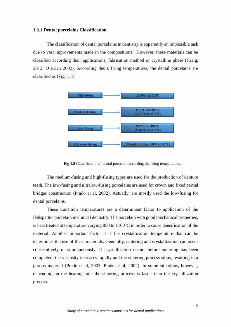
1.3.1 Dental porcelains Classification
The classification of dental porcelains in dentistry is apparently an impossible task
due to vast improvements made in the compositions. However, these materials can be
classified according their applications, fabrication method or crystalline phase (Craig,
2012; O’Brien 2002). According theirs firing temperatures, the dental porcelains are
classified as (Fig. 1.5):
Fig 1.5 Classification of dental porcelain according the firing temperatures.
The medium-fusing and high-fusing types are used for the production of denture
teeth. The low-fusing and ultralow-fusing porcelains are used for crown and fixed partial
bridges construction (Prado et al, 2002). Actually, are mostly used the low-fusing for
dental porcelains.
These transition temperatures are a determinant factor to application of the
feldspathic porcelain in clinical dentistry. The porcelain with good mechanical properties,
is heat treated at temperature varying 850 to 1100°C in order to cause densification of the
material. Another important factor it is the crystallization temperature that can be
determines the use of these materials. Generally, sintering and crystallization can occur
consecutively or simultaneously. If crystallization occurs before sintering has been
completed, the viscosity increases rapidly and the sintering process stops, resulting in a
porous material (Prado et al, 2003; Prado et al, 2002). In some situations, however,
depending on the heating rate, the sintering process is faster than the crystallization
process.

10 Study of porcelain-zirconia composites for dental applications
The sintering is the most common process applied to dental porcelains
restorations. The process consist of the firing the compacted ceramic powder at high
temperature to ensure optimal densification. The densification occurs by pore elimination
and viscous flow when the firing temperature is reached. The all-ceramic can also be
produced by sintering and use of a wider range of processing techniques such as: slip-
casting, heat-pressing and CAD/CAM machining (Craig 2012, O’Brien 2002, Anusavice,
2006).
1.3.2 Porcelains Composition
The quality of any ceramic depends on the choice of components; correct
proportioning of each component, and control of the firing procedure. Only the purest
components are used in the manufacture of dental porcelains because of the stringent
requirements of optical properties and chemical inertness, combined with adequate
strength, toughness and thermal expansion. (Fredericci et al, 2011).
The dental porcelains are composed of silica (SiO2) and alumina (Al2O3) with
various amounts of potash and soda feldspar (CaO, Na2O, K2O, B2O3, ZnO, ZrO2) for the
thermal expansion coefficient of the porcelain. The microstructure of the porcelain
consists of a vitreous phase (matrix) and some dispersed/embedded crystalline phases
(K2O.Al2O3.4SiO2 - tetragonal leucite at room temperature) (Fig. 1.6) (Fredericci et al,
2011; Li et al 2005; Tsetsekou et al, 2002;).
The glass matrix present a relatively small percentage of leucite crystal dispersed
within it (Giordano, 1996). Kaolin, is a hydrated aluminum silicate Al2O.2SiO2.2H2O act
as a binder, increasing the ability to mold the green porcelain. Thus, for dental porcelains
the kaolin was omitted and could therefore be considered a feldspathic glass with
crystalline inclusions of silica (Craig & Powers, 2002). The quartz (SiO2) remains
unchanged during the firing process and acts as a strengthening agent. It is present as a
fine crystalline dispersion throughout the glassy phase that is produced by the melting of
the feldspar. The feldspar fuses when it melts, forming a glass matrix. The feldspar are
mixtures of potassium aluminosilicate K2O.Al2O3.6SiO2 and sodium aluminosilicate, also
known as albite Na2O.Al2O3.6SiO2 (Clark et al, 2005; Noort, 2002; Barrelro et al, 1989).
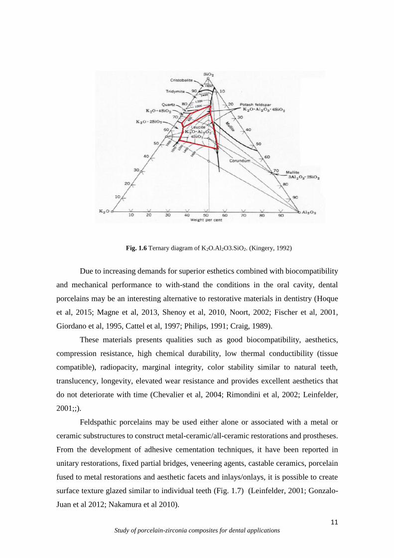
11 Study of porcelain-zirconia composites for dental applications
Fig. 1.6 Ternary diagram of K2O.Al2O3.SiO2. (Kingery, 1992)
Due to increasing demands for superior esthetics combined with biocompatibility
and mechanical performance to with-stand the conditions in the oral cavity, dental
porcelains may be an interesting alternative to restorative materials in dentistry (Hoque
et al, 2015; Magne et al, 2013, Shenoy et al, 2010, Noort, 2002; Fischer et al, 2001,
Giordano et al, 1995, Cattel et al, 1997; Philips, 1991; Craig, 1989).
These materials presents qualities such as good biocompatibility, aesthetics,
compression resistance, high chemical durability, low thermal conductibility (tissue
compatible), radiopacity, marginal integrity, color stability similar to natural teeth,
translucency, longevity, elevated wear resistance and provides excellent aesthetics that
do not deteriorate with time (Chevalier et al, 2004; Rimondini et al, 2002; Leinfelder,
2001;;).
Feldspathic porcelains may be used either alone or associated with a metal or
ceramic substructures to construct metal-ceramic/all-ceramic restorations and prostheses.
From the development of adhesive cementation techniques, it have been reported in
unitary restorations, fixed partial bridges, veneering agents, castable ceramics, porcelain
fused to metal restorations and aesthetic facets and inlays/onlays, it is possible to create
surface texture glazed similar to individual teeth (Fig. 1.7) (Leinfelder, 2001; Gonzalo-
Juan et al 2012; Nakamura et al 2010).
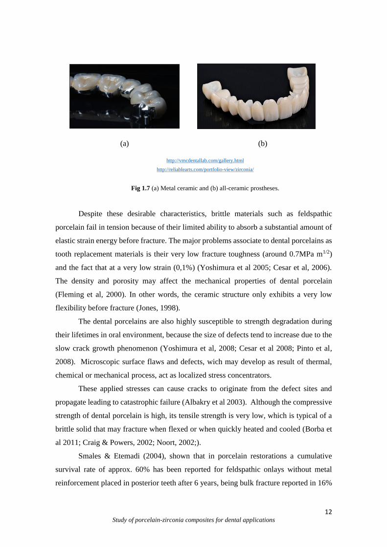
12 Study of porcelain-zirconia composites for dental applications
(a) (b)
http://vmcdentallab.com/gallery.html
http://reliablearts.com/portfolio-view/zirconia/
Fig 1.7 (a) Metal ceramic and (b) all-ceramic prostheses.
Despite these desirable characteristics, brittle materials such as feldspathic
porcelain fail in tension because of their limited ability to absorb a substantial amount of
elastic strain energy before fracture. The major problems associate to dental porcelains as
tooth replacement materials is their very low fracture toughness (around 0.7MPa m1/2)
and the fact that at a very low strain (0,1%) (Yoshimura et al 2005; Cesar et al, 2006).
The density and porosity may affect the mechanical properties of dental porcelain
(Fleming et al, 2000). In other words, the ceramic structure only exhibits a very low
flexibility before fracture (Jones, 1998).
The dental porcelains are also highly susceptible to strength degradation during
their lifetimes in oral environment, because the size of defects tend to increase due to the
slow crack growth phenomenon (Yoshimura et al, 2008; Cesar et al 2008; Pinto et al,
2008). Microscopic surface flaws and defects, wich may develop as result of thermal,
chemical or mechanical process, act as localized stress concentrators.
These applied stresses can cause cracks to originate from the defect sites and
propagate leading to catastrophic failure (Albakry et al 2003). Although the compressive
strength of dental porcelain is high, its tensile strength is very low, which is typical of a
brittle solid that may fracture when flexed or when quickly heated and cooled (Borba et
al 2011; Craig & Powers, 2002; Noort, 2002;).
Smales & Etemadi (2004), shown that in porcelain restorations a cumulative
survival rate of approx. 60% has been reported for feldspathic onlays without metal
reinforcement placed in posterior teeth after 6 years, being bulk fracture reported in 16%

13 Study of porcelain-zirconia composites for dental applications
of the restorations. It has also shown a clinical success rate of 64% for maxillary anterior
veneers after 10 years. The main reasons for failure were fracture (11%) and large
marginal defects (Peumans et al, 2004). For posterior feldspathic inlays, after an 8-year
clinical assessment period, marginal and bulk fracture were reported in 22% and 11% of
the restorations, respectively (Hayashi et al, 2000; Hayashi et al, 1998;).
Therefore, in order to increase the mechanical performance of porcelain
restorations, it is necessary to enhance their overall resistance crack propagation. The
understanding of its behavior is the key for the development of materials with longer
lifetime (Pinto et al, 2008).
1.3.3 Processing
The dental ceramics technology is one of the areas the most quick increase dental
materials research development (Denry & Holloway, 2010). Advanced ceramics
represent a significant evolution between the dental applications. The quality of the final
ceramic prostheses is dependent on each stage of the fabrication process Machining or
grinding of the core structure is of particularly importance since flaws or cracks can be
introduced that call possibly be propagate to the point of fracture during subsequent
process (Anusavice, 2006).
Dental technicians use a variety of techniques when casting dental porcelains. It
is unclear whether these techniques affect the total porosity and shrinkage of dental
porcelain (Craig, 1989).
The manufacture of porcelains restorations can be made from various techniques
such as: condensation and sintering; drying; shaping under pressure and sintering; casting
and sintering; vitreous infiltration and sintering and computer controlled machining
(CAD/CAM). The single units crowns can be a metal-ceramic crown (also called a
porcelain fused to metal crown), a traditional aluminous porcelain crown based on a core
of aluminous porcelain or based on a core of leucite reinforced porcelain (Phillips, 1996).
The methods of processing the ceramic core form and ceramic prosthesis are listed (Table
1.1):

14 Study of porcelain-zirconia composites for dental applications
Table 1.1 Techniques of processing and theirs characteristics.
Techniques Characteristics
Condensation Ceramic restorations (Dehailan, 2009)
Hot pressing crowns, inlays, on-lays, veneers and fixed partial
dentures (Ven Venkatachalam et al, 2009)
Casting Porous infrastructure (Crown and bridges) (Denry
& Holloway, 2010)
Slip-Casting Porous infrastructure (Crown and bridges
restorations) (Fonseca 2008)
Computer aided machining/milling (CAD/CAM) Fully sintered ceramic blocks (hard machining)
(Duret et al, 1988)
Computer aided machining/milling (CAD/CAM)
of pre-sintered form
Partially sintered ceramics (soft machining)
(Filser, 2001; Filser et al 2003)
In this thesis, the technique used to prepare porcelain-zirconia composites was hot
pressing.
Hot pressing is a technique consists of the application of external pressure to sinter
and shape the ceramic at high temperature (Fig. 1.8), and was first used in the fabrication
of all-ceramic restorations such as crowns, inlays, on-lays, veneers and fixed partial
dentures. Hot pressing, is characterized as avoiding large pores and promoting good
dispersion of the crystalline phase within the ceramics and flawless metal-ceramic
interfaces and is fulfilled in very fast sintering cycles.Although few, there are some
studies on the bond strength of hot pressed porcelain to metal and ceramic dental
substructures (Venkatachalam et al, 2009; Drummond et al, 2000, Henriques et al, 2012).
15 Study of porcelain-zirconia composites for dental applications
http://www.substech.com/dokuwiki/doku.php?id=solid_state_fabrication_of_metal_matrix_composites
Fig 1.8 Hot pressing process.
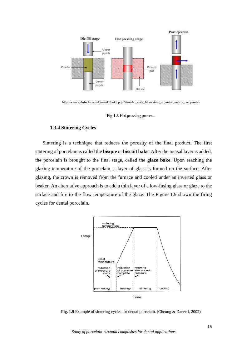
1.3.4 Sintering Cycles
Sintering is a technique that reduces the porosity of the final product. The first
sintering of porcelain is called the bisque or biscuit bake. After the incisal layer is added,
the porcelain is brought to the final stage, called the glaze bake. Upon reaching the
glazing temperature of the porcelain, a layer of glass is formed on the surface. After
glazing, the crown is removed from the furnace and cooled under an inverted glass or
beaker. An alternative approach is to add a thin layer of a low-fusing glass or glaze to the
surface and fire to the flow temperature of the glaze. The Figure 1.9 shown the firing
cycles for dental porcelain.
Fig. 1.9 Example of sintering cycles for dental porcelain. (Cheung & Darvell, 2002)
16 Study of porcelain-zirconia composites for dental applications
The purpose of feldspathic porcelain firing procedures is to densely sinter the particles
of powder together and to produce a relatively smooth, glassy layer (glaze) on the surface.
In some cases, a stain layer is applied for shade adjustment or for characterization such
as stain lines or fine cracks. Several chemical reactions occur over time at porcelain firing
temperatures and of particular importance are increases in the concentration of crystalline
leucite in the porcelains designed for fabrication of metal-ceramic and all ceramics
restorations (Cheung & Darvell, 2002, Swapan et al, 2003; Cheng et al, 2012). The
porcelain firing can occur in air or vacuum, depending on the type of furnace.
During the sintering process the pores are reduced, when the air removal. This process
create as far as possible a non-porous solid and consequently a porcelain with high
strength and aesthetics. Although sintering is often referred to as a solid-state process,
here it is driven by surface tension because of partial melting crystalline phases or
sufficient softening of glassy material (Frenkel, 1945; Mackenzie, 1949; Herring, 1951;).
This reduction of pore volume depends on sintering time (Frenkel, 1945; Herring, 1951;;
Ivensen, 1970), temperature (Frenkel, 1945; Herring, 1951;) and atmosphere (Shaler et
al, 1948; Vines et al, 1958; Wilson and Whitehead, 1967; Jones and Wilson, 1975;
Rosenkranz and Frischat, 1984;;), and the viscosity of the melt (Mackenzie, 1949;
Herring, 1951; Bottinga et al, 1972).
In this case, residual stresses can likely accumulate during the heating and cooling
firing procedures mainly because of the cooling rate and the coefficient of thermal
expansion (CTE) mismatch between core and veneer ceramics (Swain, 2009; Guazzato
et al, 2010; Tholey et al, 2010). When additional stresses are applied to the restoration,
the probability of failure to fatigue crack propagation might increase (Swain, 2009;; Della
Bona et al, 2009; Smith et al, 1994).
In all-ceramics restorations, especially when using the standard layering technique
to match the esthetics natural teeth, the multiple firing cycles can be affect the porcelain-
zirconia adhesion. Thus, the dental porcelains demonstrate an excellent aesthetics that do
not deteriorate with time due to its chemical stability. Because theirs thermal
characteristics such as conductivity and coefficient of thermal expansion being similar to
dentin and enamel, the porcelains can be employed due the good marginal sealing would
prevent percolation related problems (Henriques, 2012).
17 Study of porcelain-zirconia composites for dental applications
However, these materials receive amorphous structure that produce physical
properties typical of glass, including brittleness and lack of a definite melting
temperature. The strength of glasses and brittle materials is highly dependent on porosity
(size and number of voids) which act as crack initiation sites by the presence of small
flaws or cracks. According the crack propagation theory, when stressed in tension, small
flaws tend to open up and propagate, resulting in a low tensile strength. This factor is
different when compared as metal (ductile materials) (Anusavice, 2006).
In compression the glasses are much stronger, because compressive stresses tend
to close up flaws. Therefore, the tensile strengths of vitreous dental porcelains are around
(usually between 20 and 60MPa) as compared with compressive strengths of (517 MPa)
and very low fracture toughness (<0,1% strain) (Lawn et al, 2001). The strength of dental
porcelains is generally tested in flexure as a beam and reported as modulus of rupture.
The modulus of rupture of a vitreous body or enamel porcelain is about 13,000 psi (90
MPa). In vaccum-fired the strengths of porcelains are higher due to fewer flaws (O´Brien,
2002).
1.4 All-ceramic Restorations
Teeth play a critically important role in our lives. Loss of function reduces our
ability to eat a balanced diet, which has negative consequences for health. Loss of
aesthetics can negatively impact in social function. Both function and aesthetics can be
restored with dental crowns and bridges.
According Höland et al (2009), dental structure loss is related by a variety of
factors that range from diseases to trauma, as well as preventable hygiene-related
problems. Allied to today’s lifestyles, which require a good appearance, this problem has
lead to an increasing demand for dental restorations. This demand is translated into an
increasing need for dental materials that allow for the creation of teeth esthetically and
functionally to natural teeth.
There are many challenges for the physical properties of the ideal dental
restorative material. The ideal restorative material would be identical to natural tooth
structure in strength, adherence, and appearance.
18 Study of porcelain-zirconia composites for dental applications
In this way, ceramics are attractive dental restorations materials because of their
aesthetics, biological acceptance, and chemical durability. The all-ceramic dental
restorations are domelike structures composed of two layers of materials, namely the
reinforcing ceramic core for bearing the biting and the veneer porcelain for aesthetics.
However, ceramics are brittle and subject to premature failure, especially under repeated
contact loading in a moist environment (Lawn et al, 2001).
Despite a continuous effort in improving the strength of dental ceramics (e.g.
using a strong zirconia or alumina core to support a weak but aesthetic porcelain veneer).
All-ceramic dental prostheses (including veneered zirconia and alumina restorations)
continue to fail at a low rate each year (Burke et al, 2002).
The differences in the fabrication methods used to produce ceramic restorations
such as microstructural features, pore and crystal distribution, presence of inclusions and
flaws, composition and crystalline content, which could influence the mechanical
behavior of materials (Guazzato et al 2004; Guazzato et al,2004a).
In clinical dentistry, it is very important to obtain dental restorations that are as
dense as possible, since this will lead to better clinical performance on the long term.
Thus, the use of all-ceramic restorations is an interesting step to improve the mechanical
properties combined as aesthetics characteristics and replace metal infrastructures (Della
Bona & Kelly, 2008).
During the last two decades all-ceramic restorations have become increasingly
popular, and, particularly since the introduction of zirconia-based frameworks
(Vagkopoulou et al, 2009), a trend toward all-ceramic restorations has been observed.
Zirconia-based ceramics is being proved a successful infrastructure material.
Studies have reported no fracture of the zirconia infrastructure in short or medium-term
periods of clinical observation (Pjetursson et al, 2008).
The mechanical properties of zirconia enable clinical use of all-ceramic fixed
dental prostheses (FDP) even in the molar region (Tinschert et al 2001) and in long span
designs (Schmitter et al, 2009). The advantages of zirconia-based substructures are
biocompatibility, improved fracture strength and fracture toughness, and their unique

19 Study of porcelain-zirconia composites for dental applications
transformation toughening mechanism (Studart et al, 2007). To achieve the best aesthetic
results, however, the frameworks must be veneered using ceramics with mechanical
properties inferior to those of the substructures and, consequently, the veneering ceramic
is the weak part of the system (Al-Dohan et al, 2004; Guazzato et al 2004; Isgro et al,
2003).
The failures in porcelain-zirconia restorations, are frequently associated with
biological complications, such as secondary caries, periodontal support and tooth
fracture, which likely are not related to the materials used in fixed prostheses (Della Bona
& Kelly, 2008; Wang et al, 2014) and some structural deficiencies of the material such as
marginal discrepancies, weak performance of the veneering ceramics (Christensen &
Ploeger, 2010) and its limited bond to the zirconia substrate (Hübsch et al, 2015;
Aboushelib et al, 2009).
Clinical studies report high survival rates for all-ceramic restorations.
Infrastructure failures are unusual when the ceramic material is correctly indicated,
varying from 2 to 6% (Al-Amleh et al 2010; Sailer et al 2006, 2007, 2009; Ortorp et al
2009, Suarez et al, 2004).
The failure mode most frequently observed is chipping of the veneering material.
This type of failure has been observed mainly when zirconia-based infrastructures are
used, showing chipping rates that vary from 0 to 50%, depending on the follow-up period
(Al-Amleh et al 2010; Molin et al 2008; Tinschert et al 2008; Sailer et al 2007)). Yet,
failure have been described as crack propagation from a contact damage zone throughout
the veneering porcelain, reaching or not the interface, which can lead to delamination and
exposure of the infrastructure material in the oral environment (Borba et al, 2011).
Swain 2009, shown that residual tensile stresses are create within the porcelain at
or near the ceramic-porcelain interface and could contribute to the porcelain fracture in
zirconia systems.
Most dental materials are quite brittle; they are highly susceptible to crack
initiation in the presence of surface flaws when subjected to tensile stress, such as when
they are subjected to flexural loading. The Figure 1.10 shown the stress induced in bridges
by a flexural force (P). The stresses are believed to have multifactorial origin, potentially

20 Study of porcelain-zirconia composites for dental applications
involving the ceramic thermal history, restoration’s geometric factors (i.e. infrastructure
design and core-veneer thickness ratio), material’s elastic properties “ceramic strength”
and poor wetting of the core by the veneer (Guess et al, 2008; De Hoff & Anusavice,
2004-2004a; Benetti et al, 2010). Therefore, these factors should be hand lead carefully
to prevent development of high-magnitude tensile stresses.
http://pocketdentistry.com/mechanical-properties-of-dental-materials-2/
Fig. 1.10 A Stresses induced in a three-unit bridge by a flexural force (P). B, Stresses induced in a two-unit
cantilever bridge.
Common technical failures in clinical studies on zirconia reconstructions are
either the incidence of pure cohesive failure within the ceramic veneer or a mixed failure
mode where part of the fracture follow the frame/veneer interface (chipping) (Sailer et al
2006; Tinschert et al 2008).
In both observed failure modes, however, the frame remains intact. This type of
failure within the veneering materials is typical in daily clinical practice since load case
leading to a destruction of the frame as well, e.g., central axial loading, are associated
with force.
Nevertheless, these materials are indicate to produces dental substructures and can
be an excellent alternative to replace the natural teeth.
1.4.1 Zirconia
The zirconia (ZrO2), have received considerable attention in dental applications
due to possibility of obtaining substructures and zirconia-based materials with good
mechanical properties. This material is considered nowadays one of the most important
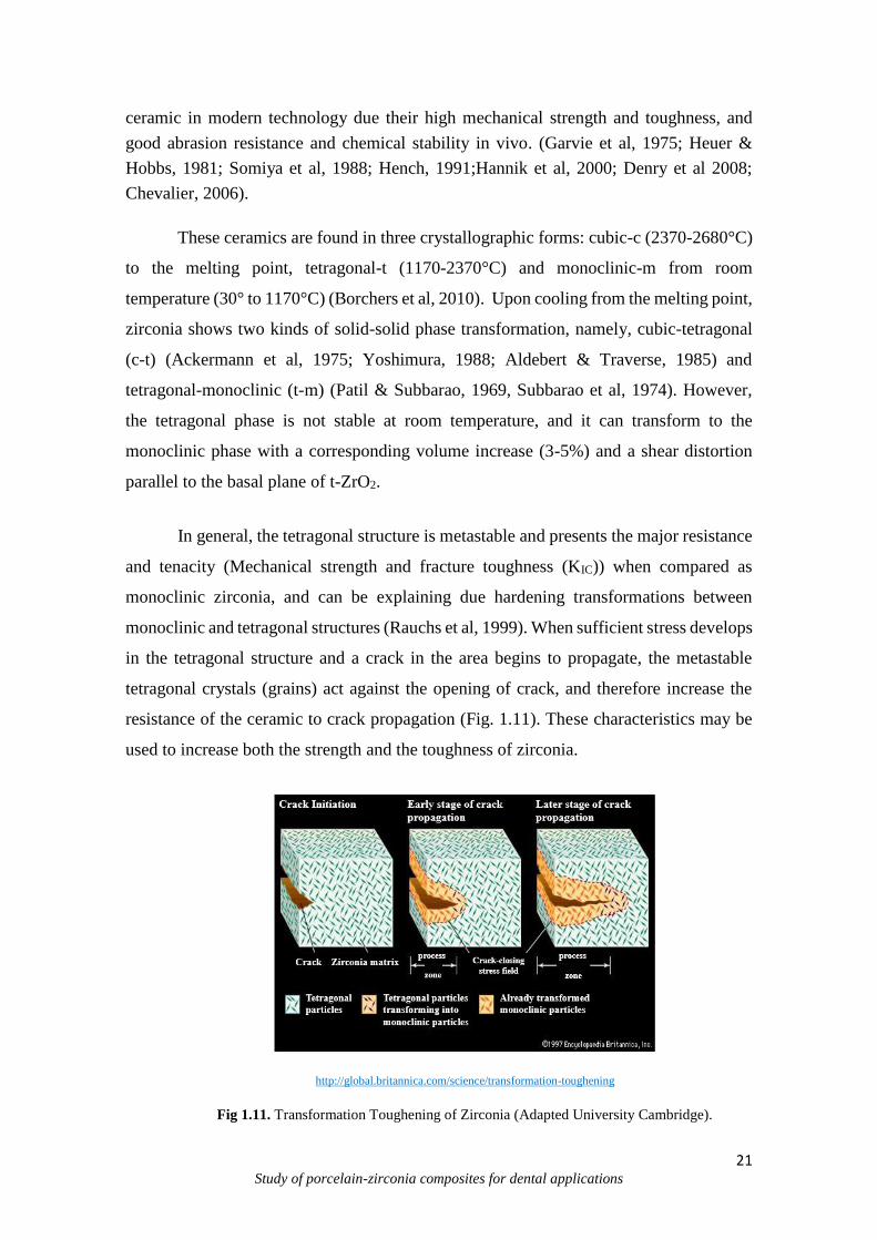
21 Study of porcelain-zirconia composites for dental applications
ceramic in modern technology due their high mechanical strength and toughness, and
good abrasion resistance and chemical stability in vivo. (Garvie et al, 1975; Heuer &
Hobbs, 1981; Somiya et al, 1988; Hench, 1991;Hannik et al, 2000; Denry et al 2008;
Chevalier, 2006).
These ceramics are found in three crystallographic forms: cubic-c (2370-2680°C)
to the melting point, tetragonal-t (1170-2370°C) and monoclinic-m from room
temperature (30° to 1170°C) (Borchers et al, 2010). Upon cooling from the melting point,
zirconia shows two kinds of solid-solid phase transformation, namely, cubic-tetragonal
(c-t) (Ackermann et al, 1975; Yoshimura, 1988; Aldebert & Traverse, 1985) and
tetragonal-monoclinic (t-m) (Patil & Subbarao, 1969, Subbarao et al, 1974). However,
the tetragonal phase is not stable at room temperature, and it can transform to the
monoclinic phase with a corresponding volume increase (3-5%) and a shear distortion
parallel to the basal plane of t-ZrO2.
In general, the tetragonal structure is metastable and presents the major resistance
and tenacity (Mechanical strength and fracture toughness (KIC)) when compared as
monoclinic zirconia, and can be explaining due hardening transformations between
monoclinic and tetragonal structures (Rauchs et al, 1999). When sufficient stress develops
in the tetragonal structure and a crack in the area begins to propagate, the metastable
tetragonal crystals (grains) act against the opening of crack, and therefore increase the
resistance of the ceramic to crack propagation (Fig. 1.11). These characteristics may be
used to increase both the strength and the toughness of zirconia.
http://global.britannica.com/science/transformation-toughening
Fig 1.11. Transformation Toughening of Zirconia (Adapted University Cambridge).

22 Study of porcelain-zirconia composites for dental applications
The toughening mechanism of crack shielding results from the controlled
transformation of the metastable tetragonal phase to the stable monoclinic phase. Several
types of crack shielding processes are possible, including micro-cracking, ductile zone
formation, and transformation zone formation. This mechanism is significantly extends
the reliability and lifetime of ZrO2 derived materials and leads to the high fracture
toughness of tetragonal zirconia (Garvie & Hannik, 1975).
In order to minimize the effect of volume expansion of zirconia structure
(transformation tetragonal to monoclinic), the addition of certain oxides (dimensional
stabilizers) is very important (Piconi et al, 1998). These oxides, act as retainers of
tetragonal form by addition (MgO and CaO) (Yoshimura et al, 2007), CeO2 (Ban et al,
2008) and especially Y2O3 (Atik et al, 1994; Garvie & Hannik, 1975, Chevalier et al,
2004), due to its large solid solubility rage in zirconia, is employed to partially or
completely stabilize the tetragonal and cubic structure.
The addition of Y2O3 (typically 3%) allows decrease the stress, due phases
transformations that occurs when cooling of transition temperature between tetragonal
and monoclinic phases (thermodynamically stable phase), or also by decrease of particle
size above a critic size (Fig 1.12) (Craska et al 2000; Chen et al, 2011; Yashima et al
1996).
http://www.doitpoms.ac.uk/tlplib/fuel-cells/sofc_electrolyte.php
Fig. 1.12. Phase Diagram of ZrO2/Y2O3 (University of Cambridge).
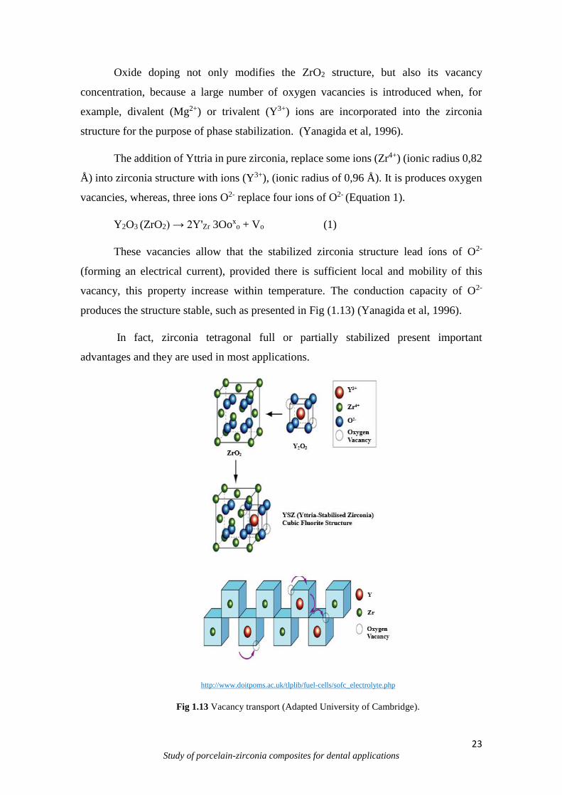
23 Study of porcelain-zirconia composites for dental applications
Oxide doping not only modifies the ZrO2 structure, but also its vacancy
concentration, because a large number of oxygen vacancies is introduced when, for
example, divalent (Mg2+) or trivalent (Y3+) ions are incorporated into the zirconia
structure for the purpose of phase stabilization. (Yanagida et al, 1996).
The addition of Yttria in pure zirconia, replace some ions (Zr4+) (ionic radius 0,82
Å) into zirconia structure with ions (Y3+), (ionic radius of 0,96 Å). It is produces oxygen
vacancies, whereas, three ions O2- replace four ions of O2- (Equation 1).
Y2O3 (ZrO2) → 2Y'Zr 3Ooxo + Vo (1)
These vacancies allow that the stabilized zirconia structure lead íons of O2-
(forming an electrical current), provided there is sufficient local and mobility of this
vacancy, this property increase within temperature. The conduction capacity of O2-
produces the structure stable, such as presented in Fig (1.13) (Yanagida et al, 1996).
In fact, zirconia tetragonal full or partially stabilized present important
advantages and they are used in most applications.
http://www.doitpoms.ac.uk/tlplib/fuel-cells/sofc_electrolyte.php
Fig 1.13 Vacancy transport (Adapted University of Cambridge).
24 Study of porcelain-zirconia composites for dental applications
One of the most popular types of doped zirconia used in technologic and
biomedical area is Tetragonal Zirconia Polycrystalline (TZP). The TZP combine a
pleasant aesthetic appearance with biocompatibility associated to superior mechanical
properties (Piconi et al, 1999; Denry et al, 2008).
With this material, it is possible produce a numerous veneering ceramics matching
the frameworks of this zirconia as have appeared on the dental market. In powder form,
the zirconia act as structural reinforcements, increasing thermal resistance, receive a
higher capacity of tenacity of formed surface, high rigidity and high fracture toughness
(Moon et al, 2002; Bulgakov et al, 2002).
In dentistry the Y-TZP (Yttria Tetragonal Zirconia Polycrystals) was first
introduced to dental clinics in the late 1990s, their microstructure is strong enough to
resist the resulting stresses.
This material are the most recent restorations in the dental ceramics family and
they have been rapidly adopted for a wide range of applications, because their superior
mechanical properties - strong (flexural strength of approximately 900-1200 MPa) and
tough (fracture toughness, KIC, of approximately 9-10MPa.m1/2) and achieve good
aesthetics results, even without veneering (Benzaid et al, 2008; Tang et al, 2012; Guess
at 2012; Egilmez et al, 2014). Thus, has considerably higher bending and fracture strength
than other ceramics (Anusavice, 2006; Guazzato et al, 2004).
The Yttria-stabilized tetragonal zirconia (Y-TZP) have a significant growing in
biomedical materials due their superior mechanical properties, chemical stability, high
fracture toughness. This material specifically with 3% of Yttria (3YSZ) for theirs
characteristics proves to be a suitable material for dental applications, industrial cutters
and grinding media (Richard et al, 2000; Chevalier et al, 2009).
Because the material provide high hardness, fracture strength, and structural
reliability and show a smaller range of strength variations than porcelain, the yttria-
stabilized zirconia ceramic is sometimes referred to as ceramic steel and have higher
importance in dental applications (Shijo et al, 2009; Fischer et al, 2008; Anusavice, 2006;
Guazzato et al, 2004a).
25 Study of porcelain-zirconia composites for dental applications
1.4.2 Synthesis of Zirconia
The synthesis optimization to obtain ceramic materials have been crucial and have
attracted special attention of the researcher’s groups and industry (Li et al, 1994). This
interest has resulted in substantial increase from the development and production of the
nanostructured materials, improving the functionally characteristics when compared as
conventional ceramics (Duncan et al, 2006).
In this context, the zirconia powders are obtained generally by solid state reactions
at high temperatures (Li et al, 2001; Politova et al 2004) or by wet methods such as:
hydrolysis (Rauchs et al, 1999; Yang et al, 1997), sol-gel method (Kessman et al, 2009;
Matsui and Ohgai, 2000), precipitation (Vargas et al, 2003; Atkinson e Segal, 1998),
hydrothermal synthesis (Aruna et al, 2011; Masala et al, 2006) and complex
polymerization method (CPM) (Razpotnik and Macek, 2007; Grzebielucka et al, 2010).
From the exposed methods above, the Complex Polymerization Method (CPM)
presents advantages such as: easy synthesis process, good chemical homogeneity, high
purity, stoichiometric control, nano-metric powders, low processing temperature and cost
(Cunha et al, 2005). This method, comprises in polymeric resin formation originated by
polyesterification process between metallic cations complexed (chelates using
hydroxycarboxylic acids) and a polyalcohol such as ethylene glycol (Pechini, 1967).
The CPM method provides a chemical way to distribute evenly the metallic ions
in a polymeric resin in atomic scale (Landeros et al, 2009). During the synthesis process
occurs the cations distribution at the molecular level in polymer structure. The heating
around 90°C, in air atmosphere, promotes the condensation reaction (alcohol and acid
chelate), with the rise of water molecules, then occurs the polyesterification and the
excess water is removal by evaporation, emerging a polymeric resin at solid state to rear
powders achievement (Quinelato et al, 2001). The decomposition of polymeric resin is
necessary to eliminate of the organic compounds by heat treatment, obtaining the desired
phase.
Besides, it is a flexible method, being directed in obtaining of various oxides, and
distinct structures modified from the parameters adjustment (Cushing, 2004). The figure
1.14 shown the reactions of the Complex Polymerization Method.

26 Study of porcelain-zirconia composites for dental applications
Fig 1.14. Complexation reaction and polymerization in the CPM. (Motta, 2008).
The organic material elimination is possible from the calcination to obtain the
desired phase. The calcination temperatures (300°C) promote the polymeric chains
rupture and the combustion of the material (pyrolysis). In temperatures between 300°C
and 900°C occurs the metallic cations oxidation (crystallites of oxides) (Motta, 2008).
One of the disadvantage of this method consist in a small control of particle size,
format and morphology. Unlike the sol-gel method when the metal it is a constituent part
of the gel network, in the CPM the synthesis is formed by chelating agent
polyesterification to a mono-hydroxylic alcohol (Cushing, 2004; Galvão, 2010).
Thus, occurs imprisonment of metallic ions in the organic compounds with weak
bonds. Also presents the large weight loss, forming agglomerates during the calcination
process, due the additional heating produced by material calcination and combustion
(sintered agglomerates) (Cushing, 2004).

27 Study of porcelain-zirconia composites for dental applications
1.5 Particulate Composites
A composite material can be defined as a combination of two or more materials
that results in better properties than those of the individual components used alone.
Differently to metal alloys, each material retains its separate chemical, physical, and
mechanical properties. The two constituents are a reinforcement and a matrix (Campbell,
2010, Callister, 2004).
Ceramics and ceramic composites are promising materials having high strength
characteristics but quite low crack resistance properties at the same time. This is one of
the major factors hindering the wide-scale application of these materials in various fields
of human activities. The main advantages of composite materials are their high strength
and stiffness, combined with low density, when compared with bulk materials, allowing
for a weight reduction in the finished part (Chawla 1998).
The crack resistance is critical not only for ceramic products operating under
extreme mechanical and thermal loads but also for structural components whose brittle
fracture is intolerable even under arbitrary loads (Gogotsi, 2002).
Effect of the dispersed particles on the composite properties depends on the
particles dimensions very small particles (less than 0.25 micron in diameter) finely
distributed in the matrix prevents movement of dislocations and deformation of the
material. Such strengthening effect is similar to the precipitation hardening.
The dispersed phase of particulate composites (ceramic particles) is usually stable
at high temperatures, so the strengthening effect is retained. Many of composite materials
are designed to work in high temperature applications. The reinforcing phase present
strength and stiffness properties. Generally, the reinforcement is harder, stronger, and
stiffer than the matrix. The reinforcement can be a (fiber or a particulate) (Campbell,
2010).
Particulate composites
Particulate composites consist of a matrix reinforced with a dispersed phase in
form particles. They may be spherical, platelets, or any other regular or irregular
geometry. Particulate composites tend to be much weaker and less stiff than continuous
fiber composites, but they are usually much less expensive. Particulate reinforced
composites usually contain less reinforcement (up to 40 to 50 volume percent) due to
processing difficulties and brittleness (Campbell, 2010).
28 Study of porcelain-zirconia composites for dental applications
Generally, these materials with improved properties, are compared with “pure”
matrix materials to understanding the mechanisms that occurs during the fabrication
process. Properties of particulate composites depend on: Concentration, size, shape,
distribution and orientation this factors can achieve a significant improvement of
hardness, stiffness, fracture toughness and/or strength of the material. It was also reported
that the decrease of inclusion size to the nanometric scale allowed extremely high values
of flexural strength and fracture toughness to be achieved. (Campbell, 2010)
The manufacturing of composites with ceramic matrix almost always leads to
residual stresses caused by the mismatch of thermal properties of constituent phases. The
difference in thermal expansion coefficients (CTE’s) could introduce stresses reaching
even hundreds of MPa to the composite system. Such a phenomenon to influence fracture
toughness of the material (Grabowski & Edzich, 2007).
These composites are designed to produce unusual combinations of properties
rather than to improve the strength (Hull, 1996). Mechanical properties, such as elastic
modulus, of particulate composites achievable are in the range defined by rule of
mixtures as follows Equations. (2 and 3):
Upper bound is represented by:
𝐸𝑐(𝑢) = 𝐸𝑚𝑉𝑚 + 𝐸𝑝𝑉𝑝 (2)
And lower bound is represented by:
𝐸𝑐(𝑙) =𝐸𝑚𝐸𝑝
𝐸𝑝𝑉𝑚+𝐸𝑚𝑉𝑝 (3)
Where:
E – elastic modulus; V – volume fractions; c, m, p – present composites, matrix and particulate
phases, respectively. A schematic diagram of these bounds is shown in the Fig. 1.15.

29 Study of porcelain-zirconia composites for dental applications
Fig. 1.15 Particulate reinforced composite.
In dentistry the use of particles as reinforcement in porcelain matrix is widely
applied to increase the mechanical properties of these composites. The possibility of some
improvement of mechanical properties of oxide ceramics by manufacturing of particulate
composites has been very well recognized recently. (Gogotsi, 2002)
The tetragonal zirconia ceramics show a relatively good reliability and widely
used as reinforcement in metals and ceramics matrix. A typical value of the Weibull
parameter (m) for TZP is about 20. However, not many data describing the reliability of
particulate composites with TZP matrix are available. It is expected microstructure of the
composite influences the material reliability (Madfa et al, 2014).Thus, the final properties
of these composites can be influence by correct choice of the some parameters such as
amount of inclusion; their size and dispersion are decisive for the final values of
mechanical properties of the material.
1.6 Ceramic-ceramic composites interaction/Adhesion
The adhesion is the attachment of materials in contact that resists the forces of
separation. Adhesive phenomena are critical in many areas of dental materials,
including the bonding of porcelains to metals and the adhesion of resins to tooth
structure (O’Brien, 2002).
Several types of adhesive bonds may be identified according to the classifications.
Mechanical adhesion depends on mechanical interlocking of the two phases and may
include microscopic attachments, as in the case of resin bonding to etched enamel or
hoop stresses of a porcelain around a metal core. Chemical adhesion relies on
chemical bonding between two phases. Diffusion bonding results when one phase
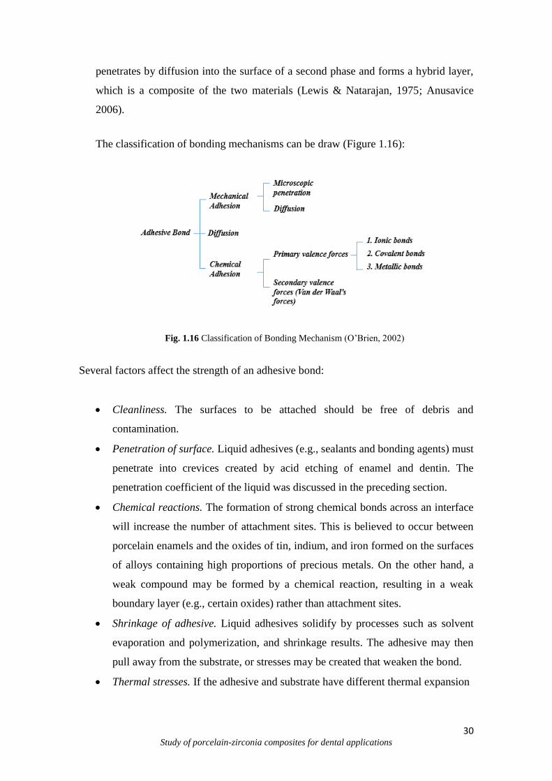
30 Study of porcelain-zirconia composites for dental applications
penetrates by diffusion into the surface of a second phase and forms a hybrid layer,
which is a composite of the two materials (Lewis & Natarajan, 1975; Anusavice
2006).
The classification of bonding mechanisms can be draw (Figure 1.16):
Fig. 1.16 Classification of Bonding Mechanism (O’Brien, 2002)
Several factors affect the strength of an adhesive bond:
Cleanliness. The surfaces to be attached should be free of debris and
contamination.
Penetration of surface. Liquid adhesives (e.g., sealants and bonding agents) must
penetrate into crevices created by acid etching of enamel and dentin. The
penetration coefficient of the liquid was discussed in the preceding section.
Chemical reactions. The formation of strong chemical bonds across an interface
will increase the number of attachment sites. This is believed to occur between
porcelain enamels and the oxides of tin, indium, and iron formed on the surfaces
of alloys containing high proportions of precious metals. On the other hand, a
weak compound may be formed by a chemical reaction, resulting in a weak
boundary layer (e.g., certain oxides) rather than attachment sites.
Shrinkage of adhesive. Liquid adhesives solidify by processes such as solvent
evaporation and polymerization, and shrinkage results. The adhesive may then
pull away from the substrate, or stresses may be created that weaken the bond.
Thermal stresses. If the adhesive and substrate have different thermal expansion
31 Study of porcelain-zirconia composites for dental applications
coefficients, changes in temperature will produce stresses in the bond. For
example, porcelain enamels are bonded to alloys at high temperatures and then
cooled to room temperature. Close matching of the thermal expansion coefficients
of porcelain enamel and alloy is required to minimize stresses.
Corrosive environment. The presence of water or corrosive liquid or vapor will
often lead to deterioration of an adhesive bond. For example, acrylic resins will
initially adhere to clean unetched tooth enamel, but the bond deteriorates upon
storage in water.
The studies of adhesive bonding started by Buonocore in 1956, has envolved through
many generations in which adhesives have become stronger, easier to use, and have
important for dental applications (Buonocore et al, 1956).
In dentistry, the ceramic-ceramic bonding specially yttria-stabilized tetragonal
zirconia polycrystal (Y-TZP) with porcelain, has been widely used in the production of
all-ceramic restorations nowadays, the relatively higher failure rate of zirconia-based
prostheses compared with porcelain-fused-to-metal (PFM) restoration is still concerned
(Guess et al, 2008, Sailer et al, 2007).
The weak bonding might be due to some factors, a mismatch in the coefficients of
thermal expansion (CTE) of the core and veneer ceramics and its resulting pre-stresses,
transformation of zirconia crystals at core-veneer interface, due to thermal influences or
stress loading (interface toughness) surface treatment, lack of chemical bonding,
unintentional formation of micro-porosities at the interface. (Aboushelib et al, 2006;
2008)
These factors, has been stated that the success of all of veneer/core bilayer restorations
depends on the mechanical adhesion between veneering ceramic and zirconia core
(Raigrodski et al, 2006). Several studies (Ozcan & Vallittu, 2003; Kern et al, 2009; Al-
Amleh et al, 2010) have investigated the bond strength and the durability of various
bonding methods to dental ceramics. Nevertheless, there is a little information about the
bond quality between veneering ceramic and zirconia core (Ozkurt et al 2010; Chaiyabutr
et al, 2008).
In oral environment, the good core-veneer bond strength is a clinical requirements to
avoid premature failure of all-ceramic restorations (Aboushelib et al, 2009).

32 Study of porcelain-zirconia composites for dental applications
1.6.1 Bond Strength Tests
To evaluate the ceramic-ceramic unions some tests are used, and are classified as
flexural strength or shear strength (Chevalier et al, 2009; Jung et al, 2000).
In brittle materials the normal tensile test cannot easily be performed because of the
presence of flaws at the surface. The lower concentration increase the probability of
failure and uniform distribution of stresses in microspecimens, in this case, holding
microtest is needed to asses theses parameters (Chevalier et al, 2009; Jung et al, 2000).
Often, just placing a brittle material in the grips of the tensile testing machine causes
cracking. These materials may be tested using the bend test. (Engineering Archives,
2012).
The flexural strength present two configurations: three or four point loading (Lavine
and Custer, 1996; O´Brien and Craig, 1977). For these type of materials the ISO 6872 it
is most appropriate. The ISO 6872 specifies the requirements and the corresponding test
methods for dental ceramic materials for fixed all ceramic and metal-ceramic restorations
and prostheses. The flexural strength testing specifications, provided by ISO 6872 include
but are not limited to ceramic dental fillings, crowns, veneers, implants, and brackets.
In this test, the rectangular specimens or flat cross-section is placed on two parallel
supporting pins. The load is applied in the center with the ceramic surface down until
failure of the ceramic occurs (Figure 1.17).
http://www.substech.com/dokuwiki/doku.php?id=flexural_strength_tests_of_ceramics
Fig. 1.17. Three point bending of a ceramic specimen according to ISO 6872.
This configuration provides uniform loading of the specimen and prevents friction
between the specimen and the supporting pins.

33 Study of porcelain-zirconia composites for dental applications
The shear bond strength is another test used to identify the adhesive parameters in
ceramic-ceramic materials. This tests is indicate due to criticism because the maximum
tensile stresses that are created at the surface porcelain, this resulting in predictable tensile
failures (Anusavice et al, 1980; McLean, 1980).
Generally, has been used the planar interface shear bond test (DeHoff et al, 1995).
The planar interface shear bond tests are bond tests with porcelain applied to a flat
zirconia surface. The ceramic-ceramic bond sites can be circular or rectangular. The tests
intends to separate porcelain from zirconia substrate (Fig. 1.18).
Fig. 1.18 Shear bond test for restorative materials.
The ISO 29022:2013 is the standard normally used for dental materials. The ISO
29022:2013 specifies a shear test method used to determine the adhesive bond strength
between direct dental restorative materials and tooth structure, e.g. dentine or enamel.
The method as described is principaly intended for dental adhesives. The method
includes substrate selection, storage and handling of tooth structure. Can be applied for
all-ceramic restorations. These tests reproduce the realistic conditions and generates
information that is useful to better predict clinical reliability of ceramic-ceramic dental
systems (De Hoff et al, 1995).

34 Study of porcelain-zirconia composites for dental applications
1.7 Clinical Applications of dental Porcelain
In recent years, the dental porcelain has found an increased number of applications
from the development of new methods for the construction of porcelain veneers, intra-
coronal restorations, jacket crowns, metal ceramic crowns and posterior/anterior bridges.
Porcelain laminate veneers were initially described by Horn 1983. It is fitted to
the surface of prepared enamel by a combination of mechanical bond to etched enamel is
by means of a composite resin (Hussin, 1979)
Porcelain Jacket Crown or All-porcelain Crown system. Porcelain jacket crowns have
been used widely in dentistry since Land developed the platinum foil technique in 1903.
The feldspathic porcelains were known for natural esthetics resulting mainly from high
translucency and the specialized laboratory skills used (Anusavice, 2006) (Fig 1.19).
Therefore, because the presence of microcracks and tendency to fracture, almost all
porcelains are strengthened with alumina crystals incorporated into the porcelain powder
(McLean and Hughes, 1965).
http://www.affinitydentalclinics.com/dentalservices/dental-crowns-jacket-crown/
Fig 1.19. Porcelain Crown.
Porcelain bonded to metal restorations. It is composed of a porcelain facing
bonded to the surface of the metal substructure or coping, which fits over the tooth
preparation. Bonding of porcelain to metal alloy during firing is achieved by Mechanical
interlocking, Chemical or molecular bonding and Van der Waals forces (Vickery 1968,
Bardirelli 1968, Cascone 1977, McLean 1979, Bagby et al. 1990).
Ceramic inlay/onlay. There are two ceramic inlay systems, the first is laboratory
made, in which the porcelain inlay is fabricated by condensing porcelain into a refractory
35 Study of porcelain-zirconia composites for dental applications
die which replicates the prepared tooth, while the second is a chairside system which
involves milling of a porcelain inlay which is designed in a computer form a three-
dimensional video image of prepared tooth (Smith, 1990).All porcelain bridges are not
yet widely accepted restoration. Al-Ajlouni et al 2004) stated that all-porcelain resin-
bonded bridges, especially the veneer type, can doubtfully sustain forces in the mouth.
Some authors mentioned that porcelain bridges can be reinforced through the
incorporation of a rod of recrystallized alumina into the structure. The porcelains also
widely used in dental implantology (Al-Ajlouni et al, 2004).
36 Study of porcelain-zirconia composites for dental applications
References
Albakry, M., Guazzato, M.G., Swain, M.V., Fracture toughness and hardness evaluation
of three pressable allceramic dental materials. J. Dent. (2003) 31, 181-188.
Aboushelib M.N., Feilzer A.J., Kleverlaan C.J. Bridging the gap between clinical failure
and laboratory fracture strength tests using a fractographic approach. Dent. Mater. (2009)
25 3:383–91.
Aboushelib MN, Kleverlaan CJ, Feilzer AJ. Microtensile bond strength of different
components of core veneered all-ceramic restorations. Part II: Zirconia veneering
ceramics. Dent. Mater. (2006) 22:857-63.
Aboushelib MN, Kleverlaan CJ, Feilzer AJ. Microtensile bond strength of different
components of core veneered all-ceramic restorations. Part 3: double veneer technique. J
Prosth. (2008)17:9-13.
Ackermann R.J., Rauh E.G., Alexander C.A., Thermodynamic properties of Zirconia.
High. Temp. Sci. 7 (1975)304.
Aldebert P., Traverse J.P., Structure and Ionic Mobility of Zirconia at High Temperature
J. Am. Ceram. Soc. 68 (1985) 34.
Al-Dohan H.M., Yaman P., Dennison J.B., Razzoog M.E., Lang B.R. Shear strength of
core–veneer interface in bi-layered ceramics. J. of Prosth. Dent. (2004) 91:349–55.
Atkinson A, Segal, D.L. Some recent developments in aqueous sol-gel processing [J]. J.
of Sol-gel Sci and Techn., 13 (1998) 133-139.
Al-Ajlouni R. F., Al Wahadni A. Khasawneh S., Dental Porcelain: Composition, Types,
Glazing and Clinical Applications. Dent Mat. 3 (2004).
Al-Amleh B., Lyons K., Swain M.. Clinical trials in zirconia: a systematic review. J Oral
Rehab 37 (2010) 8:641–52.

37 Study of porcelain-zirconia composites for dental applications
Armstrong A., Geraldeli S., Maia R. Adhesion to tooth structure: A critical review of
“micro” bond strength test methods, Dent Mater (2010) 26:50–62.
Anusavice K.J. Phillips’ science of dental materials. 11th ed. Philadelphia: W. B.
Saunders, 2006.
Anusavice K.J, De Hoff P.H., Fairhurst C.W. Comparative evaluation of ceramic-metal
bond tests using finite element stress analysis. J Dent Res, (1980) 59: 608-13.
Bagby, M., Marshall, S.J. and Marshall, G.W.. Metal ceramic compatibility: a review of
literature. J Prosth. Dent. (1990) 63, 21-25.
Ban S., Sato H., Suehiro Y., Nakahishi H., Nawa M. Biaxial flexure strength and low
temperature degradation of Ce-TZP/Al2O3 nanocomposite and Y-TZP as dental
restoratives; J. Biomed. Mater. Res. B 87 (2008) 492.
Benetti P., Kelly J.R., Della Bona A. Evaluation of thermal compatibility between core
and veneer dental ceramics using shear bond strength test and contact angle measurement.
Dent. Mater.26 (2010)8:743–50.
Benzaid R., Chevalier J. , Saaˆdaoui M., Fantozzi G., Nawa M., Diaz L.A., Torrecillas
R., Fracture toughness, strength and slow crack growth in a ceria stabilized zirconia–
alumina nanocomposite for medical applications, Biomater 29 (2008) 3636–3641.
Barrelro, M.M., Riesgo, Vicente, E.E., Phase identification in dental porcelains for
ceramometallic restorations. Dent. Mater. (1989.) 5: 51-57.
Berkovitz B.K., Holland G.R. Moxham B.J.: Oral anatomy, fisiology and embryology,
ed 3, St. Louis, 2002, Mosby.
Bottinga Y, Weill DF. The viscosity of magmatic silicate liquids: a model for calculation.
Amer J Sci (1972) 272:438-75.
38 Study of porcelain-zirconia composites for dental applications
Borba M, de Araujo MD, Fukushima KA, Yoshimura HN, Cesar PF, Griggs JA, Bona
AD. Effect of the microstructure on the lifetime of dental ceramics. Dent Mater 27 (2011)
30:710–21.
Borba M, de Araujo MD, de Lima E, Yoshimura HN, Cesar PF,Griggs JA, Della Bona
A. Flexural strength and failure modesof layered ceramic structures. Dent Mater 27
(2011) 12:1259–66.
Borchers,L. Stiesch, M., Bach, F. Influence of hydrothermal and mechanical conditions
on the strength of zirconia, Acta Biomaterialia 6 (2010) 4547–4552.
Braga R.R., Meira J.B., Boaro L.C., Xavier T.A.: Adhesion to tooth structure: A critical
review of “macro” test methods, Dent. Mater. (2010) 26:e38–e49.
Burke F.J., Fleming G.J., Nathanson D., Marquis P.M. Are adhesive technologies needed
to support ceramics? An assessment of the current evidence. J Adhes Dent (2002) 4:7–
22.
Bulgakov, N., Sadykov V., Lunin, V., Kemnitz, E., Lattice defects and oxygen
absorption/migration in ceria/ceria-zirconia solid solutions: analysis by semiempirical
interactingbonds method, React. Kinet. Catal. Lett. (2002) 76:103.
Burch, R. Zirconium in Catalysis: Its Uses and Potential; University of Liverpool:
Liverpool, U.K., 1987.
Buonocore MG, Wileman W, Brudevold F. A report on a resin composition capable of
bonding to human dentine surfaces. J Dent Res. (1956) 35:846-51.
Cattell M.J., Clarke R.L., Lynch E.J.R. The transverse strength, reliability and
microstructural features of four dental ceramics – Part I. J Dent 25 (1997) 5:399–407.

39 Study of porcelain-zirconia composites for dental applications
Cao, F., Gao, Y., Chen, H., Liu, X., Tang, X., Luo, H. Synthesis of multihierarchical
structured yttria-stabilized zirconia powders and their enhanced thermophysical
properties. J of Sol. Sta. Chem. (2013) 202:68–172.
Cascone, P.J. The theory of bonding for porcelain-to-metal systems. In: Dental porcelain.
The state of the art, Proceedings of the University of southern California. Eds. Yamada,
H. and Grenoble. (1977) 109-117.
Campbell F.C. Structural Composite Materials 2010, ASM International In:
www.asminternational.org
Cesar P.F., Yoshimura H.N., Miranda W.G. Jr., Miyazaki, C.L. Muta L.M., Relationship
between fracture toughness and flexural strength in dental porcelains, J. Biomed. Mater.
Res. Part B: Appl. Biomater. 78 (2006) 265–273.
Cesar P.F., Soki F.N., Yoshimura H.N., Gonzaga C.C., Styopkin V., Influence of leucite
content on slow crack growth of dental porcelains, Dent. Mater. 8 (2008) 24:1114–1122.
Chevalier J., Deville S., Munch E., Jullian R., Lair F. Critical effect of cubic phase on
aging in 3mol% yttria-stabilized zirconia ceramics for hip replacement prosthesis. Biomat
25 (2004) 5539-45.
Cheung K.C., Darvell B.W. Sintering of dental porcelain: effect of time and
temperatureon appearance and porosity. Dent Mater, (2002) 18:163-173.
Christensen R.P., Ploeger B.J. A clinical comparison of zirconia, metal and alumina
fixed-prosthesis frameworks veneered with layered or pressed ceramic: a three-year
report. J of the Am. Dent. Assoc 11 (2010) 141:1317–29.
Clarke D., Phill S. Mater.Today 8 (2005) 22–29.
Craig, R.G., Powers, J.M. Restorative Dental Materials, Mobsy Inc., Missouri, (2002)
11:552.
40 Study of porcelain-zirconia composites for dental applications
Craig RG, 1989. Restorative dental materials. 8th ed. St. Louis: The C.V. Mosby
Company.
Craig’s restorative dental materials, 2012. Ronald L. Sakaguchi, John M. Powers. -- 13th
ed. ELSEVIER.
Craig R.G. Restorative dental materials. 8th ed. St. Louis: The C.V. Mosby Company;
1989.226–233.
Chraska, T., King, A. H., Berndt, C. On the size-dependent phase transformation in
nanoparticulate zirconia, Mater Sci and Eng A 286 (2000) 169-178.
Chawla K.K., Composite Materials Science and Engineering, Second Edition, Springer-
Verlag, New York, 1998.
Chen, L., Mashimo, T., Omurzak, E. Okudera, H. Iwamoto, C., Yoshiasa, A. Pure
tetragonal ZrO2 nanoparticles synthesized by pulsed plasma in liquid, J of Phys Chem C
115 (2011) 9370.
Cheng X.S., Ke S.J.,Wang Q.H., Wang H., Shui A.Z., Liu P.A., Characterization of
transparent glaze for single-crystalline anorthite, Ceram Inter 38 (2002) 4901–4908.
Chevalier, J. What future for zirconia as a biomaterial. Biomat 27 (2006), 535–543.
Chevalier, J., Gremillard, L. The tetragonal-monoclinic transformation in zirconia:
Lessons learned and future trends, J of the Am Ceram Soc92 (2009) 9:1901–1920.
Cunha, J.D. Melo, D.M.A., Martinelli A.E. Ceramic Pigment Obtained by Polimeric
Precursors. Dyes and Pigments, 65 (2005) 11-14.
Cushing, B., Kolesnichenko, V. L. O’Connor, C.J. Recent advances in the liquid-phase
synthesis of inorganic nanoparticles Chem. Rev. 104 (2004) 3893–3946.
41 Study of porcelain-zirconia composites for dental applications
Denry I., Kelly J.R. State of the art of zirconia for dental applications. Dent Mat 24 (2008)
299–307.
Denry I., Holloway J.A. Ceramics for dental applications: A Review. Materials (2010)
3:351-368
Della Bona A.. Bonding to ceramics: scientific evidences for clinical dentistry. (2009) 1st
ed. São Paulo: Artes Médicas.
Della Bona A., Kelly J.R. The clinical success of all-ceramic restorations. J Amer Dent
Assoc (2008) 139:8S–13S.
Dehailan, L A. Review of the current status of all-ceramic restorations. 2009
DeHoff P.H., Anusavice A.J., Wang Z. Three-dimensional finite element analysis of the
shear bond test. Dent Mater, 1995; 11: 126-131.
DeHoff P.H., Anusavice K.J.. Creep functions of dental ceramics measured in a beam-
bending viscometer. Dent Mater 2004; 20(3):297–304.
DeHoff P.H., Anusavice J.K.. Shear relaxation of dental ceramics determined from creep
behavior. Dent Mater 20 (2004a)8:717–25.
Dr. Thomas dental implant clinic. Acessed in 13/10/2015
http://drthomasdentalimplantclinic.com/dental-fillings/
Drummond J.L., King T.J., Bapna M.S., Koperski R.D., Mechanical property evaluation
of pressable restorative ceramics. Dent. Mater. (2000) 16:226-233.
Duret F, Blouin JL, Duret B., CAD-CAM in dentistry J. Am. Dent. Assoc. 117 (1988)
715-720.
Duncan, K., Wang, Y., Bishop, S., Ebrahimi, F., Wachsman, E., Role of point defects in
the physical properties of fluorite oxides, J. Am. Ceram. Soc. 89, (2006)3162.

42 Study of porcelain-zirconia composites for dental applications
Engineering Archives, 2012.
http://www.engineeringarchives.com/les_matsci_bendtestbrittlematerials.html
Egilmez F., Ergun G., Cekic-Nagas I., Vallittu P.K., Lassila L.V., Factors affecting the
mechanical behavior of Y-TZP, J. Mech. Behav. Biomed. Mater. 37 (2014) 78–87.
Fabris S., Paxton A.T., Finnis M.W. A stabilization mechanism of zirconia based on
oxygen vacancies only. Acta Mater 50 (2000) 5171–8.
Filser F.T., Direct Ceramic Machining of dental restorations P.h.D Thesis, Swiss Federal
Institute of Technology: Zurich Switzerland, 2001.
Filser F., Kocher P., Gauckler L.J., Net-shaping of ceramic components by direct ceramic
machining. Assembly Autom. (2003) 23:382-390.
Fischer J., Stawarczyk B., Hammerle C.H. Flexural strength of veneering ceramics for
zirconia. J Dent (2008) 36:316–21.
Fischer H., Dautzenberg G., Marx R. Nondestructive estimation of the strength of dental
ceramic materials. Dent Mater (2001) 17:289–95.
Frenkel J. Viscous flow of crystalline bodies under the action of surface tension. J Tech
Phys 9 (1945) 5:385-91.
Fredericci, C., Yoshimura H.N., Molisani A.L., Pinto M.M., Cesar P.F., Effect of
temperature and heating rate on the sintering of leucite-based dental porcelains,Ceram.
Int. 37 (2011) 1073–1078.
Fleming G.J., Shaini F.J., Marquis P.M. An assessment of the influence of mixing
induced variability on the bi-axial flexure strength of dentine porcelain discs and the
implications for laboratory testing of porcelain specimens. Dent Mater (2000) 16:114–9.
Fonseca, A.S. Odontologia estética: a arte da perfeição. São Paulo: Artes Médicas (2008)
685.
43 Study of porcelain-zirconia composites for dental applications
Galvão, S.B. Obtenção de Nanopartículas de Hexaferrita de Bário pelo Método Pechini.
Dissertação de Mestrado. Universidade Federal do Rio Grande do Norte. 98 p. 2010.
Garvie R.C., Hannink R.H.J., R.T. Pascoe, Nature 258 (1975) 703.
George, A.M.,Vaidya, M.A., Phadins, A.B., Mishra, N. C. Synthesis and structural
characterization of calcia-stabilized tetragonal zirconia . J Mater Sci (1991) 26:94-41.
Giordano, R.A., Dental ceramic restorative systems. Compend. Contin. Educ. Dent. 17
(1996) 779-794.
Giordano II R.A., Pelletier L., Campbell S., Pober R. Flexural strength of an infused
ceramic, glass-ceramic and feldspathic porcelain. J Prosthet Dent (1995) 73:411–8.
Gogotsi, G.A., Fracture toughness studies on ceramics and ceramics particulate
composites at different temperatures, Fracture resistance Testing of Monolithic and
Composite Brittle Materials, ASTM STP 1409, J.A. Salem, G.D. Quinn, and M.G.
Jenkins, Eds., American Society for testing and Materials, West Conshohocken, P.A.,
2002.
Gonzalo-Juan, I., Ferrari, B., Colomer, T., Rodriguez, M. Sanchez-Herencia, A. Synthesis
and dispersion of yttria-stabilized zirconia (YSZ) nanoparticles in supercritical water.
Materials Chemistry and Physics 134 (2012) 451– 458
Grabowski G., Edzich, Z.P. Residual stresses in particulate composites with alumina and
zirconia matrices. Journal of the European Ceramic Society 27 (2007) 1287–1292.
Grzebielucka, E.C., Scoton A., Chinelatto, A., Mazurek, S., Tebcherani, A. Chinelatto,
L. Synthesis and sintering of Y2O3-doped ZrO2 powders using two Pechini-type gel routes
Ceramics International 36 (2010) 1737–1742.
44 Study of porcelain-zirconia composites for dental applications
Guazzato M., Albakry M., Ringer S.P., Swain M.V. Strength,fracture toughness and
microstructure of a selection ofall-ceramic materials, Part I. Pressable and aluminaglass-
infiltrated ceramics. Dent Mater 20 (2004) 5:441–8.
Guazzato M., Albakry M., Ringer S.P., Swain MV. Strength, fracture toughness and
microstructure of a selection of all-ceramic materials. Part II. Zirconia-based dental
ceramics. Dent Mater 20 (2004) 5:449-56.
Guazzato M., Walton T.R., Franklin W., Davis G., Bohl C., Klineberg I. Influence of
thickness and cooling rate on development of spontaneous cracks in porcelain/zirconia
structures. Austr DentJ 55 (2010) 3:306–10.
Guazzato M., Proos K., Quach L., Swain M.V. Strength, reliability and mode of fracture
of bilayered porcelain/zirconia (Y-TZP) dental ceramics. Biomater (2004) 25:5045–52.
Guess P.C., Kulis A., Witkowski S., Wolkewitz M., Zhang Y., Strub J.R. Shear bond
strengths between different zirconia cores and veneering ceramics and their susceptibility
to thermocycling. Dent Mater 24 (2008) 11:1556–67.
Guess P.C., Att W., Strub J.R., Zirconia in fixed implant prosthodontics, Clin. Implant
Dent. Relat. Res. 14 (2012) 633–645.
Guess P.C., Kulis A., Witkowski S., Wolkewitz M., Zhang Y., Strub J.R. Shear bond
strengths between different zirconia cores and veneering ceramics and their susceptibility
to thermocycling. Dent Mater (2008) 24:1556–67.
Hannik R.H., Kelly P.M., Muddle B.C. Transformation toughening in zirconia containing
ceramics; J. Am. Ceram. Soc. 83 (2000) 461.
Hayashi M. Tsuchitani, Y., Miura M., Takeshige F., Ebisu S., 6-Year clinical evaluation
of fired ceramic inlays, Oper. Dent. 23 (6) (1998) 318–326.
Hayashi M., Tsuchitani Y., Kawamura Y., Miura M., Takeshige F., Ebisu S., Eight-year
clinical evaluation of fired ceramic inlays, Oper. Dent. 25 (6) (2000) 473–481.

45 Study of porcelain-zirconia composites for dental applications
Henriques, B., Gasik, M. Soares, D., Silva, F. Experimental evaluation o f the Bond
strenght betweeen CoCrMo dental alloy and porcelain through a composite metal-ceramic
grade transition interlayer. J Mechan Beh biomechan biom Mater 13 (2012) 206-214.
Herring C. Surface tension as a motivation for sintering, Chap. 8. In: Kigston WE, editor.
The physics of powder metallurgy: a symposium held at Bayside, New York: McGraw-
Hill, (1951) 1:143-79.
Heuer A., Hobbs L.W. (Eds.), Science and Technology of Zirconia, Advances in
Ceramics, vol. 3, American Ceramic Society, Westerville, OH, 1981; Science and
Technology of Zirconia II, Advances in Ceramics, vol. 12, Amer Ceram Soc, Westerville,
OH, 1984.
Heffernan M.J., Aquilino S.A., Diaz-Arnold A.M., Haselton D.R., Stanford C.M., Vargas
M.A., Relative translucency of six all-ceramic systems. Part I: core materials,
J.Prosthet.Dent. 88 (2002) 4–9.
Höland W., Schweiger M., Rheinberger V.M., Kappert H., Bioceramics and their
application for dental restoration.Adv. Appl. Ceram. 108 (2009) 373–380.
Höland, W. Rheinberger V. Apel,E., Ritzberger Rothbrust C.,F., Kappert, H. Krumeich
F., R., Future perspectives of biomaterials for dental restoration.J. Eur. Ceram. Soc. 29
(2009) 1291–1297.
Hooshmand T., van Noort R., & Keshvad A. Bond durability of the resin-bonded and
silane treated ceramic surface. Dent Mater 18 (2002) 2:179-188.
Hoque M.E., Jin T.W., Mohamed S.H., Physical and mechanical characteristics of
conventional dental porcelain: effects of exposure environments. Mater Let (2015) 143:
67–70.
Horn, H.R.. Porcelain laminate veneers bonded to etched enamel. Dent Clin North
America, (1983) 27:671-683.
46 Study of porcelain-zirconia composites for dental applications
Hu¨ bsch C., Dellinger P., Maier H.J., Stemme F., Bruns M., Stiesch M., Borchers, L.
Protection of yttria-stabilized zirconia for dental applications by oxidic PVD coating,
Acta Biomater. 11 (2015) 488–493.
Hull D. and Clyne T.W., An Introduction to Composite Materials, Second Edition,
Cambridge University Press, New York, 1996.
Hussin, M., A.. Effect of etching on the strength of aluminous porcelain jacket crown.
British Dent J, 147(1979) 89-95.
ISO 6872
ISO 29022:2013 https://www.iso.org/obp/ui/#iso:std:iso:29022:ed-1:v1:en
Ivensen V.A. Theory and technology of sintering thermal and chemicothermal treatment
treatment processes. Powder Metallurgy (USSR) 88 (1970) 4:20-25.
Isgro G., Pallav P., van der Zel J.M., Feilzer A.J. The influence of the veneering porcelain
and different surface treatments on the biaxial flexural strength of a heat-pressed ceramic.
J Prosth Dent (2003) 90:465–73.
Jones D.W., Wilson H.J. Porosity in dental ceramics. Brit Dent J (1975) 138:16-21.
Jones, D.W., Brief overview of dental ceramics. J. Can. Dent. Assoc. 64 (1998) 648.650.
Jung Y.G., Peterson I.M., Kim D.K., Lawn B.R. Lifetime-Limiting Strength Degradation
from Contact Fatigue in Dental Ceramics. J. dent. res. (2000) 79:722-731.
Kessman, A.J., Ramji, K., Morris, M. Cairns, D. R. Zirconia sol–gel coatings on alumina–
silica refractory material for improved corrosion resistance, Surface and Coatings
Technology 204 (2009) 477–483.
Kingery, W. - Introduction to Ceramics. 1992. Wiley Interscience Publication. New York
– USA.
47 Study of porcelain-zirconia composites for dental applications
Kern M., Barloi A., & Yang B. Surface conditioning influences zirconia ceramic bonding
J Dent Res 88 (2009) 9:817-822.
Lavine, M.H., Custer, F. Variables affecting the strength of bond between porcelain and
gold. J. Dent. Res. 45 (1996) 1:32-6.
Lawn B.R., Deng Y., Thompson V.P. Use of contact testing in the characterization and
design of all-ceramic crownlike layer structures: a review. J Prosthet Dent 86 (2001);
5:495–510.
Landeros, J.O. Garcia, M.E., Pfeiffer, H. Synthesis of macroporous ZrO2–Al2O3 mixed
oxides with mesoporous walls, using polystyrene spheres as template, J. Porous Mater.
16 (2009) 473–479.
Leinfelder, K.F.,. Ask the expert, will ceramic restorations be challenged in the future?
JADA 132 (2001) 46-47.
Lewis A.F., Natarajan R.T. The attachment site theory of adhesive joint strength.
In: Lee LH (ed.). Adhesion Science and Technology. Part B. New York: Plenum Press,
(1975) 563-575.
Li, X. Shaw,L.L. Microstructure of dental porcelains in a laser-assisted rapid prototyping
process, Dent. Mater. 21 (2005) 336–346.
Li P., Chen, I., Penner-Hahn, J., Effect of dopants on zirconia stabilization – an x-ray
absorption study: I. Trivalent dopants, J. Am. Ceram. Soc. 77 (1994) 118.
Li,Y. Xie, J., Gong, J., Chen, J., Zhang, Z. Mater. Sci. Eng. B 86 (2001) 119–122.
Matsui, K., Ohgai, M. Formation mechanism of hydrous-zirconia particles produced by
hydrolysis of ZloCl2 solutions: I1 J. Amer. Ceram. Soc., 83 (2000)6:1386-1392.
48 Study of porcelain-zirconia composites for dental applications
Masala O., Hoffman D., Sundaram N., Page K., Proffen Th, Lawes G., Seshadri R.,
Preparation of magnetic spinel ferrite core/shell nanoparticles: Soft ferrites on hard
ferrites and vice-versa, Solid State Sci. 8 (2006) 1015–1022.
Mackenzie J.K., Shuttleworth R. A. phenomenological theory of sintering. Proc Phys Soc
B (1949) 62:833-52.
Madfa A.A, Al-Sanabani F.A., Al-Qudami N.A., Al-Sanabani J.S., Amran A.G. Use of
Zirconia in Dentistry: An overview. Op Biomater J, (2014) 5:1-9.
Magne P, Hanna J, Magne M. The case for moderate, “guided prep” indirect porcelain
veneers in the anterior dentition. The pendulum of porcelain veneer preparations: from
almost no-prep to over-prep to noprep, Eur J Esth Dent 8 (2013) 3:376- 388.
McLaren E.A., Whiteman Y.Y. Ceramics: rationale for material selection. Compend
Contin Educ Dent. 31 (2010) 9:666-668, 670, 672 passim; quiz 680, 700.
McLaren E.A., Cao P.T. Ceramics in dentistry – part I: classes of materials. Ins Dent. 5
(2009) 9:433-422.
McLean J.W. and Hughes T.H. The reinforcement of dental porcelain with ceramic
oxides. BDJ (1965) 113:268-272.
McLean, J.W. (1979). Science and art of dental ceramics. Vol. 1 The nature of dental
ceramics and their clinical use, 1st edn, 19-23, 49-50, 199-200. Quintessence Publishing
co, Berlin.
McLean JW. The science and art of dental ceramics. vol 1. Chicago: Quintessence Pub1
Co, Inc; (1980) 72-78.
Motta, F.V. Estrutura e preopriedade fotoluminescente do titanato de bário e cálcio obtido
pelo método de polimerização de complexos (MPC). Tese (Doutorado) – Programa de
pós-graduação em química. UFSCar, São Carlos/SP, 2008.
49 Study of porcelain-zirconia composites for dental applications
Molin M.K., Karlsson S.L. Five-year clinical prospectiveevaluation of zirconia-based
Denzir 3-unit FPDs. Int J Prosthodont 21 (2008) 3:223–7.
Moon, J., Choi, H., Kim, H., Lee, C. The effects of heat treatment on the phase
Transformation behavior of plasma-sprayed stabilized ZrO2 coatings, Surf. Coat.
Technol. (2002) 1:155.
Nakamura K, Kanno T, Milleding P, Ortengren U. Zirconia as a dental implant abutment
material: a systematic review. Int. J Prosthodont 23 (2010) 299-309.
Nielsen, L.E.:, Mechanical Properties of Particulate- Filled Systems, J. Comp. Mat.
(1967) 1:100-119.
Nelson, S. J & Ash Jr, M. M. Wheeler’s Dental Anatomy, Phisiology, and Occlusion.
9th Ed. Edition. Saunders Elsevier Misouri, 2010.
Noort, R.V. Introduction to Dental Materials, 2nd ed. Mobsy Inc., UK, (2002) 2:231.236.
O’Brien W.J., Craig R.G., ed. Dental porcelains. Dental material review. Ann Arbor:
Univ.of Michigan Press (1977) 123-35.
O’Brien, W. J. Dental Materials and their selection, Ed. Quintessence Publishing Co, Inc.
3 (2002).
Ortorp A., Kihl M.L., Carlsson G.E. A 3-year retrospective andclinical follow-up study
of zirconia single crowns performedin a private practice. J Dent 37 (2009) 9:731–6.
Ozcan M., & Vallittu P.K. Effect of surface conditioning methods on the bond strength
of luting cement to ceramics Dent Mater 19 (2003) 8:725-731.
Ozkurt Z., Kazazoglu E., Unal A. In vitro evaluation of shear bond strength of veneering
ceramics to zirconia. Dent Mater J 23 (2010) 29:138-46.
50 Study of porcelain-zirconia composites for dental applications
Patil R.N., Subbarao E.C., J. Appl. Crystallogr. 2 (1969) 281.Pechini, M. P. Method of
preparing lead and alkaline earth titanates and niobates and coating method using the
same to form a capacitor, U. S. 3 (1967) 330-697.
Peumans M., Munck J. De, Fieuws S., Lambrechts P., Vanherle G., Van Meerbeek B., A
prospective ten-year clinical trial of porcelain veneers, J. Adhes. Dent. 6 (2004) 1:65–76.
Pjetursson BE, Tan WC, Tan K, Brägger U, Zwahlen M, Lang NP. A systematic review
of the survival and complication rates of resin-bonded bridges after an observation period
of at least 5 years. Clin O Imp Res19 (2008) 2:131–41.
Pinto M.M., Cesar P.F., Rosa V., Yoshimura H.N., Influence of pH on slow crack growth
of dental porcelains, Dent. Mater. 24 (2008) 6:814–823.
Piconi C., Maccauro G. Zirconia as a ceramic biomaterial. Biomat 20 (1999) 1–25.
Philips RW. Skinner’s science of dental materials. 9th ed. Philadelphia: W.B. Saunders
Company; 1991.
Phillips, R. - Science of Dental Materials. W.B. Saunders Co., Tenth edition, Philadelphia
- USA - 1996.
Prado M. O, Fredericci C, Zanotto E. D. Glass sintering with concurrent crystallization.
Part 2. Non-isothermal sintering of jagged polydispersed particles, Phys. Chem. Glasses
43 (2002) 5:215–223.
Prado M. O, Fredericci C, Zanotto E. D. Non-isothermal sintering with concurrent
crystallization of polydispersed soda-lime–silica glass beads, J. Non-Cryst. Solids 331
(1–3) (2003) 157–167.
Politova, T.I., Irvine, T. S. Investigation of Scandia-yttria-zirconia system as an
electrolyte material for intermediate temperature fuel cells. Influence of yttria content in
system (Y2O3)x(Sc2O3)(11-x)(ZrO2). Solid State Ionics 168 (2004) 153–165.

51 Study of porcelain-zirconia composites for dental applications
Quinelato, A. L. et al. Synthesis and Sintering of ZrO2-CeO2 Powder by use of Polimeric
Precursor Based on Pechini Process. J.Mater Sci 26 (2001) 3825-3830.
Razpotnik Tanja, Macel Jadran. Synthesis of nickel oxide/zirconia powders via a
modified Pechini method. J Eur Cer Soc 27 (2007) 1405–1410.
Raigrodski A.J., Chiche G.J., Potiket N., Hochstedler J.L., Mohamed S.E., Billiot S.,
Mercante D.E. The efficacy of posterior three-unit zirconium-oxide-based ceramic fixed
partial dental prostheses: a prospective clinical pilot study. J Prosthet Dent (2006) 96:237-
44.
Rauchs, G., Munz, D., Fett, T. Calculation of crack tip phase transformation zones with
the weight function method, Comp Mat Sci 16 (1999) 213–217.
Rimondini L., Cerroni L., Carrasi A., Torricelli P. Bacterial colonization of zirconia
ceramic surfaces: an in vitro and in vivo study. Int J. Oral Maxillofac Implants 17 (2002)
793-7.
Richard, H.J. Hannik, Patrick M. Kelly, Barry C. Muddle, Transformation toughening in
zirconia-containing ceramics, JAmer Ceram Soc 83 (2000) 461–487.
Rosenkranz V., Frischat G.H. Shrinking of a gas bubble in a glass melt under
microgravity conditions. In: Material Sciences under Microgravity Results of Spacelab-
1: Fifth European Symposium Papers. Schloss Elmau, Germany, (1984) 353-7.
Sailer I., Feher A., Filser F., Luthy H., Gauckler L.J., Scharer P., Franz Hammerle C.H.
Prospective clinical study of zirconiaposterior fixed partial dentures: 3-year follow-
up.Quintessence Int 37 (2006) 9:685–93.
Sailer I., Feher A., Filser F., Gauckler L.J., Luthy H., Hammerle C.H. Five-year clinical
results of zirconia frameworks forposterior fixed partial dentures. Int J Prosthodont 20
(2007) 4:383–8.
Sailer I., Pjetursson B.E., Zwahlen M., Hämmerle C.H. A systematic review of the
survival and complication rates of all-ceramic and metal-ceramic reconstructions after an
52 Study of porcelain-zirconia composites for dental applications
observation period of at least 3 years. Part II: fixed dental prostheses. Clinical Oral
Implants Research 3 (2007) 18:86–96.
Sailer I., Gottnerb J., Kanelb S., Hammerle C.H. Randomized controlled clinical trial of
zirconia–ceramic andmetal–ceramic posterior fixed dental prostheses: a 3-yearfollow-up.
Int J Prosthodont 22 (2009) 6:553–60.
Sailer I., Feher A., Filser F., Luthy H., Gauckler L.J., Scharer P. Prospective clinical study
of zirconia posterior fixed partial dentures: 3-year follow-up. Quintessence International
(2006) 37:685–93.
Shaler A.J., Wulff J. Mechanism of sintering. J Indust. Eng. Chem. 40 (1948) 5: 838-42.
Schmitter M., Mussotter K., Rammelsberg P., Stober T., Ohlmann B., Gabbert O. Clinical
performance of extended zirconia frameworks for fixed dental prostheses: two-year
results. J O Rehab (2009) 36:610–5.
Sınmazısik G., Öveçoglu M.L.,Physical properties and microstructural characterization
of dental porcelains mixed with distilled water and modeling liquid, Dent. Mater. 22
(2006) 735–745.
Shenoy A, Shenoy N. Dental ceramics: An update. J Conserv Dent. 13 (2010) 4:195–203.
Shijo Y., Shinya A., Gomi H., Lassila L.V., Vallittu P.K., Shinya A. Studies on
mechanical strength, thermal expansion of layering porcelains to alumina and zirconia
ceramic core materials. Dent Mater J 28 (2009) 3:352–61.
Smales R.J., Etemadi S., Survival of ceramic onlays placed with and without metal
reinforcement, J. Prosthet. Dent. 91(2004) 6:548–553.
Smith T.B., Kelly J.R., Tesk J.A. In vitro fracture behavior of ceramic and metal–ceramic
restorations. JProsth 3 (1994) 3:138–44.
Somiya S., Yamamoto N., Yanagina H. (Eds.), Advances in Ceramics, vols. 24A and
24B, American Ceramic Society, Westerville, OH, 1988.
53 Study of porcelain-zirconia composites for dental applications
Subbarao E.C., Maiti H.S., Srivastava K.K., Martensitic Transformation in Zirconia.
Phys. Status Solidi A 21 (1974) 9.
Stuart C. White; Michael J. Pharoah; Oral Radiology – Principles and Interpretation; 5
th edition; Elsevier Inc.; 2004.
Studart A.R., Filser F., Kocher P., Gauckler L.J. Fatigue of zirconia under cyclic loading
in water and its implications for the design of dental bridges. Dent Mater (2007) 23:106–
14.
Strutt H. P. R. Transformation of yttrium-doped hydrated zirconium into tetragonal and
cubic nanocrystalline zirconia. JSol Sta Chem, 142 (1999) 2:409-418.
Swapan Kr.D., Kausik D., Differences in densification behavior K-and Na- feldspar-
containing porcelain bodies, Thermoch Acta 406 (2003) 199–206.
Suarez M.J., Lozano J.F., Paz Salido M., Martinez F. Three-yearclinical evaluation of In-
Ceram Zirconia posterior FPDs. Int JProsthodont 17 (2004) 1:35–8.
Swain M.V. Unstable cracking (chipping) of veneering porcelain on all-ceramic dental
crowns and fixed partial dentures. Acta Biomater (2009) 5:1668–77.
Tang X., Nakamura T., Usami H., Wakabayashi K., Yatani H., Effects of multiple firings
on the mechanical properties and microstructure of veneering ceramics for zirconia
frameworks, J. Dent. 40 (2012) 372–380.
Tinschert J., Natt G., Mautsch W., Augthun M., Spiekermann H. Fracture resistance of
lithium disilicate-, alumina-, and zirconia-based three-unit fixed partial dentures: a
laboratory study. Intern J Prosthodontics (2001) 14:231–8.
Tinschert J., Schulze K.A., Natt G., Latzke P., Heussen N., Spiekermann H. Clinical
behavior of zirconia-based fixedpartial dentures made of DC-Zirkon: 3-year results. Int
JProsthodont 21 (2008) 3:217–22.

54 Study of porcelain-zirconia composites for dental applications
Tsetsekou A, Papadopoulos T., Adamapoulos, O. Microstructure effect on the properties
of a commercial low-fusion dental porcelain ,J. Mater. Sci.: Mater. Sci. Med. 13 (2002)
407–416.
Tholey M., Thiel N., Schindler S., Rues S., Lenz J. Temperatur gradienten in Kronen mit
Gerüsten aus Zirkoniumdioxid (Y-TZP). Quintessenz der Zahntechnik 36 (2010) 9:1184–
98.
Vargas, J. T., Goto, J. Thermal barrier coatings produced by chemical vapor deposition
SciTechn Adv Mater 4 (2003) 397–402.
Venkatachalam B., Goldstein R., Pines M.S., Hittelman E.L., Int. J. Prosthodont. 22
(2009) 94–100.
Vagkopoulou T., Koutayas S.O., Koidis P., Strub J.R. Zirconia in dentistry: Part 1.
Discovering the nature of an upcoming bioceramic. Eur J Esth dent (2009) 4:130–51.
Vickery, R.C. and Badinelli, L.A. Nature of the attachment forces in porcelain gold
systems. J Dent.al Res, 47 (1968) 683-689.
Vines R.F., Semmelman JO, Lee PW, Fonvielle FP. Mechanisms involved in securing
dense vitrified ceramics from preshaped partly crystalline bodies. J Amer Ceram Soc 41
(1958) 8:304-9.
Wang, G. Zhang S., Bian C., Kong H., Fracture mechanics analyses of ceramic/veneer
interface under mixed-mode loading, J. Mech. Behav. Biomed. Mater. 39 (2014) 119–
128.Web Med, LLC. http://www.webmd.com/oral-health/picture-of-the-teeth?page=2
Wilson H.J., Whitehead F.I.H. Comparison of some physical properties of dental
porcelains. Dent Practic. 17 (1967) 10:350-4.Yanagida, H. Koumoto, K. Miyayama, M.
"The Chemistry of Ceramics", John Wiley & Sons, 1996.)
http://www.doitpoms.ac.uk/tlplib/fuel-cells/printall.php Acessed: 16/10/2014.
Yang C. L, Hsiang, H. I., Chen, C. C. Characteristics of ytria stabilized tetragonal zirconia
powder used in optical fiber connector. J Amer Cer Soc, 80 (1997) 1949-1956.
55 Study of porcelain-zirconia composites for dental applications
Yanhee Hospital, 2012 .Bradford Family dentist. Acessed in: 12/09/2015.
http://www.bradfordfamilydentist.ca/
Yashima M., Kakihana M., Yoshimura M. Metastable–stable phase diagrams in the
zirconia-containing systems utilized in solid-oxide fuel cell application. Sol Stat Ion 86
(1996) 1131–49.
Yeh, T.K., Chien, Y., Wang, B., Tsai, B.H. Electrochemical characteristics of zirconium
oxide treated Type 304 stainless steels of different surface oxide structures in high
temperature water, Cor Sci50 (2008) 2327–2337.
Yoshimura M., Phase-Stability of Zirconia. Am. Ceram. Soc. Bull. 67 (1988) 1950.
Yoshimura, H.N. Cesar P.F., Miranda W.G., Okada C.Y., Goldenstein H., Gonzaga C.C.,
Fracture toughness of dental porcelains evaluated by IF, SCF and SEPB methods, J. Am.
Ceram. Soc. 88 (2005) 6:1680–1683.
Yoshimura H .N., Molisani A. L., Narita N. E., Gonçalves M. P., Campos M. F. Low cost
partially-stabilized zirconia ceramic produced by powder mixing with additives of MgO-
Y2 O3 -CaO system) Cerâmica 53 (2007) 116-132.
Yoshimura H.N., Cesar P.F., Soki F.N., Gonzaga C.C., Stress intensity factor threshold
in dental porcelains, J. Mater. Sci.: Mater. Med. 19 (2008) 5:1945–1951.Zhao X.,
Vanderbilt D., Phonons and lattice dieletric properties of zirconia. Phys. Rev. B 65 (2002)
075105.

56 Study of porcelain-zirconia composites for dental applications
CHAPTER II
Comparative study between commercial zirconia and zirconia synthesized with
different Yttria contents and calcination temperatures by Complex Polymerization
Method (CPM).
Santos, R. L.P.a; Ferreira, C. R. D.a; Henriques, B.b,c; Silva F.b; Motta, F. V. a; Nascimento, R. M.a
a Department of Materials Engineering, Federal University of Rio Grande do Norte (UFRN), Campus
Lagoa Nova, 59078-970, Natal, Brasil.
bCenter for Microelectromechanical systems (CMEMS-UMINHO), University of Minho, Campus
Azurém, Guimarães 4800-058, Portugal.
cLaboratory of Ceramic and Composite Materials (CERMAT), Federal University of Santa Catarina
(UFSC), Campus Trindade, Florianópolis/SC, Brazil.
*Corresponding author at: Department of Materials Engineering, Federal University of Rio Grande do
Norte (UFRN), Campus Lagoa Nova, 59078-970, Natal, Brasil. Tel.: +55 (84) 3215-3826 9193-6409
E‐mail address: [email protected] (R. Santos)
Abstract
The aim of this study was to accomplish a previous characterization of the zirconia synthesized by Complex
Polymerization Method (CPM) using different yttria content (3, 4, 5 mol. %) and calcination temperatures
and compare these results with commercial zirconia. The powders were crystallized at 800, 900 and 1000
°C for 2h. The structural evolution Y-TZP powders were characterized by X-Ray Diffraction (XRD) and
Micro-Raman Spectroscopy. The thermal properties of the calcined pre-pyrolyzed (350 °C for 4 h), samples
were investigated by simultaneous thermoanalysis (TGA/DTA). After heat treatment the phase Y-TZP was
obtained of a single-phase, with absence of the deleterious phases. The results show that average crystallite
size of the powder synthesized with 3% of Yttria dopant, increased from 11.5 to 27.9 nm when the
calcination temperature increased from 800 to 1000ºC. This behavior was observed for all specimens
independent of the Yttria content. Commercial zirconia show the average crystallite size to order 37.0 nm.
The weight loss for synthesized specimens (3, 4 and 5 mol %) is major than compared with commercial
zirconia, 85, 72, 86 and 2%, respectively. The micro-Raman indicate the presence of the tetragonal phase
for all samples independ of the composition and calcination temperature employed.
Key-words: zirconia, yttria, synthesis, Complex Polymerization Method (CPM).
57 Study of porcelain-zirconia composites for dental applications
1. Introduction
Zirconia ceramic materials are widely and increasingly used in dentistry due to
their outstanding biocompatibility and aesthetics (Jing et al, 2014). This material is one
of the most employed technical oxide ceramics, especially when partially stabilized with
a suitable amount of yttrium oxide. Tetragonal zirconia polycrystals with 3 mol% of yttria
(3Y-TZP) have a metastable tetragonal structure at room temperature and exhibit very
high tensile strengths thanks mostly to their sub-micron grain size and moderate fracture
toughness (Viazzi et al, 2008; Kazemi et al, 2011).
The 3Y-TZP can be used to fabricate multiunit fixed partial dentures (Piconi et
al, 1999; Borchers et al, 2010), inlays (Piconi et al, 1999; Thompson and Rekow 2004;
Meyenberg et al, 1995) and implants (Kohal et al, 2013) due to its excellent mechanical,
thermal, and tribological properties, biocompatibility and corrosion resistance (Chevalier
et al, 2009).
From the need to improve the functional characteristics of the nanostructured
ceramic materials compared as conventional ceramic, the development and production of
these materials was expanded (Duncan et al, 2006). In nano scale, the zirconia powders
receive various advantages, especially at high temperatures such as: low thermal
conductivity, sintering temperature reduced, improvement on mechanical properties and
superplasticity (Somiya et al, 1986; Skandan et al, 1994).
Zirconia powders with different dopant contents can be produced by various
methods such as solid-state reactions at high temperatures (Li et al, 2001; Politova et al
2004) or via wet synthesis method hydrolyses (Rauchs et al, 1989; Yang et al, 1997), sol-
gel (Kessman et al, 2009; Matsui et al, 2000), co-precipitation (Atkinson et al, 1998;
Vargas and Goto, 2003), high energy milling (Masala and Seshadri, 2004), Hidrotermic
method (Politova et al, 2004; Aruna and Rajam, 2004) combustion processes
(Venkatachari et al, 1995), complex polymerization (Hajizadeh-Oghaz et al 2014), has
being applied to synthesize different types of ZrO2 nanopowders.
Serious problems in processing zirconia-based solid solutions resulted from
these high-temperature techniques, including uncontrolled grain growth, deep segregation
of dopant and possible loss of stoichiometry due to the volatilization of a reactant at high
temperatures (Salavati-Niasari et al, 2009). For this reason, based on wet chemical routes,
several increasingly important alternatives make the synthesis of zirconia-based solid
solutions at mild temperatures possible.
58 Study of porcelain-zirconia composites for dental applications
Thereat, the CPM (Complex Polymerization Method) has demonstrated the
potential choice to synthesize zirconia powders. In this method, these problems are
reduced because the synthesis occurs at a low temperature and the immobilization of the
metal complexes in such rigid organic polymeric networks can reduce the metal
segregation, thus ensuring the compositional homogeneity at the molecular scale. A poly
alcohol such as ethylene glycol (EG) is added to establish the linkages between the
chelates by a polyesterification reaction, resulting in the gelation of there action mixture.
By gel calcination, nanocrystalline powders were obtained (Sakka and Kozuka, 2005;
Hajizadeh-Oghaz et al 2013). Besides, is a low cost method when compared to others
techniques, easy to obtain, synthesis parameters controlled, facilitating the synthesis of
materials with appropriate characteristics (Motta et al, 2008).
Therefore, this work aims to compare the influence of Yttria content in zirconia
assigned by Complex Polymerization Method (CPM) with commercial zirconia and
discuss the relationships between processing parameters and calcination temperature
employed. These results can help to understand about the better materials to
manufacturing zirconia susbtructures for dental applications.
2. Experimental Procedure
2.1 Materials
In Table 2.1 are presented the precursors for Zirconia doped with yttria by CPM
and commercial Zirconia.
Table 2.1. Reagents used to obtaining the zirconia powder by CPM.
Reagents Molecular Formula Supplier Purity (%)
Zirconium Nitrate ZrO(NO3)2.3H2O Aldrich 99,0
Yttrium Chloride Hydrate Y(Cl3).6H2O Aldrich 99,9
Citric Acid C6H8O7 Vetec 99,5
Ammonium hydroxide NH4OH Aldrich 99,0
Ethylene Glycol C2H4(OH)2 Synth 99,0
Commercial Zirconia 97% ZrO2 and 3% Y2O3 Tosoh 98,0

59 Study of porcelain-zirconia composites for dental applications
2.2 Methods
The samples of zirconia in molar proportions (3, 4, and 5 mol %) were synthetized
and named as (ZY0,3), (ZY0,4), (ZY0,5) and commercial zirconia (CZ), respectively. The
Figure 2.1 show the flowchart of the zirconia synthesis by CPM. A solution was prepared
in a Becker (80mL) of destiled water and citric acid (complexing agent) (202g/mol). The
formed solution stayed stirring at 65-70°C for 15min.
Then, was added the Zirconium Nitrate (ZrO(NO3)2) (231,23g/mol) and with
homonegeity of solution was added the yttrium chloride (Y(Cl3)6H2O) (195,26g/mol)
(stabilizing volumetric). After, was added the ammonia hydroxide (25mL) to fit the pH =
8 e finally, the ethylene glycol (25mL). The solution volume was reduced during a period
of 4h, forming a resin (dark color). The resin was pyrolysed at 350°C/2h by “puff”
formation. This puff was deagglomerade in agata mortar and the powder was calcined at
800, 900 and 1000°C and a reating rate at 10°C/min for 4h to all stoichiometric
proportions (ZY0,3), (ZY0,4), (ZY0,5).
This synthesis process was accomplished according the relation citric
acid/metallic cations in proportion 3:1 mols. The amounts of acid citric, ammonium
hidroxide and glycol etilene was stoichiometrically established following the relation
citric acid/metallic cations.
Fig. 2.1. Flowchart of the zirconia synthesis by CPM.

60 Study of porcelain-zirconia composites for dental applications
2.2.2 Characterizations
The Crystal structures were identified by a powder X-ray diffractometer (XRD,
SHIMADZU XRD 6000) employing Cu K_ radiation (λ = 1.5406 Å). Powder crystallite
size was determined with X-PAS software using the Scherrer equation and X-ray
diffraction line broadening of (1 1 1), (2 2 0), (3 1 1) peaks for Yttrium doped zirconia.
The thermoanalyses were held using the powder material in a sample-port
inserted (1:1) into of the capsule for characterization with N2/O2/Air. The parameters used
were the same held for specimens calcined, the heating rate at 10ºC.min-1 and the
temperature evaluate at 1100 ºC. The Raman spectra was obtained at room temperature
using a RFS/100/S Bruker FT-Raman equipment over the scan range of 100–1400 cm−1,
using the 1064 nm exciting wavelength of a Nd:YAG laser.
3. Results and Discussion
XRD patterns of the synthesized powders by CPM with different Yttria contents
(3, 4 and 5 mol. %) are shown in Fig. 2.2. The specimens were calined at 800, 900 and
1000 °C for 4h. The figures show the most intense (1 1 1) peak in the X-Ray diffraction
patterns recorded to all calcination temperatures for zirconia powders with 3, 4 and 5 mol.
% of the Yttria content (Fig. 2.2. A, B and C), respectively. This same behavior (cubic
phase), due the crystallographic similarity between “a” cubic structuture (cfc) and
tetragonal structure, promote this identification trouble (Fig. 2.2D). The operating
temperature considered was (1000 °C) for specimens synthesized in comparison as
commercial zirconia. In this diffractogram the scan angle is slightly higher (120°), to try
found the tetragonal phase. X-Ray diffraction indicates the presence of majority cubic
phase (JCPDS No 77-2113) for all calcination temperatures and applied stoichiometry.
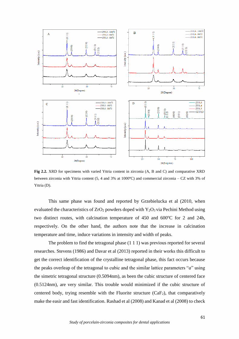
61 Study of porcelain-zirconia composites for dental applications
Fig 2.2. XRD for specimens with varied Yttria content in zirconia (A, B and C) and comparative XRD
between zirconia with Yttria content (5, 4 and 3% at 1000ºC) and commercial zirconia – CZ with 3% of
Yttria (D).
This same phase was found and reported by Grzebielucka et al (2010, when
evaluated the characteristics of ZrO2 powders doped with Y2O3 via Pechini Method using
two distinct routes, with calcination temperature of 450 and 600oC for 2 and 24h,
respectively. On the other hand, the authors note that the increase in calcination
temperature and time, induce variations in intensity and width of peaks.
The problem to find the tetragonal phase (1 1 1) was previous reported for several
researches. Stevens (1986) and Davar et al (2013) reported in their works this difficult to
get the correct identification of the crystalline tetragonal phase, this fact occurs because
the peaks overleap of the tetragonal to cubic and the similar lattice parameters “a” using
the simetric tetragonal structure (0.5094nm), as been the cubic structure of centered face
(0.5124nm), are very similar. This trouble would minimized if the cubic structure of
centered body, trying resemble with the Fluorite structure (CaF2), that comparatively
make the easir and fast identification. Rashad et al (2008) and Kanad et al (2008) to check
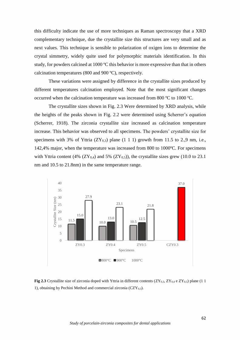
62 Study of porcelain-zirconia composites for dental applications
this difficulty indicate the use of more techniques as Raman spectroscopy that a XRD
complementary technique, due the crystallite size this structures are very small and as
next values. This technique is sensible to polarization of oxigen íons to determine the
crystal simmetry, widely quite used for polymorphic materials identification. In this
study, for powders calcined at 1000 ºC this behavior is more expressive than that in others
calcination temperatures (800 and 900 ºC), respectively.
These variations were assigned by difference in the crystallite sizes produced by
different temperatures calcination employed. Note that the most significant changes
occurred when the calcination temperature was increased from 800 ºC to 1000 ºC.
The crystallite sizes shown in Fig. 2.3 Were determined by XRD analysis, while
the heights of the peaks shown in Fig. 2.2 were determined using Scherrer’s equation
(Scherrer, 1918). The zirconia crystallite size increased as calcination temperature
increase. This behavior was observed to all specimens. The powders’ crystallite size for
specimens with 3% of Yttria (ZY0,3) plane (1 1 1) growth from 11.5 to 2.,9 nm, i.e.,
142,4% major, when the temperature was increased from 800 to 1000ºC. For specimens
with Yttria content (4% (ZY0,4) and 5% (ZY0,5)), the crystallite sizes grew (10.0 to 23.1
nm and 10.5 to 21.8nm) in the same temperature range.
Fig 2.3 Crystallite size of zirconia doped with Yttria in different contents (ZY0,3, ZY0,4 e ZY0,5) plane (1 1
1), obtaining by Pechini Method and commercial zirconia (CZY0,3).
11.510.0 10.5
15.013.0 12.5
27.9
23.121.8
37.0
0
5
10
15
20
25
30
35
40
ZY0.3 ZY0.4 ZY0.5 CZY0.3
Cry
stal
lite
Siz
e (n
m)
Specimens
800°C 900°C 1000°C

63 Study of porcelain-zirconia composites for dental applications
The results indicate that the calcination at higher temperatures enables Y-doped
zirconia to acquire larger crystallite sizes and higher crystallinity. The calcination
temperature of 1000 °C was selected for further investigation of the particle properties.
The effect of the complexation agents and metal íon concentration in the precursors, on
the crystallization behavior of Yttrium doped Zirconia was also studied. This same
behavior was reported by Davar et al (2013) and Grzebielucka et al (2010) they showed
that the increase in calcination temperature can be increased the crystallite size of the
zirconia powders. Thus, the increase in a calcination temperature promote a major atomic
diffusion, increasing the structural packing that enhaced consequently the crystallite size.
These results are in agreement as exhibit results in previous XRD (Fig 2.2). The
dopant content is also an important factor to be evaluated. The increase of dopant content
into crystal lattice promote the decrease in crystallite size for the same temperatures and
distincts compositions. Chevalier et al (2009), reported that the ZrO2 compositions
containing more than 8mol% Y2O3 stabilize the cubic phase at room temperature, while
compositions containing 2-3% stabilize the tetragonal phase, and compositions between
these values generate a mixture of tetragonal and cubic phases.
However, in addition of the 3% of Yttria content, was used in this study the
proportions (4 and 5 mol. %), this values are in a transition area to stabilization in 8%,
i.e., the structure is no totally stabilized. Comparatively with comercial zirconia (CZY0,3)
were found diferences in crystallite size, i. e., for specimens with 3% of Yttria content
assigned by CPM Method at 1000 °C the value was (27.9 nm), whereas for commercial
zirconia this value was more higher (37.0 nm) when observed to all specimens before
studieds. This result can be explain to according as achievement technique used.
The Fig. 2.4 show the calculated lattice parameters for specimens (ZY0,3, ZY0,4,
ZY0,5 and CZY0,3), respectively (FullProf – Crystalographic Server). Is it possible to
check that the lattice parameters no presents large variations with the increase of the
calcination temperature for the same stoichiometry, as the case of specimen ZY0,3 (5.18
e 5.19Å), an increase only 0.17%. The same behavior also can be seen for specimens
ZY0,4 e ZY0,5, that presents discrete difference to order 0.15%. The commercial zirconia
no presents large diferences in lattice parameters when compared with others specimens
calcined at 1000°C. Thus, the difference in lattice parameters of the specimens
synthesized and commercial are not significant.
On the other hand, the increase of Yttria content in network, promote some
changes in lattice parameters as reported by Yamagata (2008), according to the author, in
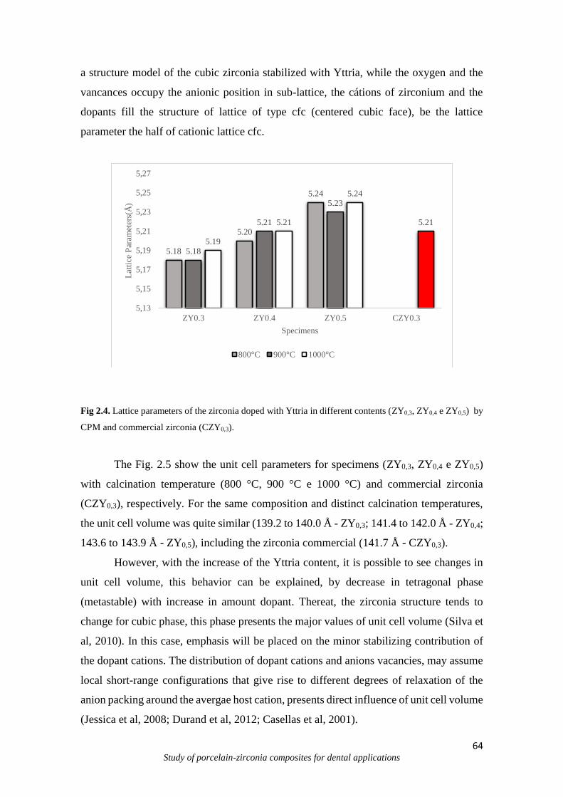
64 Study of porcelain-zirconia composites for dental applications
a structure model of the cubic zirconia stabilized with Yttria, while the oxygen and the
vancances occupy the anionic position in sub-lattice, the cátions of zirconium and the
dopants fill the structure of lattice of type cfc (centered cubic face), be the lattice
parameter the half of cationic lattice cfc.
Fig 2.4. Lattice parameters of the zirconia doped with Yttria in different contents (ZY0,3, ZY0,4 e ZY0,5) by
CPM and commercial zirconia (CZY0,3).
The Fig. 2.5 show the unit cell parameters for specimens (ZY0,3, ZY0,4 e ZY0,5)
with calcination temperature (800 °C, 900 °C e 1000 °C) and commercial zirconia
(CZY0,3), respectively. For the same composition and distinct calcination temperatures,
the unit cell volume was quite similar (139.2 to 140.0 Å - ZY0,3; 141.4 to 142.0 Å - ZY0,4;
143.6 to 143.9 Å - ZY0,5), including the zirconia commercial (141.7 Å - CZY0,3).
However, with the increase of the Yttria content, it is possible to see changes in
unit cell volume, this behavior can be explained, by decrease in tetragonal phase
(metastable) with increase in amount dopant. Thereat, the zirconia structure tends to
change for cubic phase, this phase presents the major values of unit cell volume (Silva et
al, 2010). In this case, emphasis will be placed on the minor stabilizing contribution of
the dopant cations. The distribution of dopant cations and anions vacancies, may assume
local short-range configurations that give rise to different degrees of relaxation of the
anion packing around the avergae host cation, presents direct influence of unit cell volume
(Jessica et al, 2008; Durand et al, 2012; Casellas et al, 2001).
5.18
5.20
5.24
5.18
5.21
5.23
5.19
5.21
5.24
5.21
5,13
5,15
5,17
5,19
5,21
5,23
5,25
5,27
ZY0.3 ZY0.4 ZY0.5 CZY0.3
Lat
tice
Par
amet
ers(
Å)
Specimens
800°C 900°C 1000°C
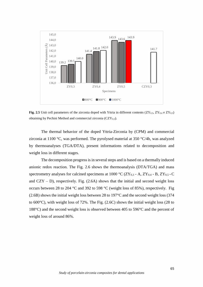
65 Study of porcelain-zirconia composites for dental applications
Fig. 2.5 Unit cell parameters of the zirconia doped with Yttria in different contents (ZY0,3, ZY0,4 e ZY0,5)
obtaining by Pechini Method and commercial zirconia (CZY0,3).
The thermal behavior of the doped Yttria-Zirconia by (CPM) and commercial
zirconia at 1100 °C, was performed. The pyrolysed material at 350 °C/4h, was analyzed
by thermoanalyses (TGA/DTA), present informations related to decomposition and
weight loss in different stages.
The decomposition progress is in several steps and is based on a thermally induced
anionic redox reaction. The Fig. 2.6 shows the thermoanalysis (DTA/TGA) and mass
spectrometry analyses for calcined specimens at 1000 °C (ZY0,3 - A, ZY0,4 - B, ZY0,5 - C
and CZY – D), respectively. Fig. (2.6A) shows that the initial and second weight loss
occurs between 28 to 204 °C and 392 to 598 °C (weight loss of 85%), respectively. Fig
(2.6B) shows the initial weight loss between 28 to 197°C and the second weight loss (374
to 600°C), with weight loss of 72%. The Fig. (2.6C) shows the initial weight loss (28 to
188°C) and the second weight loss is observed between 405 to 596°C and the percent of
weight loss of around 86%.
139.2
141.4
143.9
139.5
141.8
143.6
140.0
142.0
143.9
141.7
136,0
137,0
138,0
139,0
140,0
141,0
142,0
143,0
144,0
145,0
ZY0,3 ZY0,4 ZY0,5 CZY0,3
Un
it C
ell
Par
amet
ers
(Å)
Specimens
800°C 900°C 1000°C
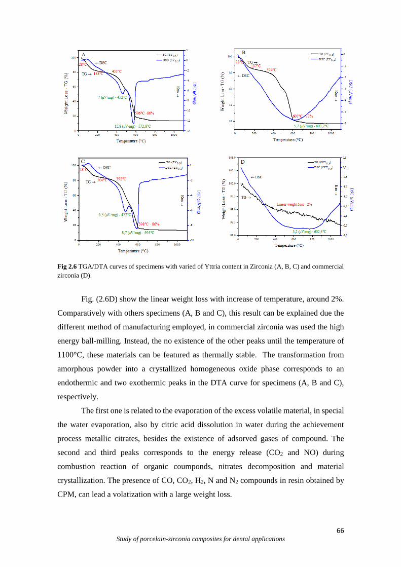
66 Study of porcelain-zirconia composites for dental applications
Fig 2.6 TGA/DTA curves of specimens with varied of Yttria content in Zirconia (A, B, C) and commercial
zirconia (D).
Fig. (2.6D) show the linear weight loss with increase of temperature, around 2%.
Comparatively with others specimens (A, B and C), this result can be explained due the
different method of manufacturing employed, in commercial zirconia was used the high
energy ball-milling. Instead, the no existence of the other peaks until the temperature of
1100°C, these materials can be featured as thermally stable. The transformation from
amorphous powder into a crystallized homogeneous oxide phase corresponds to an
endothermic and two exothermic peaks in the DTA curve for specimens (A, B and C),
respectively.
The first one is related to the evaporation of the excess volatile material, in special
the water evaporation, also by citric acid dissolution in water during the achievement
process metallic citrates, besides the existence of adsorved gases of compound. The
second and third peaks corresponds to the energy release (CO2 and NO) during
combustion reaction of organic coumponds, nitrates decomposition and material
crystallization. The presence of CO, CO2, H2, N and N2 compounds in resin obtained by
CPM, can lead a volatization with a large weight loss.
67 Study of porcelain-zirconia composites for dental applications
The TGA curves showmass change of a sample as a function of temperature or
time in a temperature controlled environment and atmosphere. The specimen with 3% of
Yttria dopant (ZY0,3 – A), present the loss energy in 6,3 and 8,7 (V/mg) at 440.5 and
595 °C, respectivelly. In specimen (ZY0,4 - B) the exothermic peak obtained an loss
energy of 5.7 (µV/mg) at 601.2 °C, this temperature refers to the energy release during
the organic material combustion. The specimen with 5% of Yttria dopant (ZY0,5 – C)
presented exothermic peaks of 7 e 12.8 (µV/mg) at 452°C and 572.8ºC, assigned by
organic material combustion (energy release) and oxidation reaction of Zr4+ íons that
remain in fluorite structure. In commercial zirconia specimen (CZY0,3 – D) occurs the
short energy loss 3.2 (µV/mg) at 610.4ºC.
The Raman spectroscopy determine the phases present is to observe the vibrations
of the atoms in the sample structure, can be used in solutions and solids as well as
characterization of molecular or crystalline structure, collects information relating to the
frequencies at which chemical bonds vibrate (Brundle et al, 1992). The broadening of the
Raman peaks is usually a result of structural disorder whithin the sample from amorphous
regions, doping, defects, or small particles sizes. The Raman spectroscopy is very flexible
and has been used by researchers to characterize the monoclinic-to-tetragonal and
tetragonal-to-cubic phase transformations that occur in zirconia materials as a result of a
change in temperature (Siu et al, 1999), the addition of dopants (Fernandez-Lopes et al
2001; Li and Li, 2002) or an applied stress (Li et al, 2003).
The Fig. 2.7 shown the Micro-Raman spectra for specimens. Were compared the
spectra with similar Yttria content in zirconia powders and varied calcinations
temperature (800, 900 and 1000 ºC). It is possible to see that independent of calcination
temperature employed, all specimens presents the monoclinic and tetragonal phases and
the mixture of both. The Fig. 2.7 (A) Raman spectra showed the ZY0,3 at 800, 900 and
1000 ºC, respectively.
According to these spectrus, in specimens calcined at 1000 °C the peaks of
100 cm-1 indicate the presence of monoclinic phase (m-phase), 171 cm-1 monoclinic phase
(m-phase), 301 cm-1 the mixture of tetragonal and monoclinic phases and 431-608 cm-1
tetragonal phase. When the calcination temperature was reduced at 900 and 800 ºC, the
characteristic of m-phase pratically no exist and the peaks located at 155, 266, 317 and
608 cm-1 confirms t-phase of zirconia successfully obtained.
The Fig. 2.7(B) show the spectra of ZY0,4 at 800, 900 and 1000 ºC, respectively.
Can be seen in this spectra the presence of monoclinic phase at 171 cm-1 tetragonal and

68 Study of porcelain-zirconia composites for dental applications
monoclinic phase at 303 cm-1, monoclinic phase at 372 cm-1 and tetragonal phase at 603
cm-1. Upon reducing the calcination temperature from 900 and 800 ºC this peaks are 100
cm-1 monoclinic phase, t-phase 140 and 155 cm-1, monoclinic phase at 800 °C - 222 cm-
1, mixture of phase tm at 301 and 314 cm-1, mixture of cubic and tetragonal phase at 542
cm-1 and tetragonal phase at 601 and 595, respectively.
Fig. 2.7 Raman spectrometry of Yttria doped zirconia in different contents and calcination temperature.
Fig. 2.7(C) show the behavior of peaks for specimens with 5% of Yttria content
in zirconia calcined at different temperatures. For calcination temperature at 1000 ºC the
monoclinic phase can be see with the peaks 100, 171 and 352 cm-1, tetragonal phase 323
and 420 cm-1 and the mixture tm at 550 cm-1. In temperatures of the 900 and 800 °C the
wavenumbers of the peaks are monoclinic phase 100 and 361 cm-1 for 900 °C and 367
cm-1 for 800 °C, tetragonal phase at 290, 584 and 645 cm-1 – 900 °C and 155 cm-1 – 800
°C and lastly the mixture of monoclinic and tetragonal phases at 550 and 667 cm-1 -800
°C.
69 Study of porcelain-zirconia composites for dental applications
The changes in zirconia bond angles and lengths allow to identify the phase
transformations (monoclinic, tetragonal and cubic), this behavior is evaluate by various
authors (Casellas et al, 2001; Bouvier et al 2001; Ishigame et al, 1977; Ramos et al, 2013),
observed that the main broad bands for zirconia were approximately 260, 464, and 642
cm-1, this values are characteristics of the tetragonal and cubic phases (Casellas et al,
2001).
In this study, the difference of calcination temperature and chemical stabilizers
(Casellas et al, 2001; Bouvier et al 2001; Ishigame et al, 1977; Ramos et al, 2013) and
crystal sizes probably caused few spectral variations at the tetragonal and cubic phases.
However, unlike indicated for standard letter (X’pert 77-2113), the phase obtained with
sucess for all calcination temperature, was the tatragonal phase and no cubic phase as
reported before. This result also was observed by Davar et al (Davar et al, 2013) where
decrease the calcination temperatures was possible to obtain the tetragonal phase.
Comparatively as X-Ray diffraction the Raman spectroscopy indentify the phases from
the vibrational modes of atomic bonds with laser radiation (visible, IR, UV) while the
XRD detect phases in quantities > 1-2 wt% and particle sizes < 100nm. Thus, the Raman
spectroscopy is considered a complementary technique to indentify the phases presents
of the materials and more indicate to identify the polymorphic phases existing in zirconia
powders.
4. Conclusions
The CPM is a good technique to produce monophasic zirconia with different Yttria
contents. Crystalographic parameters of the synthesized specimens no presents large
variations compared as zirconia commercial. Calcination temperature was a significative
factor to changes the peaks intensity and consequently the cristallinity of the specimens.
Specimens synthesized presents the large weight loss when compared as commercial
zirconia, this fact is dependent of the type of manufacturer technique used.
70 Study of porcelain-zirconia composites for dental applications
Acknowledgments
This work was supported by the CAPES-Brazil and Portuguese Foundation of Science
and Technology through the projects: EXCL/EMS-TEC/0460/2012 and
UID/EEA/04436/2013.
References
Aruna S.T., Rajam K.S., Mixture of fuels approach for the solution combustion synthesis
of Al2O3-ZrO2 nanocomposite, Mater Res Bulletin 39 (2004) 157.
Atkinson A, Segal, D. L. Some recent developments in aqueous sol-gel processing. J Sol-
gel SciTech, 13 (1998) 133-139.
Borchers L., Stiesch M., Bach F-W, Buhl J-C, Hübsch C., Kellner T., et al. Influence of
hydrothermal and mechanical conditions on the strength of zirconia. Acta Biomater
(2010) 6:4547–52.
Bouvier P., Gupta H.C., Lucazeau G. Zone center phonon frequencies in tetragonal
zirconia: lattice dynamical study and new assignment proposition. J Phys Chem Solids
(2001) 62:873-9.
Brundle C.R., Evans J. C.A. and Wilson S. Encyclopedia of Materials Characterization:
Surfaces, Interfaces, Thin Films. Butterworth-Heinemann, Stoneham, M.A., C.R. and
Manning Publishing Co., Greenwich, CT, (1992).
Casellas D, Cumbrera FL, Sanchez-Bajo F, Forsling W, Llanes L, Anglada M. On the
transformation toughening of Y-ZrO2 ceramics with mixed Y-TZP/PSZ microstuctures.
J Eur Ceram Soc (2001) 21:765-77.
71 Study of porcelain-zirconia composites for dental applications
Chevalier J., Gremillard L., Virkar A.V., Clarke D.R. The tetragonal-monoclinic
transformation in zirconia: lessons learned and future trends. J Am Ceram Soc 92 (2009)
9:1901–20.
Davar, F., Hassankhani, A., Estarki, M. Controllable synthesis of metastable tetragonal
zirconia nanocrystals using citric acid assisted sol–gel method Ceramics International 39
(2013) 2933–2941
Duncan, K., Wang, Y., Bishop, S., Ebrahimi, F., Wachsman, E., Role of point defects in
the physical properties of fluorite oxides, J. Am. Ceram. Soc. 89 (2006) 3162.
Durand J.C., Jacquot B., Salehi H., Fages M., Margerit J., Cuisinier F.J. Confocal Raman
microscopic analysis of the zirconia/feldspathic ceramic interface. Dent Mater (2012)
28:661-71.
Fernandez-Lopez E., Sanchez-Escribano V., Panizza M., Carnasciali M.M. Busca G.
"Vibrational and Electronic Spectroscopic Properties of Zirconia Powders," J Mater
Chem, 11 (2001) 1891-1897.
Grzebielucka, E. C., Scoton A., Chinelatto, A., Mazurek, S., Tebcherani, A. Chinelatto,
L. Synthesis and sintering of Y2O3-doped ZrO2 powders using two Pechini-type gel
routes. Ceram Intern 36 (2010) 1737–1742.
Hajizadeh-Oghaz M., Razavi R S. Estarki M L. Large-scale synthesis of YSZ
nanopowder by Pechini method. Mater Sci 37 (2014) 5:969–973.
HajizadehOghaz M., Razavi S.R., Loghman-Estarki M.R., Ghasemi R., Optimization of
morphology and particle size of modified sol gel synthesized YSZ Nanopowder using
Taguchi Method, J Nano Res 21 (2013) 65.
Ishigame M, Sakurai T. Temperature dependence of the Raman spectra of ZrO2. J Am
Ceram Soc (1977) 60:367-9.
72 Study of porcelain-zirconia composites for dental applications
Krogstad,J.A., Lepple M., Gao Y., Lipkin D.M., Levi, C.G.. Effect of Yttria Content on
the Zirconia Unit Cell Parameters 2008.
Jing Z., Ke Z., Yihong L., Zhijian S. Effect of multistep processing techniqueson the
formation of micro-defects and residual stresses in zirconia dentalrestoration. J
Prosthodontics (2014) 23:206–10.
Kanade, K.G., Baeg, J. Apte, J. K. Synthesis and characterization of nanocrystallined
zirconia by hydro- thermal method, Mater Res Bulletin 43 (2008) 723–729.
Kazemi F., Arianpour F, Malek-Ahmadi S., Sohrabi S., Rezaie H.R., Saberi A., novel
method for synthesis of metastable tetragonal zirconia nano powder at low temperatures,
Mater Res Bulletin (2011).
Kessman, A.J., Ramji, K., Morris, M. Cairns, D. R. Zirconia sol–gel coatings on alumina–
silica refractory material for improved corrosion resistance, Surf and Coat Tech 204
(2009) 477–483.
Kohal R-J, Patzelt S.B.M., Butz F., Sahlin H. One-piece zirconia oralimplants: one-year
results from a prospective case series. 2. Three-unitfixed dental prosthesis (FDP)
reconstruction. J Clin Periodontol (2013) 40:553–62.
Li,Y. Xie, J., Gong, J., Chen, J., Zhang, Z. Mater. Sci. Eng. B 86 (2001) 119–122.
Li C.; Li M., "UV Raman Spectroscopic Study on the Phase Transformation of ZrO2,
Y2O3-ZrO2, and SO4 2-/ZrO2," J Raman Spectroscopy, 33 (2002) 301-308.
Li M., Feng Z., Ying P., Xiu Q.; Li C., "Phase Transformation in the Surface Regionof
Zirconia and Doped Zirconia Detected by UV Raman Spectroscopy," Phys Chemistry
Chemical Physics, 5 (2003) 5326-5332.
73 Study of porcelain-zirconia composites for dental applications
Masala, O., Seshadri, R., Synthesis routes for large volumes of NPNPs. Annu. Rev.
Mater. Res. 34 (2004) 41–81.
Matsui, K., Ohgai, M. Formation mechanism of hydrous-zirconia particles produced by
hydrolysis of ZloCl2 solutions: I1 J Amer Ceram Soc, 83(6) 83 (2000) 6:1386-1392.
Meyenberg K.H., Luthy H., Schärer P.J. Zirconia posts: a new all-ceramic concept for
nonvital abutment teeth. Esthet Dent (1995) 7:73–80.
Ramos, C. M. Cesar, PF, Mondelli R F L, Tabata, A S, Santos, J S, Borges AFS. Bond
strength and Raman analysis of the zirconia-feldspathic porcelain. Bauru School of
Dentistry, Faculty of Science, University of São Paulo (USP), Bauru, SP, Brazil (2013).
Rauchs, G., Munz, D., Fett, T. Calculation of crack tip phase transformation zones with
the weight function method, Computational Materials Science 16 (1999) 213–217.
Roy M.E., Whiteside L.A., Katerberg BJ, Steiger JA. Phase transformation, roughness,
and microhardness of artificially aged yttria- and magnesia- stabilized zirconia femoral
heads. Journal of Biomedical Materials Research 83a (2007)1096–102.
Salavati-Niasari M., Dadkhah M., Davar F., Pure cubic ZrO2 nanoparticles by thermolysis
of a new precursor, Polyhedron 28 (2009) 3005.
Sakka S., Kozuka H., Handbook of Sol–Gel Science and Technology Processing,
Characterization and Applications, 2nded. Kluwer Acad Publi, Boston, (2005) 60–76.
Scherrer, P. Göttinger Nachrichten Math. Phys. 2 (1918) 98–100.
Skandan, G.,Hahn, H., Kear, B. H., Roddy, M., Cannon, M. R. Processing of
nanostructured zirconia ceramics. Mat.Res. Soc. Symp. Proc. 351 (1994) 207.
Stevens, R. Zirconia and zirconia ceramics. Magnesium elektron ltda. 2 (1986) 5-51.

74 Study of porcelain-zirconia composites for dental applications
Silva N.R.F.A., Sailer I., Zhang Y., Coelho P.G., Guess P.C., Zembic A. Performance of
Zirconia for Dental Healthcare. Mater (2010) 3:863-896.
Siu G.G., Stokes M.J.and Liu Y., "Variation of Fundamental and Higher-Order Raman
Spectra of ZrO2 Nanograins with Annealing Temperature," Phys RevB, 59 (1999)
4:3173-3179.
Somiya, S., Yamamoto, O. and Yanagida, H. Science and Technology of Zirconia III, ÕÕ
Advances in Ceramics, 24 A and 24B.American Ceramic Society, 24 (1986) Westerville,
OH.
Thompson V.P., Rekow D.E. Dental ceramics and the molar crown testingground. J Appl
Oral Sci (2004) 12:26–36.
Vargas, J. T., Goto, J. Thermal barrier coatings produced by chemical vapor deposition.
Sci Tech Adv Mater 4 (2003) 397–402.
Venkatachari K.R., Huang D., Ostrander S.P., Schulze W.A., Stangle G.C., J. Mater. A
combustion synthesis process for synthesizing. Res. 10 (1995) 3:748.
Viazzi C. Bonino, J.P. Ansart F., Barnab A., Structural study of metastable tetragonal
YSZ powders produced via a sol–gel route, J Alloys and Comp 452 (2008) 377–383.
Yamagata C., Andrade J.B., Ussui V., Lima N.B. Paschoal J.O. High purity Zirconia and
Silica Powders via wet process: Alkali fusion of zircon sand. Mater Sci F 591-593 (2008)
771-776.
Yang C. L, Hsiang, H. I., Chen, C. C. Characteristics of ytria stabilized tetragonal zirconia
powder used in optical fiber connector. J Amer Ceram Soc, 80 (1997) 1949-1956.
75 Study of porcelain-zirconia composites for dental applications
CHAPTER III
Strenghtening and Toughening of dental porcelain by the inclusion of an yttria-
stabilized zirconia reinforcing phase
Santos, R. L.P.1; Silva F.2; Nascimento, R. M.1,Souza, J.C.M. 3,4, Henriques, B.2,4*
1Department of Materials Engineering, Federal University of Rio Grande do Norte (UFRN), Campus
Lagoa Nova, 59078-970, Natal, Brasil.
2CMEMS-UMINHO, University of Minho, Campus de Azurém, 4800-058 Guimarães, Portugal.
3School of Dentistry (DODT), Post-Graduation Program in Dentistry (PPGO).
4Federal University of Santa Catarina, Campus Blumenau, 89065-300, Blumenau/SC, Brazil
Abstract
The aim of this work was to evaluate the microstructural and mechanical performance of dental porcelain
composites reinforced and toughened by 20vol.% tetragonal zirconia (3Y-TZP). Two Types of particles
were tested: yttria-stabilized zirconia (ZrO2-3%Y2O3) agglomerates and pre-sintered yttria-satabilized
zirconia (ZrO23%Y2O3) particles and processed by hot pressing technique at 970ºC. The composites were
analyzed by the means Scanning Electron Microscopy (SEM), Energy Dispersion Spectroscopy (EDS) and
XRD Diffraction (XRD). The mechanical properties were obtained by the transverse rupture strength test.
The best mechanical results were displayed by the porcelain reinforced with the pre-sintered ZrO2–3%Y2O3
particles (ZP).
Keywords—Composite, dental restoration, porcelain, strengthening, toughening, wear, zirconia.
76 Study of porcelain-zirconia composites for dental applications
1. Introduction
The use of ceramics in dentistry is nowadays the mainstream in aesthetical
restorative dentistry. Zirconia is in the forefront of this trend due to its biocompatibility,
aesthetics (Jing et al, 2014) corrosion resistance (Chevalier et al, 2009) and good
mechanical properties relative to alternative materials. Zirconia-based restorations
comprise a strong and thought zirconia substructure that is veneered with a weaker
compatible porcelain that is used to mimic the color of real teeth and eventually to reduce
the wear of the dental restoration with the opposite teeth.
Zirconia is a polymorphic ceramic with different crystal structure at different
temperatures, but keeping the same chemistry. Zirconia can be found in three crystalline
forms: monolithic; tetragonal and cubic. Pure zirconia is stable in monoclinic phase at
room temperature. At 1173°C it undergoes a phase transformation from monoclinic to
tetragonal and then to cubic at 2370°C. Above 2690°C zirconia melts.
These transformations are reversible upon cooling with the tetragonal to monoclinic (t-
m) transformation imparting compressive stresses to the material due to the volume
expansion of 4-5% that occurs with the transformation. It is possible to fully or partially
stabilize zirconia in its tetragonal or cubic form at room temperature by alloying it with
other cubic oxides termed as “stabilizers”. Calcia, magnesia, ceria, alumina or yttria are
used for that end. Zirconia is said to be partially stabilized (Partially Stabilized Zirconia
– PSZ) when a multiphase form coexists in the material: cubic as the major phase and
monoclinic and tetragonal zirconia precipitates, as the minor phase.
To date, the best properties are exhibited by zirconia stabilized with Y2O3 (yttria),
where the whole materials is constituted by transformable t-zirconia grains and called
Tetragonal Zirconia Polycrystals (TZP). The addition of 2-3 mol% yttrium oxide (Y2O3)
as stabilizing agent results in a fully tetragonal fine-grained zirconia ceramic material
with 100% small metastable tetragonal grains (Y-TZP). This type of zirconia has been
extensively used in dental frameworks for all-ceramic restorations. Partially stabilized
zirconia displays a toughening mechanism that uses the t-m transformation to arrest the
crack propagation, and is therefore termed transformation toughening. When a crack
arises in zirconia, the stress field generated at the crack tip cause a local phase
transformation from the metastable tetragonal phase to monoclinic phase. This
transformation is accompanied to a volume increase of the crystals, constrained by
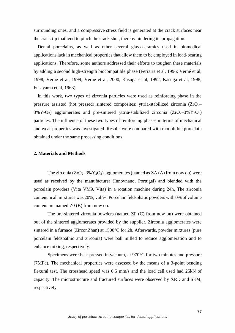
77 Study of porcelain-zirconia composites for dental applications
surrounding ones, and a compressive stress field is generated at the crack surfaces near
the crack tip that tend to pinch the crack shut, thereby hindering its propagation.
Dental porcelains, as well as other several glass-ceramics used in biomedical
applications lack in mechanical properties that allow them to be employed in load-bearing
applications. Therefore, some authors addressed their efforts to toughen these materials
by adding a second high-strength biocompatible phase (Ferraris et al, 1996; Verné et al,
1998; Verné et al, 1999; Verné et al, 2000, Kasuga et al, 1992, Kasuga et al, 1998,
Fusayama et al, 1963).
In this work, two types of zirconia particles were used as reinforcing phase in the
pressure assisted (hot pressed) sintered composites: yttria-stabilized zirconia (ZrO2–
3%Y2O3) agglomerates and pre-sintered yttria-stabilized zirconia (ZrO2–3%Y2O3)
particles. The influence of these two types of reinforcing phases in terms of mechanical
and wear properties was investigated. Results were compared with monolithic porcelain
obtained under the same processing conditions.
2. Materials and Methods
The zirconia (ZrO2–3%Y2O3) agglomerates (named as ZA (A) from now on) were
used as received by the manufacturer (Innovnano, Portugal) and blended with the
porcelain powders (Vita VM9, Vita) in a rotation machine during 24h. The zirconia
content in all mixtures was 20%, vol.%. Porcelain feldsphatic powders with 0% of volume
content are named Z0 (B) from now on.
The pre-sintered zirconia powders (named ZP (C) from now on) were obtained
out of the sintered agglomerates provided by the supplier. Zirconia agglomerates were
sintered in a furnace (ZirconZhan) at 1500°C for 2h. Afterwards, powder mixtures (pure
porcelain feldspathic and zirconia) were ball milled to reduce agglomeration and to
enhance mixing, respectively.
Specimens were heat pressed in vacuum, at 970°C for two minutes and pressure
(7MPa). The mechanical properties were assessed by the means of a 3-point bending
flexural test. The crosshead speed was 0.5 mm/s and the load cell used had 25kN of
capacity. The microstructure and fractured surfaces were observed by XRD and SEM,
respectively.
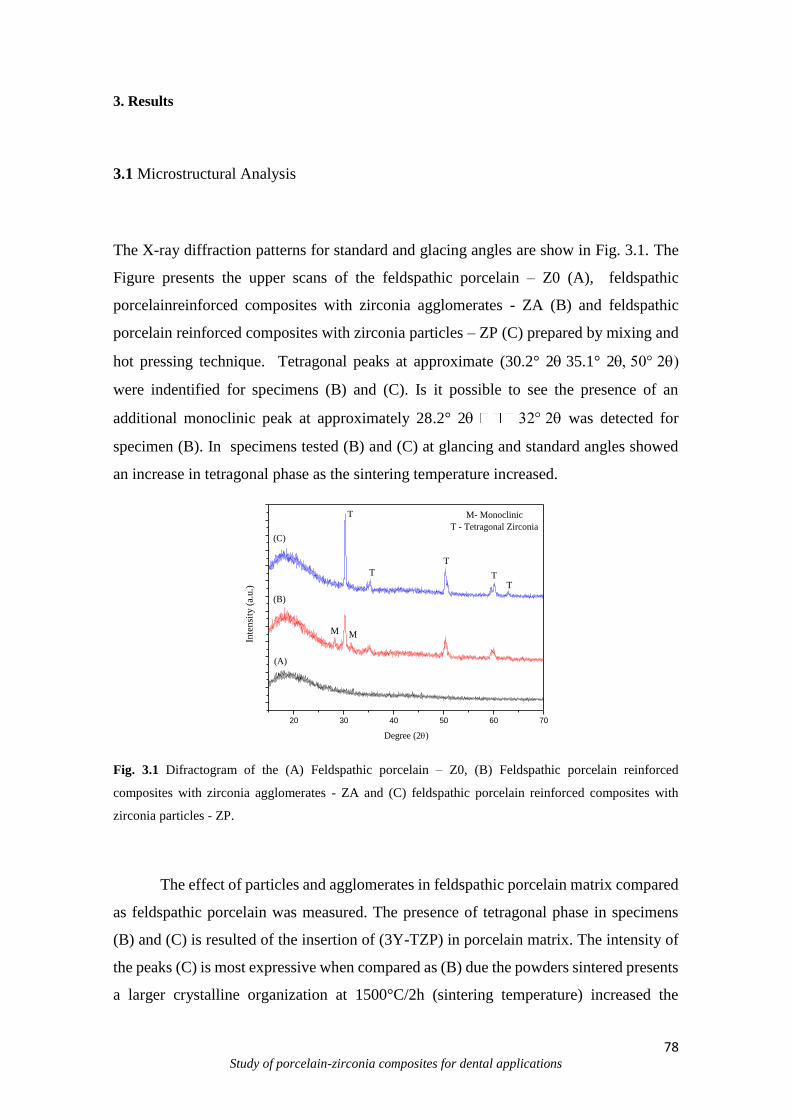
78 Study of porcelain-zirconia composites for dental applications
3. Results
3.1 Microstructural Analysis
The X-ray diffraction patterns for standard and glacing angles are show in Fig. 3.1. The
Figure presents the upper scans of the feldspathic porcelain – Z0 (A), feldspathic
porcelainreinforced composites with zirconia agglomerates - ZA (B) and feldspathic
porcelain reinforced composites with zirconia particles – ZP (C) prepared by mixing and
hot pressing technique. Tetragonal peaks at approximate (30.2° 235.1° 2
were indentified for specimens (B) and (C). Is it possible to see the presence of an
additional monoclinic peak at approximately 28.2° 2 was detected for
specimen (B). In specimens tested (B) and (C) at glancing and standard angles showed
an increase in tetragonal phase as the sintering temperature increased.
20 30 40 50 60 70
M M
M- Monoclinic
T - Tetragonal Zirconia
TT
T
T
T
(C)
(B)
(A)
Inte
nsi
ty (
a.u.)
Degree (2)
Fig. 3.1 Difractogram of the (A) Feldspathic porcelain – Z0, (B) Feldspathic porcelain reinforced
composites with zirconia agglomerates - ZA and (C) feldspathic porcelain reinforced composites with
zirconia particles - ZP.
The effect of particles and agglomerates in feldspathic porcelain matrix compared
as feldspathic porcelain was measured. The presence of tetragonal phase in specimens
(B) and (C) is resulted of the insertion of (3Y-TZP) in porcelain matrix. The intensity of
the peaks (C) is most expressive when compared as (B) due the powders sintered presents
a larger crystalline organization at 1500°C/2h (sintering temperature) increased the
79 Study of porcelain-zirconia composites for dental applications
tetragonal phase in relation the others specimens. The feldspathic porcelain (A) presents
amorphous characteristics.
Fig. 3.2 shown the micrographs of the zirconia reinforced feldspathic porcelain
composites, zirconia agglomerates (A) and zirconia particles (B). The two
microstructures produced are significantly different from each other, which might
indicate different sintering behaviors.
Fig. 3.2 Micrographs of the feldspathic porcelain composites reinforced zirconia agglomerates (A - ZA)
and zirconia particles (B - ZP). Zirconia appears as white phase and porcelain as dark phase.
In specimens with zirconia agglomerates (ZA) it is not visible the zirconia
agglomerates, suggesting that they were destroyed during the mixing procedure. The
white phase in Fig. 3.2 is the pulverized zirconia powder that covered the feldspathic
porcelain powders and negatively impacted the sintering of the composite.
On the other hand, in specimens with zirconia particles (ZP) it is possible to see
round zirconia particles spread through the feldspathic porcelain matrix. Few pores can
be detected within the porcelain matrix of feldspathic porcelain/zirconia-porcelain
samples and they are preferentially located at the vicinities of the ZPs rather than in the
porcelain matrix.
Fig. 3.3 shown the interface between the reinforcing zirconia particles phase and
feldspathic porcelain matrix. The micrographs of the fractured surfaces zirconia
reinforced feldspathic porcelain composites, (feldspathic porcelain, zirconia
agglomerates and zirconia particles), respectively.
A B
b

80 Study of porcelain-zirconia composites for dental applications
Fig. 3.3 Detail of the interface between the zirconia particle ZP (white phase) and the feldspathic porcelain
(dark phase).
In Fig. 3.3, the detail of the interface between the reinforcing Zirconia particles (ZP)
phase and the matrix is presented. It shows a good bonding between the zirconia particles
(ZP) and the matrix and a complete infiltration of the matrix inside the topological details
of the ZPs.
3.2 Mechanical Tests
The Fig. 3.4 shows the flexural strength of the composites ZA (B) and ZP (C) when
compared to the feldspathic porcelain (Z0). The zirconia agglomerates (ZA) showed to
produce a detrimental effect on the strength of the composites relative to pure porcelain.
On the other hand, the pre-sintered zirconia particles (ZP) showed to improve
significantly the flexural strength of the composite relative to pure feldspathic porcelain
(Z0).
Fig. 3.4 Flexural strength of the (A) feldspathic porcelain, (B) feldspathic porcelain reinforced composites
with zirconia agglomerates and (C) zirconia particles.
88.16
29.7
139.0
0
20
40
60
80
100
120
140
160
180
A B C
Fle
xu
ral
Str
ength
(M
Pa)
Composites
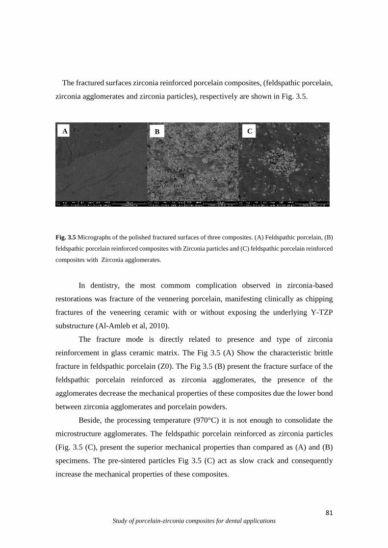
81 Study of porcelain-zirconia composites for dental applications
The fractured surfaces zirconia reinforced porcelain composites, (feldspathic porcelain,
zirconia agglomerates and zirconia particles), respectively are shown in Fig. 3.5.
Fig. 3.5 Micrographs of the polished fractured surfaces of three composites. (A) Feldspathic porcelain, (B)
feldspathic porcelain reinforced composites with Zirconia particles and (C) feldspathic porcelain reinforced
composites with Zirconia agglomerates.
In dentistry, the most commom complication observed in zirconia-based
restorations was fracture of the vennering porcelain, manifesting clinically as chipping
fractures of the veneering ceramic with or without exposing the underlying Y-TZP
substructure (Al-Amleb et al, 2010).
The fracture mode is directly related to presence and type of zirconia
reinforcement in glass ceramic matrix. The Fig 3.5 (A) Show the characteristic brittle
fracture in feldspathic porcelain (Z0). The Fig 3.5 (B) present the fracture surface of the
feldspathic porcelain reinforced as zirconia agglomerates, the presence of the
agglomerates decrease the mechanical properties of these composites due the lower bond
between zirconia agglomerates and porcelain powders.
Beside, the processing temperature (970°C) it is not enough to consolidate the
microstructure agglomerates. The feldspathic porcelain reinforced as zirconia particles
(Fig. 3.5 (C), present the superior mechanical properties than compared as (A) and (B)
specimens. The pre-sintered particles Fig 3.5 (C) act as slow crack and consequently
increase the mechanical properties of these composites.
A B C
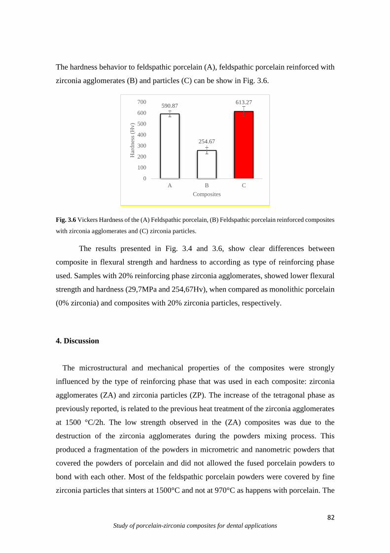
82 Study of porcelain-zirconia composites for dental applications
The hardness behavior to feldspathic porcelain (A), feldspathic porcelain reinforced with
zirconia agglomerates (B) and particles (C) can be show in Fig. 3.6.
Fig. 3.6 Vickers Hardness of the (A) Feldspathic porcelain, (B) Feldspathic porcelain reinforced composites
with zirconia agglomerates and (C) zirconia particles.
The results presented in Fig. 3.4 and 3.6, show clear differences between
composite in flexural strength and hardness to according as type of reinforcing phase
used. Samples with 20% reinforcing phase zirconia agglomerates, showed lower flexural
strength and hardness (29,7MPa and 254,67Hv), when compared as monolithic porcelain
(0% zirconia) and composites with 20% zirconia particles, respectively.
4. Discussion
The microstructural and mechanical properties of the composites were strongly
influenced by the type of reinforcing phase that was used in each composite: zirconia
agglomerates (ZA) and zirconia particles (ZP). The increase of the tetragonal phase as
previously reported, is related to the previous heat treatment of the zirconia agglomerates
at 1500 °C/2h. The low strength observed in the (ZA) composites was due to the
destruction of the zirconia agglomerates during the powders mixing process. This
produced a fragmentation of the powders in micrometric and nanometric powders that
covered the powders of porcelain and did not allowed the fused porcelain powders to
bond with each other. Most of the feldspathic porcelain powders were covered by fine
zirconia particles that sinters at 1500°C and not at 970°C as happens with porcelain. The
590.87
254.67
613.27
0
100
200
300
400
500
600
700
A B C
Har
dn
ess
(Hv)
Composites

83 Study of porcelain-zirconia composites for dental applications
work of (Verné et al 2003) suggests that if the zirconia agglomerates were maintained in
the powder mixture without being destroyed (switch the ball mill of powders by steering
suspension of the mixture in alcohol until complete evaporation) the mechanical strength
could be even increased. The mechanism was explained by the infiltration of the porcelain
phase into the zirconia agglomerates, leading to a partial spreading of each particle into
agglomerates of small zirconia grains (about 1 m size).
On the other hand, the improvement in the flexural strength of the composite (ZP)
relative to pure porcelain is explained by the action of the uniformly distributed dense
zirconia particles (pre-sintered agglomerates). The zirconia particles act as a second phase
that is more rigid than the porcelain matrix, allowing the load transfer reinforcing
mechanism to occur from matrix to the reinforcing phase. Besides the improvement
noticed in flexural strength of the (ZP) it is also expected an improvement in the toughness
behavior relative to pure feldspathic porcelain (Verné et al 2003). The toughening
mechanism is based on the crack deflection around the zirconia particles.
5. Conclusion
Two types of dental porcelain based composites reinforced by zirconia
agglomerates (ZA) and zirconia particles (ZP) were produced by pressure assisted
sintering. The best mechanical properties were found for (ZP) specimens due to a good
dispersion and good bonding between the reinforcing zirconia phase and the feldspathic
porcelain matrix. The pulverization of the zirconia micro/nano particles during the
blending process of the porcelain powders-zirconia agglomerates showed to impart
problems to the sintering and densification of (ZA) composites, reducing dramatically the
strength of the composite.
Acknowledgment
This work was supported by the CAPES-Brazil and Portuguese Foundation of Science
and Technology through the projects: EXCL/EMS-TEC/0460/2012 and
UID/EEA/04436/2013.
84 Study of porcelain-zirconia composites for dental applications
References
Al-Amleh B., Lyons K., Swain M. Clinical trials in zirconia: a systematic review. J Oral
Rehabil (2010) 37: 641-652.
Chevalier J., Gremillard L., Virkar A.V., Clarke D.R., The tetragonal-monoclinic
transformation in zirconia: lessons learned and future trends. J Am Ceram Soc, vol. 92
(2009) 9:1901–1920
Fusayama T., Katayoi T., Nomoto S., Corrosion of gold and amalgam placed in contact
with each other. J. Dent. Res., (1963) 42:1183–1197.
Ferraris M., Rabajoli P., Brossa F., Paracchini L., Vacuum plasma spray deposition of
titanium particle/glass-ceramic matrix biocomposites. J. Am. Ceram. Soc. (1996)
796:1515–1520.
Garvie R.C., Nicholson P.S. Phase analysis in zirconia systems. J. Am. Soc. (1972)
55:303-305.
Jing Z., Ke Z., Yihong, L., Zhijian S., Effect of multistep processing techniques on the
formation of micro-defects and residual stresses in zirconia dental restoration. J
Prosthodontics, (2014) 23:206–210.
Kasuga T., Nakajima K., Uno T., Yoshida M., Preparation of zirconia-toughened
bioactive glass-ceramic composite by sinter-hot isostatic pressing. J. Am. Ceram. Soc.,
75 (1992) 5.
Kasuga T., Yoshida M., Uno T., Nakajima K., Preparation of zirconia-toughened
bioactive glass-ceramics. J. Mater. Sci., (1998) 23:2255–2258.

85 Study of porcelain-zirconia composites for dental applications
Verné E., Ferraris M., Ventrella A., Paracchini L., Krajewski A., Ravaglioli A., Sintering
and plasma-spray deposition of bioactive glass-matrix composites for biomedical
applications. J. Eur. Ceram. Soc. 18 (1998) 4:363–372.
Verné E., Ferraris M., Jana C., Pressureless sintering of Bioverit1III/Ti particle
biocomposites. J. Eur. Ceram. Soc., (1999) 19: 2039–2047, 1999.
Verné E., Ferraris M., Jana C., Paracchini L., Bioverit1I base glass/Ti particulate
biocomposite: ‘‘in situ’’ vacuum plasma spray deposition. J. Eur. Ceram. Soc., (2000)
20:473–479.
Verné E., Defilippi R., Carl G, Vitale Brovarone C., Appendino P., Viscous flow sintering
of bioactive glass-ceramic composites toughened by zirconia particles, J. Eur. Cer. Soc.,
23 (2003) 5:675-683.
86 Study of porcelain-zirconia composites for dental applications
CHAPTER IV
On the mechanical properties and microstructure of zirconia-reinforced porcelain
for prosthetic dentistry
Santos, R. L.P.1; Silva F.2; Nascimento, R. M.1, Souza, J.C.M.2,3, Henriques, B.2,4*
1Department of Materials Engineering, Federal University of Rio Grande do Norte (UFRN), Campus
Lagoa Nova, 59078-970, Natal, Brazil.
2Center for Microelectromechanical systems (CMEMS_UMINHO), University of Minho, Campus
Azurém, Gumarães 4800-058, Portugal.
3Center for Research on Dental Implants (CEPID), Post-Graduation Program in Dentistry (PPGO),
Federal University of Santa Catarina, Campus Trindade, 88040-900, Florianópolis/SC, Brazil
4Laboratory of Ceramic and Composite Materials (CERMAT), Federal University of Santa Catarina
(UFSC), Campus Trindade, Florianópolis/SC, Brazil.
*Corresponding author at: Tel.: +351 253 510220; fax: +351 253 516007. E-mail address:
[email protected] (B. Henriques)
Abstract
Objective: The aim of this study was to produce and to evaluate the mechanical properties of a dental
feldspathic porcelain reinforced by zirconia particles.
Methods: The specimens were prepared by incorporating different volume fractions (10, 20, 30, 40 or
50%) of zirconia particles (3Y-TZP) in a feldspathic porcelain matrix. The mixtures were hot pressed in
vacuum at 970°C for 2 min. Flexural strength of the composites were measured using a three-point-bending
test. Micro-Hardness, Young’s modulus and fracture toughness were also measured for all the composites.
The microstructure and fracture surfaces were characterized by (FEG-SEM). Chemical analyses were
performed by Energy Dispersive Spectroscopy (EDS) and X-Ray Diffraction (XRD).
Results: The highest flexural strength (218 ± 24 MPa) and fracture toughness (2.1 ± 0.9 MPa m1/2) values
were recorded for composites containing 30% zirconia particles. That corresponded to improvement in
mechanical properties of about 2 times relative to those registered for feldspar-based porcelain.
Conclusions: The addition of zirconia particles to porcelain improved significantly the mechanical
properties of the base material. Zirconia-reinforced porcelain can constitute a promising alternative to
conventional feldspar-based porcelains to produce prosthetic restorations, mainly for posterior teeth where
high strength and toughness are demanded.
Keywords: porcelain, zirconia, heat pressing, composites, mechanical properties.

87 Study of porcelain-zirconia composites for dental applications
1. Introduction
All-ceramic restorations have been increasingly used in prosthetic dentistry
since they positively address the aesthetic and biocompatibility. Nowadays, restorations
based on zirconia substructures veneered by aesthetical porcelain are the most widely
used in single and multi-unit prostheses.
Zirconia is a polymorphic ceramic with different crystalline structures at
different temperatures, but keeping the same chemistry. Zirconia can be found in three
crystalline forms: monolithic (m-t; 1173°C); tetragonal (t-c; 2370°C) and cubic (c-
melting temperature; 2690°C). Such transformations are reversible upon cooling from
tetragonal to monoclinic (t-m) transformation resulting in compressive stresses to the
material due to the occurrence of 4-5% volume expansion. Thus, calcia (CaO), magnesia
(MgO), ceria (CeO2), alumina (Al2O3) or yttria (Y2O3) are used as stabilizers of the
tetragonal phase. Yttria-stabilized tetragonal zirconia policristal (Y-TZP) has been used
to produce dental prosthetic infrastructures, due to its excellent mechanical properties
traduced in high flexural strength (up to 900-1200 MPa) and high fracture toughness (5.9-
10 MPa m1/2) (Sevilla et al 2010; Kosmac et al 1999; Piconi and Maccauro 1999).The
addition of 2-3 mol% Y2O3 results in a fully tetragonal fine-grained zirconia ceramic with
100% small metastable tetragonal grains (Y-TZP).
Despite the aesthetical and biocompatibility improvements over metal-ceramic
systems, the failure rate of all-ceramic restorations by chipping or delamination of the
veneering porcelain are still high (Donovan 2009; Sailer et al, 2009; Tan et al, 2004
Larson et al 2013; Aboushileb et al 2009; Bonfante et al 2009; Liu et al, 2015). Both
systems present similar fracture mechanisms that rely either in edge chipping or
interfacial fracture (delamination) of the veneering porcelain (Sailer et al, 2009; Tan et
al, 2004 Larson et al 2013; Aboushileb et al 2009; Bonfante et al 2009; Liu et al, 2015).
Nevertheless, in vitro and in-vivo fatigue studies have shown that failure of
dental prostheses implies crack propagation towards the interface (Bonfante et al 2009;
Baldassari et al 2011; Coelho et a, 2009). The long-term success of the prosthesis is
achieved if the crack formation could be eliminated or delayed. The strengthening of
ceramics have been studied regarding several mechanisms such as: introduction of
compressive residual stress fields at ceramic layers, infiltration of glasses with a lower
coefficient of thermal expansion, chemical treatments, tailoring graded structures and
introduction of inorganic particles (Green et al 1999; Vhantikul et al 1979; Green et al
88 Study of porcelain-zirconia composites for dental applications
1983;Balakishnan et al 2007; Chu et al 2007; Zhang et al 2012; Suresh 2001; Chai et al
2014).
Huang et al. (Huang et al, 2014), studied the microstructure and mechanical
properties of zirconia-toughened lithium disilicate glass-ceramic composites containing
different zirconia content (0-30 wt.%) by hot pressing technique at 800ºC. The
mechanical properties of the composite containing 15%wt. zirconia increased up to 340
MPa and 3.5 MPa m1/2 relative to those of zirconia-free glass-ceramic. Verné et al. (Verné
et al 2003) also showed the improvement of the flexural strength and fracture toughness
of a glass-ceramic when reinforced with zirconia particles. In previous studies, we have
shown the influence of metallic particles in the mechanical properties of dental porcelain
(Henriques et al 2014).
The present study evaluated the influence of Y-TZP particles as a reinforcing
phase on feldspathic porcelains. It was hypothesized that the presence of zirconia particles
dispersed thought out the porcelain matrix can improve strength and toughness of the
feldspathic porcelain. Depending on the prosthesis location, zirconia-reinforced porcelain
can fully or partially replace the feldspathic porcelain. Hence, full replacement can occur
in posterior restorations where higher loads are present and lower aesthetic is demanded.
Partial replacements by using interlayer between zirconia substructure and veneering
feldspathic porcelain, can be used in anterior teeth in order to meet the esthetic
requirements.

89 Study of porcelain-zirconia composites for dental applications
2. Materials and Methods
2.1. Base materials
Dental porcelain and Y-TZP powders used in this study are show in Table 4.1.
Table 4.1. Chemical composition, material and manufacturer of porcelain and Y-TZP used in this study.
Manufacturer/Brand Chemical composition Material Batch
VITA VM9 Base
Dentim –V9 Shade
3M2 (Lot n. 44670)
SiO2 (60-64%), Al2O3 (13-
15%), K2O (7-10%), Na2O
(4-6%), TiO2 (<0,5%),
CeO2 (<0.5%), ZrO2 (0-
1%), CaO (1-2%), B2O3 (3-
5%), BaO (1-3%), SnO2
(<0,5%), Mg, Fe and P
oxides (<0,1%).
Feldspar-based
porcelain
Vita Zahnfabrik, Bad
Säckingen, Germany
3YSZGSB – Zirconia
(3%mol Yttria)
(Lot n. 1301PA046)
ZrO2 + HfO2 + Y2O3 +
Al2O3 (99,9%)
Y2O3 (5,2±0,2)
Al2O3 (<0,4).
Yttria-stabilized
tetragonal zirconia
policrystals (Y-
TZP)
Innovnano, Portugal
In order to obtain the zirconia particles, zirconia agglomerates as received by the
manufacturer were sintered in a furnace (ZircohnZhan, Germany) at 1500°C for 2h. Then,
the sintered zirconia was deagglomerated in a mortar. Irregular morphology of the
porcelain powders and the spherical-like morphology of the zirconia particles after
deagglomeration are shown in Figure 4.1.
Afterwards, the powder mixtures (feldspar-based porcelain and zirconia) were dry ball
milled to reduce agglomeration and to enhance mixing. The specimens are hereafter
named according to the volume content (xx%) of zirconia (Z) in the porcelain matrix, e.g.
Zxx%. Hence, Z0 will stand for pure feldspathic porcelain; Z10 for 10% of zirconia, and
so on.
The size distribution of the porcelain and zirconia powders were the following:
Porcelain: D10= 2.49 μm; D50=18.92 μm; D90=50.71 μm and Dave=23.47 μm; Zirconia:
D10=14.54 μm; D50 =162.64 μm; D90=336.71 μm and Dave=166.44 μm. Figure 4.1 shows
the morphology of the powders. Zirconia powders show.

90 Study of porcelain-zirconia composites for dental applications
A B
Fig. 4.1 SEM micrograph of feldspathic porcelain powders (A) and zirconia sintered particles (B).
Figure 4.1 shows the irregular morphology of the feldspathic porcelain powders and the
spherical-like morphology of the zirconia powders after deagglomeration.
2.2. Composites processing
The different mixtures of porcelain and zirconia powders (Figure 4.1) were placed in a
graphite die, previously painted with zirconia to avoid direct contact between the powder
mixtures and graphite die. The samples were prepared using the heat pressing technique
with ~7 MPa of pressure, under vacuum and 970°C during 2 min. Rectangular specimens
with 36 x 6 x 2.5 mm were synthesized for 3-point bending tests.
2.3 Chemical and Microstructural Characterization
The embedded samples in resin epoxy should first be hand polished on wet sanding paper
(SiC), grain #180. Then the surface should be ground with increasingly fine grain sand
papers, from 320, 600, 800, 1200, 2500 and ultimately to 4000. The final surface were
polished up to mirror using a diamond paste (1m) and performed the microstructural
characterizations. The microstructural inspection were performed by field emission guns
scanning electron microscopy (FEG-SEM NOVA 200 Nano SEM, FEI Company). The
atomic contrast images were performed using a Backscattering Electron Detector (BSED)
91 Study of porcelain-zirconia composites for dental applications
at an acceleration voltage between 15 and 20 kV. Chemical analyses of specimens were
performed by Energy Dispersive Spectroscopy (EDS) technique, using an EDAX Si(Li)
coupled to the FEG-SEM
X-ray diffraction (XRD) (AXS D8 Discover, Bruker, Massachusetts, USA) was used to
characterize the crystal structure of the zirconia particles present in composites. XRD data
were collected in the area 2θ =15 to 70° with a step of 0.02° using the Cu-Kα radiation
source and interaction time of 1,2 s.
2.4. Mechanical tests
The flexural strength of porcelain-zirconia composites was assessed by a three-
point bending test performed in an universal testing machine (Instron 8874, MA, USA).
Tests were carried using a load cell of 25kN capacity under a crosshead speed of 0.5mm/s,
until fracture. Flexural strength of specimens was calculated as an average of ten
specimens tested for each composition (n = 10).
The mean values of microhardness of the materials were obtained from 10
indentations for each specimen. The indentations were performed using a Vickers
hardness indenter (9,8N) (EmcoTest-DuraScan, Struers, Denmark) with a standard 136°
pyramidal diamond (HV1), oriented perpendicular to the surface of the specimen. The
fracture toughness (KIC) of specimens was determined by direct measurement of radial
crack length generated by indentation of the specimens on 9.8 N load. The following
equation was used (Blendell 1979):
𝐾𝐼𝐶 =𝐻0√𝑎
𝜑[0.055𝑙𝑜𝑔 {
8.4𝑎
𝑐} {𝐻0
𝐸𝜑}−0.4
]
where:
KIC = Fracture toughness (MPa·m1/2); H0 = Vickers hardness (GPa); a = half of the
diagonal legth of the Vickers indentation (μm); c= half average of crack length (μm); E=
Young’s modulus (GPa); 𝜑= constant value (3).
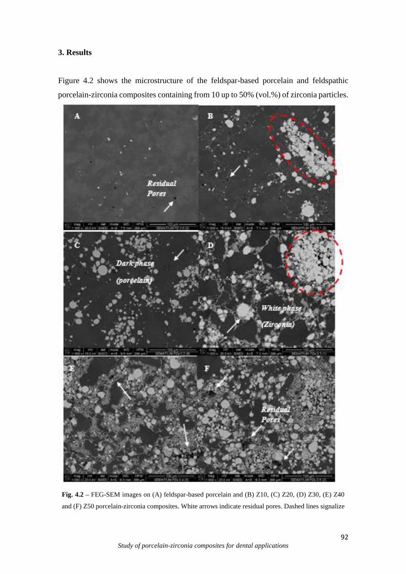
92 Study of porcelain-zirconia composites for dental applications
3. Results
Figure 4.2 shows the microstructure of the feldspar-based porcelain and feldspathic
porcelain-zirconia composites containing from 10 up to 50% (vol.%) of zirconia particles.
Fig. 4.2 – FEG-SEM images on (A) feldspar-based porcelain and (B) Z10, (C) Z20, (D) Z30, (E) Z40
and (F) Z50 porcelain-zirconia composites. White arrows indicate residual pores. Dashed lines signalize
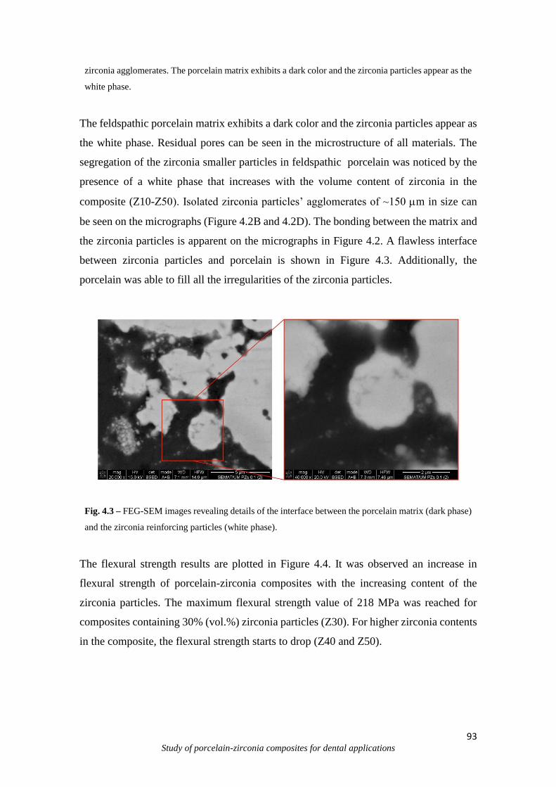
93 Study of porcelain-zirconia composites for dental applications
zirconia agglomerates. The porcelain matrix exhibits a dark color and the zirconia particles appear as the
white phase.
The feldspathic porcelain matrix exhibits a dark color and the zirconia particles appear as
the white phase. Residual pores can be seen in the microstructure of all materials. The
segregation of the zirconia smaller particles in feldspathic porcelain was noticed by the
presence of a white phase that increases with the volume content of zirconia in the
composite (Z10-Z50). Isolated zirconia particles’ agglomerates of ~150 m in size can
be seen on the micrographs (Figure 4.2B and 4.2D). The bonding between the matrix and
the zirconia particles is apparent on the micrographs in Figure 4.2. A flawless interface
between zirconia particles and porcelain is shown in Figure 4.3. Additionally, the
porcelain was able to fill all the irregularities of the zirconia particles.
Fig. 4.3 – FEG-SEM images revealing details of the interface between the porcelain matrix (dark phase)
and the zirconia reinforcing particles (white phase).
The flexural strength results are plotted in Figure 4.4. It was observed an increase in
flexural strength of porcelain-zirconia composites with the increasing content of the
zirconia particles. The maximum flexural strength value of 218 MPa was reached for
composites containing 30% (vol.%) zirconia particles (Z30). For higher zirconia contents
in the composite, the flexural strength starts to drop (Z40 and Z50).

94 Study of porcelain-zirconia composites for dental applications
Fig. 4.4 - Flexural strength mean values recorded on porcelain matrix composites containing different
volume contents of zirconia phase.
The fractures surfaces of the specimens were also analyzed and they confirmed the proper
bond strength between the porcelain matrix and the zirconia particles. The fracture surface
of a Z10 specimen is shown in Figure 4.5. A zirconia agglomerate was also inspected to
confirm the bonding between the materials. In fact, zirconia particles were veneered by
the porcelain, indicating cohesive fracture within the porcelain matrix.
Fig. 4.5 – FEG-SEM images revealing the fracture surface of a zirconia specimen containing 10%
zirconia particles (Z10). (B) Interface between the porcelain matrix and a zirconia particle agglomerate.
The hardness (HV/GPa) and fracture toughness (MPa.m1/2) results of feldspar-based
porcelain and porcelain-zirconia composites are given in Table 4.2. It was observed a
steady increase of the hardness values with the increasing content of zirconia phase up to
98
127139
218200
109
0
50
100
150
200
250
300
Z0 Z10 Z20 Z30 Z40 Z50
Fle
xu
ral
Str
ength
(M
PA
)
Type of specimen
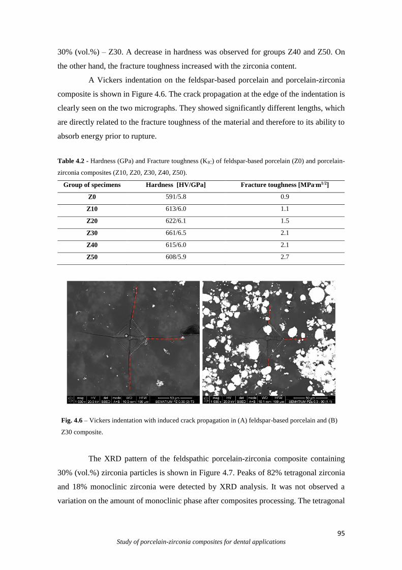
95 Study of porcelain-zirconia composites for dental applications
30% (vol.%) – Z30. A decrease in hardness was observed for groups Z40 and Z50. On
the other hand, the fracture toughness increased with the zirconia content.
A Vickers indentation on the feldspar-based porcelain and porcelain-zirconia
composite is shown in Figure 4.6. The crack propagation at the edge of the indentation is
clearly seen on the two micrographs. They showed significantly different lengths, which
are directly related to the fracture toughness of the material and therefore to its ability to
absorb energy prior to rupture.
Table 4.2 - Hardness (GPa) and Fracture toughness (KIC) of feldspar-based porcelain (Z0) and porcelain-
zirconia composites (Z10, Z20, Z30, Z40, Z50).
Group of specimens Hardness [HV/GPa] Fracture toughness [MPa.m1/2]
Z0 591/5.8 0.9
Z10 613/6.0 1.1
Z20 622/6.1 1.5
Z30 661/6.5 2.1
Z40 615/6.0 2.1
Z50 608/5.9 2.7
Fig. 4.6 – Vickers indentation with induced crack propagation in (A) feldspar-based porcelain and (B)
Z30 composite.
The XRD pattern of the feldspathic porcelain-zirconia composite containing
30% (vol.%) zirconia particles is shown in Figure 4.7. Peaks of 82% tetragonal zirconia
and 18% monoclinic zirconia were detected by XRD analysis. It was not observed a
variation on the amount of monoclinic phase after composites processing. The tetragonal

96 Study of porcelain-zirconia composites for dental applications
to monoclinic transformation occurred during the mechanical deagglomeration process
undergone by the zirconia powders after the pre-sintering treatment.
The flexural strength of the feldspathic porcelain-zirconia composites as
compared to the values estimated by the rule of Voigt and Reuss models are plotted in
Figure 4.8. The experimental values followed closely those estimated by Reuss model.
Fig. 4.7 X-Ray diffraction pattern of the feldspathic porcelain-zirconia composite, Z30.
Fig. 4.8 Plot of the flexural strength of feldspathic porcelain-zirconia composites as compared to the rule
of mixtures models, Voigt and Reuss.
97 Study of porcelain-zirconia composites for dental applications
4. Discussion
Feldspathic porcelain has been for long time the veneering material of choice
used in metal-ceramic and all-ceramic dental restorations. However, it lacks in
mechanical strength when compared to the underlying materials and therefore its
mechanical properties could be desirably improved. The incorporation of zirconia as
reinforcing phase has been successfully carried out to improve the mechanical properties
of different materials (Asopa et al 2015; Huang et al 2014; Korkmaz et al 2005; Verné et
al 2003;; Panyayong et al 2002; Zuccari et al 1997;). Zirconia has been the selected for
the reinforcing phase due to its excellent mechanical properties, corrosion resistance,
biocompatibility, aesthetics and thermal compatibility to zirconia substructures of dental
prostheses (Piconi and Maccauro 1999).
In this work, the fabrication of porcelain-zirconia composites was successfully
achieved, using the heat pressing technique to minimize the defects such pores and flaws
in the specimens (Henriques et al 2014; Henriques et al, 2012; Henriques et al, 2012b;
Henriques et al 2011;). The pre-sintering of the zirconia agglomerates supplied by the
manufacturers intended to produce bigger and dense particles that could be used as a
reinforcing phase, thus having load bearing capacity.
The addition of non pre-sintered zirconia agglomerates to feldspathic porcelain
composites had been studied and showed significantly lower mechanical properties than
those obtained in this study (Verné et al, 2003). The addition of zirconia particles to
porcelain has produced the general effect of increasing fracture strength, hardness and
toughness. This finding is in accordance with other studies that used a stronger ceramic
phase to strengthen a typically weaker ceramic (e.g. glass or porcelain) or polymer (e.g.
PMMA) (Asopa et al 2015; Huang et al 2014; Korkmaz et al 2005; Verné et al 2003;;
Panyayong et al 2002; Zuccari et al 1997;).
The microstructure analyses showed a well distribution of zirconia particles
although zirconia agglomerates and residual pores were found in the microstructure of all
specimens. Both zirconia agglomerates and small pores, produced by entrapped air during
porcelain fusing, were considered flaws. The agglomeration can be traced to the
preparation of the zirconia powders that were obtained out of the pre-sintering of the
spherical zirconia agglomerates (<60 μm) supplied by the manufacturer. After this
process, the deagglomeration of the pre-sintered powder was performed but some zirconia
agglomerates (~150 μm) remained in the powder. The preparation of powder is crucial
98 Study of porcelain-zirconia composites for dental applications
for achieving composites with high strength and, for future iterations, the larger
agglomerates should be removed from the powder.
The increase in flexural strength confirms that a good bonding between the two
phases of the composites was achieved and suggests that a load-transfer mechanism can
be active. The significant load transfer strengthening requires that the Young’s modulus
of the reinforcing phase should be at least twice of that of the matrix as well as by the
proper bonding between the two phases. The coefficient of thermal expansion (CTE) of
the reinforcing phase should not be significantly higher to that of the matrix avoiding high
stresses which could lead to the interfacial debonding and micro cracking. That could
decrease the fracture stress. All these conditions are confirmed in porcelain-zirconia
composites.
The crack deflection (Faber and Evans, 1983; Faber and Evans, 1983a) and
crack bridging (Cook et al 2011) were the toughening mechanisms observed in the
composites. The crack deflection was observed by the presence of feldspathic porcelain
material covering the zirconia particles at the fracture surface (Figure 4.5). This shows
that crack did not propagate through the zirconia particle. Instead it was deviated around
it due to the high strength of the zirconia particle and the good bonding between the
reinforcement and the matrix. Additionally, the fracture mode, which was determined by
FEG-SEM examination of fracture surfaces of the composites, showed mainly cohesive
fracture type within the porcelain. The crack bridging from particles was observed at the
zirconia agglomerates, in which mechanical strength are lower than that of the smaller
zirconia particles. This was also evidenced by damaged zirconia agglomerate (Figure
4.4). No pull outs of the zirconia particles were noticed.
The fracture toughness, KIC, is the critical stress intensity level at which a given
flaw starts extending and provides insight on the potential resistance to crack growth of a
material (Wang et al, 2005). However, the measurement of KIC is much sensitive to the
technique and procedures (Wang et al, 2005). The fracture toughness values recorded for
feldspar-based porcelain is in accordance with those reported in other studies 0.9-1.0
MPa.m1/2 (Rizkala et al, 2004). The flexural strength of the Z10-Z40 composites are
contained in the boundaries of Voigt and Reuss mixing rules. The flexural strength values
recorded for Z10 and Z20 were well predicted by Reuss rule while the values for Z30 and
Z40 composites were slightly above Reuss line. On the other hand, the flexural strength
values for composites Z50 were slightly bellow the Reuss line.
99 Study of porcelain-zirconia composites for dental applications
The Young’s moduli values of the composites were higher than that of feldspar-
based porcelain. Also, those values increased with the increasing amount of zirconia
phase in the composite. The trend observed in this study is in agreement with other studies
on glass- or porcelain-matrix composites containing reinforcement particles. Such
improvements in the strength and toughness are often associated with an increased
Young’s modulus (Henriques et al, 2014; Frey and Mackenzie 1967).
The benefits of the introduction of zirconia particles as reinforcing phase of
dental porcelain have been demonstrated in this study. The authors are conducting further
studies on the wear behavior and on the influence of the zirconia particles size on the
mechanical properties of porcelain-zirconia composites. Together with mechanical
strength and wear behavior, the aesthetic properties of dental materials are also vey
important. Therefore, colorimetric tests should also be performed.
5. Conclusions
In this study, the microstructure and mechanical properties of porcelain-zirconia
composites containing up to 50% (vol.%) zirconia have been evaluated.
The addition of zirconia phase showed to improve the overall mechanical properties of
porcelain. The highest values of flexural strength and fracture toughness were obtained
for composites containing 30% (vol.%) of zirconia particles. The flexural strength and
fracture toughness were increased by >100%, and hardness by 12% relative to feldspar-
based porcelain values. The enhanced properties displayed by such composites can
provide a high performance for dental prostheses.
Acknowledgments
The authors acknowledge the Portuguese Foundation for Science and Technology (FCT
- SFRH/BPD/87435/2012; EXCL/EMS-TEC/0460/2012; UID/EEA/04436/2013),
CAPES and CNPq (Brazil) for the financial support.
100 Study of porcelain-zirconia composites for dental applications
References
Aboushelib M.N., Feilzer A.J., Kleverlaan C.J. Bridging the gap between clinical failure
and laboratory fracture strength tests using a fractographic approach. Dent Mater (2009)
25:383–91.
Asopa V., Suresh S., Khandelwal M., Sharma V., Asopa S.S., Kaira L.S. A comparative
evaluation of properties of zirconia reinforced high impact acrylic resin with that of high
impact acrylic resin. Saudi J Dent Res (2015) 6:146–51.
Baldassarri M., Zhang Y., Thompson V.P., Rekow E.D., Stappert C.F.J. Reliability and
failure modes of implant-supported zirconium-oxide fixed dental prostheses related to
veneering techniques. J Dent (2011) 39:489–98.
Balakrishnan A., Panigrahi B.B., Chu M-C, Kim T.N., Yoon K-J, Cho S-J. Improvement
in mechanical properties of sintered zirconia (3% yttria stabilized) by glass infiltration. J
Mater Res (2007) 22:2550–7.
Blendell J.E. The origins of internal stresses in polycrystalline AL2O3 and their effects on
mechanical properties 1979.
Bonfante E.A., da Silva N.R.F.A., Coelho P.G., Bayardo-González D.E., Thompson V.P.,
Bonfante G. Effect of framework design on crown failure. Eur J Oral Sci (2009) 117:194–
9.
Chai H., Lee JJ-W., Mieleszko A.J., Chu S.J., Zhang Y. On the interfacial fracture of
porcelain/zirconia and graded zirconia dental structures. Acta Biomater (2014) 10:3756–
61.
101 Study of porcelain-zirconia composites for dental applications
Chantikul P., Marshall D.B., Lawn B.R., Drexhage M.G. Contact-Damage resistance of
resiatnce of partially leached glasses. J Am Ceram Soc (1979) 62:551–5.
Coelho P.G., Silva N.R., Bonfante E.A., Guess P.C., Rekow E.D., Thompson V.P.
Fatigue testing of two porcelain-zirconia all-ceramic crown systems. Dent Mater (2009)
25:1122–7.
Cook R.F., Fairbanks C.J., Lawn B.R., Mai Y-W. Crack resistance by interfacial bridging:
Its role in determining strength characteristics. J Mater Res (2011) 2:345–56.
Chu M.C., Panigrahi B.B., Balakrishnan A., Cho S.J., Yoon K.J., Kim T.N.,
Strengthening of alumina by a low thermal expansion glass at surface. Mater Sci Eng A
(2007) 452-453:110–5.
Donovan T.E., Swift E.J. Porcelain-Fused-To-Metal (PFM) Alternatives. J Esthet Restor
Dent (2009) 21:4–6.
Faber K.T., Evans A.G. Crack deflection processes — I. Theory. Acta Metall (1983)
31:565–76.
Faber K.T., Evans A.G. Crack deflection processes — II. Experiment. Acta Metall
(1983a) 31:577–84.
Frey W.J., Mackenzie J.D. Mechanical properties of selected glasscrystal composites. J
Mater Sci (1967) 2:124–30.
Green D.J. A Technique for Introducing Surface Compression into Zirconia Ceramics. J
102 Study of porcelain-zirconia composites for dental applications
Am Ceram Soc (1983) 66:178–9.
Green D.J. Crack Arrest and Multiple Cracking in Glass Through the Use of Designed
Residual Stress Profiles. Science 80 (1999) 283:1295–7.
Henriques B., Miranda G., Gasik M., Souza J.C.M., Nascimento R.M., Silva F.S. Finite
element analysis of the residual thermal stresses on functionally gradated dental
restorations. J Mech Behav Biomed Mater (2015) 50:123–30.
Henriques B., Gasik M., Souza J.C.M., Nascimento R.M., Soares D., Silva F.S.
Mechanical and thermal properties of hot pressed CoCrMo-porcelain composites
developed for prosthetic dentistry. J Mech Behav Biomed Mater (2014) 30:103–10.
Henriques B., Gonçalves S., Soares D., Silva F.S. Shear bond strength comparison
between conventional porcelain fused to metal and new functionally graded dental
restorations after thermal-mechanical cycling. J Mech Behav Biomed Mater (2012)
13:194–205.
Henriques B., Gasik M., Soares D., Silva F.S. Experimental evaluation of the bond
strength between a CoCrMo dental alloy and porcelain through a composite metal-
ceramic graded transition interlayer. J Mech Behav Biomed Mater (2012a)13:206–14.
Henriques B., Soares D., Silva F.S. Microstructure, hardness, corrosion resistance and
porcelain shear bond strength comparison between cast and hot pressed CoCrMo alloy
for metal-ceramic dental restorations. J Mech Behav Biomed Mater (2012) 12:83–92.
Henriques B., Soares D., Silva F.S. Shear bond strength of a hot pressed Au-Pd-Pt alloy-
porcelain dental composite. J Mech Behav Biomed Mater (2011) 4:1718–26.

103 Study of porcelain-zirconia composites for dental applications
Huang X., Zheng X., Zhao G., Zhong B., Zhang X., Wen G. Microstructure and
mechanical properties of zirconia-toughened lithium disilicate glass–ceramic composites.
Mater Chem Phys (2014) 143:845–52.
Korkmaz T., Doğan A., Usanmaz A. Dynamic mechanical analysis of provisional resin
materials reinforced by metal oxides. Biomed Mater Eng (2005) 15:179–88.
Kosmac T., Oblak C., Jevnikar P., Funduk N., Marion L. The effect of surface grinding
and sandblasting on flexural strength and reliability of Y-TZP zirconia ceramic. Dent
Mater (1999) 15:426–33.
Larsson C., Vult Von Steyern P. Implant-supported full-arch zirconia-based mandibular
fixed dental prostheses. Eight-year results from a clinical pilot study. Acta Odontol Scand
(2013) 71:1118–22.
Liu Y., Liu G., Wang Y., Shen J.Z., Feng H. Failure modes and fracture origins of
porcelain veneers on bilayer dental crowns. Int J Prosthodont (2015) 27:147–50.
Panyayong W., Oshida Y., Andres C.J., Barco T.M., Brown D.T., Hovijitra S.
Reinforcement of acrylic resins for provisional fixed restorations. Part III: effects of
addition of titania and zirconia mixtures on some mechanical and physical properties.
Biomed Mater Eng (2002) 12:353–66.
Piconi C., Maccauro G. Zirconia as a ceramic biomaterial. Biomater 1999;20:1–25.
Rizkalla A.S., Jones D.W. Indentation fracture toughness and dynamic elastic moduli for
commercial feldspathic dental porcelain materials. Dent Mater (2004) 20:198–206.
Sailer I., Gottnerb J., Kanelb S., Hammerle C.H.F. Randomized controlled clinical trial
104 Study of porcelain-zirconia composites for dental applications
of zirconia-ceramic and metal-ceramic posterior fixed dental prostheses: a 3-year follow-
up. Int J Prosthodont (2009) 22:553–60.
Sevilla P., Sandino C., Arciniegas M., Martínez-Gomis J., Peraire M., Gil F.J. Evaluating
mechanical properties and degradation of YTZP dental implants. Mater Sci Eng C (2010)
30:14–9.
Suresh S. Graded materials for resistance to contact deformation and damage. Sci (2001)
292:2447–51.
Tan K., Pjetursson B.E., Lang N.P., Chan E.S.Y. A systematic review of the survival and
complication rates of fixed partial dentures (FPDs) after an observation period of at least
5 years. Clin Oral Implants Res (2004) 15:654–66.
Verné E., Defilippi R., Carl G., Vitale Brovarone C., Appendino P. Viscous flow sintering
of bioactive glass-ceramic composites toughened by zirconia particles. J Eur Ceram Soc
(2003) 23:675–83.
Wang H., Isgrò G., Pallav P., Feilzer A.J., Chao Y. Influence of test methods on fracture
toughness of a dental porcelain and a soda lime glass. J Am Ceram Soc (2005) 88:2868–
73.
Zhang Y., Sun M-J., Zhang D. Designing functionally graded materials with superior
load-bearing properties. Acta Biomater (2012) 8:1101–8.
Zuccari A.G., Oshida Y., Moore B.K. Reinforcement of acrylic resins for provisional
fixed restorations. Part I: Mechanical properties. Biomed Mater Eng (1997) 7:327–43.
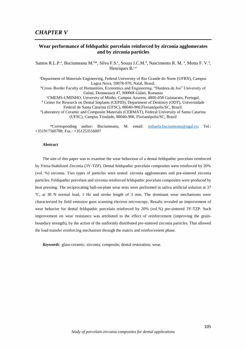
105 Study of porcelain-zirconia composites for dental applications
CHAPTER V
Wear performance of feldspathic porcelain reinforced by zirconia agglomerates
and by zirconia particles
Santos R.L.P.a, Buciumeanu M.b*, Silva F.S.c, Souza J.C.M.d, Nascimento R. M. a, Motta F. V.a,
Henriques B.c,e
aDepartment of Materials Engineering, Federal University of Rio Grande do Norte (UFRN), Campus
Lagoa Nova, 59078-970, Natal, Brasil. bCross–Border Faculty of Humanities, Economics and Engineering, “Dunărea de Jos” University of
Galaţi, Domnească 47, 800008 Galati, Romania cCMEMS-UMINHO, Unversity of Minho, Campus Azurem, 4800-058 Guimaraes, Portugal.
d Center for Research on Dental Implants (CEPID), Department of Dentistry (ODT), Universidade
Federal de Santa Catarina (UFSC), 88040-900,Florianópolis/SC, Brazil. eLaboratory of Ceramic and Composite Materials (CERMAT), Federal University of Santa Catarina
(UFSC), Campus Trindade, 88040-900, Florianópolis/SC, Brazil
*Corresponding author: Buciumeanu, M. email: [email protected]; Tel.:
+351917560788; Fax.: +351253516007
Abstract
The aim of this paper was to examine the wear behaviour of a dental feldspathic porcelain reinforced
by Yttria-Stabilized Zirconia (3Y-TZP). Dental feldspathic porcelain composites were reinforced by 20%
(vol. %) zirconia. Two types of particles were tested: zirconia agglomerates and pre-sintered zirconia
particles. Feldspathic porcelain and zirconia reinforced feldspathic porcelain composites were produced by
heat pressing. The reciprocating ball-on-plate wear tests were performed in saliva artificial solution at 37
ºC, at 30 N normal load, 1 Hz and stroke length of 3 mm. The dominant wear mechanisms were
characterized by field emission guns scanning electron microscopy. Results revealed an improvement of
wear behavior for dental feldspathic porcelain reinforced by 20% (vol.%) pre-sintered 3Y-TZP. Such
improvement on wear resistance was attributed to the effect of reinforcement (improving the grain-
boundary strength), by the action of the uniformly distributed pre-sintered zirconia particles. That allowed
the load transfer reinforcing mechanism through the matrix and reinforcement phase.
Keywords: glass-ceramic; zirconia; composite; dental restoration; wear.
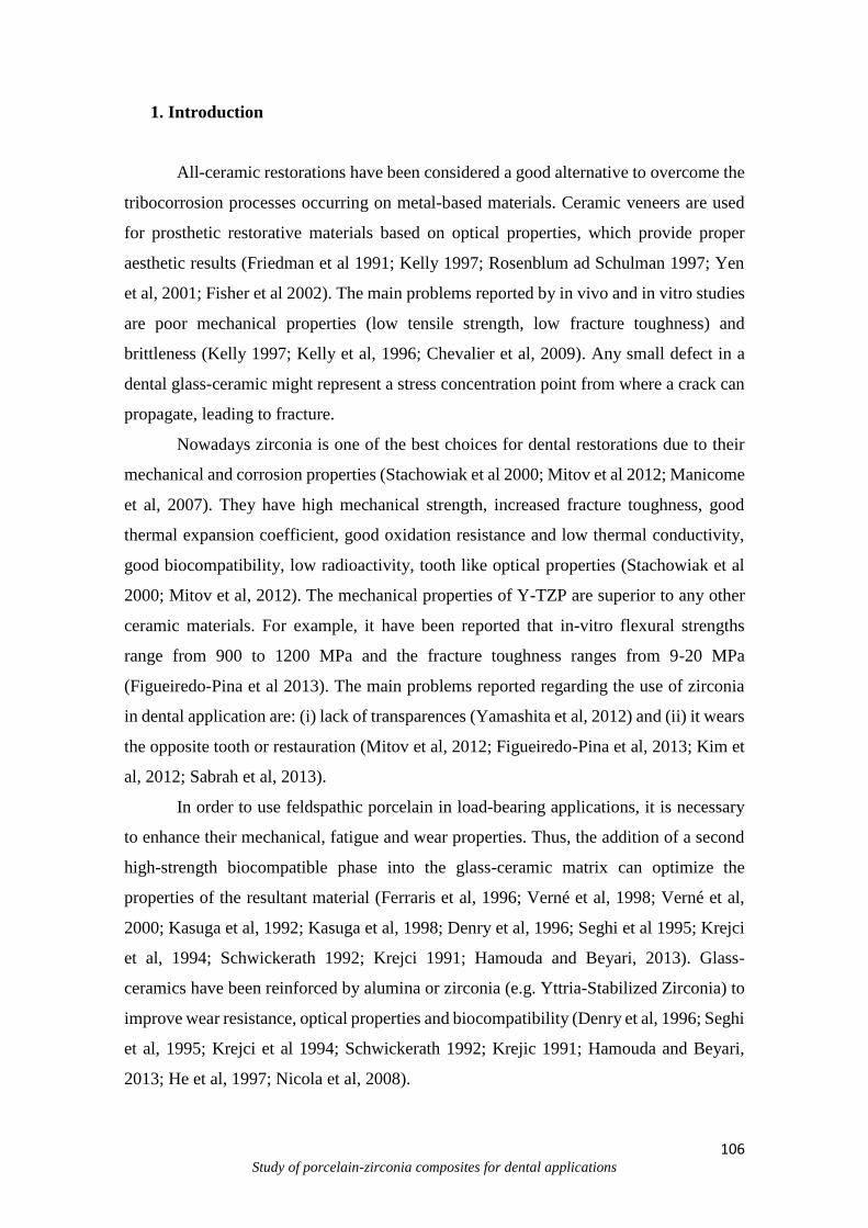
106 Study of porcelain-zirconia composites for dental applications
1. Introduction
All-ceramic restorations have been considered a good alternative to overcome the
tribocorrosion processes occurring on metal-based materials. Ceramic veneers are used
for prosthetic restorative materials based on optical properties, which provide proper
aesthetic results (Friedman et al 1991; Kelly 1997; Rosenblum ad Schulman 1997; Yen
et al, 2001; Fisher et al 2002). The main problems reported by in vivo and in vitro studies
are poor mechanical properties (low tensile strength, low fracture toughness) and
brittleness (Kelly 1997; Kelly et al, 1996; Chevalier et al, 2009). Any small defect in a
dental glass-ceramic might represent a stress concentration point from where a crack can
propagate, leading to fracture.
Nowadays zirconia is one of the best choices for dental restorations due to their
mechanical and corrosion properties (Stachowiak et al 2000; Mitov et al 2012; Manicome
et al, 2007). They have high mechanical strength, increased fracture toughness, good
thermal expansion coefficient, good oxidation resistance and low thermal conductivity,
good biocompatibility, low radioactivity, tooth like optical properties (Stachowiak et al
2000; Mitov et al, 2012). The mechanical properties of Y-TZP are superior to any other
ceramic materials. For example, it have been reported that in-vitro flexural strengths
range from 900 to 1200 MPa and the fracture toughness ranges from 9-20 MPa
(Figueiredo-Pina et al 2013). The main problems reported regarding the use of zirconia
in dental application are: (i) lack of transparences (Yamashita et al, 2012) and (ii) it wears
the opposite tooth or restauration (Mitov et al, 2012; Figueiredo-Pina et al, 2013; Kim et
al, 2012; Sabrah et al, 2013).
In order to use feldspathic porcelain in load-bearing applications, it is necessary
to enhance their mechanical, fatigue and wear properties. Thus, the addition of a second
high-strength biocompatible phase into the glass-ceramic matrix can optimize the
properties of the resultant material (Ferraris et al, 1996; Verné et al, 1998; Verné et al,
2000; Kasuga et al, 1992; Kasuga et al, 1998; Denry et al, 1996; Seghi et al 1995; Krejci
et al, 1994; Schwickerath 1992; Krejci 1991; Hamouda and Beyari, 2013). Glass-
ceramics have been reinforced by alumina or zirconia (e.g. Yttria-Stabilized Zirconia) to
improve wear resistance, optical properties and biocompatibility (Denry et al, 1996; Seghi
et al, 1995; Krejci et al 1994; Schwickerath 1992; Krejic 1991; Hamouda and Beyari,
2013; He et al, 1997; Nicola et al, 2008).

107 Study of porcelain-zirconia composites for dental applications
Guazzato et al. (Guazzato et al, 2004) reported that the zirconia reinforced
feldspathic porcelain composites are stronger and tougher materials than the conventional
feldspathic porcelain. The improvement on the properties of the zirconia reinforced
feldspathic porcelain composites was attributed to phase transformation from tetragonal
to monoclinic and to the relationship between the glassy matrix and the crystalline second
phase in the strengthening and toughening mechanisms of these ceramics. The
strengthening mechanisms of zirconia reinforced glass-ceramic composites are based on
stopping, deflection or bridging the crack propagation (Guazzato et al 2004; Scherrer et
al, 1998; Medeiros et al, 2009), either, on compressive residual stresses due to thermal
mismatch between the glass-ceramic matrix and inorganic fillers. It has been mentioned
that the optimization of the microstructure is an important factor for improvement on
friction and wear behavior of dental glass-ceramic (He et al, 1997; Yu et al, 2006). As
stated by He et al. (He et al, 1997), the amount of tetragonal zirconia transformed to the
monoclinic crystal structure increased with increasing grain size resulting in a decrease
in wear resistance. The volume increment caused by this phase transformation weakens
the grain boundaries which results in grain pull-out.
The present study reveals the wear behavior of zirconia reinfored glass-ceramic
composites in a saliva artificial solution. It was hypothesized that the addition of pre-
sintered yttria-stabilized zirconia particles into glass-ceramic is very effective in reducing
the wear loss of the resulting material.
2. Experimental procedure
2.1. Materials
A dental feldspathic porcelain powder (Vita VM9, Vita, Germany) was used as
matrix material. The chemical composition of the glass-ceramic is shown in Table 5.1.
FEGSEM images of dental feldspathic porcelain powder (raw material) as provided by
the supplier are show in Figure 5.1A. The powder size distribution of feldspathic
porcelain powder was D10= 2.49 μm; D50=18.92 μm; D90=50.71 μm and Dave=23.47 μm.
Table 5.1 Chemical composition of dental feldspathic porcelainpowder (wt.%). (VITA®)
SiO2 Al2O3 B2O3 K2O Na2O Other oxides
60-64 13-15 3-5 7-10 4-6 ~2
108 Study of porcelain-zirconia composites for dental applications
Fig. 5.1 FEGSEM images showing: A) Feldspathic porcelain; B) zirconia agglomerates; and C) pre-
sintered zirconia powders.
Yttria-stabilized zirconia particles - 3Y-TZP (Innovnano, Portugal), were used as
reinforcement (Table 5.2). FEGSEM images of the zirconia agglomerates and pre-
sintered zirconia powder are shown in Figure 5.1B and 5.1C, respectively. The powder
size distribution of pre-sintered zirconia is D10=14.54 μm; D50 =162.64 μm; D90=336.71
μm and Dave=166.44 μm.
Table 5.2 Chemical composition of 3Y-TZP powder (wt.%)
ZrO2 + HfO2 + Y2O3 + Al2O3 Y2O3 Al2O3 HfO2 Other oxides
99.9 5.2±0.2 <0.4 <0.5 <0.5
2.2. Fabrication of zirconia reinforced glass-ceramic composites
The feldspathic porcelain was used as matrix material (named as Z0 from now on).
Two types of zirconia reinforced feldspathic porcelain composites were produced:
(i). feldspathic porcelain reinforced by 20% (vol.%) Yttria-Stabilized Zirconia (ZrO2
– 3%Y2O3) agglomerates (named as ZA from now on). The 3Y-TZP agglomerates were
used asprovided by the supplier and blended with the feldspathic porcelain powders were
ball milled during 24 h.
(ii). Feldspathic porcelain reinforced by 20% (vol%) pre-sintered Yttria-Stabilized
Zirconia (ZrO2 – 3%Y2O3) particles (named as ZP from now on). The 3Y-TZP
agglomerates as provided by the supplier were sintered in a furnace at 1500°C for 2h.
Afterwards, powder mixtures (feldspathic porcelain and sintered 3Y-TZP), were ball
milled to reduce agglomeration and to enhance mixing.
A
A
A
C B
B
109 Study of porcelain-zirconia composites for dental applications
All the samples were processed by the hot pressing (HP) technique (Medeiros et al,
2009; Yu et al, 2006; Souza et al 2013; Sampaio et al 2015; Hutching, 1992; Yin et al,
2003). The compactation pressure applied was 7.0MPa in a stage of 2 minutes at 970ºC
under vacuum (1 bar).
2.3. Microstructural and chemical analysis
The obtained samples were characterized in terms of microstructure, morphology
and chemical composition by Field Emission Guns Scanning Electron Microscopy
(FEGSEM) coupled to Energy Dispersive Spectrometer (EDS) (FEGSEM, FEI Nova 200,
USA). The samples were sputter-coated with carbon.
2.4. Hardness measurements
Vicker’s microhardnessmeasurements were performed on 10 different areas of the
samples surfaces ( n = 50) using a microhardness tester (DuraScan, Emco-Test, Austria)
with a load of 1 kgf (9,8N) during 15 s. Vickers hardness mean values were obtained as
a average of 10 indentations per sample.
2.5. Wear tests
Square test samples (10 x 10 x 2 mm) were embedded in epoxy resin. Samples
were wet ground on SiC sandpapers down to 4000Mesh and then polished using diamond
paste (1m). Samples were ultrasonically cleaned in isopropyl alcohol for 15 min and
then in distilled water for 15 min. A reciprocating ball-on-plate tribometer (Bruker-UMT-
2) was used to evaluate the wear of square samples (10 x 10 x 2 mm) against Al2O3
counterbody (10 mm diameter alumina ball, Goodfellow). The samples were placed in an
acrylic electrochemical cell containing a Fusayama artificial saliva solution (Table 5.3),
that was attached to the tribometer.
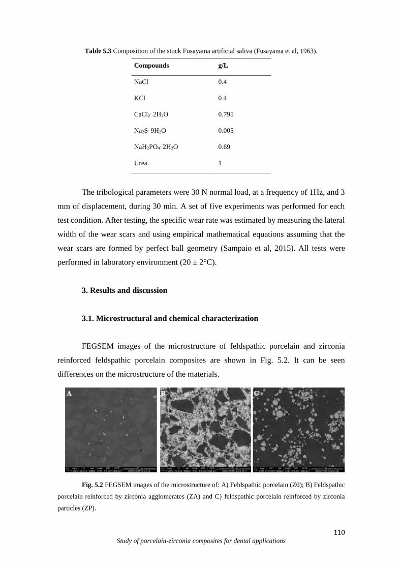
110 Study of porcelain-zirconia composites for dental applications
Table 5.3 Composition of the stock Fusayama artificial saliva (Fusayama et al, 1963).
Compounds g/L
NaCl 0.4
KCl 0.4
CaCl2. 2H2O 0.795
Na2S. 9H2O 0.005
NaH2PO4. 2H2O 0.69
Urea 1
The tribological parameters were 30 N normal load, at a frequency of 1Hz, and 3
mm of displacement, during 30 min. A set of five experiments was performed for each
test condition. After testing, the specific wear rate was estimated by measuring the lateral
width of the wear scars and using empirical mathematical equations assuming that the
wear scars are formed by perfect ball geometry (Sampaio et al, 2015). All tests were
performed in laboratory environment (20 ± 2°C).
3. Results and discussion
3.1. Microstructural and chemical characterization
FEGSEM images of the microstructure of feldspathic porcelain and zirconia
reinforced feldspathic porcelain composites are shown in Fig. 5.2. It can be seen
differences on the microstructure of the materials.
Fig. 5.2 FEGSEM images of the microstructure of: A) Feldspathic porcelain (Z0); B) Feldspathic
porcelain reinforced by zirconia agglomerates (ZA) and C) feldspathic porcelain reinforced by zirconia
particles (ZP). C
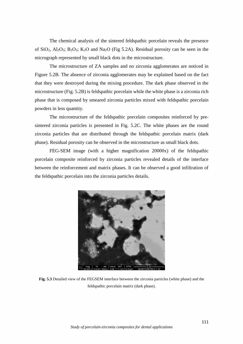
111 Study of porcelain-zirconia composites for dental applications
The chemical analysis of the sintered feldspathic porcelain reveals the presence
of SiO2, Al2O3; B2O3; K2O and Na2O (Fig 5.2A). Residual porosity can be seen in the
micrograph represented by small black dots in the microstructure.
The microstructure of ZA samples and no zirconia agglomerates are noticed in
Figure 5.2B. The absence of zirconia agglomerates may be explained based on the fact
that they were destroyed during the mixing procedure. The dark phase observed in the
microstructure (Fig. 5.2B) is feldspathic porcelain while the white phase is a zirconia rich
phase that is composed by smeared zirconia particles mixed with feldspathic porcelain
powders in less quantity.
The microstructure of the feldspathic porcelain composites reinforced by pre-
sintered zirconia particles is presented in Fig. 5.2C. The white phases are the round
zirconia particles that are distributed through the feldspathic porcelain matrix (dark
phase). Residual porosity can be observed in the microstructure as small black dots.
FEG-SEM image (with a higher magnification 20000x) of the feldspathic
porcelain composite reinforced by zirconia particles revealed details of the interface
between the reinforcement and matrix phases. It can be observed a good infiltration of
the feldspathic porcelain into the zirconia particles details.
Fig. 5.3 Detailed view of the FEGSEM interface between the zirconia particles (white phase) and the
feldspathic porcelain matrix (dark phase).
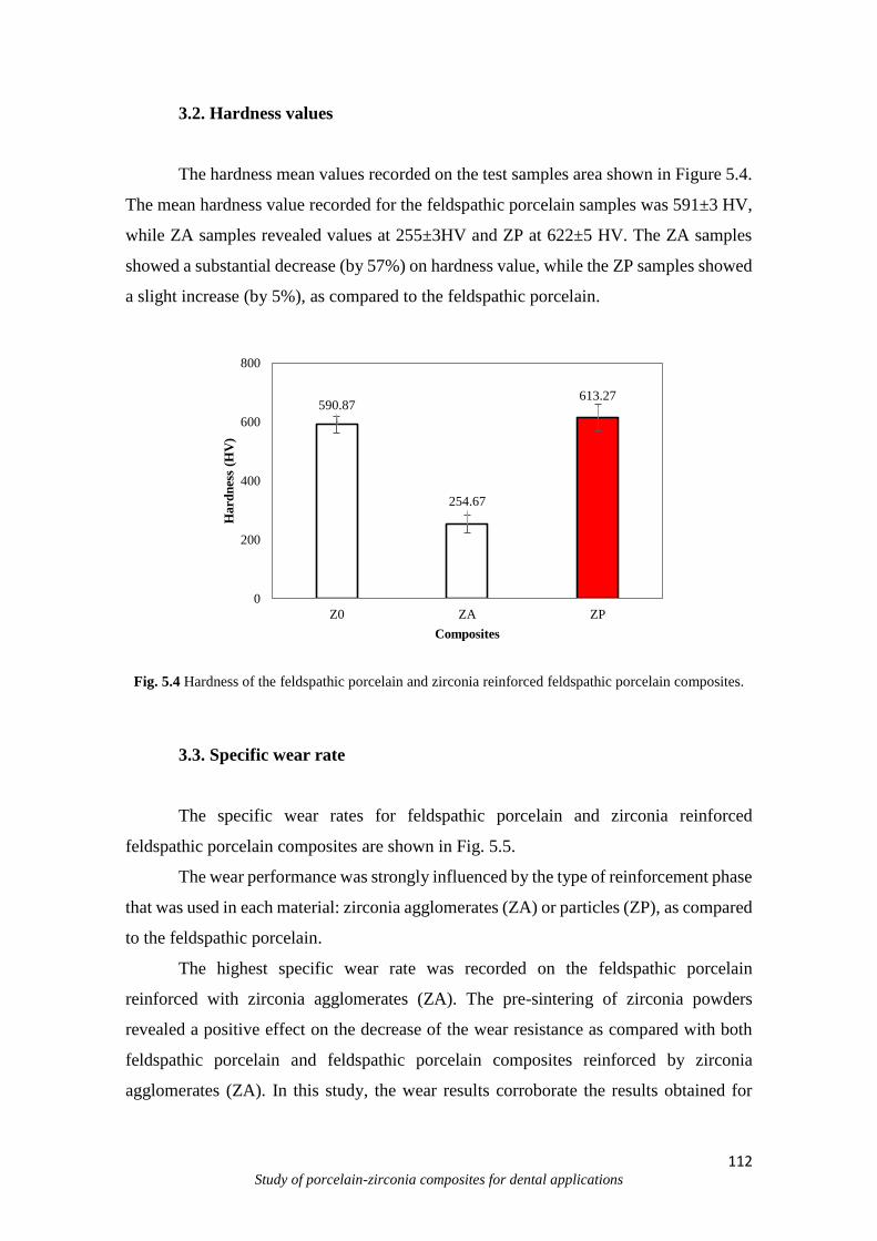
112 Study of porcelain-zirconia composites for dental applications
3.2. Hardness values
The hardness mean values recorded on the test samples area shown in Figure 5.4.
The mean hardness value recorded for the feldspathic porcelain samples was 591±3 HV,
while ZA samples revealed values at 255±3HV and ZP at 622±5 HV. The ZA samples
showed a substantial decrease (by 57%) on hardness value, while the ZP samples showed
a slight increase (by 5%), as compared to the feldspathic porcelain.
Fig. 5.4 Hardness of the feldspathic porcelain and zirconia reinforced feldspathic porcelain composites.
3.3. Specific wear rate
The specific wear rates for feldspathic porcelain and zirconia reinforced
feldspathic porcelain composites are shown in Fig. 5.5.
The wear performance was strongly influenced by the type of reinforcement phase
that was used in each material: zirconia agglomerates (ZA) or particles (ZP), as compared
to the feldspathic porcelain.
The highest specific wear rate was recorded on the feldspathic porcelain
reinforced with zirconia agglomerates (ZA). The pre-sintering of zirconia powders
revealed a positive effect on the decrease of the wear resistance as compared with both
feldspathic porcelain and feldspathic porcelain composites reinforced by zirconia
agglomerates (ZA). In this study, the wear results corroborate the results obtained for
590.87
254.67
613.27
0
200
400
600
800
Z0 ZA ZP
Hard
nes
s (H
V)
Composites
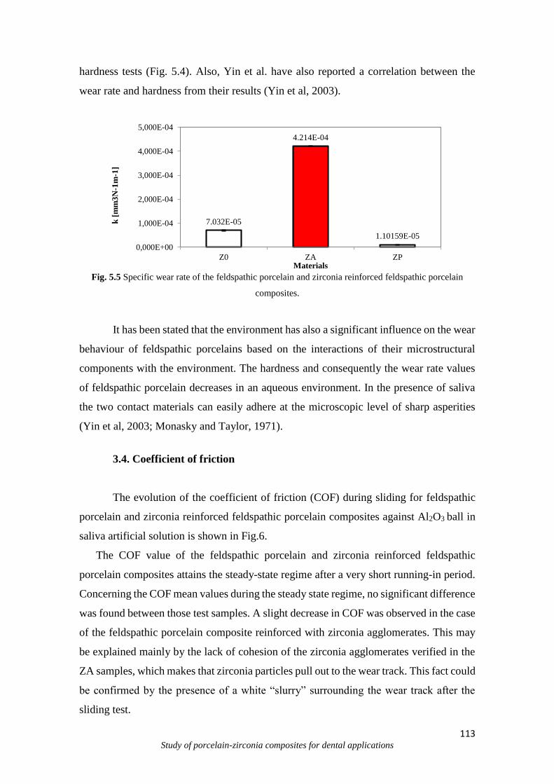
113 Study of porcelain-zirconia composites for dental applications
hardness tests (Fig. 5.4). Also, Yin et al. have also reported a correlation between the
wear rate and hardness from their results (Yin et al, 2003).
Fig. 5.5 Specific wear rate of the feldspathic porcelain and zirconia reinforced feldspathic porcelain
composites.
It has been stated that the environment has also a significant influence on the wear
behaviour of feldspathic porcelains based on the interactions of their microstructural
components with the environment. The hardness and consequently the wear rate values
of feldspathic porcelain decreases in an aqueous environment. In the presence of saliva
the two contact materials can easily adhere at the microscopic level of sharp asperities
(Yin et al, 2003; Monasky and Taylor, 1971).
3.4. Coefficient of friction
The evolution of the coefficient of friction (COF) during sliding for feldspathic
porcelain and zirconia reinforced feldspathic porcelain composites against Al2O3 ball in
saliva artificial solution is shown in Fig.6.
The COF value of the feldspathic porcelain and zirconia reinforced feldspathic
porcelain composites attains the steady-state regime after a very short running-in period.
Concerning the COF mean values during the steady state regime, no significant difference
was found between those test samples. A slight decrease in COF was observed in the case
of the feldspathic porcelain composite reinforced with zirconia agglomerates. This may
be explained mainly by the lack of cohesion of the zirconia agglomerates verified in the
ZA samples, which makes that zirconia particles pull out to the wear track. This fact could
be confirmed by the presence of a white “slurry” surrounding the wear track after the
sliding test.
7.032E-05
4.214E-04
1.10159E-05
0,000E+00
1,000E-04
2,000E-04
3,000E-04
4,000E-04
5,000E-04
Z0 ZA ZP
k [
mm
3N
-1m
-1]
Materials
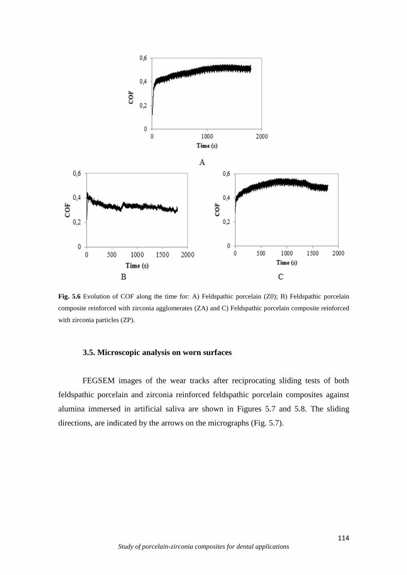
114 Study of porcelain-zirconia composites for dental applications
Fig. 5.6 Evolution of COF along the time for: A) Feldspathic porcelain (Z0); B) Feldspathic porcelain
composite reinforced with zirconia agglomerates (ZA) and C) Feldspathic porcelain composite reinforced
with zirconia particles (ZP).
3.5. Microscopic analysis on worn surfaces
FEGSEM images of the wear tracks after reciprocating sliding tests of both
feldspathic porcelain and zirconia reinforced feldspathic porcelain composites against
alumina immersed in artificial saliva are shown in Figures 5.7 and 5.8. The sliding
directions, are indicated by the arrows on the micrographs (Fig. 5.7).

115 Study of porcelain-zirconia composites for dental applications
Fig. 5.7 FEGSEM images of the midle area of the wear track: A) Feldspathic Porcelain (Z0); B)
Feldspathic porcelain composite reinforced with zirconia agglomerates (ZA) and C) Feldspathic porcelain
composite reinforced with zirconia particles (ZP).
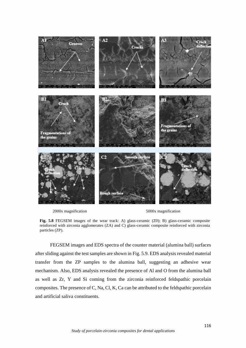
116 Study of porcelain-zirconia composites for dental applications
2000x magnification 5000x magnification
Fig. 5.8 FEGSEM images of the wear track: A) glass-ceramic (Z0); B) glass-ceramic composite
reinforced with zirconia agglomerates (ZA) and C) glass-ceramic composite reinforced with zirconia
particles (ZP).
FEGSEM images and EDS spectra of the counter material (alumina ball) surfaces
after sliding against the test samples are shown in Fig. 5.9. EDS analysis revealed material
transfer from the ZP samples to the alumina ball, suggesting an adhesive wear
mechanism. Also, EDS analysis revealed the presence of Al and O from the alumina ball
as well as Zr, Y and Si coming from the zirconia reinforced feldspathic porcelain
composites. The presence of C, Na, Cl, K, Ca can be attributed to the feldspathic porcelain
and artificial saliva constituents.
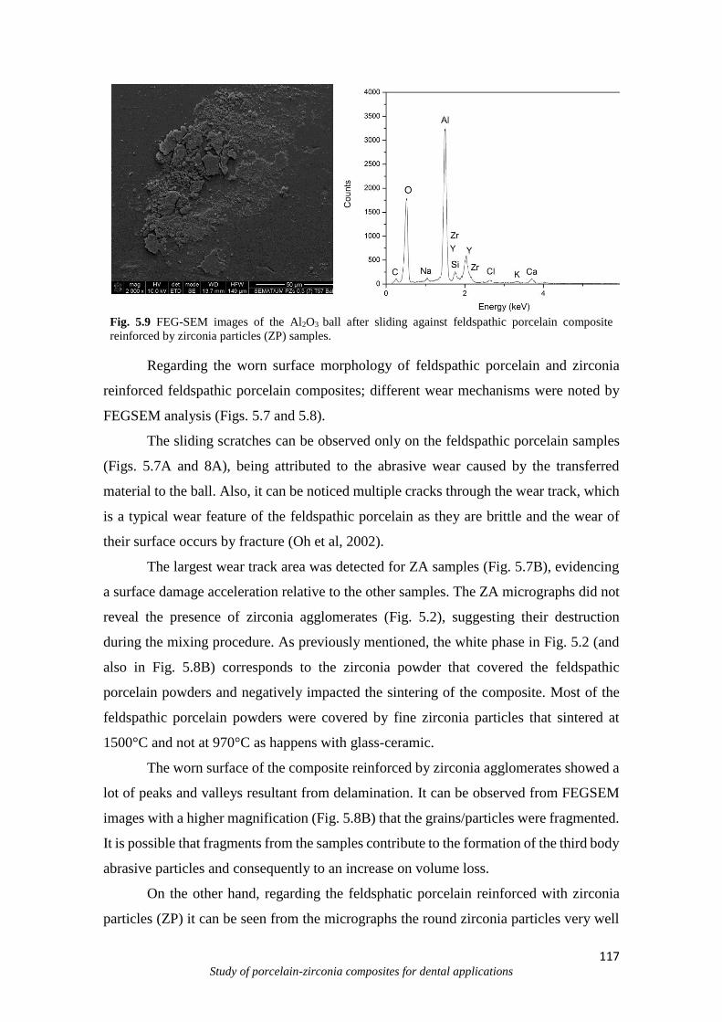
117 Study of porcelain-zirconia composites for dental applications
Fig. 5.9 FEG-SEM images of the Al2O3 ball after sliding against feldspathic porcelain composite
reinforced by zirconia particles (ZP) samples.
Regarding the worn surface morphology of feldspathic porcelain and zirconia
reinforced feldspathic porcelain composites; different wear mechanisms were noted by
FEGSEM analysis (Figs. 5.7 and 5.8).
The sliding scratches can be observed only on the feldspathic porcelain samples
(Figs. 5.7A and 8A), being attributed to the abrasive wear caused by the transferred
material to the ball. Also, it can be noticed multiple cracks through the wear track, which
is a typical wear feature of the feldspathic porcelain as they are brittle and the wear of
their surface occurs by fracture (Oh et al, 2002).
The largest wear track area was detected for ZA samples (Fig. 5.7B), evidencing
a surface damage acceleration relative to the other samples. The ZA micrographs did not
reveal the presence of zirconia agglomerates (Fig. 5.2), suggesting their destruction
during the mixing procedure. As previously mentioned, the white phase in Fig. 5.2 (and
also in Fig. 5.8B) corresponds to the zirconia powder that covered the feldspathic
porcelain powders and negatively impacted the sintering of the composite. Most of the
feldspathic porcelain powders were covered by fine zirconia particles that sintered at
1500°C and not at 970°C as happens with glass-ceramic.
The worn surface of the composite reinforced by zirconia agglomerates showed a
lot of peaks and valleys resultant from delamination. It can be observed from FEGSEM
images with a higher magnification (Fig. 5.8B) that the grains/particles were fragmented.
It is possible that fragments from the samples contribute to the formation of the third body
abrasive particles and consequently to an increase on volume loss.
On the other hand, regarding the feldsphatic porcelain reinforced with zirconia
particles (ZP) it can be seen from the micrographs the round zirconia particles very well
118 Study of porcelain-zirconia composites for dental applications
dispersed through the feldspathic porcelain matrix (Fig. 5.2). The zirconia particles act as
a second phase that is stiffer than the feldspathic porcelain matrix, allowing the load
transfer reinforcing mechanism to occur from the matrix to the reinforcing phase. There
was a good bonding between the zirconia particles and the feldspathic porcelain matrix
(Fig. 5.3) that may explained the increased extension of smooth surface noted on the worn
surface of the P/ZP samples (Figs. 5.7C and 5.8C) and consequently the improvement on
wear performance (Fig. 5.4). The presence of some voids was detected on the worn
surfaces of the P/ZP samples. These voids resulted mainly from the pull out of zirconia
larger particles and particles agglomerates as seen in the backscattered FEGSEM images
of the worn surfaces (Fig. 5.7c).
It is well known that the particles agglomerates are critic areas from the point of
view of wear resistance and mechanical strength. It is important to point out that less
cracks were observed on the worn surface of ZP samples (Fig. 5.7), as compared to the
feldspathic porcelain (Fig. 5.8). This is explained by the toughening effect produced by
the dispersed zirconia particles in the feldspathic porcelain matrix. Hence, when a crack
arises in the weaker matrix (feldspathic porcelain) it is deflected or blocked due to the
presence of a stronger zirconia phase which is well bonded to the matrix (Fig. 5.3). Crack
deflections a common effect in ceramic and metal matrix composites (Kelly and Denry,
2008; Scherrer et al, 1998; Mair et al, 1996), and it leads to a decrease of wear loss. If the
crack reaches an agglomerate (weaker zone), the propagation of the crack is accelerated
(Fig. 5.7C).
These observations are in good correlation with the specific wear rate (Fig. 5.5).
The smoother surface of the wear track (Fig. 5.7C) noticed in the case of ZP samples was
consistent with the results reported in the technical literature. As also mentioned by Kim
et al. (Kim et al, 2012), the superior properties of zirconia (such as hardness, bending
strength, fracture toughness and density) as compared to feldspathic porcelain, enabled it
to maintain a smooth surface during the wear test.
It seems that the main strengthening mechanism, as observed in the fracture
morphologic aspects after wear tests, is the strengthening effect of zirconia particles
promoted by a strong bonding between feldspathic porcelain matrix and zirconia particles
and by the load transfer mechanisms occurring from feldspathic porcelain matrix to the
zirconia particles. Also, it should be mentioned that the zirconia agglomerates may have
detrimental effect on the strengthening, and they are responsible for voids nucleation.

119 Study of porcelain-zirconia composites for dental applications
4. Conclusions
From the present investigation the following conclusions can be drawn:
- Both the feldspathic porcelain and the feldspathic porcelain composites
reinforced zirconia agglomerates and by zirconia articles were successfully synthesized
by heat pressing technique;
- the feldspathic porcelain composites reinforced by zirconia agglomerates (ZA)
exhibited inappropriate wear response. This was explained by the destruction of the
zirconia agglomerates during the mixture blending process, which resulted in sintering
instabilities of the resultant material;
- the feldspathic porcelain composites reinforced by zirconia particles (ZP)
showed the best wear response. This was attributed to the action of the uniformly
distributed zirconia particles on the feldspathic porcelain matrix, which allowed the load
transfer reinforcing mechanism to occur from the matrix to the reinforcement. Hence, it
can be concluded that the ZP composite can be a good alternative to feldspathic porcelain
especially when advanced wear properties are desired.
Acknowledgements
This work was supported by the CAPES and FCT-Portugal
(SFRH/BPD/87435/2012; EXCL/EMS-TEC/0460/2012; UID/EEA/04436/2013).
120 Study of porcelain-zirconia composites for dental applications
References
Anusavice K.J. (2004). Phillips Science of Dental materials. Amsterdam: Elsevier.
Chevalier J., Gremillard L. Ceramics for medical applications: A picture for the next 20
years. JEurop Ceram Soc 29 (2009) 7:1245-1255.
Denry I., Mackert J., Holloway L., Rosentiel S., Effect of cubic leucite stabilization on
the flexural strength of feldspathic dental glass-ceramic, J. Dent. Res. 75 (1996) 12:1928–
1935.
Fisher J, Hu X, Tipper J, Stewart T, Williams S, Stone M, Davies C, Hatto P, Bolton J,
Riley M and others. An in vitro study of the reduction in wear of metal-on-metal hip
prostheses using surface-engineered femoral heads. Proceedings of the Institution of
Mechanical Engineers, Part H: J Eng Med 216 (2002) 4:219-230.
Ferraris M., Rabajoli P., Brossa F., Paracchini L., Vacuum plasma spray deposition of
titanium particle/glass-ceramic matrix biocomposites. J. Am. Ceram. Soc. 79 (1996)
6:1515–1520.
Figueiredo-Pina C.G., Monteiro A., Guedes M., Mauricio A., Serro A.P., Ramalho A.,
Santos C., Effect of feldspar glass-ceramic coating upon the wear behavior of zirconia
dental crowns, Wear 297 (2013) 872-877.
Friedman M., Augmenting restorative dentistry with glass-ceramic veneers, J. Am. Dent.
Assoc. 122 (1991) 6:29–34;
Fusayama T., Katayori T., Nomoto S., J. Dent. Res. 42 (1963) 1183-1197.
Guazzato M., Albakry M., Ringer S.P., Swain M.V. Strength, fracture toughness and
microstructure of a selection of all-ceramic materials. Part II. Zirconia-based dental
ceramics. Dent Mater. (2004) 20: 449-456.
121 Study of porcelain-zirconia composites for dental applications
Hamouda I. M., Beyari M. M., Addition of Yttria Partially Stabilized Zirconia for
Reinforcement of Dental Glass-ceramic, Int J Sci Res Know (IJSRK), 10 (2013) 1:404-
412.
He Y.J., Winnubst A.J.A., Burggraaf A.J., Venveij H., Van der Vars P.G.T., With G.,
Sliding Wear of ZrO,-Al,O, Composite Ceramics, J Eur Ceram Soc 00 (1997) 1371-1380.
Henriques B., Soares D., Silva F.S. Microstructure, hardness, corrosion resistance and
glass-ceramic shear bond strength comparison between cast and hot pressed CoCrMo
alloy for metal-ceramic dental restorations. J. Mech. Behav. Biomed. (2012) 12:83–92.
Henriques B., Gasik M., Soares D., Silva F.S. Experimental evaluation of the bond
strength between a CoCrMo dental alloy and glass-ceramic through a composite metal-
ceramic graded transition interlayer. J Mech Behav Biomed Mater (2012a) 13:206–14.
Henriques B., Soares D., Silva F.S. Optimization of bond strength between gold alloy and
glass-ceramic through a composite interlayer obtained by powder metallurgy. Mater Sci
Eng A (2011) 528:1415–20.
Henriques B., Gasik M., Souza J.C.M., Nascimento R.M., Soares D., Silva F.S.
Mechanical and thermal properties of hot pressed CoCrMo-glass-ceramic composites
developed for prosthetic dentistry. J Mech Behav Biomed Mater (2014) 30:103–10.
Henriques B., Soares D., Silva F.S. Shear bond strength of a hot pressed Au-Pd-Pt alloy-
glass-ceramic dental composite. J Mech Behav Biomed Mater (2011a) 4:1718–26.
Henriques B., Gonçalves S., Soares D., Silva F.S. Shear bond strength comparison
between conventional glass-ceramic fused to metal and new functionally graded dental
restorations after thermal-mechanical cycling. J Mech Behav Biomed Mater (2012b)
13:194–205.
Kasuga T., Nakajima K., Uno T., Yoshida M., Preparation of zirconia toughened
bioactive glass-ceramic composite by sinter-hot isostatic pressing. J. Am. Ceram. Soc.,
75 (1992) 5.
122 Study of porcelain-zirconia composites for dental applications
Kasuga T., Yoshida M., Uno T., Nakajima K., Preparation of zirconia toughened
bioactive glass-ceramics. J. Mater. Sci., 23 (1988) 2255– 2258.
Kelly J.R., Ceramics in restorative and prosthetic dentistry, Mater. Sci. 27 (1997) 443–
468;
Kelly J.R., Nishimura I., Campbell S.D., Ceramics in dentistry: historical roots and
current perspectives, J. Prosthet. Dent. 75 (1996) 1:18–32.
Kelly, J. R. Denry, I. Stabilized zirconia as a structural ceramic: An overview. Dent
Mater, 24 (2008) 3:289-298.
Kim M.J., Oh S.H., Kim J.H., Ju S.W., Seo D, G., Ahn J.S., Ryu J.J., Jun S. H., Wear
evaluation of the human enamel opposing different Y-TZP dental ceramics and other
glass-ceramics, The J Dent 40 (2012) 979-988.
Krejci I., Lutz F., Reimer M., Wear of CAD/CAM ceramic inlays: restorations, opposing
cups, and luting cements, Quintessence Int. 25 (1994) 3:199–207.
Krejci I., in: W. Mormann (Ed.), Wear of cerec and other restorative materials,
Proceedings of the International Symposium on Computer Restorations, Quintessence
Publishing, Chicago, IL, USA (1991) 245–251.
Manicone P.F., Iommetti P.R., Raffaelli L., An overview of zirconia ceramics: Basic
properties and clinical applications, J Dent 35 (2007) 819–826.
Mair L. H., Stolarskij T.A., Vowles R.W., Lloyd C.H., Wear: mechanisms, manifestations
and measurement. Report of a workshop, J Dent 24 (1996) 1-2:141-148.
Medeiros I.S., Luz L.A., Yoshimura H.N., Cesar P.F., Hernandes A.C. Al2O3/GdAlO
fiber for dental glass-ceramic reinforcement. J Mech Behav Biomed Mater., (2009) 2:
471 – 477.
123 Study of porcelain-zirconia composites for dental applications
Mitov G., Heintze S. D., Walz S., Woll K., Muecklich F., Pospiech P., Wear behavior
of dental Y-TZP ceramic against natural enamel after different finishing procedures,
dentalmaterials 28 (2012) 909–918.
Monasky G.E., Taylor D.F., Studies on the wear of glass-ceramic, enamel, and gold, J
Prosthet Dent. 25 (1971) 3:299-306.
Nicola O., Andronesu G. Visan E, Vasi B.S., Composite ceramics reinforced with
zirconia nanoparticles, Adv Mater Res 47-50 (2008) 1059-1062.
Rosenblum M., Schulman A., A review of all-ceramic restorations, J. Am. Dent. Assoc.
128 (1997) 3:297–307.
Oh W.S., Delong R., Anusavice K.J. Factors affecting enamel and ceramic wear: a
literature review. J Prosthet Dent (2002) 87:451-9.
Sabrah A.H.A., Cook N. B., Luangruangrong P., Hara A.T., Bottino M.C., Full-contour
Y-TZP ceramic surface roughness effect on synthetic hydroxyapatite wear, Dental
Materials 29 (2013) 666-673.
Sampaio M., Buciumeanu M., Henriques B., Silva F. S., Souza J. C. M., Gomes J.R.,
Tribocorrosion behavior of veneering biomedical PEEK to Ti6Al4V structures, Journal
of the Mechanical Behavior of Biomedical Materials, accepted, 2015.
Scherrer, S.S.; Denry, I.L.; Wiskott, H.W. Comparison of three fracture toughness testing
techniques using a dental glass and a dental ceramic. Dent Mater. 14 (1998) 4:246-255.
Schwickerath H., The strength characteristics of cerec, Quintessenz 43 (1992) 669–677.
Seghi R., Denry I., Rosenstiel S., Relative fracture toughness and hardness of new dental
ceramics, J. Prosthet. Dent. 74 (1995) 2:145–150.
Stachowiak G.B., Stachowiak G.W., Evans P., Wear and friction characteristics of ion-
implanted zirconia ceramics, Wear 241 (2000) 220–227.
124 Study of porcelain-zirconia composites for dental applications
Souza J.C., Ponthiaux P., Henriques M., Oliveira R., Teughels W., Celis J.P., Rocha L.A.
Corrosion behavior of titanium in the presence of Streptococcus mutans. J Dent (2013)
41: 528‒534.
Verné E., Ferraris M., Venterella A., Paracchini L., Krajewski A., Ravaglioli A., Sintering
and plasma-spray deposition of bioactive glass-matrix composites for biomedical
applications. J. Eur. Ceram. Soc. vol. 18 (1998) 4:363–372.
Verné E., Ferraris M., Jana C., Pressureless sintering of Bioverit1III/Ti particle
biocomposites. J. Eur. Ceram. Soc., 19 (1999) 2039–2047.
Verné E., Ferraris M., Jana C., Paracchini L., Bioverit1I base glass/Ti particulate
biocomposite: ‘‘in situ’’ vacuum plasma spray deposition. J. Eur. Ceram. Soc., 20 (2000)
473–479.
Yamashita I., Kudo M., Tsukuma K., Development of highly transparent zirconia
ceramics, TOSOH Research & Technology Review 56 (2012).
Yen S.K., Guo M.J., Zan H.Z. Characterization of electrolytic ZrO2 coating on Co-Cr-
Mo implant alloys of hip prosthesis. Biomater 22 (2001) 2:125-133.
Yin L., Jahanmir S., Ives L.K., Abrasive machining of glass-ceramic and zirconia with a
dental handpiece, Wear 255 (2003) 975–989.
Yu H.Y., Ca Z.B., Ren P.D., Zhu M.H., Zhou Z.R. Friction and wear behavior of dental
feldspathic glass-ceramic, Wear 261 (2006) 611–621.
125 Study of porcelain-zirconia composites for dental applications
CHAPTER VI
Tribological behavior of zirconia-reinforced feldspathic porcelain composites
Santos R.L.P.a, Buciumeanu M.b*, Silva F.S.c, Souza J.C.M.d, Nascimento R. M. a, Motta F. V.a,
Henriques B.c,e
aDepartment of Materials Engineering, Federal University of Rio Grande do Norte (UFRN), Campus
Lagoa Nova, 59078-970, Natal, Brasil. bCross–Border Faculty of Humanities, Economics and Engineering, “Dunărea de Jos” University of
Galaţi, Domnească 47, 800008 Galati, Romania cCMEMS-UMINHO, University of Minho, Campus Azurem, 4800-058 Guimaraes, Portugal.
dCenter for Research on Dental Implants (CEPID), Department of Dentistry (ODT), Universidade Federal
de Santa Catarina (UFSC), 88040-900,Florianópolis/SC, Brazil. eLaboratory of Ceramic and Composite Materials (CERMAT), Federal University of Santa Catarina
(UFSC), Campus Trindade, 88040-900, Florianópolis/SC, Brazil
*Corresponding author: Buciumeanu, M. email: [email protected]; Tel.:
+351917560788; Fax.: +351253516007
Abstract
The purpose of this study was to characterize the wear behavior of dental feldspathic porcelain
composites reinforced with different contents of zirconia particles. The tests were carried out using a ball-
on-plate configuration, in artificial saliva at body temperature (37 °C). The zirconia particles were
incorporated in different volume contents ranging from 10 up to 50%. The results demonstrated that
zirconia plays a relevant role on wear behavior of zirconia reinforced feldspathic porcelain composites. The
worn surfaces were characterized by field emission guns scanning electron microscope (FEGSEM). The
results indicated that the addition of 10%, 20% or 30% (vol.%) zirconia into the dental feldspathic porcelain
composites resulted in increased wear resistance combined with a lower coefficient of friction and a higher
hardness. A decrease in wear resistance was recorded for higher zirconia contents, 40% or 50% (vol. %).
Keywords: wear; composite, dental restoration, glass-ceramic, zirconia.
126 Study of porcelain-zirconia composites for dental applications
1. Introduction
During mastication, the restorative materials are subjected to sliding contact under
normal and tangential loads. The friction and wear mechanisms occurring on dental
restorative materials depend on different factors including environment (e.g. pH, oxygen,
abrasive), loading and properties of the materials (Zhou and Zheng, 2008; Schuluetter et
al 2012; Zheng et al, 2007; Mair et al, 1996). The degradation process of dental materials
is also accelerated by substances from dietary and microbial metabolism (Souza et al,
2015).
Dental feldspathic porcelain are widely used to produce prosthetic restorative
materials due to their good aesthetic, chemical inertness and biocompatibility (Strang et
al, 1998). They are good choices, but it is well known that the main disadvantage of these
materials is that they have poor mechanical properties such as low fracture toughness and
tensile strength (Hamouda et al 2013; Stachowiak et al 2000; Manicone et al, 2007; Mitov
et al, 2012; Song et al, 2008). Yttria-stabilized tetragonal zirconia polycristal (3Y-TZP)
has superior physical and mechanical properties (including high hardness, wear
resistance, elastic modulus, and high melting temperature) than all ceramics (Hamouda
et al, 2013; Stachowiak et al, 2000; SerkanSaridag et al, 2013). For instance, zirconia
possesses a flexural strength ranging from 900 to 1200 MPa and the fracture toughness
ranging from 9-20 MPa (Figueiredo-Pina et al, 2013).
There are several methods reported in technical literature to improve the
properties (e.g. to prevent micro cracks) of the dental feldspathic porcelain composites
such as framework support, dispersion strengthening, transformation toughening, residual
surface stressing and surface treatment (Hamouda et al, 2013; Kasuga et al, 1992; Kasuga
et al 1988; Shenoy and Shenoy, 2010; Usha et al, 2014, Maller et al 2012; O´Brien 2002;
Atala and Gu, 2015). The incorporation of a high-strength biocompatible phase into
feldspathic porcelain have been considered as potential alternative to overcome their main
disadvantages (Hamouda et al, 2013; Kasuga et al, 1992; Kasuga et al, 1988; Ferraris et
al, 1996; Verné et al, 1998; Verné et al, 1999; Verné et al, 2000).
To the authors knowledge, the studies regarding wear/mechanical behavior of
zironia reinforced feldspathic porcelain composites and produced by hot pressing process
are scarce (Hamouda et al, 2013; Kim et al, 2012). As reported by Hamouda et al.
(Hamouda et al, 2013) by adding 3 and 5 wt. % of Yttria partially stabilized zirconia to a
127 Study of porcelain-zirconia composites for dental applications
conventional dental feldspathic porcelain, it was observed an increase in flexural strength
and fracture toughness as compared to the conventional dental feldspathic porcelain,
while the Young modulus and hardness decreased. The variation of the mechanical
properties with the amount of zirconia was also reported by Huang et al. (Huang et al,
2014). They studied the effects of zirconia content (from 0 to 30 wt.%.) on microstructure
and mechanical properties of zirconia-toughened lithium disilicate glass ceramic
composites. In the case of the composites with small zirconia content (below 10 wt.%),
zirconia acted as nucleation agent primarily and the microstructure was refined. On
composites with larger zirconia content (from 15 to 30 wt. %), the zirconia particles re-
strained the phase separation of glass. The improvement on mechanical properties was
attributed mainly to compressive stress reinforcement, phase transformation and bridging
toughening mechanisms.
In order to simulate the masticatory motion different test configurations have been
used, such as pin-on-disc (Preis et al, 2011), ball/pin-on-plate (Figueiredo-Pina et al,
2013), ball and crater (Antunes and Ramalho, 2003) etc. Concerning the environment, it
plays an important role on tribology. The wear resistance of the tooth/restorative materials
has been studied in electrolytes such as distilled water (Preis et al, 2011), artificial saliva
or other acidic substance (e.g. citric acid). The artificial saliva can play both cooling and
lubricant effect during wear process (Zhou and Zheng, 2008; Figueiredo-Pina et al, 2013;
Li and Zhou, 2002).
The main aim of this study was to investigate the wear behavior of zirconia
reinforced feldspathic porcelain with different zirconia (Y-TZP) particles contents
ranging from 10 up to 50% (vol.%) in artificial saliva solution.
2. Experimental procedure
2.1. Materials
A dental glass-ceramic powder (Vita VM9, Vita, Germany) was used as matrix
material. The chemical composition of the feldspathic porcelain is shown in Table 6.1.
FEGSEM images of dental feldspathic porcelain powder (raw material) as provided by
the supplier are shown in Figure 1A. The powder size distribution of feldspathic porcelain
powder was D10= 2.49 μm; D50=18.92 μm; D90=50.71 μm and Dave=23.47 μm.
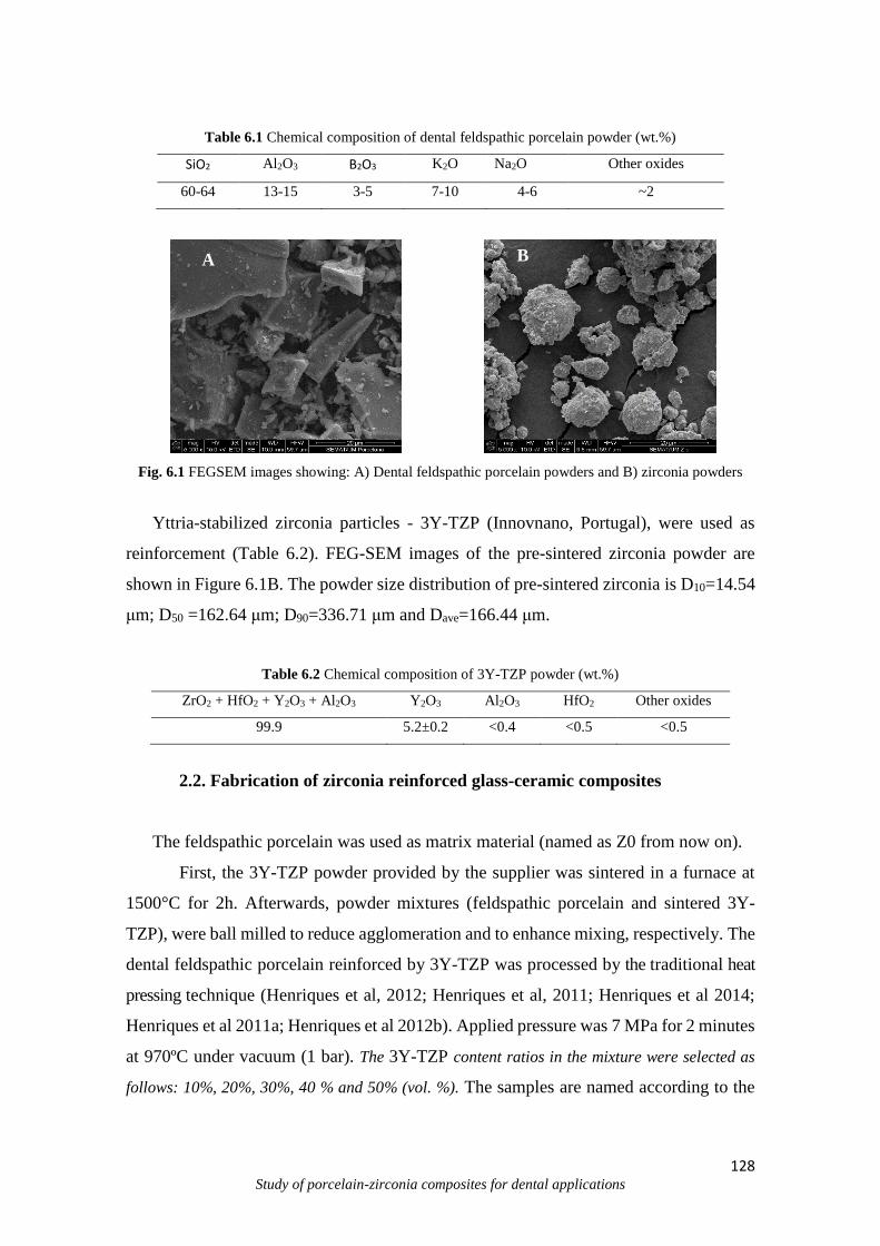
128 Study of porcelain-zirconia composites for dental applications
Table 6.1 Chemical composition of dental feldspathic porcelain powder (wt.%)
SiO2 Al2O3 B2O3 K2O Na2O Other oxides
60-64 13-15 3-5 7-10 4-6 ~2
Fig. 6.1 FEGSEM images showing: A) Dental feldspathic porcelain powders and B) zirconia powders
Yttria-stabilized zirconia particles - 3Y-TZP (Innovnano, Portugal), were used as
reinforcement (Table 6.2). FEG-SEM images of the pre-sintered zirconia powder are
shown in Figure 6.1B. The powder size distribution of pre-sintered zirconia is D10=14.54
μm; D50 =162.64 μm; D90=336.71 μm and Dave=166.44 μm.
Table 6.2 Chemical composition of 3Y-TZP powder (wt.%)
ZrO2 + HfO2 + Y2O3 + Al2O3 Y2O3 Al2O3 HfO2 Other oxides
99.9 5.2±0.2 <0.4 <0.5 <0.5
2.2. Fabrication of zirconia reinforced glass-ceramic composites
The feldspathic porcelain was used as matrix material (named as Z0 from now on).
First, the 3Y-TZP powder provided by the supplier was sintered in a furnace at
1500°C for 2h. Afterwards, powder mixtures (feldspathic porcelain and sintered 3Y-
TZP), were ball milled to reduce agglomeration and to enhance mixing, respectively. The
dental feldspathic porcelain reinforced by 3Y-TZP was processed by the traditional heat
pressing technique (Henriques et al, 2012; Henriques et al, 2011; Henriques et al 2014;
Henriques et al 2011a; Henriques et al 2012b). Applied pressure was 7 MPa for 2 minutes
at 970ºC under vacuum (1 bar). The 3Y-TZP content ratios in the mixture were selected as
follows: 10%, 20%, 30%, 40 % and 50% (vol. %). The samples are named according to the
A B
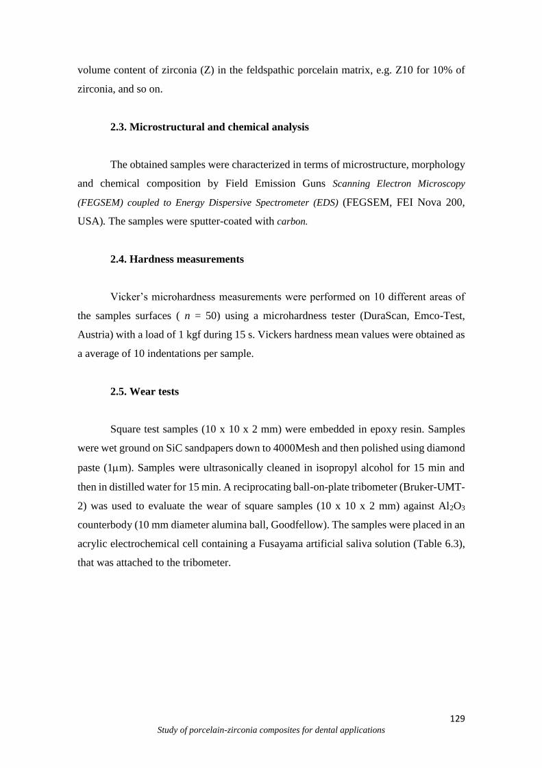
129 Study of porcelain-zirconia composites for dental applications
volume content of zirconia (Z) in the feldspathic porcelain matrix, e.g. Z10 for 10% of
zirconia, and so on.
2.3. Microstructural and chemical analysis
The obtained samples were characterized in terms of microstructure, morphology
and chemical composition by Field Emission Guns Scanning Electron Microscopy
(FEGSEM) coupled to Energy Dispersive Spectrometer (EDS) (FEGSEM, FEI Nova 200,
USA). The samples were sputter-coated with carbon.
2.4. Hardness measurements
Vicker’s microhardness measurements were performed on 10 different areas of
the samples surfaces ( n = 50) using a microhardness tester (DuraScan, Emco-Test,
Austria) with a load of 1 kgf during 15 s. Vickers hardness mean values were obtained as
a average of 10 indentations per sample.
2.5. Wear tests
Square test samples (10 x 10 x 2 mm) were embedded in epoxy resin. Samples
were wet ground on SiC sandpapers down to 4000Mesh and then polished using diamond
paste (1m). Samples were ultrasonically cleaned in isopropyl alcohol for 15 min and
then in distilled water for 15 min. A reciprocating ball-on-plate tribometer (Bruker-UMT-
2) was used to evaluate the wear of square samples (10 x 10 x 2 mm) against Al2O3
counterbody (10 mm diameter alumina ball, Goodfellow). The samples were placed in an
acrylic electrochemical cell containing a Fusayama artificial saliva solution (Table 6.3),
that was attached to the tribometer.
130 Study of porcelain-zirconia composites for dental applications
Table 6.3 Composition of the stock Fusayama artificial saliva (Fusayama et al, 1963).
Compounds g/L
NaCl 0.4
KCl 0.4
CaCl2. 2H2O 0.795
Na2S. 9H2O 0.005
NaH2PO4. 2H2O 0.69
Urea 1
The tribological parameters were 30 N normal load, at a frequency of 1Hz, and 3
mm of displacement, during 30 min. A set of five experiments was performed for each
test condition. After testing, the specific wear rate was estimated by measuring the lateral
width of the wear scars and using empirical mathematical equations assuming that the
wear scars are formed by perfect ball geometry (Sampaio et al, 2015). All tests were
performed in laboratory environment (20 ± 2°C).
3. Results and discussion
3.1. Microstructural characterization
FEGSEM images of the microstructure of feldspathic porcelain and feldspathic
porcelain composites reinforced by zirconia particles in different content ratios (10%,
20%, 30%, 40% and 50%, vol. %) are shown in Fig. 6.2. It can be seen differences on the
microstructure of the materials, with the white phase increasing with the increasing
content of zirconia particles in the feldspathic porcelain matrix (dark phase).
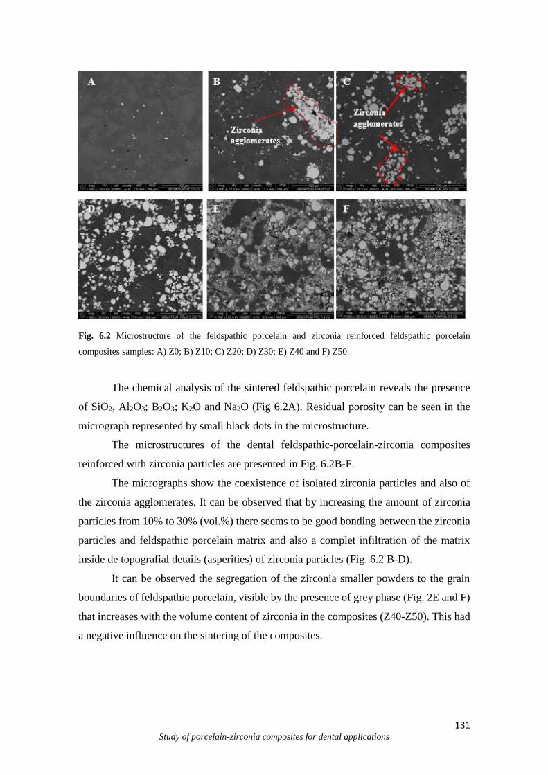
131 Study of porcelain-zirconia composites for dental applications
Fig. 6.2 Microstructure of the feldspathic porcelain and zirconia reinforced feldspathic porcelain
composites samples: A) Z0; B) Z10; C) Z20; D) Z30; E) Z40 and F) Z50.
The chemical analysis of the sintered feldspathic porcelain reveals the presence
of SiO2, Al2O3; B2O3; K2O and Na2O (Fig 6.2A). Residual porosity can be seen in the
micrograph represented by small black dots in the microstructure.
The microstructures of the dental feldspathic-porcelain-zirconia composites
reinforced with zirconia particles are presented in Fig. 6.2B-F.
The micrographs show the coexistence of isolated zirconia particles and also of
the zirconia agglomerates. It can be observed that by increasing the amount of zirconia
particles from 10% to 30% (vol.%) there seems to be good bonding between the zirconia
particles and feldspathic porcelain matrix and also a complet infiltration of the matrix
inside de topografial details (asperities) of zirconia particles (Fig. 6.2 B-D).
It can be observed the segregation of the zirconia smaller powders to the grain
boundaries of feldspathic porcelain, visible by the presence of grey phase (Fig. 2E and F)
that increases with the volume content of zirconia in the composites (Z40-Z50). This had
a negative influence on the sintering of the composites.
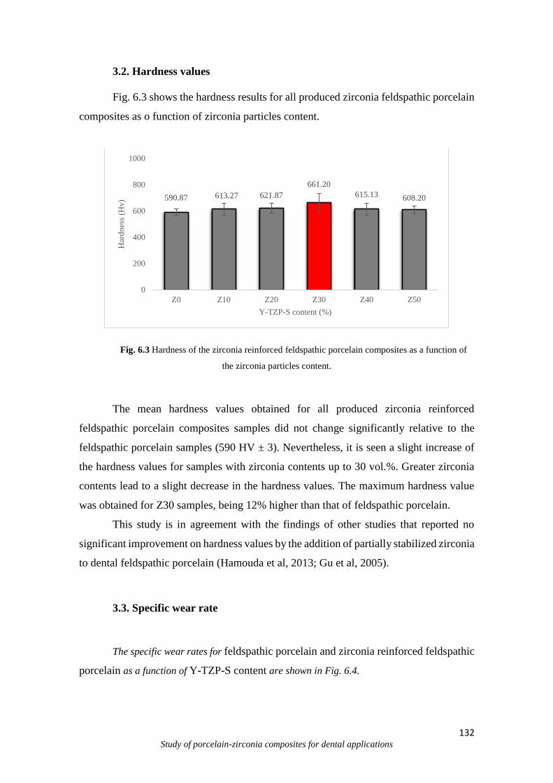
132 Study of porcelain-zirconia composites for dental applications
3.2. Hardness values
Fig. 6.3 shows the hardness results for all produced zirconia feldspathic porcelain
composites as o function of zirconia particles content.
Fig. 6.3 Hardness of the zirconia reinforced feldspathic porcelain composites as a function of
the zirconia particles content.
The mean hardness values obtained for all produced zirconia reinforced
feldspathic porcelain composites samples did not change significantly relative to the
feldspathic porcelain samples (590 HV ± 3). Nevertheless, it is seen a slight increase of
the hardness values for samples with zirconia contents up to 30 vol.%. Greater zirconia
contents lead to a slight decrease in the hardness values. The maximum hardness value
was obtained for Z30 samples, being 12% higher than that of feldspathic porcelain.
This study is in agreement with the findings of other studies that reported no
significant improvement on hardness values by the addition of partially stabilized zirconia
to dental feldspathic porcelain (Hamouda et al, 2013; Gu et al, 2005).
3.3. Specific wear rate
The specific wear rates for feldspathic porcelain and zirconia reinforced feldspathic
porcelain as a function of Y-TZP-S content are shown in Fig. 6.4.
590.87 613.27 621.87
661.20
615.13 608.20
0
200
400
600
800
1000
Z0 Z10 Z20 Z30 Z40 Z50
Har
dn
ess
(Hv)
Y-TZP-S content (%)
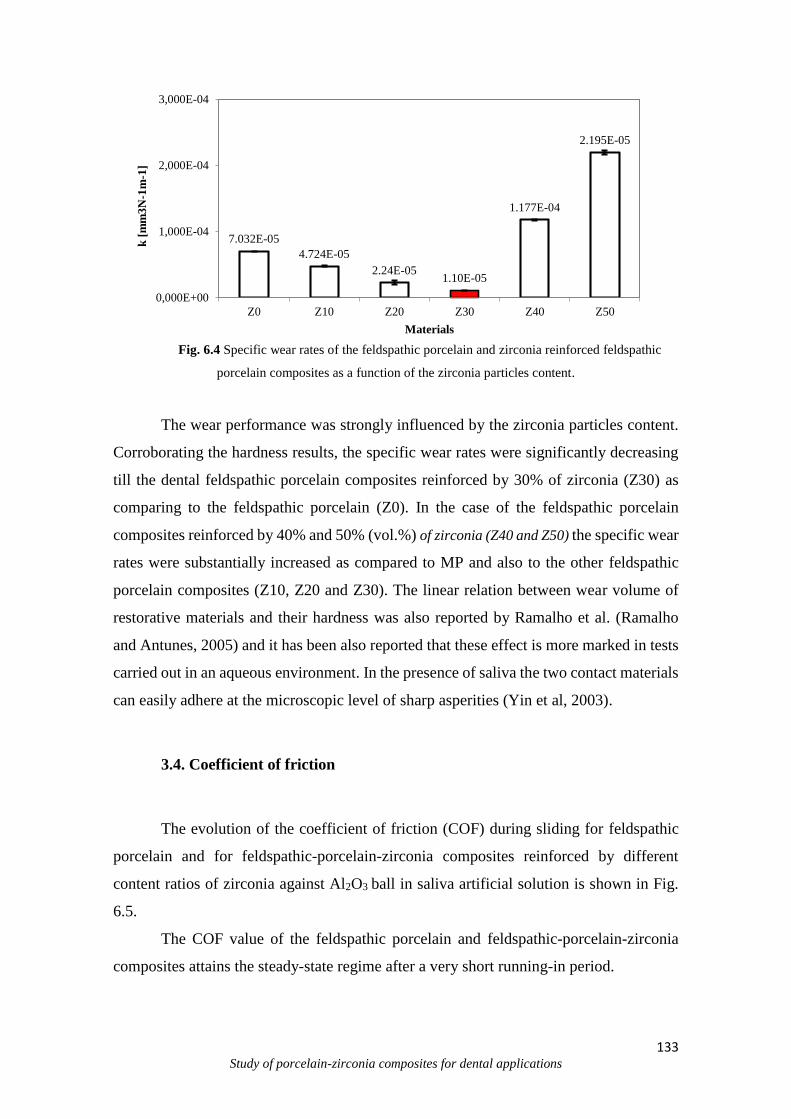
133 Study of porcelain-zirconia composites for dental applications
Fig. 6.4 Specific wear rates of the feldspathic porcelain and zirconia reinforced feldspathic
porcelain composites as a function of the zirconia particles content.
The wear performance was strongly influenced by the zirconia particles content.
Corroborating the hardness results, the specific wear rates were significantly decreasing
till the dental feldspathic porcelain composites reinforced by 30% of zirconia (Z30) as
comparing to the feldspathic porcelain (Z0). In the case of the feldspathic porcelain
composites reinforced by 40% and 50% (vol.%) of zirconia (Z40 and Z50) the specific wear
rates were substantially increased as compared to MP and also to the other feldspathic
porcelain composites (Z10, Z20 and Z30). The linear relation between wear volume of
restorative materials and their hardness was also reported by Ramalho et al. (Ramalho
and Antunes, 2005) and it has been also reported that these effect is more marked in tests
carried out in an aqueous environment. In the presence of saliva the two contact materials
can easily adhere at the microscopic level of sharp asperities (Yin et al, 2003).
3.4. Coefficient of friction
The evolution of the coefficient of friction (COF) during sliding for feldspathic
porcelain and for feldspathic-porcelain-zirconia composites reinforced by different
content ratios of zirconia against Al2O3 ball in saliva artificial solution is shown in Fig.
6.5.
The COF value of the feldspathic porcelain and feldspathic-porcelain-zirconia
composites attains the steady-state regime after a very short running-in period.
7.032E-05
4.724E-05
2.24E-051.10E-05
1.177E-04
2.195E-05
0,000E+00
1,000E-04
2,000E-04
3,000E-04
Z0 Z10 Z20 Z30 Z40 Z50
k [
mm
3N
-1m
-1]
Materials
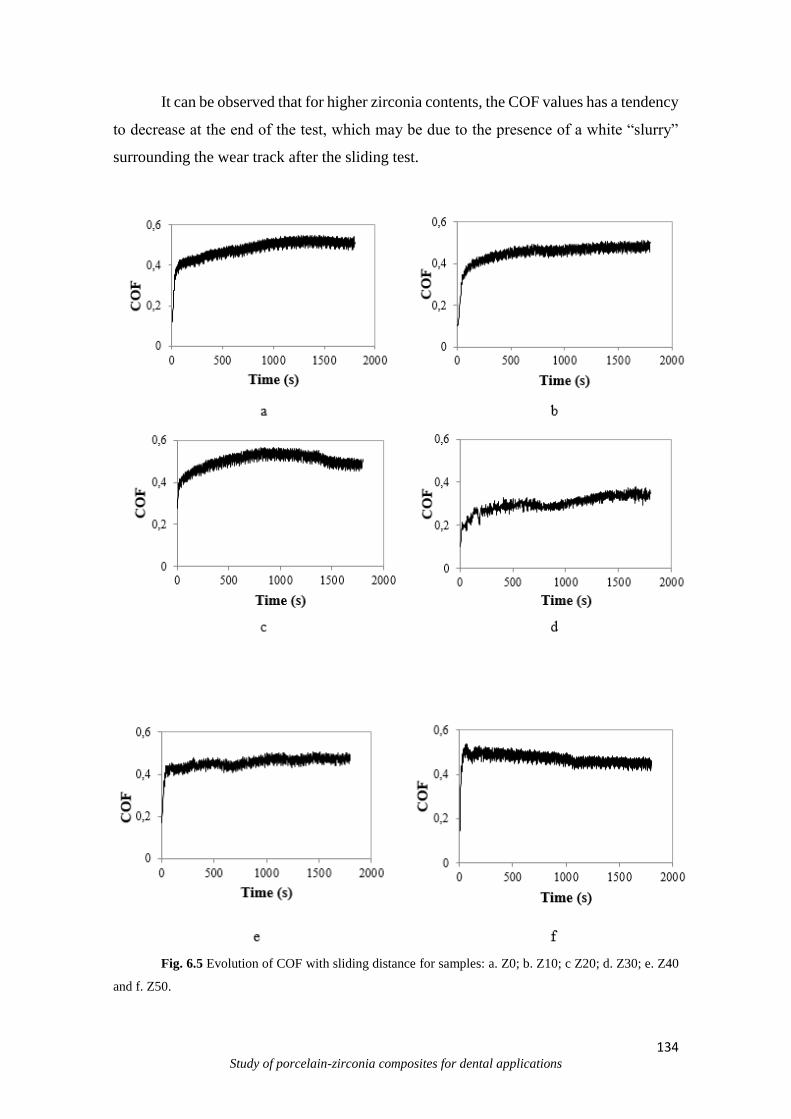
134 Study of porcelain-zirconia composites for dental applications
It can be observed that for higher zirconia contents, the COF values has a tendency
to decrease at the end of the test, which may be due to the presence of a white “slurry”
surrounding the wear track after the sliding test.
Fig. 6.5 Evolution of COF with sliding distance for samples: a. Z0; b. Z10; c Z20; d. Z30; e. Z40
and f. Z50.
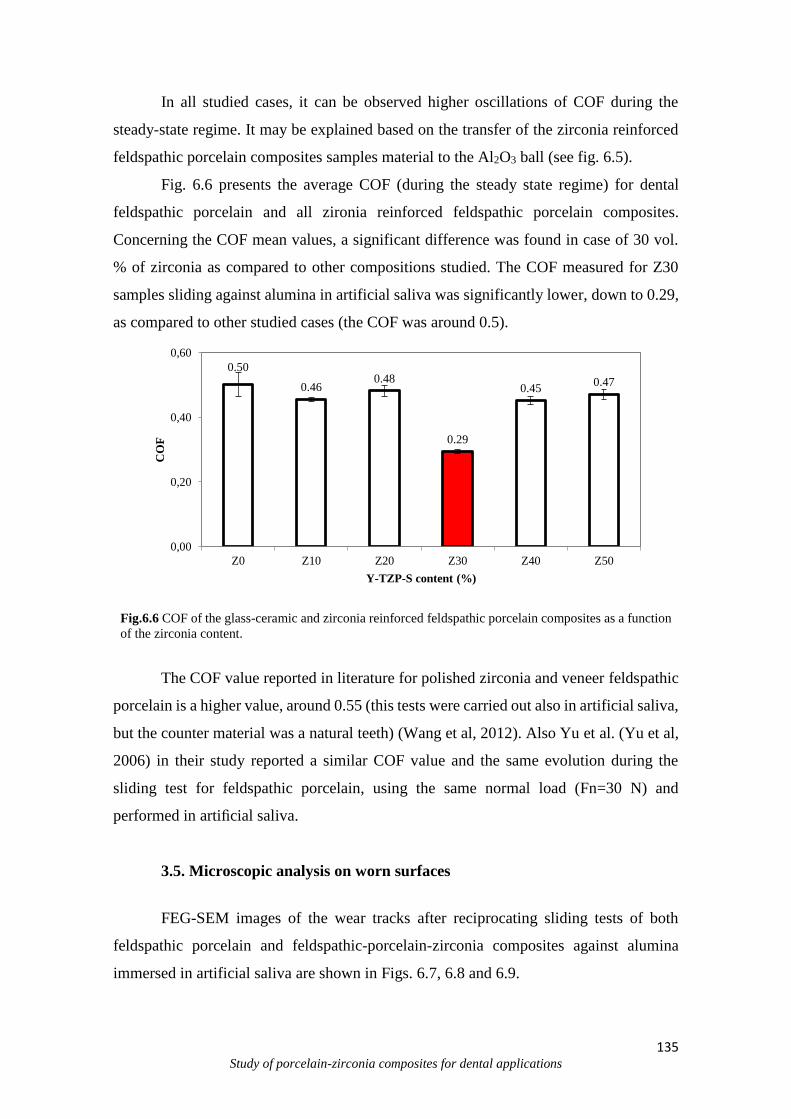
135 Study of porcelain-zirconia composites for dental applications
In all studied cases, it can be observed higher oscillations of COF during the
steady-state regime. It may be explained based on the transfer of the zirconia reinforced
feldspathic porcelain composites samples material to the Al2O3 ball (see fig. 6.5).
Fig. 6.6 presents the average COF (during the steady state regime) for dental
feldspathic porcelain and all zironia reinforced feldspathic porcelain composites.
Concerning the COF mean values, a significant difference was found in case of 30 vol.
% of zirconia as compared to other compositions studied. The COF measured for Z30
samples sliding against alumina in artificial saliva was significantly lower, down to 0.29,
as compared to other studied cases (the COF was around 0.5).
Fig.6.6 COF of the glass-ceramic and zirconia reinforced feldspathic porcelain composites as a function
of the zirconia content.
The COF value reported in literature for polished zirconia and veneer feldspathic
porcelain is a higher value, around 0.55 (this tests were carried out also in artificial saliva,
but the counter material was a natural teeth) (Wang et al, 2012). Also Yu et al. (Yu et al,
2006) in their study reported a similar COF value and the same evolution during the
sliding test for feldspathic porcelain, using the same normal load (Fn=30 N) and
performed in artificial saliva.
3.5. Microscopic analysis on worn surfaces
FEG-SEM images of the wear tracks after reciprocating sliding tests of both
feldspathic porcelain and feldspathic-porcelain-zirconia composites against alumina
immersed in artificial saliva are shown in Figs. 6.7, 6.8 and 6.9.
0.50
0.460.48
0.29
0.450.47
0,00
0,20
0,40
0,60
Z0 Z10 Z20 Z30 Z40 Z50
CO
F
Y-TZP-S content (%)
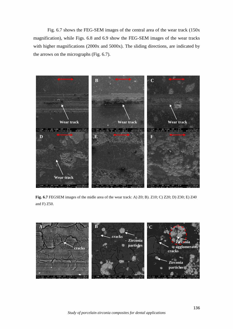
136 Study of porcelain-zirconia composites for dental applications
Fig. 6.7 shows the FEG-SEM images of the central area of the wear track (150x
magnification), while Figs. 6.8 and 6.9 show the FEG-SEM images of the wear tracks
with higher magnifications (2000x and 5000x). The sliding directions, are indicated by
the arrows on the micrographs (Fig. 6.7).
Fig. 6.7 FEGSEM images of the midle area of the wear track: A) Z0; B). Z10; C) Z20; D) Z30; E) Z40
and F) Z50.
Zirconia
particles
Zirconia
particles
Zirconia
agglomerates cracks
cracks
cracks
B C
D E F
Wear track Wear track Wear track
Wear track
A B C
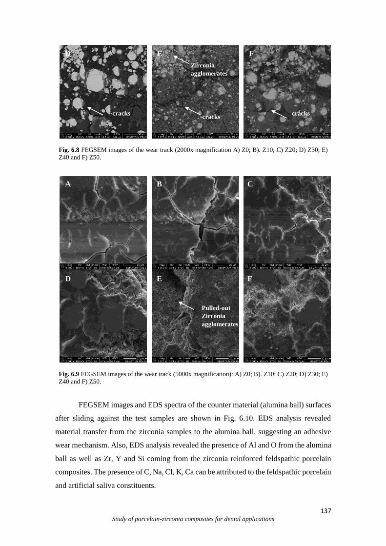
137 Study of porcelain-zirconia composites for dental applications
Fig. 6.8 FEGSEM images of the wear track (2000x magnification A) Z0; B). Z10; C) Z20; D) Z30; E)
Z40 and F) Z50.
Fig. 6.9 FEGSEM images of the wear track (5000x magnification): A) Z0; B). Z10; C) Z20; D) Z30; E)
Z40 and F) Z50.
FEGSEM images and EDS spectra of the counter material (alumina ball) surfaces
after sliding against the test samples are shown in Fig. 6.10. EDS analysis revealed
material transfer from the zirconia samples to the alumina ball, suggesting an adhesive
wear mechanism. Also, EDS analysis revealed the presence of Al and O from the alumina
ball as well as Zr, Y and Si coming from the zirconia reinforced feldspathic porcelain
composites. The presence of C, Na, Cl, K, Ca can be attributed to the feldspathic porcelain
and artificial saliva constituents.
Pulled-out
Zirconia
agglomerates
cracks
Zirconia
agglomerates
D E F
A B C
D E F
cracks cracks
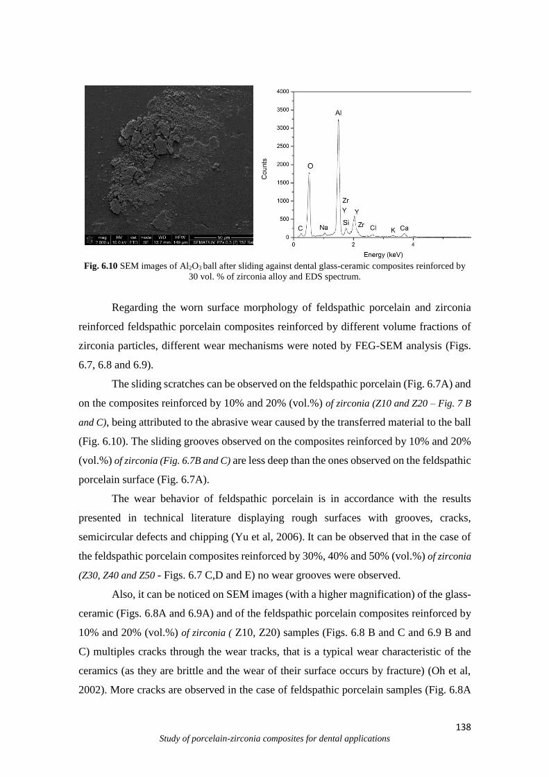
138 Study of porcelain-zirconia composites for dental applications
Fig. 6.10 SEM images of Al2O3 ball after sliding against dental glass-ceramic composites reinforced by
30 vol. % of zirconia alloy and EDS spectrum.
Regarding the worn surface morphology of feldspathic porcelain and zirconia
reinforced feldspathic porcelain composites reinforced by different volume fractions of
zirconia particles, different wear mechanisms were noted by FEG-SEM analysis (Figs.
6.7, 6.8 and 6.9).
The sliding scratches can be observed on the feldspathic porcelain (Fig. 6.7A) and
on the composites reinforced by 10% and 20% (vol.%) of zirconia (Z10 and Z20 – Fig. 7 B
and C), being attributed to the abrasive wear caused by the transferred material to the ball
(Fig. 6.10). The sliding grooves observed on the composites reinforced by 10% and 20%
(vol.%) of zirconia (Fig. 6.7B and C) are less deep than the ones observed on the feldspathic
porcelain surface (Fig. 6.7A).
The wear behavior of feldspathic porcelain is in accordance with the results
presented in technical literature displaying rough surfaces with grooves, cracks,
semicircular defects and chipping (Yu et al, 2006). It can be observed that in the case of
the feldspathic porcelain composites reinforced by 30%, 40% and 50% (vol.%) of zirconia
(Z30, Z40 and Z50 - Figs. 6.7 C,D and E) no wear grooves were observed.
Also, it can be noticed on SEM images (with a higher magnification) of the glass-
ceramic (Figs. 6.8A and 6.9A) and of the feldspathic porcelain composites reinforced by
10% and 20% (vol.%) of zirconia ( Z10, Z20) samples (Figs. 6.8 B and C and 6.9 B and
C) multiples cracks through the wear tracks, that is a typical wear characteristic of the
ceramics (as they are brittle and the wear of their surface occurs by fracture) (Oh et al,
2002). More cracks are observed in the case of feldspathic porcelain samples (Fig. 6.8A
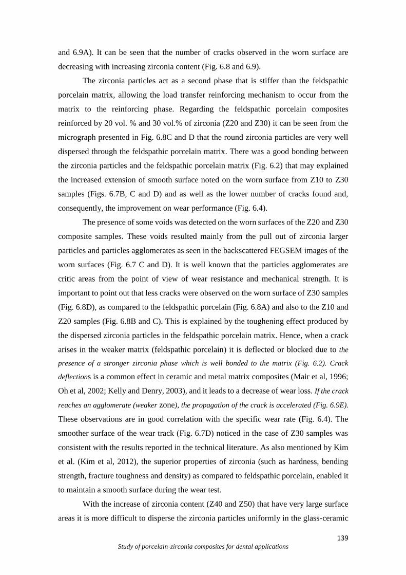
139 Study of porcelain-zirconia composites for dental applications
and 6.9A). It can be seen that the number of cracks observed in the worn surface are
decreasing with increasing zirconia content (Fig. 6.8 and 6.9).
The zirconia particles act as a second phase that is stiffer than the feldspathic
porcelain matrix, allowing the load transfer reinforcing mechanism to occur from the
matrix to the reinforcing phase. Regarding the feldspathic porcelain composites
reinforced by 20 vol. % and 30 vol.% of zirconia (Z20 and Z30) it can be seen from the
micrograph presented in Fig. 6.8C and D that the round zirconia particles are very well
dispersed through the feldspathic porcelain matrix. There was a good bonding between
the zirconia particles and the feldspathic porcelain matrix (Fig. 6.2) that may explained
the increased extension of smooth surface noted on the worn surface from Z10 to Z30
samples (Figs. 6.7B, C and D) and as well as the lower number of cracks found and,
consequently, the improvement on wear performance (Fig. 6.4).
The presence of some voids was detected on the worn surfaces of the Z20 and Z30
composite samples. These voids resulted mainly from the pull out of zirconia larger
particles and particles agglomerates as seen in the backscattered FEGSEM images of the
worn surfaces (Fig. 6.7 C and D). It is well known that the particles agglomerates are
critic areas from the point of view of wear resistance and mechanical strength. It is
important to point out that less cracks were observed on the worn surface of Z30 samples
(Fig. 6.8D), as compared to the feldspathic porcelain (Fig. 6.8A) and also to the Z10 and
Z20 samples (Fig. 6.8B and C). This is explained by the toughening effect produced by
the dispersed zirconia particles in the feldspathic porcelain matrix. Hence, when a crack
arises in the weaker matrix (feldspathic porcelain) it is deflected or blocked due to the
presence of a stronger zirconia phase which is well bonded to the matrix (Fig. 6.2). Crack
deflections is a common effect in ceramic and metal matrix composites (Mair et al, 1996;
Oh et al, 2002; Kelly and Denry, 2003), and it leads to a decrease of wear loss. If the crack
reaches an agglomerate (weaker zone), the propagation of the crack is accelerated (Fig. 6.9E).
These observations are in good correlation with the specific wear rate (Fig. 6.4). The
smoother surface of the wear track (Fig. 6.7D) noticed in the case of Z30 samples was
consistent with the results reported in the technical literature. As also mentioned by Kim
et al. (Kim et al, 2012), the superior properties of zirconia (such as hardness, bending
strength, fracture toughness and density) as compared to feldspathic porcelain, enabled it
to maintain a smooth surface during the wear test.
With the increase of zirconia content (Z40 and Z50) that have very large surface
areas it is more difficult to disperse the zirconia particles uniformly in the glass-ceramic
140 Study of porcelain-zirconia composites for dental applications
matrix. It can be observed larger wear tracks area in the case of Z40 and Z50 composites
(Fig. 6.7 E and F). It clearly showed that the surface damage was accelerated by
reinforcing the feldspathic porcelain composite by a higher amount of zirconia. The SEM
images of the Z40 and Z50 composites (Fig. 6.8 E and F and Fig. 6.9 E and F) present a
lot of peaks and valleys (delamination). There are few regions with a smoother
appearance aspect.
These smoother plateaus may correspond to some of the regions where the
zirconia reinforcement bonded to the feldspathic porcelain matrix. The extensive rougher
regions correspond to the regions where there is no bonding between the zirconia and
feldspathic porcelain matrix. These rougher regions are areas of weakness from which
failure was initiated to cause extensive wear of the composites reinforced by zirconia
agglomerates. It can be observed from SEM images with a higher magnification (Fig. 6.8
E and F) that the grains/particles are fragmented. It is possible that fragments from the
zirconia reinforced feldspathic porcelain composite contributed to the formation of the
third body abrasive particles and consequently to an increase on volume loss.
These observations are in good correlation with the specific wear rate (Fig. 6.4).
It is well known that the degree of dispersion of reinforcement in a composite has
an important role in wear resistance. Also, the agglomeration formation and second phase
segregation are important factors in controlling the mechanical, wear and corrosion
behavior (e.g. increasing stress concentrations at the grain boundaries, substantially
accelerating the crack initiation and propagation etc.) of the composites.
It seems that the main strengthening mechanism, as observed in the fracture
morphologic aspects after wear tests, is the strengthening effect of zirconia particles
promoted by a strong bonding between feldspathic porcelain matrix and zirconia particles
and by the load transfer mechanisms occurring from feldspathic porcelain matrix to the
zirconia particles. Also, it should be mentioned that the zirconia agglomerates may have
detrimental effect on the strengthening, and they are responsible for voids nucleation.
4. Conclusions
The main outcome of this work can be drawn as follows:
- The dental zirconia feldspathic porcelain reinforced composites were
successfully synthesized by heat pressing technique;
141 Study of porcelain-zirconia composites for dental applications
- The results demonstrated that the amount of zirconia plays a relevant role on wear
behavior;
- The addition of 10%, 20% and 30% (vol.%) Yttria-stabilized zirconia particles
in dental feldspathic porcelain resulted in a substantial improvement of the wear
resistance, while the 40% and 50% (vol.%) contents had a detrimental effect on the wear
resistance.
- Under saliva artificial at 37°C conditions against Al2O3, the optimal wear
properties were obtained for the feldspathic porcelain reinforced with 30% (vol.%) of
zirconia particles. The improvement on wear resistance was mainly attributed to the good
dispersion of the reinforcement phase, which allowed the load transfer reinforcing
mechanism to effectively occur from the matrix to the reinforcement.
Acknowledgements
The authors acknowledge the Portuguese Foundation for Science and Technology
(FCT - SFRH/BPD/87435/2012; EXCL/EMS-TEC/0460/2012; UID/EEA/04436/2013),
CAPES and CNPq (Brazil) for the financial support.
References
Antunes P.V., Ramalho A., Study of abrasive resistance of composites for dental
restoration by ball-cratering, Wear 255 (2003) 990–998.
Atala M.H., Gul E., How to Strengthen Dental Ceramics, Int J Dent Sci Res, 3 (2015)
2:24-27.
142 Study of porcelain-zirconia composites for dental applications
Ferraris M., Rabajoli P., Brossa F., Paracchini L., Vacuum plasma spray deposition of
titanium particle/glass-ceramic matrix biocomposites. J. Am. Ceram. Soc. 79 (1996)
6:1515–1520.
Figueiredo-Pina C.G., Monteiro A., Guedes M., Mauricio A., Serro A.P., Ramalho A.,
Santos C., Effect of feldspar glass-ceramic coating upon the wear behavior of zirconia
dental crowns, Wear 297 (2013) 872-877.
Fusayama T., Katayori T., Nomoto S., J. Dent. Res. 42 (1963) 1183-1197.
Guess P., Kulis A., Witkowotz S., Zhang Y., Strub J., Shear bond strengths between
different zirconia cores and veneering ceramics and their susceptibility to thermocycling,
Dent Mat 24 (2008) 1556-1567.
Gu Y.W., Yap A.U.J., Cheang P., Khor K.A., Zirconia–glass ionomer cement––a
potential substitute for Miracle Mix, Scrip Mater 52 (2005) 113–116.
Hamouda I. M., Beyari M. M., Addition of Yttria Partially Stabilized Zirconia for
Reinforcement of Dental Glass-ceramic, Int J Sci Res Know (IJSRK), 10 (2013) 1:404-
412.
Henriques B., Soares D., Silva F.S. Microstructure, hardness, corrosion resistance and
glass-ceramic shear bond strength comparison between cast and hot pressed CoCrMo
alloy for metal-ceramic dental restorations. J Mech Behav Biomed (2012) 12:83–92.
Henriques B., Gasik M., Soares D., Silva F.S. Experimental evaluation of the bond
strength between a CoCrMo dental alloy and glass-ceramic through a composite metal-
ceramic graded transition interlayer. J Mech Behav Biomed Mater (2012a) 13:206–14.
Henriques B., Soares D., Silva F.S. Optimization of bond strength between gold alloy and
glass-ceramic through a composite interlayer obtained by powder metallurgy. Mater Sci
Eng A (2011) 528:1415–20.

143 Study of porcelain-zirconia composites for dental applications
Henriques B., Gasik M., Souza J.C.M., Nascimento R.M., Soares D., Silva F.S.
Mechanical and thermal properties of hot pressed CoCrMo-glass-ceramic composites
developed for prosthetic dentistry. J Mech Behav Biomed Mater (2014) 30:103–10.
Henriques B., Soares D., Silva F.S. Shear bond strength of a hot pressed Au-Pd-Pt alloy-
glass-ceramic dental composite. J Mech Behav Biomed Mater (2011) 4:1718–26.
Henriques B., Gonçalves S., Soares D., Silva F.S. Shear bond strength comparison
between conventional glass-ceramic fused to metal and new functionally graded dental
restorations after thermal-mechanical cycling. J Mech Behav Biomed Mater (2012b)
13:194–205.
Huang X., Zheng X., Zhao G., Zhong B., Zhang X., Wen G., Microstructure and
mechanical properties of zirconia-toughened lithium disilicate glass ceramic composites,
Mater Chem Phys 143 (2014) 845-852.
Kasuga T., Nakajima K., Uno T., Yoshida M., Preparation of zirconia-toughened
bioactive glass-ceramic composite by sinter-hot isostatic pressing. J. Am. Ceram. Soc.,
75 (1992) 5.
Kasuga, M. Yoshida, T. Uno, K. Nakajima, Preparation of zirconia-toughened bioactive
glass-ceramics. J. Mater. Sci., 23 (1988) 2255–2258.
Kelly, J. R. & Denry, I. Stabilized zirconia as a structural ceramic: An overview. Dent
Mater, 24 (2008) 3: 289-298.
Kim M-J., Oh S-H., Kim J-H., Ju S-W., Seo D-G., Ahn J-S., Ryu J-J., Jun S-H., Wear
evaluation of the human enamel opposing different Y-TZP dental ceramics and other
glass-ceramics. The J Dent 40 (2012) 979-988.
Li H., Zhou Z.R., Wear behavior of human teeth in dry and artificial saliva conditions,
Wear 249 (2002) 980-984.
144 Study of porcelain-zirconia composites for dental applications
Maller Udita S., Thangaraj D. N., Maller S., From Ceramics to Ceramic Steel: Genesis, J
Orofacial Res, 2 (2012) 3:139-145.
Mair L. H., Stolarskij T. A., Vowles R. W., Lloyd C. H., Wear: mechanisms,
manifestations and measurement. Report of a workshop, J Dent 24 (1996) 1-2:141-148.
Manicone P. F., Iommetti P. R., Raffaelli L., An overview of zirconia ceramics: Basic
properties and clinical applications, Journal of dentistry 35 (2007) 819–826.
Mitov G., Heintze S. D., Walz S., Woll K., Muecklich F, Pospiech P., Wear behavior of
dental Y-TZP ceramic against natural enamel after different finishing procedures, Dent
Mater 28 (2012) 909–918.
O'Brien (2002) Dental glass-ceramic. In 'Dental materials and their selection'. (Ed. WJ
O'Brien) 210-224, Quintessence Publishing Co, Inc.,
Oh W.S., Delong R., Anusavice K.J. Factors affecting enamel and ceramic wear: a
literature review. J Prosthet Dent (2002) 87:451-9.
Preis V., Behr M., Kolbeck C., Hahnel S., Handel G., Rosentritt M ., Wear performance
of substructure ceramics and veneering glass-ceramics, Dental materials 27 (2011) 796–
804.
Ramalho A., Antunes P. V., Reciprocating wear test of dental composites: effect on the
antagonist, Wear 259 (2005) 1005–1011.
Sampaio M., Buciumeanu M., Henriques B., Silva F.S., Souza J. C. M., Gomes J.R.,
Tribocorrosion behavior of veneering biomedical PEEK to Ti6Al4V structures, J Mechan
BehavBiomed Mater 54 (2015) 123-130.
Shenoy A., Shenoy N. Dental ceramics: An update, J Conserv Dent. 13 (2010) 4:195–
203.
145 Study of porcelain-zirconia composites for dental applications
SerkanSaridag, OnjenTak, GamzeAlniacik, Basic properties and types of zirconia: An
overview, World J Stomatol 20 (2013) 2(3): 40-47.
Stachowiak G.B., Stachowiak G.W., Evans P., Wear and friction characteristics of ion-
implanted zirconia ceramics, Wear 241 (2000) 220–227.
Schlueter N., Jaeggi T., Lussi A., Is Dental erosion really a problem? Adv Dent Res 24
(2012) 2.
Strang R., Whitters C.J., Brown D., Clarke R.L., Curtis R.V., Hattons P.V., Irelandn A.J.,
Lloyd C.H., McCabe J.F., Nicholson J.W., Scrimgeourll S.N., Setcos J.C., M. Sherriff,
van Noort R., Watts D C, WoodsilI D, Dental materials: 1996 literature review. Part 2, J
Dent 26 (1998) 4:273-291.
Song X-F, Yin L., Han Y-G, Wang H, In vitro rapid intraoral adjustment of glass-ceramic
prostheses using a high-speed dental handpiece, Acta Biomater 4 (2008) 414–424.
Souza J. C. M., Henriques M., Teughels W., Ponthiaux P., Celis J.-P., Rocha L. A. Wear
and Corrosion Interactions on Titanium in Oral Environment: Literature Review. J. Bio-
Tribo-Corrosion, 1 (2015) 2:13.
Souza J.C., Ponthiaux P., Henriques M., Oliveira R., Teughels W., Celis J.P., Rocha L.A.
Corrosion behavior of titanium in the presence of Streptococcus mutans. Journal of
Dentistry (2013) 41: 528‒534.
Usha G., Prashanth T.R., Roopa R. N., Yashwanth G. Advanced ceramics. A review of
materials science, J Dent Sci Res (2014) 5:5-11.
Verné E., Ferraris M., Ventrella,A. Paracchini L., Krajewski A., Ravaglioli, A. Sintering
and plasma-spray deposition of bioactive glass-matrix composites for biomedical
applications. J. Eur. Ceram. Soc. 18 (1998) 4:363–372.
Verné E., Ferraris M., Jana C., Pressureless sintering of Bioverit1III/Ti particle
biocomposites. J. Eur. Ceram. Soc., 19 (1999) 2039–2047.
146 Study of porcelain-zirconia composites for dental applications
Verné E., Ferraris M., Jana C., Paracchini L., Bioverit1I base glass/Ti particulate
biocomposite: ‘‘in situ’’ vacuum plasma spray deposition. J. Eur. Ceram. Soc., 20 (2000)
473–479.
Wang W., Liao S., Zhu Y., Liu M., Zhao Q., Fu Y., Recent Applications of Nanomaterials
in Prosthodontics, J Nanomater Vol (2015) 11.
Wang, Y. Liu, W. Si, H. Feng, Y. Tao, Z. Ma, Friction and wear behaviors of dental
ceramics against natural tooth enamel, J Eur Ceram Soc 32 (2012) 2599-2606.
Yin L., Jahanmir S., Ives L.K., Abrasive machining of glass-ceramic and zirconia with a
dental handpiece, Wear 255 (2003) 975–989;
Yu H.Y., Cai Z.B., Ren P.D., Zhu M.H., Zhou Z.R., Friction and wear behavior of dental
feldspathic glass-ceramic, Wear 261 (2006) 611–621.
Zheng J., Zhou Z.R., Friction and wear behavior of human teeth under various wear
conditions, Tribol Int 40 (2007) 278–284.
Zhou Z.R., Zheng J., Tribology of dental materials: a review, Topical Review, Journal of
Physics D: Applied Physics 41 (2008) 1-22.
147 Study of porcelain-zirconia composites for dental applications
CHAPTER VII
Shear bond strength of veneering porcelain to zirconia: effect of surface treatment
by CNC-milling and composite layer deposition on zirconia
Santos R. L.P.1; Silva F.S.2; Nascimento R. M.1, Souza J.C.M.2,3, Motta F.V.1, Carvalho O.2,
Henriques B.2,4*
1Department of Materials Engineering, Federal University of Rio Grande do Norte (UFRN), Campus Lagoa
Nova, 59078-970, Natal, Brazil.
2Center for Microelectromechanical Systems (CMEMS-UMINHO), University of Minho, Campus Azurém, Guimarães 4800-
058, Portugal.
3Center for Research on Dental Implants (CEPID), Post-Graduation Program in Dentistry (PPGO), Federal
University of Santa Catarina, Campus Trindade, 88040-900, Florianópolis/SC, Brazil
4Laboratory of Ceramic and Composite Materials (CERMAT), Federal University of Santa Catarina
(UFSC), Campus Trindade, Florianópolis/SC, Brazil
*Corresponding author at: Center for Mechanical and Materials Technologies, University of Minho,
Campus de Azurém 4800‐058 Guimarães, Portugal. Tel.: +351253510220; fax: +351253516007.
E‐mail address: [email protected] (B. Henriques)
148 Study of porcelain-zirconia composites for dental applications
ABSTRACT
Objectives: The purpose of this study was to evaluate the shear bond strength of veneering porcelain to
zirconia substrates modified by CNC-milling process or by coating zirconia with a composite interlayer.
Methods: Four types of zirconia-porcelain interface configurations were tested: RZ- porcelain bonded to
rough zirconia substrate (n = 16); PZ - porcelain bonded to zirconia substrate with surface holes (n=16);
RZI - application of a composite interlayer between the veneering porcelain and the rough zirconia substrate
(n = 16); PZI - application of a composite interlayer between the porcelain and the zirconia substrate treated
by CNC-milling (n=16). Surface holes on zirconia substrates were produced by CNC-milling machine. The
composite interlayer was composed of zirconia-particles reinforced porcelain (30%, vol.%). The
mechanical properties of the ceramic composite have been determined. The shear bond strength test was
performed at 0.5 mm/min using a universal testing machine. The interfaces of fractured and untested
specimens were examined by field emission guns scanning electron microscopy coupled to energy
dispersive spectroscopy (FEG-SEM/EDS). Data was analyzed with Shapiro-Wilk test to test the assumption
of normality. The one-way ANOVA followed by Tukey HSD multiple comparison test was used to compare
shear bond strength results (α=0.05).
Results: The shear bond strength of PZ (100 ± 15 MPa) and RZI (96 ± 11 MPa) specimens were higher
than that recorded for RZ (control group) specimens (89 ± 15 MPa), although not significantly (p>0.05).
The highest shear bond strength values were recorded for PZI specimens (138 ± 19 MPa), yielding a
significant improvement of 55% relative to RZ specimens (p<0.05).
Significance: This study shows that it is possible to highly enhance the zirconia-porcelain bond strength –
even by ~55% - by combining surface holes in zirconia frameworks and the application of a proper ceramic
composite interlayer.
Keywords: Zirconia; Porcelain; Shear bond strength, Composite interlayer, CNC-created surface holes
149 Study of porcelain-zirconia composites for dental applications
1. Introduction
Zirconia can be found within three crystalline forms: monolithic (m-t; 1173°C);
tetragonal (t-c; 2370°C) and cubic (c-melting temperature; 2690°C). These
transformations are reversible upon cooling with the tetragonal to monoclinic (t-m)
transformation imparting compressive stresses to the material due to the occurrence of 4-
5% volume expansion. Calcia (CaO), magnesia (MgO), ceria (CeO2), alumina (Al2O3) or
yttria (Y2O3) are used as stabilizers of the tetragonal phase. The addition of 2-3 mol%
Y2O3 as stabilizing agent results in a fully tetragonal fine-grained zirconia ceramic
material with 100% small metastable tetragonal grains (Y-TZP). Yttria stabilized
tetragonal zirconia policristals (Y-TZP) has emerged as the most important ceramic for
the fabrication of dental all-ceramic prostheses due to its high biocompatibility, chemical
stability, aesthetics, outstanding flexural strength (up to 900-1200 MPa) and high fracture
toughness (5.9-10 MPa m1/2) (Sevilla et al 2010; Kosmac et al 1999; Piconi and
Maccauro, 1999; Denry and Kelly, 2008; Chevalier 2006).
Y-TZP is mainly used in the manufacturing of all-ceramic restorations as a
substructure material which is veneered by feldspar-based porcelains to optimize the
optical properties mimicking teeth structures. Despite the significant improvement on
aesthetic outcome over the metal-ceramic restorations, the failure rate of dental all-
ceramic restorations is high. Several studies have evaluated the clinical performance of
zirconia-based restorations and reported high stability of zirconia frameworks (exceeding
97.8% over 5-years period). However, chipping and fracture of the veneering porcelain
has been found, ranging from 6% up to 25% of the clinical cases over the same period of
observation (Sailer et al, 2007; Tinschert et al, 2008). These failure percentage contrast
with lower fracture prevalence on metal-ceramic restorations, which are reported to range
from 8% up to 10% over 10 years (Creugers et al 1994, Scurria et al, 1998).
Hence, the most common form of all ceramic restorations failure is the
delamination of the veneering porcelain. Such occurrence is considered to be a
multifactorial phenomenon caused by the lack of proper framework support,
microstructure defects, mismatch in thermal properties, occlusal overloads and poor
mechanical interlocking of the porcelain (Dündar et al 2007; Kelly et al 1996).
This way, in order to reduce clinical failures of all ceramic restorations,
considerable effort has been made in order to improve the bond strength between zirconia
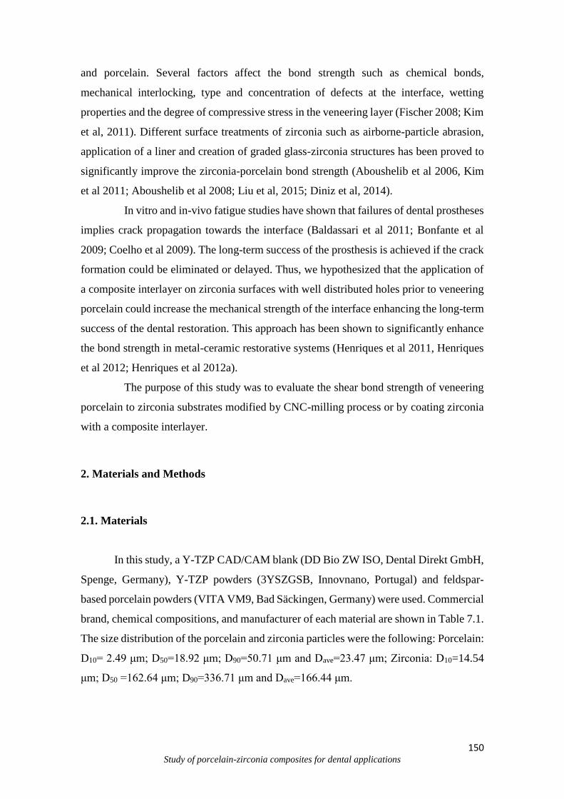
150 Study of porcelain-zirconia composites for dental applications
and porcelain. Several factors affect the bond strength such as chemical bonds,
mechanical interlocking, type and concentration of defects at the interface, wetting
properties and the degree of compressive stress in the veneering layer (Fischer 2008; Kim
et al, 2011). Different surface treatments of zirconia such as airborne-particle abrasion,
application of a liner and creation of graded glass-zirconia structures has been proved to
significantly improve the zirconia-porcelain bond strength (Aboushelib et al 2006, Kim
et al 2011; Aboushelib et al 2008; Liu et al, 2015; Diniz et al, 2014).
In vitro and in-vivo fatigue studies have shown that failures of dental prostheses
implies crack propagation towards the interface (Baldassari et al 2011; Bonfante et al
2009; Coelho et al 2009). The long-term success of the prosthesis is achieved if the crack
formation could be eliminated or delayed. Thus, we hypothesized that the application of
a composite interlayer on zirconia surfaces with well distributed holes prior to veneering
porcelain could increase the mechanical strength of the interface enhancing the long-term
success of the dental restoration. This approach has been shown to significantly enhance
the bond strength in metal-ceramic restorative systems (Henriques et al 2011, Henriques
et al 2012; Henriques et al 2012a).
The purpose of this study was to evaluate the shear bond strength of veneering
porcelain to zirconia substrates modified by CNC-milling process or by coating zirconia
with a composite interlayer.
2. Materials and Methods
2.1. Materials
In this study, a Y-TZP CAD/CAM blank (DD Bio ZW ISO, Dental Direkt GmbH,
Spenge, Germany), Y-TZP powders (3YSZGSB, Innovnano, Portugal) and feldspar-
based porcelain powders (VITA VM9, Bad Säckingen, Germany) were used. Commercial
brand, chemical compositions, and manufacturer of each material are shown in Table 7.1.
The size distribution of the porcelain and zirconia particles were the following: Porcelain:
D10= 2.49 μm; D50=18.92 μm; D90=50.71 μm and Dave=23.47 μm; Zirconia: D10=14.54
μm; D50 =162.64 μm; D90=336.71 μm and Dave=166.44 μm.
151 Study of porcelain-zirconia composites for dental applications
Table 7.1. Chemical composition and manufacturer of the materials used in this study.
Manufacturer/Brand Chemical composition
(Wt.%)
Material Type Manufacturer
VITA VM9 Base
Dentim –V9 Shade
3M2 (Lot n. 44670)
SiO2 (60-64%), Al2O3 (13-
15%), K2O (7-10%), Na2O
(4-6%), TiO2 (<0,5%), CeO2
(<0.5%), ZrO2 (0-1%), CaO
(1-2%), B2O3 (3-5%), BaO
(1-3%), SnO2 (<0,5%), Mg,
Fe and P oxides (<0,1%).
Feldspar-based porcelain
Vita Zahnfabrik, Bad
Säckingen, Germany
3YSZGSB – Zirconia
(3%mol Yttria)
(Lot n. 1301PA046)
ZrO2 + HfO2 + Y2O3 + Al2O3
(>99,9%)
Y2O3 (5,2±0,2)
Al2O3 (<0,4%)
Yttria-stabilized tetragonal
zirconia policrystals (Y-TZP)
Innovnano, Portugal
DD Bio ZW ISO
CAD/CAM blank
ZrO2+HfO2+Y2O3 >99%
Al2O3 <0,5%
Other oxides <0,25%
Yttria-stabilized tetragonal
zirconia policrystals
Dental Direkt GmbH,
Spenge, Germany
2.2. Specimens preparation
72 cylindrical (7.5mm in diameter and 5 mm in height) zirconia substrates were machined
using a CNC milling machine (DWX 50, Roland, Japan) with spindle speed of 29000
rpm. The substructures were divided in two groups according to the type of surface
treatment performed: A) CNC-miling treatment and B) grit-blasting. Homogeneous
surface holes (group A) with 300 μm diameter and 400 μm depth were carried out using
a Ø300 μm milling tool in a CNC milling machine according to a preset CAD pattern, as
shown in Figure 7.1. The holes density was 6.6 holes per mm2. After this, all substrates
were sintered in a furnace (ZircohnZhan, Germany) at 1500°C for 2 h. On the other hand,
the production of rough substrates (group B) was performed by grit-blasting the surface
of the sintered zirconia substrates with airbone-particles of alumina (Al2O3) with 110 m
diameter under 6 bar for 15 s and at a distance of 20-30 mm from the surface. The
152 Study of porcelain-zirconia composites for dental applications
sintering process produced a contraction (by 25%) of the substrates and the final
dimensions of the cylinders were 6 mm in diameter and 4 mm in height. The surface
holes of the substrates had final dimensions of 240 μm in diameter and 300 μm in depth.
The specimens were rinsed in propyl alcohol for 5 min using an ultrasonic bath and dried
at room temperature.
A B
Fig 7.1 Schematic of the cilindrical zirconia substrates and ilustration of the two types of surface
treatments performed: A) CNC-created surface holes; B) rough surfaces by grit-blasting.
Four types of veneering porcelain to zirconia interface configurations were tested
in this study: RZ- porcelain bonded to rough zirconia substrate (n = 16); PZ - porcelain
bonded to zirconia substrate with surface holes (n = 16); RZI - application of a composite
interlayer between the porcelain and the rough zirconia substrate (n = 16); PZI -
application of a composite interlayer between the porcelain and the zirconia substrate
with surface holes (n = 16).
The composite used as interlayer was a mixture of porcelain and 30% (vol.%) of zirconia
particles. The amount of porcelain powder in the mixture was selected based on other
studies conducted by the authors that showed optimal mechanical properties of the
composite with this composition. The zirconia powder was previously sintered at 1500°C
for 2 h and deagglomerated before mixing to the porcelain powder. The powder mixture
(feldspar-based porcelain and zirconia) was ball milled to reduce agglomeration and to
enhance mixing. Porcelain was bonded to zirconia substrates by hot pressing in a graphite
153 Study of porcelain-zirconia composites for dental applications
die under vacuum (1.5 Bar) at 970°C (manufacturer recommendation for porcelain
sintering temperature) and at a pressure of ~7 MPa (Fig. 7.1). That procedure was in
accordance with that described by Henriques et al. (Henriques et al, 2012). The cavity of
the graphite die was coated with a zirconia paint to prevent carbon contaminations. The
heat rate was 65°C/min up to 970oC and then, after 2 min, the power of the induction
heating furnace was shut down. The die cooled naturally at room temperature.
2.3. Mechanical tests
The shear bond strength tests were carried out at room temperature and performed in a
universal testing machine (Instron 8874, MA, USA), with a load cell of 25 kN capacity
and under a crosshead speed of 0.5mm/s. Tests were performed in a custom-made
stainless steel apparatus similar to that described by Henriques et al. (Henriques et al,
2012). The apparatus consisted in two sliding parts, each one with a hole perfectly aligned
to the other. After aligning the holes, the specimens were inserted and loaded at the
interface until fracture. The shear bond strength (MPa) was calculated by the dividing the
highest recorded load (N) by the cross sectional area of resistant porcelain (mm2).
The hardness mean values were calculated as an average of 10 indentations on the
polished surface of each material (n =5). The indentations were made with a Vickers
hardness indenter (EmcoTest-DuraScan, EMPRESA, PAIS) using a standard 136°
pyramidal diamond (HV1), oriented perpendicular to the material surface.
The fracture toughness (KIC) of the specimens was determined by direct measurement of
radial cracks generated by indentation on a load of 9.8 N. Measurements were performed
after 24 h with minimal error of crack lengths due to continuing crack propagation in the
presence of residual indentation stress (Blendell, 1979).
2.4. Analysis of the zirconia-porcelain interfaces and failure mode
The zirconia-porcelain specimens from each group were randomly selected for
microscopic analysis by field emission guns electron microscopy (FEG-SEM; NOVA 200
Nano SEM, FEI Company). For interface analysis, the specimens were embedded in self-
curing resin, wet-ground using grit SiC sand-papers down to 4000 Mesh and polished
with diamond paste (1 m). After shear bond tests, the failure modes of the test specimens
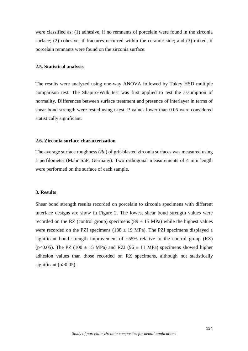
154 Study of porcelain-zirconia composites for dental applications
were classified as: (1) adhesive, if no remnants of porcelain were found in the zirconia
surface; (2) cohesive, if fractures occurred within the ceramic side; and (3) mixed, if
porcelain remnants were found on the zirconia surface.
2.5. Statistical analysis
The results were analyzed using one-way ANOVA followed by Tukey HSD multiple
comparison test. The Shapiro-Wilk test was first applied to test the assumption of
normality. Differences between surface treatment and presence of interlayer in terms of
shear bond strength were tested using t-test. P values lower than 0.05 were considered
statistically significant.
2.6. Zirconia surface characterization
The average surface roughness (Ra) of grit-blasted zirconia surfaces was measured using
a perfilometer (Mahr S5P, Germany). Two orthogonal measurements of 4 mm length
were performed on the surface of each sample.
3. Results
Shear bond strength results recorded on porcelain to zirconia specimens with different
interface designs are show in Figure 2. The lowest shear bond strength values were
recorded on the RZ (control group) specimens (89 ± 15 MPa) while the highest values
were recorded on the PZI specimens (138 ± 19 MPa). The PZI specimens displayed a
significant bond strength improvement of ~55% relative to the control group (RZ)
(p<0.05). The PZ (100 ± 15 MPa) and RZI (96 ± 11 MPa) specimens showed higher
adhesion values than those recorded on RZ specimens, although not statistically
significant (p>0.05).
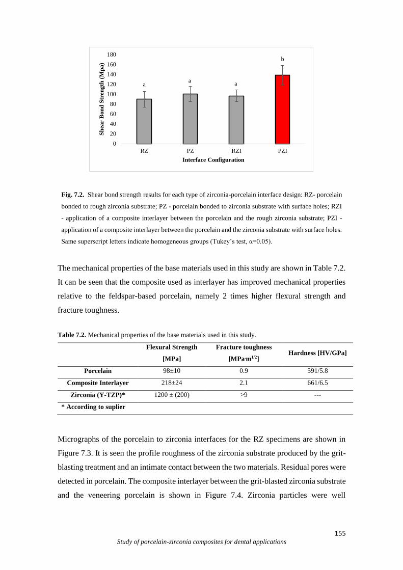
155 Study of porcelain-zirconia composites for dental applications
Fig. 7.2. Shear bond strength results for each type of zirconia-porcelain interface design: RZ- porcelain
bonded to rough zirconia substrate; PZ - porcelain bonded to zirconia substrate with surface holes; RZI
- application of a composite interlayer between the porcelain and the rough zirconia substrate; PZI -
application of a composite interlayer between the porcelain and the zirconia substrate with surface holes.
Same superscript letters indicate homogeneous groups (Tukey’s test, α=0.05).
The mechanical properties of the base materials used in this study are shown in Table 7.2.
It can be seen that the composite used as interlayer has improved mechanical properties
relative to the feldspar-based porcelain, namely 2 times higher flexural strength and
fracture toughness.
Table 7.2. Mechanical properties of the base materials used in this study.
Flexural Strength
[MPa]
Fracture toughness
[MPa.m1/2] Hardness [HV/GPa]
Porcelain 98±10 0.9 591/5.8
Composite Interlayer 218±24 2.1 661/6.5
Zirconia (Y-TZP)* 1200 ± (200) >9 ---
* According to suplier
Micrographs of the porcelain to zirconia interfaces for the RZ specimens are shown in
Figure 7.3. It is seen the profile roughness of the zirconia substrate produced by the grit-
blasting treatment and an intimate contact between the two materials. Residual pores were
detected in porcelain. The composite interlayer between the grit-blasted zirconia substrate
and the veneering porcelain is shown in Figure 7.4. Zirconia particles were well
aa
a
b
0
20
40
60
80
100
120
140
160
180
RZ PZ RZI PZI
Sh
ear
Bon
d S
tren
gth
(M
pa)
Interface Configuration
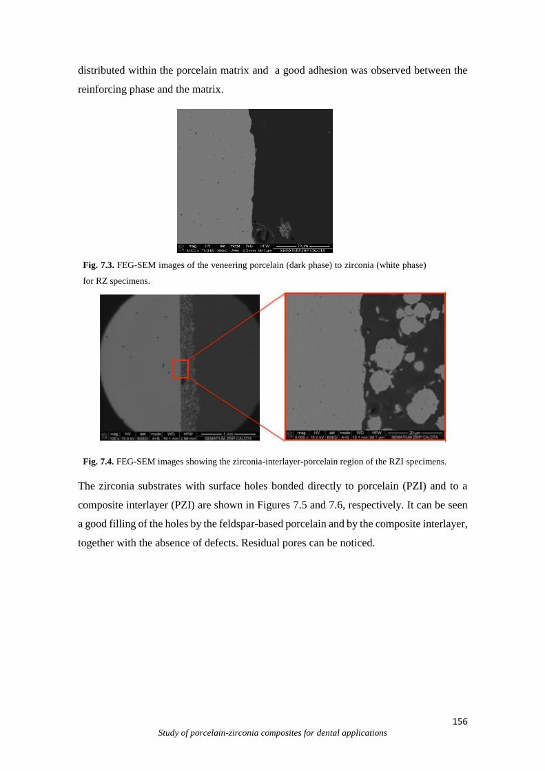
156 Study of porcelain-zirconia composites for dental applications
distributed within the porcelain matrix and a good adhesion was observed between the
reinforcing phase and the matrix.
Fig. 7.3. FEG-SEM images of the veneering porcelain (dark phase) to zirconia (white phase)
for RZ specimens.
Fig. 7.4. FEG-SEM images showing the zirconia-interlayer-porcelain region of the RZI specimens.
The zirconia substrates with surface holes bonded directly to porcelain (PZI) and to a
composite interlayer (PZI) are shown in Figures 7.5 and 7.6, respectively. It can be seen
a good filling of the holes by the feldspar-based porcelain and by the composite interlayer,
together with the absence of defects. Residual pores can be noticed.
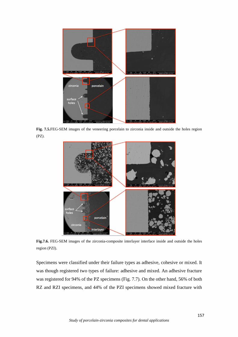
157 Study of porcelain-zirconia composites for dental applications
Fig. 7.5.FEG-SEM images of the veneering porcelain to zirconia inside and outside the holes region
(PZ).
Fig.7.6. FEG-SEM images of the zirconia-composite interlayer interface inside and outside the holes
region (PZI).
Specimens were classified under their failure types as adhesive, cohesive or mixed. It
was though registered two types of failure: adhesive and mixed. An adhesive fracture
was registered for 94% of the PZ specimens (Fig. 7.7). On the other hand, 56% of both
RZ and RZI specimens, and 44% of the PZI specimens showed mixed fracture with
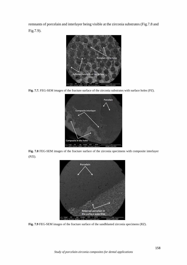
158 Study of porcelain-zirconia composites for dental applications
remnants of porcelain and interlayer being visible at the zirconia substrates (Fig.7.8 and
Fig.7.9).
Fig. 7.7. FEG-SEM images of the fracture surface of the zirconia substrates with surface holes (PZ).
Fig. 7.8 FEG-SEM images of the fracture surface of the zirconia specimens with composite interlayer
(PZI).
Fig. 7.9 FEG-SEM images of the fracture surface of the sandblasted zirconia specimens (RZ).
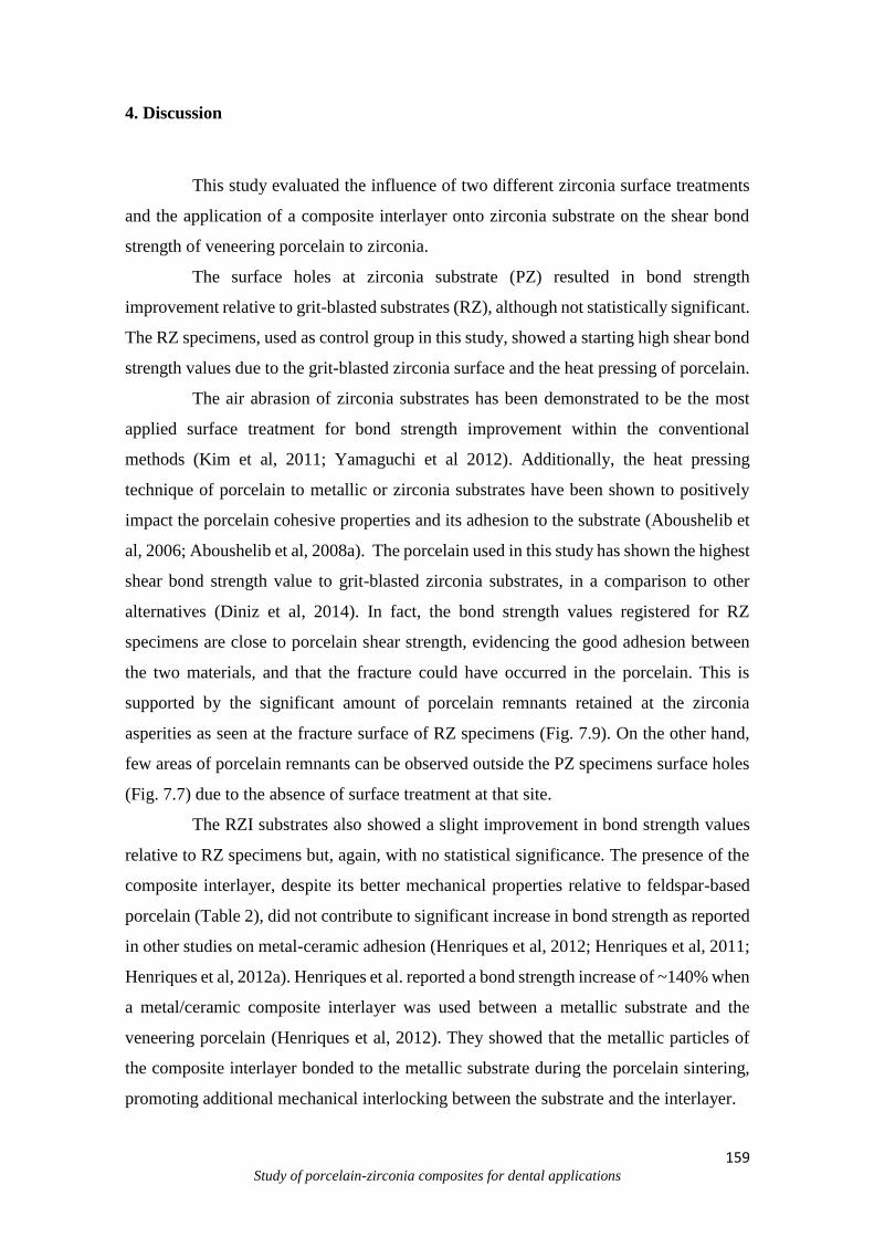
159 Study of porcelain-zirconia composites for dental applications
4. Discussion
This study evaluated the influence of two different zirconia surface treatments
and the application of a composite interlayer onto zirconia substrate on the shear bond
strength of veneering porcelain to zirconia.
The surface holes at zirconia substrate (PZ) resulted in bond strength
improvement relative to grit-blasted substrates (RZ), although not statistically significant.
The RZ specimens, used as control group in this study, showed a starting high shear bond
strength values due to the grit-blasted zirconia surface and the heat pressing of porcelain.
The air abrasion of zirconia substrates has been demonstrated to be the most
applied surface treatment for bond strength improvement within the conventional
methods (Kim et al, 2011; Yamaguchi et al 2012). Additionally, the heat pressing
technique of porcelain to metallic or zirconia substrates have been shown to positively
impact the porcelain cohesive properties and its adhesion to the substrate (Aboushelib et
al, 2006; Aboushelib et al, 2008a). The porcelain used in this study has shown the highest
shear bond strength value to grit-blasted zirconia substrates, in a comparison to other
alternatives (Diniz et al, 2014). In fact, the bond strength values registered for RZ
specimens are close to porcelain shear strength, evidencing the good adhesion between
the two materials, and that the fracture could have occurred in the porcelain. This is
supported by the significant amount of porcelain remnants retained at the zirconia
asperities as seen at the fracture surface of RZ specimens (Fig. 7.9). On the other hand,
few areas of porcelain remnants can be observed outside the PZ specimens surface holes
(Fig. 7.7) due to the absence of surface treatment at that site.
The RZI substrates also showed a slight improvement in bond strength values
relative to RZ specimens but, again, with no statistical significance. The presence of the
composite interlayer, despite its better mechanical properties relative to feldspar-based
porcelain (Table 2), did not contribute to significant increase in bond strength as reported
in other studies on metal-ceramic adhesion (Henriques et al, 2012; Henriques et al, 2011;
Henriques et al, 2012a). Henriques et al. reported a bond strength increase of ~140% when
a metal/ceramic composite interlayer was used between a metallic substrate and the
veneering porcelain (Henriques et al, 2012). They showed that the metallic particles of
the composite interlayer bonded to the metallic substrate during the porcelain sintering,
promoting additional mechanical interlocking between the substrate and the interlayer.
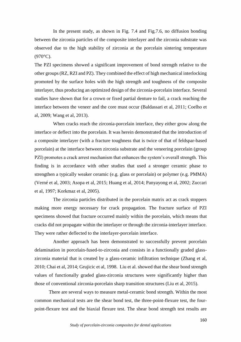
160 Study of porcelain-zirconia composites for dental applications
In the present study, as shown in Fig. 7.4 and Fig.7.6, no diffusion bonding
between the zirconia particles of the composite interlayer and the zirconia substrate was
observed due to the high stability of zirconia at the porcelain sintering temperature
(970°C).
The PZI specimens showed a significant improvement of bond strength relative to the
other groups (RZ, RZI and PZ). They combined the effect of high mechanical interlocking
promoted by the surface holes with the high strength and toughness of the composite
interlayer, thus producing an optimized design of the zirconia-porcelain interface. Several
studies have shown that for a crown or fixed partial denture to fail, a crack reaching the
interface between the veneer and the core must occur (Baldassari et al, 2011; Coelho et
al, 2009; Wang et al, 2013).
When cracks reach the zirconia-porcelain interface, they either grow along the
interface or deflect into the porcelain. It was herein demonstrated that the introduction of
a composite interlayer (with a fracture toughness that is twice of that of feldspar-based
porcelain) at the interface between zirconia substrate and the veneering porcelain (group
PZI) promotes a crack arrest mechanism that enhances the system’s overall strength. This
finding is in accordance with other studies that used a stronger ceramic phase to
strengthen a typically weaker ceramic (e.g. glass or porcelain) or polymer (e.g. PMMA)
(Verné et al, 2003; Asopa et al, 2015; Huang et al, 2014; Panyayong et al, 2002; Zuccari
et al, 1997; Korkmaz et al, 2005).
The zirconia particles distributed in the porcelain matrix act as crack stoppers
making more energy necessary for crack propagation. The fracture surface of PZI
specimens showed that fracture occurred mainly within the porcelain, which means that
cracks did not propagate within the interlayer or through the zirconia-interlayer interface.
They were rather deflected to the interlayer-porcelain interface.
Another approach has been demonstrated to successfully prevent porcelain
delamination in porcelain-fused-to-zirconia and consists in a functionally graded glass-
zirconia material that is created by a glass-ceramic infiltration technique (Zhang et al,
2010; Chai et al, 2014; Grujicic et al, 1998. Liu et al. showed that the shear bond strength
values of functionally graded glass-zirconia structures were significantly higher than
those of conventional zirconia-porcelain sharp transition structures (Liu et al, 2015).
There are several ways to measure metal-ceramic bond strength. Within the most
common mechanical tests are the shear bond test, the three-point-flexure test, the four-
point-flexure test and the biaxial flexure test. The shear bond strength test results are

161 Study of porcelain-zirconia composites for dental applications
characterized for not being influenced by the Young’s Modulus of the alloy as happens
for bending tests (Hammad and Talic, 1996); by its relatively uniform distribution of
interface stresses (Hammad and Stein, 1991) and by its suitability for evaluation of metal-
ceramic and ceramic-ceramic bonds (Hammad and Talic, 1996; Anusavice et al, 1980).
Special attention must be paid to the device configuration because it can largely
influence results. For instance, the shear device composed by a knife whithout a groove,
generates high local stresses in the porcelain caused by small area of contact that can lead
to the porcelain failure. Failure at the interface can be triggered by a cohesive failure in
the porcelain and consequent propagation through the interface. The test device used in
this study consisted on a piston-type shear device with half loop loading which reduces
the stress concentration magnitude adjacent to the interface. This is considered an
improved loading condition compared with concentrated force application at a single
point (knife whithout a groove) or distributed only over a single layer (DeHoff et al,
1995).
Based on these facts, many authors used this test configuration in metal-ceramic
bond strength characterization (Akova et al, 2008; Santos et al, 2006; Vásquez et al, 2009;
Melo et al, 2005). This study presents shear bond strength mean values that are higher
than those commonly reported for zirconia-porcelain systems in literature (Komine et al,
2012). The shear test device used can be in the origin of the values discrepancy.
Ashkanani et al. showed similar bond strength values for zirconia-porcelain specimens
using similar test protocol (Ashkanami et al, 2008). Nevertheless, more important than
the absolute values stays the relative differences between the groups with the different
variables.
5. Conclusions
From this study, the following conclusions can be drawn:
1. The addition of zirconia particles as reinforcing phase to porcelain resulted in
significant improvement of mechanical properties (tensile strength, fracture
toughness and hardness) relative to feldspar-based porcelain;
2. The surface holes (PZ) and the application of composite interlayers (RZI) on
zirconia produced separately an improvement of zirconia-porcelain bond strength

162 Study of porcelain-zirconia composites for dental applications
values relative to conventional zirconia-porcelain specimens (RZ), although not
statistically significant (p>0.05);
3. Holes produced by CNC-milling coupled with the composite interlayer produced a
significant bond strength improvement (by ~55%) relative to conventional zirconia-
porcelain specimens (RZ).
Acknowledgements
The authors acknowledge the Portuguese Foundation for Science and Technology (FCT
- SFRH/BPD/87435/2012; EXCL/EMS-TEC/0460/2012; UID/EEA/04436/2013),
CAPES and CNPq (Brazil) for the financial support.
References
Aboushelib M.N., Kleverlaan C.J., Feilzer A.J. Microtensile bond strength of different
components of core veneered all-ceramic restorations. Part II: Zirconia veneering
ceramics. Dent Mater (2006) 22:857–63.
Aboushelib M.N., Kleverlaan C.J., Feilzer A.J. Effect of zirconia type on its bond strength
with different veneer ceramics. J Prosthodont (2008)17:401–8.
Aboushelib M.N., de Kler M., van der Zel J.M., Feilzer A.J. Effect of veneering method
on the fracture and bond strength of bilayered zirconia restorations. Int J Prosthodont
(2008a) 21:237–40.
Anusavice K.J., Dehoff P.H., Fairhurst C.W. Comparative evaluation of ceramic-metal
bond tests using finite element stress analysis. J Dent Res (1980) 59:608–13.
Akova T., Ucar Y., Tukay A., Balkaya M.C., Brantley W. Comparison of the bond
strength of laser-sintered and cast base metal dental alloys to porcelain. Dent Mater
(2008) 24:1400–4.
163 Study of porcelain-zirconia composites for dental applications
Ashkanani H.M., Raigrodski A.J., Flinn B.D., Heindl H., Mancl L.A. Flexural and shear
strengths of ZrO2 and a high-noble alloy bonded to their corresponding porcelains. J
Prosthet Dent (2008) 100:274–84.
Asopa V., Suresh S., Khandelwal M., Sharma V., Asopa S.S., Kaira L.S. A comparative
evaluation of properties of zirconia reinforced high impact acrylic resin with that of high
impact acrylic resin. Saudi J Dent Res (2015) 6:146–51.
Baldassarri M., Zhang Y., Thompson V.P., Rekow E.D., Stappert C.F.J. Reliability and
failure modes of implant-supported zirconium-oxide fixed dental prostheses related to
veneering techniques. J Dent (2011)39:489–98.
Blendell J.E. The origins of internal stresses in polycrystalline AL2O3 and their effects on
mechanical properties 1979.
Bonfante E.A., da Silva N.R.F.A., Coelho P.G., Bayardo-González D.E., Thompson V.P.,
Bonfante G. Effect of framework design on crown failure. Eur J Oral Sci (2009) 117:194–
9.
Chai H., Lee JJ-W., Mieleszko A.J., Chu S.J., Zhang Y. On the interfacial fracture of
porcelain/zirconia and graded zirconia dental structures. Acta Biomater (2014)10:3756–
61.
Chevalier J. What future for zirconia as a biomaterial? Biomater (2006) 27:535–43.
Creugers N.H., Kayser A.F., Van’t Hof M.A. A meta-analysis of durability data on
conventional fixed bridges. Comm Dent Oral Epidemiol (1994) 22:448–52.
Coelho P.G., Silva N.R., Bonfante E., Guess P.C., Rekow E.D., Thompson VP. Fatigue
testing of two porcelain-zirconia all-ceramic crown systems. Dent Mater (2009) 25:1122–
7.
Coelho P.G., Silva N.R., Bonfante E.A., Guess P.C., Rekow E.D., Thompson V.P.
Fatigue testing of two porcelain-zirconia all-ceramic crown systems. Dent Mater (2009)
25:1122–7.
DeHoff P.H., Anusavice K.J., Wang Z. Three-dimensional finite element analysis of the
shear bond test. Dent Mater (1995) 11:126–31.

164 Study of porcelain-zirconia composites for dental applications
Denry I., Kelly J.R. State of the art of zirconia for dental applications. Dent Mater
(2008)24:299–307.
Diniz A.C., Nascimento R.M., Souza J.C.M., Henriques B.B., Carreiro A.F.P. Fracture
and shear bond strength analyses of different dental veneering ceramics to zirconia. Mater
Sci Eng C Mater Biol Appl (2014)38:79–84.
Dündar M., Ozcan M., Gökçe B., Cömlekoğlu E., Leite F., Valandro L.F. Comparison of
two bond strength testing methodologies for bilayered all-ceramics. Dent Mater (2007)
23:630–6.
Fischer J., Grohmann P., Stawarczyk B. Effect of zirconia surface treatments on the shear
strength of zirconia/veneering ceramic composites. Dent Mater J (2008) 27:448–54.
Grujicic M., Zhao H. Optimization of 316 stainless steel/alumina functionally graded
material for reduction of damage induced by thermal residual stresses. Mater Sci Eng A
(1998) 252:117–32.
Hammad I.A., Talic Y.F. Designs of bond strength tests for metal-ceramic complexes:
review of the literature. J Prosthet Dent (1996) 75:602–8.
Hammad I.A., Stein R.S. A qualitative study for the bond and color of ceramometals. Part
II. J Prosthet Dent (1991)65:169–79.
Henriques B., Gasik M., Soares D., Silva F.S. Experimental evaluation of the bond
strength between a CoCrMo dental alloy and porcelain through a composite metal-
ceramic graded transition interlayer. J Mech Behav Biomed Mater (2012)13:206–14.
Henriques B., Soares D., Silva F.S. Optimization of bond strength between gold alloy
and porcelain through a composite interlayer obtained by powder metallurgy. Mater Sci
Eng A (2011) 528:1415–20.
Henriques B., Gonçalves S., Soares D., Silva F.S. Shear bond strength comparison
between conventional porcelain fused to metal and new functionally graded dental
restorations after thermal-mechanical cycling. J Mech Behav Biomed Mater (2012)
13:194–205.
Huang X., Zheng X., Zhao G., Zhong B., Zhang X., Wen G. Microstructure and
mechanical properties of zirconia-toughened lithium disilicate glass–ceramic composites.
Mater Chem Phys (2014) 143:845–52.

165 Study of porcelain-zirconia composites for dental applications
Kelly J.R., Nishimura I., Campbell S.D. Ceramics in dentistry: historical roots and current
perspectives. J Prosthet Dent (1996) 75:18–32.
Kim H-J., Lim H-P., Park Y-J., Vang M-S. Effect of zirconia surface treatments on the
shear bond strength of veneering ceramic. J Prosthet Dent (2011) 105:315–22.
Komine F., Strub J.R., Matsumura H. Bonding between layering materials and zirconia
frameworks. Jpn Dent Sci Rev (2012) 48:153–61.
Kosmac T., Oblak C., Jevnikar P., Funduk N., Marion L. The effect of surface grinding
and sandblasting on flexural strength and reliability of Y-TZP zirconia ceramic. Dent
Mater (1999)15:426–33.
Korkmaz T., Doğan A., Usanmaz A. Dynamic mechanical analysis of provisional resin
materials reinforced by metal oxides. Biomed Mater Eng (2005) 15:179–88.
Liu R., Sun T., Zhang Y., Zhang Y., Jiang D., Shao L. The effect of graded glass-zirconia
structure on the bond between core and veneer in layered zirconia restorations. J Mech
Behav Biomed Mater (2015) 46:197–204.
Melo R.M., Travassos A.C., Neisser M.P. Shear bond strengths of a ceramic system to
alternative metal alloys. J Prosthet Dent (2005) 93:64–9.
Panyayong W., Oshida Y., Andres C.J., Barco T.M., Brown D.T., Hovijitra S.
Reinforcement of acrylic resins for provisional fixed restorations. Part III: effects of
addition of titania and zirconia mixtures on some mechanical and physical properties.
Biomed Mater Eng (2002) 12:353–66.
Piconi C., Maccauro G. Zirconia as a ceramic biomaterial. Biomaterials (1999) 20:1–25.
Sailer I., Fehér A., Filser F., Gauckler L.J., Lüthy H, Hämmerle C.H.F. Five-year clinical
results of zirconia frameworks for posterior fixed partial dentures. Int J Prosthodont
(2007) 20:383–8.
Santos J.G., Fonseca R.G., Adabo G.L., dos Santos Cruz C.A. Shear bond strength of
metal-ceramic repair systems. J Prosthet Dent (2006) 96:165–73.
Sevilla P., Sandino C., Arciniegas M., Martínez-Gomis J., Peraire M., Gil F.J. Evaluating
mechanical properties and degradation of Y-TZP dental implants. Mater Sci Eng C (2010)
30:14–9.

166 Study of porcelain-zirconia composites for dental applications
Scurria M.S., Bader J.D., Shugars D. Meta-analysis of fixed partial denture survival:
Prostheses and abutments. J Prosthet Dent (1998)79:459–64.
Tinschert J., Schulze K.A., Natt G., Latzke P., Heussen N., Spiekermann H. Clinical
behavior of zirconia-based fixed partial dentures made of DC-Zirkon: 3-year results. Int
J Prosthodont (2008) 21:217–22.
Vásquez V.Z.C., Ozcan M., Kimpara E.T. Evaluation of interface characterization and
adhesion of glass ceramics to commercially pure titanium and gold alloy after thermal-
and mechanical-loading. Dent Mater (2009) 25:221–31.
Verné E., Defilippi R., Carl G., Vitale Brovarone C., Appendino P. Viscous flow sintering
of bioactive glass-ceramic composites toughened by zirconia particles. J Eur Ceram Soc
(2003) 23:675–83.
Wang R., Lu C., Arola D., Zhang D. Plastic Damage Induced Fracture Behaviors of
Dental Ceramic Layer Structures Subjected to Monotonic Load. J Prosthodont (2013)
22:456–64.
Yamaguchi H., INO S., Hamano N., Okada S., Teranaka T. Examination of bond strength
and mechanical properties of Y-TZP zirconia ceramics with different surface
modifications. Dent Mater J (2012) 31:472–80.
Zhang Y., Kim J.W. Graded zirconia glass for resistance to veneer fracture. J Dent Res
(2010) 89:1057–62.
Zhang Y., Sun M-J., Zhang D. Designing functionally graded materials with superior
load-bearing properties. Acta Biomater (2012) 8:1101–8.
Zuccari A.G., Oshida Y., Moore B.K. Reinforcement of acrylic resins for provisional
fixed restorations. Part I: Mechanical properties. Biomed Mater Eng (1997) 7:327–43.
167 Study of porcelain-zirconia composites for dental applications
CHAPTER VIII
General Discussion
In order to improve the mechanical performance of the all-ceramic substructures
for dental restorations, it is critical to understand the procedures used to fabricate these
materials. From there, were evaluated the best conditions for optimization of these
substructures.
This thesis is devoted to the mechanical and microstructural study of the
feldspathic porcelain-zirconia composites for dental applications. In this thesis was
studied the influence of the several variables: Previous characterization and comparative
study between zirconia commercial and zirconia synthesized by CPM; the type of zirconia
particles (particles and agglomerates) in glass-ceramic matrix; the mechanical
performance from the zirconia content (0-50% vol.%) in feldspathic porcelain matrix, the
wear behavior for all composites; and the adhesion/interaction between porcelain-
zirconia substructures with different surface treatment. Next, a general discussion about
each of these topics is presented, focusing on the main highlights of each theme.
8.2 Commercial zirconia vs. synthesized zirconia
In this thesis was firstly studied the characteristics of the commercial zirconia and
synthesized zirconia using different Yttria contents (3, 4 and 5 mol.%) by Complex
Polymerization Method. This study had high importance in this thesis from the correct
choice of the obtaining method for zirconia particles in all-ceramic substructures, in other
words, enabled to explain the “Why?” of this selection. The effect of calcination
temperature (800 to 1000 °C) was assessed to zirconia obtained by CPM and indicates
that the increase in calcination temperature and time, induce variations in intensity and
width of peaks (Fig 2.1). Besides, the increase of the crystallite size (11.5 to 27.9 nm)
may occur due the major atomic diffusion at high calcination temperature, increasing the
structural packing that enhanced consequently the crystallite size (Davar et al, 2013;
Grzebielucka et al, 2010). Comparing the results between zirconia synthesized and

168 Study of porcelain-zirconia composites for dental applications
commercial (27.9 and 37.0 nm) an increase of 32%, this behavior can be explain from the
different manufacturing method employed, CPM and high energy milling, respectively.
Related to yttria content, the increase of dopant content (3,4 and 5 mol. %) into crystal
lattice promote the decrease in crystallite size for the same temperatures and distinct
compositions (Chevalier et al, 2009). In terms of unit cell volume, the increase of dopant
content may decrease the tetragonal phase and tends to change to cubic phase (major
values of unit cell volume) (Silva et al, 2010). In this case, the thesis emphasis will be
placed on the minor stabilizing contribution of the dopant cations, whereas, in tetragonal
phase production. The thermoanalysis presented the behavior of the specimens from the
decomposition progress in several steps and is based on a thermally induced anionic redox
reaction (Laberty-Robert et al, 2001). The materials evaluated are thermally stable at 1100
°C and the weight loss for synthesized specimens with yttria content (3, 4 and 5 mol%)
is major than compared as commercial zirconia, 85, 72, 86 and 2%, respectively (Fig 2.6).
The large weight loss present in specimens synthesized by CPM can be explained from
the volatilization of the organic compounds (CO, CO2, H2, N and N2), during combustion
reaction, decomposition of nitrates and material crystallization, unlike what happens with
commercial zirconia obtained by high-energy milling. The Raman spectroscopy was used
in this thesis to confirm the presence of tetragonal phase of synthesized materials from
the changes in the zirconia bond angles and lengths (Casellas et al, 2001; Ishigame and
Sakurai, 1977; Bouvier et al, 2001; Ramos et al 2013). The different calcination
temperature and dopant content probably caused few spectral variations at the tetragonal
and cubic phases (260, 464 and 642 cm-1) (Casellas et al, 2001). The results indicate that
the CPM is a good technique to produce monophasic zirconia with different Yttria
contents. However, independent of calcination temperature and Yttria content used, the
results not exceeded the commercial zirconia. In this context, the use of the commercial
zirconia is endorsed for dental applications.
8.3 Zirconia particle effect in feldspathic porcelain matrix
One of the aims of this thesis was to evaluate the influence of the type of zirconia
reinforcement. The use of reinforcement is widely used to improve the mechanical
properties of the dental materials, composites especially. Thus, this study was developed
in order to maximize the mechanical performance of these composites. Were used two
169 Study of porcelain-zirconia composites for dental applications
types of particles: (yttria-stabilized zirconia 3Y-TZP agglomerates (ZA) and pre-sintered
yttria-stabilized zirconia (3Y-TZP) particles (ZP) inserted in feldspathic porcelain matrix
to produce the composites by Hot pressing technique. The results indicates that
composites present different microstructures related to the type of the zirconia
reinforcement used. To (ZA), the agglomerates are not visible in feldspathic porcelain
matrix, this behavior can be confirmed due to the weakly bond within the agglomerates,
they are easily destroyed during the mixing procedure. This produced a fragmentation of
the powders in micrometric and nanometric powders that covered the porcelain powders
and negatively impacted the sintering and densification of the composites, reducing
dramatically the strength of the composite (Fig. 3.3). Most of the porcelain powders were
covered by fine zirconia particles that sinters at 1500 °C and not 970 °C as happens with
glass-ceramic. To improve the mechanical strength of the zirconia agglomerates (ZA),
these agglomerates can be ball-milled with steering suspension of the mixture in alcohol
until complete evaporation. The mechanism was explained by the infiltration of the
feldspathic porcelain phase into the zirconia agglomerates, leading to a partial spreading
of each particle into agglomerates of small zirconia grains (about 1 mm size) (Verné et
al, 2003). On the other hand, the improvement in flexural strength of the composite (ZP)
compared as Z0 (monolithic porcelain) is explained by the action of the uniformly
distributed dense zirconia particles (pre-sintered agglomerates). The zirconia particles act
as a second phase that is more rigid than the porcelain matrix, allowing the load transfer
reinforcing mechanism to occur from matrix to the reinforcing phase. It is possible to see
round zirconia particles spread through the porcelain matrix few pores can be detected
within the porcelain matrix of feldspathic porcelain/zirconia-porcelain samples and they
are preferentially located at the vicinities of the ZPs rather than in the feldspathic
porcelain matrix. The results in Fig. 3.4 and 3.5, show clear differences between the
composites in flexural strength and hardness to according as type of reinforcing phase
used. The (ZA) present lower flexural strength and hardness (29.7 MPa and 254.67 Hv),
when compared as Z0 (glass-ceramic) and (ZP), respectively. Composites hot pressed
with pre-sintered zirconia (3Y-TZP) particles (ZP) in feldspathic porcelain matrix showed
significantly higher hardness than reinforced composites with (ZA). These results are
based in a good dispersion and good bonding between the reinforcing zirconia phase and
the feldspathic porcelain matrix (Fig 3.3). Besides the improvement noticed in flexural
strength of the (ZP) it is also expected an improvement in the toughness behavior relative
to pure porcelain (Verné et al, 2003). The toughening mechanism is based on the crack
170 Study of porcelain-zirconia composites for dental applications
deflection around the zirconia particles. In all composites, it is possible to see the presence
of pores remnants the porosity found in cast and pressed substrates were less %. Thus,
these results indicate that the zirconia particles (ZP) present higher mechanical properties
can be widely used as ceramic reinforcement.
8.4 Zirconia content effect in mechanical performance of the composites feldspathic
porcelain-zirconiaIn this study was investigated the mechanical properties of the
feldspathic porcelain-zirconia composites. Porcelain has been for long time the veneering
material of choice used in metal-ceramic and all-ceramic dental restorations. However, it
lacks in mechanical strength when compared to the underlying materials and therefore its
mechanical properties could be desirably improved. Thus, the addition of zirconia
particles to porcelain affected significantly the properties of these base materials,
increasing fracture strength, hardness and toughness (Huang et al, 2014; Verné et al 2003;
Assopa et al, 2015; Panyayong et al, 2002; Zuccari et al 1997; Rorkmaz et al 2005). The
composites were prepared according the particulate composites theory and Voigt & Reuss
mixing rule, when the particulate reinforced composites usually contain less
reinforcement (up to 40 to 50 volume percent) due to processing difficulties and
brittleness (Campbell, 2010). In this work, were incorporating different volume fractions
(0-50% vol.%) of zirconia particles (3Y-TZP) in a porcelain matrix obtained by Hot
pressing that allows to minimize the defects such pores and flaws in the specimens
(Henriques et al, 2014; Henriques et al 2011; Henriques et al 2012). The pre-sintering of
the zirconia agglomerates supplied by the manufactures intended to produce larger and
dense particles that could be used as reinforcing phase. The microstructure analyses
revealed a well distribution of zirconia particles although zirconia agglomerates and
residual pores were found in the microstructure of all specimens. The segregation of the
zirconia smaller particles to the grain boundaries of feldspathic porcelain was noted by
the presence of a white phase that increases with the volume content of zirconia in the
composite (Z10-Z50) (Fig. 4.2). Both zirconia agglomerates and small pores, produced
by entrapped air during porcelain fusing, were considered flaws. In this context, the
preparation of powders is crucial for achieving composites with high strength and, for
future interactions. The best properties were obtained to composites with 30% (vol.%) of
zirconia reinforcing phase in flexural strength (218±24 MPa) and fracture toughness
(2.1±0.9 MPa m1/2). This results can be explained from the significant load transfer

171 Study of porcelain-zirconia composites for dental applications
strengthening requires that the Young’s modulus of the reinforcing phase should be at
least twice of that of the matrix as well as by the proper bonding between the two phases.
The fracture toughness, KIC, increased with the zirconia content. The hardness (HV/GPa)
Table 3.3 had a steady increase of the hardness values with the increasing content of
zirconia phase up to 30% (vol.%) – Z30 and decrease with the increase of the zirconia
content for groups Z40 and Z50. The flexural strength and fracture toughness were
increased by >100%, and hardness by 12% relative to feldspathic porcelain values. Thus,
the improvements seen in the zirconia-reinforced feldspathic porcelain can constitute a
promising alternative to current feldspathic porcelain in the manufacturing of dental
restorations, especially in posterior teeth.
8.5 Wear effect according the type of zirconia particles applied
The influence of type zirconia particles on the wear behavior for porcelain-
zirconia composites was asseessed in this thesis. The wear tests were performed with two
types of particles {Yttria-stabilized zirconia (ZrO2-3%Y2O3) agglomerates (ZA) and pre-
sintered Yttria-Stabilized Zirconia (ZrO2-3%Y2O3) (ZP)}. The results revealed an
improvement on wear behavior for dental composites reinforced by (ZP) when compared
as glass-ceramic (Z0) and (ZA). This improvement on wear resistance of the composite
reinforced by zirconia particles is attributed to the effect of reinforcement (improving the
grain-boundary strength) by the action of the uniformly distributed pre-sintered zirconia
particles, which allows the load transfer reinforcing mechanism to occur from the matrix
to the reinforcement Sug Won Kima et al (2003). The results of hardness (Fig. 5.4) may
be considered the main material parameter governing its wear behavior (Hutchings,
1992). The increase in hardness with the addition of the sintered zirconia particles may
eventually be explained based on the fact that the ZP placed at the grain interfaces of
dental porcelain, create barriers to slip thus increasing strain hardening during plastic
deformation (strengthening effects of sintered zirconia particles), and consequently an
improvement on wear performance. The hardness values of glass or ceramic decreases in
an aqueous environment. In the presence of saliva the two contact materials can easily
adhere ate the microscopic level of sharp asperities (Yin et al 2003; Monasky and Taylor,
1971) The evolution of the coefficient of friction (COF) (Fig. 5.6) can be explained based
on the fact that as there is not bond between the feldspathic porcelain and zirconia
172 Study of porcelain-zirconia composites for dental applications
agglomerates (zirconia powders that covered the porcelain powders), the zirconia
powders were released into the contact. It was observed white “slurry” around the contact
zone during the sliding test, so the zirconia powder that covered the porcelain powders
was mixed with the saliva. In feldspathic porcelain samples (Fig. 5.7a and 5.8b), the
sliding grooves being attributed to the abrasive wear (caused by the asperities of the
alumina ball – of the transferred materials to the ball). Also, it can be noticed multiples
cracks through the wear track (typical characteristic of the ceramics) (Oh et al, 2002). The
feldspathic porcelain has a small amount of zirconia beside abrasive wear and micro
cracking, also crack deflection can be observed (Fig. 5.8a3). However, to composite
reinforced by zirconia agglomerates was possible to see a largest wear track (Fig. 5.7b).
In Z0/ZA composites is not being visible any the zirconia agglomerates (Fig. 5.2). This
suggests that they were destroyed during the mixing procedure as previously mentioned
in “Zirconia particle effect in porcelain matrix”. To (ZP) composites have a stronger
bonding between feldspathic porcelain matrix and zirconia particles and the effect of the
zirconia’s reinforcement on the matrix most probably due to restrictions on matrix slip
(mainly in the zones where the zirconia particles are well dispersed). The zirconia
powders revealed a beneficial effect on the decrease of the wear resistance as compared
with both the feldspathic porcelain and also with the composites reinforced by zirconia
agglomerates (Z0/ZA). Therefore, dental feldspathic porcelain composite reinforced by
zirconia particles showed the best wear response. It can be concluded that composite
reinforced by zirconia particles can be used a restorative material, and seems to be a better
choice than the feldspathic porcelain, as it showed a less wear.
8.6 Wear effect according the zirconia content in porcelain matrix
The results presented in this study shown the wear characterization of Dental
Porcelain reinforced by Yttria-Stabilized Zirconia against alumina. The tests were carried
out using a ball-on-plate configuration, in artificial saliva at body temperature (37 ± 2°C).
The wear resistance is investigated as a function of zirconia content. The zirconia was
incorporated in 10, 20, 30, 40 and 50 percent by volume to dental porcelain. The results
demonstrated that zirconia plays a relevant role on wear behavior of dental based-
composites. The results indicated that the addition of 10, 20 and 30 vol. % yttria stabilized
zirconia to the dental porcelain resulted substantially improving the wear resistance (Fig.
6.4) combined with a lower coefficient of friction (COF) (Fig. 6.6) and a higher hardness
173 Study of porcelain-zirconia composites for dental applications
(Fig. 6.3), while by increasing more zirconia content (40 and 50 vol. %), a drop on wear
properties was observed. These results can be explained due to the strengthening of the
grain boundaries and to the good dispersion of the reinforcement, which allows the load
transfer reinforcing mechanism to occur form the matrix to the reinforcement. In all
studied cases, the coefficient of friction (COF) revealed higher oscillations during the
steady-state regime. It may be explained based on the transfer of the dental porcelain
composites to the Al2O3 ball (Fig. 6.10). The COF for feldspathic porcelain-zirconia
composites was similar to the results reported in literature (Wang et al, 2012) Also Yu et
al. (Yu et al, 2006) when used the natural teeth and 30N of load. Under saliva artificial at
37°C conditions against Al2O3, the optimal wear properties were obtained in the case of
the addition of 30 vol. % of zirconia the dental porcelain composite. The hardness values
obtained for all produced dental composites reinforced by 3Y-TZP were higher than the
glass ceramic samples (590 HV ± 3). The maximum hardness value was obtained for the
dental feldspathic porcelain composite reinforced with 30 vol. % of 3Y-TZP (increases
with 12% as compared to the feldspathic porcelain). As reported by several studies
(Hamouda et al, 2013; Gu et al, 2005) no improvement on hardness values was observed
by the addition of partially stabilized zirconia to dental feldspathic porcelain.
Corroborating the hardness results, the specific wear rates were significantly decreasing
till the dental porcelain reinforced by 30 vol. % 3Y-TZP as comparing to Z0. In the case
of the composites reinforced by 40 and 50 vol. % 3Y-TZP the specific wear rates were
substantially increased as compared to Z0 and also to the other composites (Z10, Z20 and
Z30). The linear relation between the wear volume of restorative materials and their
hardness was also reported by Ramalho et al (2005) and it was reported that these effect
is more marked in tests with saliva. As mentioned also by Huang et al (2014) as compared
to feldspathic porcelain, the superior properties of zirconia (such as hardness, bending
strength, fracture toughness and density) enabled it to maintain a smooth surface during
the wear test. Thus, the stronger bonding between feldspathic porcelain matrix and
zirconia particles (30% vol.%) guarantee the highest mechanical properties and can be
applied in all-ceramic restorations.
8.7 Surface treatment and adhesion
This study evaluated the influence of different zirconia surface treatments and the
application of a composite interlayer between the zirconia substrate and the veneering on

174 Study of porcelain-zirconia composites for dental applications
the shear bond strength of zirconia-porcelain specimens. The PZI specimens (application
of a composite interlayer between the porcelain and zirconia substrate with surface holes)
(138±19 MPa) showed a significant improvement of bond strength relative to the other
groups {RZ (conventional zirconia-porcelain specimens), RZI (application of a
composite interlayer between the porcelain the rough zirconia substrate) and PZ(porcelain
bonded to zirconia substrate with surface holes)} (p<0.05). Whereas, to the shear bond
strength of PZ (100±15 MPa) and RZI (96±11 MPa) specimens were higher than RZ
specimens (89±15 MPa) the results are not significantly (p>0.05). The application of
porosity coupled with the composite interlayer produced a significant bond strength
improvement (by ~55%) relative to conventional zirconia-porcelain specimens (RZ). The
presence of the composite interlayer, despite its better mechanical properties relative to
glass-ceramic (Table 8.2), did not contribute to significant increase in bond strength as
reported in other studies on metal-ceramic adhesion (Aboushelib et al, 2006; Henriques
et al, 2012; Henriques et al, 2011). Henriques et al. reported a bond strength increase of
~140% when a metal/ceramic composite interlayer was used between a metallic substrate
and the veneering porcelain (Aboushelib et al, 2006). The optimization design of the
zirconia-porcelain interface can be produced from the effect of high mechanical retention,
promoted by the surface holes with the high strength and toughness. The zirconia particles
distributed in the porcelain matrix acted as crack stoppers making more energy necessary
for crack propagation, resulting in a significant improvement of mechanical properties
(tensile strength, fracture toughness and hardness) relative to glass-ceramic. The heat
pressing technique of porcelain to metallic and zirconia substrates have been shown to
positively impact the porcelain cohesive properties and its adhesion to the substrate
(Henriques et al, 2012; Aboushelib et al, 2008). The shear bond strength test results are
characterized for not being influenced by the Young’s Modulus of the alloy as happens
for bending tests (Hammad and Talic, 1996) by its relatively uniform distribution of
interface stresses (Hammad and Stein, 1991) by its suitability for evaluation of metal-
ceramic and ceramic-ceramic bonds (Hammad and Talic, 1996; Anusavice et al, 1980).
The device configuration is a important factor to guarantee the correct measures of the
data. Therefore, this study showed that it is possible to highly enhance the zirconia-
porcelain bond strength - even by~55% - by combining surface holes in zirconia
substructures with the appropriate ceramic composite interlayer.
175 Study of porcelain-zirconia composites for dental applications
Summarizing
Summarizing the results of this thesis, this study supports the assumption that the zirconia
powders in proportion (30% vol.%) and the use of interlayer has a strong influence on the
mechanical properties of the porcelain-zirconia composites. The mechanical tests
showed high performance these composites emphasizing the importance of the
reinforcement in all-ceramic restorations. Improvements should be achieved by
increasing of zirconia content up to 30% vol.%. Alternative zirconia-porcelain
composites may allow to formation of a strong and aesthetics substructures. The hot
pressing technique is an adequate technique to fabricate zirconia-porcelain composites to
all-ceramic restorations. The fabrication of the restorations must be adapted and
application guidelines must be adhered in order to ensure the quality of zirconia
substructures (adhesion/interaction) and utilize the high potential of all ceramic materials.
Besides, these materials revealed the importance of the deepening in studies related as the
all-ceramic substructures and consequently increasing the mechanical characteristics and
minimize the fracture risks on replacement of the natural teeth.
176 Study of porcelain-zirconia composites for dental applications
References
Aboushelib M.N., Kleverlaan C.J., Feilzer A.J. Microtensile bond strength of different
components of core veneered all-ceramic restorations. Part II: Zirconia veneering
ceramics. Dent Mater (2006) 22:857–63.
Aboushelib M.N., de Kler M., van der Zel J.M., Feilzer A.J. Effect of veneering method
on the fracture and bond strength of bilayered zirconia restorations. Int J Prosthodont
(2008) 21:237–40.
Anusavice K.J., Dehoff P.H., Fairhurst C.W. Comparative evaluation of ceramic-metal
bond tests using finite element stress analysis. J Dent Res (1980) 59:608–13.
Asopa V., Suresh S., Khandelwal M., Sharma V., Asopa S.S., Kaira L.S. A comparative
evaluation of properties of zirconia reinforced high impact acrylic resin with that of high
impact acrylic resin. Saudi J Dent Res (2015) 6:146–51.
Bouvier P., Gupta H.C., Lucazeau G. Zone center phonon frequencies in tetragonal
zirconia: lattice dynamical study and new assignment proposition. J Phys Chem Solids
(2001) 62:873-9.
Campbell F.C. Structural Composite Materials 2010, ASM International In:
www.asminternational.org
Casellas D., Cumbrera F.L., Sanchez-Bajo F., Forsling W., Llanes L., Anglada M. On the
transformation, toughening of Y-ZrO2 ceramics with mixed Y-TZP/PSZ microstuctures.
J Eur Ceram Soc (2001) 21:765-77.
Chevalier J., Gremillard L., Virkar A.V., Clarke D.R. The tetragonal-monoclinic
transformation in zirconia: lessons learned and future trends. J Am CeramSoc 92 (2009)
9:1901–20.
177 Study of porcelain-zirconia composites for dental applications
Davar, F., Hassankhani, A., Estarki, M. Controllable synthesis of metastable tetragonal
zirconia nanocrystals using citric acid assisted sol–gel method Ceramics International 39
(2013) 2933–2941.
Gu Y.W., Yap A.U.J., Cheang P., Khor K.A., Zirconia–glass ionomer cement––a
potential substitute for Miracle Mix, Scripta Materialia (2005) 52:113–116.
Grzebielucka, E. C., Scoton A., Chinelatto, A., Mazurek, S., Tebcherani, A. Chinelatto,
L. Synthesis and sintering of Y2O3-doped ZrO2 powders using two Pechini-type gel
routes Ceramics International 36 (2010) 1737–1742.
Hammad I.A., Talic Y.F. Designs of bond strength tests for metal-ceramic complexes:
review of the literature. J Prosthet Dent (1996) 75:602–8.
Hammad I.A., Stein R.S. A qualitative study for the bond and color of ceramometals. Part
II. J Prosthet Dent (1991) 65:169–79.
Hamouda I.M., Beyari M.M., Addition of Yttria Partially Stabilized Zirconia for
Reinforcement of Dental Porcelain, Int J Sci Res Know (IJSRK) (2013) 10:404-412.
Henriques B., Soares D., Silva F.S. Optimization of bond strength between gold alloy and
porcelain through a composite interlayer obtained by powder metallurgy. Mater Sci Eng
A (2011) 528:1415–20.
Henriques B., Soares D., Silva F.S. Shear bond strength of a hot pressed Au-Pd-Pt alloy-
porcelain dental composite. J Mech Behav Biomed Mater (2011) 4:1718–26.
Henriques B., Gasik M., Soares D., Silva F.S. Experimental evaluation of the bond
strength between a CoCrMo dental alloy and porcelain through a composite metal-
ceramic graded transition interlayer. J Mech Behav Biomed Mater (2012) 13:206–14.
178 Study of porcelain-zirconia composites for dental applications
Henriques B., Soares D., Silva F.S. Microstructure, hardness, corrosion resistance and
porcelain shear bond strength comparison between cast and hot pressed CoCrMo alloy
for metal-ceramic dental restorations. J Mech Behav Biomed Mater (2012) 12:83–92.
Henriques B., Gonçalves S., Soares D., Silva F.S. Shear bond strength comparison
between conventional porcelain fused to metal and new functionally graded dental
restorations after thermal-mechanical cycling. J Mech Behav Biomed Mater (2012)
13:194–205.
Henriques B., Gasik M., Souza J.C.M., Nascimento R.M., Soares D., Silva F.S.
Mechanical and thermal properties of hot pressed CoCrMo-porcelain composites
developed for prosthetic dentistry. J Mech Behav Biomed Mater (2014) 30:103–10.
Huang X., Zheng X., Zhao G., Zhong B., Zhang X., Wen G. Microstructure and
mechanical properties of zirconia-toughened lithium disilicate glass–ceramic composites.
Mater Chem Phys (2014) 143:845–52.
Hutchings I. M., “Tribology: friction and wear of engineering materials,” 1992.
Ishigame M., Sakurai T. Temperature dependence of the Raman spectra of ZrO2. J Am
Ceram Soc (1977) 60:367-9.
Korkmaz T., Doğan A., Usanmaz A. Dynamic mechanical analysis of provisional resin
materials reinforced by metal oxides. Biomed Mater Eng (2005) 15:179–88.
Laberty-Robert, C., Ansart F., Deloget C. Gaudon, M. Rousset A. Powder’s synthesis of
nanocrystalline ZrO2-8Y2O3 via a polymerization route. Materials Research Bulletin, 36
(2001) 19: 2083-2101.
179 Study of porcelain-zirconia composites for dental applications
Monasky G.E., Taylor D.F., Studies on the wear of porcelain, enamel, and gold, J.
Prosthet Dent. 25 (1971) 3:299-306.
Oh W.S., Delong R., Anusavice K.J. Factors affecting enamel and ceramic wear: a
literature review. J Prosthet Dent (2002) 87:451-9.
Panyayong W., Oshida Y., Andres C.J., Barco T.M., Brown D.T., Hovijitra S.
Reinforcement of acrylic resins for provisional fixed restorations. Part III: effects of
addition of titania and zirconia mixtures on some mechanical and physical properties.
Biomed Mater Eng (2002) 12:353–66.
Ramos, C.M. Cesar, P.F., Mondelli R.F.L., Tabata, A.S., Santos, J.S., Borges A.F.S. Bond
strength and Raman analysis of the zirconia-feldspathic porcelain. Bauru School of
Dentistry, Faculty of Science, University of São Paulo (USP), Bauru, SP, Brazil (2013).
Ramalho, A. Vale Antunes P., Reciprocating wear test of dental composites: effect on the
antagonist, Wear (2005) 259:1005–1011.
Silva N.R.F.A, Sailer I., Zhang Y., Coelho P.G., Guess P.C., Zembic A. Performance of
Zirconia for Dental Healthcare. Mater (2010) 3:863-896.
Kim S.W., Lee U.J., Han S.W., Kim D. K., Ogi K., Heat treatment and wear
characteristics of Al/SiCp composites fabricated by duplex process. Composites: Part B
34 (2003) 737–745.
Verné E., Defilippi R., Carl G, Vitale Brovarone C., Appendino P., Viscous flow sintering
of bioactive glass-ceramic composites toughened by zirconia particles, J. Eur. Cer. Soc.,
23 (2003) 5:675-683.
Wang, Y. Liu, W. Si, H. Feng, Y. Tao, Z. Ma, Friction and wear behaviors of dental
ceramics against natural tooth enamel, J Eur Ceram Soc (2012) 32:2599-2606.
Yin L., Jahanmir S., Ives L.K., Abrasive machining of porcelain and zirconia with a dental
handpiece, Wear 255 (2003) 975–989.
180 Study of porcelain-zirconia composites for dental applications
Yu H.Y., Cai Z.B., Ren P.D., Zhu M.H., Zhou Z.R., Friction and wear behavior of dental
feldspathic porcelain, Wear (2006) 261:611–621.
Zuccari A.G., Oshida Y., Moore B.K. Reinforcement of acrylic resins for provisional
fixed restorations. Part I: Mechanical properties. Biomed Mater Eng (1997) 7:327–43.

181 Study of porcelain-zirconia composites for dental applications
Main Conclusions and future perspectives
The main conclusions and future perspectives of this thesis can be drawn:
Zirconia commercial and synthesized
The CPM is a good technique to produce monophasic zirconia with different yttria
content;
Calcination temperature affected the characteristics of the synthesized materials;
The zirconia commercial present the best characteristics to produce dental
restorations compared as zirconia synthesized by CPM.
Zirconia agglomerates and zirconia particles
The Yttria zirconia stabilized particles (ZP) presents the highest mechanical
results when compared as Yttria zirconia stabilized agglomerates (ZA)
incorporating in glass-ceramic matrix for all-ceramic substructures.
Zirconia content in porcelain matrix
The zirconia particles up to 30% vol.% improve the mechanical properties
(strength, toughness and hardness) of the porcelain-zirconia composites and lower
values of the mechanical properties to composites with 40 and 50% vol.% and
glass-ceramic (0% vol.%).
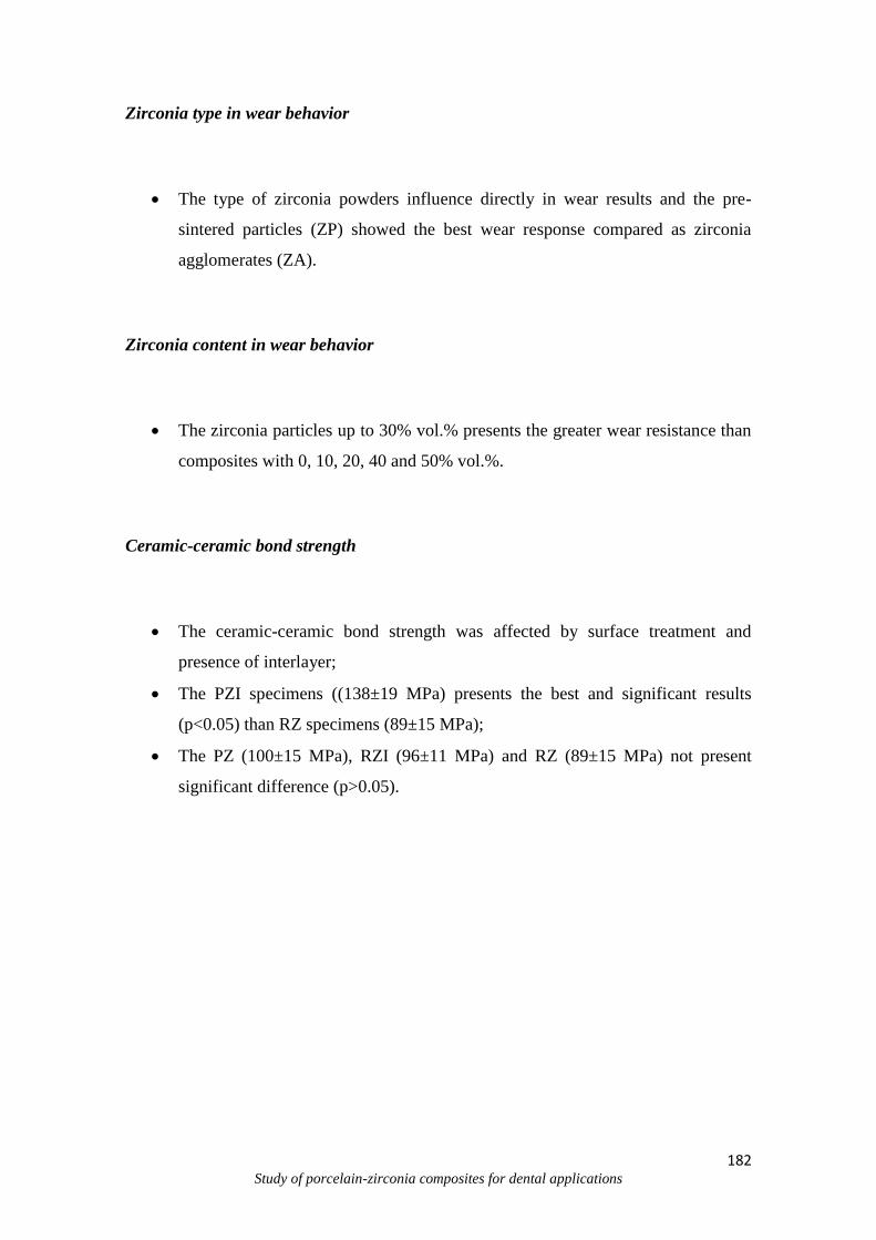
182 Study of porcelain-zirconia composites for dental applications
Zirconia type in wear behavior
The type of zirconia powders influence directly in wear results and the pre-
sintered particles (ZP) showed the best wear response compared as zirconia
agglomerates (ZA).
Zirconia content in wear behavior
The zirconia particles up to 30% vol.% presents the greater wear resistance than
composites with 0, 10, 20, 40 and 50% vol.%.
Ceramic-ceramic bond strength
The ceramic-ceramic bond strength was affected by surface treatment and
presence of interlayer;
The PZI specimens ((138±19 MPa) presents the best and significant results
(p<0.05) than RZ specimens (89±15 MPa);
The PZ (100±15 MPa), RZI (96±11 MPa) and RZ (89±15 MPa) not present
significant difference (p>0.05).

183 Study of porcelain-zirconia composites for dental applications
Future perspectives
According the results presented in this study some future perspectives can be drawn:
Influence of different contents of iron in colorimetric characteristics of yttria
stabilized zirconia by CPM for dental applications.
Influence of the zirconia particle size on the mechanical performance;
Influence of the zirconia particle size on the wear behavior;
Influence of the temperature and time of hot pressing technique on the mechanical
properties of zirconia-porcelain composites;
Influence of the yttria stabilized zirconia (3Y-TZP) content in porcelain on the
dynamic mechanical analysis (DMA);
Influence of yttria stabilized zirconia substructure surface treated on the shear
bond strength;
Recommended