FACULDADE DE ODONTOLOGIA
AVALIAÇÃO IN VITRO DA CITOTOXICIDADE, GENOTOXICIDADE E
MUTAGENICIDADE DE MATERIAIS ESTÉTICOS DE PREENCHIMENTO FACIAL
RUCHIELLI LOUREIRO BORGHETTI
2015
����

� �
RUCHIELLI LOUREIRO BORGHETTI
AVALIAÇÃO IN VITRO DA CITOTOXICIDADE, GENOTOXICIDADE E
MUTAGENICIDADE DE MATERIAIS ESTÉTICOS DE PREENCHIMENTO FACIAL
PORTO ALEGRE
2015
PONTIFÍCIA UNIVERSIDADE CATÓLICA DO RIO GRANDE DO SUL
FACULDADE DE ODONTOLOGIA
�����
�
AVALIAÇÃO IN VITRO DA CITOTOXICIDADE, GENOTOXICIDADE E
MUTAGENICIDADE DE MATERIAIS ESTÉTICOS DE PREENCHIMENTO FACIAL
Linha de pesquisa: Enfermidades da região bucomaxilofacial - estudos clínicos,
imunológicos e anátomo-patológicos.
Orientadora: Profa. Dra. Maria Antonia Zancanaro de Figueiredo
Co - orientador: Prof. Dr. João Antonio Pêgas Henriques
PORTO ALEGRE
2015
RUCHIELLI LOUREIRO BORGHETTI
Tese apresentada como requisito para obtenção do título de Doutor pelo Programa de Pós-Graduação em Odontologia, área de concentração em Estomatologia Clínica, Faculdade de Odontologia, Pontifícia Universidade Católica do Rio Grande do Sul.
����
����

Fontes de Catalogação (CIP)
B732a Borghetti, Ruchielli Loureiro Avaliação in vitro da citotoxicidade, genotoxicidade e
mutagenicidade de materiais estéticos de preenchimento facial / Ruchielli Loureiro Borghetti. – Porto Alegre, 2015. 149 f.
Tese (Doutorado) – Faculdade de Odontologia, Área de
Concentração Estomatologia Clínica, PUCRS.
Orientador: Profa. Dra. Maria Antonia Zancanaro de Figueiredo. 1. Odontologia. 2. Ácido Hialurônico. 3. Estomatologia. 4. Polimetilmetacrilato. 5. Citotoxicidade. 6. Genotoxicidade. 7. Mutagenicidade. I. Figueiredo, Maria Antonia Zancanaro de. II. Título. CDD 617.607
Bibliotecário Responsável Ginamara de Oliveira Lima
CRB 10/1204
� �
RUCHIELLI LOUREIRO BORGHETTI
AVALIAÇÃO IN VITRO DA CITOTOXICIDADE, GENOTOXICIDADE E
MUTAGENICIDADE DE MATERIAIS ESTÉTICOS DE PREENCHIMENTO FACIAL
Linha de pesquisa: Enfermidades da região bucomaxilofacial - estudos clínicos,
imunológicos e anátomo-patológicos.
BANCA EXAMINADORA
Prof. Dr. Marcus Vinicius Martins Collares (UFRGS)
Prof. Dr. Paulo Edelvar Correa Peres (UFSM)
Profa. Dra. Sabrina Pozatti Moure (ULBRA)
Prof. Dr. Vinicius Duval da Silva (PUCRS)
Profa. Dra. Maria Antonia Zancanaro de Figueiredo (orientadora)
Profa. Dra. Fernanda Gonçalves Salum (PUCRS - suplente)
Tese apresentada como requisito para obtenção do título de Doutor pelo Programa de Pós-Graduação em Odontologia, área de concentração em Estomatologia Clínica, Faculdade de Odontologia, Pontifícia Universidade Católica do Rio Grande do Sul.
�
�����

� �
EPÍGRAFE
�
� �
“Desejo que você, sendo jovem,
não amadureça depressa demais
e, sendo maduro, não insista em rejuvenescer,
e que sendo velho, não se dedique ao desespero.
Porque cada idade tem o seu prazer e a sua dor e
é preciso que eles escorram entre nós”.
Victor Hugo
�����

� �
DEDICATÓRIA
�����

�
À Deus, pela força espiritual que me move e me carrega. Pela graça da vida.
Ao meu esposo, Luiz Cesar, por ser minha fortaleza. Pelo amor e entusiasmo que
impõe em tudo que realizo. Por seu coração admirável. Por sonhar os meus sonhos.
�����

�
AGRADECIMENTOS
�����
� ���
Como grãos de areia que compõe imensas dunas, como pequenas gotas que
formam as chuvas, gestos de compreensão, ternura e apoio foram fundamentais
para a concretização desta etapa. À todos os meus familiares, amigos, professores e
colegas, agradeço com muito carinho.
Ao meu pai, Carlos Alberto, por ser um exemplo de determinação, foco e leveza.
Por me revestir de um amor incondicional, que transborda da alma. Obrigada por
nunca ter desistido de mim. Meu alicerce. Meu tesouro.
À minha mãe, Leni (in memorian). Ainda que tenha partido demasiadamente cedo,
compreendi que com dedicação e entusiasmo, conquistamos nosso objetivos. Seu
eterno sorriso nos olhos e lábios deixou um rastro de amor e saudade. Sei que estás
sempre ao meu lado. Obrigada por fazer parte da minha vida, mesmo que por um
breve período.
Às minhas avós (in memorian), Gilda e Gabriela, obrigada pela compreensão da
minha ausência em momentos importantes. Pelo imenso e minucioso cuidado a mim
dedicados. Pela força que me sustentou nos momentos de tristeza.
À Heloísa, pessoa especial em minha vida. Por ter me apresentado a Odontologia.
Pelo zelo, respeito e amor com que trata meu pai e meus irmãos. Pelo apoio,
amizade e exemplo de profissionalismo.
À minha mãe de coração, minha orientadora, Profa. Dra. Maria Antonia Zancanaro
de Figueiredo, agradeço com toda força o seu acolhimento desde o princípio,
�����

� ���
quando me formei e decidi fazer o Mestrado. O seu amparo e o seu abraço são
sinceros e repletos de amor. Engrandecem, encorajam e fortalecem. Sua sabedoria
e conhecimento são admiráveis. Sou extremamente grata pelo auxílio em cada
passo que eu dou, dentro e fora da PUCRS. Por ser a maior incentivadora dos seus
alunos e por sua preocupação em sempre nos ver bem. Por ensinar a enfrentar
todos os tipos de obstáculos. Por dividir o conhecimento e reforçar a ideia de que
somando, fazemos melhor. Obrigada pelo voto de confiança e amizade. Meu eterno
carinho!!!
Ao meu co-orientador, Prof. Dr. João Antonio Pêgas Henriques, por sua imensa
contribuição à ciência, pelos importantes momentos de discussão do estudo, ainda
que dificuldades tenham ocorrido pelo fechamento do Instituto Royal. Sei que o
esforço foi grande para conseguir levar adiante as pesquisas em andamento, mas
nunca “desistir” foi sua opção. É preciso reconhecer que a sua capacidade em
resolver as questões paralelas o torna uma pessoa ainda mais admirável. Muito
obrigada pelo carinho constante.
Ao Prof. Dr. Jose Antonio Poli de Figueiredo, entusiasta nato. Professor por
natureza. Minha eterna gratidão por me incentivar e acolher desde o Mestrado, em
todas as situações que precisei de auxílio. Por me encantar com sua paixão pelo
conhecimento. Obrigada por abraçar a minha caminhada e fazer o possível para
torná-la viável.
À Profa. Dra. Renata Medina da Silva, que orientou a pesquisa com leveduras com
brilhantismo. Por sua disposição quase sempre imediata em sanar as dúvidas ao
�����
� ���
longo do estudo. Por acolher todos os alunos de modo tão gentil e delicado. Por
possibilitar o desenvolvimento de parte da tese.
À Clarissa Castro Galvão Medeiros e Vanessa Paim Nora, que participaram dos
estudos em levedura, abdicando de seu tempo com outras tarefas, motivadas
apenas pelo sentimento de amizade. Obrigada pela disponibilidade constante,
companheirismo e por não medirem esforços para que tudo desse certo.
Aos meus pequenos, Luiza, Bernardo e Guilherme. Vocês são o orgulho da mana!!
Amo tanto que chega a doer. Obrigada por existirem na minha vida e entenderem
minha ausência.
Ao meus sogros, Luiz Cesar e Moema, à Ci e à vó Stella, por me acolherem de
forma tão calorosa. Por me fazerem sentir sua filha. Por serem meu refúgio. Muito
obrigada pela energia positiva que me passam a cada encontro. Pela eterna alegria
que contagia a todos. Por serem sinônimos de unidade. Pelo exemplo de
honestidade e compreensão.
Aos meus queridos cunhados, Carolina, Daniel, Carla e André. Por me
presentearem com afilhadas tão lindas. Obrigada pelos momentos de risada e
descontração. Pela intensa torcida e incentivo.
À toda minha família por compreenderem meu período de reclusão. Por lutarem
junto comigo. Pelo abraço afável de sempre.
�����
� ���
Às estimadas professoras do Serviço de Estomatologia do Hospital São Lucas da
PUCRS, Dras. Liliane Yurgel, Karen Cherubini e Fernanda Salum, que com muita
competência fazem do ambulatório um centro de referência. Obrigada pelos
momentos aprazíveis no Serviço e fora dele. Pelo conhecimento compartilhado. São
pessoas admiráveis pela maneira de conduzir seus trabalhos, pesquisas, família e
pacientes. Agradeço por me lembrarem diariamente que devemos aplicar a
excelência em qualquer tarefa, por mais simples que pareça. Que devemos
contornar os empecilhos e não nos abater, pois outras pessoas precisam de nós.
À secretária do Serviço de Estomatologia, Márcia Rollsing, pela simpatia e
empenho no atendimento de alunos e pacientes.
Aos amigos e colegas da “Estomato”, pelo excelente convívio, encontros de
desabafos, quando nada parecia dar certo. Em especial à Clarissa e Juliana
Spanemberg, pela amizade que permanece.
Ao Laboratório de Imunologia e Microbiologia da PUCRS e ao GENOTOX-
ROYAL da UFRGS, por gentilmente cederem o espaço para que eu pudesse
desenvolver esta tese. Bem como aos alunos e funcionários que de maneira muito
afetuosa me acolheram.
Ao mestrando André Juchem e à Dra. Miriana da Silva Machado que trabalham
no GENOTOX, pelo suporte estatístico e pelas exaustivas horas de discussão em
torno de artigos. Obrigada por “abraçarem” minha tese e me auxiliarem em todos os
�����
� ���
momentos que precisei, sempre de modo educado e gentil. Vocês foram
fundamentais nesta pesquisa.
Ao Dr. André Hermann, competente cirurgião plástico, que apoiou essa linha de
pesquisa desde sua concepção. Pela disponibilidade em nos atender. Por abrir as
portas de seu consultório e compartilhar suas experiências.
À Coordenação de Aperfeiçoamento de Pessoal de Nível Superior (CAPES), pela
possibilidade de concluir o doutorado como bolsista.
À Pontifícia Universidade Católica do Rio Grande do Sul (PUCRS), na pessoa da
Profa. Dra. Ana Maria Sphor, coordenadora do Programa de Pós-Graduação em
Odontologia, pelo aprendizado desde 2004.
Aos queridos professores e amigos da Universidade Federal do Rio Grande do Sul
(UFRGS), Dra. Manoela Martins e Dr. Marco Antonio Trevizani Martins, por
acreditarem no meu potencial e me incentivarem constantemente a buscar novos
desafios. Obrigada pela confiança e amizade. Pelo carinho com que me tratam e
pelos momentos divertidos e saborosos como são quando nos reunimos. Pela luz e
carisma que emanam ao próximo.
À Clarissa Canto de Mesquita e Deborah Stona, por me mostrarem que a
amizade é irmandade. Por serem grandes apoiadoras e motivadoras! Por
acompanharem de perto minhas conquistas e angústias. Por cada mensagem de
preocupação e encorajamento que manifestam sempre. Amo muito vocês!
�����
� ���
Ao meu grupo “TFs”, Clarissa, Cibeli, Debora, Franciele, Gabriela Azevedo,
Gabriela Costa e Juliana. “A amizade desenvolve a felicidade e reduz o sofrimento,
duplicando a nossa alegria e dividindo a nossa dor”. Obrigada pelo estímulo
constante e votos de que tudo daria certo. Pela empolgação com minhas conquistas.
Por estarem sempre ao meu lado, mesmo que distante. Por me acompanhar nas
madrugadas de estudo apenas para não me deixar esmorecer. Amo vocês!
Finalmente, ao Núcleo de Saúde Bucal do Hospital Moinhos de Vento, em
especial aos colegas Carolina, Gustavo, Luiz Henrique e Maria Paula pelo apoio
inestimável desde a época do Mestrado, contribuindo para que eu pudesse me
dedicar ao Doutorado. Por dividirem conhecimento. Muito obrigada!
�����

� ���
RESUMO
�����
� ���
RESUMO
O ácido hialurônico (AH) e o polimetilmetacrilato (PMMA) são os materiais de
preenchimento estético mais empregados na atualidade. Seu uso indiscriminado tem
evidenciado efeitos indesejáveis nos usuários destes produtos, tornando necessário
o estudo da biocompatibilidade dos mesmos, uma vez que a literatura científica
disponível não esclarece, de forma conclusiva, a etiologia das reações adversas que
podem se desenvolver a partir da sua injeção. A presente pesquisa avaliou a
resposta citotóxica, genotóxica e mutagênica, a partir de testes in vitro distintos e
efetuados de maneira independente. Utilizando a levedura Saccharomyces
cerevisiae averiguou-se a citotoxicidade do AH (16 mg/mL e 20 mg/mL) e do PMMA
(2%, 10% e 30%), por meio de experimentos qualitativos e quantitativos. O primeiro
procedeu-se pela indução de halo de inibição. Para conduzir uma análise preliminar,
as diluições 10-2 a 10-5 foram verificadas. No teste quantitativo, as colônias formadas
foram contadas em UFC/mL (unidades formadoras de colônias por mililitro). Os
dados observados em levedura demonstraram que no teste do halo de inibição, o
silicone, utilizado como controle positivo, foi o único material capaz de induzir
citotoxicidade. O exame preliminar também indicou o silicone e o AH 16 mg/mL
como indutores de toxicidade celular. Na análise quantitativa, o AH 20 mg/mL no
volume de 0,1 mL inibiu a proliferação celular (ANOVA, teste de Tukey, p≤ 0,05). O
PMMA apresentou resposta dose-dependente nas concentrações de 2% e 10%
(teste de Tukey, p≤ 0,05). Por outro lado, o PMMA 30% exibiu níveis de crescimento
celular semelhantes ao controle negativo. O silicone confirmou o impedimento de
proliferação celular em S. cerevisiae (teste de Tukey, p≤ 0,05). Numa segunda
investigação, em cultura de fibroblastos pulmonares de hamster Chinês (linhagem
V79), foram determinados os potenciais de citotoxicidade, genotoxicidade e
�����

� ��
mutagenicidade do AH 20 mg/mL e do PMMA 30%. Para esses parâmetros, a
abordagem envolveu os ensaios clonogênico, cometa e micronúcleos. O AH e o
PMMA foram capazes de diminuir o crescimento de colônias quando as culturas
foram expostas aos produtos por 24 h, seguidos por 6 dias em meio com ausência
das drogas. Não foram detectados efeitos genotóxicos em 3 h ou 24 h de exposição
ao AH ou PMMA. Da mesma maneira, ambas as substâncias não induziram
aumento na frequência de micronúcleos em células binucleadas. Os resultados
obtidos permitem sugerir que (1) o AH 20 mg/mL e o PMMA 10% são indutores de
citotoxicidade em modelo eucarioto S. cerevisiae; (2) o AH e o PMMA possuem fraca
citotoxicidade sobre a linhagem V79; (3) os materiais testados não provocam danos
no DNA e alterações cromossômicas detectáveis.
Palavras-chave: Ácido hialurônico; Polimetil metacrilato; Citotoxicidade;
Genotoxicidade; Mutagenicidade.
�����

� ��
SUMMARY
�����
� ���
SUMMARY
Hyaluronic acid (HA) and polymethylmethacrylate (PMMA) are the most used
dermal fillers nowadays. The indiscriminate use of such substances has brought to
light unwanted adverse effects. Thus biocompatibility studies are a necessity, since
the scientific literature does not clarify the etiology of these effects. The current
research has evaluated cytotoxic, genotoxic and mutagenic responses from distinct
in vitro tests performed in an independent manner. The cytotoxicity potential of the
materials was evaluated by the induction of an inhibition halo in a solid yeast
cultures. To conduce a preliminary view, the dilutions ranging from 10-2 to 10-5 were
verified. For quantitative test, the colonies were counted to estimate the CFU/mL
(colony-forming units per milliliter) values. Halo inhibition test showed that only
silicone, used as a positive control, was capable of inducing cytotoxicity in this yeast.
The preliminary experiment also indicated silicone and HA 16 mg/mL as a cellular
toxicity inductor material. Quantitative test indicated that HA 20 mg/mL and 0.1mL
volume inhibited cell proliferation (ANOVA, Tukey test, p≤ 0.05). PMMA was dose-
dependent to 2 and 10% concentrations (Tukey test, p≤ 0.05). 30% PMMA showed
cell proliferation inhibition similar to the negative control. Silicone proved to inhibit S.
cerevisiae cell proliferation (Tukey test, p≤ 0.05). In a second investigation, in
Chinese hamster lung fibroblasts (V79 cells), the cytotoxic, genotoxic and mutagenic
potentials of 20 mg/mL HA and 30% PMMA were determined. For testing these
effects, clonogenic survival, comet and micronucleus assay were performed. HA and
PMMA were able to decrease the colony formation when cultures were exposed to
compounds by 24 h followed by 6 days in drug-free media. In addition, no genotoxic
effects were detected in the 3 or 24 h of exposure to HA or PMMA. In the same
�����
� ���
manner, both dermal fillers did not induce increase in the micronucleus frequency in
binucleated cells. Taken together, these results suggest that (1) 20 mg/mL HA and
10% PMMA are cytotoxicity inductors for the eukaryotic model S. cerevisiae; (2) 20
mg/mL HA and 30% PMMA have a weak cytotoxic activity in V79 cells; (3) the tested
substances do not cause detectable DNA damage and chromosome alterations in
V79 cells.
Keywords: Hyaluronic acid; Polymethyl methacrylate; Cytotoxicity;
Genototoxicity; Mutagenicity.
�����

� ���
SUMÁRIO
�����

� ���
SUMÁRIO
1 INTRODUÇÃO ................................................................................... 25
2 ARTIGO CIENTÍFICO 1 ..................................................................... 35
3 ARTIGO CIENTÍFICO 2 .................................................................... 52
4 ARTIGO CIENTÍFICO 3 .................................................................... 76
5 DISCUSSÃO COMPLEMENTAR ..................................................... 101
REFERÊNCIAS ...................................................................................... 108
ANEXOS ................................................................................................. 121
APÊNDICES ........................................................................................... 146
�����

� ���
INTRODUÇÃO
�����
� ���
1 INTRODUÇÃO
O envelhecimento cutâneo é um processo complexo e resultado de fatores
intrínsecos ou cronológicos e extrínsecos, os quais são influenciados pela
modificação do material genético, exposição solar, tabagismo, etilismo, alimentação
e estresse. A partir dessas interações, observa-se elastose solar, presença de
colágeno desorganizado e com níveis reduzidos, redução do número de fibroblastos
diminuído e atrofia da epiderme.1,2 Como resultado macroscópico, formam-se linhas
de expressão, rítides profundas, pigmentos e evidencia-se o decréscimo da gordura
subcutânea.3,4
Tradicionalmente, a cirurgia plástica destacava-se como a principal alternativa
aos efeitos do tempo sobre a derme.5 Porém, em alguns casos, exacerbava a perda
dos contornos faciais. Assim, a restauração do volume foi reconhecida como novo
parâmetro a ser aperfeiçoado.6
Segundo o relatório da Sociedade Internacional de Cirurgia Plástica Estética,
de 2013, o Brasil ocupa o segundo lugar em número de terapias rejuvenescedoras e
a primeira posição no ranking mundial de cirurgias plásticas.7 A busca desenfreada
por procedimentos estéticos, oportunizou o desenvolvimento de técnicas não
invasivas, como a bioplastia, cujo objetivo é devolver o contorno e o volume faciais,
suavizar rítides e sulcos, corrigir defeitos cutâneos e aumentar artificialmente lábios
e região malar. Como atrativos da terapia, pode-se citar, ainda, o baixo custo, o
retorno de uma aparência satisfatória em curto prazo e a rápida recuperação.6
Os materiais de preenchimento podem ser classificados em reabsorvíveis e
não-reabsorvíveis, variando de acordo com o seu tempo de permanência nos
tecidos.8 Para que seja considerado ideal, o produto deve ser biocompatível, incapaz
� ���
de provocar reação alérgica ou carcinogênica, ter estabilidade no local implantado e
ser de fácil aplicação e remoção.9 Embora não exista, até o momento, substâncias
que contemplem todas essas características sugeridas, destacam-se na literatura
como as mais utilizadas na bioplastia o ácido hialurônico (AH) e o
polimetilmetacrilato (PMMA).
O AH tornou-se popular nas últimas décadas, por ser considerado um
preenchedor de origem natural, uma vez que é um importante componente da matriz
extracelular. Sua estrutura representa um polissacarídeo formado por unidades
repetidas de ácido glucurônico e N-acetilglicosamina, isolado pela primeira vez por
Meyer e Palmer.10 Sua molécula exibe tamanhos e agentes de cross-linking
variados, os quais afetam a durabilidade do produto.11
Para o desenvolvimento do preenchedor injetável, o AH pode ser extraído a
partir da crista de galo (origem aviária) ou da fermentação de bactérias,
especialmente Streptococcus (origem não animal).11 Alguns autores relacionam os
efeitos adversos dessa substância com a presença de impurezas decorrentes do
seu processo de fabricação.12,13
É considerado um implante biossintético temporário14,15 e sua sobrevida na
estrutura da derme varia de 3 a 12 meses.3,16 As propriedades hidrofílicas do AH
atraem água para a matriz extracelular aumentando desta forma a elasticidade da
pele.17
As indicações frequentes de uso do AH destinam-se para as regiões glabelar,
periorbital, perioral e malar. As contra-indicações relacionam-se aos portadores de
doença autoimune, presença de material de preenchimento permanente no local da
aplicação e história de alergia prévia ao AH.14,18

� ���
No mercado, há uma diversidade de produtos comercializados em distintas
concentrações, variando de 5,5 mg/mL a 30 mg/mL. A escolha da concentração do
AH em preenchimentos faciais considera a profundidade das rugas e linhas de
expressão e a quantidade de volume desejado. As anestesias tópica, infiltrativa ou
por bloqueio são meios facultativos para controle da dor durante o tratamento.14
Dos materiais permanentes disponíveis, o PMMA destaca-se no
preenchimento de rugas e/ou correção do sulco nasolabial, definição do contorno do
lábio e aumento de partes moles.19,20 Se observa um crescente uso desse recurso
nos casos de lipodistrofia facial em portadores do HIV21, especialmente devido ao
seu baixo custo.
As apresentações comerciais podem variar de acordo com a concentração de
PMMA empregada, sendo de 2%, 10% e 30%. O PMMA 2% é utilizado na região
intradérmica para minimizar rugas finas, especialmente na área labial, enquanto a
concentração de 10% é indicado para áreas móveis, visando diminuir o aspecto de
cansaço provocado pela flacidez. Já o PMMA 30% é designado para injeção onde
exista uma estrutura óssea abaixo, sempre a nível intramuscular ou justaperiostal,
com o intuito de aumentar o volume da região de interesse.20
Os distintos produtos a base de PMMA também podem diferir quanto à sua
composição.22 Existe no mercado o PMMA em gel de colágeno bovino (Artefill®), o
PMMA associado ao gel de carboximetilcelulose (Metacrill®; Biossimetric®) e o
PMMA em gel de hidroxietilcelulose (Newplastic®).20 O Metacrill® está entre os
materiais mais aplicados pelos profissionais e se apresenta na forma de
microesferas de diâmetros variados que oscilam entre 30 a 80 micras. Em virtude
do maior diâmetro e da presença de irregularidades na superfície, as microesferas
não são fagocitadas, permanecendo no local onde foram injetadas, estimulando a
� ��
colagenogênese e neovascularização induzidas por um padrão inflamatório.5,23-25
Embora tenha sido suspenso nos Estados Unidos e Brasil, o preenchimento
estético com o silicone líquido injetável ainda é amplamente realizado. Este material
é elaborado à base de polidimetilsiloxano e composto por sílica, oxigênio e metano.
As viscosidades encontradas resultam dos distintos graus de polimerização e do
número de ligações cruzadas entre as moléculas.26,27
A incessante procura por determinados padrões estéticos, muitas vezes leva
o paciente a alcançar seu objetivo de maneira clandestina, seja com profissionais
não habilitados ou por meio de substâncias ilícitas para esta finalidade, como o
silicone industrial.26,28 Esta situação evidenciou severas complicações,
especialmente granulomas de corpo estranho, denominados siliconomas.28-30
Consequências sistêmicas também foram demonstradas em relatos de casos, tais
como embolia e pneumonia, corroborando com a restrição ao uso do produto. Entre
os indivíduos mais acometidos, destacam-se os transgêneros.27,31,32
A patogênese das reações adversas das substâncias de preenchimento é
ainda desconhecida. Quando os biomateriais são injetados nos tecidos, observa-se
uma reação granulomatosa com presença de histiócitos e formação de novo
colágeno circundando a área. Alguns pacientes podem apresentar uma reação
tecidual mais severa resultando clinicamente em nódulos visíveis. Outros estudos
sugerem o desenvolvimento de lesões, onde o material de preenchimento pode agir
como um estímulo para infecção ou contaminação cruzada via técnica injetável.33-35
Além disso, alguns pesquisadores consideram que os efeitos indesejáveis possam
ser provocados pelas propriedades físico-químicas das substâncias, impurezas
presentes nas preparações, técnicas e pacientes inadequadamente selecionados,
ou, ainda, por profissionais inexperientes, sem o devido preparo.36,37

� ��
Quanto à migração do material de preenchimento introduzido nos tecidos,
sugere-se que a mesma aconteça em decorrência da injeção dentro de um vaso
sanguíneo, da fagocitose ou, ainda, da sistematização das drogas, que
eventualmente poderiam atuar como substâncias quimiotáticas, agindo à distância
em algum órgão de metabolismo e excreção.23,24,38,39
Os resultados indesejáveis observados a partir da utilização das substâncias
estéticas na bioplastia costumam ser associados à natureza do material. Os
produtos temporários ocasionam um baixo índice de complicações e com tempo de
duração limitado.40 Ao contrário, os produtos permanentes provocam alterações
muitas vezes de difícil manejo clínico, devido ao potencial de deslocamento nos
tecidos e do caráter não-absorvível da substância.
As consequências locais imediatas ao preenchimento podem ser
evidenciadas na forma de eritema, dor, prurido e edema, as quais são transitórias e
duram de horas a dias.40,41 Os efeitos tardios ocorrem a partir de 15 dias da
bioplastia e apresentam natureza inflamatória crônica.12 Alguns autores verificaram o
surgimento de lesões até 6 anos (PMMA), 7 anos (AH), e 14 anos (silicone) após o
procedimento.30 Nódulos e granulomas do tipo corpo estranho já foram identificados
nas regiões bucal e perioral, motivo de interesse do cirurgião-dentista.30,42-46
Outrossim, a literatura reporta casos de necrose,27,47 cegueira,48,49 infarto cerebral,49
embolia pulmonar50 e pneumopatia.31
A segurança na utilização dos materiais de preenchimento injetáveis é ainda
um assunto bastante discutido por diversos autores. Os métodos de investigação do
efeito tóxico de substâncias podem ser proporcionados a partir de observações
clínicas, estudos em animais e estudos in vitro.
Devido ao controle cada vez mais rigoroso em relação ao uso de animais de
� ���
laboratório, há a necessidade de desenvolver e padronizar testes in vitro que
possam detectar a toxicidade dos biomateriais. Essas metodologias possuem a
vantagem de serem relativamente simples, propiciarem o controle de variáveis,
serem reprodutíveis e de baixo custo51.
Testes in vitro, utilizando diversos organismos-modelo, podem ser
empregados para avaliação da toxicidade induzida por diferentes tipos de
biomateriais. A citotoxicidade consiste em colocar o material em contato de maneira
direta ou indireta com a cultura celular, verificando as alterações por distintos
mecanismos, sendo a viabilidade da célula um dos parâmetros mais aplicados.
A levedura Saccharomyces cerevisiae, o fermento biológico, é um modelo
experimental amplamente utilizado em trabalhos de pesquisa científica de diferentes
áreas das ciências biológicas e biomédicas.52-54 É um organismo unicelular
eucarioto, apresentando, assim, características celulares e bioquímicas muito
semelhantes às de células animais e humanas. Sua utilização para a análise de
toxicidade celular induzida por materiais de interesse clínico é, portanto, bastante
adequada.55,56 Embora os resultados de testes de citotoxicidade in vitro não possam
ser imediatamente extrapolados para a clínica, eles são importantes para definição
do comportamento biológico dos materiais.
Sob essa perspectiva, dentro das possibilidades de metodologias, a genética
toxicológica avalia as alterações induzidas por agentes xenobióticos no material
genético dos seres vivos. A exposição de um organismo a uma substância exógena,
pode desencadear uma cascata de eventos sobre o DNA, levando à formação de
mudanças nesta macromolécula.
Os agentes genotóxicos podem ser avaliados por meio da aplicação de
alguns parâmetros bem estabelecidos, como o dano de DNA primário, determinado
� ���
pelo ensaio cometa (EC), ou a frequência de micronúcleos (MN), de acordo com o
teste MN. O uso concomitante destes ensaios é recomendado na literatura, uma vez
que apresentam características complementares.56,57 Cultura de células
permanentes de fibroblastos pulmonares provenientes de hamster Chinês (linhagem
V79), tem sido utilizadas para a avaliação da genotoxicidade e mutagenicidade,
através do emprego do ensaio cometa e do teste de micronúcleos.58,59
O ensaio cometa (teste de células individualizadas em gel de agarose) é uma
técnica útil para o estudo de indução de danos no DNA e também do seu eventual
reparo. O princípio do método consiste na inclusão das células em gel sobre uma
lâmina de microscopia, por meio da qual se faz passar uma corrente elétrica. Uma
vez que o DNA tem carga negativa, se estiver rompido (clastogênese), migra para
fora do núcleo. Desta forma, as células com aumento de danos no DNA mostram um
aumento na migração de fragmentos cromossômicos do núcleo em direção ao
ânodo, evidenciando uma forma de um cometa ou cauda.56 O DNA que não estiver
rompido ou quebrado fica armazenado no núcleo, uma vez que é muito grande para
migrar.
Pode-se conceituar os micronúcleos como sendo corpúsculos extranucleares
formados durante o processo da mitose, os quais são o resultado de fragmentos
cromossômicos acêntricos ou de cromossomos inteiros que não ficaram incluídos
em nenhum dos núcleos filhos, originados no processo de divisão celular.58 Assim,
os efeitos de substâncias que provoquem quebras cromossômicas ou ainda afetem
os componentes do fuso ou da região centromérica podem ser detectados a partir da
presença de micronúcleos.
Na literatura científica, existem poucos ensaios clínicos delineados de forma
adequada que permitam determinar, de forma segura, os efeitos que o uso dos

� ���
materiais de preenchimento podem provocar a longo prazo. O que se sabe a
respeito, baseia-se quase que exclusivamente em relatos de casos clínicos.
A presente tese compreende 3 artigos científicos. O primeiro deles faz uma
revisão da literatura sobre um dos materiais utilizados e tem por objetivo
fundamentar, com base científica, o experimento realizado. O segundo averigua a
viabilidade celular em Saccharomyces cerevisiae a partir do uso destes recursos na
bioplastia. O terceiro descreve a investigação, em células V79, das alterações
citotóxicas, genotóxicas e mutagênicas a partir da utilização de AH e PMMA.

� ���
ARTIGO 1
�����
�����
� ���
2 ARTIGO 1
O artigo de revisão a seguir intitula-se “Hyaluronic acid facial filler —
Implications in dentistry” e foi submetido e formatado segundo as normas do
periódico British Journal of Oral and Maxilofacial Surgery (Anexos A e B), o qual
possui Qualis B1.
� ���
Hyaluronic acid facial filler — Implications in dentistry
Ruchielli Loureiro Borghetti, PhDa*, Karen Cherubini, PhDb, Maria Antonia Zancanaro
de Figueiredo, PhDb
aPhD student of Oral Medicine at the Pontifical Catholic University of Rio Grande do
Sul (PUCRS), Porto Alegre, Brazil
bOral Medicine Service of São Lucas Hospital, Pontifical Catholic University of Rio
Grande do Sul (PUCRS), Porto Alegre, Rio Grande do Sul, Brazil
Corresponding author:
*Ruchielli Loureiro Borghetti ([email protected])
Address: Serviço de Estomatologia do Hospital São Lucas, PUCRS, Pontifícia
Universidade Católica do Rio Grande do Sul
Av. Ipiranga, 6690 – 2º andar/sala 231, Porto Alegre, RS. Brasil.
CEP: 90610-000. Phone/Fax.: +55 51 3320.3254
Abstract
This study reviews the effects of hyaluronic acid as facial filling substance for
cosmetic improvement. The high demand for restoration of facial volume and filling of
facial depression has promoted the rapid emergence of new materials in the market.
Facial fillers represent a breakthrough in the non-invasive rejuvenation of skin and
subcutaneous tissues. Hyaluronic acid is often used in the treatment of wrinkles and
in lip augmentation. The literature published by the National Center for Biotechnology
Information (NCBI) was reviewed regarding the description, indications and adverse
side effects of hyaluronic acid. The increasing demand for cosmetic procedures and
� ���
the variety and indiscriminate use of substances currently available for these
interventions point to the need to fully investigate adverse reactions that may impair
facial esthetics and even put the patient’s general health condition at risk.
Keywords: Hyaluronic acid; Diagnosis; Granuloma; Adverse effects.
Introduction
The aging process is influenced by environmental factors, and causes
structural and functional changes in organic tissues, among which the depletion of
subcutaneous fat and skin collagen levels. The phenomenon reduces skin thickness
and elasticity, generating facial depression and folds,1,2 and affecting esthetic
appearance. This scenario has encouraged the development of numerous cosmetic
facial rejuvenation procedures.
Cosmetic surgery has for long been the most commonly adopted approach in
facial rejuvenation. Traditionally, the treatment against face aging was based on the
surgical traction of tissues.3 In this sense, facial filling techniques have been
developed to meet the increasing demand for less invasive procedures that also
afford fast recovery and satisfactory looks in the short run.4
For decades a variety of substances have been used to smooth out wrinkles
or folds in the perioral and periocular regions in the skin tissue, to artificially augment
lips and the malar region, and to correct facial defects. Ideally, these materials should
be safe, efficient, present as few adverse effects as possible, and afford long-term
esthetic outcomes.5,6
Recently the use of hyaluronic acid as facial filler has been advocated. This
review investigates the influence of this material in the diagnoses of dental conditions
� ���
and in procedures currently adopted in dentistry.
History
The augmentation of soft tissues as a means to improve facial esthetics was
first considered in 1800, when Neuber reported the use of fat collected from the arm
to fill facial depressions. Subsequently, paraffin was also used, though it was soon
discovered that it causes granuloma, and was prohibited in 1930. Additionally, soft
tissues may suffer unpredictable reactions in the long run, creating the need to study
more appropriate and suitable substances to be used as facial fillers. In 1962, liquid
silicone was launched as a cosmetic corrective agent, though years later it was
banned because of the high potential to cause adverse effects. Starting in 1980 and
until recently, bovine collagen was the biomaterial of choice in face filling
procedures.7,8 Nevertheless, its use in skin is associated to a 3 % risk of late
hypersensitivity reaction, and requires a double skin test before treatment is
started.9,10
This scenario revealed the occurrence of a set of adverse reactions that may
impair facial esthetics and even put the patient’s general health condition at risk.6,11
The most important attribute of a facial filler candidate is biocompatibility.
However, other characteristics are also important, like nontoxicity, stability to organic
fluids and tissues, absence of inflammatory or allergic reaction, resistance to
mechanical stress, easy application, and inexpensive removal. In spite of the
technological advancements and of the existence of several biomaterials in use, no
product currently available in the market meets all these requirements.12
The literature ranks cosmetic fillers into two classes, considering the time
these substances remain in tissues: temporary (or resorbable), and permanent (non-
� ��
resorbable).13 The advantage of resorbable products lies in the fact that the result
may be reverted after some period has elapsed, while permanent fillers require
surgical removal in the event of migration or tissue reaction of the material.8
Currently hyaluronic acid (HA) is the most commonly employed resorbable
biomaterial in esthetic improvement procedures. It was launched in the market in
1996, and since then several other molecules have been developed for use as facial
filler. Product safety is based on the washing-off process of these cross-linking
residues, which affords to obtain a pure, atoxic and biocompatible filling material.14,15
HA injections do not require skin testing, and the literature indicates minimal
hypersensitivity risk.1,10,16
Permanence in tissues
Due to the fact that it is resorbable, HA is metabolized by enzymes or
gradually phagocytized. These processes occur within 3 to 24 months after
applications, depending on how much HA is injected in tissues.17 Other authors have
reported a gradual absorption of the substance between 6 months and one year after
applications, and that mean HA permanence in tissues is 9 months.18-20
Indication
The concentration of HA in facial filling procedures is defined based on two
main aspects: (1) the depth of wrinkles and expression lines, and (2) the level of
augmentation desired.21 As a rule, 3 concentrations of the product are used. Low
concentration HA is used to fill the so-called “smoker lines” that form around the
upper lip, as well as crow’s feet. Intermediate concentration HA is used in lip
augmentation procedures, while high concentration HA products are injected in
nasolabial folds.15,22 Topical, infiltrative or block anesthesia are the main anesthetic

� ��
measures used to control pain during the injection of the filling material.3,23
Dentistry and facial filling materials
In recent times, dentistry professionals have to become increasingly aware of
the effects of facial filling materials, since these may affect the facial region and, as a
result, the oral cavity. Filling substances are increasingly present in esthetic
complementation and oral rehabilitation approaches.7 The presence of facial fillers
may change the oral mucosa, leading to confusion or misinterpretation in the
diagnosis of dental and oral conditions.
With the advent of dental implants, a large number of patients began to
replace their total prostheses for fixed protocol prostheses. However, this class of
prostheses does not allow the same esthetic result, since the lack of resin flank in
these fixed prostheses often increases the nasogenian fold and consequently
worsens an aged look. For this reason, the use of fillers in the lower lip and in the
nasogenian fold is often suggested to patients who replace their total prostheses with
implant-supported dentures. In this case, the aim of filling is to mitigate the aging
effect of the loss of lip support.
Clinical evaluation of hyaluronic acid filling
Although it has been classified as a non-immunogenic substance,24,25 it is
known that HA may trigger unfavorable tissue responses, usually due to presence of
the remnants of bacterial proteins in the commercial product, to incorrect application
or even the presence of a biofilm on the tissue.20 Generally speaking, filling
substances may cause a wide array of complications, from a simple inflammatory
reaction to tissue necrosis,15,26-28 which may become visible immediately or after a
longer time lapse following application.
� ���
The immediate and/or transient complications are the most common adverse
effects of HA fillings. These manifestations may last for up to 14 days, and mostly are
related to inflammatory processes or to technical problems.20 Some of these side
effects include erythema, ecchymosis and swelling in the region where the product
was applied.29-32 Due to injury to a blood vessel during the procedure, hematomas
may occur, while necrosis may appear when the injection perforates an artery. These
changes have been reported in the glabella and in the nasolabial fold.33,34
Hypersensitivity,35 vasculitis36 and ischemia37 have also been observed in some
clinical case reports.
Biopsy is only seldom prescribed in the occurrence of transient effects.38
However, the procedure is necessary when some clinical signs become apparent,
like the migration of the injected material and the formation of foreign body
granuloma.39,40 Biopsy is an indicated precautionary measure, because the effects of
filling materials may manifest as papules or nodes,39,41,42 and frequently may be
mistaken for pathologies with distinct etiology and behavior, like cysts and/or salivary
gland neoplasias.42-45
Also, an inflammatory reaction like a granuloma may be observed in the site
the exogenous material is injected.46 The process starts with the recruitment of
neutrophils and lymphocytes, which is accompanied by pain and exudation. The
material injected is invaded by inflammatory cells as soon as it is injected. However,
this foreign body is too large to allow phagocytosis by one macrophage only.
Therefore, these cells gather together to form giant cells, which measure roughly 40
to 50 μm and aim to isolate the exogenous substance. More intense signs of
fibroplasia are observed around the zone of granulomatous inflammation, in a
process that occurs in order to limit the tissue response to the presence of the filling

� ���
material and thus reduce local inflammation. In histological examinations, HA is
observed as a blue mass with a bizarre configuration and variable sizes, surrounded
by neutrophils, eosinophils and multinucleated giant cells.5,30,39
In 2003, Fernández-Aceñero, Zamora and Borbujo16 described the case of a
patient who presented several nodes in the upper lip caused by an irregular increase
in tissue volume that had been evolving for 2 months. The patient reported having
had lip augmentation injections with HA. Based on the assumed diagnosis of foreign
body, an incisional biopsy was conducted involving the epidermis, the dermis and
subcutaneous adipose tissue. A clearly outlined mass was detected in the
subcutaneous adipose tissue plane, and was diagnosed as granuloma. The
presence of exogenous material in the biopsied area was confirmed by
histopathology.
The mechanism through which filling substances trigger a foreign body
reaction, the reasons behind the variation in intensity, and the unpredictable
character of their mode of action are yet to be elucidated. The filling material often
migrates to the oral mucosa, forming a stiff nodule within tissues.47-49 This stresses
the need for a complete physical examination that should include visual inspection
and tissue palpation. Former use of filling substance is not always reported
spontaneously. Several times it is necessary to insist in collecting more thorough
information during the interview with a patient in order to obtain as many details as
possible concerning past filling material applications.
Conclusion
The unplanned use of filling materials has revealed a series of adverse
reactions that put the esthetic result and the patient’s general health at risk. Even
though many professionals of the health industry understand that bioplasty
� ���
procedures in general are safe and pose no serious hazards to the patient, adverse
effects are observed in some cases. These complications may be significant, and
include deformity and tissue destruction by an inflammatory response.
The low cost of filling substances has led to a widespread adoption of
incisionless cosmetic interventions in facial rejuvenation. In this scenario, several
professionals of different areas in the health industry have sensed the popularity of
these procedures, performing them in their patients, who are exposed to
unnecessary and sometimes serious hazards. These patients are often informed of
the advantages of these facial fillers, but are unaware of likely adverse effects they
may experience after an intervention of this kind.
When facial filling is performed by experienced professionals and the correct
biomaterial is used, it affords to minimize the effects of age on the skin.
Nevertheless, in some circumstances some sequelae may occur. The excessive and
indiscriminate utilization of filling materials may point to disappointing issues related
to safety and efficacy. Dental surgeons are required to understand these procedures,
since the possible adverse effects of filling materials may emulate various
pathologies in the orofacial region,50 making it difficult to diagnose and conduct the
appropriate clinical management of the patient.
Conflict of interest
The authors have no conflict of interest.
Funding
None.
� ���
References
1- Brandt FS, Cazzaniga A. Hyaluronic acid fillers Restylane and Perlane. Facial
Plast Surg Clin North Am. 2007; 15(1):63-76.
2- Medeiros CC, Cherubini K, Salum FG, de Figueiredo MA. Complications after
polymethylmethacrylate (PMMA) injections in the face: a literature review.
Gerodontology. 2014; 31(4):245-250.
3- Brandt FS, Cazzaniga A. Hyaluronic acid gel fillers in the management of
facial aging. Clin Interv Aging. 2008; 3(1):153-159.
4- Smith KC. Reversible vs. nonreversible fillers in facial aesthetics: concerns
and considerations. Dermatol Online J. 2008; 14(8):3.
5- Vargas KF, Borghetti RL, Moure SP, Salum FG, Cherubini K, de Figueiredo
MA. Use of polymethylmethacrylate as permanent filling agent in the jaw,
mouth and face regions-implications for dental practice. Gerodontology. 2012;
29(2):16-22.
6- Alijotas-Reig J, Fernández-Figueras MT, Puig L. Late-Onset inflammatory
adverse reactions related to soft tissue filler injections. Clin Rev Allergy
Immunol. 2013; 45(1): 97-108.
7- Dastoor SF, Misch CE, Wang HL. Dermal fillers for facial soft tissue
augmentation. J Oral Implantol. 2007; 33(4):191-204.
8- John HE, Price RD. Perspectives in the selection of hyaluronic acid fillers for
facial wrinkles and aging skin. Patient Prefer Adherence. 2009; 3:225-230.
� ���
9- Lowe NJ, Maxwell CA, Lowe P, Duick MG, Shah K. Hyaluronic acid skin fillers:
adverse reactions and skin testing. J Am Acad Dermatol. 2001; 45(6):930-933.
10- Parada MB, Michalany NS, Hassun KM, et al. A histologic study of adverse
effects of different cosmetic skin fillers. Skinmed. 2005; 4(6):345-349.
11- de Castro ACB, Collares MVM, Portinho CP, Dias PC, Pinto RA. Extensive
facial necrosis after infiltration of polymethylmethacrylate. Braz J
Otorhinolaryngol. 2007; 73(6):850.
12- Lemperle G, Morhenn V, Charrier U. Human histology and persistence of
various injectable filler substances for soft tissue augmentation. Aesthetic
Plast Surg. 2003; 27(5):354-366.
13- Edwards PC, Fantasia JE. Review of long-term adverse effects associated
with the use of chemically-modified animal and nonanimal source hyaluronic
acid dermal fillers. Clin Interv Aging. 2007; 2(4):509-519.
14- Romagnoli M, Belmontesi M. Hyaluronic acid-based fillers: theory and
practice. Clin Dermatol. 2008; 26(2):123-159.
15- Yeom J, Bhang SH, Kim BS, Seo MS, Hwang EJ, Cho IH, et al. Effect of
cross-linking reagents for hyaluronic acid hydrogel dermal fillers on tissue
augmentation and regeneration. Bioconjug Chem. 2010; 21(2):240-247.
16- Fernández-Aceñero MJ, Zamora E, Borbujo J. Granulomatous foreign body
reaction against hyaluronic acid: report of a case after lip augmentation.
Dermatol Surg. 2003; 29(12):1225-1226.
� ���
17- Vargas AF, de Amorim NG, Pitanguy I. Complicações tardias dos
preenchimentos permanentes. Rev Bras Cir Plást. 2009; 24(1):71-81.
18- Lemperle G, Morhenn VB, Pestonjamasp V, Gallo RL. Migration studies and
histology of injectable microspheres of different sizes in mice. Plast Reconstr
Surg. 2004; 113(5):1380-1390.
19- Perenack J. Treatment options to optimize display of anterior dental esthetics
in the patient with the aged lip. J Oral Maxillofac Surg. 2005; 63(11):1634-
1641.
20- Rohrich RJ, Monheit G, Nguyen AT, Brown SA, Fagien S. Soft tissue filler
complications: The important role of biofilms. Plast Reconstr Surg. 2010;
125(4):1250-1256.
21- Burgess CM. Principles of soft tissue augmentation for the aging face. Clin
Interv Aging. 2006; 1(4):349-355.
22- Saylan Z. Facial fillers and their complications. Aesthet Surg J. 2003;
23(3):221-224.
23- Matarasso SL, Carruthers JD, Jewell ML. Consensus recommendations for
soft-tissue augmentation with nonanimal stabilized hyaluronic acid
(Restylane). Plast Reconstr Surg. 2006; 117(Suppl 3):3-34.
24- Alijotas-Reig J, Fernández-Figueras MT, Puig L. Inflammatory, immune-
mediated adverse reactions related to soft tissue dermal fillers. Semin Arthritis
Rheum. 2013; 43(2): 241-258.

� ���
25- Hamilton RG, Strobos J, Adkinson NF. Immunogenicity studies of
cosmetically administered nonanimal-stabilized hyaluronic acid particles.
Dermatol Surg. 2007; 33 (Suppl 2):176-185.
26- Fernández-Cossío S, Castaño-Oreja MT. Biocompatibility of two novel dermal
fillers: histological evaluation of implants of a hyaluronic acid filler and a
polyacrylamide filler. Plast Reconstr Surg. 2006; 117(6):1789-1796.
27- Monheit GD, Rohrich RJ. The nature of long-term fillers and the risk of
complications. Dermatol Surg. 2009; 35 (Suppl 2):1598-1604.
28- Grunebaum LD, Bogdan Allemann I, Dayan S, Mandy S, Baumann L. The
risk of alar necrosis associated with dermal filler injection. Dermatol Surg.
2009; 35:1635-1640.
29- Cox SE. Clinical experience with filler complications. Dermatol Surg. 2009; 35
(Suppl 2):1661-1666.
30- Loureiro Borghetti R, de Vargas KF, Pozatti Moure S, Gonçalves Salum F, de
Figueiredo MA. Clinical and histologic evaluation of effects of hyaluronic acid
in rat tongue. Oral Surg Oral Med Oral Pathol Oral Radiol. 2012; 113(4):488-
494.
31- Rzany B, Cartier H, Kestemont P, Trevidic P, Sattler G, Kerrouche N et al.
Full-face rejuvenation using a range of hyaluronic acid fillers: efficacy, safety,
and patient satisfaction over 6 months. Dermatol Surg. 2012; 38(7 Pt 2):1153-
1161.

� ���
32- Lupo MP. Hyaluronic acid fillers in facial rejuvenation. Semin Cutan Med
Surg. 2006; 25(3):122-126.
33- Glaich AS, Cohen JL, Goldberg LH. Injection necrosis of the glabella: protocol
for prevention and treatment after use of dermal fillers. Dermatol Surg. 2006;
32(2):276-281.
34- Bellman B. Complication following suspected intra-arterial injection of
Restylane. Aesthet Surg J. 2006; 26(3):304-305.
35- Patel VJ, Bruck MC, Katz BE. Hypersensitivity reaction to hyaluronic acid with
negative skin testing. Plast Reconstr Surg. 2006;117(6):92-94.
36- Alijotas-Reig J. Recurrent urticarial vasculitis related to nonanimal hyaluronic
acid skin filler injection. Dermatol Surg. 2009; 35 (Suppl 1): 395-397.
37- Banh K. Facial ischemia after hyaluronic acid injection. J Emerg Med. 2013;
44(1):169-170.
38- Zimmermann US, Clerici TJ. The histological aspects of fillers complications.
Semin Cutan Med Surg. 2004; 23(4):241-250.
39- Moure SP, de Vargas KF, Borghetti RL, Salum FG, Cherubini K, da Silva VD
et al. Clinical and pathological characteristics of polymethylmethacrylate and
hyaluronic acid in the rat tongue. Int J Oral Maxillofac Surg. 2012; 41(10):
1296-1303.
40- Medeiros CC, Borghetti RL, Nicoletti N, da Silva VD, Cherubini K, Salum FG
et al. Polymethylmethacrylate dermal fillers: evaluation of the systemic toxicity
in rats. Int J Oral Maxillofac Surg. 2014; 43(1): 62-67.
� ��
41- Jham BC, Nikitakis NG, Scheper MA, Papadimitriou JC, Levy BA, Rivera H.
Granulomatous foreign-body reaction involving oral and perioral tissues after
injection of biomaterials: a series of 7 cases and review of the literature. J Oral
Maxillofac Surg. 2009; 67(2):280-285.
42- Da Costa Miguel MC, Nonaka CF, dos Santos JN, Germano AR, de Souza
LB. Oral foreign body granuloma: unusual presentation of a rare adverse
reaction to permanent injectable cosmetic filler. Int J Oral Maxillofac Surg.
2009; 38(4):385-387.
43- Quirino MR, Neves AC, Campos MS, Brandão AA, Anbinder AL. Oral
granuloma formation after injection of cosmetic filler. J Craniomaxillofac Surg.
2012; 40(7):194-197.
44- Gonçales ES, Almeida AS, Soares S, Oliveira DT. Silicone implant for chin
augmentation mimicking a low-grade liposarcoma. Oral Surg Oral Med Oral
Pathol Oral Radiol Endod. 2009; 107(4):21-23.
45- Maly A, Regev E, Meir K, Maly B. Tissue reaction to liquid silicone simulating
low-grade liposarcoma following lip augmentation. J Oral Pathol Med. 2004;
33(5):314-6.
46- Lombardi T, Samson J, Plantier F, Husson C, Küffer R. Orofacial granulomas
after injection of cosmetic fillers. Histopathologic and clinical study of 11
cases. J Oral Pathol Med. 2004; 33(2):115-120.
47- Lee SC, Kim JB, Chin BR, Kim JW, Kwon TG. Inflammatory granuloma
caused by injectable soft tissue filler (Artecoll). J Korean Assoc Oral Maxillofac
Surg. 2013; 39(4):193-196.

� ��
48- Eversole R, Tran K, Hansen D, Campbell J. Lip augmentation dermal filler
reactions, histopathologic features. Head Neck Pathol. 2013; 7(3):241-249.
49- Feio PS, Gouvêa AF, Jorge J, Lopes MA. Oral adverse reactions after
injection of cosmetic fillers: report of three cases. Int J Oral Maxillofac Surg.
2013; 42(4):432-435.
50- Vargas KF, Borghetti RL, Moure SP, Cherubini K, de Figueiredo MA. Local
and systemic tissue response submitted to injection of 2 and 30%
polymethylmethacrylate in rats' tongue. Gerodontology. 2014. doi:
10.1111/ger.12108.

� ���
ARTIGO 2
�����

� ���
3 ARTIGO 2
O artigo de pesquisa a seguir intitula-se “Cytotoxicity of dermal fillers
assessed by survival tests in Saccharomyces cerevisiae” e foi formatado e
submetido de acordo com as normas do periódico International Journal of Oral &
Maxillofacial Surgery (Anexos C e D), o qual possui Qualis A1.
� ���
Cytotoxicity of dermal fillers assessed by survival
tests in Saccharomyces cerevisiae
Running title: Cytotoxic effects of dermal fillers
Ruchielli Loureiro Borghetti, MSca*, Clarissa Castro Galvão Medeiros, MSca, Karen
Cherubini, PhDb, Renata Medina-Silva, PhDc , Maria Antonia Zancanaro de
Figueiredo, PhDb
aPhD student of Oral Medicine, Pontifícia Universidade Católica do Rio Grande do
Sul (PUCRS), Faculdade de Odontologia
bProfessor of Oral Medicine, Pontifícia Universidade Católica do Rio Grande do Sul
(PUCRS), Faculdade de Odontologia
cProfessor of Microbiology, Pontifícia Universidade Católica do Rio Grande do Sul
(PUCRS), Faculdade de Biociências
Corresponding author:
*Ruchielli Loureiro Borghetti ([email protected])
Address: Serviço de Estomatologia do Hospital São Lucas, PUCRS, Pontifícia
Universidade Católica do Rio Grande do Sul
Av. Ipiranga, 6690 – 2º andar/sala 231, Porto Alegre, RS. Brasil.
CEP: 90610-000. Phone/Fax.: +55 51 3320.3254
Keywords: Oral medicine; Hyaluronic acid; Polymethylmethacrylate;
Biocompatibility; Cytotoxicity.
� ���
Abstract
The purpose of this study was to evaluate the cytotoxicity potential induced by
hyaluronic acid (HA) and polymethylmethacrylate (PMMA) facial fillers through
growth inhibition and survival tests after direct exposure of Saccharomyces
cerevisiae cells to these substances. The cytotoxicity potential of the materials was
assessed by the induction of inhibition halos in solid yeast cultures and survival
experiments, in which a preliminary essay and a quantitative analysis were
performed to estimate the CFU/mL (colony-forming units per milliliter) values of all
treatments in relation to the negative control. The inhibition halo test showed that
only silicone, used as a positive control, was capable of inducing cytotoxicity in this
yeast. The preliminary survival experiment indicated silicone and 16 mg/mL HA as
cytotoxic materials. Quantitative tests confirmed that silicone, 20 mg/mL HA and 10%
PMMA were able to induce significant cytotoxicity in S. cerevisiae (ANOVA, Tukey
test, p≤ 0.05), with a dose-dependent response for 10% PMMA. The data obtained in
this study revealed that the facial fillers HA (at 20 mg/mL) and PMMA (at 10%) are
cytotoxicity inductors for the eukaryotic supermodel S. cerevisiae.
Introduction
Dermal fillers are considered an alternative to conventional plastic surgery due
to their low financial cost and less invasive technique, which allows for a prompt
recovery for the patient and provides the return to a satisfactory appearance in the
short term.1-3 The pursuit of dermatologic procedures aiming at volumetric restitution
has increased indiscriminately. On the other hand, the industry has been launching
several new products at an accelerated rate, while scientific researches cannot
effectively and safely contemplate all the necessary analyses with the same

� ���
promptitude. Hyaluronic Acid (HA) and Polymethylmethacrylate (PMMA) are featured
as the most commonly used materials. HA is a glycosaminoglycan present in the
extracellular matrix, which has the ability to connect with water, thereby providing
volume, stability, tenacity and elasticity to the skin.4 Furthermore, it is considered to
be a temporary substance that remains in the tissues for 6 to 12 months.4 In contrast,
PMMA is classified as a permanent dermal filler since its particles do not degrade.3
Both substances are used to fill in wrinkles and lines, provide soft tissue
augmentation and correction of nasolabial folds, and are also applied to treat facial
lipodystrophy in HIV patients.5,6
Dentists are now dealing with the repercussions of the adverse effects of
dermal fillers, especially if the fillers were injected into the lips and/or perioral
region.3,7,8 During physical examination, palpable nodules can be seen in the oral
submucosa, which can mimic salivary-gland cysts and neoplastic lesions.9,10 Biopsy
followed by microscopy analysis of these nodules evidences the presence of an
exogenous material surrounded by a foreign body, resulting in an inflammatory
reaction.2
Cases of complications after liquid silicone injections are still frequently
reported in the literature as well as in the media. It continues to be used illegally on a
large scale in order to add volume and shape the body, particularly in the malar,
gluteal and mammary regions, although its employment for aesthetic purposes has
been prohibited.11 Several consequences involving local and systemic inflammatory
reactions have been described after its use.12,13
Biocompatibility studies are fundamental to ensure the safety of dermal fillers.
In vitro investigation methods to detect substances’ toxic effects are being performed
since they have the advantage of being relatively simple; they enable control of the

� ���
variables and are reproducible. Among the various methods used, human cell
cultures have been the first choice of the researchers.14 However, these present an
elevated cost, have a long lifecycle, are difficult to treat and, in some cases, do not
allow quantitative analysis of a large amount of data.
An alternative for basic biocompatibility research is the use of the yeast
supermodel Saccharomyces cerevisiae.15 This organism has a low cost of cultivation,
is easily manipulated and provides a wide amount of quantitative data in a short
period of time.16,17 Furthermore, the biochemical, genetic and genomic structure of
this species have been well described. Moreover, fungi and animals are
phylogenetically very closed related and both are clustered in the group of eukaryotic
organisms called "Opisthokonta".18 Therefore, they share biochemical and genetic
similarities that justify the employment of yeast models to study scientific issues of
human interest.19 Many recent studies have highlighted the diversity of yeast models
and how they can be used for the investigation of the cytotoxicity potential of different
chemical products20 and orthodontic materials,16,17 and even can be used to elucidate
some aspects of human pathologies,15 including those of neurodegenerative
diseases.21 In this context, the main purpose of this research was to evaluate the
cytotoxicity potential induced by 2 facial fillers through growth inhibition and survival
tests after direct exposure of S. cerevisiae cells to these substances.
Materials and methods
This research was initiated after approval from the Scientific and Ethics
Committee of the Dental School of the Pontifical Catholic University of Rio Grande do
Sul (PUCRS), protocol number 0002/12. The procedures were performed in
accordance with institutional guidelines of the Laboratory of Immunology and
Microbiology at the same institution. The materials used for these experiments were
� ���
HA (Stylage®, Laboratory Vivacy, Paris, France) at two concentrations (16mg/mL- S
and 20mg/mL- M), PMMA (Metacrill®, Laboratory Nutricell, Rio de Janeiro, Brazil) at
the concentrations of 2%, 10% and 30%, since these are the concentrations most
frequently used by physicians, and also industrial liquid silicone (Jimo®,
Cachoeirinha, Brazil), which was used as a positive control in all experiments, as it
has previously been reported as highly cytotoxic. 22,23
Strains and culture medium
The wild type Saccharomyces cerevisiae strain FF18733 (mata, ura3-52, his7-
3, leu2-1, trp1-289, lys1-1) was employed in this study. YPD medium in broth (1%
yeast extract, 2% peptone and 2% glucose) or in solid form (with 2% agar) was used
for cytotoxicity tests, according to the methodological step and following the protocol
established at the laboratory.
Halo induction tests
This qualitative experiment evaluated the cytotoxicity potential of the materials
by the induction of an inhibition halo in solid yeast cultures. Cells from exponential S.
cerevisiae cultures (~106 cells/mL) at 30oC in YPD broth were plated in full coverage
on solid YPD with a sterile swab. Subsequently, a perforation of 0.5 cm diameter was
made in the medium using a sterile instrument specially developed for this purpose
and the substances were placed into the hole. For the HA experiments, 4
perforations were made, one each for HA S (16mg/mL), HA M (20mg/mL), negative
(without any material) and positive control (PC - liquid silicone). For the PMMA test, 5
perforations were made for each concentration of the substance (2%, 10%, 30%),
along with the negative and positive controls. All the holes were made with the same
� ���
instrument and filled with the same material quantity (0.4 mL). The plates were then
incubated at 30oC for 2 days, after which the presence or absence of inhibition halos
was evaluated. Four experiments were performed for each material.
Survival experiments for cytotoxicity analysis
In these experiments, S. cerevisiae was cultured in 5 mL of broth YPD
medium to exponential phase (~106 cells/mL), at 30° C. The ideal absorbance (0.8)
was measured with a spectrophotometer (Genesys 20, Thermo Fischer Scientific
Inc., MA, USA) at 600 nm. 100 μL of the pre-inoculum were then transferred to new
tubes containing liquid YPD (1 mL). HA (0.1 and 0.2 mL) and PMMA (0.1, 0.2 and 0.3
mL) were added individually to each tube. The negative control was a culture without
any material immersed, while the positive control was industrial liquid silicone (0.1,
0.2 and 0.3 mL). The samples were cultured under agitation of 180 RPM for 20 h to
exponential phase (~106 cells/mL), after which the survival analyses were performed,
according to Limberger et al. (2011),16 as described below.
To conduct a preliminary view, after reaching the appropriate cell density,
aliquots of each culture (controls and treatments) were diluted in sterile 0.9% NaCl
solution, and 5 μL from each dilution (from 10-2 to 10-5) were plated on YPD solid
medium. Plates were stored at 30oC for 2 days. This experiment verifies whether an
eventual toxicity is induced by the distinct materials tested, through the evaluation of
the appearance of small colonies, which are compared to the negative and positive
controls.
In order to perform the quantitative test, after growth to exponential phase 100
μL of the proper dilutions (10-4 and 10-5) were spread on solid YPD individual plates,
in duplicate. After incubation at 30ºC for 48 h the obtained colonies were counted to

� ��
estimate the CFU/mL (colony-forming units per milliliter) values. The statistic
comparison of CFU/mL from the different treatments in relation to the negative
control provided the quantitative data related to the toxicity of the evaluated
materials. At least 3 independent tests were performed with each concentration of
the products.
Data analysis
The data provided by each set of at least 3 quantitative experiments (CFU/mL)
were converted to a logarithmic scale to conduct a survival analysis to verify an
eventual loss of S. cerevisiae cell viability of the treatments in comparison with the
control. Then statistical differences were tested by One-way ANOVA and Tukey tests
with a confidence level of 5% (p≤ 0.05).
Results
The qualitative evaluation by the induction of inhibition halos after S.
cerevisiae exposure to the fillers allowed us to observe that only silicone, which was
used as a positive control in the experiments, seemed to be capable of inducing
cytotoxicity in this yeast. This fact was observed by the large inhibition halo formed
next to the product deposited at the culture media (Fig. 1). In relation to the other
tested materials, only small halos that were visually indistinguishable from the
negative control were produced, which indicates a possible absence of cellular
toxicity caused by these products in S. cerevisiae.

� ��
Figure 1: Qualitative test showing the inhibition halo of S. cerevisiae cell proliferation
adjacent to the industrial silicone (positive control- PC). HA S: 16mg/mL, HA M:
20mg/mL, NC: negative control.
In the survival analysis, a preliminary experiment was conducted with small
volumes to initially verify the yeast behavior in relation to the tested materials, as can
be observed in figure 2. In this test, the results also indicated silicone as a cellular
toxicity inductor material in this yeast. In the same test, HA S (16 mg/mL) seems to
present a similar cellular toxicity effect.

� ���
Figure 2: Preliminary analysis of S. cerevisiae survival after direct exposure to the
tested materials, showing the reduction of cell proliferation in different dilutions (10-2 -
10-5), especially in treatments with HA S (16 mg/mL) and with the positive control
(PC).
The quantitative test (Fig. 3) indicated that HA significantly decreased cell
viability when compared to the negative control, proving to be more cytotoxic at 20
mg/mL concentration and 0.1 mL volume (HA M1) (Fig. 3, Fig. 4). Moreover, cell
viability after PMMA exposure (Fig. 3, Fig. 5) was also significantly decreased with
the 10% concentration, which occurred in a dose-dependent manner. Conversely,
30% PMMA induced a cell proliferation inhibition similar to the negative control that
was independent of the volume used. In these experiments, the silicone (positive
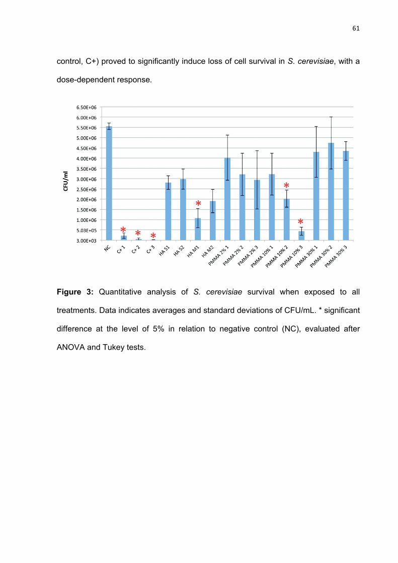
� ���
control, C+) proved to significantly induce loss of cell survival in S. cerevisiae, with a
dose-dependent response.
Figure 3: Quantitative analysis of S. cerevisiae survival when exposed to all
treatments. Data indicates averages and standard deviations of CFU/mL. * significant
difference at the level of 5% in relation to negative control (NC), evaluated after
ANOVA and Tukey tests.
� ���
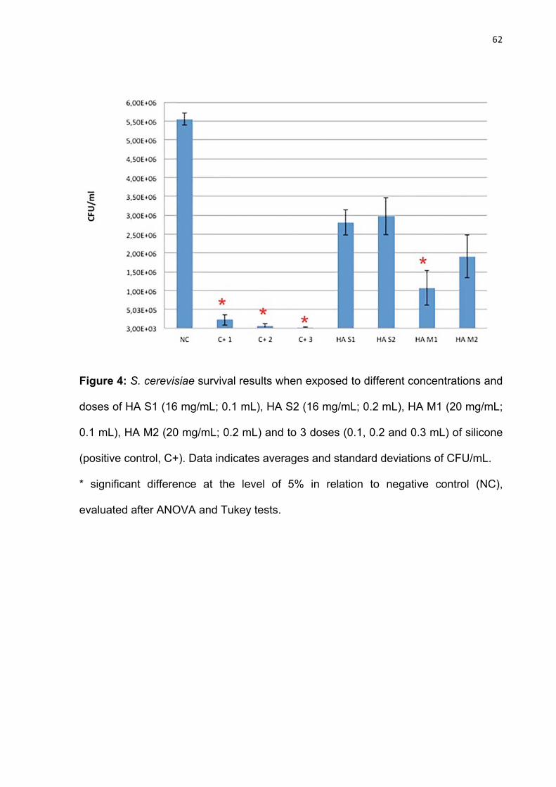
Figure 4: S. cerevisiae survival results when exposed to different concentrations and
doses of HA S1 (16 mg/mL; 0.1 mL), HA S2 (16 mg/mL; 0.2 mL), HA M1 (20 mg/mL;
0.1 mL), HA M2 (20 mg/mL; 0.2 mL) and to 3 doses (0.1, 0.2 and 0.3 mL) of silicone
(positive control, C+). Data indicates averages and standard deviations of CFU/mL.
* significant difference at the level of 5% in relation to negative control (NC),
evaluated after ANOVA and Tukey tests.
� ���
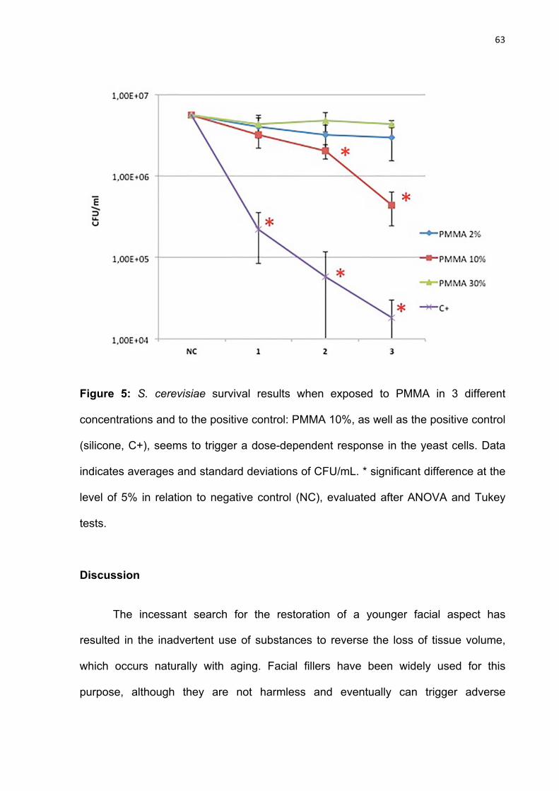
Figure 5: S. cerevisiae survival results when exposed to PMMA in 3 different
concentrations and to the positive control: PMMA 10%, as well as the positive control
(silicone, C+), seems to trigger a dose-dependent response in the yeast cells. Data
indicates averages and standard deviations of CFU/mL. * significant difference at the
level of 5% in relation to negative control (NC), evaluated after ANOVA and Tukey
tests.
Discussion
The incessant search for the restoration of a younger facial aspect has
resulted in the inadvertent use of substances to reverse the loss of tissue volume,
which occurs naturally with aging. Facial fillers have been widely used for this
purpose, although they are not harmless and eventually can trigger adverse
� ���
reactions. As a result, dentists, due to their area of expertise, have identified some of
these mentioned implications.24
With the intention of approaching another aspect of the adverse effects of filler
materials, this research aimed to identify the alterations caused by these substances
when exposed to the S. cerevisiae microbial supermodel. This choice was made
based on studies that have shown the consolidation of the use of such a yeast as an
adequate research resource15 and a cost-effective option for the evaluation of dental
materials’ cytotoxicity.16,17
A standard wild-type Saccharomyces cerevisiae was employed in this study in
order to assess the cytotoxic potential of the substances that are mainly used for
bioplasty. Initially, the inhibition halo test indicated that silicone, used as a positive
control, was capable of inhibiting the yeast proliferation. Statistical analysis from the
yeast survival data also proved that silicone was able to induce significant
cytotoxicity, in a dose-dependent manner, in S. cerevisiae cells. These data assure
the reliability of the present study, since silicone was previously verified to be
cytotoxic in other models.22,23
In contrast to those observed in the halo formation test, the results from the
survival experiments indicated that HA 20 mg/mL significantly reduced the viability of
yeast cells when used with a volume of 0.1 mL. This finding is contrary to those from
authors who defend the HA’s biocompatibility.25 Yoneda et al. (1988)26 found that HA
at the concentration of 1 mg/mL increased the proliferation of dermal fibroblasts in
rats. Park et al. (2014)27 pointed to results that indicate 90% viability of fibroblasts
exposed to HA in mice (L929). Conversely, corroborating our study, Boeckel et al.
(2014)28 analyzed the effect of HA (Teosyal®), associated or not with other materials,

� ���
as a scaffold for tissue engineering. The authors observed that the material induced
loss of pre-osteoblasts cells OFCOL II viability at a concentration of 2.97 mg/mL,
when compared to the control group, by MTT test. In our experiments, 0.1 and 0.2
mL of HA were dissolved in 1 mL of liquid YPD, thus reducing the initial treatment
concentration of the product to 2 and 4 mg/mL, respectively. Moreno et al. (2014)29
evaluated the proliferative effect and viability of mesenchymal cells from adipose
tissue after exposure to HA (10mg/mL) and found that this material was not cytotoxic
at the final doses of 0.1, 0.3, 1 mg/mL and 5 mg/mL. Likewise, for our research, the
smallest tested concentration of HA (16 mg/mL) in the final treatment doses of 1.6
and 3.2 mg/mL was not significantly toxic to S. cerevisiae cells.
There is a discussion in the literature regarding the proliferative or inhibitory
capacity of HA. It is speculated that its high molecular weight and the concentrations
of 50 μg/mL and 1 mg/mL stimulate the proliferation of fibroblasts26 and melanoma
cells.30 Other authors reported that high molecular weight HA inhibited the
proliferation of fibroblasts,31 macrophages32 and keratinocytes.33 High molecular
weight HA (> 5- 6 MDa) is of bacterial origin, while low molecular weight HA (< 0.5-
4.5 MDa) is of avian origin (cockscomb).34 The HA used in our research is of non-
animal origin-and therefore has a high molecular weight-and exhibited a induction
response of low survival at the higher concentration when compared to the negative
control. It could be assumed that the concentration of the substance also reflects its
behavior over the cells, besides the methodological variations.
The PMMA, on the other hand, presents itself as a permanent substance in
the tissues, which could lead to toxic reactions. In our study, inhibition of cell
proliferation was not observed at the concentrations of 2% and 30%. Nevertheless, at

� ���
the concentration of 10%, a cytotoxic dose-dependent response was observed.
Garner et al. (2012)35 evaluated a dental adhesive containing PMMA and found that it
did not present any significant effect on human gingival fibroblast proliferation or on
cell viability reduction after exposures of 24, 48 and 72 hours. In contrast, according
to Wang et al. (2014),36 PMMA proved to reduce HLECs (human lens epithelial cells)
viability after 24h of treatment through the survival experiment CCK-8 (cell counting
kit-8).
The concentration of the materials and their type of presentation seems to be
involved on the cytotoxicity effects in in vitro studies. HA has been investigated as a
scaffold for tissue engineering.28 In previous studies, PMMA was approached in
different ways, since it is widely employed in orthopedics and ophthalmology and
constitutes the foundation of dental prosthesis.37 According to some authors,38 the
disadvantages related to the PMMA cement are bacterial infections, leakage of the
product, toxicity and tissue necrosis. Physicians’ hypersensitivity while handling it
and steam inhalation are also attributed as a result of the monomer polymerization
process (liquid) with the polymer (powder), which may trigger high temperatures and
increase the amount of free monomer. Therefore, the occurrence of this reaction
could explain PMMA toxicity.36,37
In macroscopic analysis, the lower the concentration of the gel (2%), the more
liquid is presented, and the higher the concentration (30%) the more condensed it
appears in the syringe. This fact may justify the yeast proliferation viability revealed in
our study at the concentrations of 2% (final treatment doses of 0.2%, 0.4% and 0.6%)
and 30% (final treatment doses of 3%, 6% and 9%). Although less viscous, the lower
doses of the lower concentration could be considered insufficient to induce cellular
� ���
alterations. On the other hand, 30% PMMA is more consistent and less soluble. In
this way, it presents a less efficient contact to the cells and thus shows a low
cytotoxic potential. The intermediate concentration of 10% (final treatment doses of
1%, 2% and 3%) was more cytotoxic, probably due to its efficient contact with the
yeast, releasing a greater amount of monomers. Undesirable clinical effects
occasioned by PMMA are described in several case reports,10,39 and it should be
noted that PMMA and silicone showed potential for systemic toxicity, involving the
kidneys and liver.13,40 Thus, new approaches are necessary to achieve a better
comprehension of the adverse effects.
Within the methodology employed in this study, it is concluded that HA
20mg/mL and 10% PMMA exhibited cellular toxicity to S. cerevisiae yeast. Although
the tests performed had suggested biocompatibility of both substances at other
concentrations, new studies focusing on the elucidation of undesirable effects of
bioplasty materials are necessary.
Acknowledgments
We would like to thank Professor Brasílio Ricardo Cirillo da Silva for the statistical
support.
Funding
None.
Conflict of interests
None declared.
� ��
References
1- Loureiro Borghetti R, de Vargas KF, Pozatti Moure S, Gonçalves Salum F, de
Figueiredo MA. Clinical and histologic evaluation of effects of hyaluronic
acid in rat tongue. Oral Surg Oral Med Oral Pathol Oral Radiol.
2012;113:488-494.
2- Moure SP, de Vargas KF, Borghetti RL, Salum FG, Cherubini, da Silva VD, de
Figueiredo MAZ. Clinical and pathological characteristics of
polymethylmethacrylate and hyaluronic acid in the rat tongue. Int J Oral
Maxillofac Surg. 2012; 41:1296-1303.
3- Medeiros CC, Cherubini K, Salum FG, de Figueiredo MA. Complications
after polymethylmethacrylate (PMMA) injections in the face: a literature
review. Gerodontology. 2014;31:245-250.
4- Romagnoli M, Belmontesi M. Hyaluronic acid-based fillers: theory and
practice. Clin Dermatol. 2008;26:123-159.
5- Vargas KF, Borghetti RL, Moure SP, Cherubini K, de Figureiredo MAZ. Use of
polymethylmethacrylate as permanent filling agent in the jaw, mouth and
face regions - Implications on dental practice. Gerodontology. 2012; 29:
16-22.
6- Warde M, Gragnani A, Gomes H, Hochman B, Ferreira LM. The impact of
facial lipoatrophy treatment with polymethylmethacrylate in AIDS
patients as measured by four quality-of-life questionnaires. Int J STD
AIDS. 2011; 22: 596-599.
7- Eversole R, Tran K, Hansen D, Campbell J. Lip augmentation dermal filler
reactions, histopathologic features. Head Neck Pathol. 2013; 7: 241-249.
� ��
8- Vargas KF, Borghetti RL, Moure SP, Cherubini K, de Figueiredo MA. Local
and systemic tissue response submitted to injection of 2 and 30%
polymethylmethacrylate in rats' tongue. Gerodontology. 2014. doi:
10.1111/ger.12108.
9- Gonçales ES, Almeida AS, Soares S, Oliveira DT. Silicone implant for chin
augmentation mimicking a low-grade liposarcoma. Oral Surg Oral Med
Oral Pathol Oral Radiol Endod. 2009; 107: e21-23. doi:
10.1016/j.tripleo.2008.12.044.
10- Jham BC, Nikitakis NG, Scheper MA, Papadimitriou JC, Levy BA, Rivera H.
Granulomatous foreign-body reaction involving oral and perioral tissues
after injection of biomaterials: a series of 7 cases and review of the
literature. J Oral Maxillofac Surg. 2009; 67: 280-285.
11- Dornelas MT, Correa MPD, Barra FML, Sá Junior CAC, Dornelas MC,
Sant’Anna LL, Mendonça Neto G, de Arruda FR. Siliconomas. Rev Bras Cir
Plast. 2011; 26:16-21.
12- Mello DF, Gonçalves KC, Fraga MF, Perin LF, Helene Jr A. Local
complications after industrial liquid silicone injection – case series. Rev
Col Bras Cir. 2013; 40: 37-42.
13- Rosa SC, de Magalhães AV, de Macedo JL. An experimental study of
tissue reaction to polymethylmethacrylate (PMMA) microspheres
(Artecoll) and dimethylsiloxane (DMS) in the mouse. Am J Dermatopathol.
2008; 30: 222-227.
14- Truisi GL, Consiglio ED, Parmentier C, Savary CC, Pomponio G, Bois F,
Lauer B, Jossé R, Hewitt PG, Mueller SO, Richert L, Guillouzo A, Testai E.
Understanding the biokinetics of ibuprofen after single and repeated
� ���
treatments in rat and human in vitro liver cell systems. Toxicol Lett. 2015.
doi: 10.1016/j.toxlet.2015.01.006.
15- Simonicova L, Dudekova H, Ferenc J, Prochazkova K, Nebohakova M,
Dusinsky R, Nosek J, Tomaska L. Saccharomyces cerevisiae as a model
for the study of extranuclear functions of mammalian telomerase. Curr
Genet. 2015. doi 10.1007/s00294-014-0472-8.
16- Limberger KM, Westphalen GH, Menezes LM, Medina-Silva R. Cytotoxicity
of orthodontic materials assessed by survival tests in Saccharomyces
cerevisiae. Dent Mater. 2011; 27:81-86.
17- Gonçalves TS, de Menezes LM, Ribeiro LG, Lindholz CG, Medina-Silva R.
Differences of cytotoxicity of orthodontic bands assessed by survival
tests in Saccharomyces cerevisiae. Biomed Res Int. 2014. doi:
10.1155/2014/143283.
18- Marande W, López-García P, Moreira D. Eukaryotic diversity and
phylogeny using small-and large-subunit ribosomal RNA genes from
environmental samples. Environ Microbiol. 2009; 11: 3179-3188. doi:
10.1111/j.1462-2920.2009.02023.x.
19- Galagan JE, Henn MR, Ma LJ, Cuomo CA, Birren B. Genomics of the
fungal kingdom: Insights into eukaryotic biology. Genome Res. 2005; 15:
1620-1631.
20- Xu C, Wang J, Gao Y, Lin H, Du L, Yang S, Long S, She Z, Cai X, Zhou S, Lu
Y. The anthracenedione compound bostrycin induces mitochondria
mediated apoptosis in the yeast Saccharomyces cerevisiae. FEMS Yeast
Res. 2010; 10: 297-308. doi: 10.1111/j.1567-1364.2010.00615.x.
� ���
21- Macreadie IG. Copper transport and Alzheimer’s disease. Eur Biophys J.
2008; 37: 295-300. doi 10.1007/s00249-007-0235-2.
22- Yang CS, Chen KH, Hsu WM, Li YS. Cytotoxicity of silicone oil on
cultivated human corneal endothelium. Eye (Lond). 2008; 22: 282-288.
23- Kowalczuk D, Przekora A, Ginalska G. Biological safety evaluation of the
modified urinary catheter. Mater Sci Eng C. 2015; 49: 274-280.
24- Feio PSQ, Gouvêa AF, Jorge J, Lopes MA. Oral adverse reactions after
injection of cosmetic fillers: report of three cases. Int J Oral Maxillofac
Surg. 2013; 42: 432-435.
25- Fernández-Cossío S, Castaño-Oreja MT. Biocompatibility of two novel
dermal fillers: histological evaluation of implants of a hyaluronic acid
filler and a polyacrylamide filler. Plast Reconstr Surg. 2006; 117: 1789-
1796.
26- Yoneda M, Yamagata M, Suzuki S, Kimata K. Hyaluronic acid modulates
proliferation of mouse dermal fibroblasts in culture. J Cell Sci. 1988; 90:
265-273.
27- Park KY, Kim HK, Jim BJ. Comparative study of hyaluronic acid fillers by
in vitro and in vivo testing. J Eur Acad Dermatol Venereol; 2014; 28: 565-
568. doi:10.1111/jdv.12135.
28- Boeckel DG, Shinkai RS, Grossi ML, Teixeira ER. In vitro evaluation of
cytotoxicity of hyaluronic acid as an extracellular matrix on OFCOL II
cells by the MTT assay. Oral Surg Oral Med Oral Pathol Oral Radiol. 2014;
117: 423-428. doi: 10.1016/j.oooo.2012.07.486.
� ���
29- Moreno A, Martínez A, Olmedillas S, Bello S, de Miguel F. Hyaluronic acid
effect on adipose-derived stem cells. Biological in vitro evaluation. Rev
Esp Cir Ortop Traumatol. 2014. doi: 10.1016/j.recot.2014.10.004.
30- Ahrens T, Assmann V, Fieber C, Termeer CC, Herrlich P, Hofmann M, Simon
JC. CD44 is the principal mediator of hyaluronic-acid-induced melanoma
cell proliferation. J Invest Dermatol. 2001; 116: 93-101.
31- Bodo M, Pezzetti F, Baroni T, Carinci F, Arena N, Nicoletti I, Becchetti E.
Hyaluronic acid modulates growth, morphology and cytoskeleton in
embryonic chick skin fibroblasts. Int J Dev Biol. 1993; 37:349-352.
32- Sheehan KM, DeLott LB, West RA, Bonnema JD, DeHeer DH. Hyaluronic
acid of high molecular weight inhibits proliferation and induces cell
death in U937 macrophage cells. Life Sci. 2004; 75: 3087-3102.
33- Harvima IT, Heikura H, Hyttinen M, Naukkarinen A. Hyaluronic acid inhibits
the adherence and growth of monolayer keratinocytes but does not
affect the growth of keratinocyte epithelium. Arch Dermatol Res. 2006;
298: 207-219.
34- Marcellin E, Steen JA, Nielsen LK. Insight into hyaluronic acid molecular
weight control. Appl Microbiol Biotechnol. 2014; 98: 6947-6956. doi:
10.1007/s00253-014-5853-x.
35- Garner AD, Tucci MA, Benghuzzi HA. Functional and structural responses
associated with potential restorative dental adhesives using human
gingival fibroblasts as a model. Biomed Sci Instrum. 2012; 48: 141-148.
36- Wang B, Lin Q, Shen C, Tang J, Han Y, Chen H. Hydrophobic modification
of polymethylmethacrylate as intraocular lenses material to improve the
� ���
cytocompatibility. J Colloid Interface Sci. 2014; 431:1-7. doi:
10.1016/j.jcis.2014.05.056.
37- Gautam R, Singh RD, Sharma VP, Siddhartha R, Chand P, Kumar R.
Biocompatibility of polymethylmethacrylate resins used in dentistry. J
Biomed Mater Res B Appl Biomater. 2012; 100:1444-1450. doi:
10.1002/jbm.b.32673.
38- Dall'Oca C, Maluta T, Cavani F, Morbioli GP, Bernardi P, Sbarbati A,
Degl’Innocenti, Magnan B. The biocompatibility of porous vs non-porous
bone cements: a new methodological approach. Eur J Histochem. 2014;
58: 2255. doi: 10.4081/ejh.2014.2255.
39- De Castro ACB, Collares MVM, Portinho CP, Dias PC, di Angeli Pinto R.
Necrose facial extensa após infiltração com polimetilmetacrilato. Rev
Bras Otorrinolaringol. 2007; 73:850.
40- Medeiros CC, Borghetti RL, Nicoletti N, da Silva VD, Cherubini K, Salum FG,
de Figueiredo MAZ. Polymethylmethacrylate dermal fillers: evaluation of
the systemic toxicity in rats. Int J Oral Maxillofac Surg. 2014; 43: 62-67. doi:
10.1016/j.ijom.2013.06.009.

� ���
ARTIGO 3
�����
� ���
4 ARTIGO 3
O artigo de pesquisa a seguir intitula-se “Cytotoxic, genotoxic and mutagenic
effects of hyaluronic acid and polymethylmethacrylate dermal fillers on V79
cells” e foi formatado e submetido de acordo com as normas do periódico
International Journal of Oral & Maxillofacial Surgery (Anexos C e E), o qual possui
Qualis A1.
� ���
Cytotoxic, genotoxic and mutagenic effects of hyaluronic acid and
polymethylmethacrylate dermal fillers on V79 cells
Running title: Toxic effects of dermal fillers
Ruchielli Loureiro Borghettia, MSc, Karen Cherubinia, PhD, Miriana da Silva
Machadob, PhD, Maria Antonia Zancanaro de Figueiredoa, PhD, João Antonio Pêgas
Henriquesb, PhD
a Division of Oral Medicine, Pontifícia Universidade Católica do Rio Grande do Sul
(PUCRS), Faculdade de Odontologia.
b Departamento de Biofísica, Instituto de Biociências, Universidade Federal do Rio
Grande do Sul (UFRGS), Porto Alegre, RS, Brazil
Corresponding author:
*Ruchielli Loureiro Borghetti ([email protected])
Address: Serviço de Estomatologia do Hospital São Lucas, PUCRS, Pontifícia
Universidade Católica do Rio Grande do Sul
Av. Ipiranga, 6690 – 2º andar/sala 231, Porto Alegre, RS. Brasil.
CEP: 90610-000. Phone/Fax.: +55 51 3320.3254
Keywords: Hyaluronic acid; Polymethylmethacrylate; Biocompatibility; Cytotoxicity;
DNA damage; Mutagenicity.
� ���
Abstract
Given the growing number of therapies involving dermal fillers, and in order to
facilitate a better understanding of the biocompatibility of such material, this study
aimed to evaluate the cytotoxic, genotoxic and mutagenic properties of hyaluronic
acid (HA, 20 mg/mL) and polymethylmethacrylate (PMMA, 30%) by performing
clonogenic survival, comet and micronucleus assays in Chinese hamster lung
fibroblast - V79 cells. Results showed that HA and PMMA decreased colony
formation when cultures were exposed to compounds for 24 h followed by 6 days in
drug-free media. Moreover, no genotoxic effects were detected in the V79 cells after
either 3 or 24 h of exposure to HA or PMMA. Both substances did not induce
increase in the micronucleus frequency in binucleated cells. Taken together, these
findings suggest that HA and PMMA have weak cytotoxic activity, which is detected
only during extended periods of exposure followed by growth conditions. Though
genotoxic and mutagenic results were negatives, further investigations with different
exposure durations are necessary to warrant the biocompatibility of dermal fillers,
particularly since adverse reactions have been observed in dental practice.
� ��
Introduction
Dermal fillers are injectable substances frequently used in medicine to reduce
facial wrinkles and provide tissue volume.1 Known as bioplasty, this treatment
modality is often a substitute for traditional surgical procedures. However, even with
satisfactory esthetic results, its known adverse effects can occur both at the injection
site and in distant regions.2,3
The ideal soft tissue filler substance is biocompatible, non-immunogenic and
non-carcinogenic, as well as remains stable at the injection site. Such stability
prevents dermal protrusions, inflammatory reactions such as foreign body granuloma
and migration.4,5 However, there is no such ideal exists on the market, though
scientific literature indicates a series of cases with complications related to dermal
filler procedures.6-8
Hyaluronic acid (HA) and polymethylmethacrylate (PMMA) dermal fillers have
been used since the late 1990s to minimize signs of facial ageing and correct skin
imperfections.9,10 HA is a polysaccharide and component of the extracellular matrix
whose hydrophilic properties attract water to tissues, thereby increasing the skin’s
elasticity.5 It is thus considered to be part of a temporary biosynthetic implant lasting
from 6 to 12 months in dermal structures.9 On the other hand, PMMA is an
extensively used permanent filler for filling facial wrinkles, correcting nasolabial folds,
improving labial contour and augmenting soft parts of the skin.10,11 Among their other
uses, HA and PMMA can be employed to treat facial lipodystrophy in HIV patients, a
procedure that has recently gained popularity.12,13
In general, however, filling substances can cause an array of complications,
from simple inflammatory reactions to tissue necrosis9,14 which if not immediately
visible can appear much later after application. Though the most common adverse
� ��
effects involve edema, erythema and ecchymosis.9 Cases of foreign body reactions
in the orofacial region have also been reported.7,8 The effects of which can manifest
as either papules or nodes1,12 mimicking cysts and/or salivary gland neoplasia.7,12
Beyond the studies cited, research on the toxicological aspects and cytotoxicity of
dermal fillers remains scarce. Moreover, little is known about the genotoxic effects of
dermal fillers, despite the recognized relationship between inflammation and DNA
damage.15
Considering the growing number of therapies involving injectable dermal fillers
and in order to contribute to a better understanding of the biocompatibility of such
material, the present study aimed to evaluate the cytotoxic, genotoxic, and mutagenic
properties of HA and PMMA by using cultured mammalian cells.
Materials and methods
Chemicals
Dulbecco’s modified eagle medium (DMEM), fetal bovine serum (FBS),
trypsin-EDTA, L-glutamine and antibiotics were purchased from Gibco BRL (Grand
Island, NY, USA). Cytochalasin B, colchicine, dimethyl sulfoxide (DMSO) and
etoposide were purchased from Sigma-Aldrich (St. Louis, MO, USA). Low-melting
point agarose, normal agarose, TRIS®, and SYBR® Gold nucleic acid gel stain were
obtained from Invitrogen (Carlsbad, CA, USA). Giemsa stain was bought from Merck
(Darmstadt, Germany).
Dermal fillers
The materials used for this experiment were 20 mg/mL hyaluronic acid
(Stylage® M, Laboratory VIVACY, Paris, France) and 30% polymethylmethacrylate
� ��
(Metacrill®, Laboratory Nutricell, Rio de Janeiro, Brazil).
Cell culture and sample preparation
For this research, the Chinese hamster lung fibroblast V79 cell line was
employed as test system. V79 cells were chosen since the cell line is widely used in
cytogenetic assays, mainly for its stable karyotype and relatively short cellular cycle,
which ranges from 12 to 16 h. Moreover, V79 cells are known to form large, well-
defined colonies that, among other characteristics, are highly suitable for clonal
survival assays.16
This adherent cell line was maintained in standard culture conditions with high
glucose DMEM media supplemented with inactivate 10% FBS and antibiotics. Cells
were maintained in 25 cm² culture bottles in a humid atmosphere of 37 ºC with 5%
CO2. While harvesting and establishing the culture, phosphate buffer saline (PBS)
was used for cell washing, as was trypsin-EDTA.
In the experiments, dermal fillers were diluted in DMEM culture media to
obtain different concentrations, of which 5 μL/mL was used as the highest test
concentration, considering dermal filler solubility in culture media and Organisation
for Economic Co-operation and Development (OECD)17 suggestions for mutagenesis
analysis.
Clonogenic assay
Clonogenic assay was performed as described by Franken (2006)18 and
Machado et al. (2009)19. This assay is an in vitro cell survival based a single cell’s
ability to grow into a colony.18
V79 cells were plated onto six-well plates at a concentration of 200 cells per
� ��
well with 3 wells per treatment group. Cells were maintained in DMEM media culture
with FBS for 24 h at 37 °C and 5% CO2 and later exposed to 0.008, 0.04, 0.2, 1, and
5 μL/mL of either HA or PMMA for 24 h. For a negative control, 100% DMEM was
used.
After 24 h of treatment, the media culture containing the test substance was
removed and the cells were washed with PBS and reincubated with drug-free media
at 37 ºC and 5% CO2. After 5 days, the media were aspirated, and the cells were
fixed and stained with 0.1% crystal violet. Visible colonies were counted and M ± SD
estimated. To calculate viability, the number of colonies of the negative control was
considered to constitute 100% viability for comparison to the number of colonies in
other groups. Experiments were performed at least 3 times.
Comet assay
Comet assay was performed as suggested by Burlinson et al. (2007)20 and
Gonçalves et al. (2014)21. The assay can detect single-and double-stranded breaks,
incomplete repair sites, alkali labile sites, and possibly both DNA-protein and DNA-
DNA cross-links.19,20,22 Cells were plated in 24-well cell plates, and after 24 h they
were exposed to 0.312, 0.625, 1.25, 2.5, and 5 μL/mL HA and 0.18, 0.55, 1.66, and 5
μL/mL PMMA for 3 or 24 h at 37 ºC. Again, 100% DMEM was used as a negative
control, while 1.5 and 5 µg/mL etoposide was used as a positive control during 24- or
3-h treatment, respectively.
After exposition, cells were washed with PBS, trypsinized, resuspended in
0.75% low-melting agarose, and immediately spread onto a glass microscope slide
precoated with a layer of 1.5% normal agarose. The low-melting point layer was
allowed to set at 4 ºC for 5 min, after which the slides were incubated in ice-cold lysis
� ��
solution (2.5 M NaCl, 10 mM TRIS, 100 mM EDTA, 1% Triton X-100, and 10%
DMSO; pH 10.0) at 4 ºC and incubated for at least 1 h to remove cell membranes
and proteins, thereby leaving only nucleoid DNA. After lysis, the slides were placed
on a horizontal electrophoresis unit and covered with the electrophoresis buffer (300
M NaOH and 1 M EDTA; pH 13.0) and incubated for 20 min at 4 ºC, during which
DNA unwinding occurred. Electrophoresis was conducted for 20 min at 25 V (300
mA), after which the slides were neutralized with 0.4 TRIS and stained with a solution
containing SYBR® Gold. To evaluate DNA breaks, the fluorescence of comet cells
localized in a preselected area in the gel center was analyzed with a fully automated
scoring PathFinder™ Screen Tox system (Imstar, France).23
Since DNA fragments migrate faster than larger unfragmented DNA during
electrophoresis, damaged cells resemble a comet, with DNA fragments forming the
comet tail whose size and intensity are directly proportional to the amount of DNA
damage. In the present study, the slides were coded before analysis and the
parameters’ percentage of tail DNA and Olive tail moment were determined for 50-
300 randomly selected cells for each slide consisting of two gels by culture and two
cultures by treatment group.
Micronucleus test
In vitro micronucleus (MNvit) assay is a genotoxicity test for detecting
micronuclei in the cytoplasm of interphase cells. Micronuclei can originate from
acentric (i.e., lacking a centromere) chromosome fragments or whole chromosomes
unable to migrate to the poles during the anaphase of cell division. MNvit assay
detects the activity of clastogenic and aneugenic chemicals in cells that have
undergone cell division during or after exposure to the test substance.17
� ��
To verify the potential mutagenic effects of HA and PMMA, MNvit assay was
performed as described by OECD 487 (2014)17 and Gonçalves et al. (2014)21 with
minor modifications. For the present study, 1 × 10⁵ V79 cells were seeded per well in
6-well plates and exposed to 0.625, 1.25, 2.5, and 5 µL/mL of either HA or PMMA for
either 3 or 21 h. Such concentrations were selected based on a replication index
previously analyzed at 0.008, 0.04, 0.2, 1, and 5 μL/mL of either HA or PMMA, at
which no cytostatic effects were observed in any concentrations tested (data not
shown). As controls, 100% DMEM was used as negative control, while 0.75 µg/mL
colchicine and 0.5 µg/mL etoposide were used as positive controls in the
experiments performed with 3 - or 21- h treatment, respectively.
In the experiments with 3h treatment, after treatment the medium was
removed and replaced with a fresh medium containing 3 µg/mL cytochalasin B to
block cells in mitosis and thereafter incubated for 21 h (i.e., the period corresponding
to 1.5-2 normal cell cycles). In experiments with 21- h treatment, cultures were
exposed to either HA or PMMA in the presence of cytochalasin B. In both types of
experiments, after cytochalasin B incubation cells were incubated in hypotonic
solution (KCl 0.075 M) for 3 min at 4 ºC, prefixated, and fixed with methanol and
acetic acid (3:1) solution. This process was repeated, and the cells were stored at 4
ºC overnight. Fixed cells were dropped in microscope slides and stained with 2%
Giemsa stain.
Slide analyses were performed by means of a semi-automated scoring
PathFinder™ Screen Tox system (Imstar)24 or manually scored using an optical
microscope. Semi-automated scoring was previously validated in the laboratory,
where it demonstrated a high correlation with manual scoring results. For analysis,
cell viability was first evaluated by a replication index,17 for which the number of

� ��
mononucleated, binucleated and multinucleated cells were counted in either 500
cells per slide (i.e., in manual analysis) or all cells localized in a preselected area by
slide (i.e., for semi-automated analysis). To analyze micronucleus formation, at least
500 binucleated cells were considered to signify the presence of a micronucleus,
totalizing around 3.000 per test group (i.e., 2 slides per treatment well, 3 wells per
treatment group) when manual scoring was used. With semi-automated scoring, the
system identified in a preselected area the number of binucleated and
micronucleated binucleated cells by slide. A technician confirmed the real presence
of micronuclei in each binucleated cell previously identified by the software.
Statistical analyses
All experiments were independently repeated at least 3 times, with triplicate
samples for each treatment. Results expressed as M ± SD were analyzed by one-
way analysis of variance (ANOVA) followed by Dunnett’s multiple comparison test or
Student’s t test when needed (p ≤ 0.05 signified statistical significance). Graph Pad
Prism 5.0 software (GraphPad Inc., San Diego, CA) was used for statistical analysis.
Results
Clonogenic assay
Clonogenic assay was performed to evaluate the influence of the dermal fillers
HA and PMMA on cell viability. For this, V79 cells were exposed to HA and PMMA
for 24 h followed by drug-free media incubation for 6 days, after which viable cells
able to form colonies were counted. Results showed that both dermal fillers were
able to decrease cell viability in a dose-response manner (Fig. 1). HA induced a
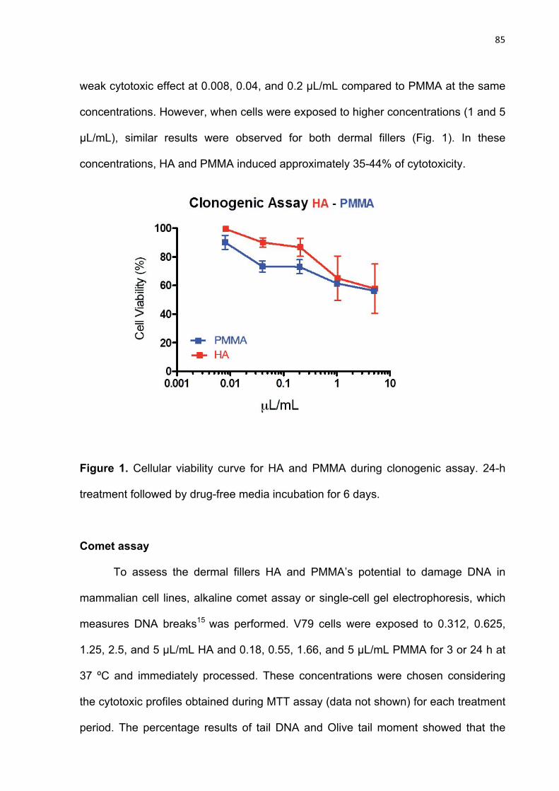
� ��
weak cytotoxic effect at 0.008, 0.04, and 0.2 μL/mL compared to PMMA at the same
concentrations. However, when cells were exposed to higher concentrations (1 and 5
μL/mL), similar results were observed for both dermal fillers (Fig. 1). In these
concentrations, HA and PMMA induced approximately 35-44% of cytotoxicity.
Figure 1. Cellular viability curve for HA and PMMA during clonogenic assay. 24-h
treatment followed by drug-free media incubation for 6 days.
Comet assay
To assess the dermal fillers HA and PMMA’s potential to damage DNA in
mammalian cell lines, alkaline comet assay or single-cell gel electrophoresis, which
measures DNA breaks15 was performed. V79 cells were exposed to 0.312, 0.625,
1.25, 2.5, and 5 μL/mL HA and 0.18, 0.55, 1.66, and 5 μL/mL PMMA for 3 or 24 h at
37 ºC and immediately processed. These concentrations were chosen considering
the cytotoxic profiles obtained during MTT assay (data not shown) for each treatment
period. The percentage results of tail DNA and Olive tail moment showed that the
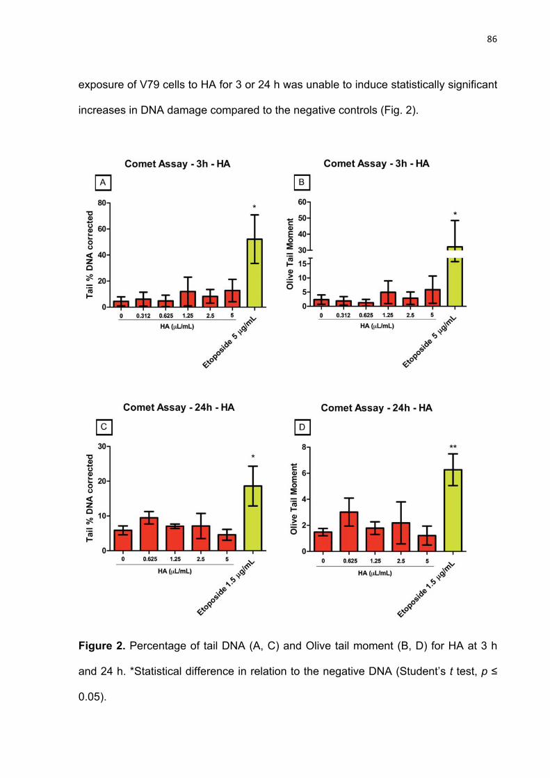
� ��
exposure of V79 cells to HA for 3 or 24 h was unable to induce statistically significant
increases in DNA damage compared to the negative controls (Fig. 2).
Figure 2. Percentage of tail DNA (A, C) and Olive tail moment (B, D) for HA at 3 h
and 24 h. *Statistical difference in relation to the negative DNA (Student’s t test, p ≤
0.05).
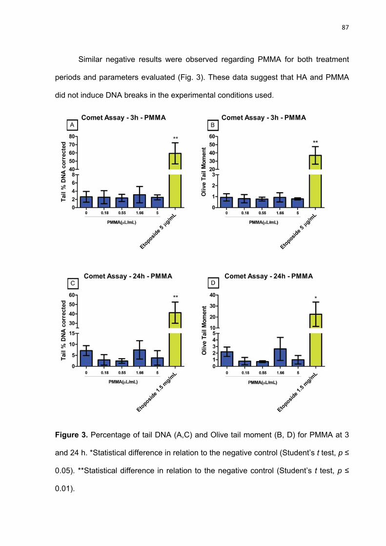
� ��
Similar negative results were observed regarding PMMA for both treatment
periods and parameters evaluated (Fig. 3). These data suggest that HA and PMMA
did not induce DNA breaks in the experimental conditions used.
Figure 3. Percentage of tail DNA (A,C) and Olive tail moment (B, D) for PMMA at 3
and 24 h. *Statistical difference in relation to the negative control (Student’s t test, p ≤
0.05). **Statistical difference in relation to the negative control (Student’s t test, p ≤
0.01).

� �
Micronucleus assay
MNvit assay was performed to evaluate the potential of the dermal fillers HA
and PMMA to induce chromosome mutation. To further warrant performing
micronucleus formation analysis in concentrations providing the appropriate range of
cytotoxicity (up to 55 ± 5% of cytotoxicity), viability was first calculated by a
replication index in all cultures treated. As illustrated in Tables 1 and 2, neither HA
nor PMMA showed mutagenic effects.
Table 1. Micronucleus assay for HA for periods of 3 and 21 h.
Group Replication index
(%)
Frequency of micronucleated
binucleated cells3
3 h 21 h 3 h 21 h
NC1 100.00 100.00 0.607 ± 0.453 1.111 ± 0.234
HA 0.625 μL/mL 100.29 96.45 0.388 ± 0.153 1.017 ± 0.044
HA 1.25 μL/mL 97.27 98.95 0.345 ± 0.346 0.883 ± 0.275
HA 2.5 μL/mL 99.71 102.54 0.515 ± 0.021 1.084 ± 0.301
HA 5 μL/mL 101.11 101.57 0.366 ± 0.057 0.805 ± 0.039
PC2 70.23 33.98 1.530 ± 0.981 3.334 ± 0.527**
1Negative control (100% Dulbecco’s modified eagle medium)
2Positive control (0.8 μg/mL colchicine)
3Frequency of micronucleated binucleated cells expressed in M ± SD
**Student’s t test (p ≤ 0.01).

� �
Table 2. Micronucleus assay for PMMA for periods of 3 and 21 h.
Group Replication index
(%)
Frequency of micronucleated
binucleated cells3
3 h 21 h 3 h 21 h
NC1 100.00 100.00 0.292 ± 0.173 1.050 ± 0.391
PMMA 0.625 μL/mL 103.51 91.52 0.238 ± 0.043 1.117 ± 0.407
PMMA 1.25 μL/mL 103.96 99.86 0.340 ± 0.067 0.950 ± 0.265
PMMA 2.5 μL/mL 101.66 98.33 0.189 ± 0.087 0.750 ± 0.180
PMMA 5 μL/mL 102.11 96.25 0.309 ± 0.014 0.933 ± 0.301
PC2 55.81 72.92 3.174 ± 1.977 23.400 ± 6.006**
1Negative control (100% Dulbecco’s modified eagle medium)
2Positive control (0.5 μg/mL etoposide)
3Frequency of micronucleated binucleated cells expressed in M ± SD
**Student’s t test (p ≤ 0.01).
Neither HA nor PMMA were able to increase micronucleus frequency in
binucleated cells compared to the negative control during both periods of treatment
evaluated (Fig. 4).

� ��
Figure 4. Mutagenicity assay of HA and PMMA during 3-h and continuous treatment.
**Statistical difference in relation to the negative control (Student’s t test, p ≤ 0.01).
Discussion
Studies evaluating the biocompatibility of HA and PMMA as dermal fillers have
remained limited. In the present research, two substances widely used by
dermatologists and plastic surgeons were examined for their viability to induce
� ��
potentially genotoxic and mutagenic effects in V79 cells. Extreme concentrations
were used given the possibility of cell damage. According to our results, both HA and
PMMA (1 and 5 μL/mL) induced a weak cell viability (35-44%) decrease when cells
were exposed for 24 h followed by incubation in drug-free media. This cytotoxic effect
corroborates the findings of Boeckel et al. (2014)25 who evaluated the osteoblastic
cell line (OFCOL II) with 48 h MTT assay and verified that groups with HA inhibited
the growth of OFCOL II cells.
The cell viability reduction verified in the present study could be related to the
molecular weight of HA applied. In support, scientific literature demonstrates that
high molecular weight HA (>1000 kDa) exhibits anti-angiogenic and anti-
inflammatory tissue effects, as well as reduces cellular migration, proliferation and
division.26 By contrast, low molecular weight HA (<50 kDa) stimulates cellular
proliferation and causes pro-angiogenic and pro-inflammatory effects.27,28 The
substance used in this research originated in bacterial fermentation and has low
molecular weight.29 Such discrepancies among the effects of molecular weight on cell
viability can be speculated upon given its influence on specific cells depending on the
HA concentrations applied. Hyaluronidase enzymes also seem to play an elucidative
role; once the substance is broken into smaller molecules, those molecules could
stimulate a biological event.28,30 Moreover, HA can bind with cellular membrane
receptors such as CD44 and receptors for HA-mediated motility (RHAMM) involved
in intra and intercellular signaling and thus regulate cellular proliferation.26,31
Via clonal survival assay, it was possible to verify a loss of 38.54% and
48.63% of V79 cell viability in the presence of 1 and 5 μL/mL PMMA, respectively. In
studying PMMA as an adjunct to bone regeneration, Savadkoohi et al. (2008)32 found
that the substance was cytotoxic by using a cell line from giant cell tumors. More
� ��
recently, Chae et al. (2014)33 detected a 10% reduction in the L929 cell line (i.e.,
mice fibroblasts) when exposed to PMMA during MTT assay. In MTT viability assays
using mesenchymal stem cells, however, such a substance could not inhibit cellular
viability.34 In sum, all of these differences can be explained by the use of a diversity
of cell lines, exposure times, cytotoxicity assays, and types of materials for bioplasty
and orthopedic techniques. In clinical reality, however, PMMA is the material that
most often triggers adverse reactions, including those involving tissue necrosis and
foreign body granuloma, when applied in bioplasty techniques.7,14,35
Concerning genotoxicity and mutagenicity, both substances tested induced
neither significant DNA damage nor micronucleus formation in the binuclear cells
analyzed, according to the experimental protocol adopted in our research. These
findings are important given the wide clinical use of HA and PMMA in both healthy
and ill patients.
Currently, little is known about the biocompatibility of dermal fillers. Since most
studies evaluate clinical benefits or esthetic satisfaction, most information about
adverse effects comes from case reports.1,7,14,35,36 HA has seemed to play a role in
preventing DNA damage in ophthalmological studies.37,38 Similarly, Lamberti et al.
(1998)39 and Chae et al. (2014)33 did not consider PMMA to be genotoxic or
mutagenic when evaluating its use in orthopedics. However, in an in vitro study,
Bigatti et al. (1994)40 observed an increase in the micronucleus frequency of cultured
human lymphocytes’ binuclear cells after exposure to PMMA cement. It is possible to
speculate that this difference stems from the material polymerization that releases
small amounts of monomer; in vivo, they are quickly eliminated, though could trigger
genotoxicity in vitro. Nevertheless, such did not apply in our study since the products
used derived from a syringe ready for use.
� ��
In sum, HA and PMMA showed weak cytotoxic effects when colony formation
was evaluated in V79 cells 6 d after 24 h exposure. Furthermore, no genotoxic or
mutagenic effects were detected after 3 or 24 h of exposure. New investigations are
necessary to clarify possible causes of unwanted effects related to these
compounds, as well as to determine their safety and efficacy when used in the
perioral region, lips, and chin, which are areas of interest to dentists.
Funding
None.
Conflict of interests
None declared.
References
1- Shahrabi Farahani S, Sexton J, Stone JD, Quinn K, Woo SB. Lip nodules
caused by hyaluronic acid filler injection: report of three cases. Head
Neck Pathol. 2012; 6 :16-20. doi: 10.1007/s12105-011-0304-9.
2- Medeiros CC, Borghetti RL, Nicoletti N, da Silva VD, Cherubini K, Salum FG,
de Figueiredo MA. Polymethylmethacrylate dermal fillers: evaluation of
the systemic toxicity in rats. Int J Oral Maxillofac Surg. 2014; 43: 62-67. doi:
10.1016/j.ijom.2013.06.009.
3- Vargas KF, Borghetti RL, Moure SP, Cherubini K, de Figueiredo MA. Local
and systemic tissue response submitted to injection of 2 and 30%
polymethylmethacrylate in rats' tongue. Gerodontology. 2014. doi:
10.1111/ger.12108.

� ��
4- Lemperle G, Morhenn V, Charrier U. Human histology and persistence of
various injectable filler substances for soft tissue augmentation.
Aesthetic Plast Surg. 2003; 27: 354-366.
5- Loureiro Borghetti R , de Vargas KF, Pozatti Moure S, Gonçalves Salum F, de
Figueiredo MA. Clinical and histologic evaluation of effects of hyaluronic
acid in rat tongue. Oral Surg Oral Med Oral Pathol Oral Radiol. 2012; 113:
488-494. doi: 10.1016/j.tripleo.2011.04.023.
6- Karagozoglu KH, van der Waal I. Polyacrylamide soft tissue filler nodule
mimicking a mucoepidermoid carcinoma. Int J Oral Maxillofac Surg. 2008;
37: 578-580.
7- da Costa Miguel MC, Nonaka CFW, dos Santos JN, Germano AR, de Souza
LB. Oral foreign body granuloma: unusual presentation of a rare adverse
reaction to permanent injectable cosmetic filler. Int J Oral Maxillofac Surg.
2009; 38:385-387.
8- Feio PSQ, Gouvêa AF, Jorge J, Lopes MA. Oral adverse reactions after
injection of cosmetic fillers: report of three cases. Int J Oral Maxillofac
Surg. 2013; 42: 432-435.
9- Romagnoli M, Belmontesi M. Hyaluronic acid-based fillers: theory and
practice. Clin Dermatol. 2008; 26: 123-159.
10- Vargas KF, Borghetti RL, Moure SP, Salum FG, Cherubini K, de Figueiredo
MA. Use of polymethylmethacrylate as permanent filling agent in the jaw,
mouth and face regions - implications for dental practice. Gerodontology.
2012; 29: 16-22. doi: 10.1111/j.1741-2358.2011.00479.x.

� ��
11- Medeiros CC, Cherubini K, Salum FG, de Figueiredo MA. Complications
after polymethylmethacrylate (PMMA) injections in the face: a literature
review. Gerodontology. 2014; 31: 245-250.
12- Moure SP, de Vargas KF, Borghetti RL, Salum FG, Cherubini K, da Silva VD,
de Figueiredo MA. Clinical and pathological characteristics of
polymethylmethacrylate and hyaluronic acid in the rat tongue. Int J Oral
Maxillofac Surg. 2012; 41: 1296-1303.
13- Pignatti M, Pedone A, Baccarani A, Guaraldi G, Orlando G, Lombardi M, de
Santis G. High-density hyaluronic acid for the treatment of HIV-related
facial lipoatrophy. Aesthetic Plast Surg. 2012; 36: 180-185. doi:
10.1007/s00266-011-9771-3.
14- De Castro ACB, Collares MVM, Portinho CP, Dias PC, di Angeli Pinto R.
Necrose facial extensa após infiltração com polimetilmetacrilato. Rev
Bras Otorrinolaringol. 2007; 73: 850.
15- Pálmai-Pallag T, Bachrati CZ. Inflammation-induced DNA damage and
damage-induced inflammation: a vicious cycle. Microbes Infect. 2014; 16:
822-832.
16- Bradley MO, Bhuyan B, Francis MC, Langenbach R, Peterson A, Huberman
E. Mutagenesis by chemical agents in V79 Chinese hamster cells: a
review and analysis of the literature. A report of the Gene-Tox Program.
Mutat Res. 1981; 87: 81-142.
17- Organisation for Economic Co-operation and Development (OECD). Test no
487: In vitro mammalian cell micronucleus test. In: Guidelines 448 for
testing of chemicals section 4 health effects. 2014; 1-26.

� ��
18- Franken NAP, Rodermond HM, Stap J, Haveman J, van Bree C. Clonogenic
assay of cells in vitro. Nat Protoc. 2006; 1: 2315- 2319.
19- Machado M da S, Villela IV, Moura DJ, Rosa RM, Salvador M, Lopes N,
Braga AL, Roesler R, Saffi J, Henriques JAP. 3'3-ditrifluoromethyldiphenyl
diselenide a new organoselenium compound with interesting
antigenotoxic and antimutagenic activities. Mutat Res. 2009; 673: 133-
140.
20- Burlinson B, Tice RR, Speit G, Agurell E, Brendler-Schwaab SY, Collins AR,
Escobar P, Honma M, Kumaravel TS, Nakajima M, Sasaki YF, Thybaud V,
Uno Y, Vasquez M, Hartmann A. In vivo comet assay workgroup, part of
the Fourth International Workgroup on Genotoxicity Testing. Fourth
international workgroup on genotoxicity testing: results of the in vivo
comet assay workgroup. Mutat Res. 2007; 627: 31-35.
21- Gonçalves TS, Menezes LM, Trindade C, Machado MS, Thomas P, Fenech
M, Henriques JAP. Cytotoxicity and genotoxicity of orthodontic bands
without silver soldered joints. Mutat Res Genet Toxicol Environ Mutagen.
2014; 762: 1-8.
22- Azqueta A, Slyskova J, Langie SA, O’Neill Gaivão I, Collins A. Comet assay
to measure DNA repair: approach and applications. Front Genet. 2014;
5:288. doi: 10.3389/fgene.2014.00288.
23- Azqueta A, Meier S, Priestley C, Gutzkow KB, Brunborg G, Sallette J,
Soussaline F, Collins A. The influence of scoring method on variability in
results obtained with the comet assay. Mutagenesis. 2011; 26: 393-399.
� ��
24- Decordier I, Papine A, Vande Loock K, Plas G, Soussaline F, Kirsch-Volders
M. Automated image analysis of micronuclei by IMSTAR for
biomonitoring. Mutagenesis. 2011; 26: 163-168.
25- Boeckel DG, Shinkai RS, Grossi ML, Teixeira ER. In vitro evaluation of
cytotoxicity of hyaluronic acid as an extracellular matrix on OFCOL II
cells by the MTT assay. Oral Surg Oral Med Oral Pathol Oral Radiol. 2014;
117: 423-428. doi: 10.1016/j.oooo.2012.07.486.
26- Yang C, Cao M, Liu H, He Y, Xu J, Du Y, Liu Y, Wang W, Cui L, Hu J, Gao F.
The high and low molecular weight forms of hyaluronan have distinct
effects on CD44 clustering. J Biol Chem. 2012; 3287: 43094-43107. doi:
10.1074/jbc.M112.349209.
27- Noble PW. Hyaluronan and its catabolic products in tissue injury and
repair. Matrix Bio.l 2002; 21: 25-29.
28- Gao F, Yang CX, Mo W, Liu YW, He YQ. Hyaluronan oligosaccharides are
potential stimulators to angiogenesis via RHAMM mediated signal
pathway in wound healing. Clin Invest Med. 2008; 31:106-116.
29- Marcellin E, Steen JA, Nielsen LK. Insight into hyaluronic acid molecular
weight control. Appl Microbiol Biotechnol. 2014; 98: 6947-6956. doi:
10.1007/s00253-014-5853-x.
30- Fuchs K, Hippe A, Schmaus A, Homey B, Sleeman JP, Orian-Rousseau V.
Opposing effects of high- and low-molecular weight hyaluronan on
CXCL12-induced CXCR4 signaling depend on CD44. Cell Death Dis. 2013;
4:819. doi: 10.1038/cddis.2013.364.

� �
31- Collins MN, Birkinshaw C. Hyaluronic acid based scaffolds for tissue
engineering-A review. Carbohydr Polym. 2013; 92: 1262-1279. doi:
10.1016/j.carbpol.2012.10.028.
32- Savadkoohi DG, Sadeghipour P, Attarian H, Sardari S, Eslamifar A,
Shokrgozar MA. Cytotoxic effect of drugs eluted from
polymethylmethacrylate on stromal giant-cell tumour cells: an in vitro
study. J Bone Joint Surg Br. 2008; 90: 973-979. doi: 10.1302/0301-
620X.90B7.19692.
33- Chae MJ, Kim DW, Kim YJ, Kweon HJ, Cho YJ, Choi SY, Lee HR. Toxicity
assessment of a novel silk fibroin and polymethylmethacrylate
composite material. Mol Cell Toxicol. 2014; 10: 277-283. doi
10.1007/s13273-014-0031-x.
34- Pauksch L, Hartmann S, Szalay G, Alt V, Lips KS. In vitro assessment of
nanosilver-functionalized PMMA bone cement on primary human
mesenchymal stem cells and osteoblasts. PLoS One. 2014; 9: e114740.
doi: 10.1371/journal.pone.0114740.
35- Jham BC, Nikitakis NG, Scheper MA, Papadimitriou JC, Levy BA, Rivera H.
Granulomatous foreign-body reaction involving oral and perioral tissues
after injection of biomaterials: a series of 7 cases and review of the
literature. J Oral Maxillofac Surg. 2009; 67: 280-285.
36- Edwards PC, Fantasia JE, Iovino R. Foreign body reaction to hyaluronic
acid (Restylane): an adverse outcome of lip augmentation. J Oral
Maxillofac Surg. 2006; 64: 1296-1299.

� �
37- Wu H, Zhang H, Wang C, Wu Y, Xie J, Jin X, Yang J, Ye J. Genoprotective
effect of hyaluronic acid against benzalkonium chloride-induced DNA
damage in human corneal epithelial cells. Mol Vis. 2011; 17: 3364-3370.
38- Ye J, Zhang H, Wu H, Wang C, Shi X, Xie J, He J, Yang J. Cytoprotective
effect of hyaluronic acid and hydroxypropyl methylcellulose against DNA
damage induced by thimerosal in Chang conjunctival cells. Graefes Arch
Clin Exp Ophthalmol. 2012; 250: 1459-1466. doi: 10.1007/s00417-012-2087-
4.
39- Lamberti L, Bigatti P, Rizzi P, Marchisio S, Rizzo E, Cannas M. In vivo
cytogenetic studies of the genotoxic effects of polymethylmethacrylate
employed in orthopaedics. J Mater Sci Mater Med. 1998; 9: 239-242.
40- Bigatti MP, Lamberti L, Rizzi FP, Cannas M, Allasia G. In vitro micronucleus
induction by polymethylmethacrylate bone cement in cultured human
lymphocytes. Mutat Res. 1994; 321: 133-137.

� ����
DISCUSSÃO COMPLEMENTAR
�����
� ����
5 DISCUSSÃO COMPLEMENTAR
Os materiais estéticos de preenchimento facial foram desenvolvidos com o
objetivo de suavizar rítides e vincos na região perioral, periorbital e glabela,
aumentar artificialmente as partes moles e corrigir defeitos faciais, sejam eles
congênitos ou provocados.60 Desde que foram disponibilizados no mercado, seu uso
tem sido vasto, posto que a técnica que os envolve é pouco invasiva, o período de
recuperação é curto, o custo é baixo quando comparado à cirurgia plástica e o
resultado é percebido com rapidez.6,61
Diante das vantagens expostas, em especial a financeira, e a valorização
excessiva da aparência rejuvenescida, o emprego da bioplastia tem sido
indiscriminado e, muitas vezes, desnecessário. Com isso, um grande número de
reações adversas é reportado na literatura62-65 e divulgado pela mídia, cuja
gravidade torna imprescindível o estudo desses biomateriais.
O AH e o PMMA podem suscitar efeitos secundários, os quais são muitas
vezes identificados por cirurgiões-dentistas, devido a sua área de atuação. Um
aspecto importante que impulsionou o prosseguimento dos estudos nessa linha de
pesquisa, foi o diagnóstico diferencial das lesões granulomatosas desencadeadas
por estes produtos, com patologias oriundas do tecido adiposo (lipossarcoma) e das
glândulas salivares (lesões císticas e neoplásicas malignas e benignas). Sabe-se
que a aplicação destes materiais nos lábios e sulcos nasogenianos, é feita em área
anatômica próxima das glândulas salivares acessórias. Assim, quando uma reação
do tipo corpo estranho ocorre, pode originar uma lesão que clinicamente confunde-
se com uma patologia de outra natureza. Soma-se a isto, a dificuldade de se obter
informações importantes durante a anamnese, pois muitas vezes o paciente omite

� ����
este tipo de procedimento, ou, quando o menciona, desconhece o nome ou o tipo do
produto que foi utilizado no seu tratamento. Outrossim, a grande procura pelo
rejuvenescimento e o excesso de preocupação estética, popularizou a bioplastia,
levando profissionais de diversas áreas de atuação a utilizarem este recurso. No
entanto, muitos não possuem suficiente conhecimento técnico e científico, o que
pode ocasionar muitos dos problemas reportados na literatura.
Considerando a discussão cada vez mais enfática acerca da ética no
emprego de animais de laboratório para os testes de substâncias, a abordagem in
vitro tem sido apreciada como alternativa ao uso dos mesmos. Apesar do enfoque
ideal para análise da biocompatibilidade ser a avaliação clínica, os percalços éticos
e legais e a padronização metodológica dificultam a sua execução,66 o que reforça a
escolha de ensaios distintos que sejam precisos e forneçam informações seguras
sobre as características dos produtos. Embora permitam a execução dos estudos de
maneira rápida e tenham baixo custo, o recurso in vitro apresenta uma técnica
sensível e um alto risco de contaminação, conforme circunstância enfrentada na
presente pesquisa. A sensibilidade do método levou à repetição de alguns
experimentos envolvendo as leveduras. Em alguns momentos pode-se detectar a
falha técnica, talvez pelo grande número de grupos e pelo fato dos testes serem
efetuados sempre em duplicata. A contaminação foi identificada pelo exame
microscópico, que verificou a presença de microorganismo do ambiente.
Dos procedimentos utilizados na averiguação qualitativa da citotoxicidade, o
halo de inibição é o experimento empregado nos testes de sensibilidade a
antimicrobianos67,68 e materiais dentários.55 O princípio desse ensaio é determinar a
� ����
toxicidade do produto por meio da ausência de crescimento celular adjacente ao
local onde o mesmo foi aplicado.
A avaliação quantitativa da viabilidade celular pode ser realizada através de
métodos colorimétricos utilizando-se corantes, tais como o brometo 3 - [4,5-dimetil-
tiazol - 2-il] - 2,5 - difenil-tetrazolio (MTT).69 O MTT consiste em um sal amarelo que
é reduzido pela atividade da enzima desidrogenase mitocondrial resultando em um
sal de formazan de cor púrpura. Sabe-se que a redução mencionada ocorre
exclusivamente nas células vivas.70,71 Desta maneira, a viabilidade celular pode ser
determinada pela intensidade da coloração púrpura, que é proporcional à quantidade
de cristais de formazan constituídos. Na presente pesquisa, o MTT foi empregado
como teste de escolha de doses não citotóxicas, de modo que permitissem o
desenvolvimento do ensaio cometa. Com finalidade semelhante, o ensaio
clonogênico também é ponderado, uma vez que é um teste de sobrevivência que
avalia a habilidade de uma única célula crescer e formar colônias.72,73 Nesse estudo,
o clonogênico foi adotado como teste de viabilidade celular, para determinar a
citotoxicidade do AH e PMMA nos períodos de exposição de 3h e 24h da linhagem
V79 às substâncias.
Deve-se considerar que a biocompatibilidade representa um processo
complexo, o qual envolve testes in vitro e in vivo. No presente trabalho, os materiais
de preenchimento foram avaliados in vitro, sob o aspecto da citotoxicidade,
genotoxicidade e mutagenicidade, utilizando-se Saccharomyces cerevisiae como
organismo modelo e optando-se pelo emprego dos ensaios de micronúcleos e
cometa em cultura celular permanente de fibroblastos pulmonares de hamster
Chinês (linhagem V79). Esses recursos são bem estabelecidos na literatura para
investigação de efeitos tóxicos oriundos de alguma substância.52,55,57,73,74

� ����
Entre os testes de análise da genotoxicidade, o ensaio de micronúcleos
identifica danos cromossômicos após a divisão celular. Essa técnica é a mais
utilizada para detecção de agentes clastogênicos (que quebram cromossomos) e
aneugênicos (que induzem aneuploidia ou segregação cromossômica anormal).75,76
Por outro lado, o ensaio cometa detecta danos no DNA, sejam por quebras de fita
simples ou dupla, sítios abásicos ou álcali-lábeis, ou, ainda, danos do tipo ligações
cruzadas.76,77
No que concerne aos efeitos citotóxicos analisados, o AH 20mg/mL e o
PMMA 10% induziram significativa perda de viabilidade celular nos testes
quantitativos em modelo eucarioto S. cerevisiae. No ensaio clonogênico da linhagem
V79, o AH 20mg/mL e o PMMA 30% demonstraram fraca citotoxicidade na maior
concentração (5 μL/mL) e, a menor (0.008 μL/mL), atingiu níveis de viabilidade
próximos ao controle negativo. No teste MTT, empregado com o objetivo de escolher
as doses para o ensaio cometa, ambas as substâncias não inviabilizaram o
crescimento dos fibroblastos (V79). Além disso, a presença de genotoxicidade não
foi percebida em 3h e 24h de tratamento com os materiais. Da mesma maneira, o
AH 20mg/mL e o PMMA 30% foram incapazes de induzir aumento na formação de
micronúcleos em células binucleadas. Alijotas - Reig et al. (2013)78 e Medeiros et al.
(2014)39 sugeriram como perspectivas, a avaliação da genotoxicidade dos materiais
de preenchimento, uma vez que os mesmos podem desencadear reações adversas
à distância, como em órgãos de metabolismo.
O AH apresenta efeitos contraditórios sobre a inibição e a proliferação celular.
De acordo com a literatura, o AH de baixo peso molecular estimula a angiogênese79
e aumenta a proliferação celular,80 enquanto o AH de alto peso molecular apresenta
efeito anti-angiogênico e inibitório sobre o crescimento celular.79 As substâncias de

� ����
preenchimento de origem animal (crista de galo) são consideradas de alto peso
molecular e apresentam baixas concentrações de AH. Os preenchedores de origem
não animal ou bacteriana (Streptococcus), porém, possuem baixo peso molecular e
concentrações maiores de AH.11,81 Atribui-se ao peso molecular, à dose e à
concentração do material, as discrepâncias encontradas sobre a citotoxicidade.
Poucos são os estudos a respeito da biocompatibilidade do PMMA como
recurso de preenchimento facial. Devido à sua presença em materiais largamente
utilizados na oftalmologia, ortopedia e odontologia, a literatura retrata as implicações
da substância dentro dessa linha de pesquisa. Wang et al. (2014)82 encontraram
redução na viabilidade de células epiteliais de lentes de contato tratadas com
PMMA. Akin et al. (2013)83 verificaram ausência de citotoxicidade em cultura de
L929 (linhagem de fibroblastos de camundongo) testada com resina acrílica a base
de PMMA, empregada em próteses dentárias. Bural et al. (2011)84 detectaram em
células L929 que quanto maior for a presença do monômero residual do PMMA,
maior é a citotoxicidade.
Ainda que não consigam captar todas as consequências adversas, os
testes in vitro85,86 constituem a base da pesquisa para o desenvolvimento de novos
produtos e funcionam como uma seleção preliminar dos materiais a fim de que
prossigam com as etapas seguintes de análise. Estudos in vivo13,24,35,38,39,87
identificaram a presença de reação inflamatória local e à distância, em órgãos de
metabolismo e excreção, a partir do uso do AH, PMMA e silicone. Da mesma
maneira, estudos clínicos45,88,89 apontaram os efeitos indesejáveis decorrentes da
bioplastia. Sob a perspectiva clínica, não parece razoável, tampouco interessante,
uma substância ter caráter definitivo no organismo, devido a sua capacidade de se
deslocar nos tecidos e provocar complicações. As alterações desfavoráveis, como o

� ����
granuloma de corpo estranho, deveriam ser facilmente removidas através de
procedimentos cirúrgicos, porém, algumas vezes, se tornam sequelas de difícil
manejo e tratamento.
Diante da metodologia empregada nesta pesquisa e, a partir dos resultados
obtidos, pode-se concluir que o AH e o PMMA se mostraram citotóxicos em S.
cerevisiae e apresentaram fraca citotoxicidade em cultura de células V79. No
entanto, efeitos genotóxicos ou mutagênicos não foram identificados em 3 h ou 24 h
de exposição das substâncias às células V79. Novas investigações com diferentes
tempos de tratamento e protocolos de mecanismo de ação, genotoxicidade e
mutagenicidade são necessários para esclarecer as possíveis causas das
repercussões desagradáveis provenientes do recurso da bioplastia. Ainda, torna-se
fundamental determinar a eficácia e segurança do AH e PMMA, uma vez que são
recursos frequentemente empregados na região perioral, reconhecida área de
interesse do cirurgião-dentista. A demanda de novos produtos é crescente e carece
de informações científicas minuciosas a respeito de sua aplicação.
�

� ����
REFERÊNCIAS
�����
� ���
REFERÊNCIAS
1- Fenske NA, Lober CW. Structural and functional changes of normal aging
skin. J Am Acad Dermatol. 1986 Oct;15(4 Pt 1):571-85.
2- Atiyeh BS, Dibo SA. Nonsurgical nonablative treatment of aging skin:
radiofrequency technologies between aggressive marketing and evidence-
based efficacy. Aesth Plast Surg. 2009 May;33(3):283-94.
3- Gold MH. Use of hyaluronic acid fillers for the treatment of the aging face. Clin
Interv Aging. 2007 Sep;2(3):369-76.
4- Rhee SM, You HJ, Han SK. Injectable tissue-engineered soft tissue for tissue
augmentation. J Korean Med Sci. 2014 Nov;29(Suppl 3):170-5.
5- Vargas AF, de Amorim NG, Pitanguy I. Complicações tardias dos
preenchimentos permanentes. Rev Soc Bras Cir Plást. 2009 Jan; 24(1): 71-
81.
6- De Maio M. The minimal approach: an innovation in facial cosmetic
procedures. Aesthetic Plast Surg. 2004 Sep-Oct;28(5):295-300.
7- Sociedade Brasileira de Cirurgia Plástica. Acessado em 15/11/2014.
Disponível em: http://www2.cirurgiaplastica.org.br/de-acordo-com-a-isaps-
brasil-lidera-ranking-de-cirurgias-plasticas-no-mundo/
8- Edwards PC, Fantasia JE. Review of long-term adverse effects associated
with the use of chemically-modified animal and nonanimal source hyaluronic
acid dermal fillers. Clin Interv Aging. 2007 Jan;2(4):509-19.
9- John HE, Price RD. Perspectives in the selection of hyaluronic acid fillers for
facial wrinkles and aging skin. Patient Prefer Adherence. 2009 Nov;3: 225-30.

� ���
10- Anderegg U, Simon JC, Averbeck M. More than just a filler - the role of
hyaluronan for skin homeostasis. Exp Dermatol. 2014 May;23(5):295-303.
11- Rzany B, Zielke H. Aspectos gerais das substâncias de preenchimento. In: de
Maio M, Rzany B, organizadores. Substâncias de preenchimento em medicina
estética. São Paulo: Livraria Santos; 2007. p. 1-9.
12- Rohrich RJ, Monheit G, Nguyen AT, Brown SA, Fagien S. Soft-tissue filler
complications: the important role of biofilms. Plast Reconstr Surg. 2010
Apr;125(4):1250-6. Erratum in: Plast Reconstr Surg. 2010 Jun;125(6):1850.
13- Loureiro Borghetti R, de Vargas KF, Pozatti Moure S, Gonçalves Salum F, de
Figueiredo MA. Clinical and histologic evaluation of effects of hyaluronic acid
in rat tongue. Oral Surg Oral Med Oral Pathol Oral Radiol. 2012
Apr;113(4):488-94.
14- Romagnoli M, Belmontesi M. Hyaluronic acid-based fillers: theory and
practice. Clin Dermatol. 2008 Mar-Apr;26(2):123-59.
15- Hedén, P, Sellman G, von Wachenfeldt M, Olenius M, Fagrell D. Body
shaping and volume restoration: the role of hyaluronic acid. Aesthetic Plast
Surg. 2009 May;33(3):274-82.
16- Bogdan Allemann I, Baumann L. Hyaluronic acid gel (Juvéderm) preparations
in the treatment of facial wrinkles and folds. Clin Interv Aging. 2008
Dec;3(4):629-34.
17- Matarasso SL, Carruthers JD, Jewell ML. Consensus recommendations for
soft-tissue augmentation with nonanimal stabilized hyaluronic acid
(Restylane). Plast Reconstr Surg 2006 Mar;117(Suppl 3):3-34.
18- Vedamurthy M. Soft tissue augmentation - Use of hyauronic acid as dermal
filler. Indian J Dermatol Venereol Leprol. 2004 Nov-Dec;70(6):383-7.
� ����
19- Nácul AM. Contour of the lower third of the face using an intramusculary
injectable implant. Aesthetic Plast Surg. 2005 Jul-Aug;29(4):222-9.
20- Vargas KF, Borghetti RL, Moure SP, Salum FG, Cherubini K, de Figueiredo
MA. Use of polymethylmethacrylate as permanent filling agent in the jaw,
mouth and face regions - Implications on dental practice. Gerodontology. 2012
Jun;29(2):16-22.
21- Quintas RC, de França ER, de Petribú KC, Ximenes RA, Quintas LF,
Cavalcanti EL et al. Treatment of facial lipoatrophy with
polymethylmethacrylate among patients with human immunodeficiency
virus/acquired immunodeficiency syndrome (HIV/AIDS): impact on the quality
of life. Int J Dermatol. 2014 Apr;53(4):497-502.
22- Medeiros CC, Cherubini K, Salum FG, de Figueiredo MA. Complications after
polymethylmethacrylate (PMMA) injections in the face: a literature review.
Gerodontology. 2014 Dec;31(4):245-50.
23- Lemperle G, Morhenn V, Charrier U. Human histology and persistence of
various injectable filler substances for soft tissue augmentation. Aesthetic
Plast Surg. 2003 Sep-Oct;27(5):354-66.
24- Lemperle G, Morhenn VB, Pestonjamasp V, Gallo RL. Migration studies and
histology of injectable microspheres of different sizes in mice. Plast Reconstr
Surg. 2004 Apr;113(5):1380-90.
25- Gelfer A, Carruthers A, Carruthers J, Jang F, Bernstein SC. The natural
history of polymethylmethacrylate microspheres granulomas. Dermatol Surg.
2007 May; 33(5):614-20.
26- Mandel L, Addison S, Clark M. Buccal reaction to silicone cosmetic filler. J
Am Dent Assoc. 2010 Feb;141(2):162-6.
� ����
27- Mello DF, Gonçalves KC, Fraga MF, Perin LF, Helene A Jr. Local
complications after industrial liquid silicone injection – case series. Rev Col
Bras Cir. 2013 Jan-Feb;40(1):37-42.
28- Dornelas MT, Correa MPD, de Sá Jr CAC, Barra FML, Dornelas MC,
Sant’Anna LL et al. Siliconomas. Rev. Bras. Cir. Plást. 2011 Jan-Mar;
26(1):16-21.
29- Maly A, Regev E, Meir K, Maly B. Tissue reaction to liquid silicone simulating
low-grade liposarcoma following lip augmentation. J Oral Pathol Med 2004
May; 33(5):314-6.
30- Sanchis-Bielsa JM, Bagan JV, Poveda R, Salvador I. Foreign body
granulomatous reactions to cosmetic fillers: a clinical study of 15 cases. Oral
Surg Oral Med Oral Pathol Oral Radiol Endod 2009 Aug; 108(2):237-41.
31- Duong T, Schonfeld AJ, Yungbluth M, Slotten R. Acute pneumopathy in a
nonsurgical transsexual. Chest. 1998 Apr;113(4):1127-9.
32- Bartsich S, Wu JK. Silicon emboli syndrome: A Sequela of clandestine liquid
silicone injections. A case report and review of the literature. J Plast Reconstr
Aesthet Surg. 2010 Jan;63(1):1-3.
33- Bigatà X, Ribera M, Bielsa I, Ferrándiz C. Adverse granulomatous reaction
after cosmetic dermal silicone injection. Dermatol Surg. 2001 Feb; 27(2):198-
200.
34- Baumann LS, Halem ML. Lip silicone granulomatous foreign body reaction
treated with Aldara (imiquimod 5%). Dermatol Surg. 2003 Apr; 29(4):429-32.
35- Moure SP, de Vargas KF, Borghetti RL, Salum FG, Cherubini K, da Silva VD,
de Figueiredo MA. Clinical and pathological characteristics of

� ����
polymethylmethacrylate and hyaluronic acid in the rat tongue. Int J Oral
Maxillofac Surg. 2012 Oct;41(10):1296-303.
36- Patel VJ, Bruck MC, Katz BE. Hipersensitivity reaction to hyaluronic acid with
negative skin testing. Plast Reconstr Surg. 2006 May;117(6):92-4.
37- Edwards PC, Fantasia JE, Iovino R. Foreign body reaction to hyaluronic acid
(Restylane): an adverse outcome of lip augmentation. J Oral Maxillofac Surg.
2006 Aug;64(8):1296-9.
38- Rosa SC, de Magalhães AV, de Macedo JL. An experimental study of tissue
reaction to polymethyl methacrylate (PMMA) microspheres (Artecoll) and
dimethylsiloxane (DMS) in the mouse. Am J Dermatopathol. 2008
Jun;30(3):222-7.
39- Medeiros CC, Borghetti RL, Nicoletti N, da Silva VD, Cherubini K, Salum FG
et al. Polymethylmethacrylate dermal fillers: evaluation of the systemic toxicity
in rats. Int J Oral Maxillofac Surg 2014 Jan; 43 (1): 62-7.
40- Sánchez-Carpintero I, Candelas D, Ruiz-Rodríguez R. Dermal fillers: types,
indications, and complications. Actas Dermosifiliogr. 2010 Jun;101(5):381-93.
41- Owosho AA, Bilodeau EA, Vu J, Summersgill KF. Orofacial dermal fillers:
foreign body reactions, histopathologic features, and spectrometric studies.
Oral Surg Oral Med Oral Pathol Oral Radiol. 2014 May;117(5):617-25.
42- Da Costa Miguel MC, Nonaka CFW, dos Santos JN, Germano AR, de Souza
LB. Oral foreign body granuloma: unusual presentation of a rare adverse
reaction to permanent injectable cosmetic filler. Int J Oral Maxillofac Surg
2009 Apr; 38 (4):385-7.
43- Jham BC, Nikitakis NG, Scheper MA, Papadimitriou JC, Levy BA, Rivera H.
Granulomatous foreign-body reaction involving oral and perioral tissues after

� ����
injection of biomaterials: a series of 7 cases and review of the literature. J Oral
Maxillofac Surg. 2009 Feb;67(2): 280-5.
44- Quirino MR, Neves AC, Campos MS, Brandão AA, Anbinder AL. Oral
granuloma formation after injection of cosmetic filler. J Craniomaxillofac Surg.
2012 Oct;40(7):194-7.
45- Feio PSQ, Gouvêa AF, Jorge J, Lopes MA. Oral adverse reactions after
injection of cosmetic fillers: report of three cases. Int J Oral Maxillofac Surg
2013 Apr; 42(4): 432-5.
46- Requena C, Requena L, Alegre V, Serra C, Llombart B, Nagore E et al.
Adverse reaction to silicone simulating orofacial granulomatosis. J Eur Acad
Dermatol Venereol. 2014 Apr; 1-4.
47- De Castro ACB, Collares MVM, Portinho CP, Dias PC, Pinto RA. Necrose
facial extensa após infiltração com polimetilmetacrilato. Rev Bras
Otorrinolaringol 2007 Nov-Dec; 73(6):850.
48- Carruthers JD, Fagien S, Rohrich RJ, Weinkle S, Carruthers A. Blindness
caused by cosmetic filler injection: a review of cause and therapy. Plast
Reconstr Surg. 2014 Dec;134(6):1197-201.
49- Kim EG, Eom TK, Kang SJ. Severe visual loss and cerebral infarction after
injection of hyaluronic acid gel. J Craniofac Surg. 2014;25(2):684-6.
50- Park HJ, Jung KH, Kim SY, Lee JH, Jeong JY, Kim JH. Hyaluronic acid
pulmonary embolism: a critical consequence of an illegal cosmetic vaginal
procedure. Thorax. 2010 Apr;65(4):360-1.
51- Rogero SO, Lugão AB, Ikeda TI, Cruz AS. Teste in vitro de citotoxicidade:
estudo comparativo entre duas metodologias. Mat. Res. 2003 Jun; 6(3): 317-
20.
� ����
52- Matuo R, Sousa FG, Soares DG, Bonatto D, Saffi J, Escargueil AE, Larsen
AK, Henriques JA. Saccharomyces cerevisiae as a model system to study the
response to anticancer agents. Cancer Chemother Pharmacol. 2012
Oct;70(4):491-502.
53- De Oliveira IM, Degrandi TH, Jorge PM, Saffi J, Rosa RM, Guecheva TN,
Henriques JA. Dicholesteroyl diselenide: cytotoxicity, genotoxicity and
mutagenicity in the yeast Saccharomyces cerevisiae and in Chinese hamster
lung fibroblasts. Mutat Res Genet Toxicol Environ Mutagen. 2014 Mar
15;763:1-11.
54- Gonçalves TS, de Menezes LM, Ribeiro LG, Lindholz CG, Medina-Silva R.
Differences of cytotoxicity of orthodontic bands assessed by survival tests in
Saccharomyces cerevisiae. Biomed Res Int. 2014 Jan;2014:1-7.
55- Limberger KM, Westphalen GH, Menezes LM, Medina-Silva R. Cytotoxicity of
orthodontic materials assessed by survival tests in Saccharomyces cerevisiae.
Dent Mater. 2011 May;27(5):81-6.
56- Westphalen GH, Menezes LM, Prá D, Garcia GG, Schmitt VM, Henriques JA
et al. In vivo determination of genotoxicity induced by metals from orthodontic
appliances using micronucleus and comet assays. Genet Mol Res. 2008
Nov;7(4):1259-66.
57- Vasquez MZ. Combining the in vivo comet and micronucleus assays: a
practical approach to genotoxicity testing and data interpretation.
Mutagenesis. 2010 Mar;25(2):187-99.
58- Rosa RM, Moura DJ, Romano e Silva AC, Saffi J, Pêgas Henriques JA.
Antioxidant activity of diphenyl diselenide prevents the genotoxicity of several

� ����
mutagens in Chinese hamster V79 cells. Mutat Res. 2007 Jul 10;631(1):44-
54.
59- Rosa RM, Moura DJ, Melecchi MI, dos Santos RS, Richter MF, Camarão EB
et al. Protective effects of Hibiscus tiliaceus L. methanolic extract to V79 cells
against cytotoxicity and genotoxicity induced by hydrogen peroxide and tert-
butyl-hydroperoxide. Toxicol In Vitro. 2007 Dec;21(8):1442-52.
60- Cox SE, Adigun CG. Complications of injectable fillers and neurotoxins.
Dermatol Ther. 2011 Nov-Dec;24(6):524-36.
61- Smith KC. Reversible vs. nonreversible fillers in facial aesthetics: concerns
and considerations. Dermatol Online J. 2008 Aug;14(8):3.
62- Alijotas-Reig J, Garcia-Gimenez V. Delayed immune-mediated adverse
effects related to hyaluronic acid and acrylic hydrogel dermal fillers: clinical
findings, long-term follow-up and review of the literature. J Eur Acad Dermatol
Venereol. 2008 Feb;22(2):150-61.
63- Alijotas-Reig J. Recurrent urticarial vasculitis related to nonanimal hyaluronic
acid skin filler injection. Dermatol Surg. 2009 Feb;35(Suppl 1):395-7.
64- Bachmann F, Erdmann R, Hartmann V, Wiest L, Rzany B. The spectrum of
adverse reactions after treatment with injectable fillers in the glabellar region:
results from the injectable filler safety study. Dermatol Surg. 2009 Oct;35
(Suppl 2):1629-34.
65- Shahrabi Farahani S, Sexton J, Stone JD, Quinn K, Woo SB. Lip nodules
caused by hyaluronic acid filler injection: report of three cases. Head Neck
Pathol. 2012 Mar;6(1):16-20.
� ����
66- Murray PE, Lumley PJ, Ross HF, Smith AJ. Tooth slice organ culture for
cytotoxicity assessment of dental materials. Biomaterials. 2000
Aug;21(16):1711-21.
67- Iyamba JM, Wambale JM, Lukukula CM, za Balega Takaisi-Kikuni N. High
prevalence of methicillin resistant staphylococci strains isolated from surgical
site infections in Kinshasa. Pan Afr Med J. 2014 Aug ;18:322.
68- Rivadeneira J, Di Virgilio AL, Audisio MC, Boccaccini AR, Gorustovich AA.
Evaluation of the antibacterial effects of vancomycin hydrochloride released
from agar-gelatin-bioactive glass composites. Biomed Mater. 2015
Jan;10(1):015011.
69- Mosmann T. Rapid colorimetric assay for cellular growth and survival:
application to proliferation and cytotoxicity assays. J Immunol Methods. 1983
Dec;65(1-2):55-63.
70- Abe K, Matsuki N. Measurement of cellular 3-(4,5-dimethylthiazol-2-yl)-2,5-
diphenyltetrazolium bromide (MTT) reduction activity and lactate
dehydrogenase release using MTT. Neurosci Res. 2000 Dec;38(4):325-9.
71- Stockert JC, Blázquez-Castro A, Cañete M, Horobin RW, Villanueva A. MTT
assay for cell viability: Intracellular localization of the formazan product is in
lipid droplets. Acta Histochem. 2012 Dec;114(8):785-96.
72- Franken NA, Rodermond HM, Stap J, Haveman J, van Bree C. Clonogenic
assay of cells in vitro. Nat Protoc. 2006 Dec;1(5):2315-9.
73- Machado M da S, Villela IV, Moura DJ, Rosa RM, Salvador M, Lopes NP et
al. 3'3-ditrifluoromethyldiphenyl diselenide: a new organoselenium compound
with interesting antigenotoxic and antimutagenic activities. Mutat Res. 2009
Mar;673(2):133-40.

� ����
74- Kawaguchi S, Nakamura T, Yamamoto A, Honda G, Sasaki YF. Is the comet
assay a sensitive procedure for detecting genotoxicity? J Nucleic Acids. 2010
Nov;2010:541050.
75- Fenech M, Kirsch-Volders M, Natarajan AT, Surralles J, Crott JW, Parry J et
al. Molecular mechanisms of micronucleus, nucleoplasmic bridge and nuclear
bud formation in mammalian and human cells. Mutagenesis. 2011
Jan;26(1):125-32.
76- Kang SH, Kwon JY, Lee JK, Seo YR. Recent advances in in vivo genotoxicity
testing: prediction of carcinogenic potential using comet and micronucleus
assay in animal models. J Cancer Prev. 2013 Dec;18(4):277-88.
77- Burlinson B, Tice RR, Speit G, Agurell E, Brendler-Schwaab SY, Collins AR
et al. In Vivo Comet Assay Workgroup, part of the Fourth International
Workgroup on Genotoxicity Testing. Fourth international workgroup on
genotoxicity testing: results of the in vivo Comet assay workgroup. Mutat Res.
2007 Feb;627(1):31-5.
78- Alijotas-Reig J, Fernández-Figueras MT, Puig L. Late-onset inflammatory
adverse reactions related to soft tissue filler injections. Clin Rev Allergy
Immunol. 2013 Aug;45(1):97-108.
79- Kim J, Park Y, Tae G, Lee KB, Hwang CM, Hwang SJ et al. Characterization
of low-molecular-weight hyaluronic acid-based hydrogel and differential stem
cell responses in the hydrogel microenvironments. J Biomed Mater Res A.
2009 Mar;88(4):967-75.
80- Huang L, Cheng YY, Koo PL, Lee KM, Qin L, Cheng JC et al. The effect of
hyaluronan on osteoblast proliferation and differentiation in rat calvarial-
derived cell cultures. J Biomed Mater Res A. 2003 Sep;66(4):880-4.

� ���
81- Marcellin E, Steen JA, Nielsen LK. Insight into hyaluronic acid molecular
weight control. Appl Microbiol Biotechnol. 2014 Aug;98(16):6947-56.
82- Wang B, Lin Q, Shen C, Tang J, Han Y, Chen H. Hydrophobic modification of
polymethyl methacrylate as intraocular lenses material to improve the
cytocompatibility. J Colloid Interface Sci. 2014 Oct; 431:1-7.
83- Akin H, Tugut F, Polat ZA. In vitro comparison of the cytotoxicity and water
sorption of two different denture base systems. J Prosthodont. 2015
Feb;24(2):152-5.
84- Bural C, Aktaş E, Deniz G, Ünlüçerçi Y, Bayraktar G. Effect of leaching
residual methyl methacrylate concentrations on in vitro cytotoxicity of heat
polymerized denture base acrylic resin processed with different polymerization
cycles. J Appl Oral Sci. 2011 Aug;19(4):306-12.
85- Boeckel DG, Shinkai RS, Grossi ML, Teixeira ER. In vitro evaluation of
cytotoxicity of hyaluronic acid as an extracellular matrix on OFCOL II cells by
the MTT assay. Oral Surg Oral Med Oral Pathol Oral Radiol. 2014
Jun;117(6):423-8.
86- Uzun N, Martins TD, Teixeira GM, Cunha NL, Oliveira RB, Nassar EJ, Dos
Santos RA. Poly(l-lactic acid) membranes: Absence of genotoxic hazard and
potential for drug delivery. Toxicol Lett. 2015 Jan;232(2):513-8.
87- Vargas KF, Borghetti RL, Moure SP, Cherubini K, de Figueiredo MA. Local
and systemic tissue response submitted to injection of 2 and 30%
polymethylmethacrylate in rats' tongue. Gerodontology. 2014 Jan.
88- Parada MB, Michalany NS, Hassun KM, Bagatin E, Talarico S. A histologic
study of adverse effects of different cosmetic skin fillers. Skinmed. 2005 Nov-
Dec;4(6):345-9.

� ���
89- Monheit GD, Rohrich RJ. The nature of long-term fillers and the risk of
complications. Dermatol Surg. 2009 Oct;35 (Suppl 2):1598-604.

� ����
ANEXOS
�����
� ����
ANEXO A
Official Journal of the British Association of Oral & Maxillofacial Surgeons Types of paper The following types of paper are published in the journal (please note that the abstract is included in the word count): Full length articles: maximum length 2500 words; abstract maximum 250 words, 25 references, normally no more than 6 figures or tables (note that composite or multi-part figures are not accepted). Short communications: maximum length 750 words; abstract maximum of 150 words, 10 references, 3 figures or tables (note that composite or multi-part figures are not accepted). Normally no more than 4 authors. Technical notes: maximum length 500 words, 5 references, 4 figures or tables (note that composite or multi-part figures are not accepted), no abstract, no introduction or discussion. Normally no more than 4 authors. Letters: Maximum length 400 words, 3 references, 1 figure or table. Letters about a published paper should be headed with the full reference to that paper. Original letters should have their own heading. Review articles are usually commissioned by the editor. Leading Articles - by invitation only: these are planned for each edition and will cover the whole remit of the specialty. They will provide up-to-date knowledge by a recognised expert in the field and are by invitation only from the editor. Length 2500 words, up to 40 references, maximum 6 figures or tables (note that composite or multi-part figures are not accepted). Please contact the Editor if you are interested in writing a leading article. Book reviews - by invitation only. PLEASE STATE WHICH TYPE OF PAPER YOU ARE SUBMITTING IN YOUR COVER LETTER. Contact The contact details for the journal's editorial office are as follows: Mrs Jacqui Merrison Administrative Editor BJOMS Editorial Office Health Sciences, Elsevier Ltd The Boulevard Langford Lane Kidlington Oxford OX5 1GB UK

� ����
Tel: +44 (0) 1865 843270 Fax: +44 (0) 1865 843992Email: [email protected]<
Editorial policy The editorial office will acknowledge receipt of all material. All submissions are subject to editorial review, and all research papers will be forwarded to at least two referees for peer review. An immediate reject decision may be given for papers that are not suitable for publication in the journal. These papers are not usually sent for further peer review, though comments may be provided for the reject decision by the Editor. The Editor will not enter into any correspondence about papers not accepted. The editors reserve the right to make editorial and literary corrections. Any opinions expressed or policies advocated do not necessarily reflect the opinions or policies of the editors, the publisher or BAOMS. Online-only publication Owing to the high volume of submissions to the journal, the Editor reserves the right to select papers for online-only publication in the electronic version of the journal at http://www.sciencedirect.com and http://bjoms.com. Online-only papers will be listed in the contents page of the print journal; they are fully citable and are indexed in PubMed/Medline. Authors will be informed if their paper is accepted to appear on-line only. The Editors decision in this regard is final. Ethics in publishing For information on Ethics in publishing and Ethical guidelines for journal publication see
http://www.elsevier.com/publishingethics and g
http://www.elsevier.com/journal-authors/ethics. Human and animal rights
If the work involves the use of animal or human subjects, the author should ensure that the work described has been carried out in accordance with The Code of Ethics of the World Medical Association (Declaration of Helsinki) for experiments involving humans
http://www.wma.net/en/30publications/10policies/b3/index.html; EU Directive 2010/63/EU for animal experiments http://ec.europa.eu/environment/chemicals/lab_animals/legislation_en.htm; Uniform Requirements for manuscripts submitted to Biomedical journals http://www.icmje.org. Authors should include a statement in the manuscript that informed consent was obtained for experimentation with human subjects. The privacy rights of human subjects must always be observed. Conflict of interest All authors must disclose any financial and personal relationships with other people or organizations that could inappropriately influence (bias) their work. Examples of potential conflicts of interest include employment, consultancies, stock ownership, honoraria, paid expert testimony, patent applications/registrations, and grants or other funding. If there are no conflicts of interest then please state this: 'Conflicts of interest: none'. See also http://www.elsevier.com/conflictsofinterest. Further information and an example of a Conflict of Interest form can be found at:
http://help.elsevier.com/app/answers/detail/a_id/286/p/7923. British Journal of Oral & Maxillofacial Surgery requires full disclosure of all potential conflicts of interest. All authors must complete and upload the official British Journal of Oral & Maxillofacial
� ����
Surgery conflict of interest form, which may be found here: conflict of Interest Form. Each and every author must disclose any potential conflict of interest as defined on the form itself.
If this form is not provided upon submission, your submission will be returned to you with a request that you provide this form. Your manuscript will not be considered for publication by editors or reviewers until the completed conflict of interest form has been received. Please note that the Conflict of Interest and Ethics Statement/confirmation of patient permission statements must also be included in your manuscript text prior to the reference list. Submission declaration Submission of an article implies that the work described has not been published previously (except in the form of an abstract or as part of a published lecture or academic thesis or as an electronic preprint, see http://www.elsevier.com/sharingpolicy), that it is not under consideration for publication elsewhere, that its publication is approved by all authors and tacitly or explicitly by the responsible authorities where the work was carried out, and that, if accepted, it will not be published elsewhere including electronically in the same form, in English or in any other language, without the written consent of the copyright-holder. Authorship All authors should have made substantial contributions to all of the following: (1) the conception and design of the study, or acquisition of data, or analysis and interpretation of data, (2) drafting the article or revising it critically for important intellectual content, (3) final approval of the version to be submitted. Minor contributors and non-contributory clinicians who have allowed their patients to be used in the paper should be acknowledged at the end of the text and before the references. Normally no more than four authors should appear on a short communication and technical note. Full length articles may contain as many authors as appropriate. Author Contribution Form. Confirmation of Authorship Before a paper is accepted all authors must sign the Confirmation of Authorship form (available here and at http://ees.elsevier.com/bjoms). Signing this form confirms that all the named authors agree to publication if the paper is accepted and that each has had significant input into the paper. The form can be downloaded and returned electronically if scanned, or the original may be posted to the Editorial Office (contact details above). It is advisable that to prevent delay this form is submitted early in the editorial process. Changes to authorship This policy concerns the addition, deletion, or rearrangement of author names in the authorship of accepted manuscripts: Before the accepted manuscript is published in an online issue: Requests to add or remove an author, or to rearrange the author names, must be sent to the Journal Manager from the corresponding author of the accepted manuscript and must include: (a) the reason the name should be added or removed, or the author names rearranged and (b) written confirmation (e-mail, fax, letter) from all authors that they agree with the addition, removal or rearrangement. In the case of addition or removal of authors, this includes confirmation from the author being added or removed. Requests that are not sent by the corresponding author will be forwarded by the Journal Manager to the corresponding author, who must follow the procedure as described above. Note that: (1) Journal Managers will inform the Journal Editors of any such requests and (2) publication of the accepted manuscript in an online issue is suspended until authorship has been agreed. After the accepted manuscript is published in an online issue: Any requests to add, delete, or rearrange author names in an article published in an online issue will follow the same policies as noted above and result in a corrigendum.
� ����
Reporting clinical trials Randomized controlled trials should be presented according to the CONSORT guidelines. At manuscript submission, authors must provide the CONSORT checklist accompanied by a flow diagram that illustrates the progress of patients through the trial, including recruitment, enrollment, randomization, withdrawal and completion, and a detailed description of the randomization procedure. The CONSORT checklist and template flow diagram can be found on http://www.consort-statement.org. Registration of clinical trials Registration in a public trials registry is a condition for publication of clinical trials in this journal in accordance with International Committee of Medical Journal Editors (ICMJE, http://www.icmje.org) recommendations. Trials must register at or before the onset of patient enrolment. The clinical trial registration number should be included at the end of the abstract of the article. A clinical trial is defined as any research study that prospectively assigns human participants or groups of humans to one or more health-related interventions to evaluate the effects of health outcomes. Health-related interventions include any intervention used to modify a biomedical or health-related outcome (for example drugs, surgical procedures, devices, behavioural treatments, dietary interventions, and process-of-care changes). Health outcomes include any biomedical or health-related measures obtained in patients or participants, including pharmacokinetic measures and adverse events. Purely observational studies (those in which the assignment of the medical intervention is not at the discretion of the investigator) will not require registration. Copyright
Upon acceptance of an article, authors will be asked to complete a 'Journal Publishing Agreement' (for more information on this and copyright, see http://www.elsevier.com/copyright). An e-mail will be sent to the corresponding author confirming receipt of the manuscript together with a 'Journal Publishing Agreement' form or a link to the online version of this agreement. Subscribers may reproduce tables of contents or prepare lists of articles including abstracts for internal circulation within their institutions. Permission of the Publisher is required for resale or distribution outside the institution and for all other derivative works, including compilations and translations (please consult http://www.elsevier.com/permissions). If excerpts from other copyrighted works are included, the author(s) must obtain written permission from the copyright owners and credit the source(s) in the article. Elsevier has preprinted forms for use by authors in these cases: please consult http://www.elsevier.com/permissions. For open access articles: Upon acceptance of an article, authors will be asked to complete an 'Exclusive License Agreement' (for more information see
http://www.elsevier.com/OAauthoragreement). Permitted third party reuse of open access articles is determined by the author's choice of user license (see
http://www.elsevier.com/openaccesslicenses). Author rights As an author you (or your employer or institution) have certain rights to reuse your work. For more information see http://www.elsevier.com/copyright. Role of the funding source You are requested to identify who provided financial support for the conduct of the research and/or preparation of the article and to briefly describe the role of the sponsor(s), if any, in study design; in the collection, analysis and interpretation of data; in the writing of the report; and in the decision to submit the article for publication. If the funding source(s) had no such involvement then this should be stated.

� ����
Funding body agreements and policies Elsevier has established a number of agreements with funding bodies which allow authors to comply with their funder's open access policies. Some authors may also be reimbursed for associated publication fees. To learn more about existing agreements please visit
http://www.elsevier.com/fundingbodies. After acceptance, open access papers will be published under a noncommercial license. For authors requiring a commercial CC BY license, you can apply after your manuscript is accepted for publication. Open access This journal offers authors a choice in publishing their research: Open access • Articles are freely available to both subscribers and the wider public with permitted reuse • An open access publication fee is payable by authors or on their behalf e.g. by their research funder or institution Subscription • Articles are made available to subscribers as well as developing countries and patient groups through our universal access programs ( http://www.elsevier.com/access). • No open access publication fee payable by authors. Regardless of how you choose to publish your article, the journal will apply the same peer review criteria and acceptance standards. For open access articles, permitted third party (re)use is defined by the following Creative Commons user licenses: Creative Commons Attribution-NonCommercial-NoDerivs (CC BY-NC-ND) For non-commercial purposes, lets others distribute and copy the article, and to include in a collective work (such as an anthology), as long as they credit the author(s) and provided they do not alter or modify the article. Creative Commons Attribution-NonCommercial-ShareAlike (CC BY-NC-SA) Creative Commons Attribution-NonCommercial-ShareAlike (CC BY-NC-SA)
The open access publication fee for this journal is USD 3000, excluding taxes. Learn more about Elsevier's pricing policy: http://www.elsevier.com/openaccesspricing. Language (usage and editing services) Please write your text in British English. Authors who feel their English language manuscript may require editing to eliminate possible grammatical or spelling errors and to conform to correct scientific English may wish to use the English Language Editing service available from Elsevier's WebShop (http://webshop.elsevier.com/languageediting/) or visit our customer support site (http://support.elsevier.com) for more information. Informed consent and patient details Studies on patients or volunteers require ethics committee approval and informed consent, which should be documented in the paper. Appropriate consents, permissions and releases must be obtained where an author wishes to include case details or other personal information or images of patients and any other individuals in an Elsevier publication. Written consents must be retained by the author and copies of the consents or evidence that such consents have been obtained must be provided to Elsevier on request. For more information, please review the Elsevier Policy on the Use of Images or Personal Information of Patients or other Individuals, http://www.elsevier.com/patient-consent-policy. Unless you have written permission from the patient (or, where applicable, the next of

� ����
kin), the personal details of any patient included in any part of the article and in any supplementary materials (including all illustrations and videos) must be removed before submission. Submission Our online submission system guides you stepwise through the process of entering your article details and uploading your files. The system converts your article files to a single PDF file used in the peer-review process. Editable files (e.g., Word, LaTeX) are required to typeset your article for final publication. All correspondence, including notification of the Editor's decision and requests for revision, is sent by e-mail. Submit your article Please submit your article via http://ees.elsevier.com/bjoms/. Style of text Papers should be presented in British English in the style of the journal; failure to do so may delay assessment and publication. The title page should contain the title of the paper, the initials, name and address of each author with higher academic degrees and current positions held, followed by the name, address, fax number, and email address of the author responsible for correspondence.
Use of Word Processing Software It is important that the file be saved in the native format of the wordprocessor used. The text should be in single-column, double-spaced format. Keep the layout of the text as simple as possible. Most formatting codes will be removed and replaced on processing the article. In particular, do not use the wordprocessor's options to justify text or to hyphenate words. However, do use bold face, italics, subscripts, superscripts etc. When preparing tables, if you are using a table grid, use only one grid for each individual table and not a grid for each row. If no grid is used, use tabs, not spaces, to align columns. The electronic text should be prepared in a way very similar to that of conventional manuscripts (see also the Guide to Publishing with Elsevier:
http://www.elsevier.com/guidepublication). Note that source files of figures, tables and text graphics will be required whether or not you embed your figures in the text. See also the section on Electronic artwork. To avoid unnecessary errors you are strongly advised to use the 'spell-check' and 'grammar-check' functions of your wordprocessor. Article structure Subdivision - unnumbered sections Divide your article into clearly defined sections. Each subsection is given a brief heading. Each heading should appear on its own separate line. Subsections should be used as much as possible when cross-referencing text: refer to the subsection by heading as opposed to simply 'the text'. Introduction State the objectives of the work and provide an adequate background, avoiding a detailed literature survey or a summary of the results.

� ����
Material and methods Provide sufficient detail to allow the work to be reproduced. Methods already published should be indicated by a reference: only relevant modifications should be described. Results Results should be clear and concise. Discussion This should explore the significance of the results of the work, not repeat them. A combined Results and Discussion section is often appropriate. Avoid extensive citations and discussion of published literature. Conclusions The main conclusions of the study may be presented in a short Conclusions section, which may stand alone or form a subsection of a Discussion or Results and Discussion section. Appendices If there is more than one appendix, they should be identified as A, B, etc. Formulae and equations in appendices should be given separate numbering: Eq. (A.1), Eq. (A.2), etc.; in a subsequent appendix, Eq. (B.1) and so on. Similarly for tables and figures: Table A.1; Fig. A.1, etc. Essential title page information
• Title. Concise and informative. Titles are often used in information-retrieval systems. Avoid abbreviations and formulae where possible. • Author names and affiliations. Where the family name may be ambiguous (e.g., a double name), please indicate this clearly. Present the authors' affiliation addresses (where the actual work was done) below the names. Indicate all affiliations with a lower-case superscript letter immediately after the author's name and in front of the appropriate address. Provide the full postal address of each affiliation, including the country name and, if available, the e-mail address of each author. • Corresponding author. Clearly indicate who will handle correspondence at all stages of refereeing and publication, also post-publication. Ensure that the e-mail address is given and that contact details are kept up to date by the corresponding author. • Present/permanent address. If an author has moved since the work described in the article was done, or was visiting at the time, a 'Present address' (or 'Permanent address') may be indicated as a footnote to that author's name. The address at which the author actually did the work must be retained as the main, affiliation address. Superscript Arabic numerals are used for such footnotes. Abstract A concise and factual abstract is required. The abstract should state briefly the purpose of the research, the principal results and major conclusions. An abstract is often presented separately from the article, so it must be able to stand alone. For this reason, References should be avoided, but if essential, then cite the author(s) and year(s). Also, non-standard or uncommon abbreviations should be avoided, but if essential they must be defined at their first mention in the abstract itself. Graphical abstract Although a graphical abstract is optional, its use is encouraged as it draws more attention to the online article. The graphical abstract should summarize the contents of the article in a concise, pictorial form designed to capture the attention of a wide readership. Graphical abstracts should be submitted as a separate file in the online submission system. Image size: Please provide an image with a minimum of 531 × 1328 pixels (h × w) or proportionally more. The image should be readable at a size of 5 × 13 cm using a regular screen resolution of 96 dpi. Preferred file types: TIFF, EPS, PDF or MS Office files. See http://www.elsevier.com/graphicalabstracts for examples. Authors can make use of Elsevier's Illustration and Enhancement service to ensure the best
� ���
presentation of their images and in accordance with all technical requirements: Illustration Service. Keywords Immediately after the abstract, provide a maximum of 6 keywords, using British spelling and avoiding general and plural terms and multiple concepts (avoid, for example, 'and', 'of'). Be sparing with abbreviations: only abbreviations firmly established in the field may be eligible. These keywords will be used for indexing purposes. Abbreviations Define abbreviations that are not standard in this field in a footnote to be placed on the first page of the article. Such abbreviations that are unavoidable in the abstract must be defined at their first mention there, as well as in the footnote. Ensure consistency of abbreviations throughout the article. Acknowledgements Collate acknowledgements in a separate section at the end of the article before the references and do not, therefore, include them on the title page, as a footnote to the title or otherwise. List here those individuals who provided help during the research (e.g., providing language help, writing assistance or proof reading the article, etc.). Units Follow internationally accepted rules and conventions: use the international system of units (SI). If other units are mentioned, please give their equivalent in SI. Math formulae Please submit math equations as editable text and not as images. Present simple formulae in line with normal text where possible and use the solidus (/) instead of a horizontal line for small fractional terms, e.g., X/Y. In principle, variables are to be presented in italics. Powers of e are often more conveniently denoted by exp. Number consecutively any equations that have to be displayed separately from the text (if referred to explicitly in the text). Numerical data Statistical analyses accepted by the journal follow the principles laid down in: Lang TA, Secic M. How to report statistics in medicine. Philadelphia: American College of Physicians, 1997 (available from the BMA Bookshop). Statistical analysis Statistical analyses accepted by the journal follow the principles laid down in: Lang TA, Secic M. How to report statistics in medicine. Philadelphia: American College of Physicians, 1997 (available from the BMA Bookshop). Footnotes Footnotes should be used sparingly. Number them consecutively throughout the article. Many word processors can build footnotes into the text, and this feature may be used. Otherwise, please indicate the position of footnotes in the text and list the footnotes themselves separately at the end of the article. Do not include footnotes in the Reference list. Artwork Electronic artwork General points • Make sure you use uniform lettering and sizing of your original artwork.
� ���
• Embed the used fonts if the application provides that option. • Aim to use the following fonts in your illustrations: Arial, Courier, Times New Roman, Symbol, or use fonts that look similar. • Number the illustrations according to their sequence in the text. • Use a logical naming convention for your artwork files. • Provide captions to illustrations separately. • Size the illustrations close to the desired dimensions of the published version. • Submit each illustration as a separate file. A detailed guide on electronic artwork is available on our website:
http://www.elsevier.com/artworkinstructions You are urged to visit this site; some excerpts from the detailed information are given here. Formats If your electronic artwork is created in a Microsoft Office application (Word, PowerPoint, Excel) then please supply 'as is' in the native document format. Regardless of the application used other than Microsoft Office, when your electronic artwork is finalized, please 'Save as' or convert the images to one of the following formats (note the resolution requirements for line drawings, halftones, and line/halftone combinations given below): EPS (or PDF): Vector drawings, embed all used fonts. TIFF (or JPEG): Color or grayscale photographs (halftones), keep to a minimum of 300 dpi. TIFF (or JPEG): Bitmapped (pure black & white pixels) line drawings, keep to a minimum of 1000 dpi. TIFF (or JPEG): Combinations bitmapped line/half-tone (color or grayscale), keep to a minimum of 500 dpi. Please do not: • Supply files that are optimized for screen use (e.g., GIF, BMP, PICT, WPG); these typically have a low number of pixels and limited set of colors; • Supply files that are too low in resolution; • Submit graphics that are disproportionately large for the content. Figures of good quality should be submitted online as separate files. Composite illustrations (several pictures presented as one) are not accepted. Photomicrographs Please make sure that photomicrographs and pictures of specimens contain a scale bar. The legends to photomicrographs should state the stain used and the original magnification. Clinical photographs Written consent should be obtained from the patient (or parent or guardian) for publication of any picture in which the patient is identifiable. The legend should include the words 'published with the patient's (or parent's or guardian's) consent'. Graphs and histograms These should be used only when it is not possible to display the data in any other way (for instance in tables), or when they are being used only for comparison with other series. In all cases it should be clear how many cases or patients are being studied in each group. Three-dimensional graphs and pie-charts are not acceptable. For guidance on the preparation of graphs and figures see: Tufte ER. The visual display of quantitative information. Cheshire, CT: Graphic Press, 1983, or Jolley D. The glitter of the t table. Lancet 1993;342:27-9. Color artwork Please make sure that artwork files are in an acceptable format (TIFF (or JPEG), EPS (or PDF) or MS Office files) and with the correct resolution. If, together with your accepted article, you submit usable color figures then Elsevier will ensure, at no additional charge, that these figures will appear in color online (e.g., ScienceDirect and other sites) in addition to color reproduction in print. For further information on the preparation of electronic artwork, please see

� ����
http://www.elsevier.com/artworkinstructions. Illustration services Elsevier's WebShop ( http://webshop.elsevier.com/illustrationservices) offers Illustration Services to authors preparing to submit a manuscript but concerned about the quality of the images accompanying their article. Elsevier's expert illustrators can produce scientific, technical and medical-style images, as well as a full range of charts, tables and graphs. Image 'polishing' is also available, where our illustrators take your image(s) and improve them to a professional standard. Please visit the website to find out more. Figure captions Ensure that each illustration has a caption. Supply captions separately, not attached to the figure. A caption should comprise a brief title (not on the figure itself) and a description of the illustration. Keep text in the illustrations themselves to a minimum but explain all symbols and abbreviations used. Tables Please submit tables as editable text and not as images. Tables can be placed either next to the relevant text in the article, or on separate page(s) at the end. Number tables consecutively in accordance with their appearance in the text and place any table notes below the table body. Be sparing in the use of tables and ensure that the data presented in them do not duplicate results described elsewhere in the article. Please avoid using vertical rules. References Citations in text Every reference in the list should be cited in the text in numerical order. References should not be given in the abstract. Abstracts presented at meetings, unpublished results, and personal communications should not be listed as references, but given in brackets in the text where they are cited (see examples below). References that were originally published in a language other than English should be cited in English with the words (in French, German, Chinese, and so on, as appropriate) following the title. All references must be easily retrievable from one of the standard databases (for example, PubMed, Google Scholar, or Science Direct). Any reference that is not retrievable will be omitted. All references should be set out in the house style of the journal (please see a copy of a recent issue). Only a reference that has been accepted for publication should be given as 'in press', together with the name of the journal in which it is to be published. Reference links Increased discoverability of research and high quality peer review are ensured by online links to the sources cited. In order to allow us to create links to abstracting and indexing services, such as Scopus, CrossRef and PubMed, please ensure that data provided in the references are correct. Please note that incorrect surnames, journal/book titles, publication year and pagination may prevent link creation. When copying references, please be careful as they may already contain errors. Use of the DOI is encouraged.
Web references As a minimum, the full URL should be given and the date when the reference was last accessed. Any further information, if known (DOI, author names, dates, reference to a source publication, etc.), should also be given. Web references can be listed separately (e.g., after the reference list) under a different heading if desired, or can be included in the reference list. References in a special issue Please ensure that the words 'this issue' are added to any references in the list (and any citations in
� ����
the text) to other articles in the same Special Issue. Reference style These should be set out in the Vancouver style, superscripted in the text after punctuation marks, and numbered in the order in which they appear in the text.Examples: Paper: Williams RW, Travess HC, Williams AC. Patients' experiences after undergoing orthognathic surgery at NHS hospitals in the south west of England. Br J Oral Maxillofac Surg 2004;42:419-31. Chapter in a book: Pollock AV. The historical evolution of clinical research. In: Troidl H, McKneally MF, Mulder DS, Wechsler AS, McPeek B, Spitzer WO, eds. Surgical research. Basic principles and clinical practice. 3rd ed. Berlin: Springer, 1997:49-56. Book: Graber TM, Neumann B. Removable orthodontic appliances. 2nd ed. Philadelphia: Saunders, 1984. Papers in electronic format: Morse SS. Factors in the emergence of infectious diseases. Emerg Infect Dis (serial online) 1995 Jan-March (cited 1996 Jan 5); 1(1):[24 screens]. Available from URL: http://www.cdc.gov/ncidod/eid/index.htm. Thesis: Author, title, degree, name of university, year of publication. Abstract: First authors name et al. Title in full. Paper presented at (name of scientific meeting) year, place. Personal communication: Name, date, personal communication. Unpublished observation: Name, date, unpublished observation. For other formats see: International Committee of Medical Journal Editors. Uniform requirements for manuscripts submitted to biomedical journals. Ann Intern Med 1997;126:36-47. Also available at: www.icmje.org. Journal abbreviations source Journal names should be abbreviated according to the List of Title Word Abbreviations:
http://www.issn.org/services/online-services/access-to-the-ltwa/. Video data Elsevier accepts video material and animation sequences to support and enhance your scientific research. Authors who have video or animation files that they wish to submit with their article are strongly encouraged to include links to these within the body of the article. This can be done in the same way as a figure or table by referring to the video or animation content and noting in the body text where it should be placed. All submitted files should be properly labeled so that they directly relate to the video file's content. In order to ensure that your video or animation material is directly usable, please provide the files in one of our recommended file formats with a preferred maximum size of 50 MB. Video and animation files supplied will be published online in the electronic version of your article in Elsevier Web products, including ScienceDirect: http://www.sciencedirect.com. Please supply 'stills' with your files: you can choose any frame from the video or animation or make a separate image. These will be used instead of standard icons and will personalize the link to your video data. For more detailed instructions please visit our video instruction pages at
http://www.elsevier.com/artworkinstructions. Note: since video and animation cannot be embedded in the print version of the journal, please provide text for both the electronic and the print version for the portions of the article that refer to this content. AudioSlides The journal encourages authors to create an AudioSlides presentation with their published article.
� ����
AudioSlides are brief, webinar-style presentations that are shown next to the online article on ScienceDirect. This gives authors the opportunity to summarize their research in their own words and to help readers understand what the paper is about. More information and examples are available at
http://www.elsevier.com/audioslides. Authors of this journal will automatically receive an invitation e-mail to create an AudioSlides presentation after acceptance of their paper. Supplementary data Elsevier accepts electronic supplementary material to support and enhance your scientific research. Supplementary files offer the author additional possibilities to publish supporting applications, high-resolution images, background datasets, sound clips and more. Supplementary files supplied will be published online alongside the electronic version of your article in Elsevier Web products, including ScienceDirect: http://www.sciencedirect.com. In order to ensure that your submitted material is directly usable, please provide the data in one of our recommended file formats. Authors should submit the material in electronic format together with the article and supply a concise and descriptive caption for each file. For more detailed instructions please visit our artwork instruction pages at
http://www.elsevier.com/artworkinstructions. Submission checklist The following list will be useful during the final checking of an article prior to sending it to the journal for review. Please consult this Guide for Authors for further details of any item. Ensure that the following items are present: One author has been designated as the corresponding author with contact details: • E-mail address • Full postal address All necessary files have been uploaded, and contain: • Keywords • All figure captions • All tables (including title, description, footnotes) Further considerations • Manuscript has been 'spell-checked' and 'grammar-checked' • References are in the correct format for this journal • All references mentioned in the Reference list are cited in the text, and vice versa • Permission has been obtained for use of copyrighted material from other sources (including the Internet) Printed version of figures (if applicable) in color or black-and-white • Indicate clearly whether or not color or black-and-white in print is required. • For reproduction in black-and-white, please supply black-and-white versions of the figures for printing purposes. For any further information please visit our customer support site at http://support.elsevier.com.
Use of the Digital Object Identifier The Digital Object Identifier (DOI) may be used to cite and link to electronic documents. The DOI consists of a unique alpha-numeric character string which is assigned to a document by the publisher upon the initial electronic publication. The assigned DOI never changes. Therefore, it is an ideal medium for citing a document, particularly 'Articles in press' because they have not yet received their full bibliographic information. Example of a correctly given DOI (in URL format; here an article in the journal Physics Letters B):
http://dx.doi.org/10.1016/j.physletb.2010.09.059 When you use a DOI to create links to documents on the web, the DOIs are guaranteed never to change.
� ����
Proofs One set of page proofs (as PDF files) will be sent by e-mail to the corresponding author (if we do not have an e-mail address then paper proofs will be sent by post) or, a link will be provided in the e-mail so that authors can download the files themselves. Elsevier now provides authors with PDF proofs which can be annotated; for this you will need to download Adobe Reader version 9 (or higher) available free from http://get.adobe.com/reader. Instructions on how to annotate PDF files will accompany the proofs (also given online). The exact system requirements are given at the Adobe site:
http://www.adobe.com/products/reader/tech-specs.html. If you do not wish to use the PDF annotations function, you may list the corrections (including replies to the Query Form) and return them to Elsevier in an e-mail. Please list your corrections quoting line number. If, for any reason, this is not possible, then mark the corrections and any other comments (including replies to the Query Form) on a printout of your proof and return by fax, or scan the pages and e-mail, or by post. Please use this proof only for checking the typesetting, editing, completeness and correctness of the text, tables and figures. Significant changes to the article as accepted for publication will only be considered at this stage with permission from the Editor. We will do everything possible to get your article published quickly and accurately. It is important to ensure that all corrections are sent back to us in one communication: please check carefully before replying, as inclusion of any subsequent corrections cannot be guaranteed. Proofreading is solely your responsibility. Offprints The corresponding author, at no cost, will be provided with a PDF file of the article via e-mail (the PDF file is a watermarked version of the published article and includes a cover sheet with the journal cover image and a disclaimer outlining the terms and conditions of use). For an extra charge, paper offprints can be ordered via the offprint order form which is sent once the article is accepted for publication. Both corresponding and co-authors may order offprints at any time via Elsevier's WebShop ( http://webshop.elsevier.com/myarticleservices/offprints). Authors requiring printed copies of multiple articles may use Elsevier WebShop's 'Create Your Own Book' service to collate multiple articles within a single cover ( http://webshop.elsevier.com/myarticleservices/booklets).
You can track your submitted article at http://help.elsevier.com/app/answers/detail/a_id/89/p/8045/. You can track your accepted article at http://www.elsevier.com/trackarticle. You are also welcome to contact Customer Support via http://support.elsevier.com. Copyright © 2015 Elsevier Inc. All rights reserved. | Privacy Policy | Terms & Conditions | About Us | Help & Contact The content on this site is intended for health professionals.
Advertisements on this site do not constitute a guarantee or endorsement by the journal, Association, or publisher of the quality or value of such product or of the claims made for it by its manufacturer.

� ����
ANEXO B
Ruchielli Borghetti <[email protected]>
Submission Confirmation1 mensagem
The British Journal of Oral & Maxillofacial Surgery <[email protected]> 22 de fevereiro de 201522:45
Para: [email protected]
Title: HYALURONIC ACID FACIAL FILLER — IMPLICATIONS IN DENTISTRYCorresponding author: Dr. Ruchielli Loureiro BorghettiThe British Journal of Oral & Maxillofacial Surgery
Dear Dr. Loureiro Borghetti,
Thank you for your submission entitled: "The British Journal of Oral & Maxillofacial Surgery" which hasbeen received by The Editorial Office of The British Journal of Oral & Maxillofacial Surgery. You will beinformed as soon as your manuscript has been given a reference number. You will receive this when yourpaper passes to the Honorary Editor.
The Editorial Assistant will carry out an initial check to ensure your paper has been submitted followingour Guidelines to Authors and our Tables Templates. If there are concerns your paper will be returned toyou for changes. Once passed on to the Editors if there are still concerns it may be returned to youagain. Please make sure you have carefully followed the Guides. These are available on the HomePage of our Website in the Author Information Section http://ees.elsevier.com/bjoms/default.asp .
Once you return your paper it will be assigned to the Honorary Editor who may handle the paper himselfor appoint an Associate Editor to handle your paper through the review process. Referees will be invitedto consider your work and the editor-in-charge will consider their recommendations. You will then beinformed of the editor's decision as soon as possible following their response.
You may check on the progress of your paper by logging on to the Elsevier Editorial System as an author.The URL is http://ees.elsevier.com/bjoms/
Your username is: [email protected]
If you need to retrieve password details please go to: http://ees.elsevier.com/bjoms/automail_query.asp
You will be informed as soon as your manuscript has been given a reference number.The editor will chosereferees who will consider your work and you will be informed of the decision as soon as possiblefollowing their response.
Thank you for submitting your work to our journal.
Kind regards,
The Editorial OfficeThe British Journal of Oral & Maxillofacial Surgery
� ����
ANEXO C
Guide for Authors Would authors please note that the reference style for the journal has now changed. Please pay special attention to the guidelines under the heading "References" below Authors wishing to submit their work to the journal are urged to read this detailed guide for authors and comply with all the requirements, particularly those relating to manuscript length and format. This will speed up the reviewing process and reduce the time taken to publish a paper following acceptance. Online Submission Submission and peer-review of all papers is now conducted entirely online, increasing efficiency for editors, authors, and reviewers, and enhancing publication speed. Authors requiring further information on online submission are strongly encouraged to view the system, including a tutorial, at
http://ees.elsevier.com/ijoms A comprehensive Author Support service is available to answer additional enquiries at [email protected]. Once a paper has been submitted, all subsequent correspondence between the Editorial Office ([email protected]) and the corresponding author will be by e-mail. Editorial Policy A paper is accepted for publication on the understanding that it has not been submitted simultaneously to another journal, has been read and approved by all authors, and that the work has not been published before. The Editors reserve the right to make editorial and literary corrections. Any opinions expressed or policies advocated do not necessarily reflect the opinions and policies of the Editors. Declarations Upon submission you will be required to complete and upload the declarations page (pdf version or word version) to declare funding, conflict of interest and to indicate whether ethical approval was sought. This information must also be inserted into your manuscript under the acknowledgements section with the headings below. Upon submission you will be required to complete and upload this form (pdf version or word version) to declare funding, conflict of interest, and to indicate whether ethical approval and patient consent were sought. Lastly you must confirm that all authors have agreed to the submission. PLEASE NOTE that all funding must be declared at first submission, as the addition of funding at acceptance stage may invalidate the acceptance of your manuscript. Authorship All authors should have made substantial contributions to all of the following: (1) the conception and design of the study, or acquisition of data, or analysis and interpretation of data (2) drafting the article or revising it critically for important intellectual content (3) final approval of the version to be submitted. Normally one or two, and no more than three, authors should appear on a short communication, technical note or interesting case/lesson learnt. Full length articles may contain as many authors as appropriate. Minor contributors and non-contributory clinicians who have allowed their patients to be
� ����
used in the paper should be acknowledged at the end of the text and before the references. The corresponding author is responsible for ensuring that all authors are aware of their obligations. Before a paper is accepted all the authors of the paper must sign the Confirmation of Authorship form. This form confirms that all the named authors agree to publication if the paper is accepted and that each has had significant input into the paper. Please download the form and send it to the Editorial Office. (pdf version or word version) It is advisable that to prevent delay this form is submitted early in the editorial process. Acknowledgements All contributors who do not meet the criteria for authorship as defined above should be listed in an acknowledgements section. Examples of those who might be acknowledged include a person who provided purely technical help, writing assistance, or a department chair who provided only general support. Authors should disclose whether they had any writing assistance and identify the entity that paid for this assistance. Conflict of interest At the end of the main text, all authors must disclose any financial and personal relationships with other people or organisations that could inappropriately influence (bias) their work. Examples of potential conflicts of interest include employment, consultancies, stock ownership, honoraria, paid expert testimony, patent applications/registrations, and grants or other funding. If an author has no conflict of interest to declare, this should be stated. Role of the funding source All sources of funding should be declared as an acknowledgement at the end of the text. Authors should declare the role of study sponsors, if any, in the study design, in the collection, analysis and interpretation of data; in the writing of the manuscript; and in the decision to submit the manuscript for publication. If the study sponsors had no such involvement, the authors should so state. Open access This journal offers you the option of making your article freely available to all via the ScienceDirect platform. To prevent any conflict of interest, you can only make this choice after receiving notification that your article has been accepted for publication. The fee of $3,000 excludes taxes and other potential author fees such as color charges. In some cases, institutions and funding bodies have entered into agreement with Elsevier to meet these fees on behalf of their authors. Details of these agreements are available at http://www.elsevier.com/fundingbodies. Authors of accepted articles, who wish to take advantage of this option, should complete and submit the order form (available at http://www.elsevier.com/locate/openaccessform.pdf). Whatever access option you choose, you retain many rights as an author, including the right to post a revised personal version of your article on your own website. More information can be found here: http://www.elsevier.com/authorsrights. Ethics Any manuscript concerned with human subjects, medical records, or human tissue that is submitted to the International Journal of Oral and Maxillofacial Surgery should comply with the principles stated in the Declaration of Helsinki Ethical Principles for Medical Research Involving Human Subjects, adopted by the 18th World Medical Assembly, Helsinki, Finland, June 1964, and as amended most recently by the 64th World Medical Assembly, Fontaleza, Brazil, October 2013. The manuscript should contain a statement that the work has been approved by the appropriate Ethical Committee related to the institution(s) in which the work was performed, and that subjects gave informed consent to the work. The International Journal of Oral and Maxillofacial Surgery requires institutional Ethics Committee approval for all human studies. For retrospective studies of records either a statement of approval or a statement of exemption from the Committee is appropriate. This statement should be provided upon submission of the manuscript. Studies involving experiments with animals must state that their care was in accordance with institution guidelines.
� ����
Patient confidentiality Patients have a right to privacy. Therefore identifying information, including patients' images, names, initials, or hospital numbers, should not be included in videos, recordings, written descriptions, photographs, and pedigrees unless the information is essential for scientific purposes and you have obtained written informed consent for publication in print and electronic form from the patient (or parent, guardian or next of kin where applicable). If such consent is made subject to any conditions, The Editor and Publisher must be made aware of all such conditions. Written consents must be provided to the Editorial Office on request. Even where consent has been given, identifying details should be omitted if they are not essential. If identifying characteristics are altered to protect anonymity, such as in genetic pedigrees, authors should provide assurance that alterations do not distort scientific meaning and editors should so note. If consent for publication has not been obtained, personal details of patients included in any part of the paper and in any supplementary materials (including all illustrations and videos) must be removed before submission. Language Editing ServicesPapers will only be accepted when they are written in an acceptable standard of English. Authors, particularly those whose first language is not English, who require information about language editing and copyediting services pre- and post-submission should visit http://www.elsevier.com/wps/find/authorshome.authors/languagepolishing or contact [email protected] for more information. Please note, Elsevier neither endorses nor takes responsibility for any products, goods or services offered by outside vendors through our services or in any advertising. For more information please refer to our Terms and Conditions http://www.elsevier.com/wps/find/termsconditions.cws_home/termsconditions. Article Types The following contributions will be accepted for publication. Please take careful note of the maximum length where applicable. Overlength articles will be returned to the authors without peer review: • editorials (commissioned by the editor) • clinical papers: no more than 5000 words and 30 references • research papers: no more than 6000 words and 40 references • review papers - no limit on length or number of references • technical notes (surgical techniques, new instruments, technical innovations) - no more than 2000 words, 10 references and 4 figures • case reports - no more than 2000 words, 10 references and 2 figures • book reviews • letters to the editor - please see detailed guidelines provided at the end of the main guide for authors • IAOMS announcements • general announcements. Please note: Case reports will be considered for publication only if they add new information to the existing body of knowledge or present new points of view on known diseases. All authors must have contributed to the paper, not necessarily the patient treatment. Technical notes and case reports are limited to a maximum of 4 authors, in exceptional circumstances, 5. Criteria for Publication Papers that will be considered for publication should be: • focused • based on a sound hypothesis and an adequate investigation method analysing a statistically relevant series, leading to relevant results that back the conclusion • well written in simple, scientific English grammar and style• presented with a clear message and containing new information that is relevant for the readership of the journal • Note the comment above relating to case reports. Following peer-review, authors are required to resubmit their revised paper within 3 months; in exceptional circumstances, this timeline may be extended at the editor's discretion. Presentation of Manuscripts General points
� ���
Papers should be submitted in journal style. Failure to do so will result in the paper being immediately returned to the author and may lead to significant delays in publication. Spelling may follow British or American usage, but not a mixture of the two. Papers should be double-spaced with a margin of at least 3 cm all round. Each line must be numbered. Format Observational or Case Cohort Studies, as well as Case Series must be presented in conformance with STROBE guidelines: www.strobe-statement.org Randomized Controlled Trials must be presented in conformance with CONSORT guidelines: www.consort-statement.org Systematic Reviews and Meta-Analyses must be presented according to PRISMA guidelines: www.prisma-statement.org Papers should be set out as follows, with each section beginning on a separate page: • title page • abstract • text • acknowledgements • references • tables • captions to illustrations. Please note that the qualifications of the authors will not be included in the published paper and should not be listed anywhere on the manuscript. Title page The title page should give the following information: • title of the article • full name of each author • name and address of the department or institution to which the work should be attributed • name, address, telephone and fax numbers, and e-mail address of the author responsible for correspondence and to whom requests for offprints should be sent • sources of support in the form of grants • key words. If the title is longer than 40 characters (including spaces), a short title should be supplied for use in the running heads. Abstract 200 words maximum. Do not use subheadings or abbreviations; write as a continuous paragraph. Must contain all relevant information, including results and conclusion. Text Please ensure that the text of your paper conforms to the following structure: Introduction, Materials and Methods, Results, Discussion. There is no separate Conclusion section. There should be no mention of the institution where the work was carried out, especially in the Materials and Methods section. Introduction • Present first the nature and scope of the problem investigated • Review briefly the pertinent literature • State the rationale for the study • Explain the purpose in writing the paper • State the method of investigation and the reasons for the choice of a particular method •; Should be written in the present tense Materials and Methods • Give the full details, limit references • Should be written in the past tense • Include exact technical specifications, quantities and generic names • Limit the number of subheadings, and use the same in the results section • Mention statistical method • Do not include results in this section

� ���
Results • Do not describe methods • Present results in the past tense • Present representations rather than endlessly repetitive data • Use tables where appropriate, and do not repeat information in the text Discussion • Discuss - do not recapitulate results • Point out exceptions and lack of correlations. Do not try to cover up or 'fudge' data • Show how results agree/contrast with previous work • Discuss the implications of your findings • State your conclusions very clearly Headings: Headings enhance readability but should be appropriate to the nature of the paper. They should be kept to a minimum and may be removed by the Editors. Normally only two categories of headings should be used: major ones should be typed in capital letters; minor ones should be typed in lower case (with an initial capital letter) at the left hand margin. Quantitative analysis: If any statistical methods are used, the text should state the test or other analytical method applied, basic descriptive statistics, critical value obtained, degrees of freedom, and significance level, e.g. (ANOVA, F=2.34; df=3,46; P<0.001). If a computer data analysis was involved, the software package should be mentioned. Descriptive statistics may be presented in the form of a table, or included in the text. Abbreviations, symbols, and nomenclature: Only standardized terms, which have been generally accepted, should be used. Unfamiliar abbreviations must be defined when first used. For further details concerning abbreviations, see Baron DN, ed. Units, symbols, and abbreviations. A guide for biological and medical editors and authors, London, Royal Society of Medicine, 1988 (available from The Royal Society of Medicine Services, 1 Wimpole Street, London W1M 8AE, UK). The minus sign should be -. If a special designation for teeth is used, a note should explain the symbols. Scientific names of organisms should be binomials, the generic name only with a capital, and should be italicised in the typescript. Microorganisms should be named according to the latest edition of the Manual of Clinical Microbiology, American Society of Microbiology. Drugs: use only generic (non-proprietary) names in the text. Suppliers of drugs used may be named in the Acknowledgments section. Do not use 'he', 'his' etc where the sex of the person is unknown; say 'the patient' etc. Avoid inelegant alternatives such as 'he/she'. Patients should not be automatically designated as 'she', and doctors as 'he'. References The journal's reference style has changed. References should be numbered consecutively throughout the article, beginning with 1 for the first-cited reference. References should be listed at the end of the paper in the order in which they appear in the text (not listed alphabetically by author and numbered as previously). The accuracy of references is the responsibility of the author. References in the text should be numbered with superscript numerals inside punctuation: for example "Kenneth and Cohen14 showed..."; "each technique has advantages and disadvantages5-13." Citations in the text to papers with more than two authors should give the name of the first author followed by "et al."; for example: "Wang et al37 identified..." All references cited in the text must be included in the list of references at the end of the paper. Each reference listed must include the names of all authors. Please see section "Article Types" for guidance on the maximum number of reference for each type of article. Titles of journals should be abbreviated according to Index Medicus (see www.nlm.nih.gov.uk) . When citing papers from monographs and books, give the author, title of chapter, editor of book, title of book, publisher, place and year of publication, first and last page numbers. Internet pages and online resources may be included within the text and should state as a minimum the author(s), title and full URL. The date of access should be supplied and all URLs should be checked again at proof stage.
� ����
Examples: Journal article: Halsband ER, Hirshberg YA, Berg LI. Ketamine hydrochloride in outpatient oral surgery. J Oral Surg 1971: 29: 472-476.When citing a paper which has a Digital Object Identifier (DOI), use the following style: Toschka H, Feifel H. Aesthetic and functional results of harvesting radial forearm flap. Int J Oral Maxillofac Surg 2001: 30: 45-51. doi: 10.1054/ijom.2000.0005 Book/monograph: Costich ER, White RP. Fundamentals of oral surgery. Philadelphia: WB Saunders, 1971: 201-220. Book chapter: Hodge HC, Smith FA. Biological properties of inorganic fluorides. In: Simons JH, ed.: Fluorine chemistry. New York: Academic Press, 1965: 135. Internet resource: International Committee of Medical Journal Editors. Uniform requirements for manuscripts submitted to biomedical journals. http://www.icmje.org [Accessibility verified March 21, 2008] Tables Tables should be used only to clarify important points. Double documentation in the form of tables and figures is not acceptable. Tables should be numbered consecutively with Arabic numerals. They should be double spaced on separate pages and contain only horizontal rules. Do not submit tables as photographs. A short descriptive title should appear above each table, with any footnotes suitably identified below. Care must be taken to ensure that all units are included. Ensure that each table is cited in the text. Figures All illustrations (e.g. graphs, drawings or photographs) are considered to be figures, and should be numbered in sequence with Arabic numerals. Each figure should have a caption, typed double-spaced on a separate page and numbered correspondingly. The minimum resolution for electronically generated figures is 300 dpi. Line illustrations: All line illustrations should present a crisp black image on an even white background (127 x 178 mm (5 x 7 in), or no larger than 203 x 254 mm (8 x 10 in). The size of the lettering should be appropriate, taking into account the necessary size reduction. Photographs and radiographs: Photomicrographs should show magnification and details of any staining techniques used. The area(s) of interest must be clearly indicated with arrows or other symbols. Colour images are encouraged, but the decision whether an illustration is accepted for reproduction in colour in the printed journal lies with the editor-in-chief. Figures supplied in colour will appear in colour in the online version of the journal. Size of photographs: The final size of photographs will be: (a) single column width (53 mm), (b) double column width (110 mm), (c) full page width (170 mm). Photographs should ideally be submitted at the final reproduction size based on the above figures. Funding body agreements and policies Elsevier has established agreements and developed policies to allow authors who publish in Elsevier journals to comply with potential manuscript archiving requirements as specified as conditions of their grant awards. To learn more about existing agreements and policies please visit http://www.elsevier.com/fundingbodies
Proofs One set of page proofs in PDF format will be sent by e-mail to the corresponding author, which they are requested to correct and return within 48 hours. Elsevier now sends PDF proofs which can be annotated; for this you will need to download Adobe Reader version 7 available free from http://www.adobe.com/products/acrobat/readstep2.html. Instructions on how to annotate PDF files will accompany the proofs. The exact system requirements are given at the Adobe site: http://www.adobe.com/products/acrobat/acrrsystemreqs.html#70win. If you do not wish to use the PDF annotations function, you may list the corrections (including replies to the Query Form) and return to Elsevier in an e-mail. Please list your corrections quoting line number. If, for any reason, this is not
� ����
possible, then mark the corrections and any other comments (including replies to the Query Form) on a printout of your proof and return by fax, or scan the pages and e-mail, or by post. Please use this proof only for checking the typesetting, editing, completeness and correctness of the text, tables and figures. Significant changes to the article as accepted for publication will only be considered at this stage with permission from the Editor. We will do everything possible to get your article published quickly and accurately. Therefore, it is important to ensure that all of your corrections are sent back to us in one communication: please check carefully before replying, as inclusion of any subsequent corrections cannot be guaranteed. Proofreading is solely your responsibility. Note that Elsevier may proceed with the publication of your article if no response is received. Offprints The corresponding authorwill be provided , at no cost, with a PDF file of the article via e-mail. The PDF file is a watermarked version of the published article and includes a cover sheet with the journal cover image and a disclaimer outlining the terms and conditions of use. Additional paper offprints can be ordered by the authors. An order form with prices will be sent to the corresponding author. Accepted Articles For the facility to track accepted articles and set email alerts to inform you of when an article's status has changed, visit: http://authors.elsevier.com/TrackPaper.html There are also detailed artwork guidelines, copyright information, frequently asked questions and more. Contact details for questions arising after acceptance of an article, especially those related to proofs, are provided after registration of an article for publication. Instructions for Letters to the Editor The IJOMS welcomes Letters to the Editor. To facilitate submission of the highest quality of Letters to the Editor, the following guidelines should be followed: 1. Letters are meant to be focus pieces and, therefore, are limited to no more than 600 words, 6 references and a maximum of 2 figures. One reference should include a reference to the IJOMS article being addressed. 2. It is recommended that you limit your letter to one or two important and critical points to which you wish to provide a clear and precise discussion regarding the previously published article. 3. One should support all assertion by peer review literature which should be a primary research or large clinical studies rather than a case report. 4. Please include any financial disclosures at the end of the letter. This would include the potential conflicts of interest not just related to the specific content of your letter but also the content of the IJOMS article and other related areas. 5. Please recognize that letters that are essentially in agreement with the author's findings and offer no additional insights provide little new information for publication. Likewise, letters that highlight the writer's own research or are otherwise self promotional will receive a low publication priority. 6. There may be a need for additional editing. Should editing be required the letter will be sent back to the author for final approval of the edited version. 7. It is important to use civil and professional discourse. It is not advisable that one adopt a tone that may be misconstrued to be in anyway insulting. 8. Finally, it is not advisable to provide a letter that is anecdotal. While personal experiences can have great value in patient care, it is generally not strong evidence to be placed in a letter to the editor.
Copyright © 2015 Elsevier Inc. All rights reserved. | Privacy Policy | Terms & Conditions | About Us | Help & Contact The content on this site is intended for health professionals.
Advertisements on this site do not constitute a guarantee or endorsement by the journal, Association, or publisher of the quality or value of such product or of the claims made for it by its manufacturer.

� ����
ANEXO D
Ruchielli Borghetti <[email protected]>
Submission Confirmation for Cytotoxicity of dermal fillers assessed bysurvival tests in Saccharomyces cerevisiae1 mensagem
International Journal of Oral & Maxillofacial Surgery <[email protected]> 22 de fevereiro de 2015 18:01Para: [email protected]
Dear Dr. Loureiro Borghetti,
We acknowledge, with thanks, the receipt of your manuscript submitted to International Journal of Oral &Maxillofacial Surgery.
You may check on the progress of your paper by logging on to the Elsevier Editorial System as an author. TheURL is http://ees.elsevier.com/ijoms/. Your username is Your username is: [email protected].
If you need to retrieve password details,please go to: http://ees.elsevier.com/ijoms/automail_query.asp
Your manuscript will be given a reference number once an Editor has been assigned. Your paper will then beforwarded to the expert reviewers of the Editorial Board for review. Once the results of the reviewing processare available we will advise you.
Thank you for showing your interest in publishing in the International Journal of Oral and Maxillofacial Surgery.
Kind regards,
Jacqui MerrisonIJOMS Editorial Office

� ����
ANEXO E
Ruchielli Borghetti <[email protected]>
Submission Confirmation for Cytotoxic, genotoxic and mutagenic effects ofhyaluronic acid and polymethylmethacrylate dermal fillers on V79 cells1 mensagem
International Journal of Oral & Maxillofacial Surgery <[email protected]> 22 de fevereiro de 2015 14:03Para: [email protected]
Dear Dr. Loureiro Borghetti,
We acknowledge, with thanks, the receipt of your manuscript submitted to International Journal of Oral &Maxillofacial Surgery.
You may check on the progress of your paper by logging on to the Elsevier Editorial System as an author. TheURL is http://ees.elsevier.com/ijoms/. Your username is Your username is: [email protected].
If you need to retrieve password details,please go to: http://ees.elsevier.com/ijoms/automail_query.asp
Your manuscript will be given a reference number once an Editor has been assigned. Your paper will then beforwarded to the expert reviewers of the Editorial Board for review. Once the results of the reviewing processare available we will advise you.
Thank you for showing your interest in publishing in the International Journal of Oral and Maxillofacial Surgery.
Kind regards,
Jacqui MerrisonIJOMS Editorial Office
� ����
ANEXO F

� ����
APÊNDICES
�����

� ����
APÊNDICE A
Quadro contemplando os experimentos desenvolvidos em Saccharomyces cerevisiae.

� ����
APÊNDICE B
Identificação e seleção de uma área a ser feita a captura de imagens na lâmina (campo branco
localizado no centro de cada quadriculado). Nesta etapa ocorrem os ajustes de parâmetros e do
foco do microscópio e, após, a captura automatizada inicia.
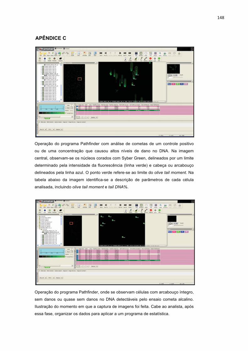
� ���
APÊNDICE C
Operação do programa Pathfinder com análise de cometas de um controle positivo
ou de uma concentração que causou altos níveis de dano no DNA. Na imagem
central, observam-se os núcleos corados com Syber Green, delineados por um limite
determinado pela intensidade da fluorescência (linha verde) e cabeça ou arcabouço
delineados pela linha azul. O ponto verde refere-se ao limite do olive tail moment. Na
tabela abaixo da imagem identifica-se a descrição de parâmetros de cada célula
analisada, incluindo olive tail moment e tail DNA%.�
Operação do programa Pathfinder, onde se observam células com arcabouço íntegro,
sem danos ou quase sem danos no DNA detectáveis pelo ensaio cometa alcalino.
Ilustração do momento em que a captura de imagens foi feita. Cabe ao analista, após
essa fase, organizar os dados para aplicar a um programa de estatística.

� ���
APÊNDICE D
Análise de micronúcleos (MN). Reconhecimento das células binucleadas e
validação do exame efetuado pelo programa, eliminando os falsos positivos. Para
confirmar um MN, o mesmo deve possuir, no máximo, 1/3 do tamanho do menor
núcleo da célula.
Recommended