Evaluation of Biochemical Biomarkers in Brain Tumors with Different Grade of Malignancy
M. Lankosz
AGH-University of Science and Technology,
Faculty of Physics and Applied Computer Science,
Al.. Mickiewicza 30, 30-059 Krakow, Poland
email: [email protected]
Cooperation
A. Wandzilak1, M. Grzelak1, B. Ostachowicz1, P. Wrobel1, M. Czyzycki1 1AGH-University of Science and Technology, Faculty of Physics and
Applied Computer Science, 30-059 Krakow, Poland
D. Adamek2 and E. Radwanska2
2 Chair of Pathomorphology, Faculty of Medicine, Jagiellonian University,
Krakow, Poland
Fe, Cu, Mn – participation in carcinogenesis for example by oxidative
stress
Cu - promotion of angiogenesis
free ionic Zn - a potent killer of neurons and glia (by oxidative stress)
Potential influence of the tumor on the surrounding tissue promoted by
metals:
Ca - excitotoxicity
Mn, Cu, Zn - disfunction of metalo-dependent enzymes like Mn-SOD (Mn)
or Cu/Zn-SOD
Se, Zn, Cu - an important role in the protection of the organism against
the mechanisms that can initiate or accelerate the carcinogenesis;
THE ROLE OF METALS IN CANCEROUS
PROCESSES
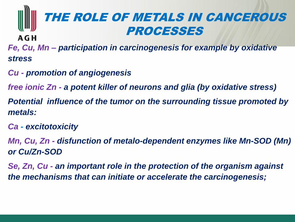
TUMOR TYPE ABBREVIATION Grade of
malignancy
glioblastoma multiforme GM IV
gemistocytic
astrocytoma AG III
oligodendroglioma O II
anaplastic
oligodendroglioma OA III
ganglioglioma G III
fibrillary astrocytoma AF II
atypical transitional
meningioma M I
control C
Types of tumors used in studies

CHEMICAL ELEMENTAL IMAGING WITH
THE USE OF X-RAY FLUORESCENCE
MICROSPECTROSCOPY

XRF equipment at the P06
beamline at Petra III
17.0 keV, 700 nm, 2 s/point
Synchrotron
1. Positron source
2. Linear accelerator
3. Booster
4. Accumulation ring
5. Beamline
6. Exprimental hutch

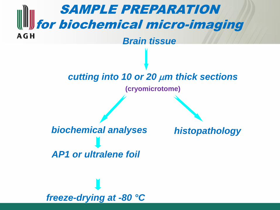
cutting into 10 or 20 m thick sections
(cryomicrotome)
Brain tissue
histopathology biochemical analyses
AP1 or ultralene foil
freeze-drying at -80 °C
SAMPLE PREPARATION
for biochemical micro-imaging

XRF spectrum
Astrocytoma diffusum XRF sum spectrum probed from 15 352 points
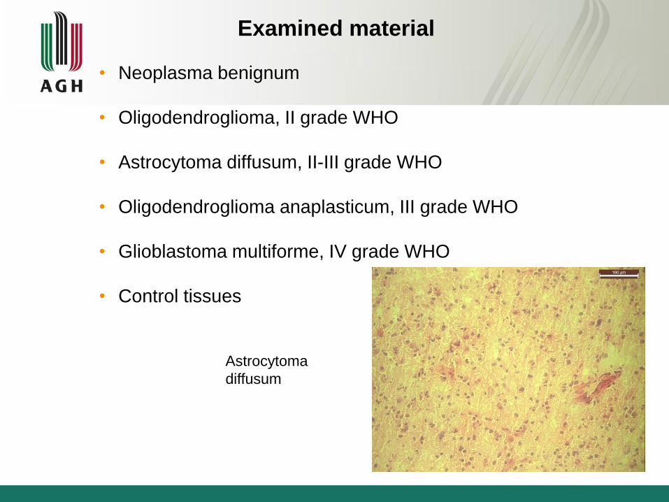
Examined material
• Neoplasma benignum
• Oligodendroglioma, II grade WHO
• Astrocytoma diffusum, II-III grade WHO
• Oligodendroglioma anaplasticum, III grade WHO
• Glioblastoma multiforme, IV grade WHO
• Control tissues
Astrocytoma
diffusum

vessels are under hypoxia.
Tumour tissue with no hypoxia (A),
moderate hypoxia (B)
and high level of hypoxia (C).
A B
C
Metallomics, 2013, 5, 1547-1553

Distribution of selected elements in glioblastoma multiforme tissue (IV WHO) in
comparison with microscopis view of hematoxylin-eosin stained section (A). Data
presented in [µg/cm2].
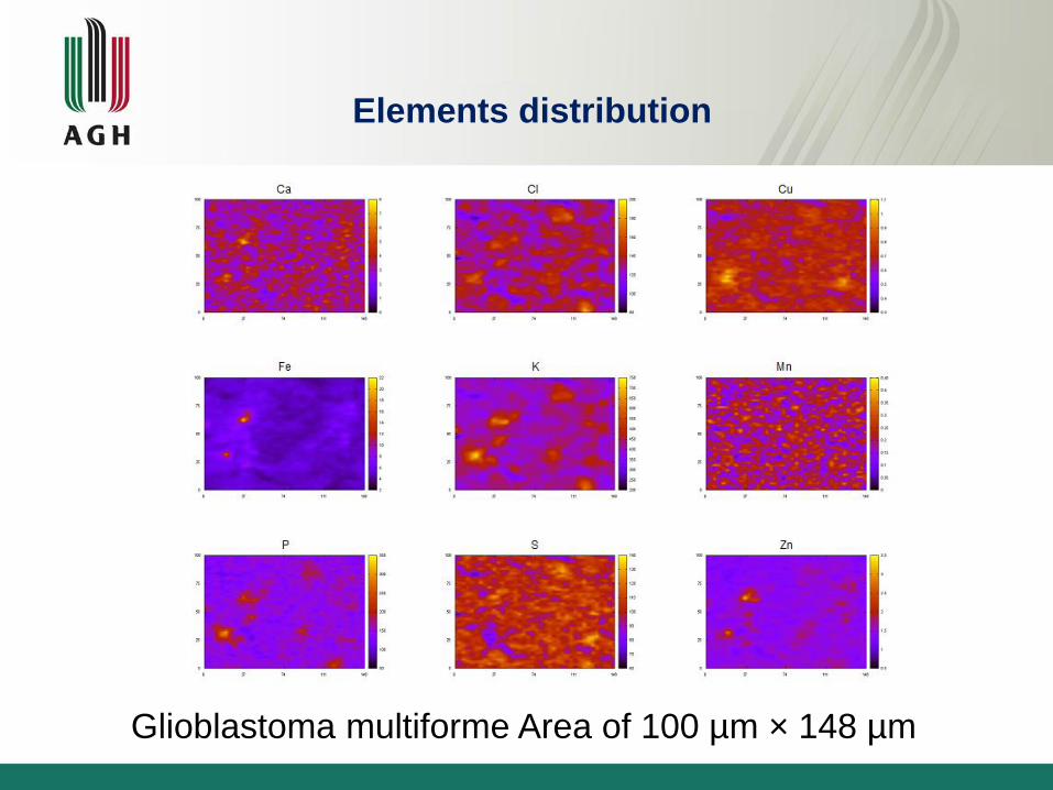
Glioblastoma multiforme Area of 100 µm × 148 µm
Elements distribution

Cancerous vs. healthy tissue
0.00
0.01
0.02
0.03
0.06
0.13
0.25
0.50
1.00
2.00
4.00
8.00
P S Cl K Ca Fe Cu Zn
ma
ss d
ep
osit p
er
un
it a
rea
, µ
g/c
m2
control III, OA IV, GM
Mean content of elements in healthy and cancerous tissues
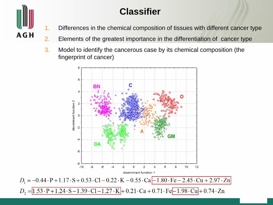
Classifier
1. Differences in the chemical composition of tissues with different cancer type
2. Elements of the greatest importance in the differentiation of cancer type
3. Model to identify the cancerous case by its chemical composition (the
fingerprint of cancer)

µ-XRF maps of elemental distribution in brain glioma tissue with areas of
calcification (see arrow) . Data presented in arbitrary units. Scale bars: 50 µm

Calcification in brain gliomas SRXRF
Scanned area: 180 µm / 120 µm.
K Fe Mn Ni
P S Zn
Ca Cl Cr Cu
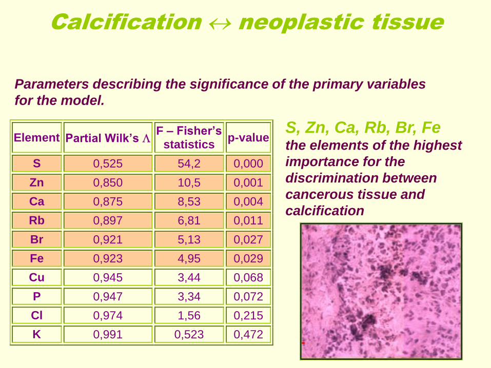
Element Partial Wilk’s F – Fisher’s
statistics p-value
S 0,525 54,2 0,000
Zn 0,850 10,5 0,001
Ca 0,875 8,53 0,004
Rb 0,897 6,81 0,011
Br 0,921 5,13 0,027
Fe 0,923 4,95 0,029
Cu 0,945 3,44 0,068
P 0,947 3,34 0,072
Cl 0,974 1,56 0,215
K 0,991 0,523 0,472
S, Zn, Ca, Rb, Br, Fe the elements of the highest
importance for the
discrimination between
cancerous tissue and
calcification
Parameters describing the significance of the primary variables
for the model.
Calcification neoplastic tissue

µ-XRF maps of elemental distribution in brain glioma tissue with blood
vessel (see arrow) . Data presented in arbitrary units. Scale bars: 100 µm
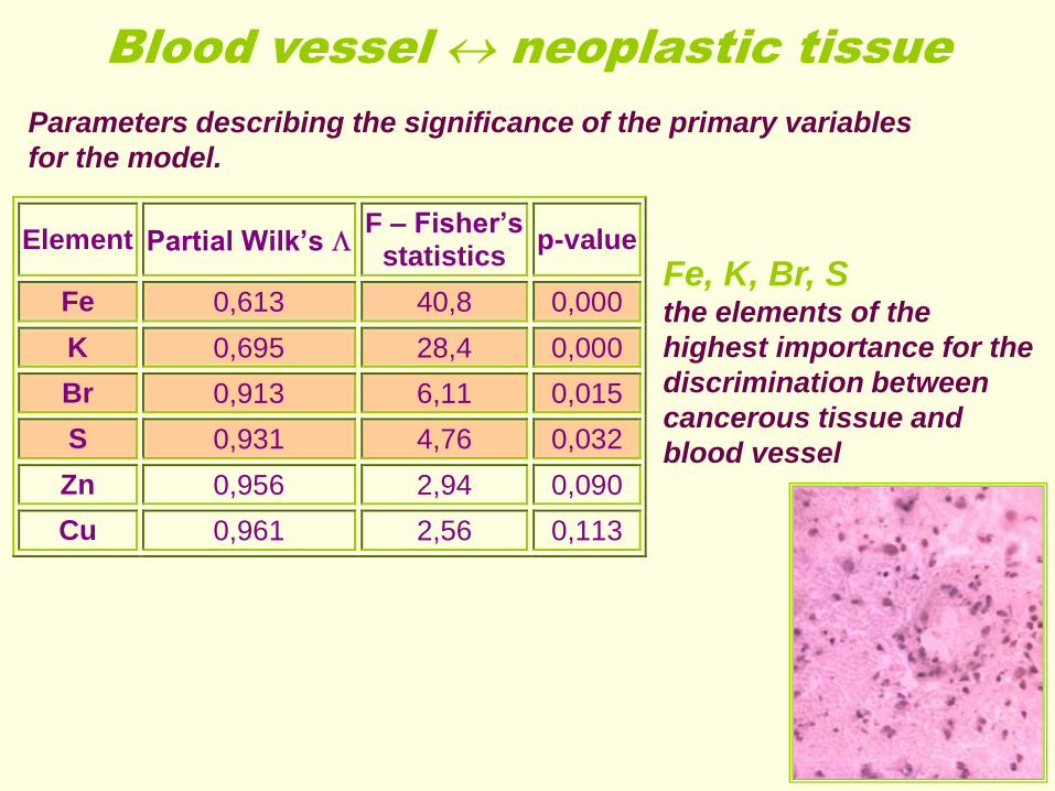
Element Partial Wilk’s F – Fisher’s
statistics p-value
Fe 0,613 40,8 0,000
K 0,695 28,4 0,000
Br 0,913 6,11 0,015
S 0,931 4,76 0,032
Zn 0,956 2,94 0,090
Cu 0,961 2,56 0,113
Fe, K, Br, S the elements of the
highest importance for the
discrimination between
cancerous tissue and
blood vessel
Parameters describing the significance of the primary variables
for the model.
Blood vessel neoplastic tissue

Blood vessel in brain cancer SRXRF
Ca Cl Cu
Fe P Zn
Scanned area: 180 µm / 120 µm.

Surface densities of Cl, Fe and Br within blood vessel area

Changes in the surface density of Cl as a function of distance from a blood vessel.
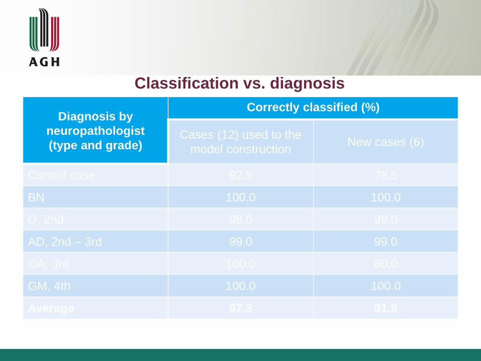
Diagnosis by
neuropathologist
(type and grade)
Correctly classified (%)
Cases (12) used to the
model construction New cases (6)
Control case 92.5 78.5
BN 100.0 100.0
O, 2nd 98.0 98.0
AD, 2nd – 3rd 99.0 99.0
OA, 3rd 100.0 80.0
GM, 4th 100.0 100.0
Average 97.3 91.9
Classification vs. diagnosis
Chemical elemental analysis of mean concentrations of elements
in brain cancers
X-ray fluorescence analysis
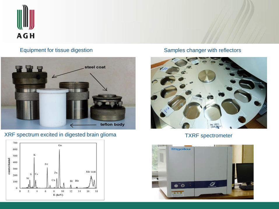
XRF spectrum excited in digested brain glioma
Samples changer with reflectors Equipment for tissue digestion
TXRF spectrometer

Concentrations of Cu for neoplastic tissues. (GM-brain
tumor, RM-cerebral abscess, OA-atypical transitional
meningioma, SA-anaplastic oligodendroglioma, SG-
oligoastrocytoma, GW-gliobastoma multiforme, GA-
anaplastic astrocytoma, OW-fibrous meningioma, RP-
metastatic carcinoma
Concentrations of Cu in brain cancers with different
WHO grade of malignancy. (0-control sample, I-IV
degrees)
Conclusions
The MDA based on the elemental composition of tissue (SRXRF)
may be a potentially valuable method in assisting the differentiation
and/or classification (diagnosis) of brain tissues including doubtful
cases
SR XRF spectroscopy allowed to determine the distribution and
amounts of trace elements in brain glioma tissues with a good
detectability and a spatial resolution at sub-micron level.
The results obtained showed that the elemental composition of a
relatively small fragment of homogeneous tissue represents
satisfactorily the biochemical ‘‘signature’’ of cancer. On the basis of
the element levels determined in such a small sample by means of
the TXRF technique it was possible to differentiate some types of
brain tumors
Cooperation
1. P06 at Petra III:
G. Falkenberg, M. Alfeld and U. Bösenberg
2. CEMO at DORIS III
E. Welter, K. Apple
3. I18 at Diamond
T. Geraki, F. Mosselmann
4. BM23 at ESRF
Olivier Mathon, Sacura Pacarelli

Acknowledgements:
The research leading to these results has received funding from:
The European Community's Seventh Framework Programme (FP7/2007-
2013) under grant agreement n° 226716,
Diamond Light Source Ltd,, Didcot Oxfordshire,
European Synchrotron Radiation Facility, Grenoble, France,
HASYLAB, Hamburg, Germany and the
Ministry of Science and Higher Education (Warsaw, Poland)
grant no N N518 377 537”
This presentation was supported by „Euro Health Care and Fitness Summit”

Thank you for your attention!
Recommended