R E S EA RCH AR T I C L E
Eukaryotes in Arctic and Antarctic cyanobacterial mats
Anne D. Jungblut1, Warwick F. Vincent2 & Connie Lovejoy3
1Departement de Biologie, Centre d’Etudes Nordiques (CEN), Institut de biologie integrative et des systemes (IBIS), Laval University, Quebec City,
QC, Canada; 2Departement de Biologie, Centre d’Etudes Nordiques (CEN), Laval University, Quebec City, QC, Canada; and 3Departement de
Biologie, Quebec-Ocean, Institut de biologie integrative et des systemes (IBIS), Laval University, Quebec City, QC, Canada
Correspondence: Anne D. Jungblut,
Department of Botany, The Natural History
Museum, Cromwell Road, London SW7 5BD,
UK. Tel.: +44 +20 7242 5285; fax: +44 +20
7242 5505; e-mail: [email protected]
Received 13 March 2012; revised 21 May
2012; accepted 21 May 2012.
DOI: 10.1111/j.1574-6941.2012.01418.x
Editor: Max Haggblom
Keywords
cyanobacterial mats; polar; biogeography;
protists; metazoa; 18S rRNA gene.
Abstract
Cyanobacterial mats are commonly found in freshwater ecosystems throughout
the polar regions. Most mats are multilayered three-dimensional structures
with the filamentous cyanobacteria embedded in a gel-like matrix. Although
early descriptions mentioned the presence of larger organisms including meta-
zoans living in the mats, there have been few studies specifically focused on the
microbial eukaryotes, which are often small cells with few morphological fea-
tures suitable for identification by microscopy. Here, we applied 18S rRNA
gene clone library analysis to identify eukaryotes in cyanobacterial mat com-
munities from both the Antarctic and the extreme High Arctic. We identified
39 ribotypes at the level of 99% sequence similarity. These consisted of taxa
within algal and other protist groups including Chlorophyceae, Prasinophyceae,
Ulvophyceae, Trebouxiophyceae, Bacillariophyceae, Chrysophyceae, Ciliophora,
and Cercozoa. Fungi were also recovered, as were 21 metazoan ribotypes. The
eukaryotic taxa appeared habitat-specific with little overlap between lake, pond,
and ice shelf communities. Some ribotypes were common to both Arctic and
Antarctic mats, suggesting global dispersal of these taxa and similarity in the
environmental filters acting on protist communities. Many of these eukaryotic
taxa likely benefit from protected, nutrient-rich microhabitats within the
cyanobacterial mat environment.
Introduction
Microbial mats dominated by oscillatorian cyanobacteria
are found in a diverse range of marine and freshwater
environments (Stal, 2000), and they are an especially
common feature of aquatic ecosystems throughout the
polar regions (Vincent, 2000a). As elsewhere, the Arctic
and Antarctic mats are multilayered three-dimensional
structures, where exo-polymer-producing cyanobacteria
create an environment than can be colonized by other
microorganisms (Zakhia et al., 2007). The polar mat
communities cope with harsh conditions typical of
cryo-ecosystems, including persistent low temperatures,
variable freeze-thaw cycles, prolonged winter darkness,
continuous solar irradiance in summer, and rapidly fluc-
tuating osmotic regimes. The phototrophic communities
in these mats rely on internal nutrient recycling and scav-
enging systems to cope with the low allochthonous input
of nutrients that is typical of ultra-oligotrophic freshwater
ecosystems in the polar desert environment (Varin et al.,
2010). The mat consortia contain diverse Bacteria (Bottos
et al., 2008) as well as Archaea and viruses (Varin et al.,
2010). Microscopic studies have long indicated that
eukaryotes including metazoa (Murray, 1910) also occur
in polar mats, but little is known about the diversity of
microbial eukaryotes that may be present and their distri-
bution across habitats, regions or continents.
Eukaryotes in general have diverse lifecycles and
include primary producers as well as primary and second-
ary consumers. Ecological processes such as competition
and environmental selection will likely operate, possibly
resulting in habitat specificity. Although some eukaryotic
taxa form readily dispersed resting stages and would be
expected to have broad distributions, other taxa may have
a more limited capacity to survive transport, resulting in
their ecological and geographic restriction to certain loca-
tions (Vincent, 2000b). The latter could promote habitat-
specific ecotypes and microbial endemism at specific
FEMS Microbiol Ecol && (2012) 1–13 ª 2012 Federation of European Microbiological SocietiesPublished by Blackwell Publishing Ltd. All rights reserved
MIC
ROBI
OLO
GY
EC
OLO
GY
isolated sites within the cold biosphere, which is defined
as the ensemble of environments on Earth characterized
by prolonged cold and freezing (Anesio & Laybourn-
Parry, 2012; Harding et al., 2011). For example, there is
evidence from maritime Antarctic lakes that the long-
distance dispersal of freshwater ciliates to these sites is
restricted and that some taxa are limited in their geo-
graphical distribution (Petz et al., 2007).
The majority of investigations using morphological and
molecular methods that have reported on the microbial
eukaryotes from the polar regions have been carried out
in Antarctica (Broady, 1996; De Wever et al., 2009;
Bielewicz et al., 2011). By comparison, little is known about
the microbial eukaryotes that inhabit similar freshwater
environments in the Arctic, which is much less isolated
from temperate continental regions than Antarctica. The
aim of this study was to evaluate the diversity of eukary-
otic communities in polar cyanobacterial mats, and it
complements a previous study that focused exclusively on
benthic polar cyanobacteria (Jungblut et al., 2010). We
determined the diversity and community structure of
eukaryotes inhabiting mats collected from lakes, ponds,
and streams on land, and from meltwater ponds on ice
shelves, at the northern limit of the North American Arc-
tic, specifically Ward Hunt Island (latitude 83.1°N) and
vicinity, in Quttinirpaaq (‘top of the world’ in Inuktitut)
National Park, Nunavut, Canada. These Arctic mats were
compared with those from analogous meltwater ponds on
the McMurdo Ice Shelf, Antarctica, at a similar latitude
and climate in the south polar region. Eukaryotic diver-
sity was determined in the microbial mats by 18S rRNA
environmental gene surveys, and their global distribution
patterns evaluated by phylogenetic analysis.
Materials and methods
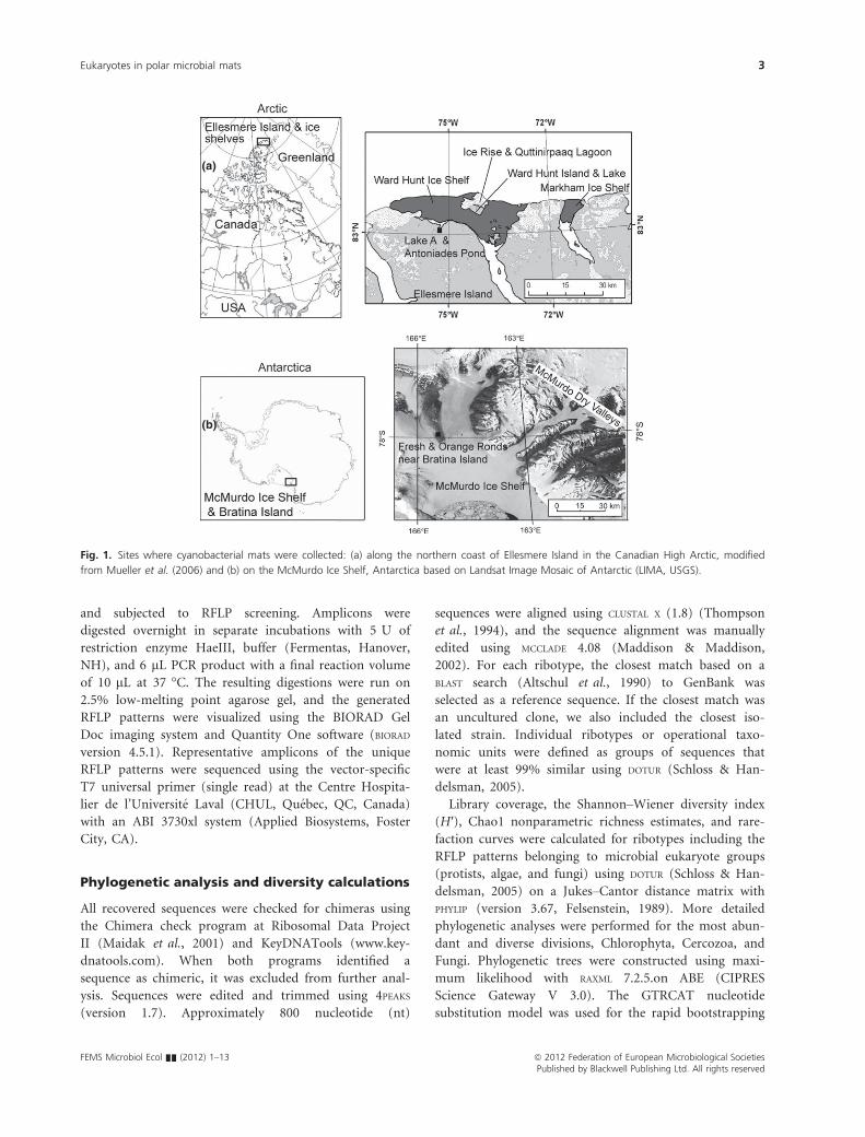
Study sites, sampling, and water analysis
Cyanobacterial mats from six Arctic and two Antarctic
freshwater systems were collected from 8 to 15 July 2007
in the Arctic and January 2005 from the Antarctic. Arctic
samples were from Quttinirpaaq National Park, Ellesmere
Island in the Canadian High Arctic (Fig. 1). The six sites
covered a range of environmental conditions. From North
to South, these were meltwater ponds on Markham Ice
Shelf (MIS) 83°01.898′N, 71°30.812′W and Ward Hunt
Ice Shelf (WIS) 83°04.949′N, 74°26.281′W along the
northern coast of Ellesmere Island; Quttinirpaaq Lagoon
(QL) 83°05.843′N, 74°15.018′W and Ward Hunt Lake
(WHL) 83°05.289′N, 74°10.048′W on Ward Hunt Island;
and Antoniades Pond (AP) 82°58.957′N, 75°24.161′Wand a stream flowing into Lake A (Inflow-A, IA) 82°58.801′N, 75°25.372′W on Ellesmere Island. Samples from
Antarctica were collected from Fresh Pond (FP) 78°00.935′S, 165°32.622′E and Orange Pond (OP) 78°00.823′ S, 165° 33.402′E on the McMurdo Ice Shelf, near
Bratina Island, January 2005. Detailed descriptions of the
Arctic and Antarctic sampling sites are given in Jungblut
et al. (2010) and Howard-Williams et al. (1990), respec-
tively.
All environmental measurements and mat samples were
from 10 to 20 cm water depth and were sampled using a
sterilized spatula and sterile-sampling containers. The
material was freeze-dried and stored at �80 °C until fur-
ther analysis. Water temperature, pH, and conductivity
were determined at each Arctic site using a portable
instrument (pH/Con 10 Series; Oakton Instruments, Ver-
non Hills, IL) and in the Antarctic as described by Hawes
et al. (1993).
DNA extraction and polymerase chain reaction
(PCR)
Total DNA was extracted from freeze-dried microbial mat
material as previously described (Jungblut et al., 2010).
The 18S rRNA gene PCR reactions were performed in
25 lL reaction volumes using 0.2 U Taq (Invitrogen,
Carlsbad, CA), 19 Buffer (Invitrogen), 2.5 mM MgCl2(Invitrogen), 5 lL BSA (20 mg L�1; Fermentas, Foster
City, CA), and 0.2 mM dNTPs (Fermentas, Foster City,
CA) and 0.5 lM of each eukaryotic-specific primer Euk
515F (5′-GTGCCAGCMGCCGCGGTA-3′) and Euk 1195
RE (5′-GGGCATCACAGACCTG-3′) (Feazel et al., 2008).
As described by Feazel et al. (2008), these primers are
general and will amplify metazoa and protist groups such
as stramenopiles, Chlorophyta, fungi, and alveolates. An
initial denaturation step at 94 °C for 2 min was followed
by 35 cycles of 94 °C for 1 min, 56 °C for 1 min, and
72 °C for 1 min, with a final extension step at 72 °C for
10 min.
Cloning, restriction fragment length
polymorphism (RFLP) analysis, and sequencing
Prior to cloning, amplified PCR products were verified by
gel electrophoresis, and amplicons of the target size were
gel purified with a Qiaquick Gel Purification Kit (Qiagen,
Mississauga, CA). For each sample, 3–5 separate PCR
reaction replicates were carried out and were pooled prior
to PCR-product purification. PCR products were cloned
using a StrataClone PCR Cloning Kit (Strategene, Cedar
Creek, TX). Ligation and transformation were performed
according to the manufacturer’s protocols. Positive clones
were transferred to 96-well plates containing LB medium
with 7% glycerol. Inserted 18S rRNA gene sequences were
amplified using vector-specific primers M13f and M13r
ª 2012 Federation of European Microbiological Societies FEMS Microbiol Ecol && (2012) 1–13Published by Blackwell Publishing Ltd. All rights reserved
2 A.D. Jungblut et al.
and subjected to RFLP screening. Amplicons were
digested overnight in separate incubations with 5 U of
restriction enzyme HaeIII, buffer (Fermentas, Hanover,
NH), and 6 lL PCR product with a final reaction volume
of 10 lL at 37 °C. The resulting digestions were run on
2.5% low-melting point agarose gel, and the generated
RFLP patterns were visualized using the BIORAD Gel
Doc imaging system and Quantity One software (BIORAD
version 4.5.1). Representative amplicons of the unique
RFLP patterns were sequenced using the vector-specific
T7 universal primer (single read) at the Centre Hospita-
lier de l’Universite Laval (CHUL, Quebec, QC, Canada)
with an ABI 3730xl system (Applied Biosystems, Foster
City, CA).
Phylogenetic analysis and diversity calculations
All recovered sequences were checked for chimeras using
the Chimera check program at Ribosomal Data Project
II (Maidak et al., 2001) and KeyDNATools (www.key-
dnatools.com). When both programs identified a
sequence as chimeric, it was excluded from further anal-
ysis. Sequences were edited and trimmed using 4PEAKS
(version 1.7). Approximately 800 nucleotide (nt)
sequences were aligned using CLUSTAL X (1.8) (Thompson
et al., 1994), and the sequence alignment was manually
edited using MCCLADE 4.08 (Maddison & Maddison,
2002). For each ribotype, the closest match based on a
BLAST search (Altschul et al., 1990) to GenBank was
selected as a reference sequence. If the closest match was
an uncultured clone, we also included the closest iso-
lated strain. Individual ribotypes or operational taxo-
nomic units were defined as groups of sequences that
were at least 99% similar using DOTUR (Schloss & Han-
delsman, 2005).
Library coverage, the Shannon–Wiener diversity index
(H′), Chao1 nonparametric richness estimates, and rare-
faction curves were calculated for ribotypes including the
RFLP patterns belonging to microbial eukaryote groups
(protists, algae, and fungi) using DOTUR (Schloss & Han-
delsman, 2005) on a Jukes–Cantor distance matrix with
PHYLIP (version 3.67, Felsenstein, 1989). More detailed
phylogenetic analyses were performed for the most abun-
dant and diverse divisions, Chlorophyta, Cercozoa, and
Fungi. Phylogenetic trees were constructed using maxi-
mum likelihood with RAXML 7.2.5.on ABE (CIPRES
Science Gateway V 3.0). The GTRCAT nucleotide
substitution model was used for the rapid bootstrapping
(a)
(b)
Fig. 1. Sites where cyanobacterial mats were collected: (a) along the northern coast of Ellesmere Island in the Canadian High Arctic, modified
from Mueller et al. (2006) and (b) on the McMurdo Ice Shelf, Antarctica based on Landsat Image Mosaic of Antarctic (LIMA, USGS).
FEMS Microbiol Ecol && (2012) 1–13 ª 2012 Federation of European Microbiological SocietiesPublished by Blackwell Publishing Ltd. All rights reserved
Eukaryotes in polar microbial mats 3
phase, and GTRGAMMA for the final tree inference (Sta-
matakis, 2006a, b). A best-scoring ML tree was obtained
with 500 bootstraps. The 18S rRNA gene sequences are
available under GenBank accession numbers JN207853–JN207906.
Results
Environmental properties
The eight collection sites spanned a range of environmen-
tal conditions (Table 1). Overlying water temperatures
ranged from �0.3 °C in FP to 6 °C in AP in the High
Arctic. OP had the highest pH of all sites (9.9), and
lowest pH values were recorded in the meltwater ponds
on the Ward Hunt (6.24) and Markham Ice Shelves
(6.52). Conductivities ranged from 3469 lS cm�1 in OP
on the McMurdo Ice Shelf, Antarctica to as low as
740 lS cm�1 on the WIS in the Arctic. Sampling in Ant-
arctic ponds was performed while the water column was
still stratified, with higher conductivities near the bottom
of the ponds. The land-based Arctic sites had lower con-
ductivities than the ice shelf meltwaters, with values of
137 lS cm�1 or less.
Microbial eukaryotic community analysis
The 18S rRNA gene clone libraries from genomic environ-
mental DNA from six Arctic (WHL, WIS, MIS, AP, QL
and IA) and two Antarctic (FP and OP) yielded a total of
464 protist clones and 326 Metazoan clones (Table 2). Rar-
efaction curves of protists suggested an incomplete sam-
pling of the diversity for most of the sites (Supporting
Information, Fig. S1). In total, 39 protist ribotypes defined
at 99% sequence similarity were retrieved, with 3–10 ribo-
types per site (Table S1). The greatest numbers retrieved
were from AP, which had a bias-corrected Chao1 average
richness of 12 (Table 2). Widely variable 18S rRNA gene
copy numbers among microbial eukaryotes might have
effected the diversity estimations (Potvin & Lovejoy, 2009).
This would have been especially the case for the micro-
fauna present in the mats, and the metazoan sequences
were therefore analyzed separately and only in broad taxo-
nomic categories (Table S3).
Among the protists, a total of 10 major groups were
represented, mostly from the chlorophyll b containing
lineages Chlorophyceae, Ulvophyceae, Prasinophyceae,
and Trebouxiophyceae. Among the chlorophyll c groups
Table 1. Environmental conditions at the Arctic and Antarctic
sampling sites
Sites
Temperature
(°C) pH
Conductivity
(lS cm�1)
Arctic
WHL +2.1 8.02 127
QL +2.2 7.51 261
WIS +1.5 6.24 740
MIS +1.1 6.52 492
AP +6.0 8.28 137
IA – – 123
Antarctica
FP �0.3 8.67 1242
OP +2.8 9.9 3469
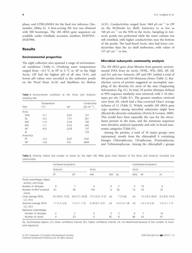
Table 2. Diversity indices and number of clones for the eight 18S rRNA gene clone libraries of the Arctic and Antarctic microbial mat
communities
Ice-based ecosystems Land-based ecosystems
Antarctic Arctic Arctic
FP OP MIS WIS QL WHL AP IA
Protist assemblages (algae,
protists, and fungi)
Number of ribotypes 9 7 4 4 7 3 10 6
Number of RFLP screened
clones
61 94 156 6 50 10 55 22
Chao average (95%
LCI, HCI)
9.3 (9.01–15.0) 8.0 (7.1–20.8) 17.3 (5.0–17.3) nd 7 (7-nd) nd 12 (10.2–26.0) 6.5 (6.0–14.3)
Shannon average (95%
LCI, HCI)
1.7 (1.5–2.0) 1.3 (1.1–1.5) 0.18 (0.1–0.3) nd 1.6 (1.4–1.8) nd 1.6 (1.4–2.0) 1.4 (1.1–1.7)
Metazoan assemblages
Number of ribotypes 2 2 0 3 3 8 7 10
Number of clones 4 23 0 95 8 86 43 67
QL, Quttinirpaaq Lagoon; LCI, lower confidence interval; HCI, higher confidence interval; nd, not determined because of low number of recov-
ered sequences.
ª 2012 Federation of European Microbiological Societies FEMS Microbiol Ecol && (2012) 1–13Published by Blackwell Publishing Ltd. All rights reserved
4 A.D. Jungblut et al.
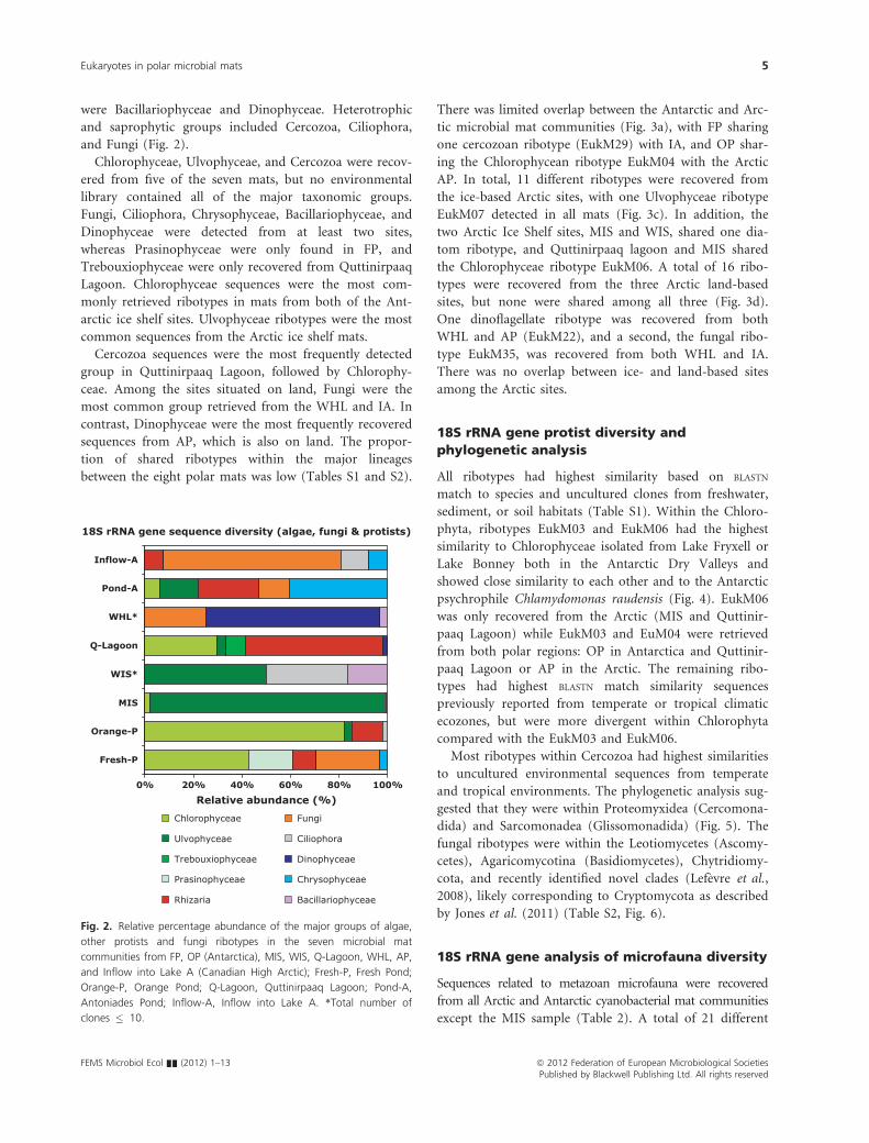
were Bacillariophyceae and Dinophyceae. Heterotrophic
and saprophytic groups included Cercozoa, Ciliophora,
and Fungi (Fig. 2).
Chlorophyceae, Ulvophyceae, and Cercozoa were recov-
ered from five of the seven mats, but no environmental
library contained all of the major taxonomic groups.
Fungi, Ciliophora, Chrysophyceae, Bacillariophyceae, and
Dinophyceae were detected from at least two sites,
whereas Prasinophyceae were only found in FP, and
Trebouxiophyceae were only recovered from Quttinirpaaq
Lagoon. Chlorophyceae sequences were the most com-
monly retrieved ribotypes in mats from both of the Ant-
arctic ice shelf sites. Ulvophyceae ribotypes were the most
common sequences from the Arctic ice shelf mats.
Cercozoa sequences were the most frequently detected
group in Quttinirpaaq Lagoon, followed by Chlorophy-
ceae. Among the sites situated on land, Fungi were the
most common group retrieved from the WHL and IA. In
contrast, Dinophyceae were the most frequently recovered
sequences from AP, which is also on land. The propor-
tion of shared ribotypes within the major lineages
between the eight polar mats was low (Tables S1 and S2).
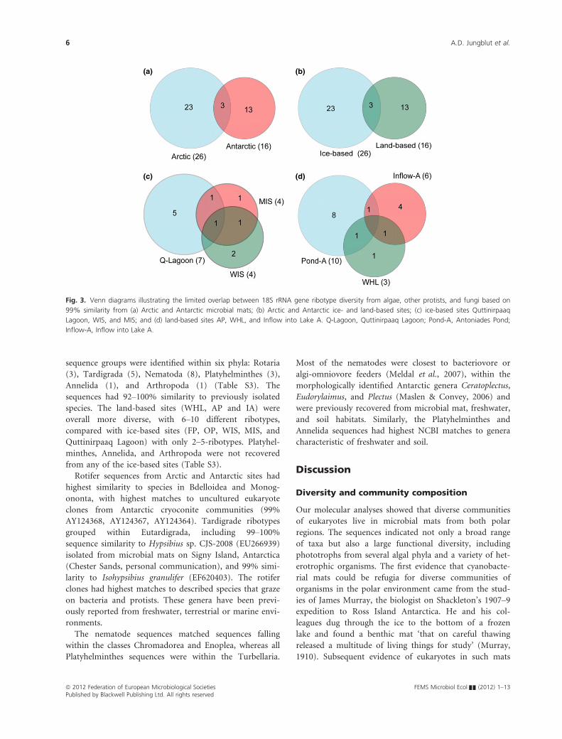
There was limited overlap between the Antarctic and Arc-
tic microbial mat communities (Fig. 3a), with FP sharing
one cercozoan ribotype (EukM29) with IA, and OP shar-
ing the Chlorophycean ribotype EukM04 with the Arctic
AP. In total, 11 different ribotypes were recovered from
the ice-based Arctic sites, with one Ulvophyceae ribotype
EukM07 detected in all mats (Fig. 3c). In addition, the
two Arctic Ice Shelf sites, MIS and WIS, shared one dia-
tom ribotype, and Quttinirpaaq lagoon and MIS shared
the Chlorophyceae ribotype EukM06. A total of 16 ribo-
types were recovered from the three Arctic land-based
sites, but none were shared among all three (Fig. 3d).
One dinoflagellate ribotype was recovered from both
WHL and AP (EukM22), and a second, the fungal ribo-
type EukM35, was recovered from both WHL and IA.
There was no overlap between ice- and land-based sites
among the Arctic sites.
18S rRNA gene protist diversity and
phylogenetic analysis
All ribotypes had highest similarity based on BLASTN
match to species and uncultured clones from freshwater,
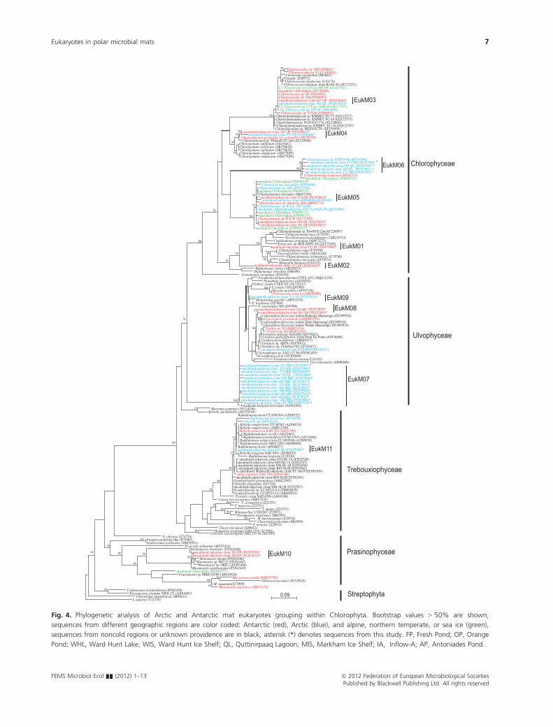
sediment, or soil habitats (Table S1). Within the Chloro-
phyta, ribotypes EukM03 and EukM06 had the highest
similarity to Chlorophyceae isolated from Lake Fryxell or
Lake Bonney both in the Antarctic Dry Valleys and
showed close similarity to each other and to the Antarctic
psychrophile Chlamydomonas raudensis (Fig. 4). EukM06
was only recovered from the Arctic (MIS and Quttinir-
paaq Lagoon) while EukM03 and EuM04 were retrieved
from both polar regions: OP in Antarctica and Quttinir-
paaq Lagoon or AP in the Arctic. The remaining ribo-
types had highest BLASTN match similarity sequences
previously reported from temperate or tropical climatic
ecozones, but were more divergent within Chlorophyta
compared with the EukM03 and EukM06.
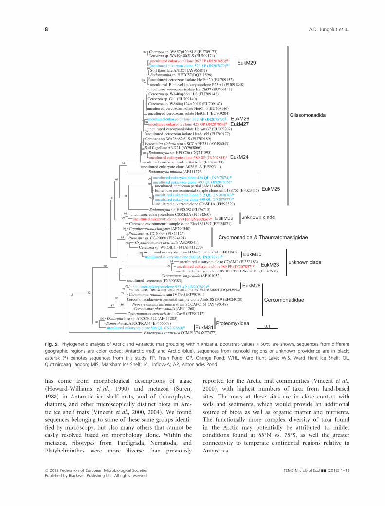
Most ribotypes within Cercozoa had highest similarities
to uncultured environmental sequences from temperate
and tropical environments. The phylogenetic analysis sug-
gested that they were within Proteomyxidea (Cercomona-
dida) and Sarcomonadea (Glissomonadida) (Fig. 5). The
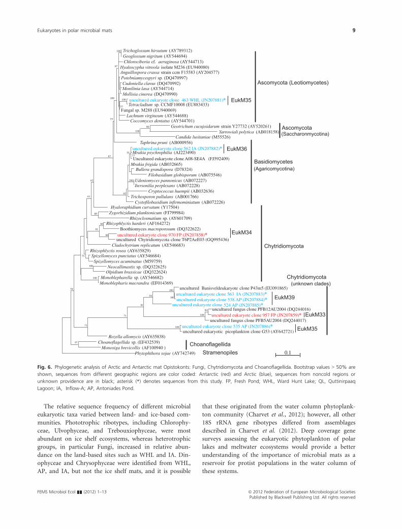
fungal ribotypes were within the Leotiomycetes (Ascomy-
cetes), Agaricomycotina (Basidiomycetes), Chytridiomy-
cota, and recently identified novel clades (Lefevre et al.,
2008), likely corresponding to Cryptomycota as described
by Jones et al. (2011) (Table S2, Fig. 6).
18S rRNA gene analysis of microfauna diversity
Sequences related to metazoan microfauna were recovered
from all Arctic and Antarctic cyanobacterial mat communities
except the MIS sample (Table 2). A total of 21 different
18S rRNA gene sequence diversity (algae, fungi & protists)
0% 20% 40% 60% 80% 100%
Fresh-P
Orange-P
MIS
WIS*
Q-Lagoon
WHL*
Pond-A
Inflow-A
Relative abundance (%)
Chlorophyceae
Ulvophyceae
Trebouxiophyceae
Prasinophyceae
Rhizaria
Fungi
Ciliophora
Dinophyceae
Chrysophyceae
Bacillariophyceae
Fig. 2. Relative percentage abundance of the major groups of algae,
other protists and fungi ribotypes in the seven microbial mat
communities from FP, OP (Antarctica), MIS, WIS, Q-Lagoon, WHL, AP,
and Inflow into Lake A (Canadian High Arctic); Fresh-P, Fresh Pond;
Orange-P, Orange Pond; Q-Lagoon, Quttinirpaaq Lagoon; Pond-A,
Antoniades Pond; Inflow-A, Inflow into Lake A. *Total number of
clones � 10.
FEMS Microbiol Ecol && (2012) 1–13 ª 2012 Federation of European Microbiological SocietiesPublished by Blackwell Publishing Ltd. All rights reserved
Eukaryotes in polar microbial mats 5
sequence groups were identified within six phyla: Rotaria
(3), Tardigrada (5), Nematoda (8), Platyhelminthes (3),
Annelida (1), and Arthropoda (1) (Table S3). The
sequences had 92–100% similarity to previously isolated
species. The land-based sites (WHL, AP and IA) were
overall more diverse, with 6–10 different ribotypes,
compared with ice-based sites (FP, OP, WIS, MIS, and
Quttinirpaaq Lagoon) with only 2–5-ribotypes. Platyhel-minthes, Annelida, and Arthropoda were not recovered
from any of the ice-based sites (Table S3).
Rotifer sequences from Arctic and Antarctic sites had
highest similarity to species in Bdelloidea and Monog-
ononta, with highest matches to uncultured eukaryote
clones from Antarctic cryoconite communities (99%
AY124368, AY124367, AY124364). Tardigrade ribotypes
grouped within Eutardigrada, including 99–100%sequence similarity to Hypsibius sp. CJS-2008 (EU266939)
isolated from microbial mats on Signy Island, Antarctica
(Chester Sands, personal communication), and 99% simi-
larity to Isohypsibius granulifer (EF620403). The rotifer
clones had highest matches to described species that graze
on bacteria and protists. These genera have been previ-
ously reported from freshwater, terrestrial or marine envi-
ronments.
The nematode sequences matched sequences falling
within the classes Chromadorea and Enoplea, whereas all
Platyhelminthes sequences were within the Turbellaria.
Most of the nematodes were closest to bacteriovore or
algi-omniovore feeders (Meldal et al., 2007), within the
morphologically identified Antarctic genera Ceratoplectus,
Eudorylaimus, and Plectus (Maslen & Convey, 2006) and
were previously recovered from microbial mat, freshwater,
and soil habitats. Similarly, the Platyhelminthes and
Annelida sequences had highest NCBI matches to genera
characteristic of freshwater and soil.
Discussion
Diversity and community composition
Our molecular analyses showed that diverse communities
of eukaryotes live in microbial mats from both polar
regions. The sequences indicated not only a broad range
of taxa but also a large functional diversity, including
phototrophs from several algal phyla and a variety of het-
erotrophic organisms. The first evidence that cyanobacte-
rial mats could be refugia for diverse communities of
organisms in the polar environment came from the stud-
ies of James Murray, the biologist on Shackleton’s 1907–9expedition to Ross Island Antarctica. He and his col-
leagues dug through the ice to the bottom of a frozen
lake and found a benthic mat ‘that on careful thawing
released a multitude of living things for study’ (Murray,
1910). Subsequent evidence of eukaryotes in such mats
(a) (b)
(c) (d)
Fig. 3. Venn diagrams illustrating the limited overlap between 18S rRNA gene ribotype diversity from algae, other protists, and fungi based on
99% similarity from (a) Arctic and Antarctic microbial mats; (b) Arctic and Antarctic ice- and land-based sites; (c) ice-based sites Quttinirpaaq
Lagoon, WIS, and MIS; and (d) land-based sites AP, WHL, and Inflow into Lake A. Q-Lagoon, Quttinirpaaq Lagoon; Pond-A, Antoniades Pond;
Inflow-A, Inflow into Lake A.
ª 2012 Federation of European Microbiological Societies FEMS Microbiol Ecol && (2012) 1–13Published by Blackwell Publishing Ltd. All rights reserved
6 A.D. Jungblut et al.
S. palustre (Y11370)Chlorokybus atmophyticus (M95612)
Pterosperma cristatum NIES 221 (AJ010407)Cymbomonas tetramitiformis (FN62438)
Mantoniella antarctica (AB017128)M. squamata(X73999)
Ostreococcus tauri (AY329635)Micromonas pusilla (DQ025753)
Crustomastix sp. MBIC10709 (AB183628)uncultured Chlorophyta (FN690727)
Monomastix opisthostigma (FN562445)Monomastix sp. OKE-1 (FJ493496)
Monomastix sp. M0722 (FN562447)Monomastix minuta (FN562446)
uncultured eukaryote clone 988 FP (JN207871)*uncultured eukaryote clone 992 FP (JN207870)*
Dolichomastix tenuilepis (FN562449)Picocystis salinarum (AF153314)
Nephroselmis pyriformis (AB058391)Pseudoscourfieldia-like (X75565)
N. olivacea (X74754)Chlorella saccharophila SAG 211-9a (X63505)
Watanabea reniformis SAG 2119 (X73991)Choricystis minor (X89012)
P. terrestre (Z28973)Characium perforatum (M62999)M. kuetzingianum (Z28974)
Friedmannia israeliensis (M62995)M.biatorellae UTEX907 (Z28971)
T. magna (Z21552)T. impressa (Z21551)
T. asymmetrica (Z21553)Coenocystis inconstans (AB017435)
Prasiola crispa SAG 4396 (AJ416106)Pseudochlorella sp. CCAP211/1A (AB488583)Pseudochlorella sp. CCAP211/1A (FM958479)uncultured eukaryote clone H04-SE1B (FJ592507)Chlorella ellipsoidea (D13324)Pseudochlorella pyrenoidosa (AM422985)
uncultured eukaryote clone B04-Se1B (FJ592461)Pabia signensis SAG 790 (AJ416108)
uncultured Banisveld eukaryote clone P1-3m10 (EU091836)uncultured eukaryote clone B05-Se1B (FJ592462)uncultured eukaryote clone F06-SE-1B (FJ592494)
uncultured eukaryote clone E09-SE-1A (FJ592347)uncultured eukaryote clone C05-SE-1A (FJ592328)
Raphidonema longiseta (U18520)Koliella longiseta SAG 4701 (AJ306535)
uncultured eukaryote clone 497 QL (JN207906)*Raphidonema nivale (AF448477)
Raphidonema nivale NIES 2290 (AB488604)Raphidonema sempervirens CCAP470/6 (AJ309939)
Raphidonema pyrenoidifera CCAP 470/5 (AJ311640)Raphidonemopsis sessilis (AJ431667)
Koliella antarctica SAG 203 (AJ311569)Koliella sempervirens (AM412750)Koliella sempervirens CCAP363 (AJ306533)
Chlorella sp. (AF514413)Raphidonema brevirostre AF514410)
Raphidonema nivale CCAP470/4 (AJ306532)Koliella spiculiformis (AF278744)
Marvania geminata (AF124336)Pseudendocloniopsis botryoides (AJ416103)
uncultured eukaryote clone 265 MIS (JN207905)*uncultured eukaryote clone 264 MIS (JN207904)*uncultured eukaryote clone 268 MIS (JN207903)*
uncultured eukaryote clone 436 MIS (JN207902)*uncultured eukaryote clone 559 WIS (JN207901)*uncultured eukaryote clone 280 MIS (JN207899)*uncultured eukaryote clone 279 MIS (JN207898)*uncultured eukaryote clone 430 MIS (JN207897)*
uncultured eukaryote clone 269 MIS (JN207896)*uncultured eukaryote clone 515 QL (JN207900)*
uncultured eukaryote clone 272 MIS (JN207895)*uncultured eukaryote clone 263 MIS (JN207894)*uncultured eukaryote clone 431 MIS (JN207893)*
Ulva intestinalis (AJ000040)Pseudoneochloris marina (U41102)
Acrosiphonia arcta (AY303600)Acrosiphonia sp. SAG-127.80 (FN562430)
uncultured eukaryote clone D2P3B04 (EF100232)Chlorothrix sp. ChloPac47SI (AY476827)
Urospora sp. AB58 (AY476812)Urospora penicilliformis (AB049417)Urospora penicilliformis strain Point No Point (AY476808)Urospora neglecta Seward8 (AY476821)
Ulothrix sp. X1 (DQ821515)Ulothrix sp. X3 (DQ821516)
Capsosiphon fulvescens isolate Wando Maesaengii (EU099919)Capsosiphon fulvescens isolate Jindo Maesaengii (EU099918)
Capsosiphon groenlandicus (DQ821514)Capsosiphon fulvescens isolate Koheung Maesaengii (EU099920)
uncultured eukaryote clone 594 OP (JN207869)*uncultured eukaryote clone 475 OP (JN207868)*P. sarcinoidea 58S (Z47998)
P. basiliense (Z47996)Monostroma grevillei (AF015279)
uncultured eukaryote clone 521 AP (JN207892)*Trichosarcina mucosa (AM109906)Hazenia mirabilis (AF387156)U.zonata 5.8S (Z47999)
Ulothrix zonata UTEX745 (AY278217)Planophila laetevirens (AJ416102)
Pseudendoclonium akinetum UTEX 1912 (DQ011230)Scenedesmus vacuolatus (X56104)
Hafniomonas reticulata (D86496)Hafniomonas conica (AB248251)
uncultured eukaryote clone 973 FP (JN207867)*Hormotila blennista (U83123)Chlamydomonas mexicana (AF39534)
Chlamydomonas asymmetrica (U70788)Fasciculochloris boldii (AB244240)Chlamydomonas rapa (U70790)
uncultured eukaryote clone 952 FP (JN207866)*Tetracystis sp. BCP-ZNP3-36 (AY377439)
Cephalomonas granulata (AB472272)Neochlorosarcina pseudominor (AB218714)Chlamydomonas baca (U70781)
Chlamydomonad sp. Tow8/8T-12w(AY220607)uncultured Chlorophyta (FN690712)*
uncultured eukaryote clone 591 OP (JN207865)*uncultured eukaryote clone 588 OP (JN207864)*
Chlamydomonas sp. ICE-W (AY731083)uncultured Chlorophyta (FN690717)
uncultured Chlorophyta (FN690713)uncultured chlamydomand clone CCCryp 002b-99 (AF514398)Chlamydomonas sp. ICE-L (AY73108)Chlamydomonas sp. Antarctic 2E9 (AB001374)
uncultured eukaryote clone D2P04F06 (EF100261)uncultured eukaryote clone 474 OP (JN207863)*
Chlamydomonas kuwadae (AB451190)uncultured Chlorophyta (FN690715)Chlamydomonas sp. ARC (EF537906)
Chlamydomonas pulsatilla (AF514404)uncultured Chlorophyta (FN690719)
uncultured Chlorophyta (FN690710)Chlamydomonas raudensis (AJ781313)uncultured eukaryote clone 271 MIS (JN207891) *uncultured eukaryote clone 485 QL (JN207862) *uncultured eukaryote clone 505 QL (JN207890) *
uncultured eukaryote clone 433 MIS (JN207889) *Chlamydomonas sp. SAG75.94 (AF514399)
Chlorogonium complexum (AB477056)Chlorogonium complexum (AB477055)Chlorogonium capillatum (AB278620)Chlorogonium euchlorum (AB278610)Chlorogonium capillatum (AJ410442)
Chlamydomonad sp. NDem9/21T-14d (AY229580)Chlamydomonas perpusilla var. perpsuilla (AB290339)
uncultured eukaryote clone 539 AP (JN207888)*uncultured eukaryote clone 597 OP (JN207861)*
Chlamydopodium sp. ML0301CTb (EF159950)Chlamydomonadaceae sp. KMMCC EC-34 (GQ122367)
Chlamydomonad sp. Pic8/18 P-17w (AY220091)Chlamydomonadaceae sp. KMMCC FC-49 (GQ122375)Chlamydomonadaceae sp. KMMCC FC-77 (GQ122377)
Chlorococcales sp. VPL96 (FJ946907)Cf. Chlorococcum sp. 105-99 (AF514408)
Cf. Pleurastrum sp. CCCryo 340b-08 (GU117573)uncultured eukaryote clone 492 QL (JN207887)*uncultured eukaryote clone 473 OP (JN207860)*Chlorococcales sp. VI8 (FJ946903)Chlorococcales sp. II4 (FJ946902)uncultured chlorophyte (AY124360)Cf. Pleurastrum sp. CCCryo 006-99 (GU117582)
Chlorococcum robustum clone Kr-86-30 (AY122332)Chlorococcum oleofaciens (U41176)
P.insigne (Z28972)Characium vacuolatum (M63001)
Chlorococcales sp. VI 12 (FJ9405)Chlorococcales sp. VII3 (FJ9404)
0.05
Chlorophyceae
Ulvophyceae
Streptophyta
Prasinophyceae
Trebouxiophyceae
79
73
100
64
82 50
96
100
90
80
10076
75
79
88
95
51
51
51
95
100
9594
9973
65
83
6363
78
100
88
100
100
79
57
80
6855
100
85
79
59
79
100
68
100
97
88
100
73
60
EukM03
EukM04
EukM06
EukM01
EukM05
EukM02
EukM09EukM08
EukM07
EukM11
EukM10
100
62
52
Fig. 4. Phylogenetic analysis of Arctic and Antarctic mat eukaryotes grouping within Chlorophyta. Bootstrap values > 50% are shown;
sequences from different geographic regions are color coded: Antarctic (red), Arctic (blue), and alpine, northern temperate, or sea ice (green),
sequences from noncold regions or unknown providence are in black; asterisk (*) denotes sequences from this study. FP, Fresh Pond; OP, Orange
Pond; WHL, Ward Hunt Lake; WIS, Ward Hunt Ice Shelf; QL, Quttinirpaaq Lagoon; MIS, Markham Ice Shelf; IA, Inflow-A; AP, Antoniades Pond.
FEMS Microbiol Ecol && (2012) 1–13 ª 2012 Federation of European Microbiological SocietiesPublished by Blackwell Publishing Ltd. All rights reserved
Eukaryotes in polar microbial mats 7
has come from morphological descriptions of algae
(Howard-Williams et al., 1990) and metazoa (Suren,
1988) in Antarctic ice shelf mats, and of chlorophytes,
diatoms, and other microscopically distinct biota in Arc-
tic ice shelf mats (Vincent et al., 2000, 2004). We found
sequences belonging to some of these same groups identi-
fied by microscopy, but also many others that cannot be
easily resolved based on morphology alone. Within the
metazoa, ribotypes from Tardigrada, Nematoda, and
Platyhelminthes were more diverse than previously
reported for the Arctic mat communities (Vincent et al.,
2000), with highest numbers of taxa from land-based
sites. The mats at these sites are in close contact with
soils and sediments, which would provide an additional
source of biota as well as organic matter and nutrients.
The functionally more complex diversity of taxa found
in the Arctic may potentially be attributed to milder
conditions found at 83°N vs. 78°S, as well the greater
connectivity to temperate continental regions relative to
Antarctica.
Phaeocystis antarctica CCMP1374 (X77477)uncultured eukaryote clone506 QL (JN207880)*Dimorpha sp. ATCCPRA54 (EF455769)Dimorpha-like sp. ATCC50522 (AF411283)
Cavernomonas stercoris strain CavE (FJ790717)Cercomonas plasmodialis (AF411268)
Neocercomonas jutlandica strain SCCAPC161 (AY496048)Cercomonadidae environmental sample clone Amb18S1509 (EF024028)
Cercomonas rotunda strain IVY9G (FJ790701)uncultured freshwater cercozoan clone PCF12AU2004 (DQ243998)uncultured eukaryote clone 527 AP (JN207879)*
uncultured cercozoan (FN690383)Cercomonas longicauda (AF101052)
uncultured eukaryote clone 051011 T2S1-W-T-SDP (FJ349632)uncultured eukaryote clone 989 FP (JN207857)*
uncultured eukaryote clone C7p3ML (FJ353183)uncultured eukaryote clone 560 IA (JN207878)*
uncultured eukaryote clone HAV-O mateuk 24 (EF032802)Cercozoa sp. WHOILI1-14 (AF411273)
Cryothecomonas aestivalis(AF290541)Protaspis sp. CC-2009a (FJ824124)Protaspis sp. CC2009b (FJ824125)Cryothecomonas longipes (AF290540)Cercozoa environmental sample clone Elev18S1397 (EF024871)
uncultured eukaryote clone 979 FP (JN207856)*uncultured eukaryote clone C05SE2A (FJ592260)
Bodomorpha sp. HFCC92 (FJ176713)uncultured eukaryote clone C06SE1A (FJ592329)uncultured eukaryote clone 488 QL (JN207877)*uncultured eukaryote clone 512 QL (JN207876)*Eimeriidae environmental sample clone Amb18S755 (EF023415)uncultured cercozoan partial (AM114807)
uncultured eukaryote clone 490 QL (JN207875)*uncultured eukaryote clone 486 QL (JN207874)*
Bodomorpha minima (AF411276)uncultured eukaryote clone A02SE1A (FJ592311)uncultured cercozoan isolate HetAus1 (EU709213)
uncultured eukaryote clone 589 OP (JN207855)*Bodomorpha sp. HFCC56 (DQ211595)
Soil flagellate AND21 (AY965866)Heteromita globosa strain SCCAPH251 (AY496043)Cercozoa sp. WA28p82t6LS (EU709189)uncultured cercozoan isolate HetAus55 (EU709177)uncultured cercozoan isolate HetAus37 (EU709207)uncultured eukaryote clone 425 OP (JN207854)*uncultured eukaryote clone 537 AP (JN207873)*uncultured cercozoan isolate HetChi1 (EU709204)uncultured cercozoan isolate HetChi6 (EU709146)Cercozoa sp. WA60ap124at20LS (EU709147)Cercozoa sp. G11 (EU709140)Cercozoa sp. WA46ap86t11LS (EU709142)uncultured cercozoan isolate HetChi37 (EU709141)uncultured Banisveld eukaryote clone P23m1 (EU091848)uncultured cercozoan isolate HetPan20 (EU709152)Bodomorpha sp. HFCC57(DQ211596)Soil flagellate AND24 (AY965867)uncultured eukaryote clone 523 AP (JN207872)*uncultured eukaryote clone 967 FP (JN207853)*
Cercozoa sp. WA49p88t2LS (EU709174)Cercozoa sp. WA57p120t8LS (EU709173)
0.1Proteomyxidea
Cercomonadidae
Cryomonadida & Thaumatomastigidae
Glissomonadida
100
100
100
95
96
96
6295
86
9977
99
86
87
93
99
99
88
80
86
69
62
68
60
82
81
unknown clade
unknown clade
EukM29
EukM26EukM27
EukM24
EukM25
EukM31
EukM28
EukM30
EukM23
EukM32
89
89
100
87
59
98100
92
81
Fig. 5. Phylogenetic analysis of Arctic and Antarctic mat grouping within Rhizaria. Bootstrap values > 50% are shown, sequences from different
geographic regions are color coded: Antarctic (red) and Arctic (blue), sequences from noncold regions or unknown providence are in black;
asterisk (*) denotes sequences from this study. FP, Fresh Pond; OP, Orange Pond; WHL, Ward Hunt Lake; WIS, Ward Hunt Ice Shelf; QL,
Quttinirpaaq Lagoon; MIS, Markham Ice Shelf; IA, Inflow-A; AP, Antoniades Pond.
ª 2012 Federation of European Microbiological Societies FEMS Microbiol Ecol && (2012) 1–13Published by Blackwell Publishing Ltd. All rights reserved
8 A.D. Jungblut et al.
The relative sequence frequency of different microbial
eukaryotic taxa varied between land- and ice-based com-
munities. Phototrophic ribotypes, including Chlorophy-
ceae, Ulvophyceae, and Trebouxiophyceae, were most
abundant on ice shelf ecosystems, whereas heterotrophic
groups, in particular Fungi, increased in relative abun-
dance on the land-based sites such as WHL and IA. Din-
ophyceae and Chrysophyceae were identified from WHL,
AP, and IA, but not the ice shelf mats, and it is possible
that these originated from the water column phytoplank-
ton community (Charvet et al., 2012); however, all other
18S rRNA gene ribotypes differed from assemblages
described in Charvet et al. (2012). Deep coverage gene
surveys assessing the eukaryotic phytoplankton of polar
lakes and meltwater ecosystems would provide a better
understanding of the importance of microbial mats as a
reservoir for protist populations in the water column of
these systems.
Phytophthora sojae (AY742749)Monosiga brevicollis (AF100940 )
Choanoflagellida sp. (EF432539)Rozella allomycis (AY635838)
uncultured eukaryotic picoplankton clone G53 (AY642721)uncultured eukaryote clone 535 AP (JN207886)*
uncultured fungus clone PFB5AU2004 (DQ244017)uncultured eukaryote clone 957 FP (JN207859)*uncultured fungus clone PFB12AU2004 (DQ244016)
uncultured eukaryote clone 524 AP (JN207885)*uncultured eukaryote clone 538 AP (JN207884)*uncultured eukaryote clone 563 IA (JN207883)*
uncultured Banisveldeukaryote clone P43m5 (EU091865)Monoblepharis macrandra (EF014369)Monoblepharella sp. (AY546682)
Olpidium brassicae (DQ322624)Neocallimastix sp. (DQ322625)
Spizellomyces acuminatus (M59759)Spizellomyces punctatus (AY546684)
Rhizophlyctis rosea (AY635829)Cladochytrium replicatum (AY546683)
uncultured Chytridiomycota clone T6P2AeE03 (GQ995436)uncultured eukaryote clone 970 FP (JN207858)*
Boothiomyces macroporosum (DQ322622)Rhizophlyctis harderi (AF164272)
Rhizoclosmatium sp. (AY601709)Zygorhizidium planktonicum (FJ799984)Hyaloraphidium curvatum (Y17504)
Cystofilobasidium infirmominiatum (AB072226)Trichosporon pullulans (AB001766)
Cryptococcus huempii (AB032636)Itersonilia perplexans (AB072228)Udeniomyces pannonicus (AB072227)
Filobasidium globisporum (AB075546)Bullera grandispora (D78324)
Mrakia frigida (AB032665)Uncultured eukaryote clone A08-SE4A (FJ592409)Mrakia psychrophilia (AJ223490)uncultured eukaryote clone 562 IA (JN207882)*
Taphrina pruni (AB000956)Candida lusitaniae (M55526)
Yarrowiali polytica (AB018158)Geotrichum cucujoidarum strain Y27732 (AY520261)
Coccomyces dentatus (AY544701)Lachnum virgineum (AY544688)
Fungal sp. M288 (EU940069)Tetracladium sp. CCMF10008 (EU883433)uncultured eukaryote clone 463 WHL (JN207881)*
Mollisia cinerea (DQ470990)Monilinia laxa (AY544714)Cudoniella clavus (DQ470992)
Potebniamycespyri sp. (DQ470997)Anguillospora crassa strain ccm F15583 (AY204577)Hyaloscypha vitreola isolate M236 (EU940080)
Chlorociboria cf. aeruginosa (AY544713)Geoglossum nigritum (AY544694)Trichoglossum hirsutum (AY789312)
0.1StramenopilesChoanoflagellida
Ascomycota (Leotiomycetes)
Ascomycota
Basidiomycetes(Agaricomycotina)
100
100
100
100
75
89
61
62
87
53
55
100
97
100
100
100
67
100
88
68
81
97
77
80
57
100
100
84
84
81
71
71
54
EukM35
EukM36
EukM34
EukM33
EukM39
EukM35
(Saccharonmycotina)
Chytridiomycota
Chytridiomycota(unknown clades)
Fig. 6. Phylogenetic analysis of Arctic and Antarctic mat Opistokonts: Fungi, Chytridiomycota and Choanoflagellida. Bootstrap values > 50% are
shown, sequences from different geographic regions are color coded: Antarctic (red) and Arctic (blue), sequences from noncold regions or
unknown providence are in black; asterisk (*) denotes sequences from this study. FP, Fresh Pond; WHL, Ward Hunt Lake; QL, Quttinirpaaq
Lagoon; IA, Inflow-A; AP, Antoniades Pond.
FEMS Microbiol Ecol && (2012) 1–13 ª 2012 Federation of European Microbiological SocietiesPublished by Blackwell Publishing Ltd. All rights reserved
Eukaryotes in polar microbial mats 9
There were large differences in the presence of eukary-
otic ribotypes among sites. This variation was striking
given the close proximity of several sites and their simi-
larity of climatic conditions and water chemistries. Fur-
thermore, earlier bacterial and cyanobacterial gene
surveys indicated only limited differences in the prokary-
otic constituents of mat communities from different High
Arctic habitats (Bottos et al., 2008; Jungblut et al., 2010;
Varin et al., 2010). In part, this may reflect greater habi-
tat selection for eukaryotes relative to prokaryotes, but it
may also be the result of dispersal limitations, and the
development of distinct populations in refugia that are
separated by even small distances. For example, geneti-
cally distinct populations of soil microinvertebrates have
been observed at different locations in the McMurdo Dry
Valleys, despite the limited spatial extent of this ice-free
area of Antarctica (McGaughran et al., 2008; and refer-
ences therein). However, additional and more compre-
hensive sampling is needed for eukaryotes in polar
microbial mats to determine the seasonality of commu-
nity composition, their small-scale spatial variations, and
the ecophysiological constraints for individual species
growing under specific local conditions within each habi-
tat (Bolhuis & Stal, 2011).
At the level of major divisions, there was little diversity
within a single mat, with one ribotype usually being far
more common than others within each system. A patchy
distribution of larger eukaryotes in the three-dimensional
mat structure could explain the few ribotypes recovered
from individual mats and also would explain the lack of
detection of usually common taxa such as tardigrades,
which are known to occur in meltwater ponds of the
McMurdo Ice Shelf. Incomplete sampling of larger
eukaryotes would also be exacerbated by inherent limita-
tions of a PCR approach, because several groups, for
example metazoans, Ulvophyceae, multinucleated fungi,
colonial species, and dinoflagellates, have higher DNA
content per individual, with multiple rRNA gene copy
numbers that would be preferentially amplified because of
primer competition (Potvin & Lovejoy, 2009). Deep sam-
pling and sequencing would therefore be needed to cover
the whole community. However, the ensemble of data
suggests that the abundant sequences could also indicate
habitat preference by certain groups. For example, Chlo-
rophyceae that are known to be rich in carotenoid pig-
ments that protect against UV stress (Vincent et al.,
2004) were found in exposed thin flaky ice shelf WIS and
MIS mats, whereas the brackish water Ulvophyceae (Van
Den Hoek et al., 1995) were the most prominent photo-
trophic group in FP, OP, and Quttinirpaaq Lagoon with
thicker more cohesive mats. Habitat stability, mat matrix
complexity, and accumulation of organic matter because
of allochthonous inputs might also be factors contributing
to the more diverse microfauna in terrestrial vs. ice shelf
ecosystems.
Geographic distribution of polar microbial
eukaryotes
Our results indicate close genetic similarity between cer-
tain taxa in the Arctic and Antarctica implying that there
may be gene flow between the polar regions or that the
rate of evolutionary divergence has been slow relative to
the timescales of isolation. Several ribotypes grouping
within Chlorophyceae were very similar, and certain Arc-
tic ribotypes clustered with taxa that have been previously
reported to be endemic to Antarctica. The 18S rRNA
gene remains a valuable choice for habitat and geographi-
cal analyses, as it has been widely used in environmental
surveys, and most polar microbial eukaryotes are uncul-
tured. However, it is a slow-evolving molecular marker,
and more complete genomic surveys are required to
assess whether there may be cryptic species exclusive to
the Arctic or Antarctica, or even different species with
disparate histories that are not resolved with the 18S
rRNA gene among the different lineages of Eukarya.
Present-day global dispersal may occur via long-range
transport processes, which have been reported for bacteria
transported across Antarctica and the Southern Hemi-
sphere (Hughes et al., 2004; Munoz et al., 2004). Such
transport would especially favor the dispersal of resting
cysts and spore-forming groups such as Chlamydomonas,
Chlorococcales, dinoflagellates, and Fungi. Over longer
timescales, microbial eukaryote evolution is likely subject
to geological and climatic processes. Thus, the present-
day distribution of some protists and polar microinverte-
brates has been linked to glaciations (McGaughran et al.,
2010), with increased genetic exchange when ranges con-
tract (Darling et al., 2000) and divergence of populations
separated by barriers such as warm oceans (Darling et al.,
2004).
Data from the increasing number of environmental
gene surveys suggest that biogeographical distribution
patterns are more complex than previously assumed.
Total numbers of species have been underestimated using
classical techniques, and some species may be numerically
rare but widespread (Pedros-Alio, 2006). The low likeli-
hood of re-collection and poor geographic coverage by
18S rRNA gene surveys such as used here mean that glo-
bal distribution patterns remain ill defined. Deeper cover-
age using high-throughput sequencing or specific tag-
targeting of protist groups at many sites, similar to the
strategy applied in the International Census of Marine
Microbes (Sogin et al., 2006; Howe et al., 2009; Stoeck
et al., 2009), would provide an improved resolution of
the diverse protist assemblages of the cold biosphere.
ª 2012 Federation of European Microbiological Societies FEMS Microbiol Ecol && (2012) 1–13Published by Blackwell Publishing Ltd. All rights reserved
10 A.D. Jungblut et al.
However, such approaches are limited by the resolving
power of the target region chosen and the availability of
reference sequences to identify short sequences (Comeau
et al., 2011).
Mats as refugia in the cryosphere
Cyanobacteria-dominated microbial mats provide micro-
habitats in the polar environment that are shielded from
many of the stresses that characterize their surroundings.
For example, cyanobacteria produce UV-screening pig-
ments, enzymes, and carotenoids that quench reactive oxy-
gen species, solute-binding materials, water absorbing gels,
antifreeze compounds, and ice-nucleating substances
(Zakhia et al., 2007), which will reduce oxidative, osmotic,
freeze-thaw, and dehydration stresses for all organisms
embedded within the microbial mat matrix. In contrast
with their overlying ultra-oligotrophic waters, the mats are
also rich in inorganic nutrients (Bonilla et al., 2005), recy-
cled organic matter and bacteria (Varin et al., 2010) that
may provide food for eukaryotic heterotrophs such as cili-
ates and the metazoan microfauna. The presence of sapro-
phytic, phagotrophic, parasitic and predatory eukaryotes
would increase the number of links within the mat for
nutrient and energy transfer, thereby increasing trophic
complexity and potential resilience to environmental
change (Duffy & Stachowicz, 2006). In addition, the
inhospitable environmental conditions outside the mats
and resulting microbe crowding provide an environment
where chemical signaling and species interactions linked to
predation, parasitism, mutualism, and symbiosis are likely
important (Pernthaler, 2005; Martinez-Garcia et al., 2012).
Bacteria within mats could also produce chemical grazing
deterrents, limiting some grazers that would otherwise
destroy mat integrity, as suggested for temperate regions
(Stal, 2000). Microbially engineered microhabitats in other
extreme environments, including hot desert soil crusts and
biofilms in geothermal habitats (Lewis & Lewis, 2005;
Aguilera et al., 2010), may also harbor a hidden world of
microbial eukaryotes that have been overlooked to date
because of their overall low total biomass and until
recently a lack of appropriate tools (Lefevre et al., 2008;
Caron et al., 2009). Descriptions of 18S rRNA gene
eukaryotic diversity of soil crusts, geothermal, and hypers-
aline mat ecosystems include the detection of various ribo-
types within Chlorophyta, stramenopiles, Alveolata, and
Rhizaria or Fungi; however, differences in methodological
approaches do not yet allow an accurate comparison of
taxon richness between these habitats (Lewis & Lewis,
2005; Feazel et al., 2008; Aguilera et al., 2010).
The presence of eukaryotes within microbial mat con-
sortia has implications for the distribution of taxa over
longer geological timescales. Cyanobacterial mats may
have acted as refugia over a wider range of latitudes dur-
ing the periods of global cooling and extreme glaciation
events such as in the Precambrian (Vincent et al., 2000).
Fossil records suggest that cyanobacteria, in particular
oscillatorian morphospecies, were present before and after
the Neoproterozoic glaciations, and perhaps during earlier
periods of global cooling (Schopf & Walter, 1982). The
cold tolerance combined with growth optima at higher
temperatures found in polar oscillatorian cyanobacteria
are ideal characteristics for surviving the ‘ice house/hot-
house’ cycles that are thought to have occurred during
the Proterozoic (Vincent & Howard-Williams, 1989), and
their exopolymeric gels may have provided a suitable
preservation medium for eukaryotic resting stages during
prolonged deep-freeze conditions.
In summary, our analysis of eukaryotic microbes in
polar cyanobacterial mats revealed an unexpected hetero-
geneity and diversity, with some taxa detected in both
Arctic and Antarctic mat consortia. These findings,
though preliminary, indicate the importance of sampling
a larger number of sites using deeper sequencing to fully
resolve the genetic characteristics of eukaryotes in the
cold biosphere. Our results show that cyanobacterial mats
are important microhabitats and refugia for Eukarya and
that they are repositories of additional microbial biodiver-
sity in polar freshwater ecosystems.
Acknowledgements
We acknowledge financial support from the Natural Sci-
ences and Engineering Research Council (NSERC), the
Canada Research Chair in Aquatic Ecosystem Studies, the
Network of Centres of Excellence program ArcticNet, and
the International Polar Year Programme MERGE. Logisti-
cal support was supplied by the Polar Continental Shelf
Project. Field assistance was provided by Denis Sarrazin,
Julie Veillette, Caroline Chenard, Dermot Antoniades, Jer-
emie Pouliot, and Alexandra Pontefract. We also thank
the staff of Quttinirpaaq National Park, Parks Canada,
for support and facilities, and three anonymous reviewers
for insightful comments and suggestions.
References
Aguilera A, Souza-Egipsy V, Gonzalez-Toril E, Rendueles O &
Amils R (2010) Eukaryotic microbial diversity of
phototrophic microbial mats in two Icelandic geothermal
hot springs. Int Microbiol 13: 21–32.Altschul SF, Gish W, Miller W, Myers EW & Lipman DJ
(1990) Basic local alignment search tool. J Mol Biol 215:
403–410.Anesio A M & Laybourn-Parry J (2012) Glaciers and ice sheets
as a biome. Trends Ecol Evol, 27: 219–225.
FEMS Microbiol Ecol && (2012) 1–13 ª 2012 Federation of European Microbiological SocietiesPublished by Blackwell Publishing Ltd. All rights reserved
Eukaryotes in polar microbial mats 11
Bielewicz S, Bell E, Kong W, Friedberg I, Priscu JC & Morgan-
Kiss RM (2011) Protist diversity in a permanently ice-
covered Antarctic lake during the polar night transition.
ISME J 5: 1559–1564.Bolhuis H & Stal LJ (2011) Analysis of bacterial and archaeal
diversity in coastal microbial mats using massive parallel
16S rRNA gene tag sequencing. ISME J 5: 1701–1712.Bonilla S, Villeneuve V & Vincent WF (2005) Benthic and
planktonic algal communities in a high Arctic lakes:
pigment structure and contrasting responses to nutrient
enrichment. J Phycol 41: 1120–1130.Bottos EM, Vincent WF, Greer CW & Whyte LG (2008)
Prokaryotic diversity of arctic ice shelf microbial mats.
Environ Microbiol 10: 950–966.Broady PA (1996) Diversity, distribution and dispersal of
Antarctic terrestrial algae. Biodivers Conserv 5: 1307–1335.Caron DA, Worden AZ, Countway PD, Demir E & Heidelberg
KB (2009) Protists are microbes too: a perspective. ISME J
3: 4–12.Charvet S, Vincent WF & Lovejoy C (2012) Chrysophytes and
other protists in High Arctic lakes: molecular gene survey,
pigment signatures and microscopy. Polar Biol 35: 733–748.Comeau AM, Li WKW, Tremblay J-E, Carmack EC & Lovejoy
C (2011) Arctic ocean microbial community structure
before and after the 2007 record sea ice minimum. PLoS
ONE 6: e27492.
Darling KF, Wade CW, Stewart IA, Kroon D, Dingle R &
Leigh Brown AJ (2000) Molecular evidence for genetic
mixing of Arctic and Antarctic subpolar populations of
planktonic foraminifers. Nature 405: 43–47.Darling KF, Kucera M, Pudsey CJ & Wade CM (2004)
Molecular evidence links cryptic diversification in polar
planktonic protists to quaternary climate dynamics. P Natl
Acad Sci USA 101: 7657–7662.De Wever A, Leliaert F, Verleyen E, Vanormelingen P, Van der
Gucht K, Hodgson DA, Sabbe K & Vyverman W (2009)
Hidden level of phylodiversity in Antarctic green algae:
further evidence for the existence of glacial refugia. Proc Biol
Sci 276: 3591–3599.Duffy JE & Stachowicz JJ (2006) Why biodiversity is important
to oceanography: potential roles of genetic, species, and
trophic diversity in pelagic ecosystem processes. Mar Ecol
Prog Ser 311: 179–189.Feazel LM, Spear JR, Berger AB, Harris JK, Frank DN, Ley RE
& Pace NR (2008) Eucaryotic diversity in a hypersaline
microbial mat. Appl Environ Microbiol 74: 329–332.Felsenstein J (1989) PHYLIP—phylogeny inference package
(version 3.2). Cladistics 5: 164–166.Harding T, Jungblut AD, Lovejoy C & Vincent WF (2011)
Microbes in High Arctic snow and implications for the cold
biosphere. Appl Environ Microbiol 77: 3234–3243.Hawes I, Howard-Williams C & Pridmore RD (1993)
Environmental control of microbial biomass in the ponds of
the McMurdo Ice Shelf, Antarctica. Arch Hydrobiol 127:
271–287.
Howard-Williams C, Pridmore R, Broady P & Vincent WF
(1990) Environmental and biological variability in the
McMurdo Ice Shelf ecosystem. Antarctic Ecosystems.
Ecological Change and Conservation (Kerry K & Hempel G,
eds), pp. 23–31. Springer Verlag, Berlin.Howe AT, Bass D, Vickerman K, Chao EE & Cavalier-Smith T
(2009) Phylogeny, taxonomy and astounding genetic
diversity of Glissmonoadida ord. nov., the dominant gliding
zooflagellates in soil (Protozoa: Cercozoa). Protist 160: 159–189.
Hughes KA, McCartney HA, Lachlan-Cope TA & Pearce DA
(2004) A preliminary study of airborne microbial
biodiversity over Peninsular Antarctica. Cell Mol Biol 50:
537–542.Jones MDM, Forn I, Gadelha C, Egan MJ, Bass D, Massana R
& Richards TA (2011) Discovery of novel intermediate
forms redefines the fungal tree of life. Nature 474: 200–2005.
Jungblut AD, Lovejoy C & Vincent WF (2010) Global
distribution of cyanobacterial ecotypes in the cold
biosphere. ISME J 4: 191–202.Lefevre E, Roussel B, Amblard C & Sime-Ngando T (2008)
The molecular diversity of freshwater picoeukaryotes reveals
high occurrence of putative parasitoids in the plankton.
PLoS ONE 3: e2324.
Lewis LA & Lewis PO (2005) Unearthing the molecular
phylodiversity of desert soil green algae (Chlorophyta). Syst
Biol 54: 936–947.Maddison D & Maddison W (2002) MacClade: Analysis of
Phylogeny and Character Evolution (Version 4.08). Sinauer
Associates, Sunderland, MA, USA.
Maidak BL, Cole JR, Lilburn TG, Parker CTJ, Saxman PR,
Farris RJ, Garrity GM, Olsen GJ, Schmidt TM & Tiedje JM
(2001) The RDP-II (Ribosomal Database Project). Nucleic
Acids Res 29: 173–174.Martinez-Garcia M, Brazel D, Poulton NJ, Swan BK, Gomez
ML, Masland D, Sieracki ME & Stephanauskas R (2012)
Unveiling in situ interactions between marine protists and
bacteria through single cell sequencing. ISME J 6: 703–707.Maslen NR & Convey P (2006) Nematode diversity and
distribution in the southern maritime Antarctic-clues to
history? Soil Biol Biochem 38: 3141–3151.McGaughran A, Hogg ID & Stevens MI (2008) Patterns of
population genetic structure for springtails and mites in
southern Victoria Land, Antarctica. Mol Phylogenet Evol 46:
606–618.McGaughran A, Torricelli G, Carapelli A, Frati F, Stevens MI,
Convey P & Hogg ID (2010) Contrasting phylogeographical
patterns for springtails reflect different evolutionary histories
between the Antarctic Peninsula and continental Antarctica.
J Biogeogr 37: 103–119.Meldal BHM, Debenham NJ, De Ley P et al. (2007) An
improved molecular phylogeny of the Nematoda with
special emphasis on marine taxa. Mol Phylogenet Evol 42:
622–636.
ª 2012 Federation of European Microbiological Societies FEMS Microbiol Ecol && (2012) 1–13Published by Blackwell Publishing Ltd. All rights reserved
12 A.D. Jungblut et al.
Mueller D, Vincent WF & Jeffries MO (2006) Environmental
gradients, fragmented habitats, and microbiota of a
northern ice shelf cryoecosystem, Ellesmere Island, Canada.
Arct Antarct Alp Res 38: 593–607.Munoz J, Felicısimo AM, Cabezas F, Burgaz AR & Martınez I
(2004) Wind as a long-distance dispersal vehicle in the
southern hemisphere. Science 304: 1144–1147.Murray J (ed) (1910) On collecting at Cape Royds. British
Antarctic Expeditions 1907–1909, Reports on Scientific
Expeditions. Heinemann, London, pp. 1–15.Pedros-Alio C (2006) Marine microbial diversity: can it be
determined? Trends Microbiol 14: 257–263.Pernthaler J (2005) Predation on prokaryotes in the water
column and its ecological implications. Nat Rev Microbiol 3:
537–546.Petz W, Valbonesi A, Schiftner U, Quesada A & Ellis-Evans JC
(2007) Ciliate biogeography in Antarctic and Arctic
freshwater ecosystems: endemism or global distribution of
species? FEMS Microbiol Ecol 59: 396–408.Potvin M & Lovejoy C (2009) PCR-based diversity estimates
of artificial and environmental 18S rRNA gene libraries.
J Eukaryot Microbiol 56: 174–181.Schloss PD & Handelsman J (2005) Introducing DOTUR, a
computer program for defining operational taxonomic units
and estimating species richness. Appl Environ Microbiol 71:
1501–1506.Schopf JW & Walter MR (1982) Origin and early evolution of
cyanobacteria: The geological evidence. The Biology of
Cyanobacteria (Carr G & Whitton BA, eds), pp. 543–564.Blackwell Scientific Publisher, Oxford.
Sogin M,Morrison HG, Huber JA, Welch DM, Huse SM, Neal
PR et al. (2006) Microbial diversity in the deep sea and the
underexplored “rare biosphere’’. P Natl Acad Sci USA 103:
12115–12120.Stal LJ (2000) Cyanobacterial mats and stromatolites. The
Ecology of Cyanobacteria (Whitton BA & Potts M, eds), pp.
61–120. Kluwer Academic Publisher, Netherlands.
Stamatakis A (2006a) RAxML-VI-HPC: maximum likelihood-
based phylogenetic analyses with thousands of taxa and
mixed models. Bioinformatics 22: 2688–2690.Stamatakis A (2006b) Phylogenetic models of rate
heterogeneity: a high performance computing perspective.
Proceedings of IPDPS.
Stoeck T, Behnke A, Christen R, Amaral-Zettler L, Rodriguez-
Mora MJ, Chistoserdov A, Orsi W & Edgcomb VP (2009)
Massively parallel tag sequencing reveals the complexity of
anaerobic marine protistan communities. BMC Biol 7: 72.
Suren A (1988) Microfauna associated with algal mats in melt
ponds of the Ross Ice Shelf. Polar Biol 10: 329–335.Thompson JD, Higgins DG & Gibson TJ (1994) CLUSTALw;
improving the sensitivity of progressive sequence alignment
through sequence weighting, position specific gap penalties
and weight matrix choice. Nucleic Acids Res 22: 4673–4680.
Van Den Hoek C, Mann DG & Jahns HM (1995) Algae: An
Introduction to Phycology. Cambridge University Press,
Cambridge, pp. 625.
Varin T, Lovejoy C, Jungblut AD, Vincent WF & Corbeil J
(2010) Metagenomic profiling of Arctic microbial mat
communities as nutrient scavenging and recycling systems.
Limnol Oceanogr 55: 1901–1911.Vincent WF (2000a) Cyanobacterial dominance in the polar
regions. The Ecology of Cyanobacteria (Whitton BA & Potts
M, eds), pp. 321–340. Kluwer Academic Publisher, the
Netherlands.
Vincent WF (2000b) Evolutionary origins of Antarctic
microbiota: invasion, selection and endemism. Antarct Sci
12: 374–385.Vincent WF & Howard-Williams C (1989) Microbial
communities in southern Victoria Land streams (Antarctica)
II. The effects of low temperature. Hydrobiologia 172: 39–49.Vincent WF, Gibson JAE, Pienitz R & Villeneuve V (2000) Ice
shelf microbial ecosystems in the high Arctic and
implications for life on Snowball Earth. Naturwissenschaften
87: 137–141.Vincent WF, Mueller DR & Bonilla S (2004) Ecosystems on
ice: the microbial ecology of Markham Ice Shelf in the high
Arctic. Cryobiology 48: 103–112.Zakhia F, Jungblut AD, Taton A, Vincent WF & Wilmotte A
(2007) Cyanobacteria in cold environments. Psychrophiles:
From Biodiversity to Biotechnology (Margesin R, Schinner F,
Marx JC & Gerday C, eds), pp. 121–135. Springer-Verlag,Berlin.
Supporting Information
Additional Supporting Information may be found in the
online version of this article:
Fig. S1. Rarefraction analysis of clones with protist
sequences recovered from 18S rRNA gene clone libraries
from Arctic and Antarctic microbial mats.
Table S1. 18S rRNA gene ribotypes within protist clades
identified in Arctic and Antarctic microbial mats.
Table S2. 18S rRNA gene ribotypes within fungi clades
(Opistokonts) identified in Arctic and Antarctic microbial
mats.
Table S3. 18S rRNA gene ribotypes within metazoa clades
(Opistokonts) identified in Arctic and Antarctic microbial
mats.
Please note: Wiley-Blackwell is not responsible for the
content or functionality of any supporting materials sup-
plied by the authors. Any queries (other than missing
material) should be directed to the corresponding author
for the article.
FEMS Microbiol Ecol && (2012) 1–13 ª 2012 Federation of European Microbiological SocietiesPublished by Blackwell Publishing Ltd. All rights reserved
Eukaryotes in polar microbial mats 13
Recommended