The Impact of Genetic and Environmental Factors in Neurodegeneration: Emerging Role of Epigenetics Lucia Migliore Fabio Copped è
225
Epigenetics and Human HealthEdited by Alexander G. Haslberger, Co-edited by Sabine GresslerCopyright © 2010 WILEY-VCH Verlag GmbH & Co. KGaA, WeinheimISBN: 978-3-527-32427-9
18
Abstract
In this chapter we provide an overview of recent advances in our understanding of genetic and environmental factors in complex neurodegenerative diseases such as Alzheimer ’ s disease, Parkinson ’ s disease and Amyotrophic Lateral Sclerosis. The discovery of several genes responsible for the familial forms has lent new insights into the molecular pathways involved in the selective neuronal degenera-tion which is specifi c for each of these disorders. Nevertheless, the vast majority of the cases occur as sporadic forms, likely resulting from complex gene – gene and gene – environment interplay. Several environmental factors, including metals, pesticides, head injuries, lifestyles and dietary habits have been associated with increased disease risk or with protection. Hundreds of genetic polymorphisms have been investigated as possible risk factors for the sporadic forms, but results are often confl icting, not confi rmed or inconclusive. It is likely that the level of expression of genes that have a fundamental role in age - related diseases, including neurodegenerative ones, can be altered due to the methylation status of their promoters. Until now only a limited number of environmental agents affecting the epigenome have been identifi ed. Dietary modifi cation can indeed have a pro-found effect on DNA methylation and genomic imprinting. Recent evidence sup-ports the importance of modifi cations of our epigenome by environmental agents acting as ROS producers. Many of the processes with a key role in neurodegenera-tion, can be now analyzed in the light of the new epigenetic knowledge to facilitate the implementation of future disease prevention strategies.
18.1 Neurodegenerative Diseases
Neurodegenerative diseases are a heterogeneous group of pathologies of the nervous system which includes complex multifactorial diseases, such as Alzheimer ’ s disease ( AD ), Parkinson ’ s disease ( PD ) and amyotrophic lateral
and
226 18 The Impact of Genetic and Environmental Factors in Neurodegeneration
sclerosis ( ALS ), monogenic disorders such as Huntington ’ s disease ( HD ), and others for whom inherited, sporadic and transmissible forms are known.
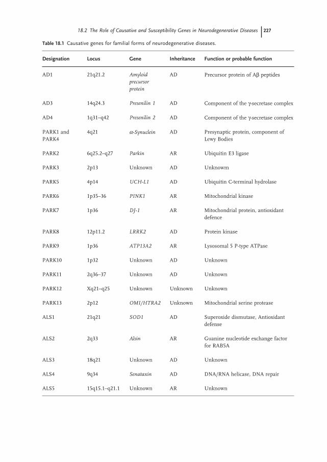
AD, PD and ALS are the three major neurodegenerative diseases, affecting several million people worldwide. They are defi ned as complex multifactorial disorders since both familial and sporadic forms are known. Familial forms represent a minority of the cases (ranging from 5 to 10% of the total), whereas the vast majority of AD, PD and ALS occurs as sporadic forms, likely resulting from the contribution of complex interactions between genetic and environ-mental factors superimposed on slow, sustained neuronal dysfunction due to aging. Several causative genes for the familial forms have been discovered in recent years, they are inherited as Mendelian traits and their discovery has led to a better comprehension of the molecular pathways responsible for the selective neuronal degeneration which is specifi c for each of these disorders (Table 18.1 ).
AD represents the most common form of dementia in the elderly, characterized by progressive loss of memory and cognitive capacity severe enough to interfere with daily functioning and the quality of life. The cardinal histopathologic lesions of AD are senile plaques, composed of extracellular deposits of amyloid beta ( A β ) peptides and neurofi brillary tangles, composed of intraneuronal tau protein aggre-gates [1] . PD is the second most common neurodegenerative disorder after AD. Pathologically, PD is characterized by progressive and profound loss of neuromela-nin containing dopaminergic neurons in the substantia nigra with the presence of cytoplasmic inclusions termed Lewy bodies ( LB ) and containing aggregates of α - synuclein as well as other substances [2] . ALS, also known as motor neuron disease ( MND ), is a progressive disorder characterized by the degeneration of motor neurons of the motor cortex, brainstem and spinal cord. The course of ALS is inexorably progressive, with 50% of the patients dying within 3 years of onset [3] . HD is a monogenic disorder transmitted as an autosomal dominant trait, meaning that all the cases result from mutations of a single gene. However, a contribution from other genes and environmental factors to age at onset and progression of the disease is indicated by several studies. The disease is character-ized by selective degeneration of medium spiny GABAergic neurons in the stria-tum, resulting in a progressive atrophy of the caudate nucleus, putamen and globus pallidus [4] .
18.2 The Role of Causative and Susceptibility Genes in Neurodegenerative Diseases
AD is a genetically complex and heterogeneous disorder. Rare, fully penetrant mutations in three genes ( APP , PSEN1 and PSEN2 ) are responsible for familial early onset ( < 65 years) autosomal dominant forms (EOAD) (Table 18.1 ). The amyloid precursor protein gene ( APP ) encodes for the amyloid precursor protein ( APP ). APP is an integral membrane protein and its cleavage mediated by β - and γ - secretases results in the production of A β peptides denoted as A β 40 and A β 42.
18.2 The Role of Causative and Susceptibility Genes in Neurodegenerative Diseases 227
Table 18.1 Causative genes for familial forms of neurodegenerative diseases.
Designation Locus Gene Inheritance Function or probable function
AD1 21q21.2 Amyloid
precursor
protein
AD Precursor protein of A β peptides
AD3 14q24.3 Presenilin 1 AD Component of the γ - secretase complex
AD4 1q31 – q42 Presenilin 2 AD Component of the γ - secretase complex
PARK1 and PARK4
4q21 α - Synuclein AD Presynaptic protein, component of Lewy Bodies
PARK2 6q25.2 – q27 Parkin AR Ubiquitin E3 ligase
PARK3 2p13 Unknown AD Unknowm
PARK5 4p14 UCH - L1 AD Ubiquitin C - terminal hydrolase
PARK6 1p35 – 36 PINK1 AR Mitochondrial kinase
PARK7 1p36 DJ - 1 AR Mitochondrial protein, antioxidant defence
PARK8 12p11.2 LRRK2 AD Protein kinase
PARK9 1p36 ATP13A2 AR Lysosomal 5 P - type ATPase
PARK10 1p32 Unknown AD Unknown
PARK11 2q36 – 37 Unknown AD Unknown
PARK12 Xq21 – q25 Unknown Unknown Unknown
PARK13 2p12 OMI/HTRA2 Unknown Mitochondrial serine protease
ALS1 21q21 SOD1 AD Superoxide dismutase, Antioxidant defense
ALS2 2q33 Alsin AR Guanine nucleotide exchange factor for RAB5A
ALS3 18q21 Unknown AD Unknown
ALS4 9q34 Senataxin AD DNA/RNA helicase, DNA repair
ALS5 15q15.1 – q21.1 Unknown AR Unknown
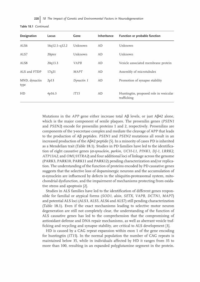
228 18 The Impact of Genetic and Environmental Factors in Neurodegeneration
Designation Locus Gene Inheritance Function or probable function
ALS6 16q12.1 – q12.2 Unknown AD Unknown
ALS7 20pter Unknown AD Unknown
ALS8 20q13.3 VAPB AD Vesicle associated membrane protein
ALS and FTDP 17q21 MAPT AD Assembly of microtubules
MND, dynactin type
2p13 Dynactin 1 AD Promotion of synapse stability
HD 4p16.3 IT15 AD Huntingtin, proposed role in vesicular traffi cking
Table 18.1 Continued.
Mutations in the APP gene either increase total A β levels, or just A β 42 alone, which is the major component of senile plaques. The presenilin genes ( PSEN1 and PSEN2 ) encode for presenilin proteins 1 and 2, respectively. Presenilins are components of the γ - secretase complex and mediate the cleavage of APP that leads to the production of A β peptides. PSEN1 and PSEN2 mutations all result in an increased production of the A β 42 peptide [5] . In a minority of cases PD is inherited as a Mendelian trait (Table 18.1 ). Studies in PD families have led to the identifi ca-tion of eight causative genes ( α - synuclein , parkin , UCH - L1 , PINK1 , DJ - 1 , LRRK2 , ATP13A2 , and OMI/HTRA2 ) and four additional loci of linkage across the genome (PARK3, PARK10, PARK11 and PARK12) pending characterization and/or replica-tion. The understanding of the function of proteins encoded by PD causative genes suggests that the selective loss of dopaminergic neurons and the accumulation of α - synuclein are infl uenced by defects in the ubiquitin - proteasomal system, mito-chondrial dysfunction, and the impairment of mechanisms protecting from oxida-tive stress and apoptosis [2] .
Studies in ALS families have led to the identifi cation of different genes respon-sible for familial or atypical forms ( SOD1, alsin, SETX, VAPB, DCTN1, MAPT ) and potential ALS loci (ALS3, ALS5, ALS6 and ALS7) still pending characterization (Table 18.1 ). Even if the exact mechanisms leading to selective motor neuron degeneration are still not completely clear, the understanding of the function of ALS causative genes has led to the comprehension that the compromising of antioxidant defense and DNA repair mechanisms, as well as aberrant vesicle traf-fi cking and recycling and synapse stability, are critical to ALS development [3] .
HD is caused by a CAG repeat expansion within exon 1 of the gene encoding for huntingtin ( IT15 ). In the normal population the number of CAG repeats is maintained below 35, while in individuals affected by HD it ranges from 35 to more than 100, resulting in an expanded polyglutamine segment in the protein.
18.2 The Role of Causative and Susceptibility Genes in Neurodegenerative Diseases 229
The age of onset of HD is inversely correlated with the CAG repeat length, and it has been hypothesized that the expanded polyglutamine segment confers a domi-nant “ gain of function ” to the protein, ultimately leading to neurodegeneration [6] . Signifi cant variance remains, however, in residual age of onset, even after CAG repeat length is factored out. Many polymorphic genes have previously shown evidence of association with age of onset of HD in several different populations, among them the GluR6 kainate glutamate receptor ( GRIK2 ), APOE , the trans-criptional coactivator CA150 ( TCERG1 ), UCHL1 , TP53 , caspase - activated DNase ( DFFB ), and the NR2A and NR2B glutamate receptor subunits ( GRIN2A,
GRIN2B ) [7] . Despite the discovery of several causative genes for the familial forms, the major-
ity of AD, PD and ALS occur as sporadic forms resulting from the contribution of several interactions between exogenous environmental factors and the individual genetic background. Over one thousand polymorphisms in almost three hundreds different genes have been analyzed in recent years as candidate AD susceptibility factors, but only the ε 4 allele of the Apolipoprotein E ( APOE ) gene has clearly emerged as an AD risk factor. The APOE - ε 4 variant is associated with higher plasma cholesterol levels, and is supposed to enhance A β deposition and the for-mation of neuritic plaques [8] . For the remaining hundreds of putative AD sus-ceptibility genes results are often confl icting, obtained in small sample - sized groups or limited to one or two papers reporting association. A recent pooled analysis of those polymorphisms which had been studied in at least three inde-pendent association studies [9] , revealed few of them as possible AD risk or protec-tive factors (Table 18.2 ). As for AD, several hundreds of association studies have been published in recent years claiming or denying association between variants in candidate genes and the risk of PD. Results published so far are often confl ict-ing and inconclusive, refl ecting the genetic heterogeneity of the studied popula-tions, inadequate sample size and the possible contribution of environmental factors. The major genes which have been analyzed in PD association studies are those related to dopamine transport and metabolism (e.g., DAT, DRD2, COMT,
MAO - B ), detoxifi cation of xenobiotics (e.g., CYP2D6, GSTs, NAT2 ) and oxidative stress (e.g., NOS, SOD2 ). Moreover, common variants of PD causative genes (e.g., SNCA, LRRK2, UCHL1 ) have been largely studied for their role as possible PD susceptibility factors. Details are shown in Table 18.2 .
Almost 95% of ALS occurs as sporadic forms; however, although several genes have been studied in recent years as possible ALS susceptibility factors, no single gene has been defi nitively shown to be consistently associated with disease risk. Recent data support a role for the DNA repair genes APE1 and hOGG1 in sporadic ALS based on their protective roles against oxidative stress [10, 11] . Confl icting or inconclusive results have been obtained for angiogenesis genes ANG and VEGF [10] . Other candidate genes are those coding for neurofi laments ( NEFL, NEFM and NEFH ), paraoxonases ( PON1, PON2 and PON3 ), survival motor neuron ( SMN1 and SMN2 ) and the hemocromatosis ( HFE ) gene. Recent pooled analyses suggest a role for the HFE H63D variant and for increased copy numbers of the SMN1 gene [10] . Details are shown in Table 18.2 .

230 18 The Impact of Genetic and Environmental Factors in Neurodegeneration
Table 18.2 Some of the proposed susceptibility genes for neurodegenerative diseases.
Genetic variant(s) Associated with
Alzheimer ’ s disease APOE - ε 4 Increased risk SORL1 variants Increased risk ACE intron 16 (ins/del) Increased risk ACE rs1800764, rs4291, rs4343 Decreased risk CHRNB2 rs4845378 Decreased risk CST3 5 ’ UTR - 157, 5 ’ UTR - 72 Increased risk CST3 A25T Increased risk ESR1 PvuII, XbaI Increased risk GAPDHS rs12984928, rs4806173 Decreased risk IDE rs2251101 Decreased risk MTHFR A1298C Decreased risk NCSTR 119 intron 16 Increased risk PRPN M129V Decreased risk PSEN1 rs165932 Decreased risk TF P570S Increased risk TFAM rs2306604 Decreased risk TNF rs4647198 ( - 1031) Increased risk GOLPH2 rs10868366 a , rs7019241 a Decreased risk Rs 9886784 (Chromosome 9) Increased risk Rs 10519262 a Increased risk
Parkinson ’ s disease SNCA Rep1 Increased risk LRRK2 G2385R Increased risk b MAPT H1 haplotype Increased risk UCHL1 S18Y Decreased risk c GSTM1 null genotype Increased risk d1 GSTP1 variants Increased risk d2 CYP2D6 variants Increased risk d3 FAM 79B Rs 1000291 a Increased risk UNC5C Rs 2241743 a Increased risk Rs 3018626 (Chromosome 11) a Increased risk
Amyotrophic lateral sclerosis APE1 D148E Increased risk c ANG G110G Increased risk c hOGG1 Ser326Cys Increased risk in males VEGF variants Inconclusive results HFE H63D Increased risk SMN1 variable copy number Increased risk DPP6 variant a Increased risk
a From WGA studies. b Only in Asiatic populations. c Confl icting results. d In combination with environmental factors ( d1 = solvents, d2 = pesticides and herbicides,
d3 = pesticides, tobacco smoking).
18.3 The Contribution of Environmental Factors to Neurodegenerative Diseases 231
Despite hundreds of association studies based on the “ candidate gene ” approach, the hottest new tool in genetics is whole - genome association ( WGA ) studies; geneticists scan patient ’ s DNA for half a million or more single nucleotide poly-morphism s ( SNP s), and then compare the results with those from a healthy control group. Unfortunately, almost none of them has highlighted genes already under suspicion by the “ candidate - gene ” approach, moreover, results from WGA studies are often confl icting and not replicating [12, 13] . Some variants associated with AD, PD or ALS by WGA studies are listed in Table 18.2 .
18.3 The Contribution of Environmental Factors to Neurodegenerative Diseases
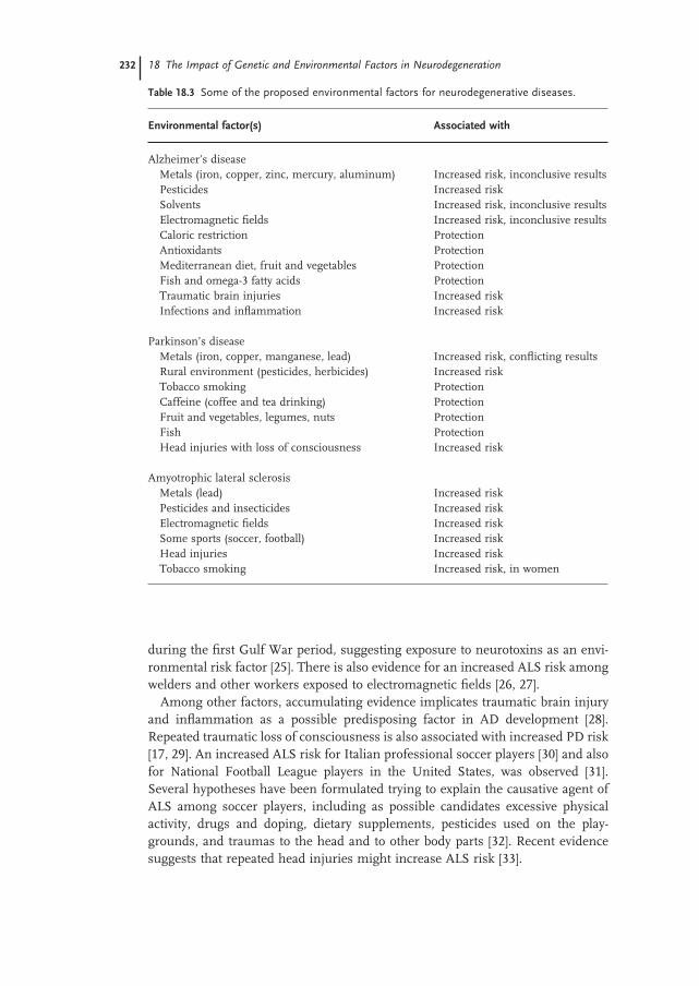
Several environmental factors have been largely studied in recent years as possible risk factors for neurodegeneration; among them metals, solvents, pesticides, elec-tromagnetic fi elds, brain injuries and physical activity, as well as drugs and dietary factors (Table 18.3 ).
Metals have been extensively studied as potential AD risk factors and even if a direct causal role for aluminum or other transition metals such as zinc, copper, iron and mercury in AD has not yet been defi nitively demonstrated, epidemiologi-cal evidence suggests that elevated levels of these metals in the brain may be linked to the development or the progression of the disease. Ingestion of aluminum in drinking water was associated with an increased risk of AD; however, other studies failed to fi nd such association [14, 15] . Another risk factor for AD is inorganic mercury, often present in dental amalgam applications, and a role for APOE as a mediator of the toxic effect of mercury has been largely suggested [16] . Human exposure to metals has been the focus of several epidemiological studies aimed at evaluating their possible contribution as PD risk factors. A recent large sample - sized study failed to fi nd association between iron, copper and manganese expo-sure and PD risk [17] . However another consistent report based on 110 000 individuals in two Canadian cities suggests that environmental manganese air pollution might contribute to neuronal loss in PD [18] . Occupational lead exposure also seems to be a risk factor for PD [19] . Increased ALS risk was observed among individuals occupationally exposed to lead [20] .
A recent analysis of 24 published studies assessing the role of occupational AD risk factors revealed a statistically consistent association only for pesticides [21] . The available evidence indicates that rural environment and pesticide exposures are associated with PD, however, no one agent has been consistently identifi ed, likely because associations with specifi c agents may be confounded by exposure to other pesticides, making it diffi cult to identify the causative agent [22] . There is also evidence suggesting that human exposures to agricultural chemicals, such as pesticides, are at increased ALS risk [23] . To support this hypothesis there is a recent report of a motor neuron disorder simulating ALS induced by chronic inhalation of pyrethroid insecticides [24] . Moreover, increased post - war risk of ALS has been observed in military personnel who were deployed to the Gulf Region
232 18 The Impact of Genetic and Environmental Factors in Neurodegeneration
Table 18.3 Some of the proposed environmental factors for neurodegenerative diseases.
Environmental factor(s) Associated with
Alzheimer ’ s disease Metals (iron, copper, zinc, mercury, aluminum) Increased risk, inconclusive results Pesticides Increased risk Solvents Increased risk, inconclusive results Electromagnetic fi elds Increased risk, inconclusive results Caloric restriction Protection Antioxidants Protection Mediterranean diet, fruit and vegetables Protection Fish and omega - 3 fatty acids Protection Traumatic brain injuries Increased risk Infections and infl ammation Increased risk
Parkinson ’ s disease Metals (iron, copper, manganese, lead) Increased risk, confl icting results Rural environment (pesticides, herbicides) Increased risk Tobacco smoking Protection Caffeine (coffee and tea drinking) Protection Fruit and vegetables, legumes, nuts Protection Fish Protection Head injuries with loss of consciousness Increased risk
Amyotrophic lateral sclerosis Metals (lead) Increased risk Pesticides and insecticides Increased risk Electromagnetic fi elds Increased risk Some sports (soccer, football) Increased risk Head injuries Increased risk Tobacco smoking Increased risk, in women
during the fi rst Gulf War period, suggesting exposure to neurotoxins as an envi-ronmental risk factor [25] . There is also evidence for an increased ALS risk among welders and other workers exposed to electromagnetic fi elds [26, 27] .
Among other factors, accumulating evidence implicates traumatic brain injury and infl ammation as a possible predisposing factor in AD development [28] . Repeated traumatic loss of consciousness is also associated with increased PD risk [17, 29] . An increased ALS risk for Italian professional soccer players [30] and also for National Football League players in the United States, was observed [31] . Several hypotheses have been formulated trying to explain the causative agent of ALS among soccer players, including as possible candidates excessive physical activity, drugs and doping, dietary supplements, pesticides used on the play-grounds, and traumas to the head and to other body parts [32] . Recent evidence suggests that repeated head injuries might increase ALS risk [33] .
18.4 Epigenetics, Environment and Susceptibility to Human Diseases 233
Dietary factors have been largely studied as possible contributors of neurode-generation; among them antioxidant compounds seem to exert a neuroprotective role (Table 18.3 ).
In transgenic HD mice models the environmental enrichment with several new different objects seems to delay the onset of motor symptoms [4] .
18.4 Epigenetics, Environment and Susceptibility to Human Diseases
Epigenetics deals with the heritable modifi cations of DNA that can infl uence the phenotype through changing gene expression without altering primary DNA sequence. The epigenetic modifi cations include DNA methylation, histone mod-ifi cations, and RNA - mediated pathways from non - coding RNAs, notably silenc-ing RNA ( siRNA ) and microRNA ( miRNA ). Epigenetic modifi cations are key regulators of important developmental events, including X - inactivation, genomic imprinting and neuronal development. Accumulating evidence indicates that variations in gene expression due to variable modifi cations in DNA methylation and chromatin structure in response to the environment also play a role in dif-ferential susceptibility to diseases. Consistent with these fundamental aspects, an increasing number of human pathologies have been found to be associated with aberrant epigenetic regulation, such as mental retardation, syndromes involving chromosomal instabilities, obesity, infertility, respiratory diseases, allergies, and a great number of age - related diseases including cancer, hearing loss and neurodegenerative diseases [34 – 38] .
Epigenetic modifi cations have been compared, in terms of phenotypic conse-quences, to genetic polymorphisms resulting in variations in gene function [39] . Recent data suggest that the epigenome is dynamic and is, therefore, responsive to environmental signals not only during the critical periods in development but also later in life. It is postulated also that not only chemicals but also exposure to social behavior, such as maternal care, could affect the epigenome [40] . Exposures to different environmental agents could lead to interindividual phenotypic diver-sity as well as differential susceptibility to disease and behavioral pathologies [39] .
The common disease genetic and epigenetic hypothesis [35] argues that, in addi-tion to genetic variation, epigenetics provides an added layer of variation that might mediate the relationship between genotype and internal and external envi-ronmental factors. This epigenetic component could help in understanding the marked increase in common diseases with age, as well as the phenotypic discor-dance between monozygotic twins [41] . It is likely that the activity of proteins that have been proved to be involved in epigenetic modifi cations, e.g. DNA methyl-transferases, could be potentially modulated by environmental factors such as diet, alcohol, cigarette smoke or environmental toxins such as heavy metals, known to disrupt DNA methylation and chromatin [42] . The fungicide vinclozolin, an endo-crine disruptor that decreases male fertility, alters DNA methylation, and changes are inherited by subsequent generations [43] .
234 18 The Impact of Genetic and Environmental Factors in Neurodegeneration
Despite a growing consensus on the importance of epigenetics in the etiology of chronic human diseases, the genes most prone to epigenetic dysregulation are incompletely defi ned. Moreover, until now only a few environmental agents affect-ing the epigenome have been identifi ed (for a review, see Ref. [44] ) and much remains to do to adequately characterize environmentally induced epigenetic alterations [45, 46] .
18.5 Epigenetics and Neurodegenerative Diseases
DNA methylation is dynamically regulated in the brain throughout the lifespan, a genome - wide decline in DNA methylation occurs during normal aging, which coincides with a functional decline in learning and memory with age [47, 48] . The emerging fi eld of studies on DNA methylation of specifi c brain regions may help account for region - specifi c functional specialization [49] . Although AD manifests in late adult life, it is not clear when the disease actually starts and how long the neuropathological processes take to develop AD. To explain the etiology of AD from an epigenetic point of view, one should consider the neuropathological fea-tures, such as neuronal cell death, tau tangles, and amyloid plaque formation, as a function of epigenome variations induced by environmental factors that have until now been associated with AD, such as diet components, or toxicological exposure (see Table 18.3 ).
In Alzheimer ’ s disease, as previously discussed, A β peptides or fragments are the major components of amyloid plaques and are produced by the amyloidogenic cutting of the amyloid precursor protein APP. APP can be alternatively processed by γ - secretases [presenilin1 (PSEN1) and 2 (PSEN2)] and α - secretases (ADAM10 and TACE) producing non - amyloidogenic peptides, or by γ - and β - secretases ( BACE ) producing A β peptides [50] . Therefore, the balance between different secretase activities is very important in the maintenance of the physiologic levels of non - amyloidogenic and amyloidogenic fragments.
We know that accumulation of oxidative stress - induced damage in brain tissue plays an important role in the pathogenesis of normal aging and neurodegenera-tive diseases, including AD. Because of its high metabolic rate the brain is believed to be particularly susceptible to reactive oxygen species ( ROS ), and the effects of oxidative stress on neurons might be cumulative. At the time oxidative damage was observed in AD, it was supposed that amyloid aggregates were the main source of oxidative stress; however, recent evidence suggests that oxidative stress is one of the earliest events in AD [51, 52] and that A β peptides might be produced to function as scavengers of reactive oxidative species. Only with the persistence of oxidative stress, does the production of A β peptides overcome their cellular turnover, so that they start to aggregate and their anti - oxidant function evolve into pro - oxidant, ultimately leading to neuronal death [53] . The connection between epigenetic mechanisms of transcriptional silencing of genes important to ROS such as MnSOD has been fi rmly established [54] . Increases in ROS can also effect
18.5 Epigenetics and Neurodegenerative Diseases 235
glutathione levels which in turn can change S - adenosylmethionine ( SAM ) synthe-sis and hence DNA methylation patterns. There are additional ROS - related mecha-nisms involving hydrogen peroxide that can lead to further changes of the chromatin structure. Interestingly Hitchler and Domann [55] proposed an epigen-etic perspective on the free radical theory of development. This theory proposes that oxygen has a key role in development by infl uencing the production of meta-bolic oxidants that would infl uence in turn the antioxidant capacity of cells throughout the production of glutathione ( GSH ). Increased GSH production infl u-ences epigenetic processes including DNA and histone methylation by limiting the availability of S - adenosylmethionine, the cofactor utilized during epigenetic control of gene expression by DNA and histone methyltransferases [55] . Glutathi-one is thus an important endogenous antioxidant, found in millimolar concentra-tions in the brain. GSH levels have been shown to decrease with aging and, in particular, are decreased in affected brain regions and peripheral cells from AD and also PD patients. Tabaton and Tamagno [56] reviewed the role of oxidative stress as a molecular link between the β - and the γ - secretase activities, and pro-vided a mechanistic explanation of the pathogenesis of sporadic late - onset AD: the overproduction of A β , dependent on the upregulation of BACE1 induced by oxida-tive stress, would contribute to the pathogenesis of the common, sporadic, late - onset form of AD, a major risk factor for which is aging. These authors suggest that an increase in the γ - secretase cleavage of APP mediated by oxidative stress (sporadic AD), or by PSEN1 mutations (FAD), fosters BACE1 expression and activity.
In general, genes involved in several pathways including antioxidant defense, detoxifi cation, infl ammation, etc., are induced in response to oxidative stress and in AD. However, genes that are associated with energy metabolism, which is necessary for normal brain function, are mostly down - regulated. The PGC - 1alpha role in regulation of ROS metabolism makes it a potential candidate player between ROS, mitochondria, and neurodegenerative diseases: down - regulated expression of PGC - 1alpha has been implicated in Huntington disease and in several Hun-tington disease animal models [57] . Lahiri et al. [58] proposed a “ Latent Early - Life Associated Regulation ” model, which postulates a latent expression of specifi c genes triggered at the developmental stage. According to this model, environmen-tal agents (e.g., heavy metals), intrinsic factors (e.g., cytokines), and dietary factors (e.g., cholesterol) perturb gene regulation in a long - term fashion, beginning in the early developmental stages, but with pathological outcomes signifi cantly later in life. For example, such actions would perturb APP gene regulation at a very early stage via its transcriptional machinery, leading to delayed overexpression of APP and subsequently of A β deposition. According to this model, promoter activity of specifi c genes, such as methyl - CpG - binding protein 2 ( MeCP2 ) and the transcrip-tion factors Sp1, can be altered by changes in the primary DNA sequence and by epigenetic changes through mechanisms such as DNA methylation at CpG dinu-cleotides or oxidation of guanosine residues [58] .
By genome scan studies increased levels of gene expression are now being discovered within specifi c classes of genes. This can be linked to a possible
236 18 The Impact of Genetic and Environmental Factors in Neurodegeneration
modulation of the methylation of promoters, such as in a study on differential expression of the ornithine transcarbamylase ( OTC ) gene, a key enzyme of the urea cycle, found expressed in AD but not in controls [59] . Currently, genome - wide technologies are available and have been utilized to examine the methylation state of cytosine bases throughout a genome (methylome). Studies involving several physiological and disease states, mainly cancer, have been performed. Although early in the process, DNA methylation is being explored as a biomarker to be used in clinical practice for early detection of disease, tumor classifi cation and for pre-dicting disease outcome or recurrence [60] . It has become increasingly evident in recent years that development is under epigenetic control. Prenatally or early life dietary and environmental exposures can have a profound effect on our epig-enome, resulting in birth defects and diseases developed later in life [61] . Studies in rodents have shown that exposure to lead (Pb) during brain development pre-determined the expression and regulation of the amyloid precursor protein and its amyloidogenic A β product in old age. The expression of AD - related genes ( APP , BACE1 ) as well as their transcriptional regulator ( Sp1 ) was elevated in aged monkeys exposed to Pb as infants. Developmental exposure to Pb altered the levels, characteristics, and intracellular distribution of A β staining and amyloid plaques in the frontal association cortex, furthermore, it induced a decrease in DNA methyltransferase activity and higher levels of oxidative damage to DNA, indicating that epigenetic imprinting in early life infl uenced the expression of AD - related genes and promoted DNA damage and pathogenesis [62] .
Wu et al. [63] propose that environmental infl uences occurring during brain development alter the methylation pattern of the APP promoter which results in a latent increase in APP and A β levels. Increased A β levels promote the production of ROS which damage DNA. Epigenetic changes in DNA methylation impact both gene transcription and the ability to repair damaged DNA and thus imprint sus-ceptibility to DNA damage. This susceptibility plus the programmed increase in A β levels, via a transcriptional pathway programmed by environmental exposures in early life, exacerbates the normal process of amyloidogenesis in the aging brain, thus accelerating the onset of AD.
Few attempts have been so far made to demonstrate the occurrence of epigenetic silencing of genes that have a fundamental role in other neurodegenerative dis-eases, such as ALS and HD. In an epigenetic context it is likely that the level of expression of several genes is altered in age - related diseases, including neurode-generative ones, due to the methylation status of their promoters. In particular, methylation levels dysregulation have been involved to explain the variable phe-notypic espressivity (age of onset, the severity and or penetrance of the pathological phenotype) [48] . Sporadic amyotrophic lateral sclerosis ( SALS ) results from the death of motor neurons in the brain and spinal cord. It has been proposed that epigenetic silencing of genes vital for motor neuron function could underlie SALS. Oates and Pamphlett [64] therefore examined the methylation status of two genes, SOD1 and VEGF , which are implicated in ALS. Methylation in the promoters of these genes was determined in white cell DNA and brain DNA of ALS patients. However the promoter regions were found to be largely unmethylated in all
18.6 The Epigenetic Role of the Diet in Neurodegenerative Diseases 237
patients [64] . The metallothionein ( MT ) family of proteins are the primary detoxi-fi cation mechanism for heavy metals and MT - Ia and MT - IIa are the most common human isoforms. It was hypothesized that inappropriate methylation at the pro-moters of these genes could lead to silencing of transcription and reduce the availability of MTs. The level of methylation in the promoters of genes encoding MT - Ia and MT - IIa in leukocyte and brain DNA samples from SALS patients was measured and compared with controls, but again no promoter methylation of these genes was evident in any SALS or control samples [65] .
18.6 The Epigenetic Role of the Diet in Neurodegenerative Diseases
Various environmental and dietary agents and lifestyles are suspected to be impli-cated in the development of a wide range of human cancers through epigenetic changes (for a review see Ref. [44] ). Very few data are available in this regard in the fi eld of neurodegeneration. Dietary modifi cation can indeed have a profound effect on DNA methylation and genomic imprinting. DNA methylation is regu-lated through cellular levels of S - adenosyl - methionine. The conversion of homo-cysteine ( HCY ) to methionine requires folate metabolites and is an essential step in the production of SAM. Recent studies of fundamental importance have shown that a variation of the diet can lead to an alteration of the phenotype in mice or in their offspring. Defi ciency in folate and methionine, necessary for normal biosyn-thesis of SAM, the methyl donor for methylcytosine, leads to aberrant imprinting of insulin - like growth factor 2 in mice [66] , maternal methyl donor supplementa-tion during gestation can alter the offspring phenotype by methylating a transpos-able element in mice with silencing of the nearby agouti coat - color gene [67] . Moreover, the same maternal dietary supplementation, with either methyl donors like folic acid or the phytoestrogen genistein, showed a protective role in counter-acting the DNA hypomethylating effect of bisphenol A, a chemical with carcino-genic properties, used in the manufacture of polycarbonate plastic [68] . Genetic polymorphisms can alter the response to dietary components (nutrigenetic effect) by infl uencing the absorption, metabolism, or site of action. Analogously, variation in DNA methylation patterns and other epigenetic events that infl uence overall gene expression can infl uence the biological response to food components and vice versa [69] .
AD is characterized by high HCY and low folate blood levels, meaning that the conversion of HCY to methionine is altered in AD, as is the production of SAM. DNA methylation is regulated through cellular levels of SAM. The conversion of homocysteine to methionine requires folate metabolites and is an essential step in the production of SAM. Fuso and collaborators [50] studied the levels of meth-ylation of CpG islands in the promoters of the APP and the PSEN1 gene (PSEN1 is one of the components of the γ - secretases which cleave APP, producing amyloid fragments), on human neuroblastoma cell lines, observing that, in conditions of folate and vitamin B12 deprivation from the media, the status of methylation of

238 18 The Impact of Genetic and Environmental Factors in Neurodegeneration
the promoter of the PSEN1 gene was changed, with a subsequent deregulation of the production of PS1, BACE (the β - secretase) and APP proteins [50] . This experi-ment has provided evidence that some of the genes responsible for the production of A β fragments in AD can be regulated through epigenetic mechanisms which are regulated by the cellular availability of folates and B12 vitamins, and involve the production of SAM and the status of methylation of CpG islands in the DNA. More recent results by the same authors indicate that homocysteine accumulation induced through vitamin B deprivation could impair the “ methylation potential ” with consequent presenilin 1, BACE and amyloid - beta upregulation. Moreover, they found that homocysteine alterations had an effect on neuroblastoma but not on glioblastoma cells; this suggested a possible differential role of the two cell types in Alzheimer ’ s disease [70] . Studies in vivo , on a murine model of Alzheimer ’ s disease, confi rmed that a combined folate, B12 and B6 dietary defi -ciency induced hyperhomocysteinemia and imbalance of S - adenosylmethionine and S - adenosylhomocysteine. This effect was associated with PSEN1 and BACE up - regulation and amyloid - β deposition [71] .
Folate defi ciency seems to contribute to a variety of age - related neurological and psychological disorders, including amyotrophic lateral sclerosis. Key nutritional defi ciencies could potentiate the impact of enrivonmental neurotoxins. The envi-ronmental neurotoxin arsenic has recently been linked with decreased neurofi la-ment ( NF ) content in peripheral nerves. Supplementation with S - adenosyl methionine (SAM) attenuated the impact of folate deprivation on arsenic neuro-toxicity, consistent with the decrease in SAM following folate deprivation and the requirement for SAM - mediated methylation for arsenic bioelimination [72] .
18.7 Concluding Remarks
Many of the processes with a key role in neurodegeneration, such as the formation of senile plaques, the accumulation of ROS, the cleavage of APP by neuroscretases, can now be analyzed in the light of the new epigenetic knowledge, to facilitate the implementation of future disease prevention strategies. Since epigenetic altera-tions are reversible, modifying epigenetic marks contributing to disease develop-ment may provide an approach to designing new therapies such as the use of inhibitors of enzymes controlling epigenetic modifi cations [73] .
However, we have to take into account that epigenetic therapy has its limitations, such as the non - specifi c activation of genes and transposable elements in normal cells, and also has the potential for mutagenicity and carcinogenicity. It is also possible that corrected epigenetic modifi cations may revert to their previous state because of the reversible nature of DNA methylation and histone modifi cation patterns, although this may be prevented with continued treatment, or corrected again with retreatment [34] .
The emerging fi eld of environmental epigenomics deals with the study of meta-stable epialleles as epigenetically labile genomic targets [74] . Among those alleles
References 239
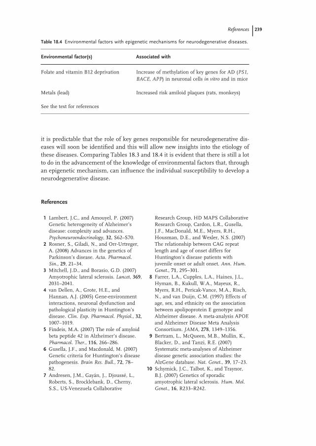
it is predictable that the role of key genes responsible for neurodegenerative dis-eases will soon be identifi ed and this will allow new insights into the etiology of these diseases. Comparing Tables 18.3 and 18.4 it is evident that there is still a lot to do in the advancement of the knowledge of environmental factors that, through an epigenetic mechanism, can infl uence the individual susceptibility to develop a neurodegenerative disease.
Table 18.4 Environmental factors with epigenetic mechanisms for neurodegenerative diseases.
Environmental factor(s) Associated with
Folate and vitamin B12 deprivation Increase of methylation of key genes for AD ( PS1,
BACE, APP ) in neuronal cells in vitro and in mice
Metals (lead) Increased risk amiloid plaques (rats, monkeys)
See the text for references
References
1 Lambert , J.C. , and Amouyel , P. ( 2007 ) Genetic heterogeneity of Alzheimer ’ s disease: complexity and advances . Psychoneuroendocrinology , 32 , S62 – S70 .
2 Rosner , S. , Giladi , N. , and Orr - Urtreger , A. ( 2008 ) Advances in the genetics of Parkinson ’ s disease . Acta. Pharmacol.
Sin. , 29 , 21 – 34 . 3 Mitchell , J.D. , and Borasio , G.D. ( 2007 )
Amyotrophic lateral sclerosis . Lancet , 369 , 2031 – 2041 .
4 van Dellen , A. , Grote , H.E. , and Hannan , A.J. ( 2005 ) Gene - environment inter actions, neuronal dysfunction and pathological plasticity in Huntington ’ s disease . Clin. Exp. Pharmacol. Physiol. , 32 , 1007 – 1019 .
5 Findeis , M.A. ( 2007 ) The role of amyloid beta peptide 42 in Alzheimer ’ s disease . Pharmacol. Ther. , 116 , 266 – 286 .
6 Gusella , J.F. , and Macdonald , M. ( 2007 ) Genetic criteria for Huntington ’ s disease pathogenesis . Brain Res. Bull. , 72 , 78 – 82 .
7 Andresen , J.M. , Gay á n , J. , Djouss é , L. , Roberts , S. , Brocklebank , D. , Cherny , S.S. , US - Venezuela Collaborative
Research Group, HD MAPS Collaborative Research Group , Cardon , L.R. , Gusella , J.F. , MacDonald , M.E. , Myers , R.H. , Housman , D.E. , and Wexler , N.S. ( 2007 ) The relationship between CAG repeat length and age of onset differs for Huntington ’ s disease patients with juvenile onset or adult onset . Ann. Hum.
Genet. , 71 , 295 – 301 . 8 Farrer , L.A. , Cupples , L.A. , Haines , J.L. ,
Hyman , B. , Kukull , W.A. , Mayeux , R. , Myers , R.H. , Pericak - Vance , M.A. , Risch , N. , and van Duijn , C.M. ( 1997 ) Effects of age, sex, and ethnicity on the association between apolipoprotein E genotype and Alzheimer disease. A meta - analysis APOE and Alzheimer Disease Meta Analysis Consortium . JAMA , 278 , 1349 – 1356 .
9 Bertram , L. , McQueen , M.B. , Mullin , K. , Blacker , D. , and Tanzi , R.E. ( 2007 ) Systematic meta - analyses of Alzheimer disease genetic association studies: the AlzGene database . Nat. Genet. , 39 , 17 – 23 .
10 Schymick , J.C. , Talbot , K. , and Traynor , B.J. ( 2007 ) Genetics of sporadic amyotrophic lateral sclerosis . Hum. Mol.
Genet. , 16 , R233 – R242 .
240 18 The Impact of Genetic and Environmental Factors in Neurodegeneration
11 Copped è , F. , Mancuso , M. , Lo Gerfo , A. , Carlesi , C. , Piazza , S. , Rocchi , A. , Petrozzi , L. , Nesti , C. , Micheli , D. , Bacci , A. , Migliore , L. , Murri , L. , and Siciliano , G. ( 2007 ) Association of the hOGG1 Ser326Cys polymorphism with sporadic amyotrophic lateral sclerosis . Neurosci.
Lett. , 420 , 163 – 168 . 12 Evangelou , E. , Maraganore , D.M. , and
Ioannidis , J.P. ( 2007 ) Meta - analysis in genome - wide association datasets: strategies and application in Parkinson disease . PLoS ONE , 2 , e196 .
13 Garber , K. ( 2008 ) The elusive ALS genes . Science , 319 , 20 .
14 McLachlan , D.R. , Bergeron , C. , Smith , J.E. , Boomer , D. , and Rifat , S.L. ( 1996 ) Risk for neuropathologically confi rmed Alzheimer ’ s disease and residual aluminum in municipal drinking water employing weighted residential histories . Neurology , 46 , 401 – 405 .
15 Martyn , C.N. , Coggon , D.N. , Inskip , H. , Lacey , R.F. , and Young , W.F. ( 1997 ) Aluminum concentrations in drinking water and risk of Alzheimer ’ s disease . Epidemiology , 8 , 281 – 286 .
16 Mutter , J. , Naumann , J. , Sadaghiani , C. , Schneider , R. , and Walach , H. ( 2004 ) Alzheimer disease: mercury as patho-genetic factor and apolipoprotein E as a moderator . Neuro. Endocrinol. Lett. , 25 , 331 – 339 .
17 Dick , F.D. , De Palma , G. , Ahmadi , A. , Scott , N.W. , Prescott , G.J. , Bennett , J. , Semple , S. , Dick , S. , Counsell , C. , Mozzoni , P. , Haites , N. , Wettinger , S.B. , Mutti , A. , Otelea , M. , Seaton , A. , S ö derkvist , P. , Felice , A. , and Geoparkinson Study Group ( 2007 ) Environmental risk factors for Parkinson ’ s disease and parkinsonism: the Geoparkinson study . Occup. Environ.
Med. , 64 , 666 – 672 . 18 Finkelstein , M.M. , and Jerrett , M. ( 2007 )
A study of the relationships between Parkinson ’ s disease and markers of traffi c - derived and environmental manganese air pollution in two Canadian cities . Environ. Res. , 104 , 420 – 432 .
19 Coon , S. , Stark , A. , Peterson , E. , Gloi , A. , Kortsha , G. , Pounds , J. , Chettle , D. , and Gorell , J. ( 2006 ) Whole - body lifetime
occupational lead exposure and risk of Parkinson ’ s disease . Environ. Health
Perspect. , 14 , 1872 – 1876 . 20 Kamel , F. , Umbach , D.M. , Hu , H. ,
Munsat , T.L. , Shefner , J.M. , Taylor , J.A. , and Sandler , D.P. ( 2005 ) Lead exposure as a risk factor for amyotrophic lateral sclerosis . Neurodegener. Dis. , 2 , 195 – 201 .
21 Santib á ñ ez , M. , Bolumar , F. , and Garc í a , A.M. ( 2007 ) Occupational risk factors in Alzheimer ’ s disease: a review assessing the quality of published epidemiological studies , Occup. Environ. Med. , 64 , 723 – 732 .
22 Elbaz , A. ( 2007 ) Parkinson ’ s disease and rural environment . Rev. Prat. , 57 , 37 – 39 .
23 Govoni , V. , Granieri , E. , Fallica , E. , and Casetta , I. ( 2005 ) Amyotrophic lateral sclerosis, rural environment and agricultural work in the Local Health Discrict of Ferrara, Italy, in the years 1964 – 1998 . J. Neurol. , 252 , 1322 – 1327 .
24 Doi , H. , Kikuchi , H. , Murai , H. , Kawano , Y. , Shigeto , H. , Ohyagi , Y. , and Kira , J. ( 2006 ) Motor neuron disorder simulating ALS induced by chronic inhalation of pyrethroid insecticides . Neurology , 67 , 1894 – 1895 .
25 Horner , R.D. , Kamins , K.G. , Feussner , J.R. , Grambow , S.C. , Hoff - Lindquist , J. , Harati , Y. , Mitsumoto , H. , Pascuzzi , R. , Spencer , P.S. , Tim , R. , Howard , D. , Smith , T.C. , Ryan , M.A.K. , Coffman , C.J. , and Kararskis , E.J. ( 2003 ) Occurrence of amyotrophic lateral sclerosis among Gulf War veterans . Neurology , 61 , 742 – 749 .
26 Li , C.Y. , and Sung , F.C. ( 2003 ) Association between occupational exposure to power frequency electromagnetic fi elds and amyotrophic lateral sclerosis: a review . Am. J. Ind.
Med. , 43 , 212 – 220 . 27 H å kansson , N. , Gustavsson , P. , Johansen ,
C. , and Floderus , B. ( 2003 ) Neurodegenerative diseases in welders and other workers exposed to high levels of magnetic fi elds . Epidemiology , 14 , 420 – 426 .
28 Van Den Heuvel , C. , Thornton , E. , and Vink , R. ( 2007 ) Traumatic brain injury and Alzheimer ’ s disease: a review . Prog.
Brain Res. , 161 , 303 – 316 .
References 241
29 Goldman , S.M. , Tanner , C.M. , Oakes , D. , Bhudhikanok , G.S. , Gupta , A. , and Langston , J.W. ( 2006 ) Head injury and Parkinson ’ s disease risk in twins . Ann.
Neurol. , 60 , 65 – 72 . 30 Chi ò , A. , Benzi , G. , Dossena , M. , Mutani ,
R. , and Mora , G. ( 2005 ) Severely increased risk of amyotrophic lateral sclerosis among Italian professional football players . Brain , 128 , 472 – 476 .
31 Abel , E.L. ( 2007 ) Football increases the risk for Lou Gehrig ’ s disease, amyotrophic lateral sclerosis . Percept. Mot.
Skills , 104 , 1251 – 1254 . 32 Belli , S. , and Vanacore , N. ( 2005 )
Proportionate mortality in italian soccer players: is amyotrophic lateral sclerosis an occupational disease? Eur. J. Epidemiol. , 20 , 237 – 242 .
33 Chen , H. , Richard , M. , Sadler , D.P. , Umbach , D.M. , and Kamel , F. ( 2007 ) Head injury and amyotrophic lateral sclerosis . Am. J. Epidemiol. , 166 , 810 – 816 .
34 Lu , Q. , Qiu , X. , Hu , N. , Wen , H. , Su , Y. , and Richardson , B.C. ( 2006 ) Epigenetics, disease, and therapeutic interventions . Ageing Res. Rev. , 5 , 449 – 467 .
35 Feinberg , A.P. ( 2007 ) Phenotypic plasticity and the epigenetics of human disease . Nature , 447 , 433 – 440 .
36 Jirtle , R.L. , and Skinner , M.K. ( 2007 ) Environmental epigenomics and disease susceptibility . Nat. Rev. Genet. , 8 , 253 – 262 .
37 Provenzano , M.J. , and Domann , F.E. ( 2007 ) A role for epigenetics in hearing: establishment and maintenance of auditory specifi c gene expression patterns . Hear Res. , 233 , 1 – 13 .
38 Santos - Rebou ç as , C.B. , and Pimentel , M.M. ( 2007 ) Implication of abnormal epigenetic patterns for human diseases . Eur. J. Hum. Genet. , 15 , 10 – 17 .
39 Szyf , M. ( 2007 ) The dynamic epigenome and its implications in toxicology . Toxicol.
Sci. , 100 , 7 – 23 . 40 Szyf , M. , Weaver , I. , and Meaney , M.
( 2007 ) Maternal care, the epigenome and phenotypic differences in behaviour . Reprod. Toxicol. , 24 , 9 – 19 .
41 Poulsen , P. , Esteller , M. , Vaag , A. , and Fraga , M.F. ( 2007 ) The epigenetic basis of
twin discordance in age - related diseases . Pediatr. Res. , 61 , 38R – 42R
42 Sutherland , J.E. , and Costa , M. ( 2003 ) Epigenetics and the environment . Ann. N.
Y. Acad. Sci. , 983 , 151 – 160 . 43 Anway , M.D. , Rekow , S.S. , and Skinner ,
M.K. ( 2008 ) Transgenerational epigenetic programming of the embryonic testis transcriptome . Genomics , 91 , 30 – 40 .
44 Herceg , Z. ( 2007 ) Epigenetics and cancer: towards an evaluation of the impact of environmental and dietary factors . Mutagenesis , 22 , 91 – 103 .
45 Edwards , T.M. , and Myers , J.P. ( 2007 ) Environmental exposures and gene regulation in disease etiology . Environ.
Health Perspect. , 115 , 1264 – 1270 . 46 Weidman , J.R. , Dolinoy , D.C. , Murphy ,
S.K. , and Jirtle , R.L. ( 2007 ) Cancer susceptibility: epigenetic manifestation of environmental exposures . Cancer J. , 13 , 9 – 16 .
47 Liu , L. , van Groen , T. , Kadish , I. , and Tollefsbol , T.O. ( 2008 ) DNA methylation impacts on learning and memory in aging . Neurobiol. Aging , 30 , 549 – 560 .
48 Siegmund , K.D. , Connor , C.M. , Campan , M. , Long , T.I. , Weisenberger , D.J. , Biniszkiewicz , D. , Jaenisch , R. , Laird , P.W. , and Akbarian , S. ( 2007 ) DNA methylation in the human cerebral cortex is dynamically regulated throughout the life span and involves differentiated neurons . PLoS ONE , 2 , e895 .
49 Ladd - Acosta , C. , Pevsner , J. , Sabunciyan , S. , Yolken , R.H. , Webster , M.J. , Dinkins , T. , Callinan , P.A. , Fan , J.B. , Potash , J.B. , and Feinberg , A.P. ( 2007 ) DNA methylation signatures within the human brain . Am. J. Hum. Genet. , 81 , 1304 – 1315 .
50 Fuso , A. , Seminara , L. , Cavallaro , R.A. , D ’ Anselmi , F. , and Scarpa , S. ( 2005 ) S - adenosylmethionine/homocysteine cycle alterations modifyDNA methylation status with consequent deregulation of PS1 and BACE and beta - amyloid production . Mol.
Cell Neurosci. , 28 , 195 – 204 . 51 Migliore , L. , Fontana , I. , Trippi , F. ,
Colognato , R. , Copped è , F. , Tognoni , G. , Nucciarone , B. , and Siciliano , G. ( 2005 ) Oxidative DNA damage in peripheral leukocytes of mild cognitive impairment
242 18 The Impact of Genetic and Environmental Factors in Neurodegeneration
and AD patients . Neurobiol. Aging , 26 , 567 – 573 .
52 Zhu , X. , Su , B. , Wang , X. , Smith , M.A. , and Perry , G. ( 2007 ) Causes of oxidative stress in Alzheimer disease . Cell Mol. Life
Sci. , 64 , 2202 – 2210 . 53 Moreira , P.I. , Nunomura , A. , Honda , K. ,
Aliev , G. , Casadenus , G. , Zhu , X. , Smith , M.A. , and Perry , G. ( 2007 ) The key role of oxidative stress in Alzheimer ’ s disease , in Oxidative Stress and Neurodegenerative
Disorders (eds G. Ali Qureshi , and S. Hassan Parvez ), Elsevier , Amsterdam , pp. 451 – 466 .
54 Hitchler , M.J. , Wikainapakul , K. , Yu , L. , Powers , K. , Attatippaholkun , W. , and Domann , F.E. ( 2006 ) Epigenetic regulation of manganese superoxide dismutase expression in human breast cancer cells . Epigenetics , 1 , 163 – 171 .
55 Hitchler , M.J. , and Domann , F.E. ( 2007 ) An epigenetic perspective on the free radical theory of development . Free Radic.
Biol. Med. , 43 , 1023 – 1036 . 56 Tabaton , M. , and Tamagno , E. ( 2007 ) The
molecular link between beta - and gamma - secretase activity on the amyloid beta precursor protein . Cell Mol. Life Sci. , 64 , 2211 – 2218 .
57 McGill , J.K. , and Beal , M.F. ( 2006 ) PGC - 1alpha, a new therapeutic target in Huntington ’ s disease? Cell , 127 , 465 – 468 .
58 Lahiri , D.K. , Maloney , B. , Basha , M.R. , Ge , Y.W. , and Zawia , N.H. ( 2007 ) How and when environmental agents and dietary factors affect the course of Alzheimer ’ s disease: the “ LEARn ” model (latent early - life associated regulation) may explain the triggering of AD . Curr.
Alzheimer Res. , 4 , 219 – 228 . 59 Bensemain , F. , Hot , D. , Ferreira , S. ,
Dumont , J. , Bombois , S. , Maurage , C.A. , Huot , L. , Hermant , X. , Levillain , E. , Hubans , C. , Hansmannel , F. , Chapuis , J. , Hauw , J.J. , Schraen , S. , Lemoine , Y. , Bu é e , L. , Berr , C. , Mann , D. , Pasquier , F. , Amouyel , P. , and Lambert , J.C. ( 2008 ) Evidence for induction of the ornithine transcarbamylase expression in Alzheimer ’ s disease . Molecular Psychiatry , 14 , 106 – 116 .
60 Zilberman , D. ( 2007 ) The human promoter methylome . Nat. Genet. , 39 , 442 – 443 .
61 Reamon - Buettner , S.M. , and Borlak , J. ( 2007 ) A new paradigm in toxicology and teratology: altering gene activity in the absence of DNA sequence variation . Reprod. Toxicol. , 24 , 20 – 30 .
62 Wu , J. , Basha , M.R. , Brock , B. , Cox , D.P. , Cardozo - Pelaez , F. , McPherson , C.A. , Harry , J. , Rice , D.C. , Maloney , B. , Chen , D. , Lahiri , D.K. , and Zawia , N.H. ( 2008 ) Alzheimer ’ s disease (AD) - like pathology in aged monkeys after infantile exposure to environmental metal lead (Pb): evidence for a developmental origin and environmental link for AD . J. Neurosci. , 28 , 3 – 9 .
63 Wu , J. , Basha , M.R. , and Zawia , N.H. ( 2008 ) The environment, epigenetics and amyloidogenesis . J. Mol. Neurosci. , 34 , 1 – 7 .
64 Oates , N. , and Pamphlett , R. ( 2007 ) An epigenetic analysis of SOD1 and VEGF in ALS . Amyotroph. Lateral Scler. , 8 , 83 – 86 .
65 Morahan , J.M. , Yu , B. , Trent , R.J. , and Pamphlett , R. ( 2007 ) Are metallothionein genes silenced in ALS? Toxicol. Lett. , 168 , 83 – 87 .
66 Waterland , R.A. , Lin , J.R. , Smith , C.A. , and Jirtle , R.L. ( 2006 ) Post - weaning diet affects genomic imprinting at the insulin - like growth factor 2 (Igf2) locus . Hum. Mol. Genet. , 15 , 705 – 716 .
67 Waterland , R.A. , and Jirtle , R.L. ( 2003 ) Transposable elements: targets for early nutritional effects on epigenetic gene regulation . Mol. Cell Biol. , 23 , 5293 – 5300 .
68 Dolinoy , D.C. , Huang , D. , and Jirtle , R.L. ( 2007 ) Maternal nutrient supplementation counteracts bisphenol A - induced DNA hypomethylation in early development . Proc. Natl. Acad. Sci. U. S. A. , 104 , 13056 – 13061 .
69 Milner , J.A. ( 2006 ) Diet and cancer: facts and controversies . Nutr. Cancer , 56 , 216 – 224 .
70 Fuso , A. , Cavallaro , R.A. , Zampelli , A. , D ’ Anselmi , F. , Piscopo , P. , Confaloni , A. , and Scarpa , S. ( 2007 ) Gamma - Secretase is differentially modulated by alterations of homocysteine cycle in neuroblastoma and glioblastoma cells . J. Alzheimers Dis. , 1 ( 3 ), 275 – 290 .
71 Fuso , A. , Nicolia , V. , Cavallaro , R.A. , Ricceri , L. , D ’ Anselmi , F. , Coluccia , P. ,

References 243
Calamandrei , G. , and Scarpa , S. ( 2008 ) B - vitamin deprivation induces hyperhomocysteinemia and brain S - adenosylhomocysteine, depletes brain S - adenosylmethionine, and enhances PS1 and BACE expression and amyloid - beta deposition in mice . Mol. Cell Neurosci. , 37 , 731 – 746 .
72 Dubey , M. , and Shea , T.B. ( 2007 ) Potentiation of arsenic neurotoxicity by
folate deprivation: protective role of S - adenosyl methionine . Nutr. Neurosci. , 10 , 199 – 204 .
73 Allen , A. ( 2007 ) Epigenetic alterations and cancer: new targets for therapy . IDrugs , 10 , 709 – 712 .
74 Dolinoy , D.C. , and Jirtle , R.T. ( 2008 ) Environmental epigenomics in human health and disease . Environ. Mol.
Mutagen. , 49 , 4 – 8 .
Recommended