Effects of the exposure of roots of Alnusglutinosa to light on flavonoids and nodulation1
M. Hughes, C. Donnelly, A. Crozier, and C.T. Wheeler
Abstract: Exposure of the roots of Alnus glutinosa (L.) Gaertn. to white light stimulated within 5 days a substantialincrease in the content of quercetin and kaempferol, two major flavonols in acid hydrolysates of both roots and rootexudates. Both compounds were detected also in hydrolysates of root extracts of Myrica gale and Casuarinaequisetifolia, the latter a species not nodulated by Frankia strains effective on Alnus. A 7-fold reduction in nodulationof seedlings 12 days after inoculation with Frankia preincubated for 24 h with kaempferol supported the possibilitythat flavonols might be involved in the regulation of nodulation. Nodulation of seedling roots that were inoculated withFrankia and then exposed to light was inhibited by 50% after 11 days compared with seedlings with darkened roots.This effect of light treatment was preceded by a 30-fold increase in quercetin and kaempferol. The inhibitory effects onnodulation of preincubation of Frankia with kaempferol persisted for 18 days after inoculation, but there was nosignificant effect on nodulation after prolonging exposure of the root system to light for 40 days. The data supportindirectly the suggestion that the balance between stimulatory and inhibitory flavonoids in roots and root exudates maycontribute to the regulation of nodulation of actinorhizal plants.
Key words: Alnus glutinosa, flavonoids, Frankia, kaempferol, nodulation, roots.
Résumé : L’exposition à la lumière blanche pendant 5 jours des racines de l’Alnus glutinosa (L.) Gaertn. stimule uneaugmentation substantielle de la teneur en quercétine et en kampférol, deux flavonols majeurs qu’on retrouve dans leshydrolysats des racines ainsi que des exsudats. On retrouve également ces deux composés dans les hydrolysatsd’extraits racinaires du Myrica gale et du Casuarina equisetifolia, cette dernière espèce de formant pas de nodules avecles souches de Frankia actives sur Alnus. Une réduction de sept fois dans la nodulation de plantules âgées de 12 joursaprès inoculation avec du Frankia, pré-incubé pendant 24 h dans du kampférol, supporte la possibilité que desflavonols pourraient être impliqués dans la régulation de la nodulation. La nodulation de racines de plantules, inoculéesavec du Frankia et exposées par la suite à la lumière, a été inhibée à 50% après 11 jours, comparativement à desracines maintenues à l’obscurité. Cet effet du traitement à la lumière est précédé d’une augmention de 30 fois de laquercétine et du kamférol. Les effets inhibiteurs sur la nodulation, d’une pré-incubation des Frankia avec le kampférol,persistent pendant 18 jours après l’inoculation, mais il n’y a pas d’effet significatif sur la nodulation lorsqu’onprolonge l’exposition à la lumière du système racinaire pendant 40 jours. Les données supportent indirectement lasuggestion que la balance entre les flavonoïdes stimulateurs et inhibiteurs, dans les racines et les exsudats racinaires,pourrait contribuer à la régulation de la nodulation chez les plantes actinorhiziennes.
Mots clés : Alnus glutinosa, flavonoïdes, Frankia, kampférol, nodulation, racines.
[Traduit par la Rédaction] Hughes et al. 1315
Introduction
Flavonoids are involved in the initiation of the legume–rhizobium symbiosis through their action as specific reg-ulators of the expression of bacterial nodulation genes(Redmond et al. 1986). The development of a nod gene re-porter system to screen potential regulatory compounds ex-creted by legume roots was of major importance for the
success of these studies. In actinorhizal symbioses, Frankiagenes with similar functions to the nod genes of rhizobiumhave not yet been identified with certainty and consequentlysuitable reporter gene systems have not been developed.However, there has been much interest in the possibility thatflavonoids, or other phenolics excreted by seedling roots,may have regulatory roles similar to that in legumes. Thispossibility was strengthened by observations that nodulationof Alnus rubra Bong. is enhanced or inhibited by compo-nents of seed wash, tentatively identified as flavonones andisoflavones (Benoit and Berry 1997). In the absence of suit-able molecular methods to screen for nodulation gene regu-lators, an alternative approach to investigate the involvementof flavonoids in nodulation is described here.
Flavonoid biosynthesis genes are transcriptionally acti-vated by light in the plant, where flavonoids may provideprotection against ultraviolet light (Schmelzer et al. 1988;Stapleton 1992; Kubasek et al. 1992). Exposure of root sys-tems to light can stimulate the synthesis of flavonoids, and
Can. J. Bot. 77: 1311–1315 (1999) © 1999 NRC Canada
1311
Received June 25, 1998.
M. Hughes, C. Donnelly, A Crozier, and C.T. Wheeler.2
Plant Science Group, Division of Biochemistry & MolecularBiology, Glasgow University, Glasgow G12 8QQ, UnitedKingdom.
1This paper was presented at the 11th InternationalConference on Frankia and Actinorhizal Plants, June 7–11,1998, University of Illinois at Urbana–Champaign.
2Author to whom all correspondence should be addressed.e-mail: [email protected]
J:\cjb\cjb77\cjb-09\B99-077.vpThursday, December 16, 1999 11:56:26 AM
Color profile: DisabledComposite Default screen

accumulation of anthocyanins in both roots and nodules ofalders can be observed soon after exposure to light. In peas,suppression of nodulation by exposure of roots to light wasascribed to increased ethylene production (Lee and LaRue1992). The possibility that inhibition of nodulation could bedue to other causal or contributory events, such as changesin flavonoid biosynthesis, was not considered.
The aim of the present study was to investigate whethernodulation of Alnus glutinosa (L.) Gaertn. is inhibited bylight and whether such inhibition could be related to changesin flavonoid biosynthesis. The occurrence of flavonols in se-cretions of seedling roots has been determined and the re-sults of preliminary experiments to examine the effect onnodulation of preincubation of Frankia with flavonols aredescribed.
Methods
Germination and growth of seedlingsSeed of A. glutinosa was sown in presterilized seed trays con-
taining a 1:1 mixture of medium- and fine-grade Perlite moistenedwith water containing 0.25 g Crone’s N-free salts and 0.2 mLHoagland’s A–Z nutrient solution per litre (Hooker and Wheeler1987). The trays were placed in a growth room under Osram warmwhite fluorescent tubes (irradiance: 120 mmol·m�2·s�1, photoperiod16 h, 23°C light : 20°C dark). Seedlings with well-developed firsttrue leaves (6–8 weeks old) were transferred to ceramic pots con-taining 0.5 g Crone’s N-free salts, 0.5 mL A–Z nutrients solution,and 4 drops of Liquinure (Fisons Ltd., U.K.) per 2 L of water. Theroots were kept in darkness.
For light treatment for anthocyanin analysis, plants were trans-ferred after 6–8 weeks to 2-L clear glass jars with fresh culturemedium and grown for 3 weeks prior to harvest.
Growth of sterile seedlings and collection of rootexudate
Alnus glutinosa seeds were sterilized by agitating in sterile dis-tilled water + 5 drops Tween 80 per litre for 5 min, soaking in96% propan-2-ol for 1 min, and then agitating in 10% bleach for10 min. Seeds were then rinsed three times with 5% hydrogenperoxide, followed by five rinses with sterile distilled water. Thetreated seeds were plated on 9 cm diameter Petri dishes containing0.8% water agar, 15 seeds per dish, and incubated in the dark at25°C for 3�4 days. Germinating seeds were then transferred to thegrowth room (conditions as above). When roots were 1 cm inlength, seedlings were transferred to sterile cellulose fibre rods(“Sorbarod,” Ilacon Ltd., Kent, U.K.) contained in 28-mL glass vi-als and moistened with 5 mL filtered Crone’s + Hoagland’s A–Znutrients and grown on for a further 8 weeks.
Seedlings were carefully removed from the Sorbarods, whichwere then squeezed and washed with distilled water. Liquid con-taining root exudate from 20 vials was combined and concentratedby lyophilisation.
HPLC analysis of root exudates (Crozier et al. 1997)Hydrolyzed root exudates were analysed using a Shimadzu
(Kyoto, Japan) LC-10A series liquid chromatograph consisting ofan SCL-10A VP system controller, two LC-10AT VP pumps, anSIL-10AD VP autoinjector with sample cooler, a CTO-10A VPcolumn oven, an SPD-10A VP UV-VIS detector, and an RF-10AXL fluorimeter linked to a Reeve Analytical (Glasgow, U.K.) 2700data handling system. Reverse phase separations were carried outat 40°C using a 150 × 3.0 mm i.d. 4-mm Genesis C18 cartridge col-umn fitted with a Genesis guard cartridge (Jones Chromatography,
Mid-Glamorgan, U.K.). The mobile phase was a 20-min, 20–40%gradient of acetonitrile in water adjusted to pH 2.5 with trifluoro-acetic acid pumped at a flow rate of 0.5 mL·min�1. Column eluentwas first directed to the UV absorbance monitor operating at365 nm. Postcolumn derivatization was achieved by the addition ofmethanolic aluminium nitrate containing 7.5% glacial acetic acid(Hollman et al. 1996), pumped at 0.5 mL·min�1 by a Reeve analyt-ical model 9802 pump. The mixture was directed to a RF-10Afluorimeter, and fluorescent flavonol complexes were detected atexcitation 420 nm and emission 495 nm. The limits of detectionwere 5 ng with UV and 0.1 ng with fluorescence. Flavonoid stan-dards for calibration and co-chromatography were purchased fromSigma Chemicals, Poole, U.K.
Extraction and acid hydrolysis of flavonoidsTissues were frozen in liquid nitrogen and lyophilised. Samples
were ground to a fine powder in liquid nitrogen and stored at�70°C. Dried sample (15 mg) was hydrolyzed in a 5-mLReactiVial (Pierce, Rockford, U.S.A.) in 2 mL of 1.2 M hydrochlo-ric acid in 50% methanol, containing 25 mM sodium diethyl di-thiocarbamate as an antioxidant and 100 ng morin as internalstandard, at 90°C in a ReactiTherm heating–stirring module. Ali-quots of unhydrolyzed samples were removed prior to heating.Samples were centrifuged at 13 000 rpm in a Sanyo Micro-Centaurcentrifuge before HPLC, as above. Root exudates, 300 µL, werehydrolyzed similarly in 2 mL 1.2 M hydrochloric acid in 50%methanol and 25 mM sodium diethyl dithiocarbamate but withoutmorin.
Exposure of seedling roots to white light andinoculation with Frankia
Seedlings (8 weeks old) were suspended in UV transparent cel-lulose acetate film covering the top of 250-mL glass beakers withtheir roots partially in filtered Crone’s solution. Dark seedlingswere grown with their roots suspended through black plastic topscovering filtered Crone’s solution in opaque jars. Frankia strainUGL010710 was cultured in propionate medium as described pre-viously (Hooker and Wheeler 1987), with or without kaempferol.Kaempferol was added by filter sterilization to 100-mL cultures toa final concentration of 10�6 M. Cultures with kaempferol were in-cubated in the dark at 24°C for 24 h. All cultures were washed se-quentially with water on harvest, with centrifugation at 1200 × gbetween each wash. The final pellet (2.5 mL) was suspended in50 mL water and gently homogenized. Seedlings were inoculatedwith 0.5 mL of this suspension. Seedlings were maintained in thegrowth room, lit by Osram warm white fluorescent tubes with 16-hphotoperiod, as above.
Results
Flavonoids of roots of alder seedlings
AnthocyaninsThe roots of seedlings grown on Sorbarods exposed to
light showed high pigmentation, characteristic of anthocy-anins. Indication of the identity of the major anthocyaninswas obtained by co-chromatography of tissue extracts (15%acetic acid in 85% methanol) by (i) two-dimensional chro-matography on Polygram Cel 300 (Machery-Nagel) utilisingn-butanol – acetic acid – water (4:1:5) and (ii) HPLC (30°C,absorbance detector 535 nm) on a 150 × 4.6 mm i.d. Waters5-µm symmetry C18 reverse phase column eluted with a 5-min linear gradient of 0–20% methanol in water (pH 1.5with trifluoracetic acid), followed by a 35-min gradient of20–55% methanol. The major compounds detected in pre-
© 1999 NRC Canada
1312 Can. J. Bot. Vol. 77, 1999
J:\cjb\cjb77\cjb-09\B99-077.vpThursday, December 16, 1999 11:56:27 AM
Color profile: DisabledComposite Default screen

hydrolyzed or hydrolyzed (1 M HCl in methanol for 2 hat 90°C) extracts were indicative of cyanidin-3-glucoside,cyanidin-3-galactoside, and delphinidin as components ofleaf, stem, root, and nodule extracts.
FlavonolsThe occurrence of quercetin and kaempferol in hydro-
lyzed alder root extract was indicated by co-chromatographywith authentic standards, utilising both absorbance and fluo-rescence detection. The latter technique is highly specific forthe detection of flavonols (Hollman et al. 1996) and thepresence of other peaks suggests that extracts contained sev-eral unidentified flavonols (Fig. 1). There were no signifi-cant differences in the chromatograms of root extracts fromnodulated and non-nodulated plants. Comparison of chro-matographs with those from hydrolyzed root extracts of nod-ulated, 1-year-old Myrica gale and Casuarina equisetifoliashowed that quercetin and kaempferol were present in allthree species but that several peaks, indicated with arrows,were unique to a particular species.
Flavonoids of seedling root exudatesAnalysis by HPLC of hydrolyzed root exudates showed
several peaks (Fig. 2), with the two main peaks co-chro-matographing with quercetin and kaempferol. The averagecontent of flavonols in root exudate from one seedling calcu-lated from peak areas and calibration curves for authenticstandards, was 1.3 ng for quercetin and 0.12 ng for kaemp-ferol. No free or conjugated anthocyanins were detected inroot exudates.
Effect on nodulation of preincubation of Frankia withkaempferol
Preincubation of Frankia with kaempferol for 24 h priorto inoculation reduced nodulation of seedlings 7-fold 12 daysafter inoculation (Table 1). The inhibitory effects on nod-ulation of preincubation of Frankia with kaempferol stillpersisted after 18 days, when the number of nodules on rootsof kaempferol-treated seedlings was almost 4 times less thanthat of the controls.
Effects of light on the flavonol content and nodulationof alder seedling roots
Exposure of seedling roots to white light during the 16-hphotoperiod stimulated after 5 days a 46- and 4-fold increasein free quercetin and kaempferol, respectively. Quercetinconjugate was not detected but kaempferol conjugates in-creased 70-fold (Table 2). Seedling roots, inoculated withFrankia at the time of exposure to light, showed a 50% re-duction in nodule number after 11 days but there was no sig-
© 1999 NRC Canada
Hughes et al. 1313
Fig. 2. Chromatography by reversed phase HPLC of acidhydrolyzed extract of root exudate of Alnus glutinosa (Q,quercetin; K, kaempferol).
Fig. 1. Chromatography by reversed phase HPLC of(A) standard flavonols (M, myricetin; Q, quercetin; K,kaempferol), (B) hydrolyzed extract of roots of Casuarinaequisetifolia, (C) hydrolyzed extract of roots of Myrica gale, and(D) hydrolyzed extract of roots of Alnus glutinosa. Arrowsindicate some peaks with retention times particular to onespecies. IS, morin, internal standard.
Number of nodules per seedling
Days postinoculation Control Treated
12 3.44±0.52 0.47±0.0314 6.12±0.31 1.40±0.0716 15.0±3.16 2.34±0.3218 18.8±3.75 5.03±0.70
Note: Frankia UGL010710 was preincubated with 10�6 M kaempferolfor 24 h prior to inoculation of 8-week-old water culture seedlings ofAlnus glutinosa. Data are means of nodule number per seedling ± standarderror (n = 14).
Table 1. Effect on nodulation of alder seedlings of incubation ofFrankia with kaempferol prior to inoculation.
J:\cjb\cjb77\cjb-09\B99-077.vpThursday, December 16, 1999 11:56:28 AM
Color profile: DisabledComposite Default screen
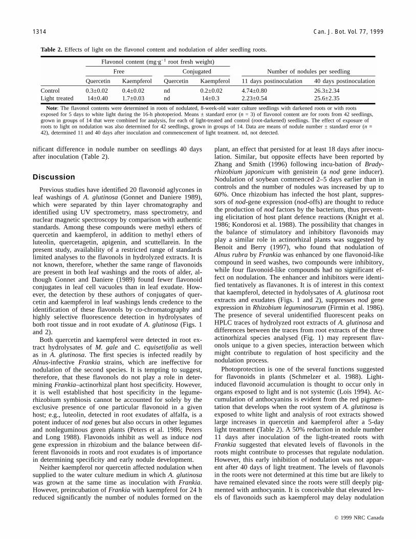
nificant difference in nodule number on seedlings 40 daysafter inoculation (Table 2).
Discussion
Previous studies have identified 20 flavonoid aglycones inleaf washings of A. glutinosa (Gonnet and Daniere 1989),which were separated by thin layer chromatography andidentified using UV spectrometry, mass spectrometry, andnuclear magnetic spectroscopy by comparison with authenticstandards. Among these compounds were methyl ethers ofquercetin and kaempferol, in addition to methyl ethers ofluteolin, quercetagetin, apigenin, and scuttellarein. In thepresent study, availability of a restricted range of standardslimited analyses to the flavonols in hydrolyzed extracts. It isnot known, therefore, whether the same range of flavonoidsare present in both leaf washings and the roots of alder, al-though Gonnet and Daniere (1989) found fewer flavonoidconjugates in leaf cell vacuoles than in leaf exudate. How-ever, the detection by these authors of conjugates of quer-cetin and kaempferol in leaf washings lends credence to theidentification of these flavonols by co-chromatography andhighly selective fluorescence detection in hydrolysates ofboth root tissue and in root exudate of A. glutinosa (Figs. 1and 2).
Both quercetin and kaempferol were detected in root ex-tract hydrolysates of M. gale and C. equisetifolia as wellas in A. glutinosa. The first species is infected readily byAlnus-infective Frankia strains, which are ineffective fornodulation of the second species. It is tempting to suggest,therefore, that these flavonols do not play a role in deter-mining Frankia–actinorhizal plant host specificity. However,it is well established that host specificity in the legume-rhizobium symbiosis cannot be accounted for solely by theexclusive presence of one particular flavonoid in a givenhost; e.g., luteolin, detected in root exudates of alfalfa, is apotent inducer of nod genes but also occurs in other legumesand nonleguminous green plants (Peters et al. 1986; Petersand Long 1988). Flavonoids inhibit as well as induce nodgene expression in rhizobium and the balance between dif-ferent flavonoids in roots and root exudates is of importancein determining specificity and early nodule development.
Neither kaempferol nor quercetin affected nodulation whensupplied to the water culture medium in which A. glutinosawas grown at the same time as inoculation with Frankia.However, preincubation of Frankia with kaempferol for 24 hreduced significantly the number of nodules formed on the
plant, an effect that persisted for at least 18 days after inocu-lation. Similar, but opposite effects have been reported byZhang and Smith (1996) following incu-bation of Brady-rhizobium japonicum with genistein (a nod gene inducer).Nodulation of soybean commenced 2–5 days earlier than incontrols and the number of nodules was increased by up to60%. Once rhizobium has infected the host plant, suppres-sors of nod-gene expression (nod-offs) are thought to reducethe production of nod factors by the bacterium, thus prevent-ing elicitation of host plant defence reactions (Knight et al.1986; Kondorosi et al. 1988). The possibility that changes inthe balance of stimulatory and inhibitory flavonoids mayplay a similar role in actinorhizal plants was suggested byBenoit and Berry (1997), who found that nodulation ofAlnus rubra by Frankia was enhanced by one flavonoid-likecompound in seed washes, two compounds were inhibitory,while four flavonoid-like compounds had no significant ef-fect on nodulation. The enhancer and inhibitors were identi-fied tentatively as flavanones. It is of interest in this contextthat kaempferol, detected in hydolysates of A. glutinosa rootextracts and exudates (Figs. 1 and 2), suppresses nod geneexpression in Rhizobium leguminosarum (Firmin et al. 1986).The presence of several unidentified fluorescent peaks onHPLC traces of hydrolyzed root extracts of A. glutinosa anddifferences between the traces from root extracts of the threeactinorhizal species analysed (Fig. 1) may represent flav-onols unique to a given species, interaction between whichmight contribute to regulation of host specificity and thenodulation process.
Photoprotection is one of the several functions suggestedfor flavonoids in plants (Schmelzer et al. 1988). Light-induced flavonoid accumulation is thought to occur only inorgans exposed to light and is not systemic (Lois 1994). Ac-cumulation of anthocyanins is evident from the red pigmen-tation that develops when the root system of A. glutinosa isexposed to white light and analysis of root extracts showedlarge increases in quercetin and kaempferol after a 5-daylight treatment (Table 2). A 50% reduction in nodule number11 days after inoculation of the light-treated roots withFrankia suggested that elevated levels of flavonols in theroots might contribute to processes that regulate nodulation.However, this early inhibition of nodulation was not appar-ent after 40 days of light treatment. The levels of flavonolsin the roots were not determined at this time but are likely tohave remained elevated since the roots were still deeply pig-mented with anthocyanin. It is conceivable that elevated lev-els of flavonoids such as kaempferol may delay nodulation
© 1999 NRC Canada
1314 Can. J. Bot. Vol. 77, 1999
Flavonol content (mg·g�1 root fresh weight)
Number of nodules per seedlingFree Conjugated
Quercetin Kaempferol Quercetin Kaempferol 11 days postinoculation 40 days postinoculation
Control 0.3±0.02 0.4±0.02 nd 0.2±0.02 4.74±0.80 26.3±2.34Light treated 14±0.40 1.7±0.03 nd 14±0.3 2.23±0.54 25.6±2.35
Note: The flavonol contents were determined in roots of nodulated, 8-week-old water culture seedlings with darkened roots or with rootsexposed for 5 days to white light during the 16-h photoperiod. Means ± standard error (n = 3) of flavonol content are for roots from 42 seedlings,grown in groups of 14 that were combined for analysis, for each of light-treated and control (root-darkened) seedlings. The effect of exposure ofroots to light on nodulation was also determined for 42 seedlings, grown in groups of 14. Data are means of nodule number ± standard error (n =42), determined 11 and 40 days after inoculation and commencement of light treatment. nd, not detected.
Table 2. Effects of light on the flavonol content and nodulation of alder seedling roots.
J:\cjb\cjb77\cjb-09\B99-077.vpThursday, December 16, 1999 11:56:29 AM
Color profile: DisabledComposite Default screen

© 1999 NRC Canada
Hughes et al. 1315
in a manner diametrically opposite to the stimulation of nod-ulation reported for genistein pretreated Bradyrhizobium(Zhang and Smith 1996). It is unlikely that changes in fla-vonoid content of roots and (or) root exudates are the solecause of such inhibition of nodulation, however, and in peas,suppression of nodulation by exposure of roots to light hasbeen ascribed to increased ethylene production (Lee andLaRue 1992). The data presented support the possibility thatflavonoids influence nodulation processes in actinorhizalplants but proof of their involvement in the regulation of in-fection requires identification and manipulation of Frankianod genes.
References
Benoit, L.F., and Berry, A.M. 1997. Flavonoid-like compoundsfrom seeds of red alder (Alnus rubra) influence host nodulationby Frankia (Actinomycetales). Physiol. Plant. 99: 588–593.
Crozier, A., Jensen, E., Lean, M., and McDonald, M. 1997. Quantita-tive analysis of flavonoids by reversed-phase high-performance liq-uid chromatography. J. Chromatogr. 761: 315–321.
Firmin, J.L., Wilson, K.E., Rossen, L., and Johnston, A.W.B. 1986.Flavonoid activation of nodulation genes in Rhizobium reversedby other compounds present in plants. Nature (London), 324:90–92.
Gonnet, J.F., and Daniere, C. 1989. Individual variation in theflavonoid aglycones excreted on the leaves of Alnus glutinosa(Betulaceae). Biochem. Syst. Ecol. 17: 239–247.
Hollman, P.C.H., van Trio, J.M.P., and Buysman, M.N.C.P. 1996.Fluorescence detection of flavonols by HPLC with post columnchelation with aluminium. Anal. Chem. 68: 3511–3515.
Hooker, J.E., and Wheeler, C.T. 1987. The effectivity of Frankiafor nodulation and nitrogen fixation in Alnus rubra and Alnusglutinosa. Physiol. Plant. 70: 333 –341.
Knight, C.D., Rossen, L., Robertson, J.G., Wells, B., and Downie,J.A. 1986. Nodulation inhibition by Rhizobium leguminosarum
multicopy nodABC genes and an analysis of early stages ofplant infection. J. Bacteriol. 166: 552–558.
Kondorosi, E., Gyuris J., Schmidt, J., John, M., Duda, E., Schell,J., and Kondorosi, A. 1988. Positive and negative control ofnodulation genes in Rhizobium meliloti strain 41. In Molecularplant–microbe interactions. Edited by D.P.S. Verma and R.Palacios. APS Press, St. Paul, Minn. p. 73.
Kubasek, W.L., Shirley, B.W., McKillop, A., Goodman, H.M.,Briggs, W., and Ausubel, F.M. 1992. Regulation of flavonoidbiosynthetic genes in germinating Arabidopsis seedlings. PlantCell, 4: 1229–1236.
Lee, K.H., and Larue, T.A. 1992. Ethylene as a possible mediatorof light induced and nitrate induced inhibition of nodulation ofPisum sativum cv Sparkle. Plant Physiol. 100: 1334–1338.
Lois, R. 1994 Accumulation of UV-absorbing flavonoids induced byUV-B radiation in Arabidopsis thaliana L. Planta, 194: 498–503.
Peters, N.K., and Long, S.R. 1988. Alfalfa root exudates and com-pounds which promote or inhibit induction of Rhizobiummeliloti nodulation genes. Plant Physiol. 88: 396–400.
Peters, N.K., Frost, J.W., and Long, S.R. 1986. A plant flavone,luteolin, induces expression of Rhizobium meliloti nodulationgenes. Science (Washington, D.C.), 233: 977–980.
Redmond, J.W., Batley, M., Djordjevic, M.A., Innes, R.W.,Kuempel, P.L., and Rolfe, B.G. 1986. Flavones induce expres-sion of nodulation genes in Rhizobium. Nature (London), 323:632–635.
Schmelzer, E., Jahnen, W., and Hahlbrock, K. 1988. In situ lo-calisation of light-induced chalcone synthase mRNA, chalconesynthase, and flavonoid end-products in epidermal cells of pars-ley leaves. Proc. Natl. Acad. Sci. U.S.A. 85: 2989–2993.
Stapleton, A.E. 1992. Ultraviolet radiation and plants: burningquestions. Plant Cell, 4: 1353–1358.
Zhang, F., and Smith, D.L. 1996. Inoculation of soybean withgenistein preincubated Bradyrhizobium japonicum or genisteindirectly applied into soil increases soybean protein content anddry matter yield. Plant Soil, 179: 233–241.
J:\cjb\cjb77\cjb-09\B99-077.vpThursday, December 16, 1999 11:56:29 AM
Color profile: DisabledComposite Default screen
Recommended