Echocardiographic
Evaluation of Mitral Valve
Prostheses
Dennis A. Tighe, M.D., FACC, FACP, FASE
Cardiovascular Medicine
University of Massachusetts Medical School
Worcester, MA

Which of the following statements
regarding the obstructed/thrombosed
prosthetic mitral valve is correct?
• 1. A pressure half-time ˃130 msec is the single best indicator
of prosthetic obstruction.
• 2. Taking into account heart rate is not necessary when
assessing trans-mitral gradients.
• 3. Pannus in-growth is more common in the mitral position
than with aortic PHVs.
• 4. A peak velocity ≥2.5 m/sec suggests significant stenosis.
• 5. Randomized, controlled trials have demonstrated that bolus
infusion of rt-PA is the fibrinolytic regimen of choice.

Which of the following statements
concerning prosthetic mitral
regurgitation is correct?
• 1. Pseudo-regurgitation is an issue most often
encountered during performance of TEE.
• 2. Any degree of regurgitation indicates dysfunction of a
mechanical valve.
• 3. Structural valve deterioration is an uncommon cause
of pathological regurgitation.
• 4. Mitral bioprostheses are less prone to suffer structural
valve deterioration than are aortic bioprostheses.
• 5. Annular dehiscence most often is a consequence of
infective endocarditis.
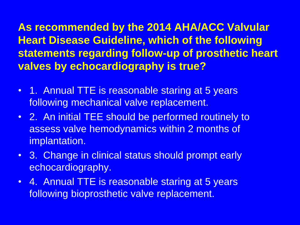
As recommended by the 2014 AHA/ACC Valvular
Heart Disease Guideline, which of the following
statements regarding follow-up of prosthetic heart
valves by echocardiography is true?
• 1. Annual TTE is reasonable staring at 5 years
following mechanical valve replacement.
• 2. An initial TEE should be performed routinely to
assess valve hemodynamics within 2 months of
implantation.
• 3. Change in clinical status should prompt early
echocardiography.
• 4. Annual TTE is reasonable staring at 5 years
following bioprosthetic valve replacement.
www.asecho.org
Nishimura RA et al. Circulation 2014;129:e521-e643.

Overview
• Description of the various types of
prosthetic heart valves
• Echocardiographic evaluation of
normally-functioning prosthetic heart
valves
• Evaluation of prosthetic heart valve
dysfunction
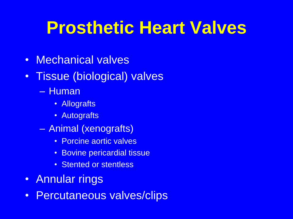
Prosthetic Heart Valves
• Mechanical valves
• Tissue (biological) valves
– Human
• Allografts
• Autografts
– Animal (xenografts)
• Porcine aortic valves
• Bovine pericardial tissue
• Stented or stentless
• Annular rings
• Percutaneous valves/clips

Mechanical Heart Valves
• Ball-in-cage– Starr Edwards valve
• Single tilting disc– Medtronic Hall valve
– OmniScience valve
– Bjork-Shiley valve
• Bileaflet tilting disc– St. Jude Medical valve
– Carbomedics valve/Sorin
– On-X
– ATS
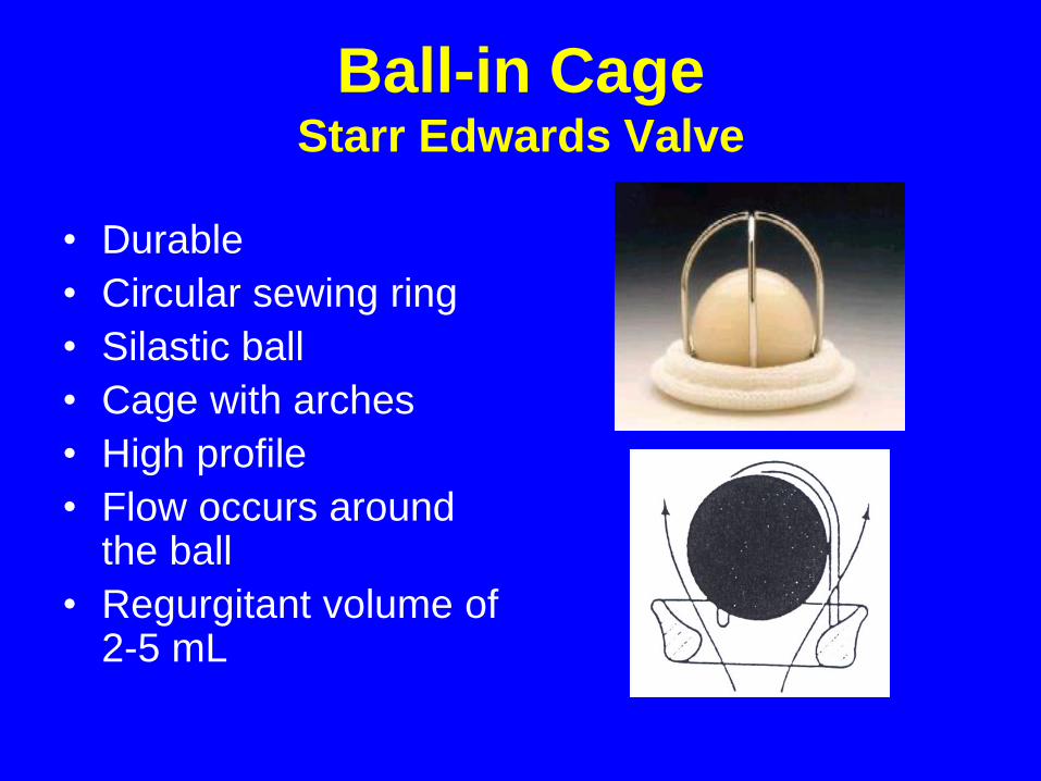
Ball-in CageStarr Edwards Valve
• Durable
• Circular sewing ring
• Silastic ball
• Cage with arches
• High profile
• Flow occurs around the ball
• Regurgitant volume of 2-5 mL

Single Tilting Disc Valves
• Circular sewing ring
• Circular disc eccentrically
attached by metal struts
• Opening angle 60o to 80o
• Flow occurs through
major and minor orifices
• Regurgitant volume of 5-
9 mL

Bileaflet Tilting Disc Valves
• 2 semicircular discs
attached to rigid valve
ring by small hinges
• Opening angle 75o to 90o
• 3 orifices
– Central and 2 lateral
orifices
• Regurgitant volume of 5-
10 mL
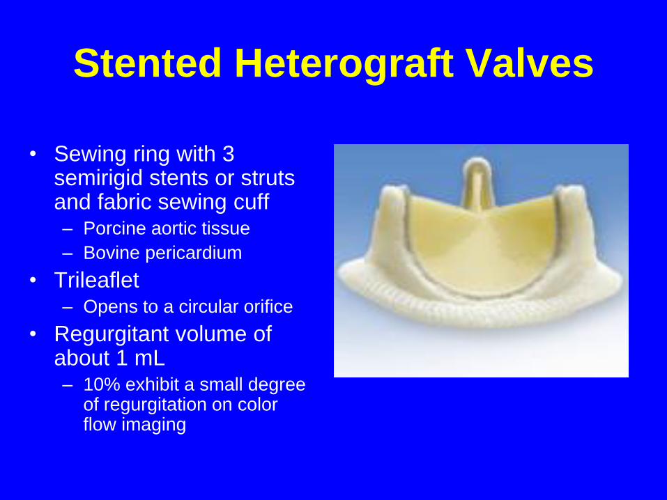
Stented Heterograft Valves
• Sewing ring with 3 semirigid stents or struts and fabric sewing cuff– Porcine aortic tissue
– Bovine pericardium
• Trileaflet– Opens to a circular orifice
• Regurgitant volume of about 1 mL– 10% exhibit a small degree
of regurgitation on color flow imaging

Percutaneous Clip
• Mitra-Clip®
• Perctaneous edge-to-
edge technique to
reduce MR
• FDA-approved for
degenerative MR

Echocardiographic Approach to
Assessment of Prosthetic Heart Valves
• Evaluation similar to that of native
valves
• Reverberations and shadowing play a
significant role
• Fluid dynamics of each specific valve
prosthesis influences the Doppler
findings

Echocardiographic Approach to
Prosthetic Heart Valves—All Valve Types
• Complete 2D/3D imaging
• Determine transvalvular pressure gradients
• Estimate valve orifice area
• Evaluate severity and location of regurgitation
• Estimate pulmonary artery systolic pressure
• Assess chamber sizes and function
• Evaluate other valves
• Clinical data– Size and type of prosthesis
– HR, BP, BSA
• ALWAYS COMPARE TO BASELINE STUDY!

Echocardiographic Approach to
Prosthetic Heart Valves—Caveats
• “Normal” Doppler values based on: – Prosthesis size
– Prosthesis type
– Position
• Higher gradients compared to native valves
• Reverberation artifacts/shadowing
• Differential diagnosis of high valve gradients:– True stenosis
– High cardiac output states
– Significant regurgitation
– Patient-prosthesis mismatch
– Pressure recovery
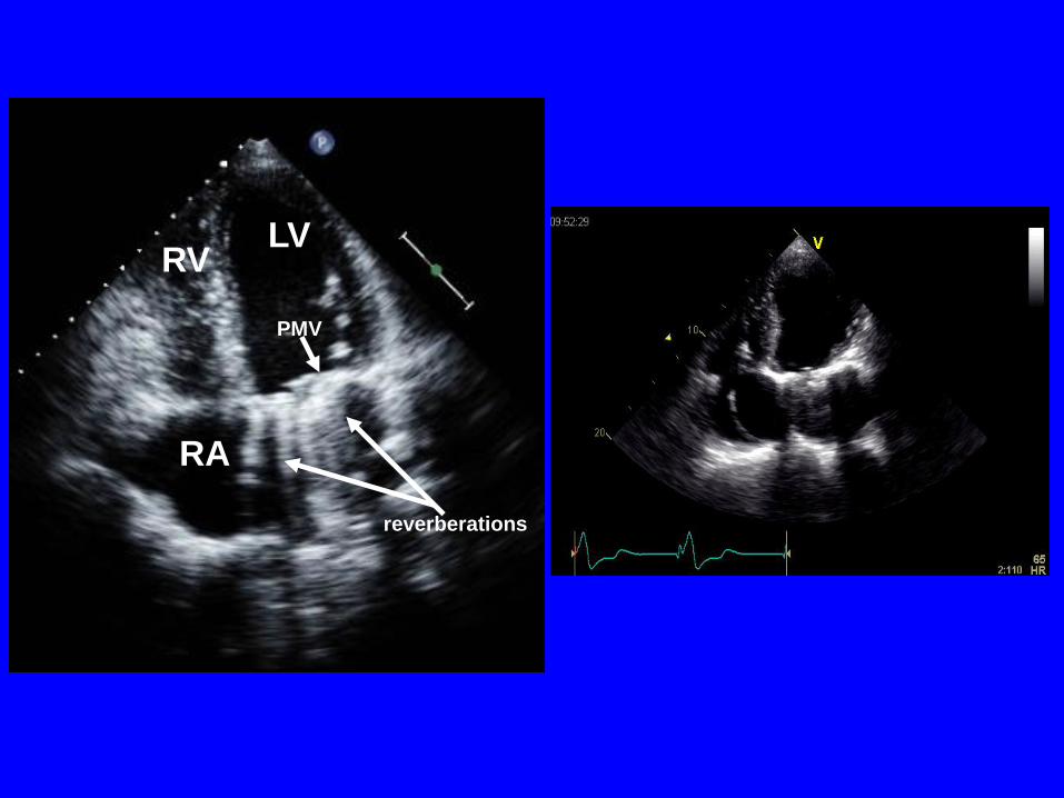
LV
RA
RV
PMV
reverberations
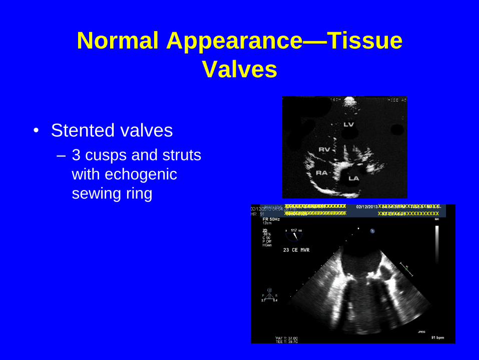
Normal Appearance—Tissue
Valves
• Stented valves
– 3 cusps and struts
with echogenic
sewing ring

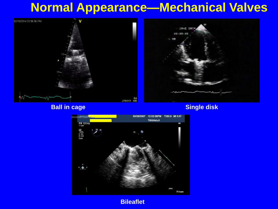
Normal Appearance—Mechanical Valves
Ball in cage Single disk
Bileaflet

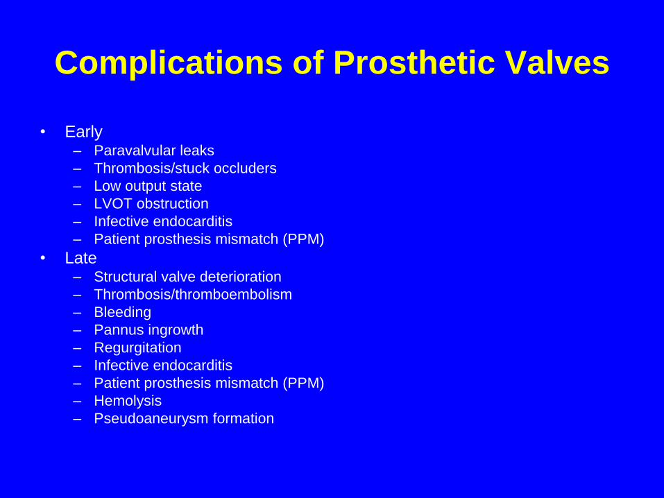
Complications of Prosthetic Valves
• Early– Paravalvular leaks
– Thrombosis/stuck occluders
– Low output state
– LVOT obstruction
– Infective endocarditis
– Patient prosthesis mismatch (PPM)
• Late– Structural valve deterioration
– Thrombosis/thromboembolism
– Bleeding
– Pannus ingrowth
– Regurgitation
– Infective endocarditis
– Patient prosthesis mismatch (PPM)
– Hemolysis
– Pseudoaneurysm formation

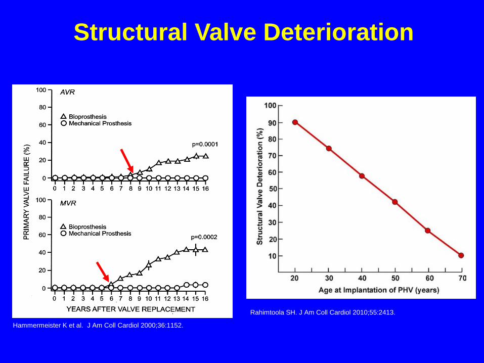
Hammermeister K et al. J Am Coll Cardiol 2000;36:1152.
Probability of an Event at 15-years
(SVD)

Prosthetic Valve Dysfunction
• Approach to suspected dysfunction– TTE/Doppler
– TEE• Atrial side of mitral prosthesis
– Cine fluoroscopy• May provide superior assessment of mechanical valve
opening and closing motion
• No assessment of pressure gradients
– Cardiac CT
– Cardiac catheterization
– Stress echocardiography

Structural Valve Deterioration
• Tissue Valves– More common
• Younger patients
• Altered Ca++ metabolism
• Valve type
– Thickening, calcification, perforation, or spontaneous tissue degeneration of leaflets
– Regurgitation• Usually gradual
• Can be acute and massive
– Stenosis
– Combination
LA
LV
LA
LV
Hammermeister K et al. J Am Coll Cardiol 2000;36:1152.
Structural Valve Deterioration
Rahimtoola SH. J Am Coll Cardiol 2010;55:2413.

Valve Thrombosis
• Incidence – 0.3% to 1.3%/yr
• Highest risk– Mitral and tricuspid positions
• Inadequate anticoagulation– Mechanical valves
• Clinical manifestations– Incidental finding
– Peripheral embolization
– Stenosis
– Regurgitation
– Heart failure
• Gradual or acute symptom onset
• Treatments– Anticoagulants
– Thrombolysis
– Surgery
Bileaflet MVR


Non-obstructive Thrombosis
Mechanical Prosthesis Bioprosthesis
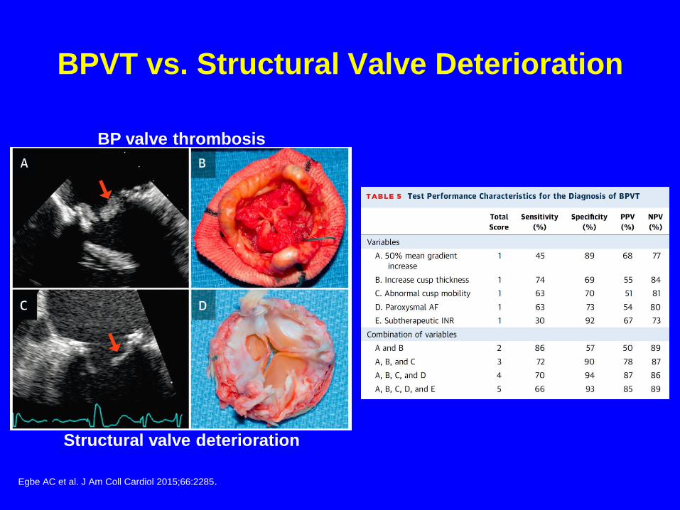
BPVT vs. Structural Valve Deterioration
BP valve thrombosis
Structural valve deterioration
Egbe AC et al. J Am Coll Cardiol 2015;66:2285.
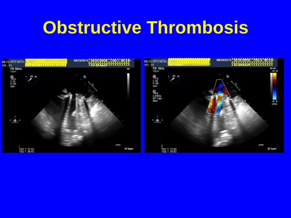
Obstructive Thrombosis
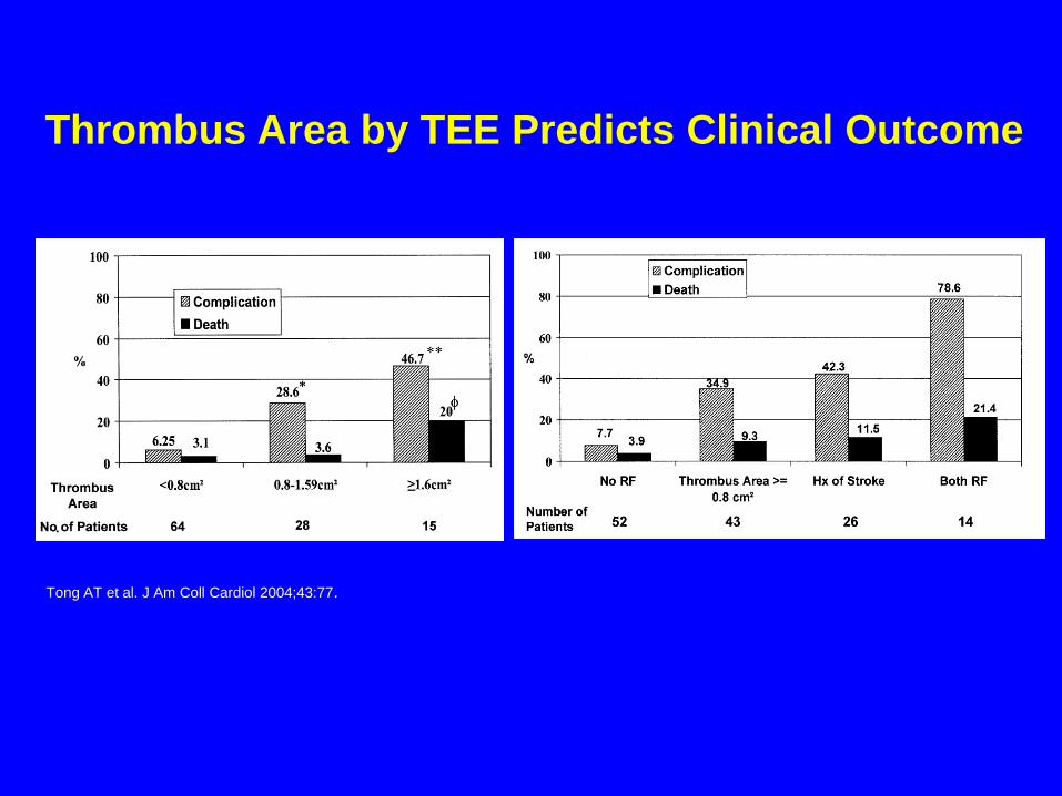
Tong AT et al. J Am Coll Cardiol 2004;43:77.
Thrombus Area by TEE Predicts Clinical Outcome

Nishimura RA et al. Circulation 2014 ;129:e586.
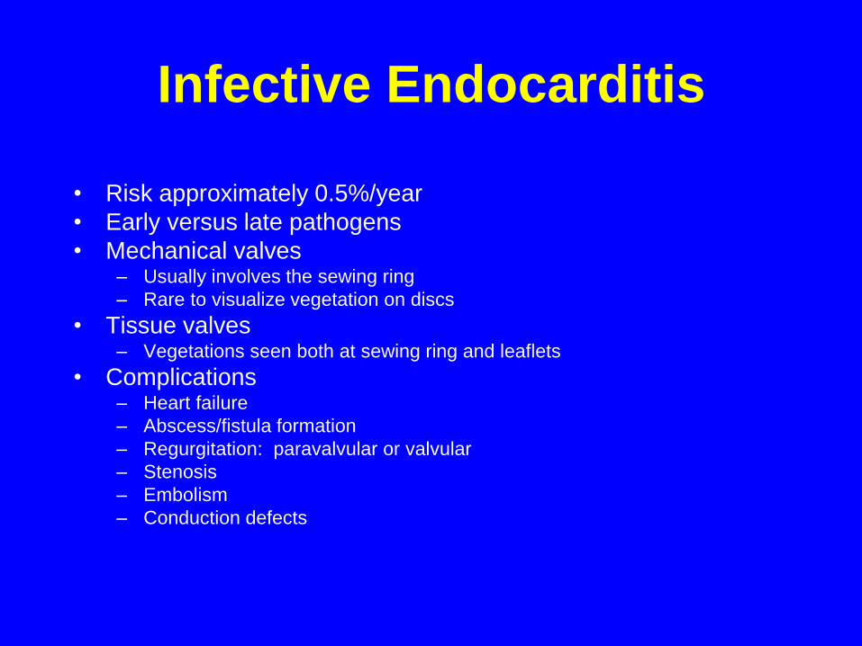
Infective Endocarditis
• Risk approximately 0.5%/year
• Early versus late pathogens
• Mechanical valves– Usually involves the sewing ring
– Rare to visualize vegetation on discs
• Tissue valves– Vegetations seen both at sewing ring and leaflets
• Complications– Heart failure
– Abscess/fistula formation
– Regurgitation: paravalvular or valvular
– Stenosis
– Embolism
– Conduction defects


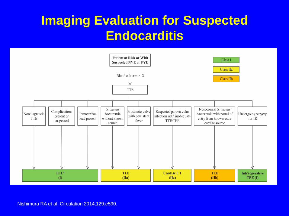
Imaging Evaluation for Suspected
Endocarditis
Nishimura RA et al. Circulation 2014;129:e590.

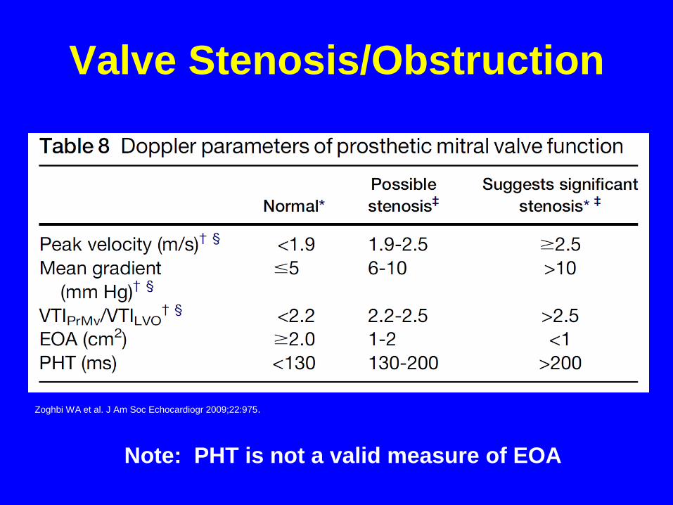
Valve Stenosis/Obstruction
• Tissue valves– Thickening, calcification
and restricted motion
– Pannus in-growth
– Thrombosis
• Mechanical valves– Restriction of disc/ball
motion
• Thrombus
• Pannus in-growth
• Combination
• Vegetations
– Restriction of annular area
• Pannus in-growth
LV
LA
RV
RA

Valve Stenosis/Obstruction
• Mitral valve parameters
– Peak E-wave velocity
– Mean gradient
– Pressure half-time
– Effective orifice area
• Continuity equation area
– DVI
• VTIprosthesis/ VTILVOT
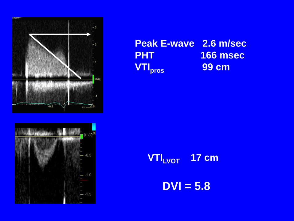
Peak E-wave 2.6 m/sec
PHT 166 msec
VTIpros 99 cm
VTILVOT 17 cm
DVI = 5.8

Fernandes V et al. Am J Cardiol 2002;89:704.


Pannus In-growth
Versus Thrombosis
-Anticoagulation usually adequate
-Greater time from implant to presentation
-More echo-dense
-Aortic position more common
Zoghbi WA et al. J Am Soc Echocardiogr 2009;22:975.

Valve Stenosis/Obstruction
• Differential Diagnosis– High cardiac output states
• Anemia, fever, hypovolemia, thyrotoxicosis
– Significant regurgitation
– Patient-prosthesis mismatch
– Pressure recovery
• Caveats– Compare to baseline study
– Take into account: • Size/type of prosthesis
• Cardiac output
• Heart rate
– Be aware of pressure recovery • Bileaflet mechanical valves primarily in aortic position
Valve Stenosis/Obstruction
Note: PHT is not a valid measure of EOA
Zoghbi WA et al. J Am Soc Echocardiogr 2009;22:975.

Prosthetic Regurgitation
• Tissue valves– Degenerative/calcific changes
– Infective endocarditis
– Pannus in-growth
– Paravalvular
• Mechanical valves– Paravalvular
• Dehiscence
• Poor seating
• Infection
– Incomplete closure• Pannus in-growth
• Thrombosis
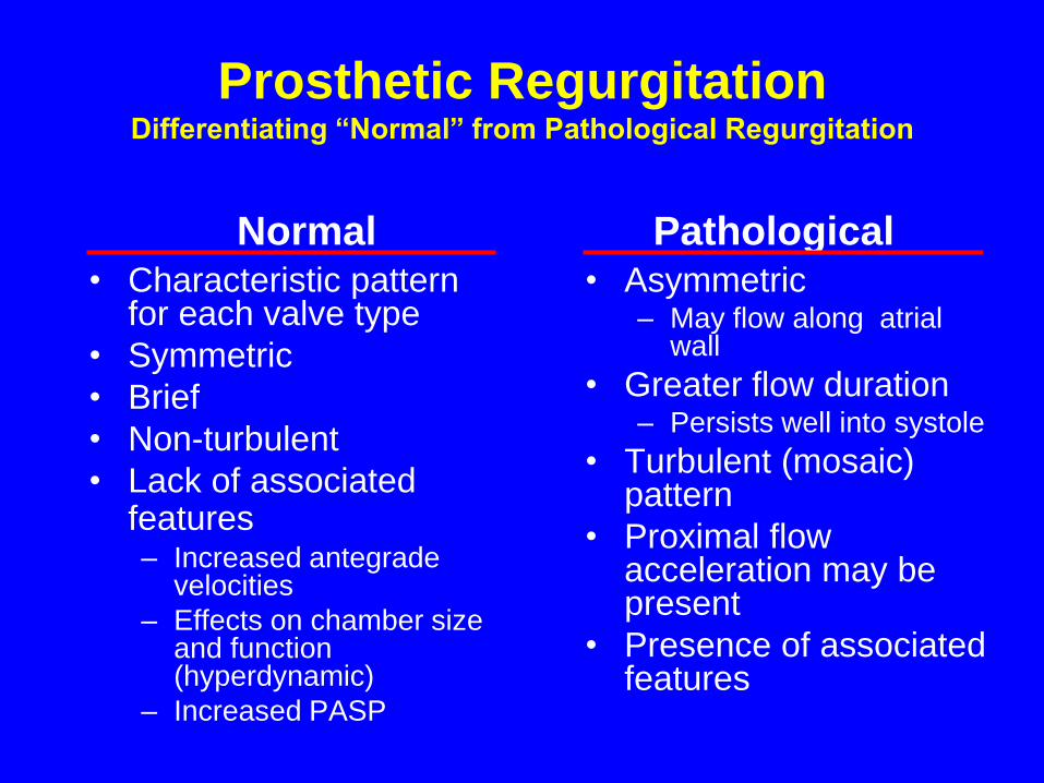
Prosthetic RegurgitationDifferentiating “Normal” from Pathological Regurgitation
• Characteristic pattern for each valve type
• Symmetric
• Brief
• Non-turbulent
• Lack of associated features– Increased antegrade
velocities
– Effects on chamber size and function (hyperdynamic)
– Increased PASP
• Asymmetric– May flow along atrial
wall
• Greater flow duration– Persists well into systole
• Turbulent (mosaic) pattern
• Proximal flow acceleration may be present
• Presence of associated features
Normal Pathological

Evaluation of Prosthetic
Regurgitation
• Similar to native valve evaluation
• Prosthetic shadowing limits evaluation
– Mitral: TEE superior to evaluate LA aspect
• “Pseudo-regurgitation”


Pseudo-regurgitation
Rudski LG et al. J Am Soc Echocardiogr 2004;17:829.

Immediate Post-operative Paravalvular MR


*PHT <130 msec
Zoghbi WA et al. J Am Soc Echocardiogr 2009;22:975.

Prosthesis-Patient Mismatch
• Effective orifice area (EOA) of the prosthetic valve is less than that of the normal native valve
– PPM occurs when EOA is smaller than expected for BSA
• High transvalvular gradients in normally functioning valves
• EOA indexed to body surface area (EOAi)– Mitral valve:
• Non-significant >1.2 cm2/m2
• Moderate ˃0.9 cm2/m2 to ≤1.2 cm2/m2
• Severe <0.9 cm2/m2
• Consequences may include:– Exercise intolerance
– Higher pulmonary artery pressures
– Heart failure
– Increased mortality

Follow-up of Prosthetic Heart ValvesACC/AHA Guidelines
Nishimura RA et al. Circulation 2014;129:e577-e578.
• Class I– Initial TTE is recommended after prosthesis implantation (6 wks
to 3 mos) for assessment of valve hemodynamics (LOE: B).
– Repeat TTE is recommended with a change in clinical symptoms
or signs suggesting prosthetic valve dysfunction (LOE: C).
– TEE is recommended when clinical symptoms or signs suggest
prosthetic valve dysfunction (LOE: C).
• Class IIa– Annual TTE is reasonable in patients with a bioprosthetic valve
after the first 10 years, even in the absence of a change in
clinical status (LOE: C).

Which of the following statements
regarding the obstructed/thrombosed
prosthetic mitral valve is correct?
• 1. A pressure half-time ˃130 msec is the single best indicator
of prosthetic obstruction.
• 2. Taking into account heart rate is not necessary when
assessing trans-mitral gradients.
• 3. Pannus in-growth is more common in the mitral position
than with aortic PHVs.
• 4. A peak velocity ≥2.5 m/sec suggests significant stenosis.
• 5. Randomized, controlled trials have demonstrated that bolus
infusion of rt-PA is the fibrinolytic regimen of choice.

Which of the following statements
concerning prosthetic mitral
regurgitation is correct?
• 1. Pseudo-regurgitation is an issue most often
encountered during performance of TEE.
• 2. Any degree of regurgitation indicates dysfunction of a
mechanical valve.
• 3. Structural valve deterioration is an uncommon cause
of pathological regurgitation.
• 4. Mitral bioprostheses are less prone to suffer structural
valve deterioration than are aortic bioprostheses.
• 5. Annular dehiscence most often is a consequence of
infective endocarditis.

As recommended by the 2014 AHA/ACC Valvular
Heart Disease Guideline, which of the following
statements regarding follow-up of prosthetic heart
valves by echocardiography is true?
• 1. Annual TTE is reasonable staring at 5 years
following mechanical valve replacement.
• 2. An initial TEE should be performed routinely to
assess valve hemodynamics within 2 months of
implantation.
• 3. Change in clinical status should prompt early
echocardiography.
• 4. Annual TTE is reasonable staring at 5 years
following bioprosthetic valve replacement.

Thank you for your attention
Recommended