102 Draft for pre-publication check
Venous thromboembolic diseases Clinical evidence tables
E.2 DVT diagnosis (D-dimer)
In people with suspected DVT, what is the effectiveness of D-dimer in ruling out deep vein thrombosis?
Study
details
Patients Diagnostic tools Outcomes Results Comments
Study name:
Goodacre 2006
170
HTA report
Study design:
Systematic review – 99 cohorts included for clinically suspected DVT, 13 for
Patient group: clinically suspected DVT
Setting:
Outpatient clinic (21), inpatients (9), emergency department (16), mixed (29), and not stated (14).
Recruitment was reported to be consecutive in 76 and prospective in 68.
Exclusion criteria:
Assessment tool under investigation:
Threshold value defined before analysis in 82 cohorts, after analysis in 10, and not clear in 7 cohorts.
Reference standard:
(number in brackets denote number of cohorts)
All assays
Pooled sensitivity
90.5% (95% CI 90% to 91%), range 48% to 100%. Heterogeneity: p<0.001
Variation predicted by an outpatient or a mixed setting for patient recruitment, exclusion of patients who were pregnant, anticoagulated or had a long history of symptoms, age, prospective analysis, the D-dimer threshold used and whether the D-dimer threshold was determined before or after the study.
Funding:
HTA analysis
Limitations: In about half of
included studies, it was unclear whether D-dimer tests and reference standards were interpreted blind to the results of the other test
Various standard references used

103 Draft for pre-publication check
Venous thromboembolic diseases Clinical evidence tables
Study
details
Patients Diagnostic tools Outcomes Results Comments
asymptomatic DVT
Duration of follow-up:
Not reported.
No exclusion reported by 50 cohorts.
The following criteria were excluded by the number of cohorts in brackets:
Postoperative patients(10), pregnant patients(19), anticoagulated patients (33), previous VTE(23), recent trauma(3), sepsis(4), prolonged history (18)
All patients
N: 8752
DVT prevalence: 2 to 78% median 36%
Age range: 51 to 69, median 59 years, except one cohort which exclusively recruited people over the age of 70
Venography(34), ultrasound(28), ultrasound with clinical follow up (10), serial ultrasound (6), ultrasound or venography (13), others – combinations of ultrasound and plethysmography (8)
Reference standard applied independent of D-dimer results in 86 cohorts, dependent in 4 and unclear in 9 cohorts
D-dimer was measured blind to reference standard in 43 cohorts and unclear in 56.
Reference standard was interpreted blind to D-dimer results in 50 cohorts and unclear in 49 cohorts.
Pooled specificity
54.7% (95% CI 54% to 55%), range 5% to 100%. Heterogeneity: p<0.001
Variation predicted by an outpatient, an emergency department or a mixed setting, exclusion of patients who were pregnant, anticoagulated or had a past history of thromboembolism, age, consecutive recruitment, prospective analysis, the reference standard used, and quality criteria relating to blinding of observers measuring D-dimer and blinding or observers interpreting the reference standard.
Heterogeneity not explained when subgroup analysis according to predictors of variability was conducted
Additional
tests:
Notes: *
ELISAs
Pooled sensitivity
Pooled specificity
91 analyses in 58 cohorts (35 reporting proximal and distal
94% (95% CI 93% to 95%),. Heterogeneity: p<0.001
45% (95% CI 44% to 46%), Heterogeneity: p<0.001
ELISAs
Pooled sensitivity
74 analyses in 52 cohorts
89% (95% CI 88% to 90%),

104 Draft for pre-publication check
Venous thromboembolic diseases Clinical evidence tables
Study
details
Patients Diagnostic tools Outcomes Results Comments
% Males: 17 to 62% (median 42%) – reported by 81 cohorts
% of proximal DVT ( out of all DVT) : 27% to 100% (median 77%)- reported by 51 cohorts
Drop outs: N/R
Pooled specificity
Heterogeneity: p<0.001
55% (95% CI 54% to 56%),. Heterogeneity: p<0.001
ELISAs
Pooled sensitivity
Pooled specificity
87% (95% CI 85% to 88%),. Heterogeneity: p<0.001
68% (95% CI 67% to 69%), Heterogeneity: p<0.001
Study
details
Patients Diagnostic tools Measure of Disorders Results Comments
Study name:
Anoop2009 27
Study design:
Prospective cohort (diagnostic)
Patient group:
Consecutive patient sent for D-dimer testing
Setting:
District general hospital, UK. Conducted from Dec 2007 to March 2008
Assessment tool under investigation:
MDA Autodimer ® (immunoturbidimetric assay using monoclonal antibody)
Cut off point: 0.50mcg FEU/mL determined based
Deep vein thrombosis:
(proximal )
Sensitivity
Specificity
RS + RS- Total
D-Di + 16 67 83
D-Di - 0 23 23
Total 16 90 106
100.0%
25. 6% (95% CI: 17.2-36%)
Funding:
Limitations: Patients recruited at
the point of referral for D-dimer testing, instead of at the point of symptom presentation ie could have missed

105 Draft for pre-publication check
Venous thromboembolic diseases Clinical evidence tables
Study
details
Patients Diagnostic tools Measure of Disorders Results Comments
Evidence level:
Duration of follow-up:
Inclusion criteria:
In and out patients
Exclusion criteria: Intensive care unit
patients Specimen error: D-
dimer levels not quantifiable; patients not receiving reference tests or inconclusive results from scans
All patients
N: Total 197 patients, 90 were for suspected DVT, 91 suspected PE
Population characteristics (for patients with suspected DVT)
Median age (range): 70(17-97)
Inpatient/Outpatientr: 41/65
on manufacturer recommendation
Performed by: laboratory personnel blinded to results of pre-test probability score (Wells score)
Reference standard:
Compression ultrasound -
Whole leg (9 common and superficial femoral veins, poplitial trifucation and all three deep vein sets) Unclear whether only symptomatic leg scanned
Performed by:
Not stated
PPV
NPV
ROC for varying cut offs
3 month VTE rate
Mortality
% negative test result*
Prevalence
Positive LR
Negative LR
FP
FN
19.8%
100.0%
Not reported
Not reported
Not reported
23/106 (21.7%)
16/106(15.1%)
1.34
0.00
67
0
patients who were symptomatic and not sent for D-dimer testing
Unclear how many patients were excluded because of non interpretable results
Results of D-dimer and imaging interpreted together by haematologists to diagnose DVT (not blinded)
Additional
tests:
Junior doctors completed Wells score for DVT or PE
Notes:
Deep vein thrombosis:
Patients with Intermediate to High PTP ( wells score)
(proximal )
Sensitivity
RS + RS- Total
D-Di + 14 65 79
D-Di - 0 22 22
Total 14 87 101

106 Draft for pre-publication check
Venous thromboembolic diseases Clinical evidence tables
Study
details
Patients Diagnostic tools Measure of Disorders Results Comments
Drop outs: not stated
Specificity
PPV
NPV
ROC for varying cut offs
3 month VTE rate
Mortality
% negative test result*
Prevalence
Positive LR
Negative LR
FP
FN
100.0%
25.3%
17.7%
100.0%
Not reported
Not reported
Not reported
21.8%
13.9%
1.34
0.0
65/79 (82.3%)
0
Deep vein thrombosis:
Patients with Low PTP ( wells score)
(proximal )
RS + RS- Total
D-Di + 2 2 4
D-Di - 0 1 1

107 Draft for pre-publication check
Venous thromboembolic diseases Clinical evidence tables
Study
details
Patients Diagnostic tools Measure of Disorders Results Comments
Sensitivity
Specificity
PPV
NPV
ROC for varying cut offs
3 month VTE rate
Mortality
% negative test result*
Prevalence
Positive LR
Negative LR
FP
FN
Total 2 3 5
100.0%
33.3%
50.0%
100.0%
Not reported
Not reported
Not reported
20.0%
40.0%
1.50
0
2/4 (50%)
0/2(0)

108 Draft for pre-publication check
Venous thromboembolic diseases Clinical evidence tables
Study
details
Patients Diagnostic tools Measure of Disorders Results Comments
Study name: Dempfle 2006
106
Study design:
Prospective cohort (diagnostic)
Evidence level:
Duration of follow-up: not reported
Patient group: Patients with clinically suspected acute DVT.
Setting: multi-centre 19 study sites in 3 countries.
Inclusion criteria:
clinically suspected acute (defined as clinical symptoms for 7 days or less) DVT.
Exclusion criteria: unclear duration of
symptoms violated the single entry criterium ‘acute deep venous thrombosis’ ( clinical symptoms were present for more than seven days)
hospitalised for more than 72 hours at the time of inclusion;
Assessment tool under investigation:
Cardiac D-dimer assay (POCT)
Cut off point: 0.5ug/ml prespecified
Performed by: not reported. Blood was drawn into heparinised syringe, and test were performed within 4 hours
Tina-quant D-dimer
Cut off point: 0.5ug/ml determined on ROC curve
Performed by: ** see notes
VIDAS D-dimer
Cut off point: 0.5ug/ml determined on ROC curve
Performed by: **see notes
Reference standard(RS):
Ultrasound (US)- including
Cardiac C® D-dimer assay (POCT)
Deep vein thrombosis:
all samples available for individual assays
RS + RS-
D-Di + 216 132 348
D-Di - 7 205 212
223 337 560
Funding: not reported.
Limitations: Time between
withdrawal of blood sample and ultrasound not reported
Unclear if person performing D-dimer test blinded to results US
Additional tests:
The sensitivity and specificity of D-dimer grouped by Wells scores (>2 vs ≤2).
Notes:
* % of people with negative test result
Sensitivity
Specificity
PPV
NPV
ROC for varying cut offs
3 month VTE rate
Mortality
% negative test result *
Prevalence
Positive LR
Negative LR
96.9% (95% CI 93.6 to 98.7)
60.8% (95% CI 55.4 to 66.1)
62.1 % (95% CI 56.7 to 67.2)
96.7% (95% CI 93.3 to 98.7)
Not reported
Not reported
Not reported
212/560(37.9%)
223/560 (39.8%)
2.47
0.05 2

109 Draft for pre-publication check
Venous thromboembolic diseases Clinical evidence tables
Study
details
Patients Diagnostic tools Measure of Disorders Results Comments
treated with therapeutic doses of UFH or LMWH for more than 24 hours, or vitamin K antagonists
surgical interventions within 30 days before
earlier proven DVT in the same leg (patients not excluded if earlier DVT had been in opposite leg)
trauma needing medical attention
Pregnancy patients younger than 18
years
compression ultrasound and color Doppler of the the symptomatic leg (minimum specification was B mode ultrasnography with high resolution real time scanner equipped with a 5Mhz electronially focused linear array transducer – better equipments could be used)
Veins examined:
Common femoral vein,
False positive
False negative
132/348 (37.9%)
7/216(3.2%)
indicates the % of patients who will not be undergoing further diagnostic imaging if test is used as a “rule out” criteria.
**The remaining whole blood sampled were centrifuged. The heparinised plasma were frozen at -20C, before thawed in the central lab for analysis using 37C water bath.

110 Draft for pre-publication check
Venous thromboembolic diseases Clinical evidence tables
Study
details
Patients Diagnostic tools Measure of Disorders Results Comments
If exclusion criteria were discovered after blood sampling patients were excluded from further analysis s.
All patients
N: 637 recruited
Drop outs: 77/637 (12.1%) mainly due to quality control measures inadequate (34/637)
Mean age (range): 57.7 (18-93)
DVT diagnosed by reference test: 223/560(39.4%)
Malignant disease: 37/560(6.6%)
Treatment with heparin (less than therapeutic dose): 40/560(7.1%)
Mean symptom onset mean±SD (days): 3.1±1.80
Previous DVT: 29/560(5.2%)
popliteal vein at the popliteal fossa down to point of the trifurcation in the prone position.
Performed by:
“local experts” according to standardised protocol
Patients classified as DVT, no DVT or “unclear”. “unclear” patients excluded from analysis.
Negative results documented.
VIDAS-D-dimer
Deep vein thrombosis:
all samples available for individual assays
Sensitivity
Specificity
PPV
NPV
ROC for varying cut offs
3 month VTE rate
Mortality
% negative test result*
Prevalence
Positive LR
Negative LR
FP
FN
RS + RS-
D-Di + 160 160 320
D-Di - 3 110 113
163 270 433
98.2% (95% CI 94.7 to 99.6)
40.7% (95% CI 34.8 to 46.9)
97.3% (95% CI 92.4 to 99.5)
50% (95% CI 44.4 to 55.6)
Not reported
Not reported
Not reported
113/433(26.1%)
163/433(37.6%)
1.66
0.05
160/433
3/113
The D-dimer values were corrected for the difference in plasma dilution resulting from the use of heparinised plasma rather than citrated plasma.
111 Draft for pre-publication check
Venous thromboembolic diseases Clinical evidence tables
Study
details
Patients Diagnostic tools Measure of Disorders Results Comments
Tina-quant D-dimer
Deep vein thrombosis:
Total per protocol population (all samples available for individual assays)
RS + RS-
D-Di + 204 116 320
D-Di - 11 214 225
215 330 545
Sensitivity
Specificity
PPV
NPV
ROC for varying cut offs
3 month VTE rate
Mortality
% negative test result*
Prevalence
Positive LR
Negative LR
FP
FN
94.9% (95% CI 91.0 to 97.4)
64.8% (95% CI 59.4 to 70)
63.8% (95% CI 58.2 to 69.0)
95.1% (95% CI 91.4 to 97.5)
Not reported
Not reported
Not reported
225/545 (41.3%)
215/545 (39.4%)
2.70
0.08
116/320
11/225

112 Draft for pre-publication check
Venous thromboembolic diseases Clinical evidence tables
Study
details
Patients Diagnostic tools Measure of Disorders Results Comments
Study name:
Diamond 2005110
Study design: Diagnostic study
Evidence level:
Duration of follow-up: not reported.
Patient group: Patients in the emergency department with suspected DVT.
Exclusion criteria: not reported
All patients
N: 148
Mean age (range): 57.2 years (18 - 92)
Drop outs: not reported.
Assessment tool under investigation:
D-dimer – Tina-quant immunoturbidimetric test using latex agglutination (ATL HDI 5000 scanner). The common femoral, deep femoral, femoral, popliteal, posterior tibial, peroneal, gastrocnemious and soleus veins were scanned in the transverse and longitudinal plane.
D-dimer less than 0.5ug/ML was assessed as negative.
Performed by: not stated. Not stated if blinded to reference standard.
Reference standard: Venous duplex by colorflow Doppler. Criteria for diagnosing acute DVT included visualisation of thrombius on B-mode, lack of venous compressibility, and the absence of doppler flow signals distal to the site of suspected thrombosis.
Whole leg- the common femoral, deep femoral, femoral, popliteal,
Deep vein thrombosis:
Sensitivity
Specificity
PPV
NPV
ROC for varying cut offs
3 month VTE rate
Mortality
% negative test result*
Prevalence
Positive LR
Negative LR
RS + RS-
D-Di + 19 66 85
D-Di - 0 63 63
19 129 148
100%
48.8%
22.4%
100%
Not reported
Not reported
Not reported
63/148(42.6%)
12.8%
1.95
0
Funding: Not reported.
Limitations: Does not report who
undertook the D-dimer assay and whether they were blinded to the gold standard results. Study did not report the time period between the index test and reference standard.
Additional
tests: None.
Notes: four patients had a clot limited to the calf veins and 15 had clot extending into the ileofemoral system.

113 Draft for pre-publication check
Venous thromboembolic diseases Clinical evidence tables
Study
details
Patients Diagnostic tools Measure of Disorders Results Comments
posterior tibial, peroneal, gastrocnemious, and soleus veins were scanned in the tranverse and longitudinal plane.
Performed by: physicians and technologist who were blinded to the results of the D-dimer assay.
FP
FN
66/85 (77.6%)
0/63
Study
details
Patients Diagnostic tools Measure of Disorders Results Comments
Study name:
Dinisio 2006108
Study design:
Prospective diagnostic study
Patient group:
Consecutive outpatients referred for clinically suspected DVT from November 1995 to December 2004 in the Netherlands.
Cancer status was recorded at presentation and patients were considered to have active cancer if they were
Assessment tool under investigation:
Test1: Clincial pretest propbabiity (wells score) .
Test 2: D-dimer test (SimpliRED test).
Cut off point: DD concentrations >200 mg L
-
1 within 2 min, determined
based on agglutination
D-dimer test
Patients with cancer
Proximal DVT
False positive
False negative
True negative
RS + RS- Total
D-Di + 96 75 171
D-Di - 4 69 73
Total 100 144 244
96
75
4
Funding: not reported. Authors declared no financial interest in the article.
Limitations:
Additional
tests:
114 Draft for pre-publication check
Venous thromboembolic diseases Clinical evidence tables
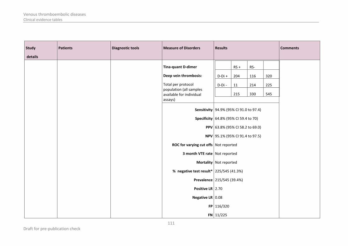
Duration of follow-up:
3 months
receiving treatment for malignancy, if treatment for cancer was stopped within the last 6 months, or if they were receiving palliative treatment for cancer.
Setting: Netherlands.
Exclusion criteria: None reported.
All patients N: 2066 With cancer: 244 Without cancer: 1822 Mean Age: With/without cancer 64/58 years
Performed and interpreted by: the technicians who were unaware of the results of the diagnostic tests for DVT as well as of the cancer status.
Reference standard: Serial compression ultrasound. In case of an initial normal ultrasound, serial testing was performed 1 week later and if still negative, patients were followed up for 3 months.
The compression ultrasound; performed on the transverse plane of the common femoral vein and the popliteal vein down to the trifurcation of the calf veins.
Performed by: not reported
(blinded to index test)
Sensitivity
Specificity
NPV
PPV
3 month VTE rate
Mortality
% negative test result*
Prevalence
Positive LR
Negative LR
69
96
47
95
56
Not reported
Not reported
73/244 (29.9%)
100/244 (41.0%)
1.8
0.08
Notes:
D-dimer test
Patients without cancer
Proximal DVT
True positive
False positive
False negative
True negative
RS + RS- Total
D-Di + 375 534 909
D-Di - 30 883 913
Total 405 1417 1822
375
534
30
883

115 Draft for pre-publication check
Venous thromboembolic diseases Clinical evidence tables
Sensitivity
Specificity
NPV
PPV
3 month VTE rate
Mortality
% negative test result*
Prevalence
Positive LR
Negative LR
92.5%
62.3%
96.7
41.3
Not reported
Not reported
913/1822 (50.1%)
405/1822(22.2%)
2.46
0.12
D-dimer test
All patients
Proximal DVT
True positive
False positive
False negative
True negative
Sensitivity
RS + RS- Total
D-Di + 471 609 1080
D-Di - 34 952 986
Total 505 1561 2066
471
609
34
952
93.27%

116 Draft for pre-publication check
Venous thromboembolic diseases Clinical evidence tables
Specificity
NPV
PPV
3 month VTE rate
Mortality
% negative test result*
Prevalence
Positive LR
Negative LR
60.99%
96.6%
43.6%
Not reported
Not reported
986/2066 (47.7%)
505/2066 (24.4%)
2.4
0.11
Study
details
Patients Diagnostic tools Measure of Disorders Results Comments
Study name: Fukuda 2007
148
Study design: diagnostic study
Patient group: consecutive outpatients with clinically suspected DVT of a lower limb.
Exclusion criteria: Previous episode of DVT, stable symptoms at presentation or prophylactic anticoagulants already applied at presentation.
Assessment tool under investigation:
PATHFAST D-dimer assay (chemiluminescent enzyme immunoassay)
Cut off point: 0.570ug/mL, determined based on ROC curve from 124 healthy
PATHFAST D-dimer assay
Cut off point 0.570ug/mL
Sensitivity
Specificity
PPV
NPV
AUV of ROC curve
100% (87.7 to 100)
63.2% (46 to 78.2)
66.%
100%
0.957(0.918 to 0.996)
Funding:
Test kits provided by Mitsubishi Kagaku Iatron Inc, Japan. Staff of manufacturer provided blood samples for the “healthy control”.
117 Draft for pre-publication check
Venous thromboembolic diseases Clinical evidence tables
Study
details
Patients Diagnostic tools Measure of Disorders Results Comments
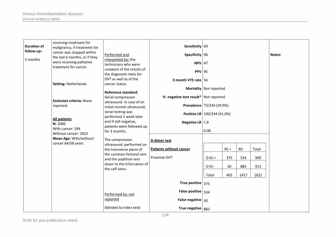
Evidence level:
Duration of follow-up:
All patients
N: 82
Age range: 23-85 years
Drop outs: not stated
Prevalence: 28/82(34.1%)
volunteers (see “Funding”). No specific cut off point recommended by manufacturer
Performed by: not reported.
Reference standard:
Compression ultrasonography
Performed by: not reported.
Prevalence
Positive LR
Negative LR
Efficiency
28/82(34.1%)
78.8%
Limitations:
No description of how reference test was conducted, or basis of classifying patients as having DVT or not
Additional
tests:
Venography
computed tomography
VIDAS D-dimer assay.
ELFA assay principle, combining the ELISA test method with a final blue fluorescent reading
Correlation between the two tests noted
Cut off point at 0.800ug/mL
Sensitivity
Specificity
PPV
NPV
Positive LR
Negative LR
Efficiency
(95% CI)
96.4 (81.7 to 99.9)
71.1 (54.1 to 84.6)
71.1%
96.4%
81.8%
Cut off point at 1.280
Sensitivity
Specificity
PPV
(95% CI)
92.9 (76.5 to 99.1)
84.2 (68.7 to 94.0)
81.3%

118 Draft for pre-publication check
Venous thromboembolic diseases Clinical evidence tables
Study
details
Patients Diagnostic tools Measure of Disorders Results Comments
NPV
Positive LR
Negative LR
Efficiency
94.1%
87.9%
Notes:
Cut off point at 1.500
Sensitivity
Specificity
PPV
NPV
Positive LR
Negative LR
Efficiency
(95% CI)
92.9% (76.5 TO 99.1)
86.8% (71.9 TO 95.6)
83.9%
94.3%
89.4%

119 Draft for pre-publication check
Venous thromboembolic diseases Clinical evidence tables
Study
details
Patients Diagnostic tools Measure of Disorders Results Comments
Study name:
Ilkhanipour 2004
203
Study design:
Prospective diagnostic study
Duration of follow-up:
None
Patient group: adult (over 18 years) emergency department patients suspected of having lower extremity acute DVT, and had symptoms for less than 1 month.
Setting: conducted at 2 sites, a university hospital and a community teaching hospital in US. From June 2000 and February 2002.
Exclusion criteria: excluded if refused to participate, or had symptoms for longer than one month.
All patients N: 336 (365 before excluded) Mean age (range):54 (19-95) F/M ratio: 65/35
Assessment tool under investigation:
VIDAS D-dimer
Cut off point: 0.5ug/ml determined on ROC curve
Performed by: not reported
Reference standard:
Duplex ultrasound of the lower extremities using a 128 XP scanner with a 5 MHz linear array probe. The pelvic inguinal, and femoral veins were evaluated with the patient in a supine position.
Patients with a high Wells
D-dimer test
All patients
DVT
Sensitivity
Specificity
PPV
NPV
% negative test result*
Prevalence
3 month VTE rate
Mortality
Positive LR
Negative LR
RS + RS- Total
D-Di + 31 159 190
D-Di - 2 144 146
Total 33 303 336
93.9%
47.5%
16.3%
98.6%
43.5%
9.8%
Not reported
Not reported
1.79
0.13
Funding: unrestricted educational grant and D-dimer kits from bioMerieux Vitek, Inc manufacturer of test kits
Limitations:
Blinding unclear
Additional
tests:
Wells score performed by emergency care physicians, residents or certfied nurse practitioners
Notes: Rapid ELISA D-dimer
D-dimer test
Low pretest probability(Wells score)
RS + RS- Total

120 Draft for pre-publication check
Venous thromboembolic diseases Clinical evidence tables
Study
details
Patients Diagnostic tools Measure of Disorders Results Comments
Drop outs: 29 incomplete data
clinical probability for DVT but had a negative ultrasound were recommended to have a repeat duplex ultrasound study in one week.
Performed by: experienced vascular technicians blinded to the results of ELISA test and clinical probability score. Vascular surgeon over read the initial classifications and classify these into acute or chronic thrombosis
DVT
Sensitivity
Specificity
PPV
NPV
% negative test result*
Prevalence
3 month VTE rate
Mortality
Positive LR
Negative LR
D-Di + 2 56 58
D-Di - 0 60 60
Total 2 116 118
100.0%
51.7%
3.4%
100.0%
50.8%
1.7%
Not reported
Not reported
2.07
0.0
test carried out.
D-dimer test
Intermediate to high pretest probability (Wells Score)
DVT
RS + RS- Total
D-Di + 29 103 132
D-Di - 2 84 86

121 Draft for pre-publication check
Venous thromboembolic diseases Clinical evidence tables
Study
details
Patients Diagnostic tools Measure of Disorders Results Comments
Sensitivity
Specificity
PPV
NPV
% negative test result*
Prevalence
3 month VTE rate
Mortality
Positive LR
Negative LR
Total 31 187 218
93.5%
44.9%
22.0%
97.7%
39.4%
14.2%
Not reported
Not reported
1.70
0.1436
122 Draft for pre-publication check
Venous thromboembolic diseases Clinical evidence tables
Study
details
Patients Diagnostic tools Measure of Disorders Results Comments
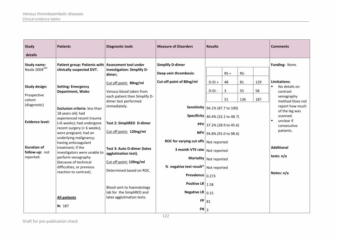
Study name: Neale 2004
293
Study design:
Prospective cohort (diagnostic)
Evidence level:
Duration of follow-up: not reported.
Patient group: Patients with clinically suspected DVT.
Setting: Emergency Department, Wales
Exclusion criteria: less than 18 years old; had experienced recent trauma (<6 weeks); had undergone recent surgery (< 6 weeks); were pregnant; had an underlying malignancy; having anticoagulant treatment; if the investigators were unable to perform venography (because of technical difficulties, or previous reaction to contrast).
All patients
N: 187
Assessment tool under investigation: Simplify D-dimer;
Cut off point: 80ng/ml
Venous blood taken from each patient then Simplify D-dimer test performed immediately.
Test 2: SimpliRED D-dimer
Cut off point: 120ng/ml
Test 3: Auto D-dimer (latex agglutination test).
Cut off point: 120ng/ml
Determined based on ROC.
Blood sent to haematology lab for the SimpliRED and latex agglutination tests.
Simplify D-dimer
Deep vein thrombosis:
Cut-off point of 80ng/ml
Sensitivity
Specificity
PPV
NPV
ROC for varying cut offs
3 month VTE rate
Mortality
% negative test result*
Prevalence
Positive LR
Negative LR
FP
FN
RS + RS-
D-Di + 48 81 129
D-Di - 3 55 58
51 136 187
94.1% (87.7 to 100)
40.4% (32.2 to 48.7)
37.2% (28.9 to 45.6)
94.8% (91.0 to 98.6)
Not reported
Not reported
Not reported
Not reported
0.273
1.58
0.15
81
3
Funding: None.
Limitations: No details on
contrast venography method-Does not report how much of the leg was scanned.
unclear if consecutive patients.
Additional
tests: n/a
Notes: n/a

123 Draft for pre-publication check
Venous thromboembolic diseases Clinical evidence tables
Study
details
Patients Diagnostic tools Measure of Disorders Results Comments
Mean age (range): not reported.
Drop outs: n/a
Performed by: haematology staff who were blinded to the venogram results.
Reference standard: Contrast venography
Performed by:
Radiology staff who were blinded to D-dimer results.
AUTO D-dimer
Cut-off point of 120ng/ml
Deep vein thrombosis
Sensitivity
Specificity
PPV
NPV
ROC for varying cut offs
3 month VTE rate
Mortality
% negative test result*
Prevalence
Positive LR
Negative LR
FP
FN
RS + RS-
D-Di + 46 77 123
D-Di - 5 59 64
51 136 187
90.2 (82.0 to 98.4)
43.4 (35.1 to 51.7)
37.4 (28.8 to 45.9)
92.2 (87.4 to 96.9)
Not reported
Not reported
Not reported
Not reported
0.273
1.59
0.23
77
5

124 Draft for pre-publication check
Venous thromboembolic diseases Clinical evidence tables
Study
details
Patients Diagnostic tools Measure of Disorders Results Comments
SimpliRED D-dimer
Deep vein thrombosis:
Cut-off point of 120ng/ml
Sensitivity
Specificity
PPV
NPV
ROC for varying cut offs
3 month VTE rate
Mortality
% negative test result*
Prevalence
Positive LR
Negative LR
FP
FN
RS + RS-
D-Di + 38 23 61
D-Di - 13 113 126
51 136 187
74.5 (62.5 to 86.5)
83.1 (76.8 to 89.4)
62.3 (50.1 to 74.5)
89.7 (82.0 to 97.3)
Not reported
Not reported
Not reported
Not reported
0.273
4.41
0.31
23
13

125 Draft for pre-publication check
Venous thromboembolic diseases Clinical evidence tables
Study
details
Patients Diagnostic tools Measure of Disorders Results Comments
Study name:
Palen 2005310
Study design:
Observational cohort (diagnostic)
Evidence level:
Duration of follow-up: 1
st
part of the study; 3 months (in most cases 12 months)
Patient group: Outpatients in a large group model managed healthcare organization with clinically suspended DVT, who were referred to the radiology department for lower extremity compression ultrasound.
Setting:
Inclusion criteria:
Exclusion criteria:
All patients
1st
study period
Assessment tool under investigation: Vidas D-Di
TM
bioMerieux (Marcy l’Etoile, France), D-Di Lia® test Diagnostica Stago (Asnieres, France), MiniQuant
TM D-
dimer Assay Biopool International (Ventura, CA).
Cut off point: different cut off points used in the three D-dimer types. Results are reported by D-dimer type and cut off point.
Performed by: (blinded to reference standard?)
Reference standard: duplex ultrasound imaging of lower
1st
study period (n=117)
Vidas D-dimer (cut off point 500 ng/Ml FEU)
Deep vein thrombosis:
Sensitivity
Specificity
PPV
NPV
94.7 (71.9-99.7)
39.8 (30.2-50.2)
23.4 (14.8-34.7)
97.5 (90.2-99.6)
Funding:
Not reported
Limitations: very poor methodology.
Additional
tests:
Notes: no clear if patients were consecutive.
*study reported 100% sensitivity, which is not possible for the other values provided
b)Vidas D-dimer (cut off point 1000 ng/Ml FEU)
Deep vein thrombosis:
Sensitivity
Specificity
PPV
NPV
ROC (largest area under the curve)
3 month VTE rate
94.7 (71.9-99.7)
39.8 (30.2-50.2)
23.4 (14.8-34.7)
97.5 (90.2-99.6)
0.821 (0.746-0.941)

126 Draft for pre-publication check
Venous thromboembolic diseases Clinical evidence tables
Study
details
Patients Diagnostic tools Measure of Disorders Results Comments
N: 117 patients were evaluated by Vidas D-dimer test, 76 patients received both Vidas and BioPool Miniquant tests, and 80 patients received both the Vidas and the Stago LIA tests.
Proportion of patients under 65 years: 43.5%
Drop outs: not reported (1 patient was found to have undergone a follow up ultrasound exam for a previously diagnosed DVT
extremity
Performed by:
Radiologists were blinded to results of the D-dimmer assays.
Miniquant D-dimer (cut off point 500 ng/Ml FEU)
Deep vein thrombosis:
Sensitivity
Specificity
PPV
NPV
92.3 (62.1-99.6)
60.3(47.2-72.2)
32.4 (18.6-49.9)
97.4 (84.4-99.9)

127 Draft for pre-publication check
Venous thromboembolic diseases Clinical evidence tables
Study
details
Patients Diagnostic tools Measure of Disorders Results Comments
and was excluded from the study).
Miniquant D-dimer (cut off point 800 ng/Ml FEU)
Deep vein thrombosis:
Sensitivity
Specificity
PPV
NPV
ROC (largest area under the curve)
3 month VTE rate
Mortality
% negative test result*
Prevalence
Positive LR
Negative LR
FP
FN
92.3 (62.1-99.6)
74.6(61.8-84.4)
42.9 (25.0-62.6)
97.9 (81.9-100)
0.800 (0.744-0.950)

128 Draft for pre-publication check
Venous thromboembolic diseases Clinical evidence tables
Study
details
Patients Diagnostic tools Measure of Disorders Results Comments
Stago D-dimer (cut off point 400 ng/Ml FEU)
Deep vein thrombosis:
Sensitivity
Specificity
PPV
NPV
100 (73.2-99.3)
72.7(60.2-82.6)
43.8 (26.8-62.1)
100 (86.7-99.7)
f)Stago D-dimer (cut off point 500 ng/Ml FEU)
Deep vein thrombosis:
Sensitivity
Specificity
PPV
NPV
Reporting error *(73.2-99.3)
77.3(65.0-86.3)
48.3 (29.9-67.1)
Reporting error *(85.4-99.7)
0.885 (0.723-0.938)

129 Draft for pre-publication check
Venous thromboembolic diseases Clinical evidence tables
Study
details
Patients Diagnostic tools Measure of Disorders Results Comments
Study name:
Rectenwald 2005
349
Study design:
Prospective (diagnostic)
Evidence level:
Duration of follow-up:
Patient group: Patients who completed a lower extremity duplex ultrasound examination. Controls were recruited randomly from the laboratory of one of the co-authors. 3 groups were included; Group 1 (normal volunteers), Group 2 (patients positive for DVT on duplex ultrasound), Group 3 (patients with symptoms of leg pain but negative duplex ultrasound for DVT)
Symptomatic patients: those who exhibited unilateral leg pain or swelling, or bilateral leg pain or swelling with a compelling history for DVT and the absence of uncompensated congestive heart failure, hypoalbuminemic state, or anasarca.
Criteria for a positive duplex ultrasound:
1) Incompressibility of the dilated vein 2) lack of color flow and pulse wave Doppler signal with distal augmentation in the vein congruent with a significant lack of echogenicity of the thrombus, 3) presence of few collateral veins.
Assessment tool under investigation: Advanced D-dimer; a latex enhanced automated turbidometric assay (Dade-Behring, Deerfield, IL).
Cut off point: 3 mg/l
Performed by: all analyses performed in a blinded fashion
Reference standard: duplex ultrasound imaging of lower extremity
Performed by: not reported
Deep vein thrombosis:
(cut off point:3 mg/l)
Use of D-dimer as dichotomous variable
Sensitivity
Specificity
PPV
NPV
64%
76%
not reported
not reported
Funding: not reported
Limitations: no information on prevalence, unable to calculate the true, false positve and negative, PPV, NPV, PLR, NLP.
Additional
tests: Soluble P-selectin, Total microparticles
Notes: not clear if patients were consecutive

130 Draft for pre-publication check
Venous thromboembolic diseases Clinical evidence tables
Study
details
Patients Diagnostic tools Measure of Disorders Results Comments
Setting: University of Michigan Diagnostic Vascular Unit
Inclusion criteria: 1) aged 18 years or over, 2) confirmed diagnosis of iliofemoral or femoropopliteal DVT by duplex ultrasound or symptomatic for DVT clinically but negative for DVT by duplex ultrasound 3) willingness to sign informed consent and 4) control subjects with no clinical signs, symptoms or history of DVT
Exclusion criteria: 1) pregnancy 2) immunosuppressive medications 3) presence of calf level venous thrombosis only without more proximal location.
All patients
N: 73, Group 1 (30), Group 2 (22), Group 3 (21).
Mean age (sd): Group 1; 28.7 (11), Group 2; 48.2 (19), Group 3; 51.1 (17)
Drop outs: not reported

131 Draft for pre-publication check
Venous thromboembolic diseases Clinical evidence tables
Study
details
Patients Diagnostic tools Measure of Disorders Results Comments
Study name:
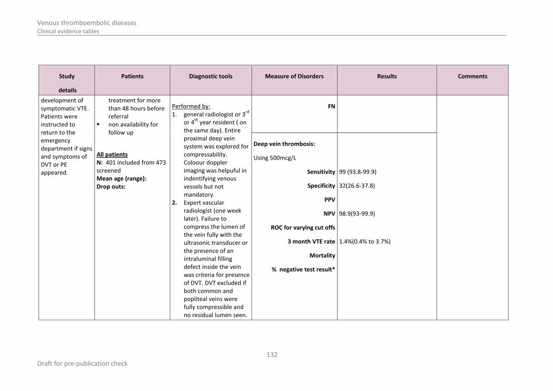
RUIZGIMENEZ2004 368
Study design:
Prospective cohort (diagnostic)
Evidence level:
Duration of follow-up:
Up to 3 months
Patients with DVT excluded were followed up by phone or medical reports to monitor
Patient group:
Consecutive outpatients with suspected DVT of the lower limbs
Setting:
Emergency department
May 2000 to Sept 2001
Inclusion criteria:
Presented with signs and symptoms of DVT – pain swelling, and/or erythema in the lower extremity.
Exclusion criteria: Pregnant women and
children Clinical suspicion of
pulmonary embolism Anticoagulant
Assessment tool under investigation:
Test1:
Wells score (original model Wells1995)
Performed by: Not stated
Test 2:
VIDAS D-dimer Assay (bioMerieux, France)
Cut off point:
1mcg/mL determined based on ROC, &
500ng/mL (manufacturer recommendation)
Performed by: Not stated
Reference standard:
Ultra sonography
Deep vein thrombosis:
Cut off point of 1mcg/mL for VIDAS D-dimer
2x2 table is based on follow up of up to 3 months.
Sensitivity
Specificity
PPV
NPV
3 month VTE rate
Mortality
% negative test result*
Prevalence
Positive LR
Negative LR
FP
RS + RS-
D-Di + 100 136 236
D-Di - 2 145 147
102 281 383
98.0%
51.6%
42.4%
98.6%
102/383
Not reported
38.4%
26.6%
2.03
0.0380
Funding:
Limitations:
Additional
tests:
Notes: * % of people with negative test result indicates the % of patients who will not be undergoing further diagnostic imaging if test is used as a “rule out” criteria.
132 Draft for pre-publication check
Venous thromboembolic diseases Clinical evidence tables
Study
details
Patients Diagnostic tools Measure of Disorders Results Comments
development of symptomatic VTE. Patients were instructed to return to the emergency department if signs and symptoms of DVT or PE appeared.
treatment for more than 48 hours before referral
non availability for follow up
All patients N: 401 included from 473 screened Mean age (range): Drop outs:
Performed by: 1. general radiologist or 3
rd
or 4th
year resident ( on the same day). Entire proximal deep vein system was explored for compressability. Coloour doppler imaging was helpuful in indentifying venous vessels but not mandatory.
2. Expert vascular radiologist (one week later). Failure to compress the lumen of the vein fully with the ultrasonic transducer or the presence of an intraluminal filling defect inside the vein was criteria for presence of DVT. DVT excluded if both common and popliteal veins were fully compressible and no residual lumen seen.
FN
Deep vein thrombosis:
Using 500mcg/L
Sensitivity
Specificity
PPV
NPV
ROC for varying cut offs
3 month VTE rate
Mortality
% negative test result*
99 (93.8-99.9)
32(26.6-37.8)
98.9(93-99.9)
1.4%(0.4% to 3.7%)

133 Draft for pre-publication check
Venous thromboembolic diseases Clinical evidence tables
Study
details
Patients Diagnostic tools Measure of Disorders Results Comments
Study name: Subramaniam2006C
415
Study design:
Prospective cohort (diagnostic)
Evidence level:
Duration of follow-up: 3 months
Patient group: Ambulatory outpatients with suspected lower limb DVT.
Setting: emergency department of a tertiary centre.
Inclusion criteria: consecutive patients at the Emergency department with suspected lower limb DVT.
Exclusion criteria: on current anticoagulation therapy (n=7); failure to perform a D-dimer blood test before sonographic examination (n=5); technical inability to perform an adequate complete compression sonographic examination (n=4).
Assessment tool under investigation:
Simplify D-dimer
Cut off point: not reported.
Performed by: (blinded to reference standard?)
Hamilton score
Cut off point: not reported.
Performed by: (blinded to reference standard?)
Hamilton score and simplify D-dimer
Cut off point: not reported.
Reference standard:
Deep vein thrombosis:
Simplify D-dimer
Sensitivity
Specificity
PPV
NPV
ROC for varying cut offs
3 month VTE rate
Mortality
% negative test result*
Prevalence
RS + RS-
D-Di + 59 109 168
D-Di - 8 136 144
67 245 312
88.00% (77.82 to 94.74)
55.51% (49.0 to 61.8)
35.12% (27.93 to 42.85)
94.44% (89.35 to 97.57)
1 patient at follow up was diagnosed with PE. 10 patients who had second sonography had no DVT.
0/312
0.215
1.98
Funding: Not reported.
Limitations: Unclear reporting of first 214 patients.
Additional
tests: Hamilton score and Hamilton score plus simplify D-dimer
Notes: the first 214 patients recruited for the study were given D-dimer testing. On the basis of this analysis, the Hamilton score was developed (aim of study) which was then validated and compared with the modified Wells scores in another 312 patients. The D-dimer results are given for this population of 312.

134 Draft for pre-publication check
Venous thromboembolic diseases Clinical evidence tables
Study
details
Patients Diagnostic tools Measure of Disorders Results Comments
All patients
N: 542 (recruited), n=526 after excluded (see above); 214 entered the D-dimer test and 312 were tested with the Hamilton score.
Mean age range (in patients tested with D-dimer ): 18-88 years
Drop outs:
Duplex compression sonography
Whole leg from inguinal ligament to the medial malleolus
Performed by:
Experienced sonographers and radiology residents under supervision of consultant radiologists.
Doppler examination of veins performed as supplemental information as a road map but not for deciding the result.
Positive LR
Negative LR
FP
FN
Accuracy
0.22
0.625
Deep vein thrombosis:
Hamilton score and simplify D-dimer
Sensitivity
Specificity
PPV
NPV
ROC for varying cut offs
3 month VTE rate
Mortality
RS + RS-
D-Di +
D-Di -
98.51 (92.0 to 99.96)
41.63 (35.4 to 48.0)
31.60 (25.34 to 38.35)
99.00 (94.71 to 99.98)

135 Draft for pre-publication check
Venous thromboembolic diseases Clinical evidence tables
Study
details
Patients Diagnostic tools Measure of Disorders Results Comments
Deep vein thrombosis:
Hamilton score
Sensitivity
Specificity
PPV
NPV
ROC for varying cut offs
RS + RS-
D-Di +
D-Di -
66.67 (54.0 to 77.8)
71.14 (65.64 to 76.72)
38.26 (29.35 to 47.79)
88.83 (83.58 to 92.87)

136 Draft for pre-publication check
Venous thromboembolic diseases Clinical evidence tables
Study
details
Patients Diagnostic tools Measure of Disorders Results Comments
Study name: Subramaniam 2006A
416
Study design:
Prospective cohort (diagnostic)
Evidence level:
Duration of follow-up: 3 months
Patient group: Patients with suspected lower limb DVT
Setting: Emergency department, Australia.
Inclusion criteria: referred by gps to the emergency department with suspected lower limb DVT.
Exclusion criteria: history of objectively confirmed lower limb DVT; currently on anticoagulation, failure to perform immunochromatographic D-dimer assay before ultrasound examination and inability to perform an adequate complete lower limb compression ultrasound examination.
Assessment tool under investigation:
Simplify D-dimer (Agen Biochemical, Australia)
Cut off point: not reported.
Performed by: Department of Haematology staff with minimal training.
Reference standard: duplex compression ultrasound (Acuson Sequoia 512, USA).
”Doppler examination used as a road map but not to decide result”??
Whole leg – from the level of inguinal ligament to
Deep vein thrombosis:
Proximal and distal
Simplify D-dimer
Sensitivity
Specificity
PPV
NPV
ROC for varying cut offs
3 month VTE rate
Mortality
% negative test result*
Prevalence
Positive LR
Negative LR
RS + RS-
D-Di + 74 152 226
D-Di - 13 214 227
87 366 453
85.1% (75.8 to 91.8)
58.5% (53.4 to 63.5)
32.7% (26.6 to 38.9)
94.3% (90.9 to 96.9)
11/453 readmitted with suspected episodes for DVT or PE and 0/453
found to have by US or US + CTPA
0/453
0.192
2.05
0.26
Funding: Department of radiology research fund, New Zealand. States no funding from manufacturers of D-dimer.
Limitations:
Additional
tests: Hamilton score for DVT.
Notes: Of 227 with negative D-dimer, 13 had isolated calf DVT. States in conclusion that D-dimer has a very high NPV for both proximal and isolated calf DVT.

137 Draft for pre-publication check
Venous thromboembolic diseases Clinical evidence tables
Study
details
Patients Diagnostic tools Measure of Disorders Results Comments
All patients
N: 453
Mean age (s.d): 55.8 years (20.3)
Drop outs:
medial malleolus.
Performed by:
7 consultant radiologists who were blinded to the D-dimer results.
FP
FN

138 Draft for pre-publication check
Venous thromboembolic diseases Clinical evidence tables
Studies with cut off levels determined based on predetermined sensitivity levels
Study
details
Patients Diagnostic tools Measure of Disorders Results Comments
Study name: Stevens 2005
413
Study design:
Prospective cohort (diagnostic)
Evidence level:
Duration of follow-up: 3 months.
Patient group: Inpatients and outpatients with susepected lower extremity DVT.
Setting: LDS Hospital, USA.
Inclusion criteria: 18 years of age or older; who provided informed consent and were referred to the peripheral vascular laboratory of the LDS Hospital, because symptoms suggested a first-episode of lower extremity DVT.
Exclusion criteria: pregnant; referred to the
Assessment tool under investigation:
Test 1: VIDAS D-dimer assay (bioMErieux, USA)
Cut off point: 160ng/ml
Test 2: STA LIATEST D-DI (Diagnostica Stago, USA)
Cut off point: 530ng/ml
Test 3: MiniQuant (BioPool International Inc, USA)
Cut off point: 160ng/ml
Deep vein thrombosis:
VIDAS D-dimer
Sensitivity
Specificity
PPV
NPV
ROC for varying cut offs
3 month VTE rate
Mortality
% negative test result*
Prevalence
Positive LR
Negative LR
RS + RS-
D-Di + 53 166 218
D-Di - 1 158 159
54 323 377
0.982
0.488 (0.434 to 0.542)
Not reported
0.994
Not reported
Not reported
Not reported
42.1%
14.2%
1.92
0.04
Funding: Not stated apart from the provision of anallyszers and reagents from companies.
Limitations: The sensitivity was
‘chosen’ for all tests and cut off points derived from ROC curve
We had to calculate the results from the sensitivity and specificity given.
Additional
tests:
Notes: A blood sample was taken and an aliquot of plasma frozen at -70

139 Draft for pre-publication check
Venous thromboembolic diseases Clinical evidence tables
Study
details
Patients Diagnostic tools Measure of Disorders Results Comments
peripheral vascular laboratory for any reason other than a first episode lower extremity DVT; anticipated geographical inaccessibility for follow-up; treatment with therapeutic doses of heparin or low molecular weight heparin for greater than 24 hours prior to enrolment; a requirement for long-term anticoagulation for any other cause, technical inability to perform duplex ultrasonography or lack of informed consent.
All patients
N: 436
Mean age, s.d (range): 56 +/-17.3 (19-94)
Drop outs:
Test 4: MDA D-dimer assay (bioMeieux)
Cut off point: 520ng/ml
Test 5: AUTO D-dimer
(Sigma, USA)
Cut off point: 220 FEU
Performed by: Technicians who were blinded to the ultrasound results
Reference standard: Comprehensive duplex ultrasonography (CDU)
Performed by:
Vascular surgical staff
FP
FN
degrees centigrade and stored. The D-dimer assays were performed in batches with thawed specimens.
The sensitivity was chosen for all tests at 0.982.
Deep vein thrombosis:
STA LIATEST D-DI
Sensitivity
Specificity
PPV
NPV
ROC for varying cut offs
3 month VTE rate
Mortality
% negative test result*
Prevalence
RS + RS-
D-Di + 53 149 201
D-Di - 1 175 176
54 323 377
0.982
0.540 (0.486 to 0.594)
Not reported
0.994
Not reported
Not reported
Not reported
46.6%
14.2%
2.13

140 Draft for pre-publication check
Venous thromboembolic diseases Clinical evidence tables
Study
details
Patients Diagnostic tools Measure of Disorders Results Comments
Refusal of phlebotomy, failure to report for phlebotomy after enrolment and errors in specimen processing resulted in inability to analyse all five D-dimer assays in 59 specimens (13.5%).
377 had all assays performed.
interpreted the CDU according to clinical protocols;
Positive LR
Negative LR
FP
FN
0.03
Deep vein thrombosis:
MiniQuant
Sensitivity
Specificity
PPV
NPV
ROC for varying cut offs
3 month VTE rate
Mortality
% negative test result*
RS + RS-
D-Di + 53 231 283
D-Di - 1 93 94
54 323 377
0.982
0.287 (0.238 to 0.336)
Not reported
0.989
Not reported
Not reported
Not reported
24.9%

141 Draft for pre-publication check
Venous thromboembolic diseases Clinical evidence tables
Study
details
Patients Diagnostic tools Measure of Disorders Results Comments
Prevalence
Positive LR
Negative LR
FP
FN
14.2%
1.38
0.06
Deep vein thrombosis:
MDA D-dimer
Sensitivity
Specificity
PPV
NPV
ROC for varying cut offs
3 month VTE rate
RS + RS-
D-Di + 53 164 216
D-Di - 1 160 161
54 323 377
0.982
0.494 (0.440 to 0.548)
Not reported
0.994
Not reported
Not reported
Not reported

142 Draft for pre-publication check
Venous thromboembolic diseases Clinical evidence tables
Study
details
Patients Diagnostic tools Measure of Disorders Results Comments
Mortality
% negative test result*
Prevalence
Positive LR
Negative LR
FP
FN
42.6%
14.2%
1.94
0.04
Deep vein thrombosis:
AUTO D-dimer
Sensitivity
Specificity
PPV
NPV
RS + RS-
D-Di + 53 111 164
D-Di - 1 213 213
54 323 377
98.2%
65.7% (60.5% to 70.9%)
Not reported
99.5%
Not reported

143 Draft for pre-publication check
Venous thromboembolic diseases Clinical evidence tables
Study
details
Patients Diagnostic tools Measure of Disorders Results Comments
ROC for varying cut offs
3 month VTE rate
Mortality
% negative test result*
Prevalence
Positive LR
Negative LR
FP
FN
Not reported
Not reported
63.8%
14.2%
2.86
0.03
Recommended