Draft
Variation in stiffness regulates cardiac myocyte hypertrophy
via signaling pathways
Journal: Canadian Journal of Physiology and Pharmacology
Manuscript ID cjpp-2015-0578.R2
Manuscript Type: Article
Date Submitted by the Author: 09-May-2016
Complete List of Authors: Li, Jieli; University of Illinois at Chicago College of Medicine, Physiology and Biophysics Mkrtschjan, Michael; University of Illinois at Chicago, Bioengineering Lin, Ying-Hsi; University of Illinois at Chicago College of Medicine, Physiology and Biophysics Russell, Brenda; University of Illinois at Chicago, Physiology and Biophysics
Keyword: Mechano-transduction, focal adhesion kinase, lipid signaling, actin assembly, substrate stiffness
https://mc06.manuscriptcentral.com/cjpp-pubs
Canadian Journal of Physiology and Pharmacology
Draft
1
Variation in stiffness regulates cardiac myocyte hypertrophy via signaling pathways
Jieli Li1, Michael Mkrtschjan2, Ying-Hsi Lin1, Brenda Russell1,2*
1Department of Physiology and Biophysics, Center for Cardiovascular Research, University of
Illinois at Chicago
2Department of Bioengineering, University of Illinois at Chicago
851 South Morgan Street
Chicago, IL 60607
*corresponding author
Mailing Address
University of Illinois at Chicago, MC 901
835 South Wolcott Avenue
Chicago, IL 60612-7342
Tel: 1-312-413-0407
Email: [email protected]
Page 1 of 28
https://mc06.manuscriptcentral.com/cjpp-pubs
Canadian Journal of Physiology and Pharmacology

Draft
2
Abstract
Much diseased human myocardial tissue is fibrotic and stiff, which increases the work that the
ventricular myocytes must perform to maintain cardiac output. The hypothesis tested is that the
increased load due to greater stiffness of the substrata drives sarcomere assembly of cells, thus
strengthening them. Neonatal rat ventricular myocytes (NRVM) were cultured on polyacrylamide
or polydimethylsiloxane substrates with stiffness of 10kPa, 100kPa, 400kPa, or glass with
stiffness of 61.9 GPa. Cell size increased with stiffness. Two signaling pathways were explored,
phosphorylation of focal adhesion kinase (p-FAK) and lipids by phosphatidylinositol 4,5-
bisphosphate (PIP2). Subcellular distributions of both were determined in the sarcomeric
fraction by antibody localization, and total amounts were measured by Western or dot blotting,
respectively. More p-FAK and PIP2 distributed to the sarcomeres of NRVM grown on stiffer
substrates. Actin assembly involves the actin capping protein Z, CapZ. Both actin and CapZ
dynamic exchange were significantly increased on stiffer substrates when assessed by
fluorescence recovery after photobleaching (FRAP) of green fluorescent protein tags. Blunting
of actin FRAP by FAK inhibition implicates linkage from mechano-signalling pathways to cell
growth. Thus, increased stiffness of cardiac disease can be modeled with polymeric materials to
understand how the microenvironment regulates cardiac hypertrophy.
Key words: Mechano-transduction; focal adhesion kinase; lipid signaling; actin assembly;
substrate stiffness
Page 2 of 28
https://mc06.manuscriptcentral.com/cjpp-pubs
Canadian Journal of Physiology and Pharmacology
Draft
3
Introduction
The mechanical properties of the local microenvironment influence the function of cells
(Yang 2014). This is particularly critical following a myocardial infarction, in which stiff, fibrotic
scar tissue replaces the normally compliant ventricle with adverse functional consequences.
The ventricular myocytes must work harder to maintain cardiac output, which they mainly
accomplish by cell hypertrophy. Thus, the processes that link mechanosensing of increased
load to the strengthening of myocytes by cell hypertrophy are of major significance in heart
diseases. Multiple mechanosensors detect increased mechanical loading (Hoshijima 2006), but
the feedback linking sensing to local actin filament assembly is not yet fully understood. Here,
we culture cardiac myocytes on substrata of defined stiffness to analyze cell responses.
The study of mechanisms of cell growth requires altering the load in a controlled manner,
which is difficult to do at the cellular level since cells are usually cultured on hard, plastic
surfaces, which poorly mimic the external forces existing in living tissue. Stiffness of the surface
on which cells grow significantly affects maturation and differentiation into myocytes (Jacot 2010)
and also force generation (Bhana 2010; Broughton 2015). The stiffness in the heart can vary
from embryonic/neonatal of 5-10 kPa (Bhana 2010; de Tombe 2003) to the normal adult rat
myocardium of 10-70kPa (Borbély 2005). Infarct stiffness and collagen content increased with
time post-infarct up to 400kPa (Fomovsky 2010A; Fomovsky 2010B; Holmes 2005). Therefore,
we fabricated novel substrata out of polyacrylamide (PAA) (Engler 2008) and
polydimethylsiloxane (PDMS) in the physiologic and pathologic range (10 to 400 kPa)
(Broughton 2015) in order to model loading of cardiac myocytes and resultant signaling
pathways.
In muscle, focal adhesions are the primary biomechanical sensors found at the Z discs
where integrins are anchored at the costamere to the extracellular matrix. The focal adhesion
kinase, FAK, binds to the cytoskeletal domain of the integrin complex and responds to
Page 3 of 28
https://mc06.manuscriptcentral.com/cjpp-pubs
Canadian Journal of Physiology and Pharmacology
Draft
4
mechanical stimuli (Senyo 2007). Mechanical stimulation rapidly phosphorylates and activates
FAK possibly by unfolding of the protein to expose a phosphorylation site (Chu 2011; Franchini
2000). Interestingly, FAK is activated by cyclic strain at Tyr-397 and distributes along the
myofilaments (Torsoni 2003), which might suggest that the distribution of p-FAK regulates actin
assembly. Another actin assembly site is at the Z-disc where the actin capping protein, CapZ, is
able to slow down filament assembly (Edwards 2014). On mechanical stimulation of myocytes,
the CapZβ1 C-terminus may control capping of the actin filament (Lin 2013). Moreover,
mechano-transduction arising from stress or strain may also modify the function of CapZ by
phosphatidylinositol 4,5-bisphosphate (PIP2), a phospholipid (Li 2013).
In this report, cell signaling and actin, thin filament assembly was assessed by
fluorescence recovery after photobleaching (FRAP) to determine the capping dynamics of CapZ
and actin. Understanding how fibrotic stiffness in the microenvironment regulates cardiac
hypertrophy may be important in cardiac disease states.
Materials and methods
Substrata fabrication
Polymers with varying stiffness were used to coat glass surfaces with a layer
approximately 100 microns thick. The goals were to attain the stiffness range from physiologic
(10 kPa) to pathologic (400 kPa) while retaining cell adhesion for culture, good optics for
immuno-localization, and protein isolation for Western and dot blotting. PDMS is viscoelastic
below 100 kPa, requiring the use of PAA at lower stiffnesses (Wei SC 2015). Cells can be
scraped for Western and dot blotting from stiff PDMS but not from soft PAA below 100 kPa. To
control for potential differences in material properties, 100 kPa surfaces were produced with
both PDMS and PAA.
Page 4 of 28
https://mc06.manuscriptcentral.com/cjpp-pubs
Canadian Journal of Physiology and Pharmacology
Draft
5
Preparation of glass base for application of polymer layer
Cell culture glass bottom dishes and 10 mm circular coverslips were treated using a
modified protocol (Tse and Engler 2010; Poellmann and Wagoner-Johnson 2013). The center
portions of glass bottom dishes were treated for 30 minutes with 10N NaOH to expose hydroxyl
groups, then washed thoroughly with deionized water. The glass was silanated with 3-
(trimethoxysilyl) propyl methacrylate (Cat# M6514, Sigma USA) for 90 minutes. Dishes were
washed 3 times with 70% EtOH and dried on a 100ºC hotplate. Coverslips necessary for
creating flat surfaces were washed with 70% EtOH in petri dishes, air-dried, then silanated by
placing them in a desiccator with 20 µl of tridecafluoro-(1,1,2,2-tetrahydrooctyl)-1-trichlorosilane
(UCT T2492) for 90 minutes. Following treatment, coverslips were washed with 70% EtOH and
dried on a 100ºC hotplate.
Polyacrylamide substrata
40% unpolymerized acrylamide (Cat#161-0140, Bio-Rad, USA) and 2% Bis solution
(Cat#161-0142, Bio-Rad, USA) were diluted in water to concentrations necessary to develop 10
kPa (final concentration 5% acrylamide, 0.3% Bis) and 100 kPa (final concentration 30%
acrylamide, 0.3% Bis) substrata, respectively. Ammonium persulfate was added at 1% by
volume and tetraethylmethylenediamine at 0.1% by volume in order to begin the polymerization
reaction. 10 µl of solutions were then added to glass bottom dishes and each covered with a
treated coverslip. Substrata were allowed to polymerize for 10 minutes, then coverslips were
gently pried up, leaving behind a flat, circular substrate. Dishes were washed 3 times in
deionized water for 10 minutes at a time to remove unpolymerized acrylamide.
Page 5 of 28
https://mc06.manuscriptcentral.com/cjpp-pubs
Canadian Journal of Physiology and Pharmacology
Draft
6
To functionalize polyacrylamide substrata for cell adhesion, they were treated twice by
drying sulfo-SANPAH (Cat#22589, Thermo Fisher, USA) in HEPES (50 mM, pH 8.5) on each
for 90 minutes at 57ºC. A UV cross-linker (UV Crosslinker, Spectronics Corporation, USA) with
a 365 nm bulb, placed approximately 10 cm away for 10 minutes, was then used to cross-link
sulfo-SANPAH to the substrate surface. Substrates were washed with HEPES between
treatments. Following sulfo-SANPAH treatment, substrates were washed 3 times using 50 mM
HEPES, then HEPES containing fibronectin (10µg/ml) was added to dishes and incubated at
37ºC for 2 hours before UV-sterilizing in water for 20 minutes.
Polydimethylsiloxane substrata
Polydimethylsiloxane (PDMS) (DowCorning, Midland, MI) was mixed in a 10:1 (400kPa)
or 50:1 (100 kPa) elastomer base to curing agent ratio for approximately 10 min. PDMS
mixtures were degassed using a vacuum desiccator for approximately 30 min. PDMS was then
spun onto cell culture glass-bottom dishes (In Vitro Scientific, CA), creating a PDMS thickness
of approximately 50 µm, or added to 6-well plates. PDMS substrata were then cured for 24 h in
a 57ºC oven. After curing, PDMS was cooled and ready for activation for fibronectin coating.
PDMS substrata were treated with 5% (3-Aminopropyl) triethoxysilane in 90.25% ethanol
solution for 10 min, then washed with 100% ethanol. Substrata were placed in a 57ºC oven for
20 min, washed with 95% ethanol and twice with PBS, then coated with 10µg/ml fibronectin in
DMEM for 2 hours at 37ºC.
Measurement of stiffness
Stiffness of both materials was measured by Young’s Modulus using atomic force
microscopy. For PDMS, our lab found 50:1 ratio to yield a Young’s Modulus of 98.4 +/- 7.36
Page 6 of 28
https://mc06.manuscriptcentral.com/cjpp-pubs
Canadian Journal of Physiology and Pharmacology

Draft
7
kPa, while a 10:1 ratio yields a 397.4 +/- 30.79, which are rounded off to 100 and 400 kPa,
respectively (Broughton 2015); for PAA, mixtures of 5%/0.3% and 30%/0.3% acrylamide/Bis
yielded ~9.3+/- 0.26 and 101.4 +/- 1.6 kPa, respectively by atomic force microscopy ([Asylum 1-
D, Asylum Research; Santa Barbara, CA) and indented by a pyramid-tipped probe (Veeco;
Santa Barbara, CA)] (data provided by Dr. Engler at UCSD, Engler 2006). The glass stiffness
reported as 61.9 GPa (Wang 2012).
Neonatal rat ventricular myocyte culture
Primary heart cultures were obtained from neonatal rats according to Institutional Animal
Care and Use Committee and NIH guidelines for the care and use of laboratory animals that are
equivalent to the Canadian Council on animal care (CCAC) regulations. Hearts were removed
and cells isolated from 1-2 day old Sprague-Dawley rats with collagenase type II (Worthington,
Lakewood, NJ USA) as previously described (Boateng 2003). Neonatal rat ventricular myocytes
(NRVMs) were re-suspended, filtered through a metal sieve to remove large material and plated
at high density (1,000 cells/mm2) in PC-1 medium (Lonza Group Ltd, U.S.A). Unattached cells
were removed by aspiration and PC-1 media was replenished. Myocytes were plated on
fibronectin coated (10µg/ml) dishes at 1560 cells/mm2 for Western blotting or 520 cells/mm2 for
immunostaining. Stiffnesses used for PAA were 10 and 100kPa; for PDMS were 100 and
400kPa; and on glass-bottom dishes (61.9 GPa). Cells were cultured for 3 days in a 5% CO2
incubator.
Localization of p-FAK (Tyr397) or PIP2 in the cytoskeletal fraction by immunostaining and
microscopy
Formatted: Font: (Default) Arial, 11 pt
Page 7 of 28
https://mc06.manuscriptcentral.com/cjpp-pubs
Canadian Journal of Physiology and Pharmacology
Draft
8
For isolation of the cytoskeletal fraction, the Calbiochem® ProteoExtract® Subcellular
Proteome Extraction Kit was used (EMD Millipore, Billerica, MA), following a previously
described detergent-based protocol (Boateng 2007). The remaining myofibrillar cytsokeleton
was immunostained with α-actinin antibody (1: 200, Cell Signaling Technology, Inc., Danvers,
MA USA), and either with a p-FAK (Tyr397) antibody (1: 200, Cell Signaling Technology, Inc.,
Danvers, MA USA) or a PIP2 antibody (1:200, mouse IgG, Abcam, Cambridge, MA USA).
Cardiomyocytes were observed by microscopy (Observer Z1, Zeiss), and by confocal
microscopy (LSM 710, Zeiss). Cell surface areas were measured by ImageJ software. In each
case, three independent experiments were performed, values were calculated and 20 cells from
each condition were randomly chosen and used to calculate cell areas. Experiments were
repeated at least three times on PDMS (100kPa, 400kPa) or glass. The selective extraction
method for the subcellular components on the polyacrylamide hydrogels detached the cells from
the surface. Efforts to retain cell attachment by shortening the exposure times and altering the
detergents were unsuccessful.
PIP2 levels by dot blots
Whole cell lysates extracted from NRVMs grown on 100 kPa, 400 kPa, PDMS substrata,
or 61.9 GPa (glass) were spotted onto nitrocellulose membranes (Bio-Rad Laboratories,
Hercules, CA, USA). These were probed with PIP2 antibody (mouse IgG, Abcam, Cambridge,
MA USA) at a 1:500 dilution and detected using a horseradish peroxidase conjugated
secondary antibody (anti-mouse, HRP, Cell Signaling Technology, Boston, MA USA) and ECL.
To exclude any non-specific binding of the PIP2 antibodies, we detected the signal with a
positive control PIP2 (Echelon Biosciences, Cat#P-4516) (data not shown). Experiments were
Page 8 of 28
https://mc06.manuscriptcentral.com/cjpp-pubs
Canadian Journal of Physiology and Pharmacology
Draft
9
repeated at least three times. Cell scraping for traditional dot blots could not be performed on
PAA (10 kPa or 100 kPa) due to the structurally weak nature of polyacrylamide hydrogels.
Fluorescence recovery after photobleaching (FRAP) for actin dynamics
Microscopic techniques, such as FRAP have yielded quantitative information about the
processes that regulate actin polymerization in living myocytes. The methods and analysis for
FRAP of actin-GFP were described by our lab (Lin 2013). NRVMs were treated with a FAK
inhibitor PF-573228 (30 µM) (Cat#PZ0117, Sigma USA) (Slack-Davis 2007) for one hour prior to
the FRAP experiment. In the present study, five myocytes were analyzed per culture, and at
least three separate cultures were studied for 100 kPa, 400kPa and glass. Stiffness affects
embryonic cardiomyocyte structure and contractility (Engler 2008). We also found very rapid
beating of the neonatal myocytes on 10 kPa substrates, which did not permit the imaging quality
and time resolution necessary for FRAP analysis.
Statistics
Data are presented as mean ± SEM. Statistical significance was determined by One-
Way ANOVA with Tukey’s multiple comparison tests. Significance was taken as p<0.05.
RESULTS
NRVM cell size increase with substrata stiffness
NRVMs grown for 3 days were beating well in culture on the various substrata and had
good sarcomere striations (Fig 1A). The NRVM area was greatest for cells grown on the hard
glass surface and least for cells on PAA substrates of 10 kPa. The ratios of cell area normalized
Page 9 of 28
https://mc06.manuscriptcentral.com/cjpp-pubs
Canadian Journal of Physiology and Pharmacology
Draft
10
to the 10 kPa were 1.76, 1.80, 2.23, and 2.69 for stiffness of PAA substrates of 100 kPa, PDMS
substrates of 100 kPa and 400 kPa, and glass (61.9 GPa), respectively with statistical
significance levels as shown in Fig 1B. Also, the area of NRVMs on glass were significantly
increased by 1.52 and 1.50 times respectively, see Fig 1B, compared with that on 100kPa-PAA
and 100kPa-PDMS. The areas of myocytes were similar when grown on the same stiffness (100
kPa) but on two different polymers (PAA and PDMS).
FAK activation by substrate stiffness
The tyrosine residue Tyr-397 is phosphorylated via an autophosphorylation process,
leading to an increase in FAK enzymatic activity (Chen 1996). The effect of the stiffness on FAK
activity was assessed by Western blotting, which was detected with a phosphospecific antibody
against the autophosphorylation site of FAK (FAK-Tyr-397). The level of FAK phosphorylation at
Tyr397 was significantly increased (p<0.05, n=3) in myocytes on stiffer PDMS substrata (400
kPa) and glass (61.9 GPa), compared to PDMS (100 kPa), (Fig 2A). The protein level of total
FAK in all the groups was not significantly changed when normalized to histone 2B (H2B) (Fig
2B).
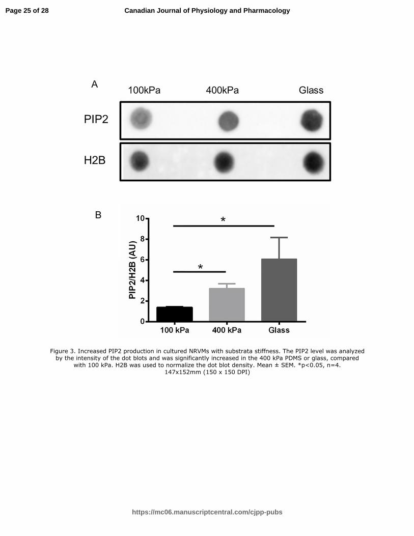
PIP2 increases with substrate stiffness
The effect of stiffness on total PIP2 abundance in cultured NRVMs was measured by dot
blotting (Fig 3). After 3 days culture, the total abundance of PIP2 in NRVMs varied with the
stiffness of the underlying PDMS or glass substratum. The PIP2 level was significantly
increased (p<0.05) in PDMS (400kPa) and glass (61.9 GPa) substrates compared to the softer
PDMS (100kPa).
Page 10 of 28
https://mc06.manuscriptcentral.com/cjpp-pubs
Canadian Journal of Physiology and Pharmacology

Draft
11
Sarcomeric distribution of phosphorylated FAK with substrate stiffness
The sarcomeric distribution p-FAK in NRVMs was evaluated by confocal microscopy
using anti-p-FAK (Tyr397), Fig 4. An α-actinin antibody was used to identify the repeating
sarcomeric structure of the Z discs. Overall, there appeared to be less staining for p-FAK on
PDMS (100kPa), and the most staining intensity for NRVM grown on glass (61.9 GPa) but
immunostaining is a semi-quantitative method (Fig 4A). However, line scans provide a
quantitative method for analysis of the distribution of the immuno-label in the various sarcomere
bands, Fig 4B. At all stiffnesses the peaks of the α-actinin at the Z-disc coincided with the peaks
of p-FAK. Line scans intensity also suggested that the p-FAK was lowest in NRVM on the
PDMS (100kPa) substrata. The glass (61.9 GPa) had the highest amount of p-FAK signaling,
which accumulated at the Z-disc.
Sarcomeric distribution of PIP2 with substrate stiffness
Similarly, the sarcomeric distribution of PIP2 in NRVMs was evaluated by confocal
microscopy with α-actinin for identification of the Z-disc and with double antibody staining for
PIP2 signaling (Fig 5). Here too, the typical sarcomeric pattern was seen for NRVM on all
stiffnesses. Overall, the PIP2 was higher on glass than for the 100 kPa or 400kPa PDMS
substrata. Furthermore, line scans showed clear co-localization of the peak intensity of PIP2
with the α-actinin at the Z-discs in myocytes, Fig 5B.
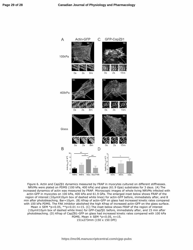
CapZβ1 and actin dynamics increase with substrate stiffness that FAK inhibition blunts
FRAP experiments revealed differences in protein dynamics on different substrates. The
actin-GFP and GFP-CapZβ1 had strong striations in NRVMs and signals were detected in a
10µm X10µm square region of interest (Fig 6). Actin-GFP had a faster dynamic protein
Page 11 of 28
https://mc06.manuscriptcentral.com/cjpp-pubs
Canadian Journal of Physiology and Pharmacology

Draft
12
exchange in myocytes on glass compared to those on 100 kPa PDMS substrata (6.90±0.99 vs
2.28±0.54, ×10 -4 sec-1, p<0.01) (Fig 6 A&B). Consistently, the dynamics of GFP-CapZβ1 was
significantly higher in myocytes on glass compared to those on 100 kPa substrata (3.05±0.67 vs
1.52±0.22, ×10 -3 sec-1, p<0.05) (Fig 6 C&D), meaning that a faster protein exchange was
occurring in myocytes working against the increased load due to the stiffness of the substrata.
Furthermore, to study the role of p-FAK at Z-disc, NRVMs grown on glass were treated
with 30 µM FAK inhibitor PF-573228 for one hour, then FRAP experiments were performed. The
increased actin dynamics dramatically decreased (2.55±0.97 vs 6.90±0.99, ×10-4 sec-1, p<0.01),
suggesting that increased p-FAK was involved in regulating actin dynamics.
Discussion
Our novel bioengineering approaches with two polymers (PAA and PDMS) permitted
fabrication of substrata with varying physiologic or pathologic stiffness for the study of NRVM in
culture. Hence, we could determine how sarcomere assembly of cardiomyocytes depends on
chronic loading. Interestingly, cell size depended on the stiffness, not on the polymer itself,
suggesting that stiffness is more important than other polymer-specific properties. Cardiac
hypertrophy measured by cell size increased with the stiffness of the culture substrata. Analysis
showed significant changes with stiffness for two components of the signaling pathways, namely
phosphorylation of FAK at Y397 and the PIP2 production level. Furthermore, both the FAK and
PIP2 signaling molecules dramatically increased at the Z-disc of the myofibrils. Moreover, the
actin and CapZβ1 dynamics increased with substrata stiffness as measured by FRAP but actin
dynamics were blunted by FAK inhibition implicating linkage from mechano-signalling to cell
growth. Taken together, results suggest that increased levels of p-FAK and PIP2 at the Z-disc
are related to actin assembly, and hence to myocyte hypertrophy.
Page 12 of 28
https://mc06.manuscriptcentral.com/cjpp-pubs
Canadian Journal of Physiology and Pharmacology

Draft
13
Integrin transmembrane receptors on the cell surface regulates attachment to substrat or
matrix resulting in activation and focal adhesion formation. With suitable functionalization of the
fibronectin binding, we found the surface chemistry of the PAA and PDMS polymers was
excellent for NRVM adhesion in culture. However, cell detachment during processing for
extraction of the membrane fraction differed and we were unable to process cells for the
cytoskeletal distribution on the PAA substrata. FAK was activated downstream of integrins, and
its signaling was especially important for cardiac hypertrophy (Peng 2008; Clemente 2012). For
example, tyrosine-phosphorylated FAK dramatically increased in the isolated rat heart perfused
to produce pressure-loading (Domingos 2002). p-FAK at Tyr-397 redistributed to the Z-disc
when myocytes were activated by mechanical stretch (Torsoni 2003). FAK was also involved in
increasing adhesion strength, particularly in response to tension forces (Wang 2001). Our data
confirm similar activation of p-FAK by chronic loading with increased stiffness.
Mechanical stimulation also engaged PIP2 signaling, which was a key determinant of
sarcomeric assembly (Li 2013). PIP2 is the most abundant of the phosphoinositides that binds
cellular proteins and accounts for approximately 1% of lipid content in the plasma membrane of
a typical mammalian cell because PIP2 has a phosphinositol head and two fatty acid chains,
making it highly hydrophobic and lipophilic (Lemmon 2008). However, PIP2 also binds to
hydrophobic pockets found within proteins, such as CapZ. Thus, PIP2 binding may result in a
reduction in binding affinity of CapZ dimers to the actin filament (Hartman 2009; Li 2013). We
have shown that there is more PIP2 at Z-disc when the cell size is bigger (Li 2013 and 2014).
Both actin and CapZ dynamics were also greater with hypertrophy. Neomycin, the PIP2
scavenger, abolished the increased cell size. The multi-step relationship between PIP2 and cell
size is proposed so that PIP2 binds CapZ, which loosens the cap enabling more actin filaments
to form resulting in cell hypertrophy. In this study, PIP2 antibody staining of fixed cells showed
PIP2 increased at the Z-discs with chronic mechanical loading by stiffness (Fig 5). Also, actin
Page 13 of 28
https://mc06.manuscriptcentral.com/cjpp-pubs
Canadian Journal of Physiology and Pharmacology

Draft
14
and CapZβ1 FRAP data showed their significantly increased dynamics (Fig 6), which was
consistent with the increased PIP2 localization at the Z-discs. These results suggest that the
increased PIP2 at Z-discs may bind CapZβ1, which regulates actin filament assembly.
The present study demonstrates that the effect of stiffness of the microenvironment in
heart tissue is very important in regulating actin assembly in cardiac myocytes. Changes in FAK
and PIP2 signaling at the Z-disc indicate involvement as mechanical sensors in response to the
stiffer environment in load-induced cardiac myocyte hypertrophy. These two signaling pathways
may have independent targets or they may interact. Indeed, FAK interacts with PIP2, which
affects FAK clustering on the lipid membrane (Chen 1996; Goñi 2014). PIP2 is produced by
PIP5K an important mediator of the integrin/FAK signaling link (Goñi 2014). Additionally,
activated FAK might phosphorylate and activate local PIP5K (Brancaccio 2006). The latter could
lead to local synthesis of high amounts of PIP2 binding to CapZβ1, resulting in the high
assembly and destabilization of the actin filaments (Xu 2014). FAK might interact directly or
indirectly with PIP2. Therefore, we interpret our data to suggest that FAK/PIP2 signaling
pathways interact both upstream at the membrane as well described previously but also
downstream in the sarcomere, leading to integrated mechanical transduction for increased
filament assembly resulting from chronic loading by external stiffness. Many other signaling
pathways are known to be involved in mechanotransduction signaling leading to induced
cardiac hypertrophy, such as calcineurin, a cytoplasmic Ca2+/calmodulin-dependent protein
phosphatase (Liu HB 2010). CapZ is critical for actin capping but is not itself calcium dependent
(Li 2013). However, partner signaling proteins and lipids do have calcium dependence.
Changes in actin capping dynamics with stiffness further suggest that thin filament
assembly depends on cell tension, which feeds back via signaling pathways onto sarcomere
assembly. It seems likely that filaments are built to serve the functional work being demanded
by the myocyte, and that local mechanical conditions ultimately regulate filament assembly and
Page 14 of 28
https://mc06.manuscriptcentral.com/cjpp-pubs
Canadian Journal of Physiology and Pharmacology
Draft
15
muscle mass. Despite the vast amount of knowledge of the multi-protein complexes of the
costamere and Z-disc, there is currently no clinical strategy to reduce or prevent the
maladaptive cardiac remodeling that occurs within each myocyte of the heart. Our findings may
provide a better understanding of fundamental processes in local fibrotic or stiff scar tissue
based on modeling of physiologic and pathologic stiffness modeled with substrata for myocyte
culture.
Acknowledgments
We gratefully acknowledge Jennifer Wen in the laboratory of Dr. Adam Engler,
University of California at San Diego for conducting atomic force microscopy for stiffness
assessment of the polyacrylamide substrata. We thank Sagar Dommaraju for cell area
measurements.
Grants
This work was supported by NIH HL62426.
Disclosures
No conflicts of interest, financial or otherwise, are declared by the authors.
References
Bhana B, Iyer RK, Chen WL, Zhao R, Sider KL, Likhitpanichkul M, Simmons CA, Radisic M.
2010. Influence of substrate stiffness on the phenotype of heart cells. Biotechnol Bioeng.
105:1148-60. PMID: 20014437.
Page 15 of 28
https://mc06.manuscriptcentral.com/cjpp-pubs
Canadian Journal of Physiology and Pharmacology
Draft
16
Boateng SY, Belin RJ, Geenen DL, Margulies KB, Martin JL, Hoshijima M, de Tombe PP,
Russell B. 2007. Cardiac dysfunction and heart failure are associated with abnormalities in the
subcellular distribution and amounts of oligomeric muscle LIM protein. Am J Physiol Heart Circ
Physiol. 292:H259-69. PMID: 16963613.
Boateng SY, Hartman TJ, Ahluwalia N, Vidula H, Desai TA, Russell B. Inhibition of fibroblast
proliferation in cardiac myocyte cultures by surface microtopography. 2003. Am J Physiol Cell
Physiol. 285:C171-82. PMID: 12672651.
Borbely A, van der Velden J, Papp Z, Bronzwaer JGF, Edes I, Stienen GJM, Paulus WJ.
Cardiomyocyte stiffness in diastolic heart failure. 2005. Circulation.111:774-81. PMID:
15699264.
Brancaccio M, Hirsch E, Notte A, Selvetella G, Lembo G, Tarone G. Integrin signalling: the tug-
of-war in heart hypertrophy. 2006. Cardiovasc Res. 70:422-33. PMID: 16466704.
Broughton KM, Russell B. Cardiomyocyte subdomain contractility arising from
microenvironmental stiffness and topography. 2015. Biomech Model Mechanobiol. 14:589-602.
PMID: 25273278.
Chen HC, Appeddu PA, Isoda H, Guan JL. Phosphorylation of tyrosine 397 in focal adhesion
kinase is required for binding phosphatidylinositol 3-kinase. 1996. J Biol Chem. 271:26329-34.
PMID: 8824286.
Chu M, Iyengar R, Koshman YE, Kim T, Russell B, Martin JL, Heroux AL, Robia SL, Samarel
AM. Serine-910 phosphorylation of focal adhesion kinase is critical for sarcomere reorganization
in cardiomyocyte hypertrophy. 2011. Cardiovasc Res. 92:409-19. PMID: 21937583.
Clemente CF, Xavier-Neto J, Dalla Costa AP, Consonni SR, Antunes JE, Rocco SA, Pereira MB,
Judice CC, Strauss B, Joazeiro PP, Matos-Souza JR, Franchini KG. Focal adhesion kinase
Page 16 of 28
https://mc06.manuscriptcentral.com/cjpp-pubs
Canadian Journal of Physiology and Pharmacology
Draft
17
governs cardiac concentric hypertrophic growth by activating the AKT and mTOR pathways.
2012. J Mol Cell Cardiol. 52:493-501. PMID: 22056317.
de Tombe PP. Cardiac myofilaments: mechanics and regulation. 2003. J Biomech. 36:721-30.
PMID: 12695002.
Domingos PP, Fonseca PM, Nadruz W Jr, Franchini KG. Load-induced focal adhesion kinase
activation in the myocardium: role of stretch and contractile activity. 2002. Am J Physiol Heart
Circ Physiol. 282:H556-64. PMID: 11788403.
Edwards M, Zwolak A, Schafer DA, Sept D, Dominguez R, Cooper JA. Capping protein
regulators fine-tune actin assembly dynamics. 2014. Nat Rev Mol Cell Biol. 15:677-89. PMID:
25207437.
Engler AJ, Carag-Krieger C, Johnson CP, Raab M, Tang HY, Speicher DW, Sanger JW, Sanger
JM, Discher DE. Embryonic cardiomyocytes beat best on a matrix with heart-like elasticity: scar-
like rigidity inhibits beating. 2008. J Cell Sci. 121:3794-802. PMID: 18957515.
Engler AJ, Sen S, Sweeney HL, Discher DE, Matrix elasticity directs stem cell lineage
specification. 2006. Cell. 126:677-89. PMID: 16923388.
Fomovsky GM, Holmes JW. Evolution of scar structure, mechanics, and ventricular function
after myocardial infarction in the rat. 2010(A). Am J Physiol Heart Circ Physiol. 298:H221-8.
PMID: 19897714.
Fomovsky GM, Thomopoulos S, Holmes JW. Contribution of extracellular matrix to the
mechanical properties of the heart. 2010(B). J Mol Cell Cardiol. 48:490-6. PMID: 19686759.
Franchini KG, Torsoni AS, Soares PH, Saad MJ. Early activation of the multicomponent
signaling complex associated with focal adhesion kinase induced by pressure overload in the rat
heart. 2000. Circ Res. 87:558-65. PMID: 11009560.
Page 17 of 28
https://mc06.manuscriptcentral.com/cjpp-pubs
Canadian Journal of Physiology and Pharmacology

Draft
18
Goñi GM, Epifano C, Boskovic J, Camacho-Artacho M, Zhou J, Bronowska A, Martín MT, Eck
MJ, Kremer L, Gräter F, Gervasio FL, Perez-Moreno M, Lietha D. Phosphatidylinositol 4,5-
bisphosphate triggers activation of focal adhesion kinase by inducing clustering and
conformational changes. 2014. Proc Natl Acad Sci U S A. 111:E3177-86. PMID: 25049397.
Hartman TJ, Martin JL, Solaro RJ, Samarel AM, Russell B. CapZ dynamics are altered by
endothelin-1 and phenylephrine via PIP2- and PKC-dependent mechanisms. 2009. Am J
Physiol Cell Physiol. 296:C1034-9. PMID: 19295171.
Holmes JW, Borg TK, Covell JW. Structure and mechanics of healing myocardial infarcts. 2005.
Annu Rev Biomed Eng. 7:223-53. PMID: 16004571.
Hoshijima M. Mechanical stress-strain sensors embedded in cardiac cytoskeleton: Z disk, titin,
and associated structures. 2006. Am J Physiol Heart Circ Physiol. 290:H1313-25. PMID:
16537787.
Jacot JG, Martin JC, Hunt DL. Mechanobiology of cardiomyocyte development. 2010. J
Biomech. 43:93-8. PMID: 19819458.
Lemmon MA. Membrane recognition by phospholipid-binding domains. 2008. Nat Rev Mol Cell
Biol. 9:99-111. PMID: 18216767.
Li J, Russell B. Phosphatidylinositol 4,5-bisphosphate regulates CapZβ1 and actin dynamics in
response to mechanical strain. 2013. Am J Physiol Heart Circ Physiol. 305:H1614-23. PMID:
24043251.
Lin YH, Li J, Swanson ER, Russell B. CapZ and actin capping dynamics increase in myocytes
after a bout of exercise and abates in hours after stimulation ends. 2013. J Appl Physiol
114:1603-9. PMID: 23493359.
Page 18 of 28
https://mc06.manuscriptcentral.com/cjpp-pubs
Canadian Journal of Physiology and Pharmacology

Draft
19
Liu HB, Yang BF, Dong DL, Calcineurin and electrical remodeling in pathologic cardiac
hypertrophy. Trends Cardiovasc Med. 2010 Jul;20(5):148-53. PMID: 21742270.
Peng X, Wu X, Druso JE, Wei H, Park AY, Kraus MS, Alcaraz A, Chen J, Chien S, Cerione RA,
Guan JL. Cardiac developmental defects and eccentric right ventricular hypertrophy in
cardiomyocyte focal adhesion kinase (FAK) conditional knockout mice. 2008. Proc Natl Acad
Sci U S A. 105:6638-43. PMID: 18448675.
Poellmann MJ, Wagoner Johnson AJ. Characterizing and Patterning Polyacrylamide Substrates
Functionalized with N-Hydroxysuccinimide. 2013. Cellular and Molecular Bioengineering 3: 299-
309.
Roy P, Rajfur Z, Pomorski P, Jacobson K. Microscope-based techniques to study cell adhesion
and migration. 2002. Nat Cell Biol. 4:E91-6. PMID: 11944042.
Senyo SE, Koshman YE, Russell B. Stimulus interval, rate and direction differentially regulate
phosphorylation for mechanotransduction in neonatal cardiac myocytes. 2007. FEBS Lett.
581:4241-7. PMID: 17698065.
Slack-Davis JK, Martin KH, Tilghman RW, Iwanicki M, Ung EJ, Autry C, Luzzio MJ, Cooper B,
Kath JC, Roberts WG, Parsons JT. Cellular characterization of a novel focal adhesion kinase
inhibitor. 2007. J Biol Chem. 282:14845-52. PMID: 17395594.
Tse JR, Engler AJ. Preparation of hydrogel substrates with tunable mechanical properties. 2010.
Curr Protoc Cell Biol. Chapter 10:Unit 10.16.
Torsoni AS, Constancio SS, Nadruz W Jr, Hanks SK, Franchini KG. Focal adhesion kinase is
activated and mediates the early hypertrophic response to stretch in cardiac myocytes. 2003.
Circ Res. 93:140-7. PMID: 12805241.
Page 19 of 28
https://mc06.manuscriptcentral.com/cjpp-pubs
Canadian Journal of Physiology and Pharmacology

Draft
20
Wang HB, Dembo M, Hanks SK, Wang Y. Focal adhesion kinase is involved in
mechanosensing during fibroblast migration. 2001. Proc Natl Acad Sci U S A. 98:11295-300.
PMID: 11572981.
Wei SC, Fattet L, Tsai JH, Guo Y, Pai VH, Majeski HE, Chen AC, Sah RL, Taylor SS, Engler AJ,
Yang J. Matrix stiffness drives epithelial-mesenchymal transition and tumour metastasis through
a TWIST1-G3BP2 mechanotransduction pathway. 2015. Nat Cell Biol. 17:678-88. PMID:
25893917.
Xu JX, Si M, Zhang HR, Chen XJ, Zhang XD, Wang C, Du XN, Zhang HL. Phosphoinositide
kinases play key roles in norepinephrine- and angiotensin II-induced increase in
phosphatidylinositol 4,5-bisphosphate and modulation of cardiac function. 2014. J Biol Chem.
289:6941-8. PMID: 24448808.
Yang C, Tibbitt MW, Basta L, Anseth KS. Mechanical memory and dosing influence stem cell
fate. 2014. Nat Mater. 13:645-52. PMID: 24633344.
Figure legends
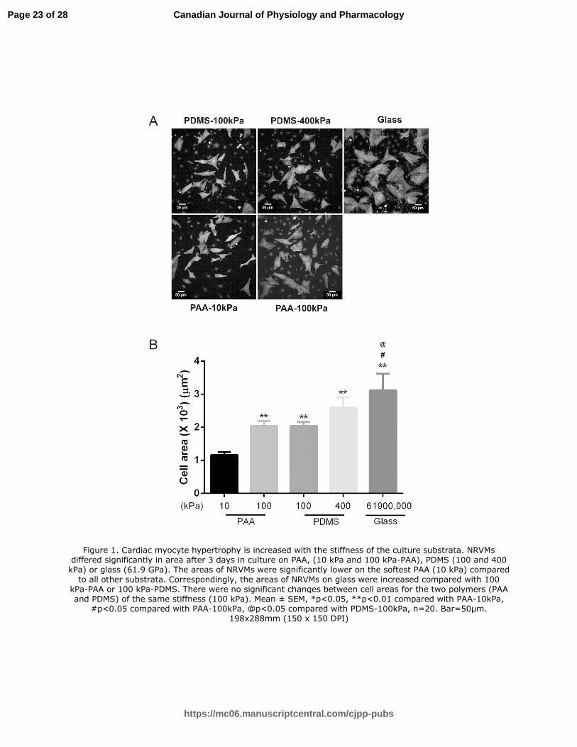
Figure 1. Cardiac myocyte hypertrophy is increased with the stiffness of the culture substrata.
NRVMs differed significantly in area after 3 days in culture on PAA, (10 kPa and 100 kPa-PAA),
PDMS (100 and 400 kPa) or glass (61.9 GPa). The areas of NRVMs were significantly lower on
the softest PAA (10 kPa) compared to all other substrata. Correspondingly, the areas of NRVMs
on glass were increased compared with 100 kPa-PAA or 100 kPa-PDMS. There were no
significant changes between cell areas for the two polymers (PAA and PDMS) of the same
stiffness (100 kPa). Mean ± SEM, *p<0.05, **p<0.01 compared with PAA-10kPa, #p<0.05
compared with PAA-100kPa, @p<0.05 compared with PDMS-100kPa, n=20. Bar=50µm.
Page 20 of 28
https://mc06.manuscriptcentral.com/cjpp-pubs
Canadian Journal of Physiology and Pharmacology
Draft
21
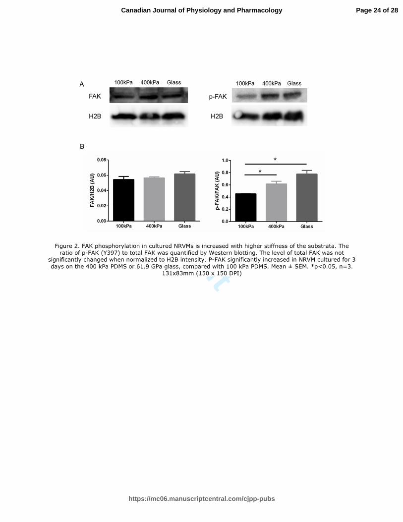
Figure 2. FAK phosphorylation in cultured NRVMs is increased with higher stiffness of the
substrata. The ratio of p-FAK (Y397) to total FAK was quantified by Western blotting. The level
of total FAK was not significantly changed when normalized to H2B intensity. P-FAK
significantly increased in NRVM cultured for 3 days on the 400 kPa PDMS or 61.9 GPa glass,
compared with 100 kPa PDMS. Mean ± SEM. *p<0.05, n=3.
Figure 3. Increased PIP2 production in cultured NRVMs with substrata stiffness. The PIP2 level
was analyzed by the intensity of the dot blots and was significantly increased in the 400 kPa
PDMS or glass, compared with 100 kPa. H2B was used to normalize the dot blot density. Mean
± SEM. *p<0.05, n=4.
Figure 4. Sarcomeric distribution of phosphorylated FAK in NRVMs with varied substrata
stiffness. Immunofluorescence images of the cytoskeletal/myofibrillar fraction of NRVMs grown
for 3 days on 100 kPa, 400 kPa PDMS or glass, 61.9 GPa (Fig 4A). NRVM were stained for p-
FAK (Tyr397) (green) and α-actinin (red). A greater intensity of green striations from p-FAK is
seen by NRVM grown on glass (61.9 GPa) than on the 100 and 400 kPa PDMS substrata. Line
scans quantify the proteins along the myofibril (10 µm long white line), Fig 4B. Note the peaks at
the two micron spacing of the sarcomeric repeat in the α-actinin (red line scan) of the Z-disc.
The peaks of pFAK (green lines) in the line scan colocalize with the Z-disc. Note, the intensity of
the p-FAK and ratio of pFAK to α-actinin (red) is lower in the 100 kPa and 400 kPa cells than in
those grown on glass, 61.9 GPa. Taken together, these findings suggest that the FAK is
involved in the pathway for signaling stiffness. . Bar=20µm in low magnification (top row).
Bar=10µm in high magnification (bottom 3 rows).
Page 21 of 28
https://mc06.manuscriptcentral.com/cjpp-pubs
Canadian Journal of Physiology and Pharmacology
Draft
22
Figure 5. Subcellular distribution of PIP2 to the sarcomeres of in NRVMs with stiffness.
Immunofluorescence images of the cytoskeletal/myofibrillar fraction of NRVM grown for 3 days
on 100 kPa, 400 kPa PDMS or glass, 61.9 GPa (Fig 5A). NRVM were stained for PIP2 (green)
and α-actinin (red). More PIP2 (green line) co-localized with α-actinin (red line) in the Z-disc in
the 61.9 GPa group, as quantified by line scan, Fig 5B. Glass (61.9 GPa) has greater intensity
of PIP2 staining suggesting that the PIP2 is involved in the stiffness-signaling pathway.
Bar=20µm in low magnification (top row). Bar=10 µm in high magnification (bottom 3 rows).
Figure 6. Actin and CapZβ1 dynamics measured by FRAP in myocytes cultured on different
stiffnesses. NRVMs were plated on PDMS (100 kPa, 400 kPa) and glass (61.9 Gpa) substrates
for 3 days. (A) The increased dynamics of actin was measured by FRAP. Microscopic images of
whole living NRVMs infected with actin-GFP in myocytes on 100 kPa, 400 kPa and 61.9 GPa.
The enlarged inset below shows FRAP of the region of interest (10µmX10µm box of dashed
white lines) for actin-GFP before, immediately after, and 8 min after photobleaching. Bar=10µm.
(B) Kfrap of actin-GFP on glass had increased kinetic rates compared with 100 kPa PDMS. The
FAK inhibitor abolished the high Kfrap of increased actin-GFP on the glass surface. Mean ±
SEM *p<0.05, **p<0.01 n=15. (C) The inset below shows FRAP of the region of interest
(10µmX10µm box of dashed white lines) for GFP-CapZβ1 before, immediately after, and 15 min
after photobleaching. (D) Kfrap of CapZB1-GFP on glass had increased kinetic rates compared
with 100 kPa PDMS. Mean ± SEM *p<0.05, n=15.
Page 22 of 28
https://mc06.manuscriptcentral.com/cjpp-pubs
Canadian Journal of Physiology and Pharmacology
Draft
Figure 1. Cardiac myocyte hypertrophy is increased with the stiffness of the culture substrata. NRVMs differed significantly in area after 3 days in culture on PAA, (10 kPa and 100 kPa-PAA), PDMS (100 and 400 kPa) or glass (61.9 GPa). The areas of NRVMs were significantly lower on the softest PAA (10 kPa) compared
to all other substrata. Correspondingly, the areas of NRVMs on glass were increased compared with 100 kPa-PAA or 100 kPa-PDMS. There were no significant changes between cell areas for the two polymers (PAA and PDMS) of the same stiffness (100 kPa). Mean ± SEM, *p<0.05, **p<0.01 compared with PAA-10kPa,
#p<0.05 compared with PAA-100kPa, @p<0.05 compared with PDMS-100kPa, n=20. Bar=50µm. 198x288mm (150 x 150 DPI)
Page 23 of 28
https://mc06.manuscriptcentral.com/cjpp-pubs
Canadian Journal of Physiology and Pharmacology
Draft
Figure 2. FAK phosphorylation in cultured NRVMs is increased with higher stiffness of the substrata. The ratio of p-FAK (Y397) to total FAK was quantified by Western blotting. The level of total FAK was not
significantly changed when normalized to H2B intensity. P-FAK significantly increased in NRVM cultured for 3
days on the 400 kPa PDMS or 61.9 GPa glass, compared with 100 kPa PDMS. Mean ± SEM. *p<0.05, n=3. 131x83mm (150 x 150 DPI)
Page 24 of 28
https://mc06.manuscriptcentral.com/cjpp-pubs
Canadian Journal of Physiology and Pharmacology
Draft
Figure 3. Increased PIP2 production in cultured NRVMs with substrata stiffness. The PIP2 level was analyzed by the intensity of the dot blots and was significantly increased in the 400 kPa PDMS or glass, compared
with 100 kPa. H2B was used to normalize the dot blot density. Mean ± SEM. *p<0.05, n=4. 147x152mm (150 x 150 DPI)
Page 25 of 28
https://mc06.manuscriptcentral.com/cjpp-pubs
Canadian Journal of Physiology and Pharmacology

Draft
Figure 4. Sarcomeric distribution of phosphorylated FAK in NRVMs with varied substrata stiffness. Immunofluorescence images of the cytoskeletal/myofibrillar fraction of NRVMs grown for 3 days on 100 kPa,
400 kPa PDMS or glass, 61.9 GPa (Fig 4A). NRVM were stained for p-FAK (Tyr397) (green) and α-actinin
(red). A greater intensity of green striations from p-FAK is seen by NRVM grown on glass (61.9 GPa) than on the 100 and 400 kPa PDMS substrata. Line scans quantify the proteins along the myofibril (10 µm long white
line), Fig 4B. Note the peaks at the two micron spacing of the sarcomeric repeat in the α-actinin (red line
scan) of the Z-disc. The peaks of pFAK (green lines) in the line scan colocalize with the Z-disc. Note, the intensity of the p-FAK and ratio of pFAK to α-actinin (red) is lower in the 100 kPa and 400 kPa cells than in
those grown on glass, 61.9 GPa. Taken together, these findings suggest that the FAK is involved in the pathway for signaling stiffness. . Bar=20µm in low magnification (top row). Bar=10µm in high magnification
(bottom 3 rows). 142x214mm (150 x 150 DPI)
Page 26 of 28
https://mc06.manuscriptcentral.com/cjpp-pubs
Canadian Journal of Physiology and Pharmacology
Draft
Page 27 of 28
https://mc06.manuscriptcentral.com/cjpp-pubs
Canadian Journal of Physiology and Pharmacology

Draft
Figure 5. Subcellular distribution of PIP2 to the sarcomeres of in NRVMs with stiffness. Immunofluorescence images of the cytoskeletal/myofibrillar fraction of NRVM grown for 3 days on 100 kPa, 400 kPa PDMS or
glass, 61.9 GPa (Fig 5A). NRVM were stained for PIP2 (green) and α-actinin (red). More PIP2 (green line)
co-localized with α-actinin (red line) in the Z-disc in the 61.9 GPa group, as quantified by line scan, Fig 5B. Glass (61.9 GPa) has greater intensity of PIP2 staining suggesting that the PIP2 is involved in the stiffness-signaling pathway. Bar=20µm in low magnification (top row). Bar=10 µm in high magnification (bottom 3
rows). 144x219mm (150 x 150 DPI)
Page 28 of 28
https://mc06.manuscriptcentral.com/cjpp-pubs
Canadian Journal of Physiology and Pharmacology
Draft
Figure 6. Actin and CapZβ1 dynamics measured by FRAP in myocytes cultured on different stiffnesses. NRVMs were plated on PDMS (100 kPa, 400 kPa) and glass (61.9 Gpa) substrates for 3 days. (A) The
increased dynamics of actin was measured by FRAP. Microscopic images of whole living NRVMs infected with
actin-GFP in myocytes on 100 kPa, 400 kPa and 61.9 GPa. The enlarged inset below shows FRAP of the region of interest (10µmX10µm box of dashed white lines) for actin-GFP before, immediately after, and 8
min after photobleaching. Bar=10µm. (B) Kfrap of actin-GFP on glass had increased kinetic rates compared with 100 kPa PDMS. The FAK inhibitor abolished the high Kfrap of increased actin-GFP on the glass surface.
Mean ± SEM *p<0.05, **p<0.01 n=15. (C) The inset below shows FRAP of the region of interest (10µmX10µm box of dashed white lines) for GFP-CapZβ1 before, immediately after, and 15 min after photobleaching. (D) Kfrap of CapZB1-GFP on glass had increased kinetic rates compared with 100 kPa
PDMS. Mean ± SEM *p<0.05, n=15. 151x273mm (150 x 150 DPI)
Page 29 of 28
https://mc06.manuscriptcentral.com/cjpp-pubs
Canadian Journal of Physiology and Pharmacology
Draft
Page 30 of 28
https://mc06.manuscriptcentral.com/cjpp-pubs
Canadian Journal of Physiology and Pharmacology
Recommended