Int.J.Curr.Microbiol.App.Sci (2014) 3(3): 739-746
739
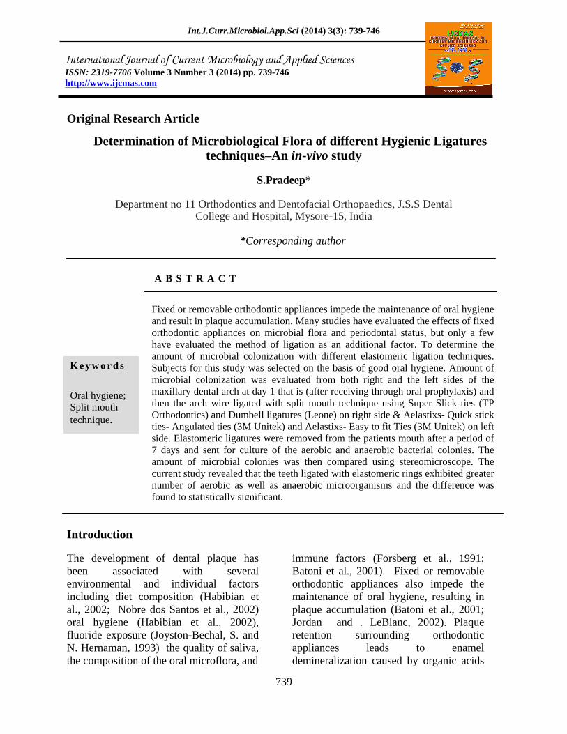
Original Research Article
Determination of Microbiological Flora of different Hygienic Ligatures techniques An in-vivo study
S.Pradeep*
Department no 11 Orthodontics and Dentofacial Orthopaedics, J.S.S Dental College and Hospital, Mysore-15, India
*Corresponding author
A B S T R A C T
Introduction
The development of dental plaque has been associated with several environmental and individual factors including diet composition (Habibian et al., 2002; Nobre dos Santos et al., 2002) oral hygiene (Habibian et al., 2002), fluoride exposure (Joyston-Bechal, S. and N. Hernaman, 1993) the quality of saliva, the composition of the oral microflora, and
immune factors (Forsberg et al., 1991; Batoni et al., 2001). Fixed or removable orthodontic appliances also impede the maintenance of oral hygiene, resulting in plaque accumulation (Batoni et al., 2001; Jordan and . LeBlanc, 2002). Plaque retention surrounding orthodontic appliances leads to enamel demineralization caused by organic acids
ISSN: 2319-7706 Volume 3 Number 3 (2014) pp. 739-746 http://www.ijcmas.com
K e y w o r d s
Oral hygiene; Split mouth technique.
Fixed or removable orthodontic appliances impede the maintenance of oral hygiene and result in plaque accumulation. Many studies have evaluated the effects of fixed orthodontic appliances on microbial flora and periodontal status, but only a few have evaluated the method of ligation as an additional factor. To determine the amount of microbial colonization with different elastomeric ligation techniques. Subjects for this study was selected on the basis of good oral hygiene. Amount of microbial colonization was evaluated from both right and the left sides of the maxillary dental arch at day 1 that is (after receiving through oral prophylaxis) and then the arch wire ligated with split mouth technique using Super Slick ties (TP Orthodontics) and Dumbell ligatures (Leone) on right side & Aelastixs- Quick stick ties- Angulated ties (3M Unitek) and Aelastixs- Easy to fit Ties (3M Unitek) on left side. Elastomeric ligatures were removed from the patients mouth after a period of 7 days and sent for culture of the aerobic and anaerobic bacterial colonies. The amount of microbial colonies was then compared using stereomicroscope. The current study revealed that the teeth ligated with elastomeric rings exhibited greater number of aerobic as well as anaerobic microorganisms and the difference was found to statistically significant.
Int.J.Curr.Microbiol.App.Sci (2014) 3(3): 739-746
740
produced by bacteria in the dental plaque (Arends, and Christofferson, 1986; O'Reilly, and Featherstone, 1987). Recently, fluoride-releasing elastomeric modules (Wiltshire, 1999; Banks et al., 2000; Mattick et al., 2001) and chlorhexidine varnish (Beyth et al., 2003) were suggested for reducing plaque accumulation and decalcification.
Fixed orthodontic appliances create new retention areas, which are suitable for bacterial colonization and lead to an increase in the absolute number and percentage of Streptococcus mutans and lactobacilli (Forsberg et al., 1991; Sakamaki, and Bahn, 1968; Balenseifen, and Madonia, 1970; Corbett,et al., 1981 Mattingly et al., 1983; Scheie et al., 1984; Diamanti-Kipioti et al., 1987; Lundström, and Krasse, 1987; Sinclair, et al., 1987; Svanberg et al., 1987; Rosenbloom, and Tinanoff, 1991; Sandham, et al., 1992; Chang, et al., 1999). A lot of studies have evaluated the effect of fixed orthodontic appliances on microbial flora and periodontal status, (Forsberg et al., 1991; Sakamaki, and . Bahn, 1968; Balenseifen, and Madonia, 1970; Corbett,et al., 1981 Mattingly et al., 1983; Scheie et al., 1984; Diamanti-Kipioti et al., 1987; Lundström, and Krasse, 1987; Sinclair, et al., 1987; Svanberg et al., 1987; Rosenbloom, and Tinanoff, 1991; Sandham, et al., 1992; Chang, et al., 1999; Pender, 1986; Huser, et al., 1990; Glans, et al., 2003). but only a few studies evaluated the methods of commercially available elastomeric ligations as an additional factor (Forsberg et al., 1991; Sukontapatipark, et al., 2001) . So this study was done to determine the amount microbial colonization with different elastomeric ligation techniques available.
The main aim of the present study to determine the amount of microbial
colonization with different elastomeric ligation techniques was used.
Assess which elastomeric ligature attracts the least microbial flora and fulfills the criteria for Hygienic ligatures
Materials and Methods
Twenty subjects were selected for the present study. Informed consent was obtained from all subjects.
Following inclusion criteria were used for patient selection:
Patients of age group 18-25years.
All patients were in the permanent dentition, free of dental plaque, and motivated for good oral hygiene.
Patient undergoing fixed orthodontic treatment with standardized 3M MBT Gemini Series 0.022 slots.
Following exclusion criteria were used for patient selection
Presence of decalcification or increased risk to caries (high caries index)Subjects who had used antibiotics during the three-month period before the study were excluded.
Poor periodontal status.
Halitosis.
The patients were instructed to brush once in the morning before breakfast and once in the evening before bed time. They were instructed to brush a minimum of three minutes to ensure thorough brushing. The patients were asked to thoroughly rinse
Int.J.Curr.Microbiol.App.Sci (2014) 3(3): 739-746
741
with water after every meal. The investigation was designed as a split-mouth study
A total of five ligature systems divided into five individual groups were chosen for the study.
Group I and II were ligated over the premolars on the right side of the patient both in the upper and lower arch with stainless steel ties i.e. Group V (Control group) over the cuspids on either side while Group III and IV were ligated over the premolars on the left side of the patient both in the upper and lower arch.
Elastomeric ligatures were removed from the patient s day and collected in labeled sterile vials with normal saline and sent to the department of Microbiology to assess the amount of microbial colonization on different elastomeric ligature groups as obtained from the patients mouth.
The elastomeric sample groups were collected under aseptic conditions. Serial 10-fold dilutions of the transport medium with the sample of plaque were prepared to 10 , and 0.1-mL samples were inoculated on sheep blood agar, Chocolate agar & Robertson cooked meat broth for number of total bacteria and Mitis-Salivarius agar (Difco Laboratories Inc., Detroit, MI, USA) containing 0.001% Chapman Tellurite solution (Difco), 150 g sucrose, and 3.33 mg bacitracin (Sigma Diagnostics, St. Louis, MO, USA) per liter agar for number of S mutans. The samples were even inoculated on MacConkey agar (HiMEDIA) for secondary bacterial infections. The agar plates were incubated for 48 hours at 37°C in anaerobic jars. Subsequently, colonies were counted under a stereomicroscope. Also, serial 10-fold dilutions were prepared to 10 , and 0.1-mL samples were inoculated on two
Rogosas agar plates for number of lactobacilli. Both plates were incubated for 48 hours at 37°C, one plate in aerobic conditions and the other in an anaerobic jar. The number of colonies were then determined under a stereomicroscope. Results are expressed as colony-forming units per milliliter
Results and Discussion
Primary dental care begins at home. Practicing satisfactory oral hygiene, such as adequate tooth brushing, mouth rinsing, and dental flossing, plays a vital role in maintaining healthy teeth, especially in the orthodontic patients (O'Reilly, and Featherstone, 1987). It is a well known fact that the placement of fixed orthodontic appliances generally hinders good oral hygiene ,and the appliance component can cause alteration in oral micro flora by reducing pH , increasing affinity of bacteria to the metallic surface because of electrostatic reactions, and causing retention areas for microorganisms. . Thus they lead to plaque accumulation around the bracket base.
The literature clearly demonstrates that fixed orthodontic appliances increase plaque accumulation, bacterial colonization, and resultant enamel decalcification. (Forsberg et al., 1991; Sakamaki, and . Bahn, 1968; Balenseifen, and Madonia, 1970; Corbett,et al., 1981 Mattingly et al., 1983; Scheie et al., 1984; Diamanti-Kipioti et al., 1987; Lundström, and Krasse, 1987; Sinclair, et al., 1987; Svanberg et al., 1987; Rosenbloom, and Tinanoff, 1991; Sandham, et al., 1992; Chang, et al., 1999; Pender, 1986; Huser, et al., 1990; Glans, et al., 2003). However, the contribution of ligation materials to this increase has only been evaluated in a few studies (Forsberg et al., 1991; Sukontapatipark, et al., 2001) evaluated.
Int.J.Curr.Microbiol.App.Sci (2014) 3(3): 739-746
742
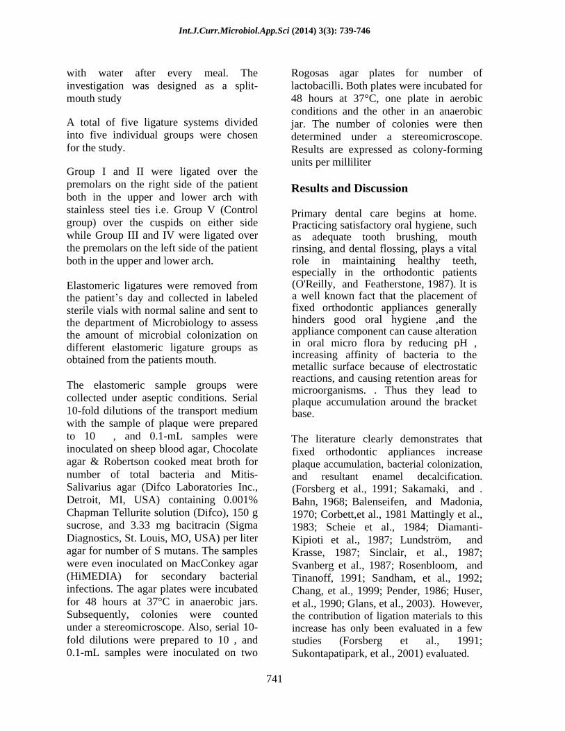
Table.1 Samples of elastomeric ties selected for microbial colony assessment.
Group I Super Slick (TP Orthodontics) Group II Dumb bell ligatures (Leone) Group III Easy to Tie- Angulated Ties (3 M Unitek) Group IV Quick Stick Ties (3 M Unitek) Group V
(Control group) Stainless steel
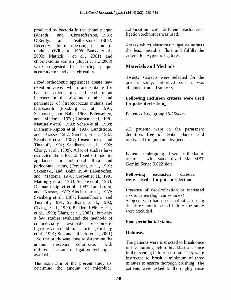
Fig.1&2 Patient in permanent dentition, free of dental plaque, and motivated for good oral hygiene
Fig.3 Group I - Super Slick (TP Orthodontics) Fig 4: Group II- Dumb bell ligatures (Leone)
Int.J.Curr.Microbiol.App.Sci (2014) 3(3): 739-746
743
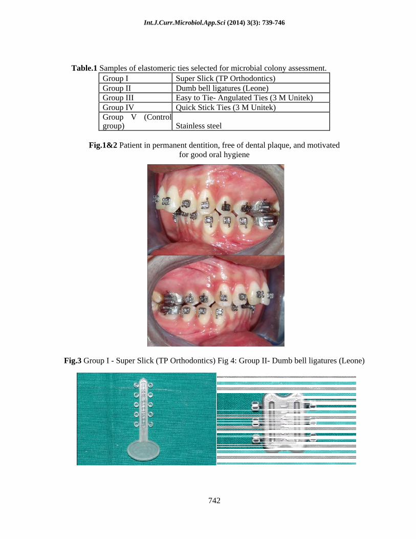
Fig.5 Group III- Easy to Tie- Angulated Tie. Fig.6 Group IV - Quick Stick Ties (3 M Unitek) (3 M Unitek)
Figure.7 Group I and II ligated over the premolars on the right side.
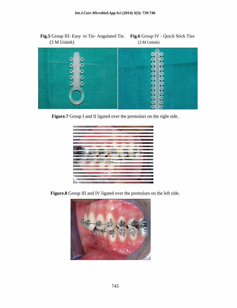
Figure.8 Group III and IV ligated over the premolars on the left side.
Int.J.Curr.Microbiol.App.Sci (2014) 3(3): 739-746
744
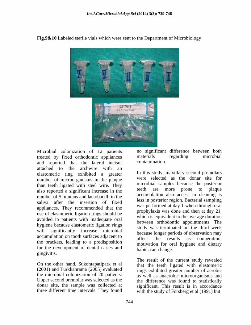
Fig.9&10 Labeled sterile vials which were sent to the Department of Microbiology
Microbial colonization of 12 patients treated by fixed orthodontic appliances and reported that the lateral incisor attached to the archwire with an elastomeric ring exhibited a greater number of microorganisms in the plaque than teeth ligated with steel wire. They also reported a significant increase in the number of S. mutans and lactobacilli in the saliva after the insertion of fixed appliances. They recommended that the use of elastomeric ligation rings should be avoided in patients with inadequate oral hygiene because elastomeric ligation rings will significantly increase microbial accumulation on tooth surfaces adjacent to the brackets, leading to a predisposition for the development of dental caries and gingivitis.
On the other hand, Sukontapatipark et al (2001) and Turkkahrama (2005) evaluated the microbial colonization of 20 patients. Upper second premolar was selected as the donar site, the sample was collected at three different time intervals. They found
no significant difference between both materials regarding microbial contamination.
In this study, maxillary second premolars were selected as the donar site for microbial samples because the posterior teeth are more prone to plaque accumulation also access to cleaning is less in posterior region. Bacterial sampling was performed at day 1 when through oral prophylaxis was done and then at day 21, which is equivalent to the average duration between orthodontic appointments. The study was terminated on the third week because longer periods of observation may affect the results as cooperation, motivation for oral hygiene and dietary habits can change.
The result of the current study revealed that the teeth ligated with elastomeric rings exhibited greater number of aerobic as well as anaerobic microorganisms and the difference was found to statistically significant. This result is in accordance with the study of Forsberg et al (1991) but
Int.J.Curr.Microbiol.App.Sci (2014) 3(3): 739-746
745
is in contrast with the study of Turkkahraman et al (2001) who found statistically not significant difference between elastomers and ligatures. A feasible explanation may be due to the difference in the study design i.e. in the present study at the 21 day the elastomeric ring and the ligature was cultured unlike the previous study were the swab from the labial surface of the tooth was taken and in this study anaerobic as well as aerobic culture was done whereas in previous study specifically the growth of streptococcus mutants and lactobacillus was evaluated. .
References
Habibian, M. , D. Beighton , R. Stevenson , M. Lawson , and G. Roberts . Relationships between dietary behaviors, oral hygiene and mutans streptococci in dental plaque of a group of infants in southern England. Arch Oral Biol 2002;47:491 8.
Nobre dos Santos, M. , L. Melo dos Santos , S. B. Francisco , and J. A. Cury. Relationship among dental plaque composition, daily sugar exposure and caries in the primary dentition. Caries Res 2002;36:347 52.
Joyston-Bechal, S. and N. Hernaman . The effect of a mouthrinse containing chlorhexidine and fluoride on plaque and gingival bleeding. J Clin Periodontol 1993; 20:49 53.
Forsberg, C. M. , V. Brattström , E. Malmberg , and C. E. Nord . Ligature wires and elastomeric rings: two methods of ligation, and their association withmicrobialcolonization
of Streptococcus mutans and lactobacilli. Eur J Orthod 1991;13:416 20.
Batoni, G. , M. Pardini , A. Giannotti , F. Ota , M. R. Giuca , M. Gabriele et al . Effect of removable orthodontic appliances on oral colonization by mutans streptococci in children. Eur J
Oral Sci 2001;109:388 92. Jordan, C. and D. J. LeBlanc . Influences of
orthodontic appliances on oral populations of mutans streptococci. Oral Microbiol Immunol 2002;17:65 71.
Arends, J. and I. Christofferson . The nature of early caries lesions in enamel. J Dent Res 1986;65:2 11.
O'Reilly, M. M. and J. D. Featherstone . Demineralization and remineralization around orthodontic appliances: an in vivo study. Am J Orthod Dentofacial Orthop 1987;92:33 40.
Wiltshire, W. A. In vitro and in vivo fluoride release from orthodontic elastomeric ligature ties. Am J Orthod Dentofacial Orthop1999;115:288 92.
Banks, P. A. , S. M. Chadwick , C. Asher-McDade , and J. L. Wright . Fluoride- releasing elastomerics a prospective controlled clinical trial. Eur J Orthod 2000;22:401 7.
Mattick, C. R. , L. Mitchell , S. M. Chadwick , and J. Wright . Fluoride-releasing elastomeric modules reduce decalcification: a randomized controlled trial. J Orthod 2001. 28:217 9.
Beyth, N. , M. Redlich , D. Harari , M. Friedman , and D. Steinberg . Effect of sustained-release chlorhexidine varnish onStreptococcus mutans and Actinomyces viscosus in orthodontic patients. Am J Orthod Dentofacial Orthop 2003;123:345 8.
Sakamaki, S. T. and A. N. Bahn . Effect of orthodontic banding on localized oral lactobacilli. J Dent Res 1968;47:275 9.
Balenseifen, J. W. and J. V. Madonia . Study of dental plaque in orthodontic patients. J Dent Res 1970;49:320 4.
Corbett, J. A. , L. R. Brown , H. J. Keene , and I. M. Horton . Comparison of Streptococcus mutans concentrations in non-banded and banded orthodontic patients. J Dent Res 1981;60:1936 42.
Mattingly, J. A. , G. J. Sauer , J. M. Yancey , and R. R. Arnold . Enhancement of Streptococcus mutans colonization by direct bonded orthodontic appliances. J
Int.J.Curr.Microbiol.App.Sci (2014) 3(3): 739-746
746
Dent Res 1983;62:1209 11.
Scheie, A. A. , P. Arneberg , and O.
Krogstad . Effect of orthodontic treatment on prevalence of Streptococcus mutans in plaque and saliva. Scand J De Res 1984;92:211 7.
Diamanti-Kipioti, . , F. usbert and N. P. Lang . Clinical andicrobiological effects of fixed rthodontic appliances. J Cli Periodontol 1987;14:326 3
Lundström, F. and B. Krasse . Caries incidence in orthodontic patients with high levels of Streptococcus mutans. Eur J Orthod1987;9:117-21.
Sinclair, P. M. , C. W. Berry , C. L. Bennett , and H. Israelson . Changes in gingiva and gingival flora with bonding and banding. Angle Orthod 1987; 57:271 8.
Svanberg, M. , C. Jacobson , and B. Hager . Streptococcus mutans, lactobacilli and Streptococcus sanguis in plaque from abutment teeth of cemented and of loose retainers. Caries Res 1987;21:474 80.
Rosenbloom, R. G. and N. Tinanoff . Salivary Streptococcus mutans levels in patients before, during, and after orthodontic treatment. Am J Orthod Dentofacial Orthop 1991; 100:35 7.
Sandham, H. J. , L. Nadeau , and H. I. Phillips . The effect of chlorhexidine varnish treatment on salivary mutans streptococcal levels in child orthodontic patients. J Dent Res 1992;71:32 5.
Chang, H. S. , L. J. Walsh , and T. J. Freer . The effect of orthodontic treatment on salivary flow, pH, buffer capacity, and levels of mutans streptococci. and lactobacilli. Aust Orthod J 1999;15:22934.
Pender, N. Aspects of oral health in orthodontic patients. Br J Orthod 1986;13:95 103.
Huser, M. C. , P. C. Baehni , and R. Lang . Effects of orthodontic bands on microbiologic and clinical parameters. Am J Ortho Dentofacial Orthop 1990;97:213 8.
Glans, R., E. Larsson, and B. Ogaard. Longitudinal changes in gingival
condition in crowded and noncrowded dentitions subjected to fixed orthodontic treatment. Am J Orthod Dentofacial Orthop 2003;124:679 82.
Sukontapatipark, W. , M. A. el-Agroudi , N. J. Selliseth , K. Thunold , and K. A. Selvi. Bacterial colonization associated with fixed orthodontic appliances. A scanning electron microscopy study. Eur J Orthod 2001;23:475 84.
Loe, H. The gingival index, the plaque index and the retention index systems. J Periodontol 1967; 38:(suppl). 610 6.
Hannah, J. J. , J. D. Johnson , and M. M. Kuftinec . Long term clinical evaluation of toothpaste and oral rinse containing sanguinaria extract in controlling plaque, gingival inflammation and sulcus bleeding during orthodontic treatment. Am J Orthod Dentofacial Orthop 1989; 96:199 207.
Greenstein, G. The role of bleeding upon probing in the diagnosis of periodontal disease. A literature review. J Periodontol1984; 55:684 8.
Lundstrom, F. and S. E. Hamp . Effect of oral hygiene education on children with and without subsequent orthodontic treatment. Scand J Dent Res 1980;88:53 9.
Yeung, S. C. , S. Howell , and P. Fahey . Oral hygiene program for orthodontic patients. Am J Orthod Dentofacial Orthop 1989;96:208 13.
Turkkahraman H, Sayin O, Bozkurt Yesim F, Yetkin Z, Kaya S, Onal S. Archwire ligation technique , microbial colonization and periodontal status in orthodontically treated patients . Angle Orthod 2005;75:231-6
Recommended