Case report
Congenital asymmetric crying facies and agenesis of corpus callosum
Konstantinos A. Voudrisa,*, Angeliki Skardoutsoub, Eleni A. Vagiakouc
aDepartment of Neurology, ‘P & A Kyriakou’ Children’s Hospital, Thivon & Levadeias Street, 115 27 Athens, GreecebSecond Department of Paediatrics-University of Athens, ‘P & A Kyriakou’ Children’s Hospital, Thivon & Levadeias Street, 115 27 Athens, Greece
cDepartment of Microbiology, ‘G. Gennimatas’ General Hospital, Athens, Greece
Received 18 April 2002; received in revised form 29 August 2002; accepted 6 September 2002
Abstract
Although association of congenital asymmetric crying facies (CACF) with major congenital anomalies of central nervous system (CNS)
has been described, brain magnetic resonance imaging (MRI) studies have not been reported. Two children who had CACF associated with
agenesis of corpus callosum (ACC) diagnosed by MRI are described. Neurofibromatosis type 1 (NF-1) was diagnosed in one case. Both
patients had developmental delay. To the best of our knowledge, only one previous case with CACF associated with ACC has been reported,
but our cases are the first cases reported with the characteristic findings of ACC on MRI. Although cafe-au-lait spots have been described in
previous cases, the coexistence of CACF and NF-1 has not previously been reported. Although these associations may be coincidental,
clinicians should be aware of the potential link between these entities. Furthermore, these findings emphasize the importance of MRI studies
for detecting brain anomalies in cases with CACF and suspected CNS involvement.
q 2002 Elsevier Science B.V. All rights reserved.
Keywords: Congenital asymmetric crying facies; Depressor anguli oris muscle; Agenesis of corpus callosum; Neurofibromatosis type 1; Magnetic resonance
imaging
1. Introduction
Congenital asymmetric crying facies (CACF) is a minor
congenital anomaly characterized by a failure of one corner
of the mouth to move downward and outward with a cry or
grimace, while forehead wrinkling, nasolabial fold depth,
and eye closure remain intact on both sides [1]. In this
clinical presentation, only the lower lip is involved and
palpable thinning of the lateral portion of the lower lip is
usually present on the affected side. Associations of this
minor facial defect with major congenital anomalies have
been reported, most commonly in the cardiovascular system
and less frequently involving the genitourinary, skeletal,
and/or respiratory system, and, rarely, the central nervous
system (CNS) [2–4]. Two children who had CACF asso-
ciated with agenesis of corpus callosum (ACC) are
described. To our knowledge, there is only one previous
reported patient with CACF and ACC [3]; however, our
cases are the first cases reported with CACF associated
with ACC detected on brain magnetic resonance imaging
(MRI).
2. Case reports
2.1. Case 1
A 10-month-old girl was referred to our hospital for
evaluation of facial asymmetry. This defect had been noted
by her parents since birth, only on crying or smiling, and no
improvement was seen in subsequent months. She is the third
child in the family and was born at term by vaginal delivery.
No maternal use of medication or antepartum illness was
reported. Outlet forceps were not applied, and no neonatal
complications were recognized. There was no parental
consanguinity and family history was non-contributory.
She had a mild developmental delay; she was able to hold
her head at 5 months, and could not sit at the time of exam-
ination. The physical and neurological examinations was
unremarkable except for a failure of right corner of the
mouth to move downward and outward with a cry or grimace,
while forehead wrinkling, nasolabial fold depth, and eye
closure remained intact on both sides. Palpable thinning of
the right lower lip near its right margin was also noted.
Brain & Development 25 (2003) 133–136
0387-7604/02/$ - see front matter q 2002 Elsevier Science B.V. All rights reserved.
doi:10.1016/S0387-7604(02)00165-1
www.elsevier.com/locate/braindev
* Corresponding author. Tel.: 13-010-6211354/7793000; fax: 13-010-
7774383.
E-mail address: [email protected] (K.A. Voudris).
Abbreviations: CACF, congenital asymmetric crying facies; ACC, agenesis
of corpus callosum; CC, corpus callosum; MRI, magnetic resonance
imaging; NF-1, neurofibromatosis type 1; CNS, central nervous system

Laboratory investigations, such as full blood count,
routine blood chemistry and urinalysis were normal. Facial
electromyography showed the intactness of the facial nerve
on the affected side. Ultrasonographic examination of the
abdomen, brain-stem auditory evoked potentials, and a
complete cardiac evaluation were normal. The patient had
a normal karyotype and the electroencephalogram was
within normal limits. Brain MRI revealed ACC, with a
poorly formed rostrum, widely separated and parallel bodies
of the lateral ventricles, and dilatation of the trigones. The
sagittal plane scan of the medial surface of the cerebral
hemispheres showed sulci and gyri having an abnormal
pattern, radiating toward the missing corpus callosum
(CC) into the 3rd ventricle (Fig. 1).
The patient underwent follow-up in the out-patient clinic,
and at present, 3 years after the initial investigation, she has
language delay with persisted facial asymmetry on crying or
smiling, which is less obvious than on earlier examinations.
2.2. Case 2
A 9-year-old boy had a facial weakness on the left side
from birth. The patient underwent periodical examinations
in our outpatient clinic from the age of 4 years, and his facial
defect presented some improvement in the subsequent years
of life. He is the second child of the family and the product
of a full-term uncomplicated gestation, labor, and delivery.
No maternal use of medication or antepartum illness was
reported. Past medical history indicates some delay achiev-
ing the milestones, while family history was unremarkable
for similar facial defect. On examination, when he smiled or
cried, the right corner of the mouth drew right and down-
ward, while the left moved slightly. Extra-ocular move-
ments, eyelid closure, nasolabial fold depths, and forehead
elevation were intact and symmetric. Furthermore, the
patient had had mild mental retardation, learning disabil-
ities, several cafe-au-lait spots, axillary and inguinal freck-
ling and involvement of the iris by pigmented hamartomas
(Lisch nodules). Because of these findings, neurofibromato-
sis type 1 (NF-1) was diagnosed.
Laboratory investigations, such as full blood count,
routine blood chemistry and urinalysis were normal. Elec-
trical testing confirmed the intactness of the facial nerve.
The ultrasonographic examination of the abdomen, brain-
stem auditory evoked potentials, karyotype, and complete
cardiac evaluation were normal. Brain MRI revealed partial
ACC with a poorly formed genu and a small part of the body
of the CC, widely separated and parallel bodies of the lateral
ventricles, dilatation of the trigones and occipital horns,
upward, crescentic lateral ventricles, and upward extension
of cavity of third ventricle. The sagittal plane scan of the
medial surface of the cerebral hemispheres showed sulci and
gyri having an abnormal pattern, radiating toward the miss-
ing CC (Fig. 2).
3. Discussion
CACF is a relatively common minor anomaly, which is
K.A. Voudris et al. / Brain & Development 25 (2003) 133–136134
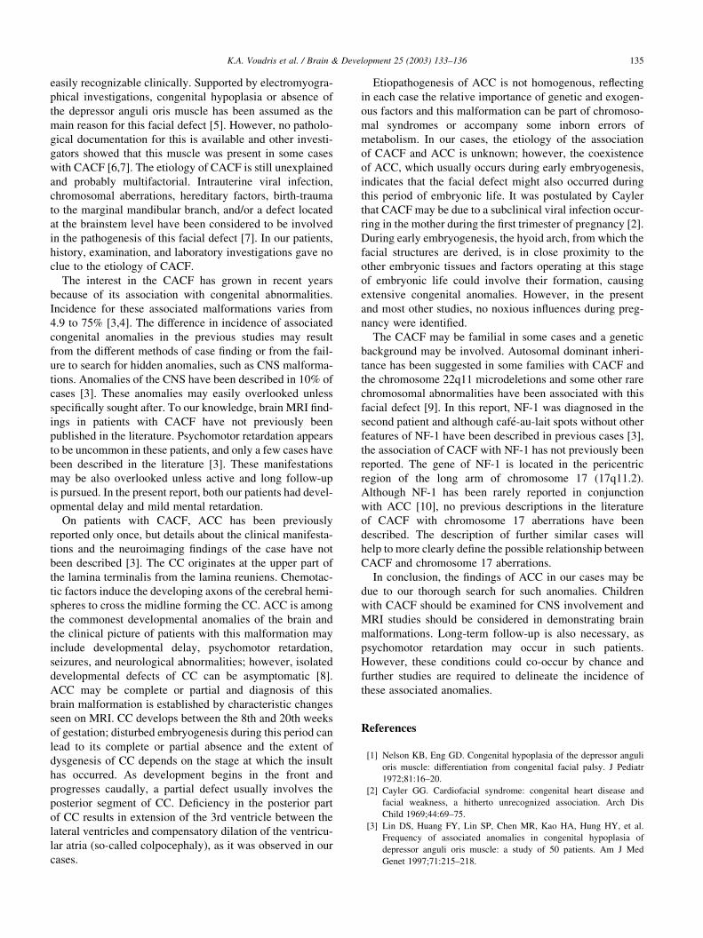
Fig. 1. (a) Midsagittal brain T1-weighted MRI of the first case showing
ACC, with a poorly formed rostrum, and radiating sulci and gyri. (b) Axial
brain T2-weighted MRI of the first case showing dilatation of the trigones.
Fig. 2. Midsagittal brain T1-weighted MRI of the second case showing
partial ACC, with a poorly formed genu and a small part of the body of
CC, and radiating sulci and gyri.
easily recognizable clinically. Supported by electromyogra-
phical investigations, congenital hypoplasia or absence of
the depressor anguli oris muscle has been assumed as the
main reason for this facial defect [5]. However, no patholo-
gical documentation for this is available and other investi-
gators showed that this muscle was present in some cases
with CACF [6,7]. The etiology of CACF is still unexplained
and probably multifactorial. Intrauterine viral infection,
chromosomal aberrations, hereditary factors, birth-trauma
to the marginal mandibular branch, and/or a defect located
at the brainstem level have been considered to be involved
in the pathogenesis of this facial defect [7]. In our patients,
history, examination, and laboratory investigations gave no
clue to the etiology of CACF.
The interest in the CACF has grown in recent years
because of its association with congenital abnormalities.
Incidence for these associated malformations varies from
4.9 to 75% [3,4]. The difference in incidence of associated
congenital anomalies in the previous studies may result
from the different methods of case finding or from the fail-
ure to search for hidden anomalies, such as CNS malforma-
tions. Anomalies of the CNS have been described in 10% of
cases [3]. These anomalies may easily overlooked unless
specifically sought after. To our knowledge, brain MRI find-
ings in patients with CACF have not previously been
published in the literature. Psychomotor retardation appears
to be uncommon in these patients, and only a few cases have
been described in the literature [3]. These manifestations
may be also overlooked unless active and long follow-up
is pursued. In the present report, both our patients had devel-
opmental delay and mild mental retardation.
On patients with CACF, ACC has been previously
reported only once, but details about the clinical manifesta-
tions and the neuroimaging findings of the case have not
been described [3]. The CC originates at the upper part of
the lamina terminalis from the lamina reuniens. Chemotac-
tic factors induce the developing axons of the cerebral hemi-
spheres to cross the midline forming the CC. ACC is among
the commonest developmental anomalies of the brain and
the clinical picture of patients with this malformation may
include developmental delay, psychomotor retardation,
seizures, and neurological abnormalities; however, isolated
developmental defects of CC can be asymptomatic [8].
ACC may be complete or partial and diagnosis of this
brain malformation is established by characteristic changes
seen on MRI. CC develops between the 8th and 20th weeks
of gestation; disturbed embryogenesis during this period can
lead to its complete or partial absence and the extent of
dysgenesis of CC depends on the stage at which the insult
has occurred. As development begins in the front and
progresses caudally, a partial defect usually involves the
posterior segment of CC. Deficiency in the posterior part
of CC results in extension of the 3rd ventricle between the
lateral ventricles and compensatory dilation of the ventricu-
lar atria (so-called colpocephaly), as it was observed in our
cases.
Etiopathogenesis of ACC is not homogenous, reflecting
in each case the relative importance of genetic and exogen-
ous factors and this malformation can be part of chromoso-
mal syndromes or accompany some inborn errors of
metabolism. In our cases, the etiology of the association
of CACF and ACC is unknown; however, the coexistence
of ACC, which usually occurs during early embryogenesis,
indicates that the facial defect might also occurred during
this period of embryonic life. It was postulated by Cayler
that CACF may be due to a subclinical viral infection occur-
ring in the mother during the first trimester of pregnancy [2].
During early embryogenesis, the hyoid arch, from which the
facial structures are derived, is in close proximity to the
other embryonic tissues and factors operating at this stage
of embryonic life could involve their formation, causing
extensive congenital anomalies. However, in the present
and most other studies, no noxious influences during preg-
nancy were identified.
The CACF may be familial in some cases and a genetic
background may be involved. Autosomal dominant inheri-
tance has been suggested in some families with CACF and
the chromosome 22q11 microdeletions and some other rare
chromosomal abnormalities have been associated with this
facial defect [9]. In this report, NF-1 was diagnosed in the
second patient and although cafe-au-lait spots without other
features of NF-1 have been described in previous cases [3],
the association of CACF with NF-1 has not previously been
reported. The gene of NF-1 is located in the pericentric
region of the long arm of chromosome 17 (17q11.2).
Although NF-1 has been rarely reported in conjunction
with ACC [10], no previous descriptions in the literature
of CACF with chromosome 17 aberrations have been
described. The description of further similar cases will
help to more clearly define the possible relationship between
CACF and chromosome 17 aberrations.
In conclusion, the findings of ACC in our cases may be
due to our thorough search for such anomalies. Children
with CACF should be examined for CNS involvement and
MRI studies should be considered in demonstrating brain
malformations. Long-term follow-up is also necessary, as
psychomotor retardation may occur in such patients.
However, these conditions could co-occur by chance and
further studies are required to delineate the incidence of
these associated anomalies.
References
[1] Nelson KB, Eng GD. Congenital hypoplasia of the depressor anguli
oris muscle: differentiation from congenital facial palsy. J Pediatr
1972;81:16–20.
[2] Cayler GG. Cardiofacial syndrome: congenital heart disease and
facial weakness, a hitherto unrecognized association. Arch Dis
Child 1969;44:69–75.
[3] Lin DS, Huang FY, Lin SP, Chen MR, Kao HA, Hung HY, et al.
Frequency of associated anomalies in congenital hypoplasia of
depressor anguli oris muscle: a study of 50 patients. Am J Med
Genet 1997;71:215–218.
K.A. Voudris et al. / Brain & Development 25 (2003) 133–136 135

[4] Lahat E, Heyman E, Barkay A, Goldberg M. Asymmetric crying
facies and associated congenital anomalies: prospective study and
review of the literature. J Child Neurol 2000;15:808–810.
[5] Renault F. Facial electromyography in newborn and young infants
with congenital facial weakness. Dev Med Child Neurol
2001;43:421–427.
[6] Monreal FJ. Asymmetric crying facies: an alternative interpretation.
Pediatrics 1980;65:146–149.
[7] Roedel R, Christen HJ, Laskawi R. Aplasia of the depressor anguli
oris muscle: a rare cause of congenital lower lip palsy? Neuropedia-
trics 1998;29:215–219.
[8] Marszal E, Jamroz E, Pilch J, Kluczewska E, Jablecka-Deja H,
Krawczyk R. Agenesis of corpus callosum: clinical description and
etiology. J Child Neurol 2000;15:401–405.
[9] Innes AM. Asymmetric crying facies and associated congenital
anomalies: the contribution of 22q11 microdeletions. J Child Neurol
2001;16:778.
[10] Atlas SW, Zimmerman RA, Bruce D, Schut L, Bilaniuk LT, Hackney
DB, et al. Neurofibromatosis and agenesis of the corpus callosum in
identical twins: MR diagnosis. AJNR Am J Neuroradiol 1988;9:598–
601.
K.A. Voudris et al. / Brain & Development 25 (2003) 133–136136
Recommended