COMPARATIVE STUDY BETWEEN LIQUID BASED
CYTOLOGY AND CONVENTIONAL SMEAR IN FNA
SAMPLES OF BREAST LESIONS
Dissertation submitted in
Partial fulfillment of the regulations required for the award of
M.D.Degree
in PATHOLOGY – BRANCH III
April 2017
THE TAMILNADU Dr. M.G.R. MEDICAL UNIVERSITY CHENNAI,
TAMILNADU
DECLARATION
I solemnly declare that the dissertation entitled COMPARATIVE
STUDY BETWEEN LIQUID BASED CYTOLOGY AND CONVENTIONAL
SMEAR IN FNA SAMPLES OF BREAST LESIONS was done by me in the
Department of Pathology at Coimbatore Medical College, Coimbatore during
the period of July 2015 to July 2016 under the guidance and supervision of DR.
C. LALITHA, MD., Professor and Head, Department of pathology,
Coimbatore Medical College, Coimbatore.
This dissertation is submitted to the Tamilnadu Dr. M.G.R. Medical
University, Chennai towards the partial fulfillment of the requirement for the
award of M.D., Degree in Pathology.
Place:
Date: Dr.R.KOUSALYA

CERTIFICATE
This is to certify that the dissertation entitled COMPARATIVE STUDY
BETWEEN LIQUID BASED CYTOLOGY AND CONVENTIONAL
SMEAR IN FNA SAMPLES OF BREAST LESIONS is a record of bonafide
work done by Dr.R.Kousalya, post graduate student in the Department of
Pathology, Coimbatore Medical College and Hospital, Coimbatore under the
supervision and guidance of Dr.C.Lalitha, M.D., Professor and Head,
Department of Pathology, Coimbatore medical college and Hospital,
Coimbatore in partial fulfillment of the regulations of the Tamilnadu Dr.M.G.R.
Medical University, Chennai towards the award of M.D. Degree (Branch III) in
Pathology.
Dr.Edwin Joe, M.D.,B.L., Dr.C.Lalitha, M.D.,
Dean, Coimbatore Medical College Professor and Head,
Coimbatore. Department of Pathology,
Coimbatore Medical College,
Coimbatore.

ACKNOWLEDGMENT
To begin with, I thank almighty GOD in making this project a successful
one.
I wish to express my sincere gratitude to the honourable Dean Dr.Edwin
Joe, M.D.,B.L., Coimbatore medical college, Coimbatore for permitting me to
conduct this study.
It’s a great pleasure to express my humble gratitude to the most
respectable teacher and my guide Dr.C.Lalitha, M.D., Professor and Head,
Department of Pathology, Coimbatore medical college, Coimbatore for her able
guidance and support. This dissertation bears her valuable suggestions and
highly professional advice.
I thank Professor Dr.A.Arjunan, M.D., all Associate Professor and
Assistant Professors and Tutors of Pathology department, Coimbatore medical
college, Coimbatore for their encouragement and valuable opinion.
I extend my heartfelt thanks to all my colleagues for their timely help,
comments and support.
I thank all the technicians for the kind cooperation.
I sincerely thank all the patients who consented to participate in the study.
I thank my lovable parents Mrs and Mr. Rajasekar, and my lovable
brothers R.Partheeban, R.Arun Kumar for their extreme patience and their
support while pursuing this study.

CONTENTS
SI.NO PARTICULARS
PAGE.NO
1.
INTRODUCTION 1
2.
AIM AND OBJECTIVES 4
3.
REVIEW OF LITERATURE 5
4.
MATERIALS AND METHODS 30
5.
OBSERVATION AND RESULTS 37
6.
DISCUSSION 62
7.
SUMMARY 74
8.
CONCLUSION 76
9.
BIBLIOGRAPHY
10.
APPENDICES
Annexure – I (Profoma)
Annexure – II (Consent form)
Annexure – III (Master chart)
Annexure – IV (Glossary)

LIST OF TABLES
SI.NO
CONTENTS
PAGE.NO
1. Total number of cases done in Conventional method and
Liquid based cytology method.
37
2. Comparison of cellularity between Liquid based method
and Conventional method.
40
3. Comparison of background material (blood, cell debris)
between Liquid based and Conventional method
41
4. Comparison of Informative background between Liquid
based method and Conventional method.
42
5. Comparison of monolayer arrangement between Liquid
based method and Conventional method.
43
6. Comparison of Cell architecture recognized by Liquid
based method and Conventional method.
44
7. Comparison of Nuclear detail between Liquid based
method and Conventional method
45
8. Comparison of cytoplasmic details between Liquid based
method and Conventional method.
46
9. Comparison of liquid based and Conventional method in
Bengin lesions.
47
10. Comparison of liquid based method and conventional
methods in malignant lesions.
48
11. Comparison of Liquid based method and Conventional
method in Fibroadenoma (FA) cases.
49
12. Comparison of Liquid based method and Conventional
method in Fibrocystic disease of breast (FCD) cases.
50
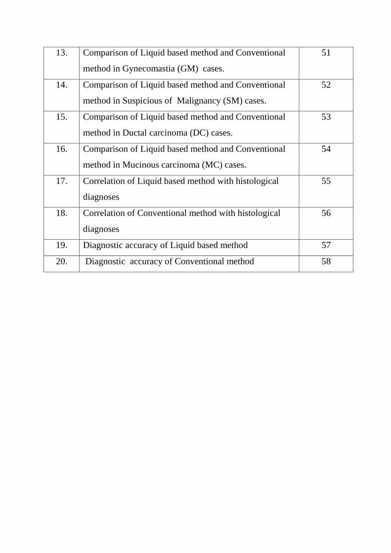
13. Comparison of Liquid based method and Conventional
method in Gynecomastia (GM) cases.
51
14. Comparison of Liquid based method and Conventional
method in Suspicious of Malignancy (SM) cases.
52
15. Comparison of Liquid based method and Conventional
method in Ductal carcinoma (DC) cases.
53
16. Comparison of Liquid based method and Conventional
method in Mucinous carcinoma (MC) cases.
54
17. Correlation of Liquid based method with histological
diagnoses
55
18. Correlation of Conventional method with histological
diagnoses
56
19. Diagnostic accuracy of Liquid based method 57
20. Diagnostic accuracy of Conventional method 58
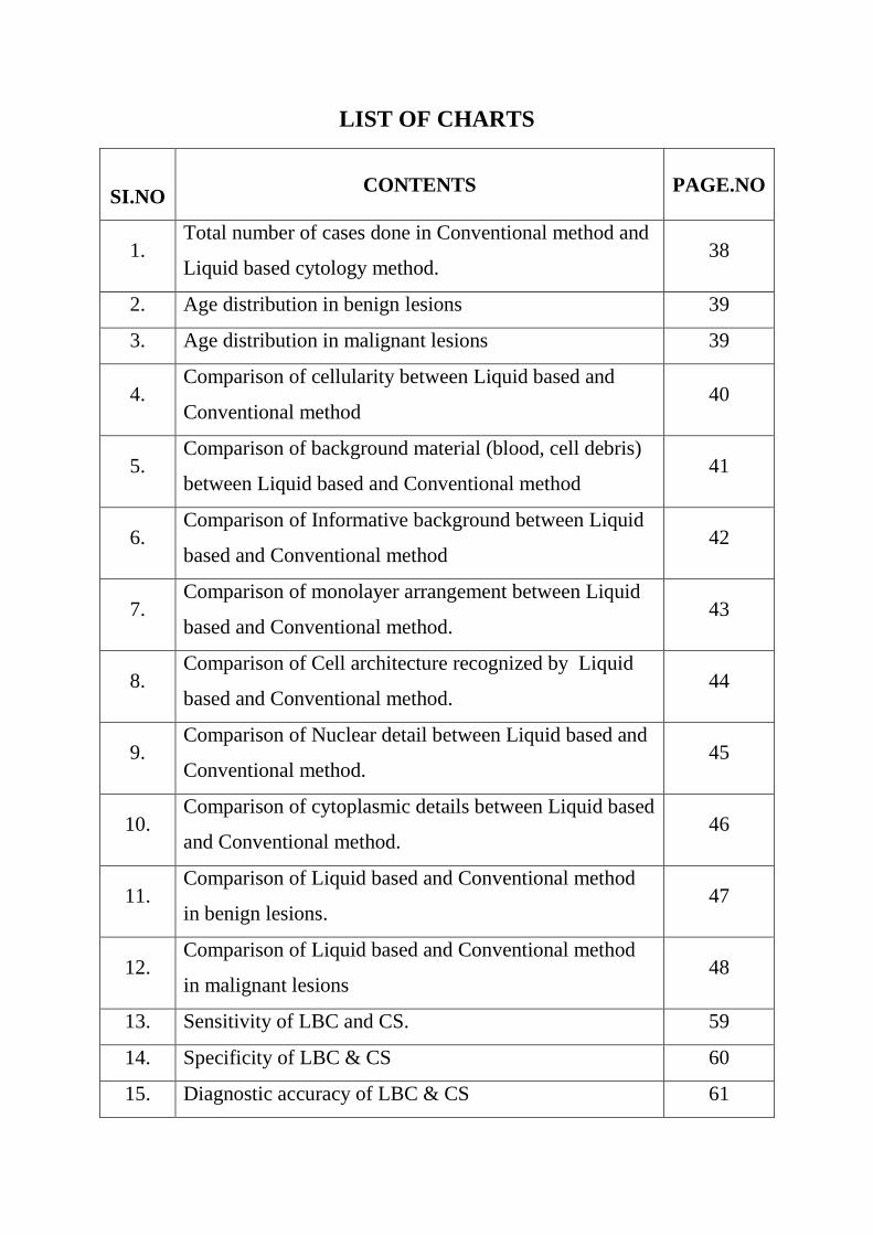
LIST OF CHARTS
SI.NO
CONTENTS
PAGE.NO
1. Total number of cases done in Conventional method and
Liquid based cytology method. 38
2. Age distribution in benign lesions 39
3. Age distribution in malignant lesions 39
4. Comparison of cellularity between Liquid based and
Conventional method 40
5. Comparison of background material (blood, cell debris)
between Liquid based and Conventional method 41
6. Comparison of Informative background between Liquid
based and Conventional method 42
7. Comparison of monolayer arrangement between Liquid
based and Conventional method. 43
8. Comparison of Cell architecture recognized by Liquid
based and Conventional method. 44
9. Comparison of Nuclear detail between Liquid based and
Conventional method. 45
10. Comparison of cytoplasmic details between Liquid based
and Conventional method. 46
11. Comparison of Liquid based and Conventional method
in benign lesions. 47
12. Comparison of Liquid based and Conventional method
in malignant lesions 48
13. Sensitivity of LBC and CS. 59
14. Specificity of LBC & CS 60
15. Diagnostic accuracy of LBC & CS 61
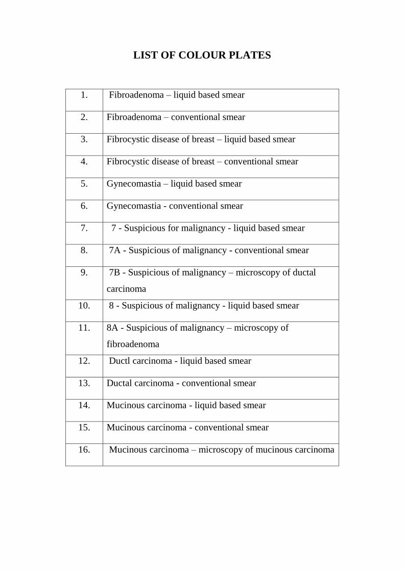
LIST OF COLOUR PLATES
1. Fibroadenoma – liquid based smear
2. Fibroadenoma – conventional smear
3. Fibrocystic disease of breast – liquid based smear
4. Fibrocystic disease of breast – conventional smear
5. Gynecomastia – liquid based smear
6. Gynecomastia - conventional smear
7. 7 - Suspicious for malignancy - liquid based smear
8. 7A - Suspicious of malignancy - conventional smear
9. 7B - Suspicious of malignancy – microscopy of ductal
carcinoma
10. 8 - Suspicious of malignancy - liquid based smear
11. 8A - Suspicious of malignancy – microscopy of
fibroadenoma
12. Ductl carcinoma - liquid based smear
13. Ductal carcinoma - conventional smear
14. Mucinous carcinoma - liquid based smear
15. Mucinous carcinoma - conventional smear
16. Mucinous carcinoma – microscopy of mucinous carcinoma

1
INTRODUCTION
Breast lesions can be of various types from inflammatory to malignant.
Some lesions are common in younger age group while others are more common
in elderly age group.
Benign lesions of the breast are the most common lesions which account
for 90% of the clinical presentation related to breast1. Patients present with
palpable breast lump and pain2. Breast lumps besides creating anxiety may
result in carcinoma and deformity3. Breast tissue is under hormonal influence
which causes changes in breast, present throughout life4. Fibroadenoma of the
breast is the common cause of benign breast lump5. Breast cancer is the frequent
cancer in women worldwide. Incidence rate differs worldwide from 27/100000
females in Eastern Africa to 96/100000 females in Europe.
Breast cancer is common among women in India according to National
cancer registry programme 2011 report. A woman has, one in eight chance of
developing breast cancer during her lifetime. By the year 2030 global burden of
breast cancer will be more than two million every year.
In India the incidence of carcinoma of breast is increasing and the
mortality rate for breast cancer in India is 11.1 per 10,000. Overall, breast
nodules are more common in women.
2
Fine-needle aspiration cytology is a safe and cost-effective, first line
diagnostic tool in diagnosing breast lumps. FNAC helps in reducing the number
of unnecessary surgeries in benign breast lesions.
For many years, conventional smears have been regarded as the gold
standard technique for diagnosing breast lesions in cytology. From the aspirated
material, smears of variable number are prepared. It consumes time and has
been tedious for the cytologists to screen the slides. Technical aspects add to the
problem, which include improper smear preparation and fixation leading to poor
preservation of cellular details. Thick smears, cellular overlapping and
obscuring inflammatory infiltrate all interfere in reporting the smears.
To overcome the above disadvantages posed by conventional method,
the Liquid-based technique has been used with increasing frequency worldwide
in most of the centers for gynecological as well as non-gynecological samples.
Two systems named Thin-prep and Sure-path approved by US Food and Drug
Administration (FDA) are commonly used. Both represent first generation
liquid based cytology systems, and they consist of automated equipments,
filters, plastic containers, and vacuum devices. When compared with
conventional smear, the cost of preparing slides using the above two systems
was increased to a greater extent, making the improved method potentially
inaccessible.
3
Liquid-PrepTM, the second generation technique is a simpler one. It
requires low cost, because most of the automated machines and devices are not
used in this liquid based cytology system. It also enhances clear visualization
due to monolayer spread of cells.
The objective of the investigator in this study, is to use a commonly
available instrument (centrifuge), and to prepare slides from fine needle
aspirates. The results are interpreted using standard morphologic criteria
proposed for liquid based smears.
Finally, the left over liquid based sample can be used for ancillary tests
such as immunocytochemistry and molecular studies. Cell block could be
prepared as well from them. However, in this study they are not included.

4
AIM AND OBJECTIVES
AIM:
To compare liquid based cytology technique with conventional smear
method and to correlate with histopathological diagnosis.
OBJECTIVES:
To compare the cyto-morphological features of manual liquid based
preparation with conventional cytology in breast lesions using FNA
technique.
To compare the diagnostic value of Liquid based method with
Conventional one.
To correlate with final histopathological diagnosis whenever available.

5
REVIEW OF LITERATURE
Over the past three decades, in the work-up of breast lesions, fine needle
aspiration has been commonly used as first line diagnostic tool. It is widely
accepted as simple, safe and cost effective and helps in selecting the patients for
surgical excision instead of managing them clinically6 .
A low false-negative rate (FNR) and high true positive rate (TPR)
indicates the success of breast fine needle aspiration. A low FNR depends upon
the high quality of samples that are procured by technically-skilled persons to
prepare slides that are representative of the lesion with sufficiently good
quantity and quality for the interpretation of results. A high TPR depends upon
the accurate interpretations using established criteria for sample adequacy and
cellular morphology7,8
The smear can be prepared by Conventional method or can be processed
by either of the following methods - liquid preparations or cytospin8,10,45
.
For years, conventional smears (CS) have been the gold standard
technique in diagnosing breast lesions. The main drawback with conventional
smear is bloody background, which hinders the evaluation of breast ductal cells;
it also requires some skill. Finally slide transportation can be an issue6,9
.

6
In 1996, to overcome the problems faced by conventional method, a newer
technique called liquid based cytology has been applied in the cytological
sample collection and preparation. This method originally developed for
cervical cytology smears10
, has gradually been applied to non gynaecological
specimens7,11,12
and especially to aspiration cytology samples with better
outcome.
In LBPs, instead of smearing, the sample is collected are rinsed in
preservative medium and processed in automated or semi-automated machines
or they are processed manually 12
.
A newer LBC method, liquid-PREP (LP) was introduced in the recent
years because of low cost when compared with the older liquid based
method13,14
.
A decreased non-diagnostic rate and an increased rate of accurate diagnosis
were observed with conventional smears in some studies. More comparable or
better results with liquid-based methods were obtained in later studies.
7
ADVANTAGES OF LBP 6,10
Liquid based preparations are considered best alternative method to
conventional smear because of its easy processing technique, faster
screening time and other interpretation advantages. They include:
Procedure of collecting the sample is uniform and 100%; collection can be
done by cytologists or by the clinicians;
Hazards of handling needle while preparing conventional smears are
minimized;
Transportation even from remote places to the diagnostic centre is easy;
The technique can be done in automated (ThinPrep) or semi-automated
(SurePath) equipments or can be processed manually (Liquid-prep);
Air drying artifact is avoided and the morphology is preserved well due to
immediate fixation in the liquid based solution;
Processing technique is standardized and is uniform which gives an enriched
cell sample with uniform distribution of cells
Enables the cytologists to examine less number of slides for each case.
Easier for the cytologists to interpret liquid based slides because the smear is
spread has bloodless background and the cells are spread in monolayer.
The time required for interpretation is less;

8
Leftover sample can be stored at room temperature for few months
(average-six months). This provides the chance of making additional slides
or cell block and immunohistochemistry/ molecular studies15,16,17,18,19
to be
performed at a later date.
Many studies reported that the diagnostic accuracy of liquid based technique
has improved or remained equal to that of conventional method; almost all
acknowledge that liquid based preparations produced cytological changes in
the morphology of cells.
With all the advantages discussed above, the cyto-pathologist should be
aware and be familiar with the minor cyto-morphologic alterations produced
by LBPs, although the architectural features are maintained6,20
.
ALTERATIONS RECOGNISED IN LBP
The alterations noted in liquid based preparations are in the cellularity,
distribution of cells, cellular architecture, morphology and the background
elements7. Michael et al (2000)
21 stated that these alterations are attributed by
processing techniques involved in preparing the liquid based slides.
Cellularity
A high cell yield with minimum loss of cells can be obtained when the
aspiration sample is processed wholly in liquid based technique. This can be
achieved when the sample is collected through a special pass21
.
9
Background Elements
Liquid based preparations provide a clean background which is achieved by
adding lytic agents to the sample that reduces the background blood cells. This
has been quoted in almost all reference studies.
Architecture
LBP usually retains the architectural patterns like syncytial cell clusters.
However, apparent discohesion with more single cells are noted with breakage
of large sheets. The cell dispersion is due to the processing technique involved
in liquid based preparations.
Distribution of cells
The cell distribution is almost uniform, as thin monolayer with minimal
overlapping21,22
.
Cellular morphology
Morphology of cells are well preserved and are seen enhanced in liquid
based preparations21,22
. The cell shape and the nuclear details are usually
retained but the nucleus appears slightly shrunken in liquid based preparations.
Nuclear features like pleomorphism, irregular nuclear membrane, chromatin
appearance are better visualized21,22,23
. Cytoplasm looks denser and is readily
seen in lymphocyte21
. The artefacts introduced by liquid based preparation
emphasizes the need to develop better experience in reporting the liquid based

10
smears to avoid diagnostic errors. These artifacts have also been observed in
few of the studies done on fine needle aspiration specimens as well as with
other non-gynecologic samples10,11,12
.
ANATOMY OF NORMAL BREAST 24
The female breast is modified sweat glands. Nonlactating adult breast
contains glandular portion. It is made up of clusters of small secretory lobules.
These lobules connected to main excretory duct. The stroma is made up of
loose connective tissue and fat. After menopause these glandular portion
undergoes atrophy.
Each acinus or lobule in the resting state is composed of small cuboidal
cells. The small ducts, lined by small cuboidal cells, with an outer layer of
myoepithelial cells. The large lactiferous ducts are lined by one to two layers of
cuboidal cells. The lining of the smaller ducts may undergo apocrine
metaplasia. Nipple is made up of thick epidermis. The male breast contains
sparse duct, scarce fat and connective tissue. The lobular apparatus is absent in
male breast.
Normal cytology 6,20,24
Normal breast is difficult to aspirate. In conventional cytologic
preparations, normal breast usually shows scant cellularity cohesive ductal
11
fragments with uniform round nuclei with dense chromatin and small
inconspiscous nucleoli.
The breast ductal epithelial cells in the liquid based preparations appear
as small clusters with three dimensional arrangement. The cells are round with
scant basophillic cytoplasm. Nucleus look small, regular and round with
clumped chromatin. There is elimination of obscuring elements such as blood,
excessive inflammation and cellular debris.
DIAGNOSTIC TERMINOLOGY 46
Accurate diagnosis of breast lesions depend upon triple assessment
approach. It has clinical, imaging and pathologic examination. Fine needle
aspiration cytology is widely adopted for pathologic assessment because of the
accuracy and ease of use. In literature, few of the classification schemes are
recommended in the reporting of breast aspiration cytology. Each one of them
was based on their individual or institutional experience and also on clinical
organisation. Perceptions of these diagnostic terminology and reporting
according to the classification scheme, lead to disordence between the clinician
and cytopathologist which altered the patient management.
The most commonly used reporting system is “Five tier method of
reporting system” (C1-C5). Categories ranges from insufficient materials (C1),
benign (C2), atypical(C3), suspicious of malignancy (C4), frankly malignant

12
(C5). This categorisation was initiated by the NATIONAL COORDINATING
COMMITTEE FOR BREAST SCREENING and UK NATIONAL BREAST
SCREENING PROGRAM. Diagnostic Terminology Reporting terminology.
The M.D. Anderson Cancer Center proposal. The category also includes the
following points
Adequacy/diagnostic category
Specimen adequacy
Satisfactory for evaluation.
Insufficient for evaluation - scant cellularity (<4–6 cell groups).
Unsatisfactory for evaluation - distortion artifact, obscuring blood
(1) Benign lesions
Specification of lesion (e.g., fibroadenoma, adenosis, FCD)
(2) Proliferative breast lesion
A. Presence of cytological atypia (e.g., crowded, pleomorphic nucleus, loss of
cohesion, hyperchromasia)
B. Architectural pattern.
a. Ductal hyperplasia
b. Atypical hyperplasia/low-grade carcinoma in situ
(3)Suspicious for carcinoma
- Insufficient cellularity.
13
- Benign ductal elements with low grade carcinoma in same slide.
(4) Malignant
- specification of type (e.g., ductal, lobular, mucinous)
- Other types (sarcoma, metastasis, lymphoma etc.)
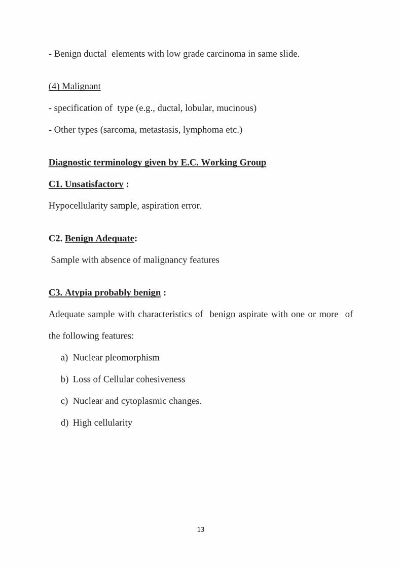
Diagnostic terminology given by E.C. Working Group
C1. Unsatisfactory :
Hypocellularity sample, aspiration error.
C2. Benign Adequate:
Sample with absence of malignancy features
C3. Atypia probably benign :
Adequate sample with characteristics of benign aspirate with one or more of
the following features:
a) Nuclear pleomorphism
b) Loss of Cellular cohesiveness
c) Nuclear and cytoplasmic changes.
d) High cellularity
14
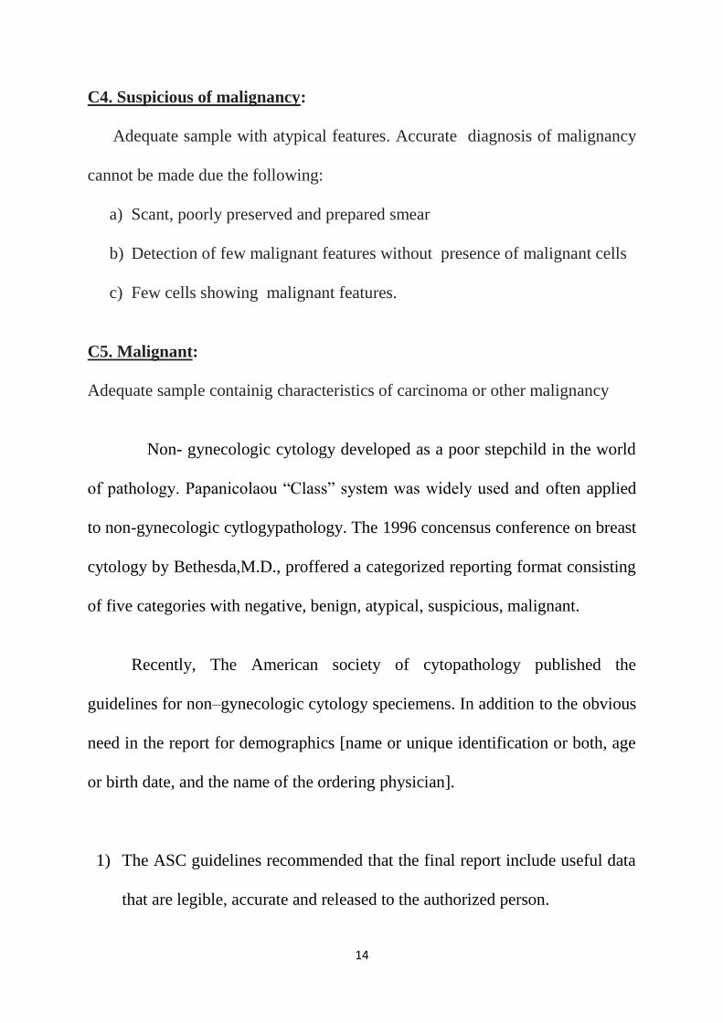
C4. Suspicious of malignancy:
Adequate sample with atypical features. Accurate diagnosis of malignancy
cannot be made due the following:
a) Scant, poorly preserved and prepared smear
b) Detection of few malignant features without presence of malignant cells
c) Few cells showing malignant features.
C5. Malignant:
Adequate sample containig characteristics of carcinoma or other malignancy
Non- gynecologic cytology developed as a poor stepchild in the world
of pathology. Papanicolaou “Class” system was widely used and often applied
to non-gynecologic cytlogypathology. The 1996 concensus conference on breast
cytology by Bethesda,M.D., proffered a categorized reporting format consisting
of five categories with negative, benign, atypical, suspicious, malignant.
Recently, The American society of cytopathology published the
guidelines for non–gynecologic cytology speciemens. In addition to the obvious
need in the report for demographics [name or unique identification or both, age
or birth date, and the name of the ordering physician].
1) The ASC guidelines recommended that the final report include useful data
that are legible, accurate and released to the authorized person.
15
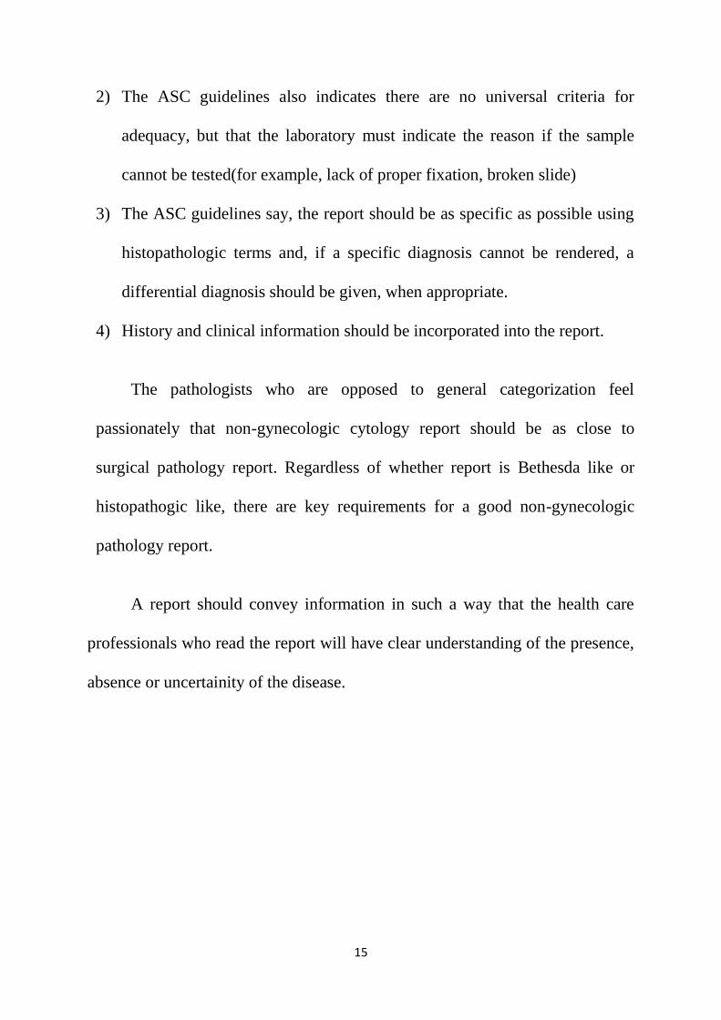
2) The ASC guidelines also indicates there are no universal criteria for
adequacy, but that the laboratory must indicate the reason if the sample
cannot be tested(for example, lack of proper fixation, broken slide)
3) The ASC guidelines say, the report should be as specific as possible using
histopathologic terms and, if a specific diagnosis cannot be rendered, a
differential diagnosis should be given, when appropriate.
4) History and clinical information should be incorporated into the report.
The pathologists who are opposed to general categorization feel
passionately that non-gynecologic cytology report should be as close to
surgical pathology report. Regardless of whether report is Bethesda like or
histopathogic like, there are key requirements for a good non-gynecologic
pathology report.
A report should convey information in such a way that the health care
professionals who read the report will have clear understanding of the presence,
absence or uncertainity of the disease.
16
Speciemen adequacy for breast fine needle aspiration cytology
Solid lesions
There is no specific requirement.
Sample adequacy depends upon aspiration.
Benign breast lesions
The amount of epithelial cells present has to be reported.
Individual laboratory may consider specific cell count, as their own criteria.
Cystic lesions
There is no minimal criteria for cell count.
If the fluid is thin, watery, and not bloody, the fluid is examined or discarded at
the aspirators decision, provided the FNA completely evacuates the cyst and
there is no residual palpable mass left.
Any residual mass or nodule requires FNA from the residual mass
Cysts with brown reddish fluid (not related to trauma of the FNA) require
careful evaluation or further workup.
BENIGN CONDITIONS :
Fibrocystic Changes
Fibroadenoma
Fat Necrosis
Pregnancy and Lactational Changes
Mastitis
17
Radiation Change
Gynecomastia
PAPILLARY NEOPLASMS
PHYLLODES TUMOR
Breast carcinomas
Ductal Carcinoma (invasive)
Lobular Carcinoma (invasive)
Mucinous Carcinoma
Medullary Carcinoma
Metaplastic Carcinomas
Tubular Carcinoma
UNCOMMON BREAST TUMORS
Sarcoma
Apocrine Carcinoma
NHL
Adenoid Cystic Carcinoma
METASTATIC TUMORS
18
Benign breast lesions:
Fibrocystic changes:
One of the common breast lesion is fibrocystic change. It is composed of
small cysts, large cysts, focal fibrosis, apocrine metaplasia, adenosis, intraductal
hyperplasia. Moderate amount of ductal hyperplasia is seen. Two types of
fibrocystic changes are seen proliferative and non proliferative. This is based on
presence of ductal hyperplasia24,47
Cytological features:
Clusters of cells without nuclear overlap with fine granular chromatin and
inconspiscous nucleoli.
Fibroadenoma:
Fibroadenoma, the most common benign tumor of the female breast. Can occur
in any age group. They are solitary, well circumscribed lesion, which is freely
mobile and rubbery due to stromal and glandular proliferation. They can occur
as multiple lesions.
Cytological features of fibroadenoma:
• Hypercellular lesions, with large sheets of three-dimensional clusters. Two cell
population epithelial and myoepithelial. Bipolar cells with oval nuclei. Stromal
fragments are fibrillar. Nuclear atypia is present. Epithelial cohesion is lost.
Nuclear spacing is regular. Chromatin is finely granular. Nucleolus is small and
round 24
.
19
Generally both conventional smears and liquid based preparations show similar
cytological features with minor differences, which includes
1. Benign appearing ductal cells in LBPs are arranged in flat sheets, clusters
or aggregates. They are small uniform in size with rare small nucleoli.
Myoepithelial cells are present admixed with the benign epithelial cells.
2. Compared to conventional smears, liquid based preparations have decreased
or absent stromal components.
Many authors observed loss of stromal components as limitations of
thinprep. Dey et al (2000) & Ali et al (2004) observed similar features as in
conventional smears like cells arranged in staghorn clusters, isolated
myoepithelial cells and stromal fragments.
Mygdakos et al (2009) stated that stromal elements were reduced or
absent, but the diagnosis of fibroadenoma is made based on the features like
ductal cell aggregates and bipolar cells.
Michael et al (2000), Leung et al (1997) & Perez-Reyes et al (1994)
observed that cells are arranged in small aggregates, with decrease in
myoepithelial cells and paucity or loss of stromal fragments.
Pregnancy, Lactational Changes:
Pregnancy and lactation causes hyperplasia of terminal lobular unit.

20
Cytological features of pregnancy and lactational changes:
Smears are moderately cellular. Smear contains many isolated epithelial
cells. Nucleus is enlarged with no change in size and shape. Prominent
nucleolus is seen. Cytoplasm is abundant and finely vacuolated. Proteinaceous
foamy material is seen in the background. Numerous naked nuclei are seen.
The lactational adenoma may be confused with ductal carcinoma but, carcinoma
do not have foamy background 6,24,26,35
.
Fat Necrosis:
Fat necrosis can mimic carcinoma. Most common in patients with
previous history of biopsy and trauma to the breast.
Cytological features of fat necrosis:
Hypocellular smears.
Contains many histiocytes.
Round to kidney-bean shaped nucleus.
Low N/C ratio.
Multinucleated, atypical cells are seen.
Neutrophils, plasma cells and lymphocytes seen in the background28,29
.
21
Radiation Changes:
Cytological features of radiation changes:
Hypocellular smears with low N/C ratio. Hyperchromatic nucleus with
prominent nucleoli. Cytoplasmic vacuolization is seen 30
.
Mastitis:
Acute mastitis:
Bacterial infection is the most common cause.
Cytological features of acute mastitis:
Numerous neutrophils with ductal cells showing reactive changes.
Chronic mastitis:
It is due to complication of acute mastitis. Etiology is unknown31
.
Cytological features of chronic mastitis:
Cellular smears.
Inspissated ducts produce amorphous granular debris.
Lymphocytes, plasma cells are common inflammatory infiltrate.
Both acute and chronic mastitis on LBC preparations are classified based
on the inflammatory cells.
22
Kalpalata Tripathy et al (2015) described that the chronic mastitis is
diagnosed on LBC preparations due to clarity of the nuclear features and also
the presence of inflammatory cells.
Granulomatous mastitis:
The term granulomatous lobular mastitis is a clinical syndrome of
unknown etiology. Most common in pregnancy age group 31
.
Cytological features of granulomatous mastitis:
Clusters of epithelioid histiocytes.
Cytoplasm is vacuolated.
Folded or round nucleus.
Dispersed chromatin.
Nucleoli is large.
Lymphocytes, eosinophils, plasma cells and giant cells are seen in the
stroma.
Subareolar Abscess:
It is also known as “recurring subareolar abscess”. It arises in areola due
to squamous metaplasia of lactiferous ducts. It is also due to keratin plugging,
rupture and dilatation of ducts. If lesions are not completely exicised, it will
recur.
23
Cytological features of subareolar abscess:
Anucleate squames are many with histiocytes, neutrophils and
multinucleated giant cells 32
. Granulation tissue fragments are noted.
Gynecomastia:
Gynecomastia is a common lesion of male breast. It is diffuse or nodular
enlargement of breast and is frequently bilateral.
Cytological features of gynecomastia:
Most commonly resembles Fibroadenoma. Cellularity is variable. Ductal
cells arranged in groups with small, oval nucleus and scant cytoplasm 33
.
Isolated bipolar cells are seen.
Papillary Neoplasms:
They are solitary tumors. They arise in subareolar lactiferous ducts. The
common presentation is bloody nipple discharge. So nipple discharge cytology
should be done. Papillary carcinoma represents 1% to 2% of breast carcinomas.
It may be cystic or solid, invasive or noninvasive. The prognosis is favourable.
The distinction between malignant and benign are difficult to diagnose in fine
needle aspiration. Papillary carcinomas show, singly scaterred columnar cells.
Sclerosing papillary lesions may mimic as malignant lesion in fine needle
aspiration 32
. Therefore excision biopsy should be done to confirm whether the
lesion is benign or malignant.
24
Cytological features of benign papillary neoplasm:
Smears are moderate to high cellular. Cells are arranged in three-
dimensional papillary groups with fibrovascular core. Cuboidal to columnar
cells. Nucleus is round to oval. Chromatin is finely granular32
.
Cytological features of malignant papillary neoplasm :
Smears are moderate to markedly cellular. Cells are arrangend in
papillary pattern, cribriform, tubular pattern. Absence of myoepithelial cells.
Tall columnar cells are commonly seen with elongated uniform nuclei 24
. Many
naked nuclei and blood and hemosiderin-laden macrophages seen.
Phyllodes Tumor:
It is a biphasic tumour. It has epithelial and stromal component with
increased stromal cellularity. The incidence is less than 1% of breast tumors.
Many grow as massive masses most commonly infiltrating the skin. They can
be classified as benign, borderline and malignant.
Cytological features of phyllodes tumor:
Cytological features are similar to fibroadenoma but phyllodes tumor is
more cellular with more cellular stromal component24,35
. Epithelial atypia also
noted and it will mimics like carcinoma 34
.
25
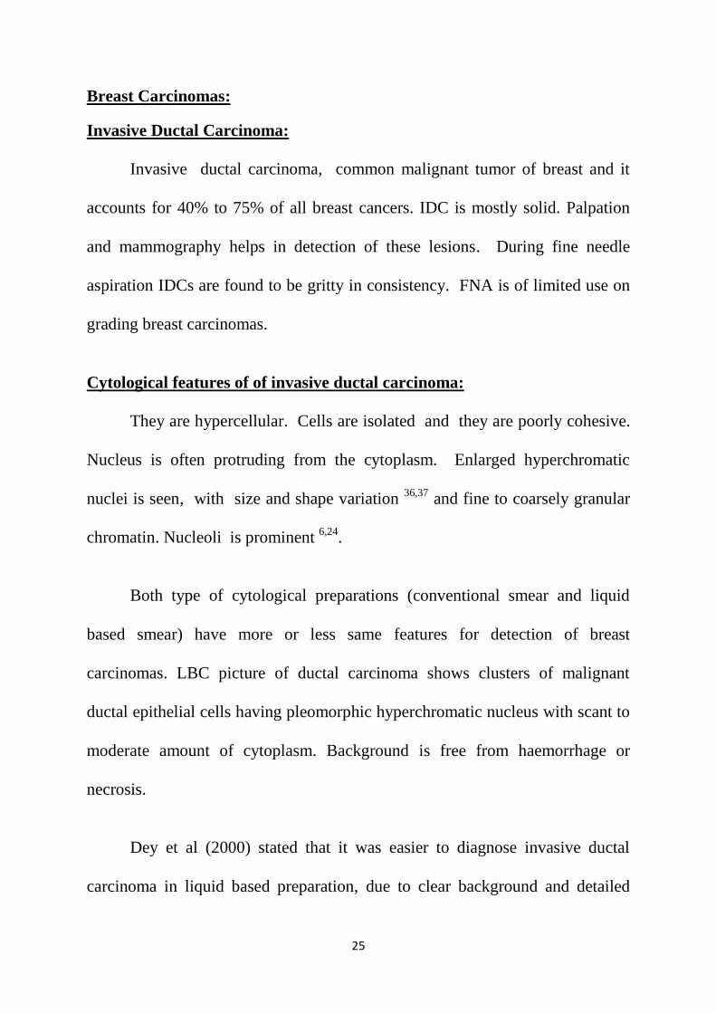
Breast Carcinomas:
Invasive Ductal Carcinoma:
Invasive ductal carcinoma, common malignant tumor of breast and it
accounts for 40% to 75% of all breast cancers. IDC is mostly solid. Palpation
and mammography helps in detection of these lesions. During fine needle
aspiration IDCs are found to be gritty in consistency. FNA is of limited use on
grading breast carcinomas.
Cytological features of of invasive ductal carcinoma:
They are hypercellular. Cells are isolated and they are poorly cohesive.
Nucleus is often protruding from the cytoplasm. Enlarged hyperchromatic
nuclei is seen, with size and shape variation 36,37
and fine to coarsely granular
chromatin. Nucleoli is prominent 6,24
.
Both type of cytological preparations (conventional smear and liquid
based smear) have more or less same features for detection of breast
carcinomas. LBC picture of ductal carcinoma shows clusters of malignant
ductal epithelial cells having pleomorphic hyperchromatic nucleus with scant to
moderate amount of cytoplasm. Background is free from haemorrhage or
necrosis.
Dey et al (2000) stated that it was easier to diagnose invasive ductal
carcinoma in liquid based preparation, due to clear background and detailed
26
nuclear features of the neoplastic cells. However clean background means an
uninformative background because, traditional diagnostic clues associated with
malignancy like blood and necrotic material are lost in liquid based
preparations.
Ryu et al (2013) found that there are remarkable differences of nuclear
features in breast carcinomas processed by liquid based cytology in comparison
to conventional smear, including more prominent nucleoli, hyperchromasia and
less coarse chromatin.
Michael et al (2000), Dey et al (2000) stated that large clusters of cells
are reduced to smaller aggregates. Ryu et al (2013) described that three
dimensional clusters are more common in liquid based preparations in contrast
with Michael et al (2000) which revealed the more common flattened cell
aggregates present in TP slides.
Mygdakos et al (2009), Kalpalata Tripathy et al (2015), Gerhard et al
(2014) described that ductal carcinoma can easily be diagnosed by liquid based
preparation than that of conventional smear because of clean background and
better nuclear features.
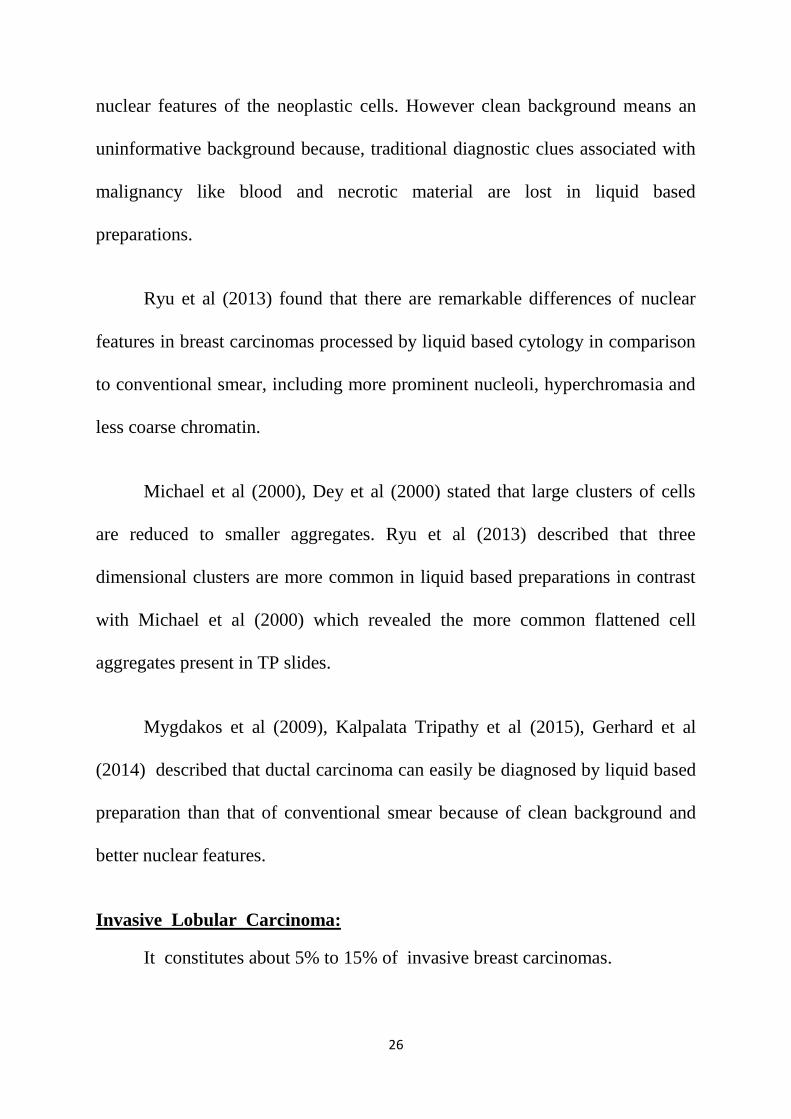
Invasive Lobular Carcinoma:
It constitutes about 5% to 15% of invasive breast carcinomas.

27
Cytlogical features of invasive lobular carcinoma:
Smears are hypocellular due to stromal fibrosis. Cells arranged in singles,
small groups and linear arrays. The cells are small to medium sized with
cytoplasmic vacuole 37
. The nucleus is hyperchromatic with small nucleolus.
Medullary Carcinoma:
Incidence of medullary carcinoma is 1% to 7% of breast tumors.
Cytological features of medullary carcinoma:
Smears are hypercellular with isolated cells and loose clusters.
Macronucleolus is more prominent and irregular 37
. Numerous mitoses are seen.
Cytoplasm is granular. Many lymphocytes are seen.
Mucinous Carcinoma (colloid ):
Incidence of mucinous carcinoma is 2% of invasive breast. Distinction
between mucinous carcinoma and IDC with mucinous change is not possible
with help of FNA.
Cytological features of mucinous carcinoma:
Cohesive tight clusters of cells with three-dimensional balls like
arrangement 24,37
. Capillary structures are branched. Nucleus is uniform with
small cytoplasmic vacuolisation.

28
Mucinous carcinoma cannot be diagnosed with the help of LBC because
the important feature of mucinous carcinoma, the mucoid background is lost
during liquid based preparation.
Michael et al (2000) described that mucinous carcinoma and low grade
carcinomas(invasive and in situ ductal carcinomas, tubular and lobular
carcinomas) of the breast are reported to be difficult to diagnose using LBC
preparation.
Veneti et al (2003) described that the mucinous carcinoma diagnosed by
cytology depends upon the presence of mucus in the background which is lost
during LBC preparations. In one study this diagnosis was missed because this
material was not present on the slides.
Tubular Carcinoma:
Incidence of this carcinoma is less than 2%. Prognosis is favourable. The
sensitivity for diagnosing tubular carcinoma is lower for FNA than for core
biopsy.
Cytological features of tubular carcinoma:
Hypocellular smears due to dense fibrosis. Cohesive angular clusters of
cells. Peripherally perpendicular cells arranged around tubular clusters 41
.
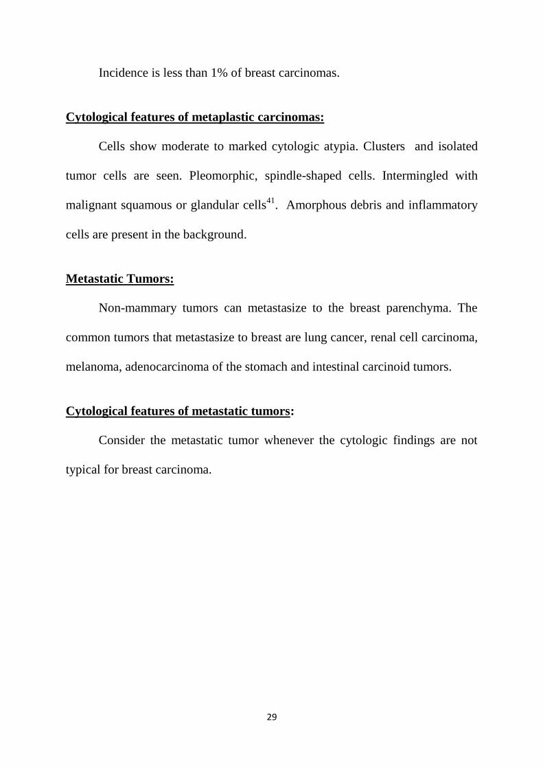
Metaplastic Carcinomas:
29
Incidence is less than 1% of breast carcinomas.
Cytological features of metaplastic carcinomas:
Cells show moderate to marked cytologic atypia. Clusters and isolated
tumor cells are seen. Pleomorphic, spindle-shaped cells. Intermingled with
malignant squamous or glandular cells41
. Amorphous debris and inflammatory
cells are present in the background.
Metastatic Tumors:
Non-mammary tumors can metastasize to the breast parenchyma. The
common tumors that metastasize to breast are lung cancer, renal cell carcinoma,
melanoma, adenocarcinoma of the stomach and intestinal carcinoid tumors.
Cytological features of metastatic tumors:
Consider the metastatic tumor whenever the cytologic findings are not
typical for breast carcinoma.
30
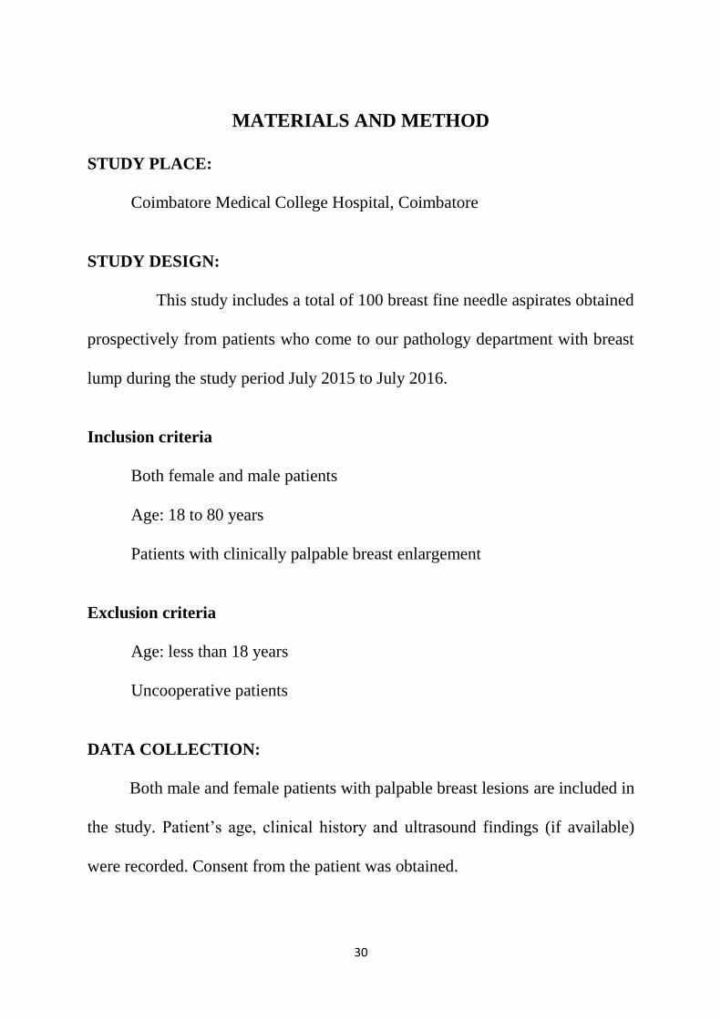
MATERIALS AND METHOD
STUDY PLACE:
Coimbatore Medical College Hospital, Coimbatore
STUDY DESIGN:
This study includes a total of 100 breast fine needle aspirates obtained
prospectively from patients who come to our pathology department with breast
lump during the study period July 2015 to July 2016.
Inclusion criteria
Both female and male patients
Age: 18 to 80 years
Patients with clinically palpable breast enlargement
Exclusion criteria
Age: less than 18 years
Uncooperative patients
DATA COLLECTION:
Both male and female patients with palpable breast lesions are included in
the study. Patient’s age, clinical history and ultrasound findings (if available)
were recorded. Consent from the patient was obtained.

31
METHODOLOGY AND TECHNIQUE USED:
Conventional and liquid based smears were prepared using cytological
material obtained by separate needle passes. The aspirates were performed using
26 gauge needle connected to 10ml syringe. Non-aspiration technique was
followed to minimize bloody samples.
Liquid based cytology smears were prepared using centrifuge machine.
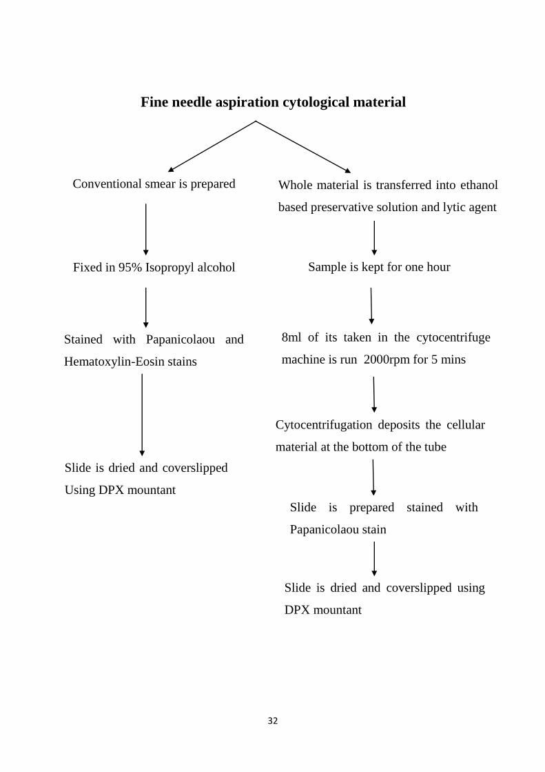
32
Conventional smear is prepared Whole material is transferred into ethanol
based preservative solution and lytic agent
Fixed in 95% Isopropyl alcohol Sample is kept for one hour
Stained with Papanicolaou and
Hematoxylin-Eosin stains
8ml of its taken in the cytocentrifuge
machine is run 2000rpm for 5 mins
Slide is dried and coverslipped
Using DPX mountant
Cytocentrifugation deposits the cellular
material at the bottom of the tube
Slide is prepared stained with
Papanicolaou stain
Slide is dried and coverslipped using
DPX mountant
Fine needle aspiration cytological material
33
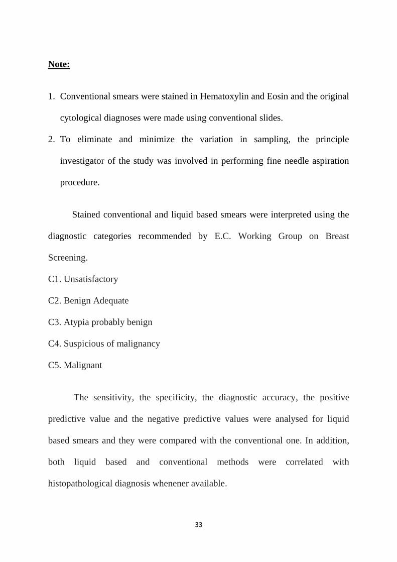
Note:
1. Conventional smears were stained in Hematoxylin and Eosin and the original
cytological diagnoses were made using conventional slides.
2. To eliminate and minimize the variation in sampling, the principle
investigator of the study was involved in performing fine needle aspiration
procedure.
Stained conventional and liquid based smears were interpreted using the
diagnostic categories recommended by E.C. Working Group on Breast
Screening.
C1. Unsatisfactory
C2. Benign Adequate
C3. Atypia probably benign
C4. Suspicious of malignancy
C5. Malignant
The sensitivity, the specificity, the diagnostic accuracy, the positive
predictive value and the negative predictive values were analysed for liquid
based smears and they were compared with the conventional one. In addition,
both liquid based and conventional methods were correlated with
histopathological diagnosis whenener available.
34
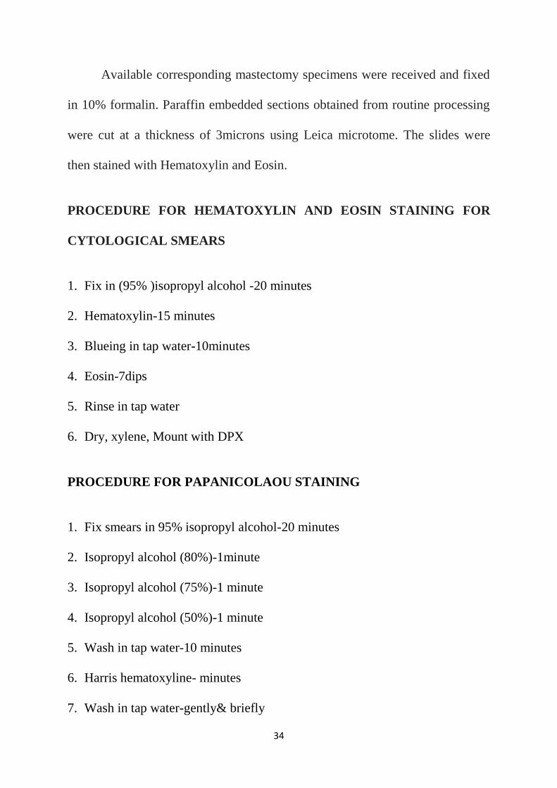
Available corresponding mastectomy specimens were received and fixed
in 10% formalin. Paraffin embedded sections obtained from routine processing
were cut at a thickness of 3microns using Leica microtome. The slides were
then stained with Hematoxylin and Eosin.
PROCEDURE FOR HEMATOXYLIN AND EOSIN STAINING FOR
CYTOLOGICAL SMEARS
1. Fix in (95% )isopropyl alcohol -20 minutes
2. Hematoxylin-15 minutes
3. Blueing in tap water-10minutes
4. Eosin-7dips
5. Rinse in tap water
6. Dry, xylene, Mount with DPX
PROCEDURE FOR PAPANICOLAOU STAINING
1. Fix smears in 95% isopropyl alcohol-20 minutes
2. Isopropyl alcohol (80%)-1minute
3. Isopropyl alcohol (75%)-1 minute
4. Isopropyl alcohol (50%)-1 minute
5. Wash in tap water-10 minutes
6. Harris hematoxyline- minutes
7. Wash in tap water-gently& briefly
35
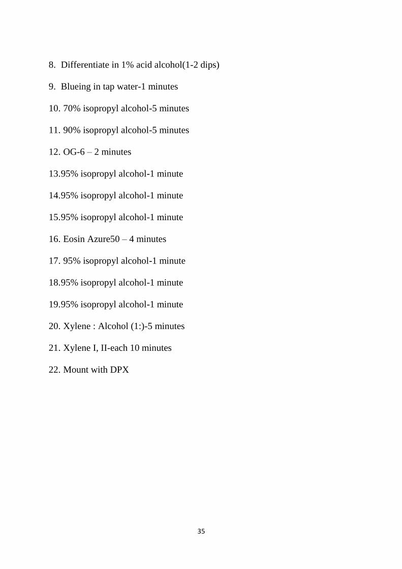
8. Differentiate in 1% acid alcohol(1-2 dips)
9. Blueing in tap water-1 minutes
10. 70% isopropyl alcohol-5 minutes
11. 90% isopropyl alcohol-5 minutes
12. OG-6 – 2 minutes
13. 95% isopropyl alcohol-1 minute
14. 95% isopropyl alcohol-1 minute
15. 95% isopropyl alcohol-1 minute
16. Eosin Azure50 – 4 minutes
17. 95% isopropyl alcohol-1 minute
18. 95% isopropyl alcohol-1 minute
19. 95% isopropyl alcohol-1 minute
20. Xylene : Alcohol (1:)-5 minutes
21. Xylene I, II-each 10 minutes
22. Mount with DPX
36
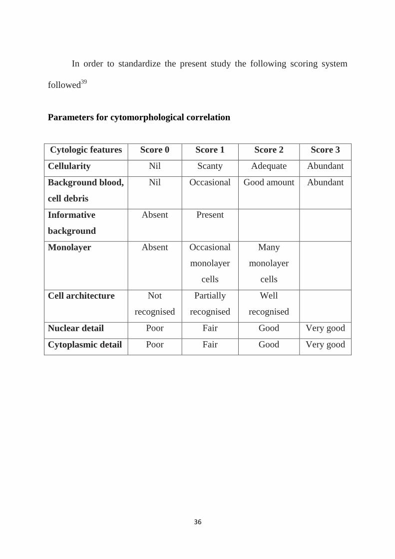
In order to standardize the present study the following scoring system
followed39
Parameters for cytomorphological correlation
Cytologic features Score 0 Score 1 Score 2 Score 3
Cellularity Nil Scanty Adequate Abundant
Background blood,
cell debris
Nil Occasional Good amount Abundant
Informative
background
Absent Present
Monolayer Absent Occasional
monolayer
cells
Many
monolayer
cells
Cell architecture Not
recognised
Partially
recognised
Well
recognised
Nuclear detail Poor Fair Good Very good
Cytoplasmic detail Poor Fair Good Very good

37
RESULTS
Total of 100 fine needle aspiration samples (98 from women and 2 from
men) were included in the study. Both conventional and liquid based smears
were prepared for all 100 fine needle aspiration specimens.
Table – 1: Total number of cases done in Conventional method and Liquid
based cytology method.
S.No Cytological
diagnosis
No. of CS with
percentage
No.of LBC smears
with percentage
1. FA 30(30%) 29(29%)
2. FCD 4(4%) 4(4%)
3. GM 2(2%) 2(2%)
4. SM 1(1%) 2(2%)
5. DC 61(61%) 61(61%)
6. MC 2(2%) 2(2%)
TOTAL NO. OF
CASES 100 100
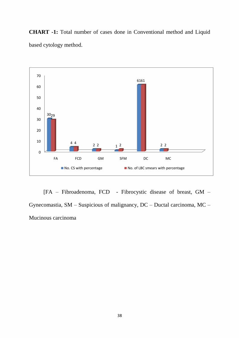
The above table shows total number of cases in each one of the study
category. Benign category includes fibroadenoma, fibrocystic disease of breast
and gynecomastia constituting about 35% of cases in each method. Suspicious
of malignant category constitutes 2% cases in LBC preparation and 1% case in
conventional method. Malignant category had equal incidence in liquid based
and conventional methods, constituting 61 cases of ductal carcinoma and 2
cases of mucinous carcinoma.
38
CHART -1: Total number of cases done in Conventional method and Liquid
based cytology method.
[FA – Fibroadenoma, FCD - Fibrocystic disease of breast, GM –
Gynecomastia, SM – Suspicious of malignancy, DC – Ductal carcinoma, MC –
Mucinous carcinoma
0
10
20
30
40
50
60
70
FA FCD GM SFM DC MC
30
42 1
61
2
29
42 2
61
2
No. CS with percentage No. of LBC smears with percentage
39
0 0 0
6
14
25
11
4
10
Age distribution in Mlignant lesions
0
3
19
9
10 0 0 0 00
10
21
0 0 0 0 001
0 0 0 0 0 0 0 0
Age distribution in Benign lesions FA Age distribution in Benign lesions FCD
Age distribution in Benign lesions GM Age distribution in Benign lesions GM
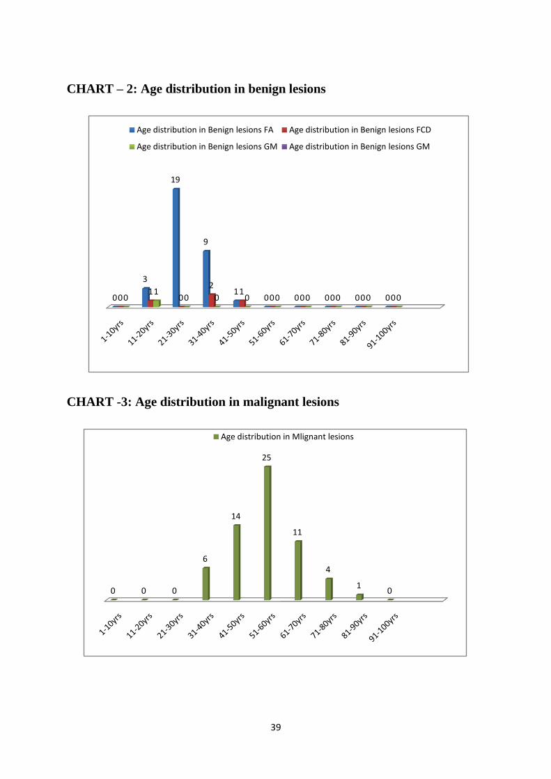
CHART – 2: Age distribution in benign lesions
CHART -3: Age distribution in malignant lesions
40
0
10
20
30
40
50
60
70
80
90
100
0-ZERO 1-SCANTY 2-ADEQUATE 3-ABUNDANT
CELLULARITY LBC
CELLULARITY CS
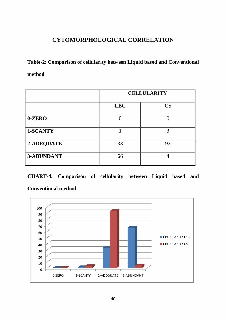
CYTOMORPHOLOGICAL CORRELATION
Table-2: Comparison of cellularity between Liquid based and Conventional
method
CELLULARITY
LBC CS
0-ZERO 0 0
1-SCANTY 1 3
2-ADEQUATE 33 93
3-ABUNDANT 66 4
CHART-4: Comparison of cellularity between Liquid based and
Conventional method

41
Table -3: Comparison of background material (blood, cell debris) between
Liquid based and Conventional method
LBC CS
0-ZERO 100 0
1-OCCASIONAL 0 16
2-GOOD AMOUNT 0 40
3-ABUNDANT 0 44
CHART-5: Comparison of background material (blood, cell debris)
between Liquid based and Conventional method
0
20
40
60
80
100
0-ZERO 1-OCCASIONAL 2-GOOD AMOUNT
3-ABUNDANT
LBC
CS
42
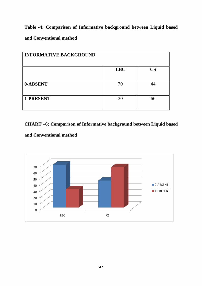
Table -4: Comparison of Informative background between Liquid based
and Conventional method
INFORMATIVE BACKGROUND
LBC CS
0-ABSENT 70 44
1-PRESENT 30 66
CHART –6: Comparison of Informative background between Liquid based
and Conventional method
0
10
20
30
40
50
60
70
LBC CS
0-ABSENT
1-PRESENT
43
0
10
20
30
40
50
60
70
80
90
0- ABSENT 1-OCCASIONAL 2-GOOD AMOUNT
MONOLAYER ARRANGEMENT LBC
MONOLAYER ARRANGEMENT CS
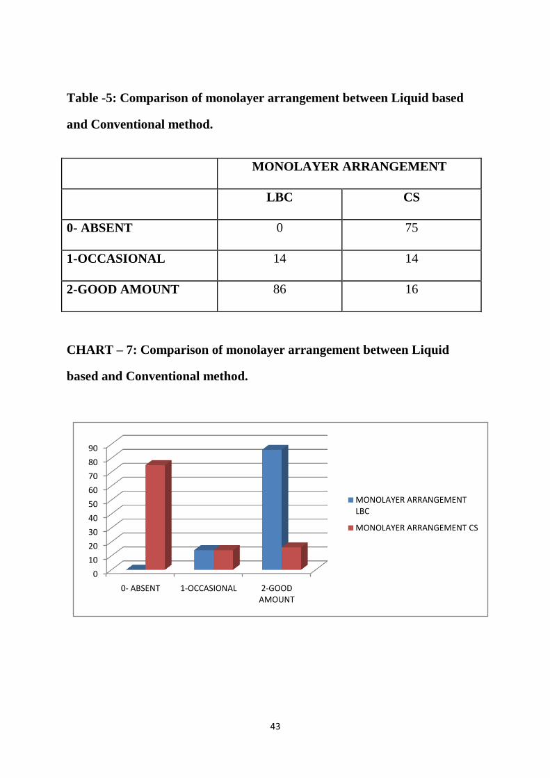
Table -5: Comparison of monolayer arrangement between Liquid based
and Conventional method.
MONOLAYER ARRANGEMENT
LBC CS
0- ABSENT 0 75
1-OCCASIONAL 14 14
2-GOOD AMOUNT 86 16
CHART – 7: Comparison of monolayer arrangement between Liquid
based and Conventional method.

44
Table -6: Comparison of Cell architecture recognized by Liquid based and
Conventional method.
CELL ARCHITECTURE
LBC LBC
0-NOT RECOGNISED 0 0
1-PARTIALLY RECOGNISED 14 26
2-WELL RECOGNISED 86 74
CHART – 8: Comparison of Cell architecture recognized by Liquid based
and Conventional method.
0
10
20
30
40
50
60
70
80
90
0-NOT RECOGNISED
1-PARTIALLY RECOGNISED
2-WELL RECOGNISED
CELL ARCHITECTURE LBC
CELL ARCHITECTURE LBC
45
0
10
20
30
40
50
60
70
80
90
0-Poor 1-Fair 2-Good 3-Very good
Nuclear detail LBC
Nuclear detail CS
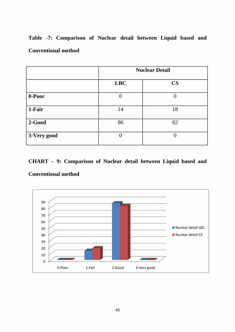
Table -7: Comparison of Nuclear detail between Liquid based and
Conventional method
Nuclear Detail
LBC CS
0-Poor 0 0
1-Fair 14 18
2-Good 86 82
3-Very good 0 0
CHART – 9: Comparison of Nuclear detail between Liquid based and
Conventional method

46
Table -8: Comparison of cytoplasmic details between Liquid based and
Conventional method.
Cytoplasmic Details
LBC CS
0-Poor 0 0
1-Fair 0 0
2-Good 100 100
3-Very good 0 0
CHART – 10
0
10
20
30
40
50
60
70
80
90
100
0-Poor 1-Fair 2-Good 3-Very good
Cytoplasmic details LBC
Cytoplasmic details CS

47
Table – 9:
Comparison of liquid based method and Conventional method in
Bengin lesions
CS
LBC Positive Negative Total
Positive 32 3 35
Negative 0 63 63
TOTAL 32 66 98
Sensitivity - 100.00%
Specificity - 95.45%
Positve Predictive value - 91.42%
Negative Predictive value - 100.00%
Diagnostic Accuracy - 96.93%
CHART - 11
Positive Negative
LBC Positive 100% 0%
LBC Negative 0% 100%
0%20%40%60%80%
100%120%
Comparison of liquid based cytology and Conventional method in Benign Lesion
[N=100][p<0.05]
48
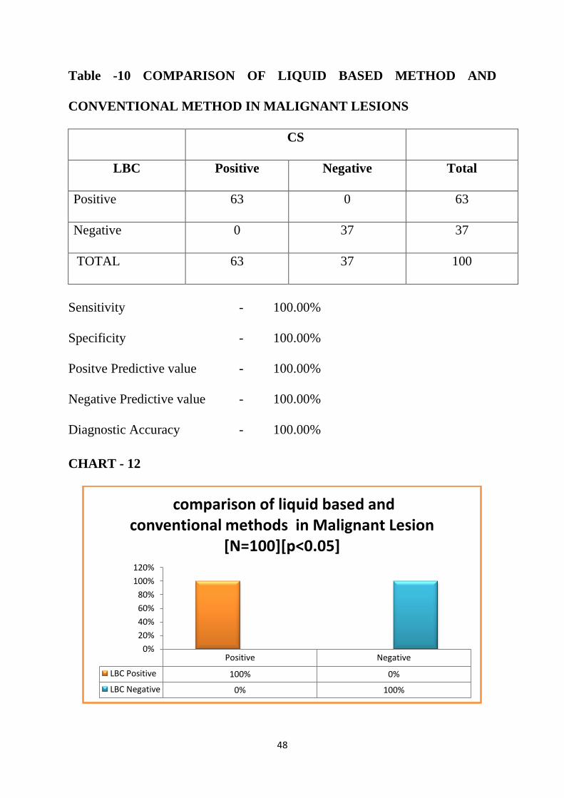
Table -10 COMPARISON OF LIQUID BASED METHOD AND
CONVENTIONAL METHOD IN MALIGNANT LESIONS
CS
LBC Positive Negative Total
Positive 63 0 63
Negative 0 37 37
TOTAL 63 37 100
Sensitivity - 100.00%
Specificity - 100.00%
Positve Predictive value - 100.00%
Negative Predictive value - 100.00%
Diagnostic Accuracy - 100.00%
CHART - 12
Positive Negative
LBC Positive 100% 0%
LBC Negative 0% 100%
0%
20%
40%
60%
80%
100%
120%
comparison of liquid based and conventional methods in Malignant Lesion
[N=100][p<0.05]

49
Table -11: Comparison of Liquid based method and Conventional method
in Fibroadenoma (FA) cases.
Comparison of Liquid based method Conventional method in FA
CS
LBC Positive (%) Negative (%)
Positive 27 100% 2 3%
Negative 0 0% 69 97%
TOTAL 27
71
Sensitivity of LBC - 100.00%
Specificity of LBC - 97.18%
Positve Predictive value of LBC - 93.55%
Negative Predictive value of LBC - 100.00%
Diagnostic Accuracy of LBC - 98.00%

50
Table -12: Comparison of Liquid based method and Conventional method
in Fibrocystic disease of breast (FCD) cases.
Comparison of Liquid based method and Conventional method in FCD
CS
LBC Positive (%) Negative (%)
Positive 3 100% 1 1%
Negative 0 0% 96 99%
TOTAL 3
97
Sensitivity of LBC - 100.00%
Specificity of LBC - 98.96%
Positve Predictive value of LBC - 75.00%
Negative Predictive value of LBC - 100.00%
Diagnostic Accuracy of LBC - 99.00%

51
Table -13: Comparison of Liquid based method and Conventional method
in Gynecomastia (GM) cases.
Comparison of Liquid based method and Conventional method in GM
CS
LBC Positive (%) Negative (%)
Positive 2 100% 0 0%
Negative 0 0% 98 100%
TOTAL 2
98
Sensitivity of LBC - 100.00%
Specificity of LBC - 100.00%
Positve Predictive value of LBC - 100.00%
Negative Predictive value of LBC - 100.00%
Diagnostic Accuracy of LBC - 100.00%
Table 11, 12 & 13 – show that Liquid based method was 100% sensitive in
detecting the benign cases (Fibroadenoma, Fibrocystic disease of breast,
Gynecomastia). The diagnostic accuracy of Liquid based smears was 98%-
100% comparable to the Conventional smears.

52
Table -14: Comparison of Liquid based and Conventional method in
Suspicious of Malignancy (SM) cases.
Comparison of Liquid based method and Conventional method in SM
CS
LBC Positive (%) Negative (%)
Positive 1 100% 1 1%
Negative 0 0% 98 99%
TOTAL 1
99
Sensitivity of LBC - 100.00%
Specificity of LBC - 98.99%
Positve Predictive value of LBC - 50.00%
Negative Predictive value of LBC - 100.00%
Diagnostic Accuracy of LBC - 99.00%
53
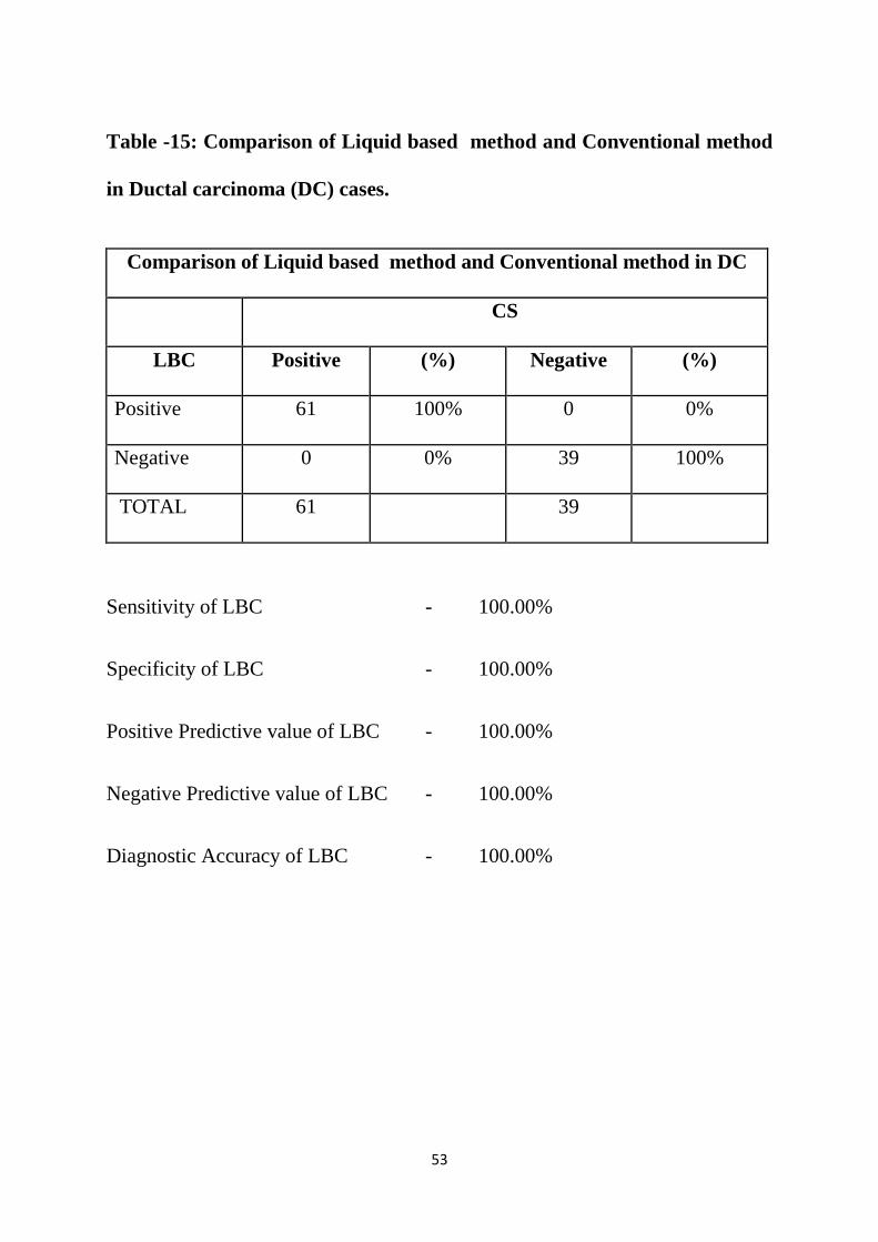
Table -15: Comparison of Liquid based method and Conventional method
in Ductal carcinoma (DC) cases.
Comparison of Liquid based method and Conventional method in DC
CS
LBC Positive (%) Negative (%)
Positive 61 100% 0 0%
Negative 0 0% 39 100%
TOTAL 61
39
Sensitivity of LBC - 100.00%
Specificity of LBC - 100.00%
Positive Predictive value of LBC - 100.00%
Negative Predictive value of LBC - 100.00%
Diagnostic Accuracy of LBC - 100.00%
54
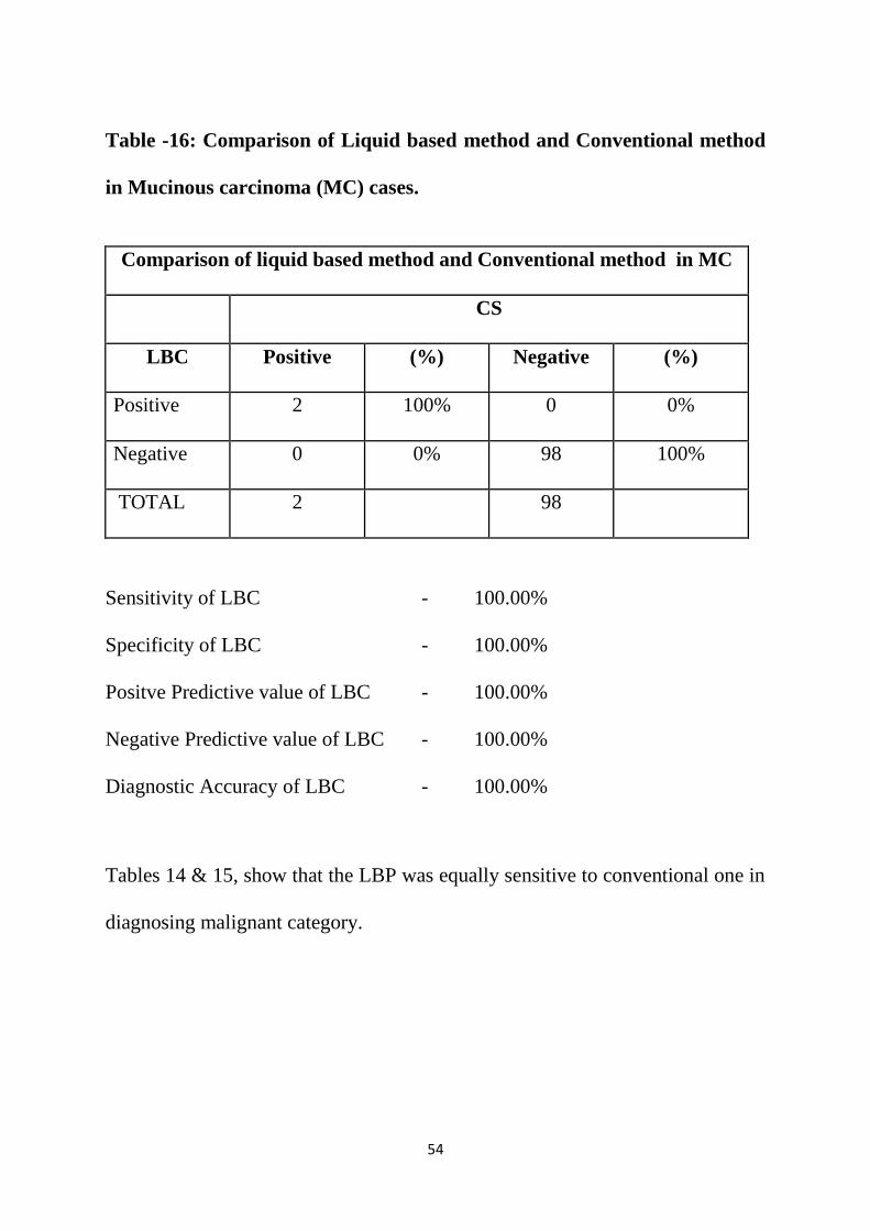
Table -16: Comparison of Liquid based method and Conventional method
in Mucinous carcinoma (MC) cases.
Comparison of liquid based method and Conventional method in MC
CS
LBC Positive (%) Negative (%)
Positive 2 100% 0 0%
Negative 0 0% 98 100%
TOTAL 2
98
Sensitivity of LBC - 100.00%
Specificity of LBC - 100.00%
Positve Predictive value of LBC - 100.00%
Negative Predictive value of LBC - 100.00%
Diagnostic Accuracy of LBC - 100.00%
Tables 14 & 15, show that the LBP was equally sensitive to conventional one in
diagnosing malignant category.

55
Table -17: Correlation of Liquid based method with histological diagnoses
LBC
method
Hist.
Diagnosis
FA FCD GM SM DC MC TOTAL
FA 10 0 0 1 0 0 11
FCD 0 1 0 0 0 0 1
GM 0 0 1 0 0 0 1
DC 0 0 0 1 20 0 21
MC 0 0 0 0 0 1 1
TOTAL 10 1 1 2 20 1 35
[FA – Fibroadenoma, FCD - Fibrocystic disease of breast, GM –
Gynecomastia, SM – Suspicious of malignancy, DC – Ductal carcinoma, MC –
Mucinous carcinoma, LBC- Liquid based cytology]
Among 100 fine needle aspiration cases, it was possible to compare
cytology results with mastectomy speciemens in 36 cases only. Correlating LBP
with available histological diagnoses, the following results were inferred. Out of
10 cases of fibroadenoma, all the 10 correctly diagnosed. 1 case of FCD
correctly interpreted. 1 case of GM correctly diagnosed. 1 case of SM in LBC
diagnosed as fibroadenoma in histopathology and another case of SM in LBC
preparation diagnosed as ductal carcinoma in histopathology.

56
Table -18: Correlation of Conventional method with histological diagnoses.
CS
Hist.
Diagnoses FA FCD GM SM DC MC TOTAL
FA 8 0 0 0 0 0 8
FCD 0 1 0 0 0 0 1
GM 0 0 1 0 0 0 1
DC 0 0 0 1 21 0 22
MC 0 0 0 0 0 1 1
TOTAL 8 1 1 1 21 1 33
[FA – Fibroadenoma, FCD - Fibrocystic disease of breast, GM –
Gynecomastia, SM – Suspicious of malignancy, DC – Ductal carcinoma, MC –
Mucinous carcinoma, CS- conventional smear]
Totally 33 cases of Conventional preparation were correlated with
histopathology findings. Malignant category of conventional smears correlated
very well with histopathological diagnosis. Out of 10 cases of fibroadenoma 8
cases correlated with histopathology. 2 cases of fibroadenoma were not
diagnosed in CS method due to very low cellularity. Gynecomastia and
fibrocystic change cases of conventional smear preparation correlated very well
with histopathology findings.
57
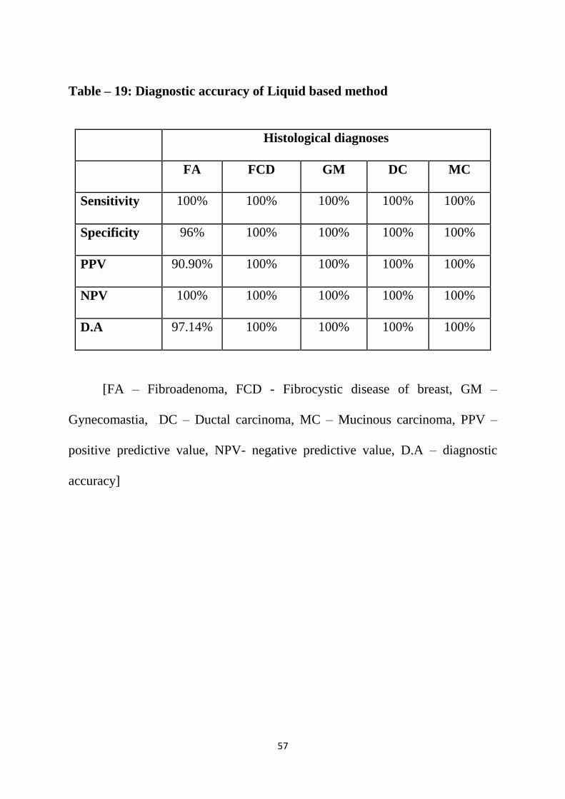
Table – 19: Diagnostic accuracy of Liquid based method
Histological diagnoses
FA FCD GM DC MC
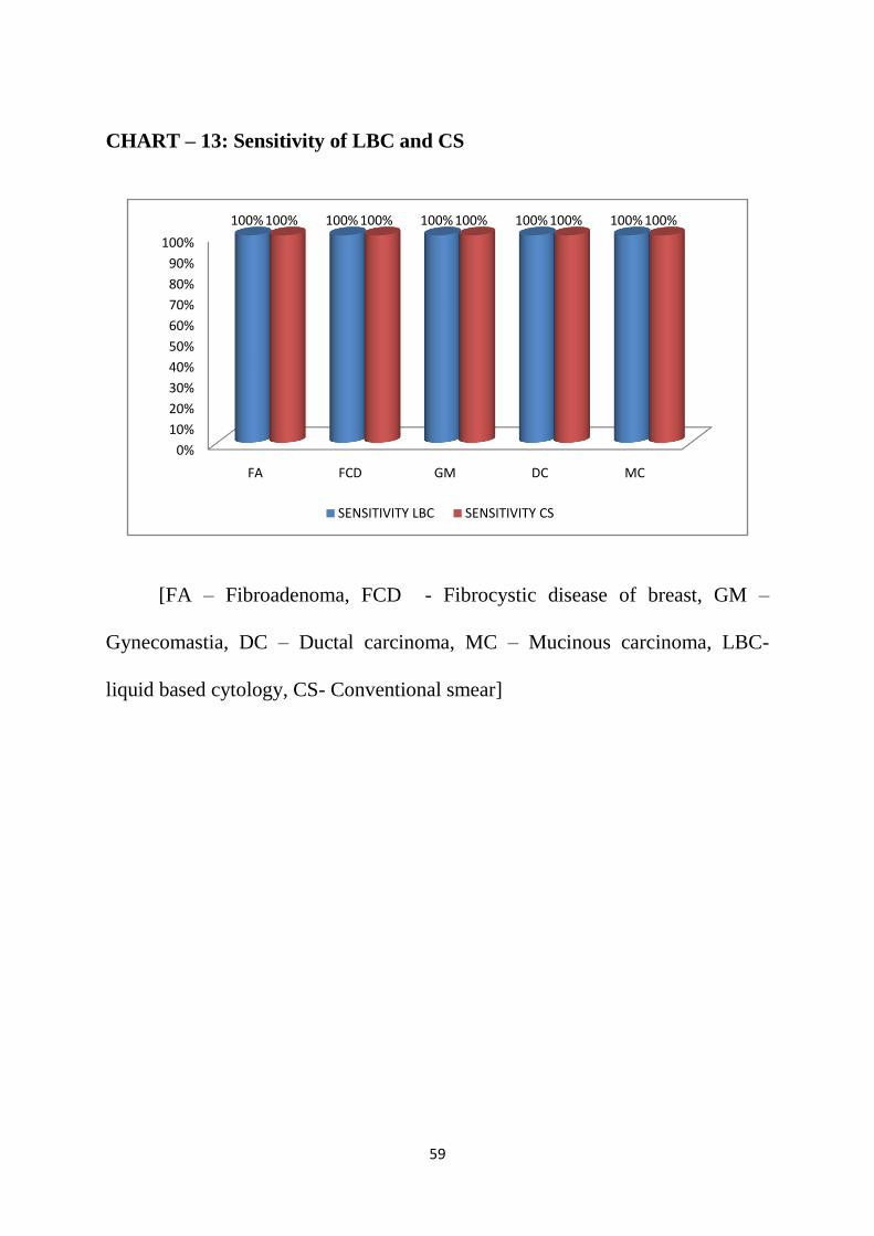
Sensitivity 100% 100% 100% 100% 100%
Specificity 96% 100% 100% 100% 100%
PPV 90.90% 100% 100% 100% 100%
NPV 100% 100% 100% 100% 100%
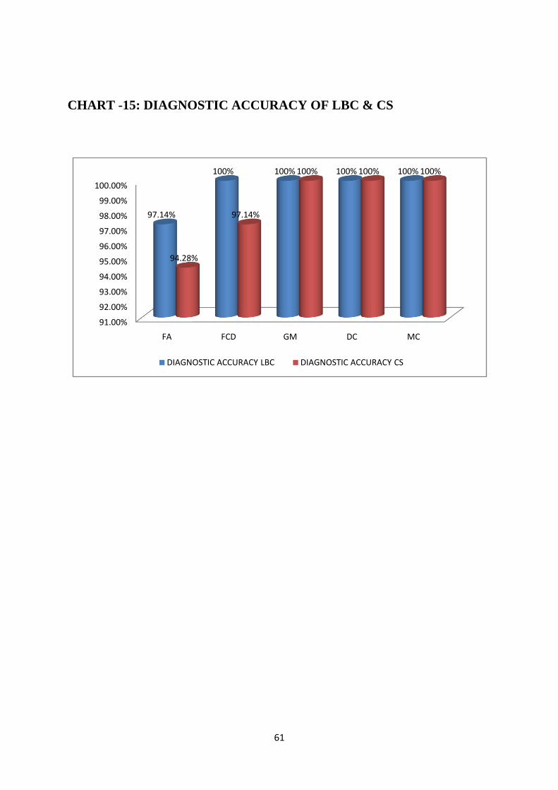
D.A 97.14% 100% 100% 100% 100%
[FA – Fibroadenoma, FCD - Fibrocystic disease of breast, GM –
Gynecomastia, DC – Ductal carcinoma, MC – Mucinous carcinoma, PPV –
positive predictive value, NPV- negative predictive value, D.A – diagnostic
accuracy]
58
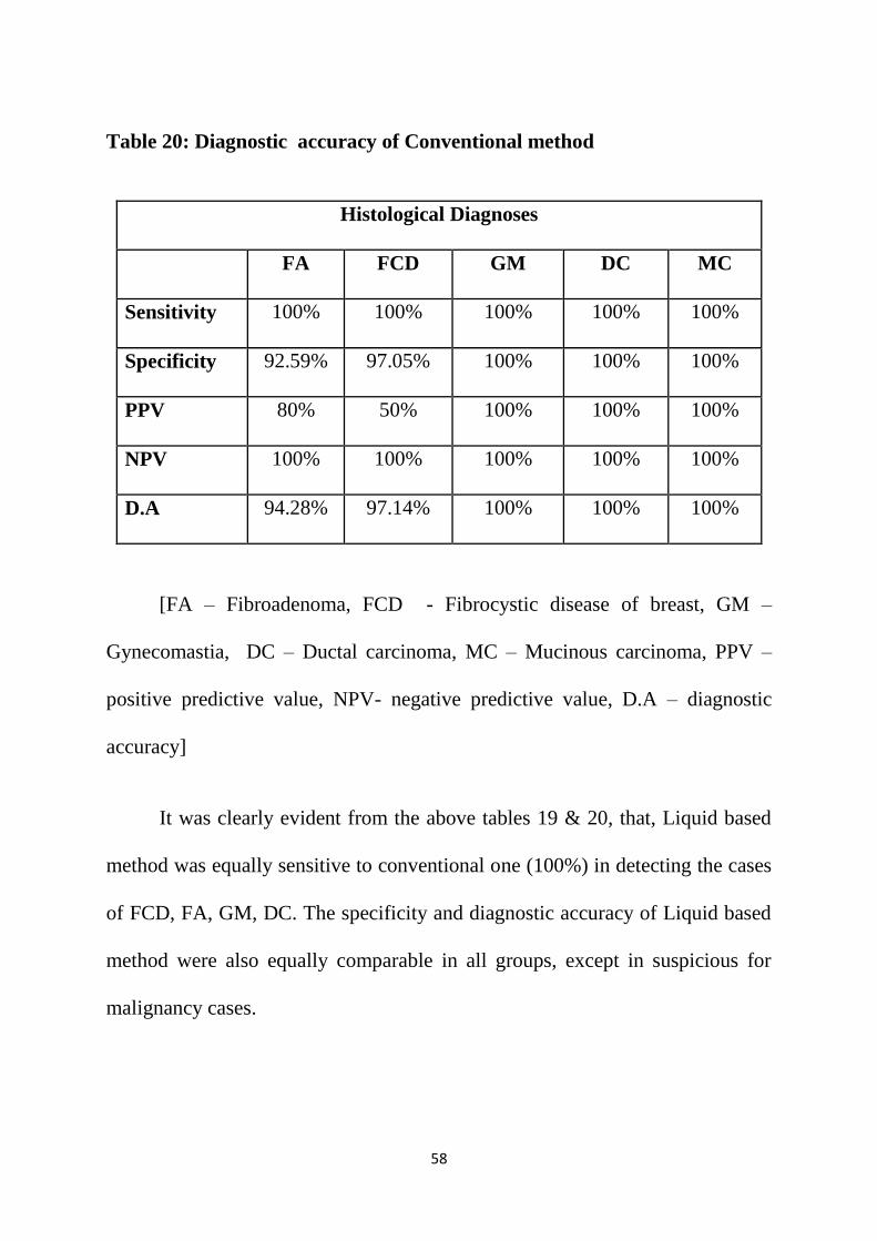
Table 20: Diagnostic accuracy of Conventional method
Histological Diagnoses
FA FCD GM DC MC
Sensitivity 100% 100% 100% 100% 100%
Specificity 92.59% 97.05% 100% 100% 100%
PPV 80% 50% 100% 100% 100%
NPV 100% 100% 100% 100% 100%
D.A 94.28% 97.14% 100% 100% 100%
[FA – Fibroadenoma, FCD - Fibrocystic disease of breast, GM –
Gynecomastia, DC – Ductal carcinoma, MC – Mucinous carcinoma, PPV –
positive predictive value, NPV- negative predictive value, D.A – diagnostic
accuracy]
It was clearly evident from the above tables 19 & 20, that, Liquid based
method was equally sensitive to conventional one (100%) in detecting the cases
of FCD, FA, GM, DC. The specificity and diagnostic accuracy of Liquid based
method were also equally comparable in all groups, except in suspicious for
malignancy cases.
59
CHART – 13: Sensitivity of LBC and CS
[FA – Fibroadenoma, FCD - Fibrocystic disease of breast, GM –
Gynecomastia, DC – Ductal carcinoma, MC – Mucinous carcinoma, LBC-
liquid based cytology, CS- Conventional smear]
0%
10%
20%
30%
40%
50%
60%
70%
80%
90%
100%
FA FCD GM DC MC
100% 100% 100% 100% 100%100% 100% 100% 100% 100%
SENSITIVITY LBC SENSITIVITY CS

60
CHART – 14: SPECIFICITY OF LBC & CS
[FA – Fibroadenoma, FCD - Fibrocystic disease of breast, GM –
Gynecomastia, DC – Ductal carcinoma, MC – Mucinous carcinoma, LBC-
liquid based cytology, CS- Conventional smear]
88%
90%
92%
94%
96%
98%
100%
FA FCD GM DC MC
96%
100% 100% 100% 100%
92.59%
97.05%
100% 100% 100%
Specificity LBC Specificity CS
61
CHART -15: DIAGNOSTIC ACCURACY OF LBC & CS
91.00%
92.00%
93.00%
94.00%
95.00%
96.00%
97.00%
98.00%
99.00%
100.00%
FA FCD GM DC MC
97.14%
100% 100% 100% 100%
94.28%
97.14%
100% 100% 100%
DIAGNOSTIC ACCURACY LBC DIAGNOSTIC ACCURACY CS
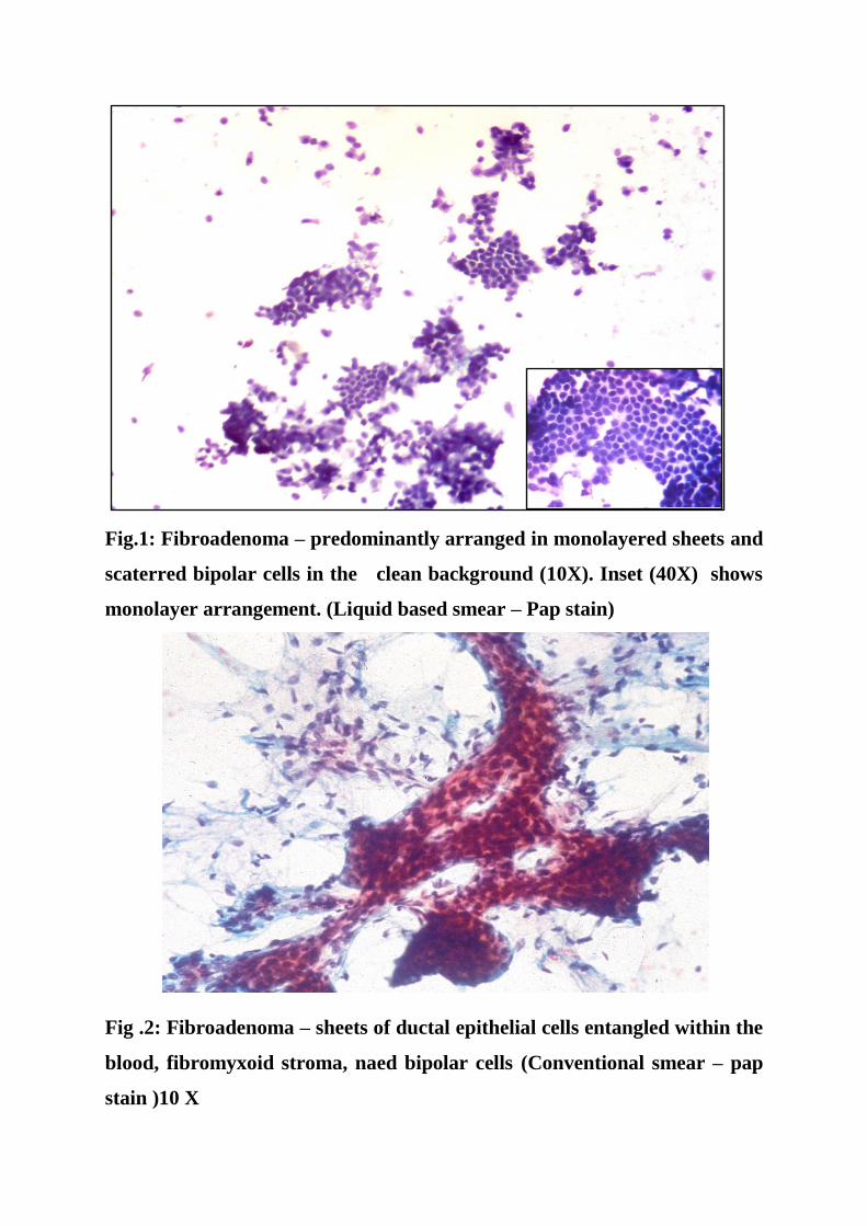
Fig.1: Fibroadenoma – predominantly arranged in monolayered sheets and
scaterred bipolar cells in the clean background (10X). Inset (40X) shows
monolayer arrangement. (Liquid based smear – Pap stain)
Fig .2: Fibroadenoma – sheets of ductal epithelial cells entangled within the
blood, fibromyxoid stroma, naed bipolar cells (Conventional smear – pap
stain )10 X
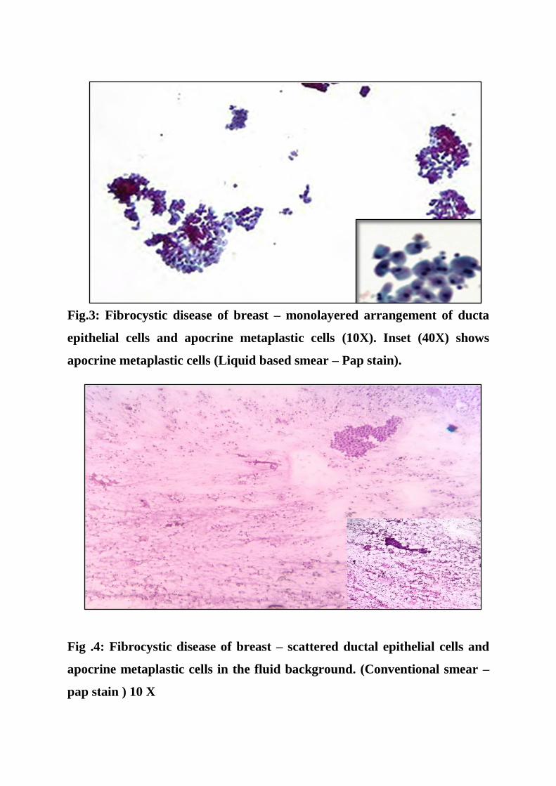
Fig.3: Fibrocystic disease of breast – monolayered arrangement of ducta
epithelial cells and apocrine metaplastic cells (10X). Inset (40X) shows
apocrine metaplastic cells (Liquid based smear – Pap stain).
Fig .4: Fibrocystic disease of breast – scattered ductal epithelial cells and
apocrine metaplastic cells in the fluid background. (Conventional smear –
pap stain ) 10 X
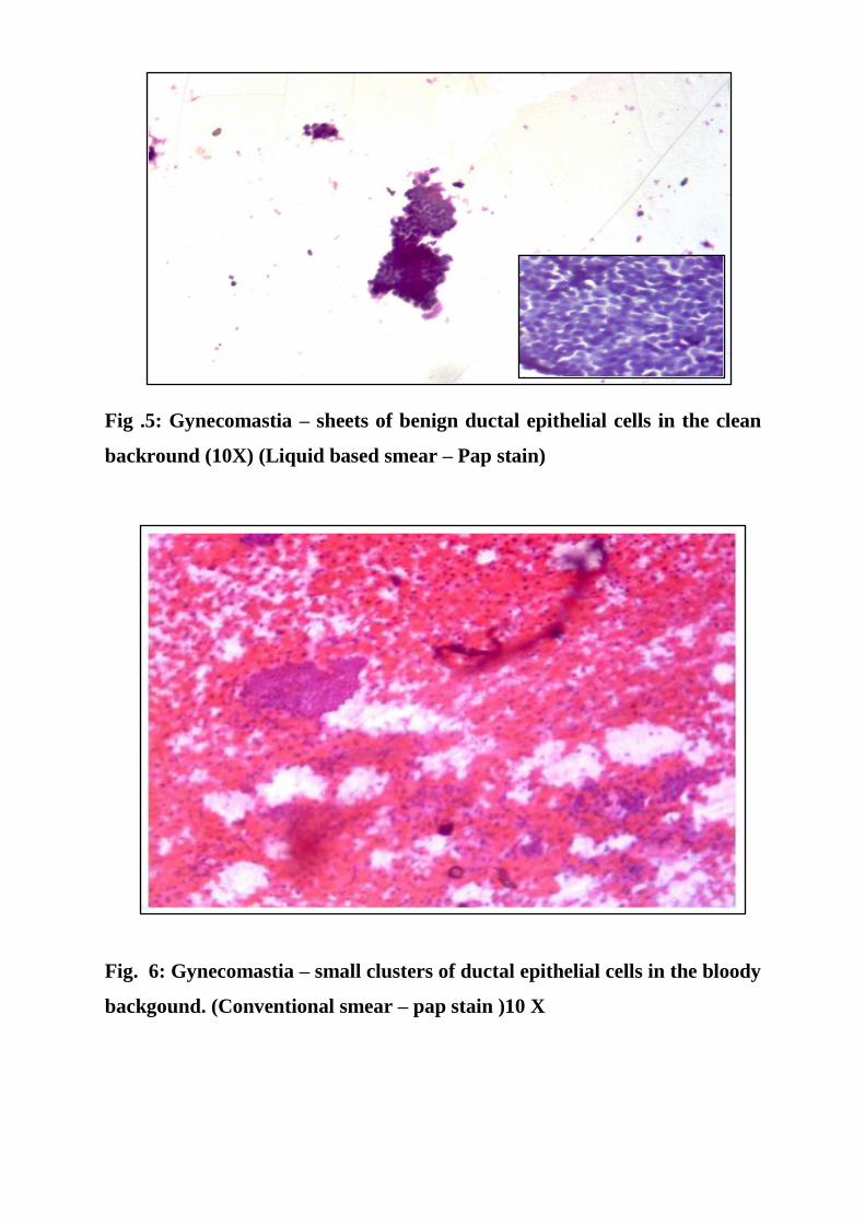
Fig .5: Gynecomastia – sheets of benign ductal epithelial cells in the clean
backround (10X) (Liquid based smear – Pap stain)
Fig. 6: Gynecomastia – small clusters of ductal epithelial cells in the bloody
backgound. (Conventional smear – pap stain )10 X
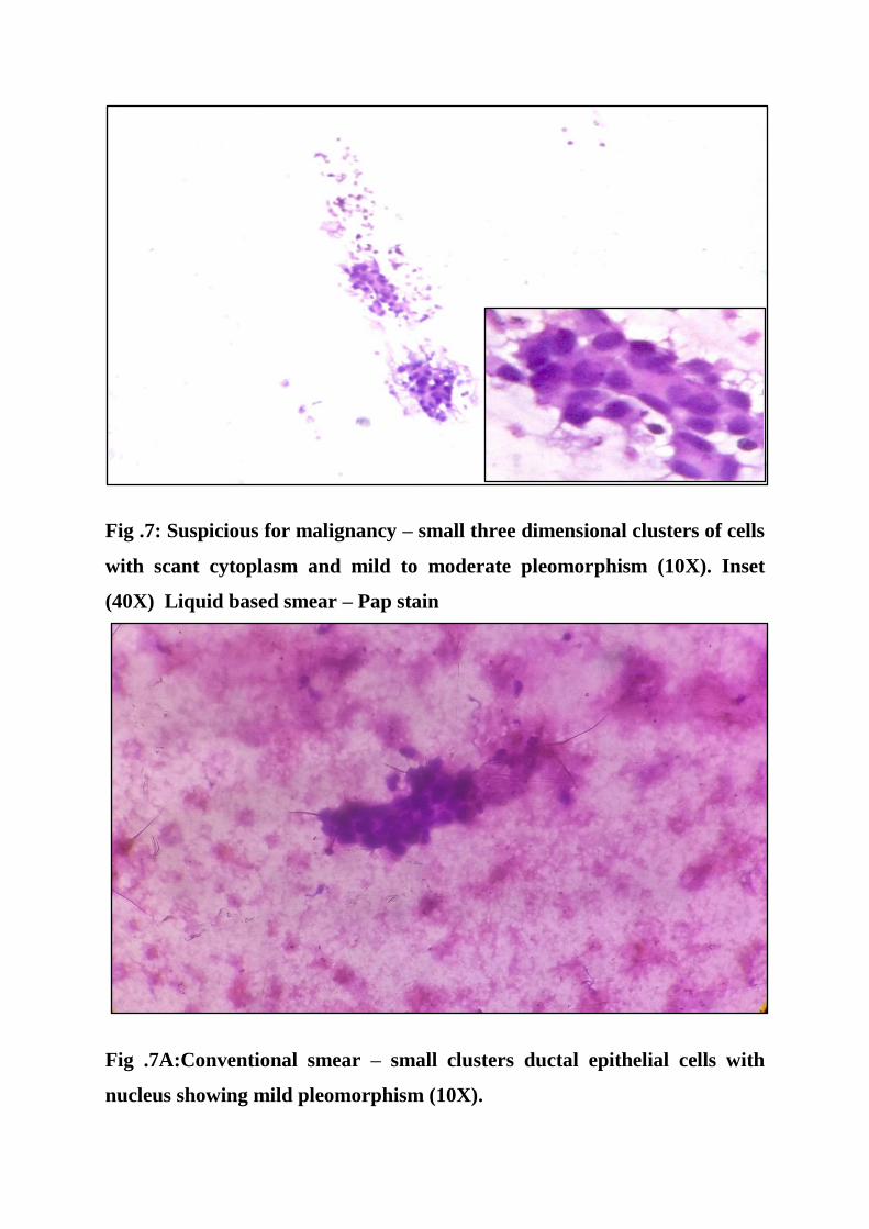
Fig .7: Suspicious for malignancy – small three dimensional clusters of cells
with scant cytoplasm and mild to moderate pleomorphism (10X). Inset
(40X) Liquid based smear – Pap stain
Fig .7A:Conventional smear – small clusters ductal epithelial cells with
nucleus showing mild pleomorphism (10X).
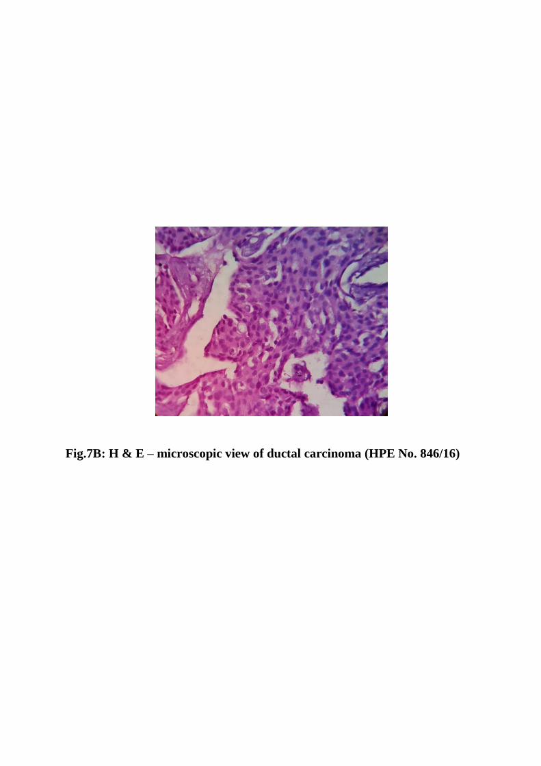
Fig.7B: H & E – microscopic view of ductal carcinoma (HPE No. 846/16)

8: Suspicious for malignancy – small three dimensional clusters of cells
with scant cytoplasm and mild to moderate pleomorphism (10X). Inset
(40X) (Liquid based smear – Pap stain)
Fig.8A: Microscopic view of fibroadenoma with epithelial hyperplasia
(40X). (HPE No.1335/16)
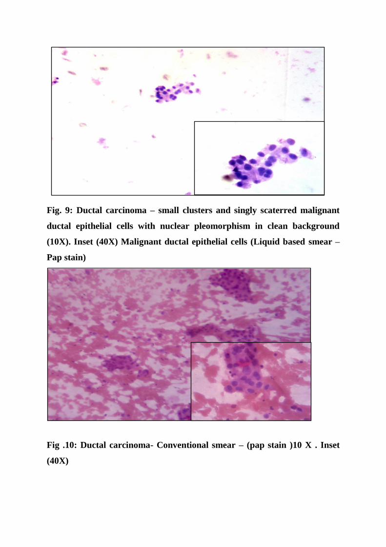
Fig. 9: Ductal carcinoma – small clusters and singly scaterred malignant
ductal epithelial cells with nuclear pleomorphism in clean background
(10X). Inset (40X) Malignant ductal epithelial cells (Liquid based smear –
Pap stain)
Fig .10: Ductal carcinoma- Conventional smear – (pap stain )10 X . Inset
(40X)
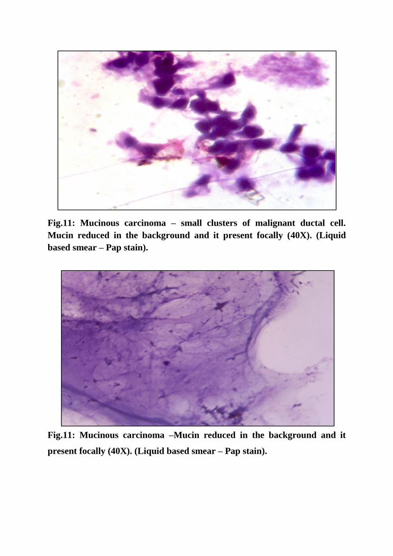
Fig.11: Mucinous carcinoma – small clusters of malignant ductal cell.
Mucin reduced in the background and it present focally (40X). (Liquid
based smear – Pap stain).
Fig.11: Mucinous carcinoma –Mucin reduced in the background and it
present focally (40X). (Liquid based smear – Pap stain).
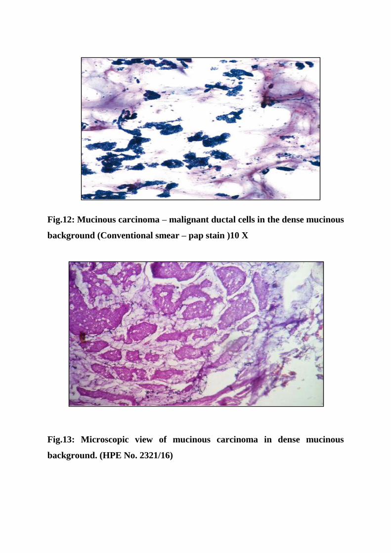
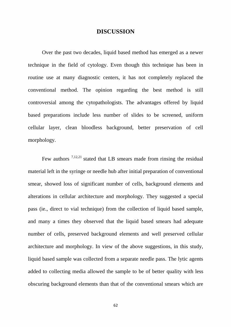
Fig.12: Mucinous carcinoma – malignant ductal cells in the dense mucinous
background (Conventional smear – pap stain )10 X
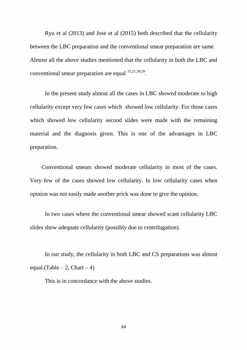
Fig.13: Microscopic view of mucinous carcinoma in dense mucinous
background. (HPE No. 2321/16)
62
DISCUSSION
Over the past two decades, liquid based method has emerged as a newer
technique in the field of cytology. Even though this technique has been in
routine use at many diagnostic centers, it has not completely replaced the
conventional method. The opinion regarding the best method is still
controversial among the cytopathologists. The advantages offered by liquid
based preparations include less number of slides to be screened, uniform
cellular layer, clean bloodless background, better preservation of cell
morphology.
Few authors 7,12,21
stated that LB smears made from rinsing the residual
material left in the syringe or needle hub after initial preparation of conventional
smear, showed loss of significant number of cells, background elements and
alterations in cellular architecture and morphology. They suggested a special
pass (ie., direct to vial technique) from the collection of liquid based sample,
and many a times they observed that the liquid based smears had adequate
number of cells, preserved background elements and well preserved cellular
architecture and morphology. In view of the above suggestions, in this study,
liquid based sample was collected from a separate needle pass. The lytic agents
added to collecting media allowed the sample to be of better quality with less
obscuring background elements than that of the conventional smears which are

63
thick with obscuring blood and inflammatory cells. The nature of the liquid
based processing technique allows a thin layer of representative sample to be
deposited on liquid based slide in a well defined area and enables cytologist to
screen the slides at a faster rate.
The results obtained in our study, implies that during the initial
introduction period, liquid based preparation has to be combined with
conventional one for the purpose of gaining experience in interpretation and
also to avoid errors in the final diagnosis. This would lower the individual
diagnostic differences.
CELLULARITY:
In a broader terminology, sample is said to be adequate if it is cellular and
of good quality with well preserved cellular morphology. Also it should be,
representative of the lesion.
Dey P et al (2000), reported the cellularity in LBC was equal to
conventional preparation.
Gerhard et al, Ryu et al (2013) described that cellularity in LBC and
conventional preparations was same for both.
Michael et al (2000) and Leung et al (1997) reported that the cellularity in
LBC preparations is slightly inferior or superior to the conventional smears.
64
Ryu et al (2013) and Jose et al (2015) both described that the cellularity
between the LBC preparation and the conventional smear preparation are same.
Almost all the above studies mentioned that the cellularity in both the LBC and
conventional smear preparation are equal 15,21,38,39
.
In the present study almost all the cases in LBC showed moderate to high
cellularity except very few cases which showed low cellularity. For those cases
which showed low cellularity second slides were made with the remaining
material and the diagnosis given. This is one of the advantages in LBC
preparation.
Conventional smears showed moderate cellularity in most of the cases.
Very few of the cases showed low cellularity. In low cellularity cases when
opinion was not easily made another prick was done to give the opinion.
In two cases where the conventional smear showed scant cellularity LBC
slides show adequate cellularity (possibly due to centrifugation).
In our study, the cellularity in both LBC and CS preparations was almost
equal.(Table – 2, Chart – 4)
This is in concordance with the above studies.
65
Background material (blood, necrosis, debris):
Gerhard et al (2013), Dey P et al (2000) describe that the background
material like blood and necrosis are lost in LBC preparation, which gives clean
background and helps in easier screening.
In our study, most of cases of LBC preparation showed clean background
with absence of blood, cell debris and necrosis in the background. This is one of
the advantages of the LBC preparation which helps in easy screening. In CS
preparation most of the cases show bloody material in the background which
obscures the cells. This is one of the disadvantages of the CS method (Table 3,
chart 5). This is in concordance with the above studies.
Informative background:
Veneti et al (2003), Dey P et al (2000) and many authors described that
the informative background was lost in LBC preparation which is one of the
disadvantages in diagnosing the benign cases like fibroadenoma and malignant
cases like mucinous carcinoma.
Informative background is one of the most important clue in diagnosing
the lesions in cytology preparation. In our study informative background was
found to be reduced but not lost in cases of LBC. This is one of the
disadvantages in LBC method as described by many authors 21,38,39,40,48
. But in
66
CS method informative background is preserved which helps in
diagnoses.(Table – 4, Chart – 6)
Cell architecture:
In the present study cell architecture was well recognized with LBC
(86/100). Conventional smear showed well recognizable architecture in 74/100
cases. This is probably due to there being less overlapping of cells in LBC,
which resulted in better assessment of cell morphology and architecture in LBC
method (table-6). Many authors described the same features 21,43
. (table – 6,8
and chart – 6,8)
Nuclear detail and cytoplasmic detail:
Nuclear and cytoplasmic details are of equally good quality in almost all
cases in both LBC and CS method (table 7,8 and chart 9,10).
BENIGN LESIONS:
The benign category included in the present study are fibroadenoma and
fibrocystic disease of breast and gynecomastia. They constitute about 30 %, 4%
and 2% respectively of all the breast lesions diagnosed in conventional and
LBC methods.
67
FIBROADENOMA:
Current study showed that the diagnostic accuracy for LBC and CS
preparation are 97% and 94 % respectively. These values imply that our study
results are almost equal to that observed in many studies. The sensitivity and
specificity in our study is 100% and 96%.
LBC preparation in fibroadenoma showed benign looking ductal
epithelial cells, arranged in sheets, small clusters and three dimensional clusters.
Some of the cases showed staghorn clusters. Isolated myoepithelial cells are
seen. Most of the cases showed loss or paucity of the stromal elements like
fibromyxoid stroma. Benign looking ductal epithelial cells without increase in
nuclear cytoplasmic ratio arranged in small clusters and isolated myoepithelial
cells helps us to diagnose fibroadenoma, eventhough there is loss or paucity of
fibromyxoid stroma in the background. Continuous practice helps one to
diagnose fibroadenoma.
Mygdakos et al (2009), Michael et al (2000), Leung et al (1997) and
many other authors also observed decrease in myoepithelial cells and paucity or
loss of stromal elements in fibroadenoma cases.
Ryu et al (2013) has interpreted some of the breast lesions, which showed
false increase in ductal epithelial cells due to decrease in the fibromyxoid

68
stroma and myoepithelial cells, thus misdiagnosing these cases as suspicious for
malignancy.
Leung et al (1992), Perez et al (1994), Kollur et al (2006) and many
authors encountered same problem.
We also encountered a similar problem, but upon review of the doubtful
cases, we could identify the predominance of cell clusters arranged in small
clusters and three dimensional clusters without crowding or overlapping.
Eventhough there is loss or paucity of background material, presence of uniform
cell morphology, without increase in nuclear cytoplasmic ratio and the
arrangement helps us to diagnose fibroadenoma in LBC.
In conventional preparation the diagnosis of fibroadenoma is easily done
because of the staghorn arrangement of the cells with myoepithelial cells and
background fibromyxoid stroma. But some cases are difficult due to the bloody
background, the nuclear features are not seen clearly and the whole slide has to
be searched. Two to three slides might have been made and all have to be
screened, which consumed more time when compared to LBC were most of the
cases are reported with a single slide. Two cases in conventional preparation
had insufficient material to interpret.
The diagnosis of fibrocystic disease of breast by CS and LBP preparation
shows similar features in both method. Ductal epithelial cells and scattered
69
apocrine metaplastic cells. But the cellularity in the conventional preparation is
low. So another prick is usually done to diagnose the case. But LBC preparation
shows moderate cellularity in such cases due to centrifugation which helps in
diagnosing the case, one of the advantages of LBC.
Gynecomastia cases in both the method showed ductal epithelial cells. In
CS preparation cellularity is low to moderate, so two to three slides are needed
to report whereas in LBC, the cells are subjected to centrifugation and the
diagnosis is made with a single slide itself.
SUSPICIOUS OF MALIGNANCY:
Two cases of the category, suspicious of malignancy were encountered in
our study. One case in LBC method showed small clusters, three dimensional
clusters and singly scaterred epithelial cells with moderate amount of cytoplasm
and nucleus showing mild pleomorphism. Features in CS preparation showed
sheets of epithelial cells and with nucleus showing mild pleomorphism. On
histopathological correlation one of the two turned out to be fibroadenoma,
other case was diagnosed as ductal carcinoma.
Bedard et al described that in their study 75% and 71% of the category
suspicious for malignancy in CS and LBC preparation turned out to be
malignant in histopathology study.
70
MALIGNANT CASES:
Most of the breast carcinomas are easily diagnosed with the help of FNA.
Veneti et al(2003), Biscotti et al (1999), Ryu et al(2013) described that both the
types of cytological preparations CS and LBC preparations have comparable
features for detection of ductal carcinoms.
Dey et al (2000) stated that it was easier to diagnose the ductal carcinoma
in LBC preparation because of the clean background. They also described that
the clean background means uninformative background because main features
of carcinomas like blood and necrosis are lost in LBC preparations.
The results of the present study is in concordance with the observation
made by most of the above authors.
The sensitivity, specificity and the diagnostic accuracy for ductal
carcinoma in CS and LBC preparations showed 100%.
LBC preparation showed malignant ductal epithelial cells arranged in
three dimensional clusters, small clusters and also singly scattered in a clean
background. Cells have scant to moderate amount of cytoplasm with nucleus
showing marked pleomorphism. Most of the cases show fine chromatin.

71
CS preparation showed sheets of ductal epithelial cells in the background
of blood. Nucleus features are almost the same for both preparations as
described by Dey et al, Ryu et al and many authors 12,38,39,40
.
But cytology preparation may not help to categorise the ductal
carcinomas which is a major disadvantage of all FNA samples of both LBC and
CS preparations.
Two cases of mucinous carcinomas were encountered in our study.
Komastsu et al detected mucinous carcinoma in a single case by the
presence of mucous.
Michael et al(2000), Veneti et al(2003) and many other authors described
that the mucinous carcinomas diagnosed by cytology depends largely on the
presence of mucous in the background, which can be reduced or lost in LBC
preparation. But this feature is preserved in CS preparation.
In our study both the cases in LBC preparation revealed 3D clusters of
malignant ductal epithelial cells with the background showing focal areas of
mucous and entrapped capillaries. This helped to make diagnosis as mucinous
carcinomas. One of the cases was confirmed with histopathological diagnosis.
In CS preparations background mucus is preserved with sheets of
malignant ductal epithelial cells which makes it easier to interpret.
Our study is in concordance with the study by the above authors.
72
Advantages of liquid based cytology:
1. Less time consuming.
2. Less number of slides - mostly single slide is enough for reporting.
3. Absence of artifact in preservation.
4. Absence of obscuring background elements (RBCs, necrosis)
5. Presence of cells in monolayers.
6. The remaining sample can be used for adjuvant study like
immunocytochemistry, cell block preparation, immunohistochemistry.
7. Cell morphology and nuclear details are similar in both the preparations.
8. Abundant cellularity in LBC with no overlapping of the cells along with
absence of obscuring background material is very helpful in diagnoses with
some exception.
9. Cytoplasmic and nuclear details are similar to conventional smear.
Disadvantages Liquid based cytology:
1. Loss or paucity of informative background (fibromyxoid stroma, mucus).
2. Loss of architectural pattern.
Advantages of conventional preparation:
1. Preserved architectural arrangement.
2. Presence of informative background.
73
Disadvantages of conventional smears:
1. Presence of obscuring background material.
2. Screening time for the number of slides is long and exhaustive, especially
when the smears are paucicellular.
3. Two or more slides needed on an average.
4. If cellularity is low second prick has to be made.
5. Less monolayering with more overlapping of cells.

74
SUMMARY
Liquid based cytology has evolved as a newer method in the field of fine
needle aspiration cytology. The introduction of liquid based technique tries to
overcome the limitations faced by conventional method. Liquid based smears
are easier to screen and less time consuming due to spread of cells in monolayer
with a clean background. The cellular morphology is always well preserved.
Only a small amount of is used for LBC preparation and the remaining solution
is stored in appropriate preservative solution for few months at an appropriate
temperature for future studies.
In the present study, the diagnostic rate observed using liquid based
method is equally comparable with that of conventional one. It is also found to
be a reliable diagnostic method to differentiate between benign and malignant
lesions.
The relatively low cost and easier method of preparing the slides are
added advantages of this method. In addition, Immunohistochemistry can be
performed on liquid based samples in equivocal cases or to confirm the nature
of neoplasm.
The results obtained in our study, implies that during the initial
introduction period, liquid based preparation has to be combined with

75
conventional one for the purpose of gaining experience in interpretation and
also to avoid errors in the final diagnosis. This would lower the individual
diagnostic differences.
To conclude, both the techniques have both advantages and
disadvantages. Liquid based method may become a promising and reliable one
for preoperative diagnosis of palpable breast lesions. But alterations in smear
background and minor changes in the cellular architecture and morphology
should be borne in mind while interpreting liquid based smears to avoid false
diagnosis in general cytological practice.
Further studies with larger sample sizes are necessary to demonstrate the
real diagnostic significance of this method among other liquid based techniques.

76
CONCLUSION
Fine needle aspiration method is a safer and cost effective method for the
diagnosis of breast lesions. However, proper preparation of cytological smears
determine the quality of diagnosis. Liquid based cytology of breast aspirates
provides a better cellular preservation, less cellular overlapping and elimination
of obscuring background when compared to that of Conventional smears. But
the loss or paucity of informative background is a disadvantage in Liquid based
cytology. Studies have shown similar accuracy between Liquid based cytology
and Conventional smear for the diagnosis of breast lesions. Thus, Liquid based
cytology can be used as important ancillary technique along with the
Conventional smear for diagnostic accuracy.
BIBLIOGRAPHY
1. Pawar PS, Gadkari RU, Swami SY, Joshi AR. Comparative study of
manual liquid based cytology (MLBC) technique and direct smear
technique (conventional) on fine needle cytology/fine needle aspiration
cytology samples. J Cytol 2014 Apr-Jun; 31(2): 83-6.
2. Muritto Ortiz, et al. BBD Clinical, radiological and pathological
correlation. Gynecol. Obstet. Mex 2002;70:613-618.
3. Kumar A, L.S. Vohra, K. Bhargava,P.S. Reddy. Investigation of Breast
lump An evaluation. MJAFI 1999;55:299-302.
4. Mehmood A, M. Ahmed, S. Jamal. Role of cytological grading in the
management of breast lump. JCPSP 2003;13:150-152.
5. Galvan Portillo, M. L. Torres Sanchez, L. Lopez Carrillo. Dietary and
reproductive factors associated with benign breast disease in Maxican
women. Nutr. Cancer 2002;43:133-140.
6. Marluce Bibbo, David Wilbur, Comprehensive cytopathology, 3rd
edition,
Elsevier.
7. Nasuti JF, Tam D, Gupta PK, Diagnostic value of liquid-based (Thinprep)
preparations in nongynecologic cases, Diagnostic Cytopathology
2001;24:17-141.

8. Jose Eleuterio Jr, Alessa Aragao, Adequacy of Fine-Needle Aspiration
Cytology for Breast Lesions: The SurePath Liquid-Based Technique
versus Conventional Smears, Acta Cytologica 210559:253-257.
9. S.Z. Ali and A.V. Parwani: Breast Cytopathology. Springer, 2007.
10. M Tunc Canda, Namik Demir, Orcum Sezur et al, clinical Results of the
Liquid-based cervical ctolog Tool, Liqui-PREP TM, in comparison with
Conventional Smears for Detection of Squamous Cell Abnormalities Asian
Pacific J Cancer Prev, 2009; 10:399-402.
11. N. Mygdakos, Sylvia niolaidou, Anna tzilivaki, D. Tamiolakis liquid based
preparation (lbp) cytology versus conventional cytology (cs) in FNA
samples from breast, thyroid, salivary glands and soft tissues. Our
experiences in crete (Greece), Romanian journal o morphology and
embryology 2009;50(2):245-250.
12. Rana S. Hoda, Non-Gynecologic Cytology on Liquid –Based
Preparations:A Morhologic Review o facts and artifacts Diagn.
Cytopathol. 2007;35:621-634.
13. Patel SJ, Katz RL, Sneige N. Use of thin layers as an adjunct to fine needle
aspiration: a side by side comparison with cytocentrifuge preparations
(abstract). Acta Cytol 1993;37: 800.
14. James Geyer, James Marino, Evaluation of the Liqui-Prep tm
Encapsulation system by comparision with the Surepath TM prep, Acta
Cytol 2004.
15. Gerhard R, Schmitt FC: Liquid-based cytology in fine needle aspiration of
breast lesions: a review. Acta Cytol 2014;58:533-542.
16. Bedard YC, Pollett AF. Breast fine-needle aspiration. A comparison of
ThinPrep and conventional smears. Am J Clin Pathol 1999;111:523-7.
17. Kaufman Z, Shpitz B, Shapiro M, Rona R, Lew S, Dinbar A. Triple
approach in the diagnosis of dominant breast masses: combined physical
examination, mammography, and fine-needle aspiration. J Surg Oncol
1994;56:254-7 .
18. Tisserand P, Fouquet C, Marck V, et al. ThinPrep-processed fine-needle
samples of breast are effective material for RNA- and DNA-based
molecular diagnosis: application to p53 mutation analysis. Cancer
2003;99:223-32.
19. Pollett A, Bedard YC, Li SQ, Rohan T, Kandel R. Correlation of p53
mutations in ThinPrep-processed fine needle breast aspirates with
surgically resected breast cancers 2000;13:11739.
20. Cibas, Barbara S.Ducatmaan, Cytology:Diagnostic principles and clinical
correlates, 3rd
edition, Elsevier.
21. Michael CW, Hunter B: Interpretation of fine-needle aspirates
processed by the Thin Prep® technique: cytologic artifacts and
diagnostic pitfalls. Diagn Cytopathol 2000;23:6-13.
22. Claire W. Michel, James Mc Connel, James Pecott et al, Comparison of
Thin prep and Tripath PREP Liquid Based preparations in Nongynecologic
speciemens: A pilot study design. Cytopathol. 2001;25:177-184.
23. Elsheikh, Tarik M, Joseph L, Kirkpatrick, Hoard H, Wu, Comparison of
Thin Prep and Cytospin Preparation in the evaluation of Exfoliative
cytology speciemens (Cancer Cytoathology) 2006;108:144-9.
24. Koss, Leopold G.; Melamed, Myron R. Koss' Diagnostic Cytology and Its
Histopathologic Bases, 5th Edition
25. G.Gherardi Fine-Needle Biopsy of Superficial and Deep Masses.
Interventional Approach and Interpretation Methodology by Pattern
Recognition. Springer-Verlag Italia 2009 . pages 17-47.
26. Cibas ES: Cervical and vaginal cytology; in Cibas ES,Ducatman BS
(eds): Cytology: Diagnostic Principles and Clinical Correlates.
Philadelphia, Saunders, 2009, pp 1-63.
27. Saqi A, Mercado CL, Hamele-Bena D: Adenoid cystic carcinoma of the
breast diagnosed by fine-needle aspiration. Diagn Cytopathol
2004;30(4):271-274.
28. Dodd LG, Sneige N, Reece GP, Fornage B: Fine-needle aspiration
cytology of silicone granulomas in the augmented breast. Diagn
Cytopathol 1993;9(5):498-502.

29. Balco MT, Ali SZ: Asteroid bodies in silicone lymphadenitis on fine-
needle aspiration. Diagn Cytopathol 2007; 35(11):715-716 158. Ku NN,
Mela NJ, Fiorica JV, et al.: Role of fine needle aspiration.
30. Dornfeld JM, Thompson SK, Shurbaji MS: Radiation-induced changes in
the breast: A potential diagnostic pitfall on fine-needle aspiration. Diagn
Cytopathol 1992;8(1):79-80; discussion 81.
31. Hirata S, Saito T, Kiyanagi K, et al.: Granulomatous mastitis diagnosed by
core-needle biopsy and successfully treated with corticosteroid therapy: A
case report. Breast Cancer 2003;10(4):378-381.
32. Silverman JF, Lannin DR, Unverferth M, Norris HT: Fine needle
aspiration cytology of subareolar abscess of the breast. Spectrum of
cytomorphologic findings and potential diagnostic pitfalls. Acta Cytol
1986;30(4):413-419.
33. Kapila K, Verma K: Cytomorphological spectrum in gynaecomastia: A
study of 389 cases. Cytopathology 2002; 13(5):300-308.
34. Bhattarai S, Kapila K, Verma K: Phyllodes tumor of the breast. A
cytohistologic study of 80 cases. Acta Cytol 2000; 44(5):790-796.
35. Saqi A, Mercado CL, Hamele-Bena D: Adenoid cystic carcinoma of the
breast diagnosed by fine-needle aspiration. Diagn Cytopathol
2004;30(4):271-274.

36. Garg S, Mohan H, Bal A, et al.: A comparative analysis of core needle
biopsy and fine-needle aspiration cytology in the evaluation of palpable
and mammographically detected suspicious breast lesions. Diagn
Cytopathol 2007;35(11):681-689.
37. Tavassoli FA, Devilee P: Tumours of the Breast and Female Genital
Organs. Lyon, France, IARC Press, 2003.
38. Ryu HS, Park S, Jung Y, Park S, Shin H: A pilot study evaluating liquid-
based fine needle aspiration cytology of breast lesions: a
cytomorphological comparison of SurePath liquid-based preparations and
conventional smears. Acta Cytol 2013;57:391-399.
39. Dey P, Luthra UK, George J, Zuhairy F, George SS, Haji BI. Comparison
of ThinPrep and conventional preparations on fine needle aspiration
cytology material. Acta Cytol 2000; 44:46-50.
40. Veneti S, Daskalopoulou D, Zervoudis S, Papasotiriou E, Ioannidou-
Mouzaka L. Liquid-based cytology in breast fine needle aspiration.
Comparison with the conventional smear. Acta Cytol 2003;47:188-92.
41. Dawson AE, Logan-Young W, Mulford DK: Aspiration cytology of
tubular carcinoma: Diagnostic features with mammographic correlation.
Am J Clin Pathol 1994;101(4):488-492.
42. Leung CS, Chiu B, Bell V. Comparison of ThinPrep and conventional
preparations: nongynecologic cytology evalu-ation. Diagn Cytopathol
1997;16:368-71.
43. Komatsu K, Nakanishi Y, Seki T, Yoshino A, Fuchinoue F, Amano S,
Komatsu A, Sugitani M, Nemoto N: Application of liquid-based
preparation to fine needle aspiration cytology in breast cancer. Acta
Cytol 2008;52:591-59.
44. Kollur SM, El Hag IA: FNA of breast fibroadenoma: observer
variability and review of cytomorphology with cytohistological
correlation. Cytopathology 2006;17:239-244.
45. Warren GP, Gatscha RM, Saigo PE. A comparison of non-gynecologic
specimens prepared byy ThinPrep to conventional methods:Direct smears,
cytospin preparations and cell blocks: A review of the Memorial Hospital
experience (abstract). Acta cytol 1994; 49: 824.
46. G.Gherardi Fine-Needle Biopsy of Superficial and Deep Masses.
Interventional Approach and Interpretation Methodology by Pattern
Recognition. Springer-Verlag Italia 2009 . pages 17-47.
47. Orell and Sterrett’s fine needle aspiration cytology. – 5th ed.
48. Leung CS, Chiu B, Bell V: Comparison of ThinPrep and conventional
preparations: nongynecologic cytology evaluation. Diagn Cytopathol
1997;16:368-371.
49. Perez-Reyes N, Mulford DK, Rutkowski MA, Logan-Young W,
Dawson AE: Breast fine-needle aspiration: a comparison of thin-layer
and conventional preparation. Am J Clin Pathol 1994;102:349-353.
50. Biscotti CV, Shorie JH, Gramlich TL, Easley KA: ThinPrep versus
conventional smear cytologic preparations in analyzing fine-needle
aspiration specimens from palpable breast masses. Diagn Cytopathol
1999;21:137-141.
51. Ali AS, Yin D, Yao D, Vazquez M: Criteria for the diagnosis of
fibroepithelial lesions of the breast with liquid-based cytology. Acta
Cytol 2004;48:481-486.

PROFORMA
Name : IP/OP No:
Age : FNAC No:
Sex:
Presenting Complaints :
Breast Lump :
Duration :
Pain :
Nipple Discharge :
Past History:
Family History :
History of previous surgery / FNAC for breast lumps :
Examination of breast :
Right side / left side breast :
Single / multiple swelling :
Size :
Consistency :
Mobile / fixed to underlying structures :
Nipple Discharge :
Examination of axillary nodes :
Ultrasound breast (if available) :
Previous FNAC report (if available) :
Previous surgery- HPE report (if available) :
Conventional smear diagnosis :
Liquid based cytology diagnosis :
xg]g[jy; gotk;
vdJ khh]gf fl]oapy; ,Ue;J Crpapd; \yk; jpR / ePh] vLj;J
ghpnrhjid bra;tjw;F KG kdJld; rk;kjpf;fpnwd;.
,g]gof;F/

CS LBP CS LBP CS LBP CS LBP CS LBP CS LBP CS LBP
1 1014/15 3983/15 67 FEMALE DC DC DC 2 2 2 0 1 0 1 2 2 2 2 2 2 2
2 1133/15 60 FEMALE DC DC 2 2 3 0 1 0 1 2 2 2 2 2 2 2
3 1144/15 2216/15 57 FEMALE DC DC DC 2 2 2 0 1 0 2 2 2 2 2 2 2 2
4 1146/15 29 FEMALE FA FA 2 2 3 0 1 1 2 2 2 2 2 2 2 2
5 1148/16 34 FEMALE FA FA 2 3 2 0 1 0 2 2 2 2 2 2 2 2
6 1152/16 60 FEMALE DC DC 2 3 2 0 1 0 1 2 2 2 2 2 2 2
7 1154/15 24 FEMALE FA DC 2 3 2 0 1 0 2 2 2 2 2 2 2 2
8 1160/15 2312/15 63 FEMALE DC DC 2 3 2 0 1 1 2 2 2 2 2 2 2 2
9 1164/15 52 FEMALE DC DC 2 3 2 0 1 0 2 2 2 2 2 2 2 2
10 1165/15 55 FEMALE DC DC 2 3 2 0 1 0 2 2 2 2 2 2 2 2
11 1167/15 37 FEMALE FCD FCD 1 1 3 0 1 0 2 2 2 2 2 2 2 2
12 1173/15 3067/15 72 FEMALE DC DC DC 2 3 3 0 1 0 2 2 2 2 2 2 2 2
13 1188/15 51 FEMALE DC DC 2 2 2 0 1 0 2 2 2 2 2 2 2 2
14 1194/15 55 FEMALE DC DC 2 2 3 0 1 0 2 2 2 2 2 2 2 2
15 1201/15 24 FEMALE FA FA 2 2 3 0 1 0 1 2 2 2 2 2 2 2
16 1208/15 82 FEMALE DC DC 2 3 3 0 1 0 2 2 2 2 2 2 2 2
17 1209/15 70 FEMALE DC DC 2 3 3 0 1 0 1 2 2 2 2 2 2 2
18 1210/15 19 FEMALE - FCD 1 1 3 0 1 0 1 2 2 2 2 2 2 2
19 1222/15 29 FEMALE FA FA 2 3 3 0 1 1 2 2 2 2 2 2 2 2
20 1231/15 45 FEMALE DC DC 2 2 3 0 1 0 1 2 2 2 2 2 2 2
21 1239/15 2353/15 45 FEMALE FA FA FA 2 3 2 0 1 1 2 2 2 2 2 2 2 2
22 1242/15 55 FEMALE DC DC 2 3 2 0 1 0 1 2 2 2 2 2 2 2
SCORING SYSTEM
0=ZERO
1=OCCASIONAL
2=GOOD AMOUNT
3=ABUNDANT
BACKGROUND
BLOOD DEBRISCELLULARITY
0=ZERO
1=SCANTY
2=ADEQUATE
3=ABUNDANT
0=ABSENT
1=PRESENT
INFORMATIVE
BACKGROUNDMONOLAYER
0=ABSENT
1=OCCASIONAL
2=GOOD AMOUNT
CELL ARCHITECTURE
0=NON-REC0GNIZED
1=MODERATELY RECOGONISED
2=WELL RECOGONISED
3=EXCELLENT
CYTOPLASMIC
DETAILS
0=POOR
1=FAIR
2=GOOD
3=EXCELLENT
NUCLEAR
DETAILS
0=POOR
1=FAIR
2=GOOD
LBC HPES.NO FNAC.NO HPE.NO AGE SEX CS

23 1298/15 55 FEMALE DC DC 2 2 2 0 1 0 1 2 2 2 2 2 2 2
24 1320/15 14 MALE GM GM 2 3 3 0 1 0 1 2 2 2 2 2 2 2
25 1372/15 2316/15 29 FEMALE FA FA FA 2 2 2 0 1 1 2 2 2 2 2 2 2 2
26 1340/15 2380/15 75 FEMALE DC DC DC 2 3 2 0 1 0 1 2 2 2 2 2 2 2
27 1426/15 2429/15 27 FEMALE FA FA FA 2 3 3 0 1 1 2 2 2 2 2 2 2 2
28 1439/15 43 FEMALE DC DC 2 2 3 0 1 0 1 2 2 2 2 2 2 2
29 1446/15 37 FEMALE FA FA 2 3 3 0 1 1 2 2 2 2 2 2 2 2
30 1453/15 32 FEMALE No cells FA 2 2 3 0 1 0 2 2 2 2 2 2 2 2
31 1459/15 30 FEMALE FA FA 2 3 2 0 1 0 2 2 2 2 2 2 2 2
32 1496/15 2581/15 43 FEMALE FA FA FA 2 2 2 0 1 0 2 2 2 2 2 2 2 2
33 1499/15 55 FEMALE DC DC 2 3 3 0 1 0 1 2 2 2 2 2 2 2
34 1504/15 21 FEMALE FA FA 2 2 3 0 1 0 2 2 2 2 2 2 2 2
35 1574/15 2612/15 24 FEMALE FA FA FA 2 3 2 0 1 0 2 2 2 2 2 2 2 2
36 1601/15 46 FEMALE DC DC 2 3 3 0 1 0 1 2 2 2 2 2 2 2
37 1602/15 28 FEMALE FA FA 2 3 2 0 1 0 2 2 2 2 2 2 2 2
38 1663/15 25 FEMALE No cells FA 2 3 3 0 1 0 2 2 2 2 2 2 2 2
39 1701/15 2778/15 55 FEMALE DC DC DC 2 2 3 0 1 0 1 2 2 2 2 2 2 2
40 1718/15 2728/15 30 FEMALE FA FA FA 2 3 2 0 1 1 2 2 2 2 2 2 2 2
41 1736/15 38 FEMALE DC DC 2 2 3 0 1 0 1 2 2 2 2 2 2 2
42 1753/15 48 FEMALE DC DC 2 3 3 0 1 0 1 2 2 2 2 2 2 2
43 1775/15 40 FEMALE FA FA 2 3 3 0 1 1 1 2 2 2 2 2 2 2
44 1786/15 24 FEMALE FA FA 2 2 2 0 1 1 2 2 2 2 2 2 2 2
45 1855/15 3058/15 56 FEMALE DC DC DC 2 3 2 0 1 0 1 2 2 2 2 2 2 2
46 2022/15 4045/15 70 FEMALE DC DC DC 2 3 2 0 1 0 1 2 2 2 2 2 2 2
47 26/16 23 FEMALE FA FA 2 3 3 0 1 1 2 2 2 2 2 2 2 2
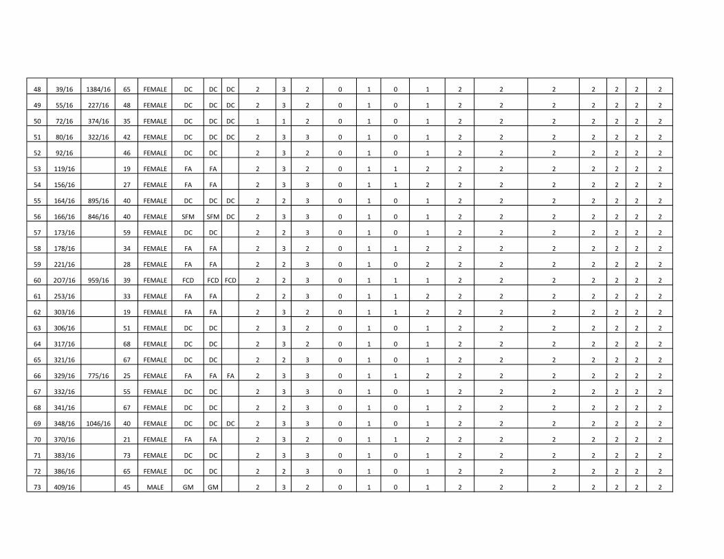
48 39/16 1384/16 65 FEMALE DC DC DC 2 3 2 0 1 0 1 2 2 2 2 2 2 2
49 55/16 227/16 48 FEMALE DC DC DC 2 3 2 0 1 0 1 2 2 2 2 2 2 2
50 72/16 374/16 35 FEMALE DC DC DC 1 1 2 0 1 0 1 2 2 2 2 2 2 2
51 80/16 322/16 42 FEMALE DC DC DC 2 3 3 0 1 0 1 2 2 2 2 2 2 2
52 92/16 46 FEMALE DC DC 2 3 2 0 1 0 1 2 2 2 2 2 2 2
53 119/16 19 FEMALE FA FA 2 3 2 0 1 1 2 2 2 2 2 2 2 2
54 156/16 27 FEMALE FA FA 2 3 3 0 1 1 2 2 2 2 2 2 2 2
55 164/16 895/16 40 FEMALE DC DC DC 2 2 3 0 1 0 1 2 2 2 2 2 2 2
56 166/16 846/16 40 FEMALE SFM SFM DC 2 3 3 0 1 0 1 2 2 2 2 2 2 2
57 173/16 59 FEMALE DC DC 2 2 3 0 1 0 1 2 2 2 2 2 2 2
58 178/16 34 FEMALE FA FA 2 3 2 0 1 1 2 2 2 2 2 2 2 2
59 221/16 28 FEMALE FA FA 2 2 3 0 1 0 2 2 2 2 2 2 2 2
60 2O7/16 959/16 39 FEMALE FCD FCD FCD 2 2 3 0 1 1 1 2 2 2 2 2 2 2
61 253/16 33 FEMALE FA FA 2 2 3 0 1 1 2 2 2 2 2 2 2 2
62 303/16 19 FEMALE FA FA 2 3 2 0 1 1 2 2 2 2 2 2 2 2
63 306/16 51 FEMALE DC DC 2 3 2 0 1 0 1 2 2 2 2 2 2 2
64 317/16 68 FEMALE DC DC 2 3 2 0 1 0 1 2 2 2 2 2 2 2
65 321/16 67 FEMALE DC DC 2 2 3 0 1 0 1 2 2 2 2 2 2 2
66 329/16 775/16 25 FEMALE FA FA FA 2 3 3 0 1 1 2 2 2 2 2 2 2 2
67 332/16 55 FEMALE DC DC 2 3 3 0 1 0 1 2 2 2 2 2 2 2
68 341/16 67 FEMALE DC DC 2 2 3 0 1 0 1 2 2 2 2 2 2 2
69 348/16 1046/16 40 FEMALE DC DC DC 2 3 3 0 1 0 1 2 2 2 2 2 2 2
70 370/16 21 FEMALE FA FA 2 3 2 0 1 1 2 2 2 2 2 2 2 2
71 383/16 73 FEMALE DC DC 2 3 3 0 1 0 1 2 2 2 2 2 2 2
72 386/16 65 FEMALE DC DC 2 2 3 0 1 0 1 2 2 2 2 2 2 2
73 409/16 45 MALE GM GM 2 3 2 0 1 0 1 2 2 2 2 2 2 2
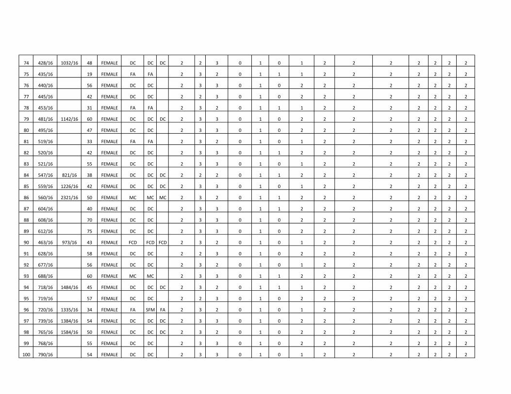
74 428/16 1032/16 48 FEMALE DC DC DC 2 2 3 0 1 0 1 2 2 2 2 2 2 2
75 435/16 19 FEMALE FA FA 2 3 2 0 1 1 1 2 2 2 2 2 2 2
76 440/16 56 FEMALE DC DC 2 3 3 0 1 0 2 2 2 2 2 2 2 2
77 445/16 42 FEMALE DC DC 2 2 3 0 1 0 2 2 2 2 2 2 2 2
78 453/16 31 FEMALE FA FA 2 3 2 0 1 1 1 2 2 2 2 2 2 2
79 481/16 1142/16 60 FEMALE DC DC DC 2 3 3 0 1 0 2 2 2 2 2 2 2 2
80 495/16 47 FEMALE DC DC 2 3 3 0 1 0 2 2 2 2 2 2 2 2
81 519/16 33 FEMALE FA FA 2 3 2 0 1 0 1 2 2 2 2 2 2 2
82 520/16 42 FEMALE DC DC 2 3 3 0 1 1 2 2 2 2 2 2 2 2
83 521/16 55 FEMALE DC DC 2 3 3 0 1 0 1 2 2 2 2 2 2 2
84 547/16 821/16 38 FEMALE DC DC DC 2 2 2 0 1 1 2 2 2 2 2 2 2 2
85 559/16 1226/16 42 FEMALE DC DC DC 2 3 3 0 1 0 1 2 2 2 2 2 2 2
86 560/16 2321/16 50 FEMALE MC MC MC 2 3 2 0 1 1 2 2 2 2 2 2 2 2
87 604/16 40 FEMALE DC DC 2 3 3 0 1 1 2 2 2 2 2 2 2 2
88 608/16 70 FEMALE DC DC 2 3 3 0 1 0 2 2 2 2 2 2 2 2
89 612/16 75 FEMALE DC DC 2 3 3 0 1 0 2 2 2 2 2 2 2 2
90 463/16 973/16 43 FEMALE FCD FCD FCD 2 3 2 0 1 0 1 2 2 2 2 2 2 2
91 628/16 58 FEMALE DC DC 2 2 3 0 1 0 2 2 2 2 2 2 2 2
92 677/16 56 FEMALE DC DC 2 3 2 0 1 0 1 2 2 2 2 2 2 2
93 688/16 60 FEMALE MC MC 2 3 3 0 1 1 2 2 2 2 2 2 2 2
94 718/16 1484/16 45 FEMALE DC DC DC 2 3 2 0 1 1 1 2 2 2 2 2 2 2
95 719/16 57 FEMALE DC DC 2 2 3 0 1 0 2 2 2 2 2 2 2 2
96 720/16 1335/16 34 FEMALE FA SFM FA 2 3 2 0 1 0 1 2 2 2 2 2 2 2
97 739/16 1384/16 54 FEMALE DC DC DC 2 3 3 0 1 0 2 2 2 2 2 2 2 2
98 765/16 1584/16 50 FEMALE DC DC DC 2 3 2 0 1 0 2 2 2 2 2 2 2 2
99 768/16 55 FEMALE DC DC 2 3 3 0 1 0 2 2 2 2 2 2 2 2
100 790/16 54 FEMALE DC DC 2 3 3 0 1 0 1 2 2 2 2 2 2 2

KEY TO MASTER CHART
1. FA- Fibroadenom
2. FCD- Fibrocystic disease of breast
3. GM – Gynecomastia
4. SM – Suspicious of malignancy
5. DC – Ductal carcinoma
6. MC – Mucinous carcinoma
7. LBC – Liquid based cytology
8. CS – Conventional smear
9. FNAC – Fine needle aspiration cytology
10. HPE – Histopathological examination
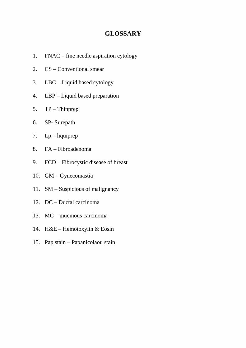
GLOSSARY
1. FNAC – fine needle aspiration cytology
2. CS – Conventional smear
3. LBC – Liquid based cytology
4. LBP – Liquid based preparation
5. TP – Thinprep
6. SP- Surepath
7. Lp – liquiprep
8. FA – Fibroadenoma
9. FCD – Fibrocystic disease of breast
10. GM – Gynecomastia
11. SM – Suspicious of malignancy
12. DC – Ductal carcinoma
13. MC – mucinous carcinoma
14. H&E – Hemotoxylin & Eosin
15. Pap stain – Papanicolaou stain
Recommended