COMMON MIDWIFE TOAD VIRUS: OUTBREAKS IN EUROPE, PATHOLOGY AND
GENOMIC ANALYSESSERIDA
GENOMIC ANALYSESAna Balseiro1, Carla Mavián2, Alberto López-Bueno2, Antonio Alcamí2, Alí Alejo3, Debra Miller4, Rosa Casais1
Centro de Biotecnología Animal, SERIDA, 33394 Gijón, Spain. Centro de Biología Molecular Severo Ochoa (CSIC-UAM), Universidad Autónoma de Madrid, Madrid, Spain. Centro de investigación en sanidad animal, Instituto Nacional de Investigación y Tecnología Agraria y
Ana Balseiro , Carla Mavián , Alberto López-Bueno , Antonio Alcamí , Alí Alejo , Debra Miller , Rosa Casais1Centro de Biotecnología Animal, SERIDA, 33394 Gijón, Spain. 2Centro de Biología Molecular Severo Ochoa (CSIC-UAM), Universidad Autónoma de Madrid, Madrid, Spain. 3Centro de investigación en sanidad animal, Instituto Nacional de Investigación y Tecnología Agraria y
Alimentaria, Valdeolmos, Spain.4Center for Wildlife Health, University of Tennessee, Knoxville, Tennessee, USA.
In Europe, the first known outbreak of common midwife toad virus (CMTV) occurred in northern Spain in 2007
(Balseiro et al., 2009). Since then, three more outbreaks have occurred in Spain and in The Netherlands (Balseiro et
OUTBREAKS IN EUROPECommon midwife toad (Alytes obstetricans)
Picos de Europa (Spain)
(Balseiro et al., 2009). Since then, three more outbreaks have occurred in Spain and in The Netherlands (Balseiro et
al., 2010; Kik et al., 2011, 2012), affecting both wild and captive amphibians, and indicating that the host range and
geographic distribution of CMTV are much wider than previously suspected. CMTV is known to infect wild tadpolesgeographic distribution of CMTV are much wider than previously suspected. CMTV is known to infect wild tadpoles
of the common midwife toad (Alytes obstetricians) and juvenile alpine newts (Triturus alpestris) in Spain; and adult
wild water frogs (Pelophylax spp.) and common newts (Lissotriton vulgaris) in The Netherlands. Recently it has been
reported in captive frogs in the former country.reported in captive frogs in the former country.
GENOMIC ANALYSES
Outbreaks of CMTV in Europe:
Spain and The Netherlands
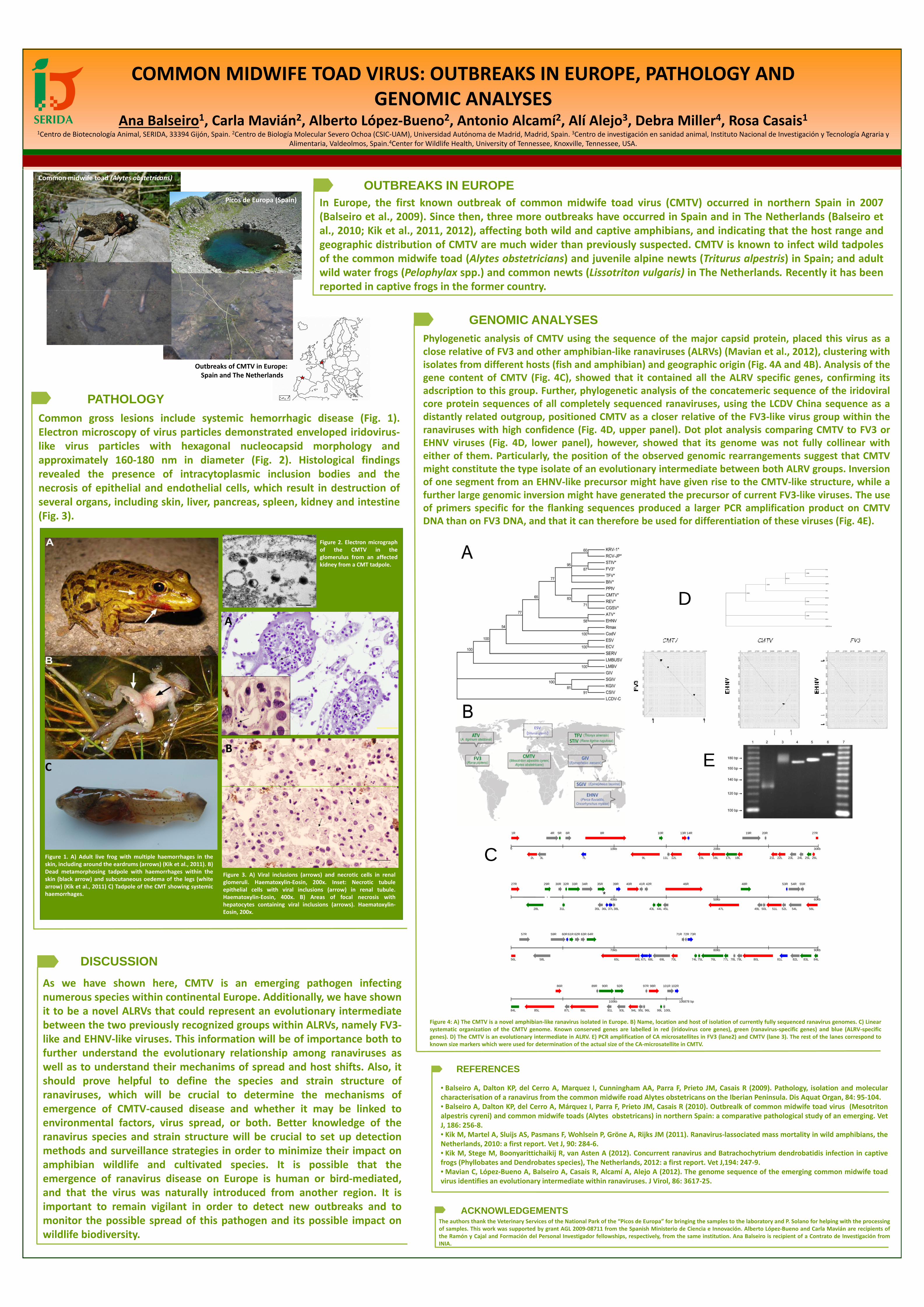
Phylogenetic analysis of CMTV using the sequence of the major capsid protein, placed this virus as a
close relative of FV3 and other amphibian-like ranaviruses (ALRVs) (Mavian et al., 2012), clustering with
isolates from different hosts (fish and amphibian) and geographic origin (Fig. 4A and 4B). Analysis of the
gene content of CMTV (Fig. 4C), showed that it contained all the ALRV specific genes, confirming its
PATHOLOGYCommon gross lesions include systemic hemorrhagic disease (Fig. 1).
Spain and The Netherlands gene content of CMTV (Fig. 4C), showed that it contained all the ALRV specific genes, confirming its
adscription to this group. Further, phylogenetic analysis of the concatemeric sequence of the iridoviral
core protein sequences of all completely sequenced ranaviruses, using the LCDV China sequence as a
distantly related outgroup, positioned CMTV as a closer relative of the FV3-like virus group within theCommon gross lesions include systemic hemorrhagic disease (Fig. 1).
Electron microscopy of virus particles demonstrated enveloped iridovirus-
like virus particles with hexagonal nucleocapsid morphology and
distantly related outgroup, positioned CMTV as a closer relative of the FV3-like virus group within the
ranaviruses with high confidence (Fig. 4D, upper panel). Dot plot analysis comparing CMTV to FV3 or
EHNV viruses (Fig. 4D, lower panel), however, showed that its genome was not fully collinear with
either of them. Particularly, the position of the observed genomic rearrangements suggest that CMTVlike virus particles with hexagonal nucleocapsid morphology and
approximately 160-180 nm in diameter (Fig. 2). Histological findings
revealed the presence of intracytoplasmic inclusion bodies and the
necrosis of epithelial and endothelial cells, which result in destruction of
either of them. Particularly, the position of the observed genomic rearrangements suggest that CMTV
might constitute the type isolate of an evolutionary intermediate between both ALRV groups. Inversion
of one segment from an EHNV-like precursor might have given rise to the CMTV-like structure, while a
further large genomic inversion might have generated the precursor of current FV3-like viruses. The usenecrosis of epithelial and endothelial cells, which result in destruction of
several organs, including skin, liver, pancreas, spleen, kidney and intestine
(Fig. 3).
further large genomic inversion might have generated the precursor of current FV3-like viruses. The use
of primers specific for the flanking sequences produced a larger PCR amplification product on CMTV
DNA than on FV3 DNA, and that it can therefore be used for differentiation of these viruses (Fig. 4E).
Figure 2. Electron micrograph
of the CMTV in the
glomerulus from an affected
kidney from a CMT tadpole.
DA
C:
A
C:
ESV
(Silurus glanis)
C EB
(Silurus glanis)
1R 4R 5R 6R 8R 10R 13R 14R 19R 20R
10kb0 20kb 30kb
27R
Figure 1. A) Adult live frog with multiple haemorrhages in the C
v2L 3L 7L 9L 11L 12L 15L 16L 17L 18L 21L 22L 23L 24L 25L 26L
27R 29R 30R 32R 33R 34R 35R 39R 40R 41R 42R 46R 48R 53R 54R 55R
*
Figure 1. A) Adult live frog with multiple haemorrhages in the
skin, including around the eardrums (arrows) (Kik et al., 2011). B)
Dead metamorphosing tadpole with haemorrhages within the
skin (black arrow) and subcutaneous oedema of the legs (white
arrow) (Kik et al., 2011) C) Tadpole of the CMT showing systemic
haemorrhages.
CFigure 3. A) Viral inclusions (arrows) and necrotic cells in renal
glomeruli. Haematoxylin-Eosin, 200x. Inset: Necrotic tubule
epithelial cells with viral inclusions (arrow) in renal tubule.
Haematoxylin-Eosin, 400x. B) Areas of focal necrosis with v28L 31L 35L 36L 37L 38L 43L 44L 45L 47L 49L 50L 51L 52L 54L 56L
40kb 50kb 60kb
57R 59R 60R61R 62R 64R 71R 72R 73R63R
*haemorrhages.Haematoxylin-Eosin, 400x. B) Areas of focal necrosis with
hepatocytes containing viral inclusions (arrows). Haematoxylin-
Eosin, 200x.
DISCUSSION
As we have shown here, CMTV is an emerging pathogen infecting
57R 59R 60R61R 62R 64R 71R 72R 73R63R
58L 65L 67L 69L 70L 74L 76L 77L 78L 79L 80L 81L 82L 83L 84L68L66L 75L
70kb 80kb 90kb
56L
As we have shown here, CMTV is an emerging pathogen infecting
numerous species within continental Europe. Additionally, we have shown
it to be a novel ALRVs that could represent an evolutionary intermediate 85L 87L 88L 91L
92R
93L 94L 95L 99L 100L
86R 89R 90R 97R 98R 101R 102R
96L
100kb 106878 bp
84Lit to be a novel ALRVs that could represent an evolutionary intermediate
between the two previously recognized groups within ALRVs, namely FV3-
like and EHNV-like viruses. This information will be of importance both to
further understand the evolutionary relationship among ranaviruses as
Figure 4: A) The CMTV is a novel amphibian-like ranavirus isolated in Europe. B) Name, location and host of isolation of currently fully sequenced ranavirus genomes. C) Linear
systematic organization of the CMTV genome. Known conserved genes are labelled in red (iridovirus core genes), green (ranavirus-specific genes) and blue (ALRV-specific
genes). D) The CMTV is an evolutionary intermediate in ALRV. E) PCR amplification of CA microsatellites in FV3 (lane2) and CMTV (lane 3). The rest of the lanes correspond to
known size markers which were used for determination of the actual size of the CA-microsatellite in CMTV.
REFERENCES
further understand the evolutionary relationship among ranaviruses as
well as to understand their mechanims of spread and host shifts. Also, it
should prove helpful to define the species and strain structure of
ranaviruses, which will be crucial to determine the mechanisms of• Balseiro A, Dalton KP, del Cerro A, Marquez I, Cunningham AA, Parra F, Prieto JM, Casais R (2009). Pathology, isolation and molecular
characterisation of a ranavirus from the common midwife road Alytes obstetricans on the Iberian Peninsula. Dis Aquat Organ, 84: 95-104.ranaviruses, which will be crucial to determine the mechanisms of
emergence of CMTV-caused disease and whether it may be linked to
environmental factors, virus spread, or both. Better knowledge of the
ranavirus species and strain structure will be crucial to set up detection
characterisation of a ranavirus from the common midwife road Alytes obstetricans on the Iberian Peninsula. Dis Aquat Organ, 84: 95-104.
• Balseiro A, Dalton KP, del Cerro A, Márquez I, Parra F, Prieto JM, Casais R (2010). Outbrealk of common midwife toad virus (Mesotriton
alpestris cyreni) and common midwife toads (Alytes obstetricans) in northern Spain: a comparative pathological study of an emerging. Vet
J, 186: 256-8.
• Kik M, Martel A, Sluijs AS, Pasmans F, Wohlsein P, Gröne A, Rijks JM (2011). Ranavirus-lassociated mass mortality in wild amphibians, theranavirus species and strain structure will be crucial to set up detection
methods and surveillance strategies in order to minimize their impact on
amphibian wildlife and cultivated species. It is possible that the
emergence of ranavirus disease on Europe is human or bird-mediated,
• Kik M, Martel A, Sluijs AS, Pasmans F, Wohlsein P, Gröne A, Rijks JM (2011). Ranavirus-lassociated mass mortality in wild amphibians, the
Netherlands, 2010: a first report. Vet J, 90: 284-6.
• Kik M, Stege M, Boonyarittichaikij R, van Asten A (2012). Concurrent ranavirus and Batrachochytrium dendrobatidis infection in captive
frogs (Phyllobates and Dendrobates species), The Netherlands, 2012: a first report. Vet J,194: 247-9.
• Mavian C, López-Bueno A, Balseiro A, Casais R, Alcamí A, Alejo A (2012). The genome sequence of the emerging common midwife toad
virus identifies an evolutionary intermediate within ranaviruses. J Virol, 86: 3617-25.emergence of ranavirus disease on Europe is human or bird-mediated,
and that the virus was naturally introduced from another region. It is
important to remain vigilant in order to detect new outbreaks and to
monitor the possible spread of this pathogen and its possible impact onACKNOWLEDGEMENTS
The authors thank the Veterinary Services of the National Park of the “Picos de Europa” for bringing the samples to the laboratory and P. Solano for helping with the processing
virus identifies an evolutionary intermediate within ranaviruses. J Virol, 86: 3617-25.
monitor the possible spread of this pathogen and its possible impact on
wildlife biodiversity.
The authors thank the Veterinary Services of the National Park of the “Picos de Europa” for bringing the samples to the laboratory and P. Solano for helping with the processing
of samples. This work was supported by grant AGL 2009-08711 from the Spanish Ministerio de Ciencia e Innovación. Alberto López-Bueno and Carla Mavián are recipients of
the Ramón y Cajal and Formación del Personal Investigador fellowships, respectively, from the same institution. Ana Balseiro is recipient of a Contrato de Investigación from
INIA.
Recommended