CLASSIFICATION OF
GLOMERULAR DISEASE
Wesam Ismail, MD Pathology department
Beni-Suef University

GLOMERULAR DISEASES Glomerulonephritis (GN), a complex syndrome encompassing a variety of individual
disorders, is associated with significant morbidity and mortality (ESRD, hospitalization
or death)
Rare Disease???
Initial estimates of presumed GN incidence and period prevalence in the USA
o GN may be more common than is traditionally appreciated and increase
substantially with age
o Type of GN, classified as primary or secondary, is associated with both age and
sex
o GN is associated with a substantial hospitalization burden, progression to ESRD,
and death
o Tens of thousands of people appear to be affected by GN in the USA alone,
making GN an important public health concern
Kidney international. Article in press. July 2016

GLOMERULAR DISEASES
? Am I missing something?
? Should I biopsy?
? If yes, When?
? Should I treat first, then biopsy?
Diseases of the glomerulus although complex have always held a
special place of interest for nephrologists
Renal Biopsy
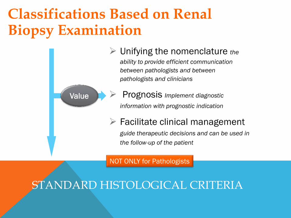
STANDARD HISTOLOGICAL CRITERIA
Classifications Based on Renal Biopsy Examination
Unifying the nomenclature the
ability to provide efficient communication
between pathologists and between
pathologists and clinicians
Prognosis Implement diagnostic
information with prognostic indication
Facilitate clinical management guide therapeutic decisions and can be used in
the follow-up of the patient
Value
NOT ONLY for Pathologists
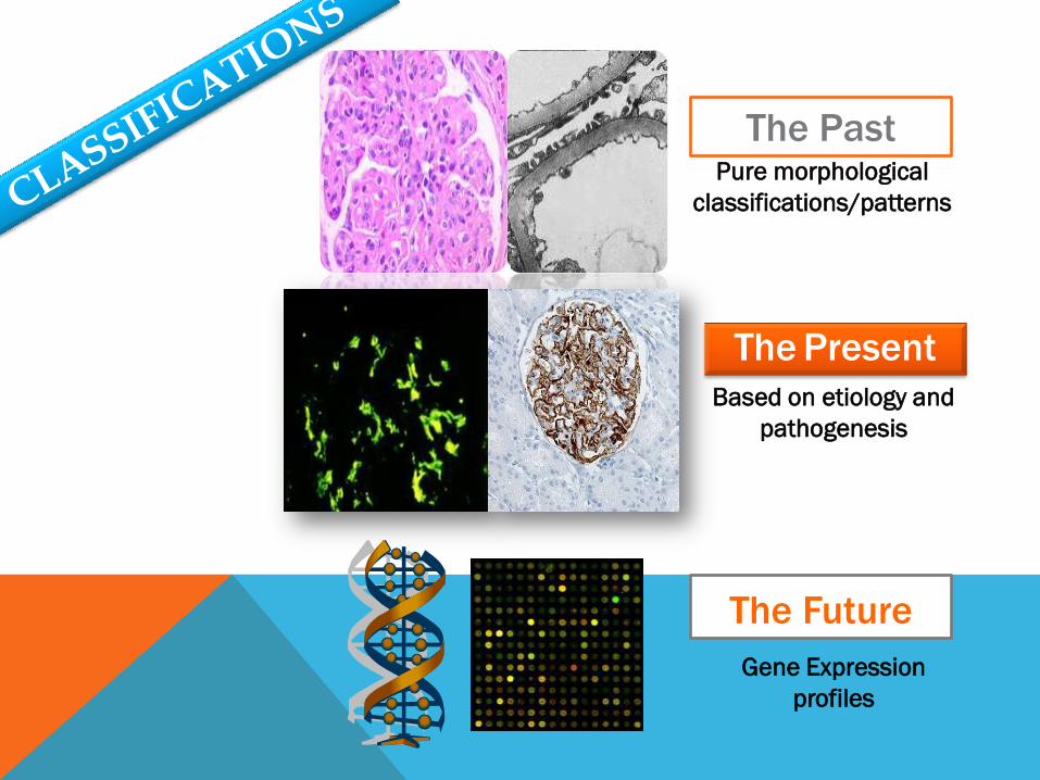
The Past
Based on etiology and
pathogenesis
The Present
Gene Expression
profiles
The Future
Pure morphological
classifications/patterns

Renee Habib
1970s
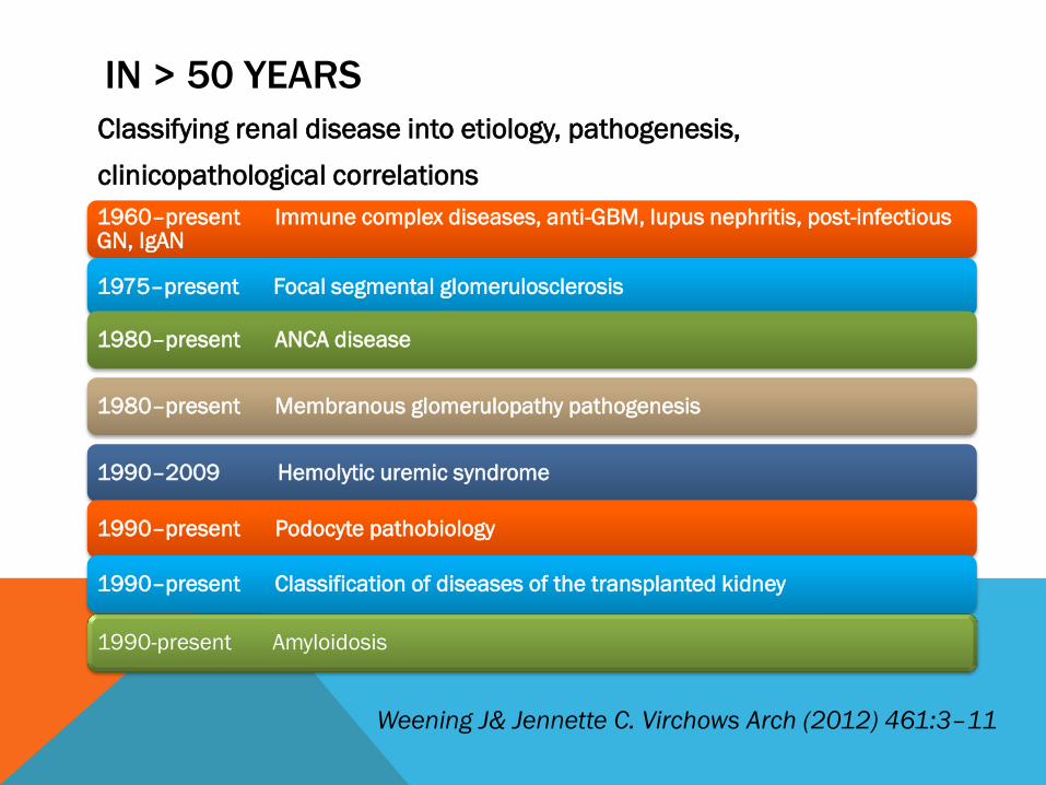
IN > 50 YEARS
Classifying renal disease into etiology, pathogenesis,
clinicopathological correlations
1960–present Immune complex diseases, anti-GBM, lupus nephritis, post-infectious GN, IgAN
1975–present Focal segmental glomerulosclerosis
1980–present ANCA disease
1980–present Membranous glomerulopathy pathogenesis
1990–2009 Hemolytic uremic syndrome
1990–present Podocyte pathobiology
1990–present Classification of diseases of the transplanted kidney
1990-present Amyloidosis
Weening J& Jennette C. Virchows Arch (2012) 461:3–11
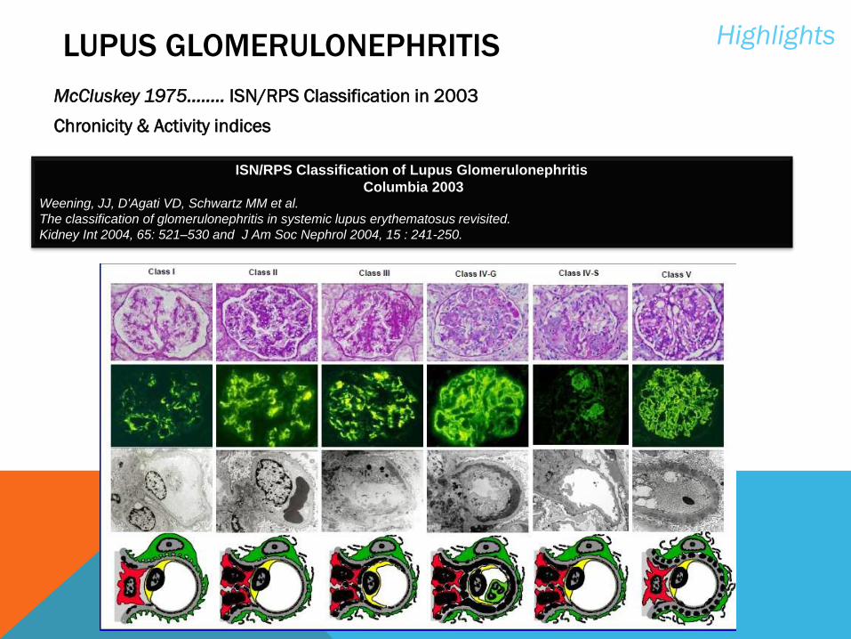
LUPUS GLOMERULONEPHRITIS
McCluskey 1975…..... ISN/RPS Classification in 2003
Chronicity & Activity indices
Highlights
ISN/RPS Classification of Lupus Glomerulonephritis
Columbia 2003 Weening, JJ, D'Agati VD, Schwartz MM et al.
The classification of glomerulonephritis in systemic lupus erythematosus revisited.
Kidney Int 2004, 65: 521–530 and J Am Soc Nephrol 2004, 15 : 241-250.

„The major objective is to standardize definitions,
emphasize clinically relevant lesions, and
encourage uniform and reproducible reporting
between centers.”
MEMBRANOUS GLOMERULOPATHY Highlights
Neonatal, alloimmune : NEP
Early childhood MN : BSA
Primary « Idiopathic » MN
- 70-80% : PLA2R (+ other specificities:AR, SOD2, enolase..?)
- 20-30% : THSD7A,food/environmental Ag (BSA)
« Secondary » MN
Prognostic significance
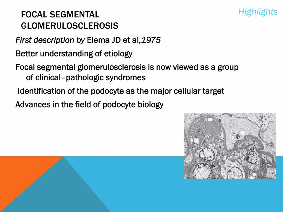
First description by Elema JD et al,1975
Better understanding of etiology
Focal segmental glomerulosclerosis is now viewed as a group
of clinical–pathologic syndromes
Identification of the podocyte as the major cellular target
Advances in the field of podocyte biology
FOCAL SEGMENTAL
GLOMERULOSCLEROSIS
Highlights
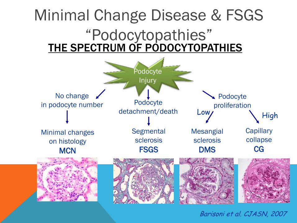
No change
in podocyte number
THE SPECTRUM OF PODOCYTOPATHIES
Low High
Barisoni et al. CJASN, 2007
Minimal Change Disease & FSGS
“Podocytopathies”
Podocyte
detachment/death
Podocyte
proliferation
Minimal changes
on histology
MCN
Segmental
sclerosis
FSGS
Mesangial
sclerosis
DMS
Capillary
collapse
CG
Podocyte
Injury
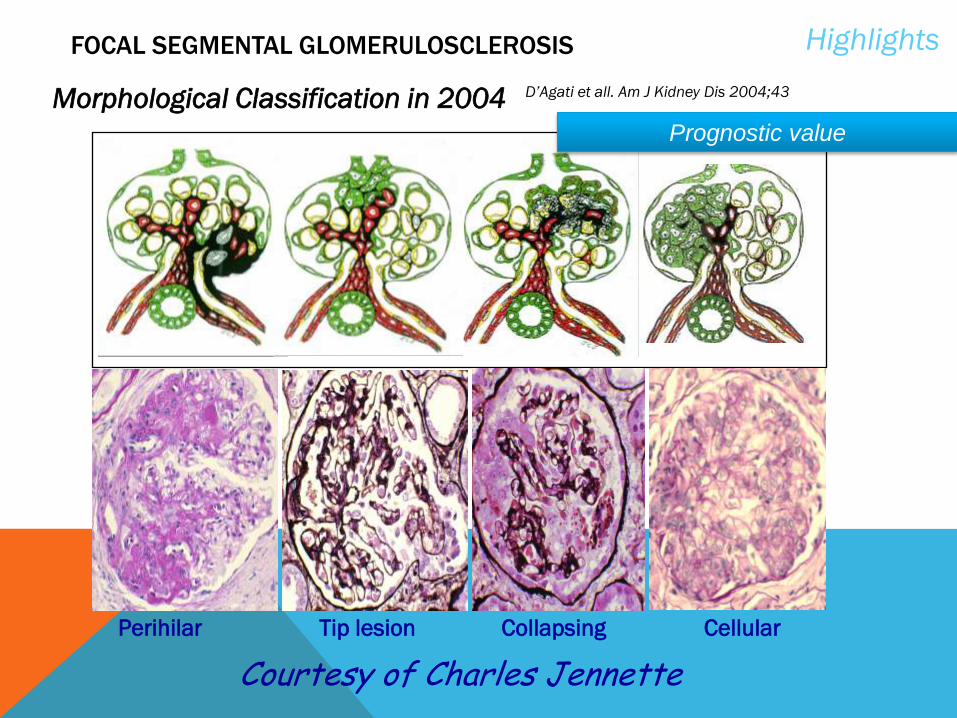
Morphological Classification in 2004 D’Agati et all. Am J Kidney Dis 2004;43
FOCAL SEGMENTAL GLOMERULOSCLEROSIS Highlights
Perihilar Tip lesion Collapsing Cellular
Courtesy of Charles Jennette
Prognostic value

Mesangial hypercellularity M0: <4 mesangial/cells/area in > 50% of the glomeruli
M1: 4 mesangial cells/area in >50% of the glomeruli
Segmental glomerulosclerosis or adhesion S0: absent
S1 present
Endocapillary hypercellularity E0: absent
E1: present
Tubular atrophy/interstitial fibrosis T0: 0-25%
T1: 26-50%
T2: >50%
THE OXFORD CLASSIFICATION (EVIDENCE BASED)
Independent value in predicting renal outcome
IgA NEPHROPATHY Highlights
Several Classifications
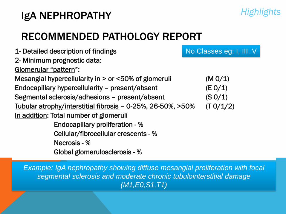
RECOMMENDED PATHOLOGY REPORT
1- Detailed description of findings
2- Minimum prognostic data:
Glomerular “pattern”:
Mesangial hypercellularity in > or <50% of glomeruli (M 0/1)
Endocapillary hypercellularity – present/absent (E 0/1)
Segmental sclerosis/adhesions – present/absent (S 0/1)
Tubular atrophy/interstitial fibrosis – 0-25%, 26-50%, >50% (T 0/1/2)
In addition: Total number of glomeruli
Endocapillary proliferation - %
Cellular/fibrocellular crescents - %
Necrosis - %
Global glomerulosclerosis - %
Example: IgA nephropathy showing diffuse mesangial proliferation with focal
segmental sclerosis and moderate chronic tubulointerstitial damage
(M1,E0,S1,T1)
No Classes eg: I, III, V
IgA NEPHROPATHY Highlights

In 1959, Gellman et al. first reported findings
Gambara et al. and Fioretto et al made basic
distinctions between typical and atypical DN
Glomerular diseases superimposed on DN
Podocytopathies
Morphological Classification in 2010 (Research
committee of RPS)
J Am Soc Nephrol 21: 556–563, 2010
I Mild or nonspecific LM changes and EM-proven GBM thickening
IIa Mild mesangial expansion
IIb Severe mesangial expansion
III Nodular sclerosis (Kimmelstiel– Wilson lesion)
IV Advanced diabetic glomerulosclerosis
TERVAERT’S PATHOLOGIC CLASSIFICATION OF DIABETIC NEPHROPATHY
Diabetic NEPHROPATHY Highlights
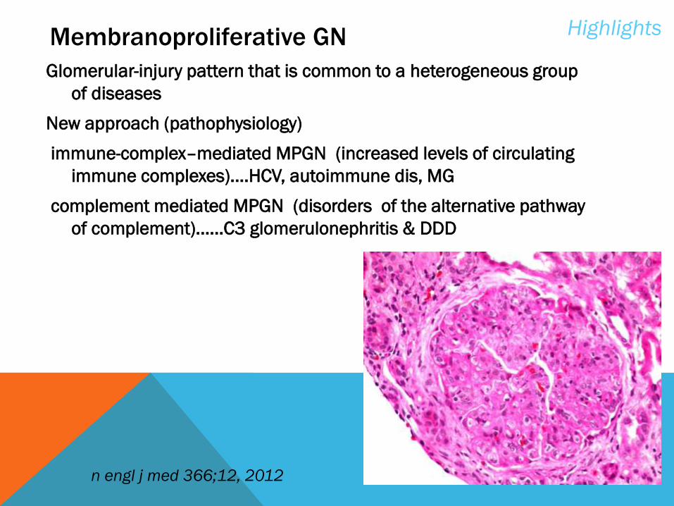
Glomerular-injury pattern that is common to a heterogeneous group
of diseases
New approach (pathophysiology)
immune-complex–mediated MPGN (increased levels of circulating
immune complexes)….HCV, autoimmune dis, MG
complement mediated MPGN (disorders of the alternative pathway
of complement)……C3 glomerulonephritis & DDD
n engl j med 366;12, 2012
Membranoproliferative GN Highlights
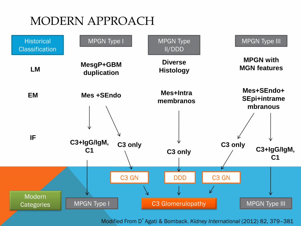
MODERN APPROACH
Historical
Classification
MPGN Type I MPGN Type
II/DDD
MPGN Type III
LM
EM
IF
Modern
Categories
Mes +SEndo
MPGN with
MGN features Diverse
Histology MesgP+GBM
duplication
Mes+SEndo+
SEpi+intrame
mbranous
Mes+Intra
membranos
C3 only C3+IgG/IgM,
C1 C3 only C3 only
C3+IgG/IgM,
C1
C3 GN DDD C3 GN
MPGN Type I C3 Glomerulopathy MPGN Type III
Modified From D’Agati & Bomback. Kidney International (2012) 82, 379–381
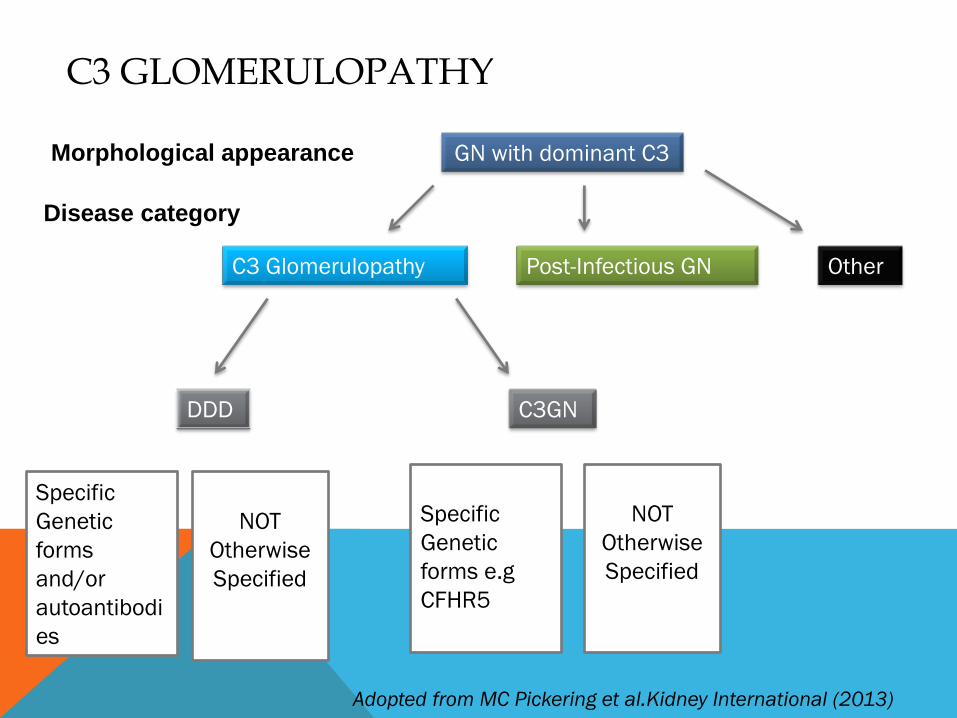
C3 GLOMERULOPATHY
Morphological appearance GN with dominant C3
Disease category
C3 Glomerulopathy Post-Infectious GN Other
DDD C3GN
Specific
Genetic
forms
and/or
autoantibodi
es
NOT
Otherwise
Specified
Specific
Genetic
forms e.g
CFHR5
NOT
Otherwise
Specified
Adopted from MC Pickering et al.Kidney International (2013)
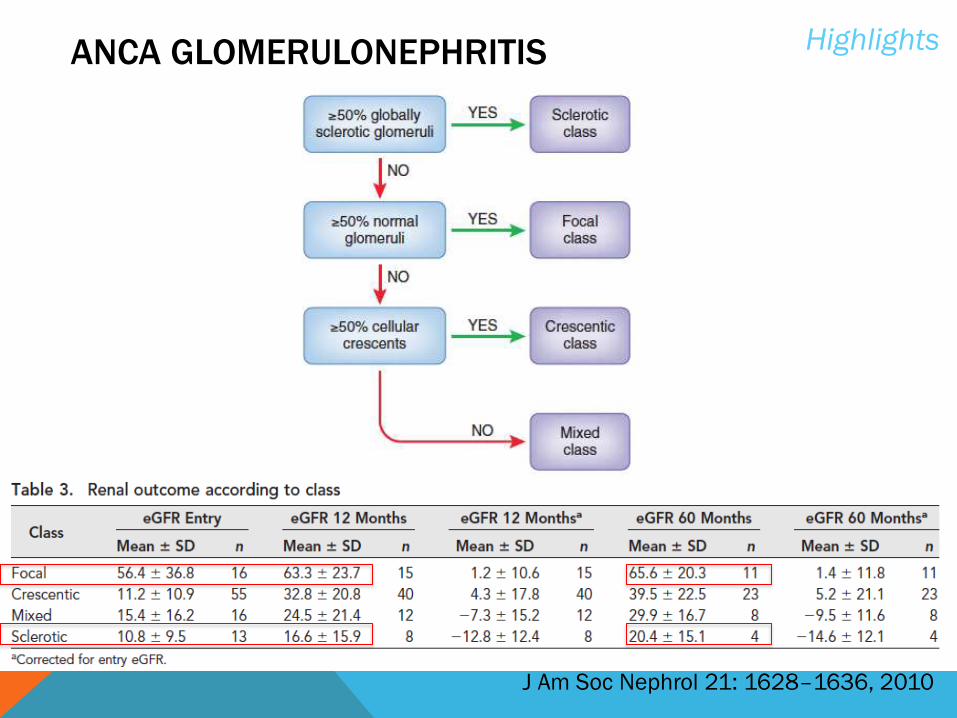
J Am Soc Nephrol 21: 1628–1636, 2010
ANCA GLOMERULONEPHRITIS Highlights
MAYO CLINIC/RENAL PATHOLOGY SOCIETY CONSENSUS
REPORT ON PATHOLOGIC CLASSIFICATION, DIAGNOSIS, AND
REPORTING OF GN

Classification of GN on the basis of etiology/pathogenesis
GLOMERULONEPHRITIS
is primarily on the basis of the findings by immunofluorescence microscopy
(IF) or immunohistochemistry (IHC) integrated with light microscopy (LM) and
electron microscopy (EM)
The manuscript does not extend to other forms of glomerular diseases,
such as membranous nephropathy, podocytopathies, and
thrombotic microangiopathy

On the basis of etiology/pathogenesis
Five pathogenic types, each with specific disease
entities:
1. Immune-complex GN
2. Pauci-immune GN
3. Antiglomerular basement membrane GN
4. Monoclonal IgGN
5. C3 glomerulopathy
GLOMERULONEPHRITIS

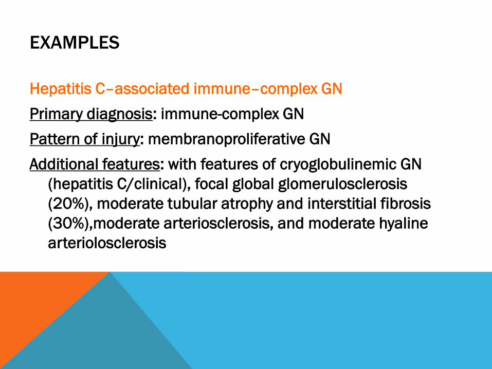
Hepatitis C–associated immune–complex GN
Primary diagnosis: immune-complex GN
Pattern of injury: membranoproliferative GN
Additional features: with features of cryoglobulinemic GN
(hepatitis C/clinical), focal global glomerulosclerosis
(20%), moderate tubular atrophy and interstitial fibrosis
(30%),moderate arteriosclerosis, and moderate hyaline
arteriolosclerosis
EXAMPLES
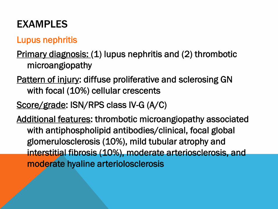
Lupus nephritis
Primary diagnosis: (1) lupus nephritis and (2) thrombotic
microangiopathy
Pattern of injury: diffuse proliferative and sclerosing GN
with focal (10%) cellular crescents
Score/grade: ISN/RPS class IV-G (A/C)
Additional features: thrombotic microangiopathy associated
with antiphospholipid antibodies/clinical, focal global
glomerulosclerosis (10%), mild tubular atrophy and
interstitial fibrosis (10%), moderate arteriosclerosis, and
moderate hyaline arteriolosclerosis
EXAMPLES
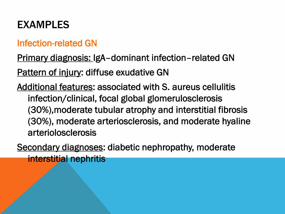
Infection-related GN
Primary diagnosis: IgA–dominant infection–related GN
Pattern of injury: diffuse exudative GN
Additional features: associated with S. aureus cellulitis
infection/clinical, focal global glomerulosclerosis
(30%),moderate tubular atrophy and interstitial fibrosis
(30%), moderate arteriosclerosis, and moderate hyaline
arteriolosclerosis
Secondary diagnoses: diabetic nephropathy, moderate
interstitial nephritis
EXAMPLES
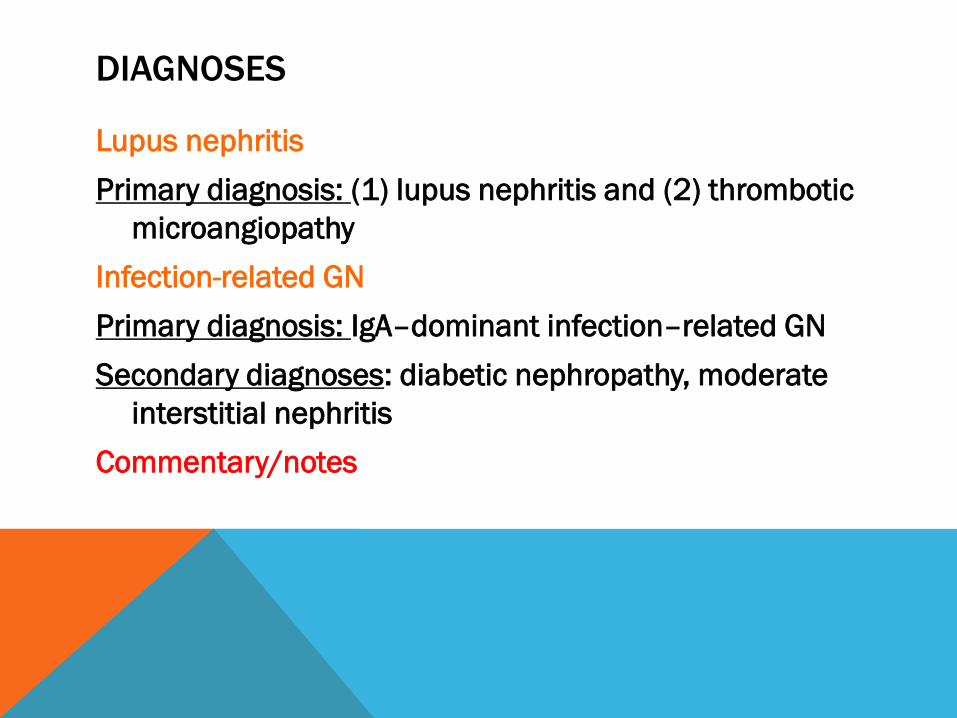
Lupus nephritis
Primary diagnosis: (1) lupus nephritis and (2) thrombotic
microangiopathy
Infection-related GN
Primary diagnosis: IgA–dominant infection–related GN
Secondary diagnoses: diabetic nephropathy, moderate
interstitial nephritis
Commentary/notes
DIAGNOSES
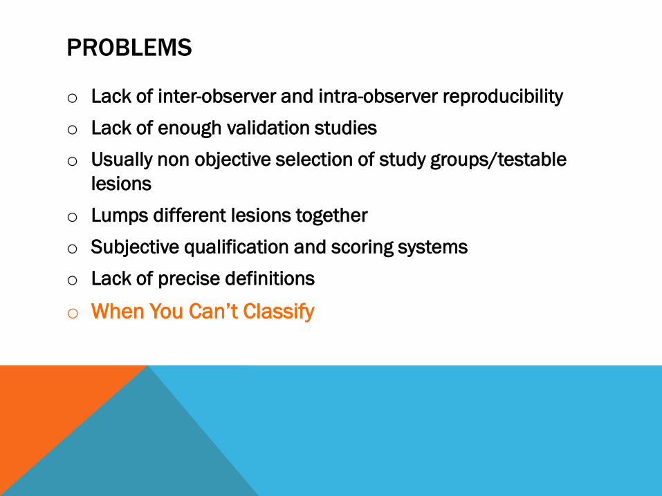
PROBLEMS
o Lack of inter-observer and intra-observer reproducibility
o Lack of enough validation studies
o Usually non objective selection of study groups/testable
lesions
o Lumps different lesions together
o Subjective qualification and scoring systems
o Lack of precise definitions
o When You Can’t Classify
FUTURE APPROACHES “VISION”
The challenge of functional genomics in pathology is to turn
expression and sequence data into information that can be used
to help diagnose disease
Laser-assisted microdissection allowed the evaluation of mRNA
expression on material fixed and processed for routine diagnostic
evaluation
Integrated Diagnosis
Gene expression profiles performed in parallel to routine work-up of biopsies
giving independent information in the diagnostic process using microarrays..high
quality RNA
Add on technique
applied to defined differential diagnostic problems after completion
of the routine diagnostic work-up, e.g. FSGS vs Minimal Change
Gene Expression Profiles
PROSPECTIVE
In the last decade
Tremendous advances have been made in our understanding of the
pathology and pathophysiology of kidney disease as a result of
intense collaboration between nephrologists and nephropathologists
Over the next years
The generation of comprehensive expression profiles for the most
frequent renal diseases can be expected and may allow the
definition of clinical subgroups with different disease courses

Thank you
Recommended