1
Supporting Information for:
Chromate reduction in Fe(II)-containing soil affected by hyperalkaline
leachate from chromite ore processing residue
Robert A. Whittleston1, Douglas I. Stewart2*, Robert J. G. Mortimer1, Zana C. Tilt1, Andrew P. Brown3, Kalotina Geraki4, and Ian T. Burke1*.
1 School of Earth and Environment,
2 School of Civil Engineering,
3 School of Process, Environmental and
Materials Engineering, University of Leeds, Leeds LS2 9JT, UK
4 Diamond Light Source, Harwell Science and Innovation Campus, Didcot, Oxfordshire, OX11 0DE, UK
* Corresponding Author’s E-mail: [email protected] / [email protected]
This section consists of 13 pages, 4 tables, and 3 figures.
Section 1. Sample characterisation.
X-ray powder diffraction (XRD) analysis of the soils (ground to < 75 µm) was performed on a
Philips PW1050 Goniometer, and X-ray fluorescence (XRF) analysis was undertaken using a fused
sample on a Philips PW2404 wavelength dispersive sequential X-ray spectrometer (data were
corrected for loss on ignition). Approximately 25g of homogenised soil was oven dried at 105C and
disaggregated with a mortar and pestle for carbon content determination. A portion of each sample
was pre-treated with 10% HCl to remove any carbonates present [1]. The total organic and inorganic
carbon contents of oven dried and HCl treated subsamples were measured using Carlo-Erba 1106
elemental analyser.
Soil pH was measured following the ASTM standard method for pH in soils, with 10g soil made up
to a 1:1 suspension with deionised water. The suspension was left for 1 hour before measurement
was taken using a Orion bench top meter and calibrated electrodes [2]. Water soluble Cr(VI) content
in soils was measured using 10:1 suspension of soil to deionised water, shaken for 24 hours at 150

2
rpm in 15ml centrifuge tubes. 1.5 ml was then extracted and centrifuged (3 min, 16,000 g). Aqueous
Cr(VI) concentration in the supernatant was then determined using standard UV/VIS spectroscopy
methods based described in main paper [3].
A freeze dried sample of soil from B2 310 was embedded in a block of epoxy resin under a
vacuum and prepared for SEM analysis by polishing in non-aqueous oils and carbon coating
(approximately 5-10 nm thickness). Scanning Electron Microscopy (SEM) was performed on a FEI
Quanta 650 FEG-ESEM to produce backscatter electron images. Elemental quantification was carried
out on the ESEM using an electron probe micro analyser (EPMA). Thirty-three 15 x 15 µm regions of
the fine matrix material of B2 310 resin embedded thin section were analysed for common rock
forming oxides. The measured elemental wt% were then corrected by assuming a total oxide weight
of 100% to account for the influence of resin infilling into pore spaces. An average composition of
these 33 regions is reported in the results section, with error given as the range of the results.
Soil B2 310 was prepared for (S)TEM imaging by freeze drying and sieving at 150 mesh to collect
the less than 104 µm sized fraction. Approximately 0.05 g was then mixed with 1 ml of LR WhiteTM
resin and polymerised for 24 hours at 70°C. A thin section of resin embedded soil was then cut using
a microtome (80 nm) and placed on a Cu support grid (Agar Scientific, UK) and carbon coated (~5
um) prior to (S)TEM analysis. The specimen was examined using a Philips/FEI CM200 field emission
gun TEM fitted with a scanning (STEM) unit and an ultra thin window energy dispersive X-ray
detector (Oxford Instruments ISIS EDX). The total Cr concentration in this soil was ~0.3 % w/w, which
if homogeneously dispersed is close to the limit of detection for EDX analysis.
Section 2. XAS analysis.
Samples for XAS analysis were chosen from a range of borehole samples, and from reoxidation
experiments on soil B2-310. , Soil slurry from one reoxidation experiment was centrifuged (10 min,
8,000 g), and the soil was recovered and stored in the dark at -80°C. All other samples for XAS
analysis were stored at -80°C in the dark under a nitrogen atmosphere.
Cr K-edge spectra were collected on beamline I18 at the Diamond Light Source in November
2009, operating at 3 GeV with a typical current of 200 mA, using a nitrogen cooled Si(111) double
crystal monochromator and focussing optics. A pair of plane mirrors was used to reduce the
harmonic content of the beam and microfocusing was done using Kirkpatrick-Baez mirrors. The

3
beam size at the sample was approximately 150 µm. For standards the Cr K-edge spectra were
collected in transmission mode at room temperatures (~295 °K). Standards were prepared as 50 mg
pressed pellets and diluted with BN as required. For soil samples data were collected in fluorescence
mode using a 9 element solid state Ge detector. Soil samples were centrifuged to form a moist pellet
and were mounted at 1 mm thickness in aluminium holders with Kapton™ widows. Experiments
were performed at liquid nitrogen temperatures (approximately 78 °K) and multiple scans averaged
to improve the signal to noise ratio using Athena version 0.8.056 [4]. For XANES spectra absorption
was also normalised in Athena over the full data range and plotted from 5980 eV to 6030 eV with no
correction required for drift in E0. For EXAFS analysis data was background subtracted using PySpline
v1.1 [5].
Section 3 EXAFS Analysis.
Background subtracted EXAFS spectra were analysed in DLexcurv v1.0 [6] using full curved wave
theory [7]. Phaseshifts were derived from ab initio calculations using Hedin-Lundqvist potentials and
von-Barth ground states [8]. Fourier transforms of the EXAFS spectra were used to obtain an
approximate radial distribution function around the central Cr atom (the absorber atom); the peaks
of the Fourier transform can be related to “shells” of surrounding backscattering ions characterised
by atom type, number of atoms, absorber-scatterer distance, and the Debye-Waller factor (± 25%),
2σ2. Atomic distances calculated by DLexcurv have an error of approximately ± 0.02 and ± 0.05 Å in
the first and outer shells respectively [9]. The data were fitted for each sample by defining a
theoretical model and comparing the calculated EXAFS spectrum with experimental data and with
published spectra for Cr-substituted compounds [10]. Shells of backscatterers were added around
the Cr and by refining an energy correction Ef (the Fermi Energy; which for final fits typically varied
between -19 and -17), the absorber-scatterer distance, and the Debye-Waller factor for each shell, a
least squares residual (the R factor [11]) was minimised. The amplitude factor (or AFAC in DLexcurv
V1.0) was retained as the default of 1 throughout. Shells or groups of shells were only included if the
overall fit (R-factor) was reduced overall by >5%. For shells of scatterers around the central Cr, the
number of atoms in the shell was chosen as an integer to give the best fit and further refined.

4
Section 4 Microbial community analysis.
4.1 Iron reducing alkaline media
To culture alkaliphilic anaerobic iron reducers indigenous to the B2 310 soil layer an alkaline
media containing Fe(III) was developed. The base media contained NaH2PO4.H2O (0.356 g/l), KCl (0.1
g/l) and 10 ml of standard vitamin and mineral mixtures [12]. Fe(III) citrate (2 g/l) and yeast extract
(2 g/l) were added as the sole sources of electron acceptors and donors. The media was made using
DIW that was first deoxygenated by boiling and nitrogen purging. The pH was adjusted to 9.2 using a
1M solution of sodium carbonate and 4.5 ml aliquots transferred to 5 ml glass serum bottles. These
were then sealed using butyl rubber stoppers and aluminium crimps, and heat sterilised at 120°C for
20 minutes.
4.2. DNA extraction and cloning procedure
Microbial DNA was extracted from a 400 mg sample of B2-310 initial soil using a FastDNA spin kit
(Qbiogene, Inc.) and FastPREP instrument (unless explicitly stated, the manufacturer’s protocols
supplied with all kits employed were followed precisely). In order to extract microbial DNA from the
iron reducing consortiums, a culture bottle was opened, transferred into a 15 ml centrifuge tube and
centrifuged at 3500 rpm for 10 minutes. Supernatant was then discarded and pellet resuspended in
978 l of sodium phosphate buffer supplied in the FastDNA spin kit. The suspension was then
transferred to a Lysing Matrix E tube also supplied in the FastDNA spin kit, and manufacturers’
protocols subsequently followed precisely. DNA fragments in the size range 3 kb ~20 kb were
isolated on a 1% “1x” Tris-borate-EDTA (TBE) gel stained with ethidium bromide to enable viewing
under UV light (10x TBE solution from Invitrogen Ltd., UK). The DNA was extracted from the gel using
a QIAquick gel extraction kit (QIAGEN Ltd., UK.).
A fragment of the 16s rRNA gene of approximately 500bp was amplified using using broad-
specificity bacterial primers 8f (5´-AGAGTTTGATCCTGGCTCAG-3´) [13] and 519r (5´-
GWATTACCGCGGCKGCTG-3´) where K = G or T, W = A or T [14]. Each PCR reaction mixture contained
20 μl of purified DNA solution, GoTaq DNA polymerase (5 units), 1× PCR reaction buffer, MgCl2
(1.5mM), PCR nucleotide mix (0.2 mM), T4 Gene 32 Protein (100 ng/µl) and 8f and 519r primers (0.6
μM each) in a final volume of 50 μl. The reaction mixtures were incubated at 95C for 2 min, and

5
then cycled 30 times through three steps: denaturing (95C, 30 s), annealing (50C, 30s), primer
extension (72C, 45 s). This was followed by a final extension step at 72C for 7min. The PCR
products were purified using a QIAquick PCR Purification Kit. Amplification product sizes were
verified by electrophoresis of 10 μl samples in a 1.0% agarose TBE gel with ethidium bromide
straining.
The PCR product was ligated into the standard cloning vector (pGEM-T Easy; Promega Corp.,
USA), and transformed into E. coli competent cells (XL1-Blue; Agilent Technologies UK Ltd).
Transformed cells were grown on LB-agar plates containing ampicillin (100 µg.ml-1) at 37C for 17
hours. The plates were surfaced dressed with IPTG and X-gal (as per the XL1-Blue protocol) for blue-
white colour screening. For each sample, 48 colonies containing a plasmid with the 16s rRNA gene
insert were restreaked on LB-ampicillin agar plates and incubated at 37C. 48 single colonies for
each condition from restreaked plates were stab inoculated into an LB-agar ampicillin 96 well plate
and sent for sequencing (GATC Biotech Ltd). In order to increase confidence that a suitable
representation of the highly diverse B2 310 soil population had been collected, a further 16 plasmids
were sent for sequencing. Single colonies from restreaked plates were incubated overnight at 37°C
in LB media. Bacterial plasmids were then extracted from the resulting broth using the Promega
Pureyield™ Plasmid Miniprep Purification system (Promega Corp., USA). The concentration of
plasmid DNA was then determined using UV spectroscopy, and adjusted to 50 ng.µl-1 with deionised
water. 12µl of each plasmid mixture was then sent for sequencing at the Faculty of Biological
Sciences, University of Leeds, Leeds, UK.
4.3 Phylogenic assignment.
16s rRNA bacterial sequence fragments, ~500 bp, of cloned DNA from B2 310 initial soil sample
and isolated iron reducing consortium were checked with both Bellerophon[15] and Mallard v
1.02[16] online chimera checkers in order to exclude putative chimeras from subsequent analyses.
Sequences were then assigned to bacterial phyla using the online Ribosomal Database Project (RDP)
naïve Bayesian Classifier version 2.2 (available online: http://rdp.cme.msu.edu/classifier/
classifier.jsp) in August 2010. The sequences were assigned to the taxonomical hierarchy: RDP
training set 6, based on nomenclatural taxonomy and Bergey’s Manual, with a confidence threshold

6
of 95% (assignments to a phylum reported in Figure 4 are based on >98% confidence). Full details of
this algorithm, its development and its performance can be found in Wang et al., (2007)[17]
Supporting Information References
[1] B.A. Schumacher, Methods for the Determination of Total Organic Carbon (TOC) in Soils and Sediments, in, United States Environmental Protection Agency, Las Vegas, 2002. [2] ASTM, D4972-01: standard test method for pH of soils. Annual book of ASTM standards, American Society for Testing and Materials, 4 (2006) 963-965. [3] USEPA, SW-846 Manual: Method 7196a. Chromium hexavalent (colorimetric). Retrieved 6th Jan 2006, (1992). [4] B. Ravel, M. Newville, ATHENA, ARTEMIS, HEPHAESTUS: data analysis for X-ray absorption spectroscopy using IFEFFIT, J. Synchrot. Radiat., 12 (2005) 537-541. [5] A. Tenderholt, B. Hedman, K.O. Hodgson, PySpline: A modern, cross-platform program for the processing of raw averaged XAS edge and EXAFS data, in: B. Hedman, P. Painetta (Eds.) X-Ray Absorption Fine Structure-XAFS13, 2007, pp. 105-107. [6] S. Tomic, B.G. Searle, A. Wander, N.M. Harrison, A.J. Dent, J.F.W. Mosselmans, J.E. Inglesfield, CCLRC Technical Report DL-TR-2005-001, ISSN 1362-0207(2005), in, 2005. [7] S.J. Gurman, N. Binsted, I. Ross, A Rapid, Exact Curved-Wave Theory for Exafs Calculations, J. Phys. C. Solid State, 17 (1984) 143-151. [8] N. Binsted, CLRC Daresbury Laboratory EXCURV98 program, CLRC Daresbury Laboratory, Warrington, UK, 1998. [9] I.T. Burke, C. Boothman, J.R. Lloyd, R.J.G. Mortimer, F.R. Livens, K. Morris, Effects of progressive anoxia on the solubility of technetium in sediments, Environ. Sci. Technol., 39 (2005) 4109-4116. [10] L. Charlet, A. Manceau, X-Ray Absorption Spectroscopic Study of the Sorption of Cr(Iii) at the Oxide Water Interface .2. Adsorption, Coprecipitation, and Surface Precipitation on Hydrous Ferric-Oxide, J. Colloid Interface Sci., 148 (1992) 443-458. [11] N. Binsted, R.W. Strange, S.S. Hasnain, Constrained and Restrained Refinement in Exafs Data-Analysis with Curved Wave Theory, Biochemistry-US, 31 (1992) 12117-12125. [12] R.A. Bruce, L.A. Achenbach, J.D. Coates, Reduction of (per)chlorate by a novel organism isolated from paper mill waste, Environ. Microbiol., 1 (1999) 319-329. [13] P.A. Eden, T.M. Schmidt, R.P. Blakemore, N.R. Pace, Phylogenetic Analysis of Aquaspirillum-Magnetotacticum Using Polymerase Chain Reaction-Amplified 16s Ribosomal-Rna-Specific DNA, Int. J. Syst. Bacteriol., 41 (1991) 324-325. [14] D.J. Lane, B. Pace, G.J. Olsen, D.A. Stahl, M.L. Sogin, N.R. Pace, Rapid determination of 16S ribosomal RNA sequences for phylogenetic analysis, P. Natl. Acad. Sci. USA, 82 (1985) 6955-6959. [15] T. Huber, G. Faulkner, P. Hugenholtz, Bellerophon: a program to detect chimeric sequences in multiple sequence alignments, Bioinformatics, 20 (2004) 2317-2319. [16] K.E. Ashelford, N.A. Chuzhanova, J.C. Fry, A.J. Jones, A.J. Weightman, New screening software shows that most recent large 16S rRNA gene clone libraries contain chimeras, Appl. Environ. Microbiol., 72 (2006) 5734-5741. [17] Q. Wang, G.M. Garrity, J.M. Tiedje, J.R. Cole, Naive Bayesian classifier for rapid assignment of rRNA sequences into the new bacterial taxonomy, Appl. Environ. Microbiol., 73 (2007) 5261-5267. [18] W.D. Derbyshire, H.J. Yearian, X-Ray Diffraction and Magnetic Measurements of the Fe-Cr Spinels, Phys. Rev., 112 (1958) 1603-1607. [19] E. Doelsch, I. Basile-Doelsch, J. Rose, A. Masion, D. Borschneck, J.L. Hazemann, H. Saint Macary, J.Y. Borrero, New combination of EXAFS spectroscopy and density fractionation for the speciation of chromium within an andosol, Environ. Sci. Technol., 40 (2006) 7602-7608.
7
Table S1. Selected XRF major and trace elemental composition of soil samples reported as component oxide weight percent and parts per million (ppm) respectively. Major element data corrected for loss on ignition.
Borehole Depth (cm) Soil type SiO2 MgO CaO Fe2O3
Cr As Ba Cu Ni Pb Sn Zn Zr
B2 190-210 COPR Waste 2.25 6.31 46.63 5.54
12716 331.5 481 17.6 512.1 21.9 2.9 100.8 5.8
B2 310 Grey clay 58.79 1.21 6.12 5.70
3436 86.3 501.3 67.3 78.1 56 43.2 167.7 323.9
B2 320-340 Grey grey 56.81 1.39 4.23 6.13
3946 41.5 552.2 26.7 67.1 44.5 15.8 123.3 278.6
B2 365 Grey clay 78.61 0.36 0.69 4.39
846 17.1 348.9 23.5 28.8 21.2 9.9 76.6 393.2
B3 150-200 Topsoil 53.37 0.7 0.47 5.71
4890 432.6 631.5 655.1 35 596.9 1242.6 152.1 200.2
B3 200-240 Topsoil 54.92 0.73 0.96 6.08
4321 374.7 511.2 537.4 70.2 529 1412.4 710.5 201.9
B3 240-250 Brown clay 63.95 0.82 1.41 5.30
1431 109.5 528.3 38.7 44.9 69 68.6 155.4 322.7
B3 280-333 Brown clay 59.54 0.98 1.65 7.48
1511 28.9 448.5 31.7 46.8 28.3 2.1 140.2 290.9
B3 360-365 Grey clay 76.64 0.65 0.88 3.66
1379 26.2 371.7 22.4 25.2 25.2 12.8 64.5 401.7
B4 240-270 Grey clay 57.29 0.89 0.73 8.88
336 16.8 447.8 65.2 49.3 56.7 48.2 252.7 279.2
B4 270-280 Grey clay 81.57 0.39 0.19 3.61
847 6 361.6 176 40.6 27.2 10 652.6 308.9
B5 190-200 Brown clay 61.75 0.84 0.23 5.82
1599 6.7 467.6 138.5 103.6 42.2 1.3 984.2 298
B5 220-230 Grey clay 60.52 0.81 0.44 7.58
1278 6.6 491.6 226.6 128.3 47.4 1.5 2215 287.3
B6 220 Brown clay 81.1 0.4 0.16 3.97
162 6.2 341.2 36 38.7 21 4.5 491.6 339.4
BH402 8.8-9.0 m Brown clay 53.43 0.98 3.02 8.71
8846 15.9 499.2 40.5 60 81 4.6 186.1 227.4
BH402 9.5-10.8 m Grey clay 58.35 0.89 1.4 7.94 868 29.9 420 39.1 37.3 62.5 24 118.5 270.5

8
Table S2. RDP classification with 95% confidence threshold and OTU assignment for sequences obtained from B2 310 initial sample.
ID* Accession number
Sequence length
Classification using the RDP classifier[17] (95% Confidence threshold)
BHL3-310I-1 FR695903 536 Firmicutes, Bacilli, Bacillales, Bacillaceae
BHL3-310I-2 FR695904 527 Proteobacteria, Betaproteobacteria
BHL3-310I-3 FR695905 547 Verrucomicrobia, Subdivision3, Subdivision3_genera_incertae_sedis
BHL3-310I-4 FR695906 470 Proteobacteria, Alphaproteobacteria
BHL3-310I-5 FR695907 517 Actinobacteria, Actinobacteria, Rubrobacteridae, Solirubrobacterales
BHL3-310I-6 FR695908 527 Proteobacteria, Betaproteobacteria
BHL3-310I-7 FR695909 535 Firmicutes, Bacilli, Bacillales, Bacillaceae, Bacillus
BHL3-310I-8 FR695910 527 Proteobacteria, Betaproteobacteria
BHL3-310I-9 FR695911 527 Proteobacteria, Betaproteobacteria
BHL3-310I-10 FR695912 499 Firmicutes, Clostridia, Clostridiales
BHL3-310I-11 FR695913 545 Verrucomicrobia, Subdivision3, Subdivision3_genera_incertae_sedis
BHL3-310I-12 FR695914 510 Firmicutes, Clostridia, Natranaerobiales, Natranaerobiaceae, Dethiobacter
BHL3-310I-14 FR695915 526 Nitrospira, Nitrospira, Nitrospirales, Nitrospiraceae, Nitrospira
BHL3-310I-15 FR695916 527 Bacteroidetes, Sphingobacteria, Sphingobacteriales, Chitinophagaceae
BHL3-310I-16 FR695917 510 Firmicutes, Clostridia, Natranaerobiales, Natranaerobiaceae, Dethiobacter
BHL3-310I-17 FR695918 531 Proteobacteria
BHL3-310I-18 FR695919 510 Firmicutes, Clostridia, Clostridiales, IncertaeSedisXIV, Anaerobranca
BHL3-310I-19 FR695920 517 Bacteroidetes, Flavobacteria, Flavobacteriales, Flavobacteriaceae, Flavobacterium
BHL3-310I-20 FR695921 525 Proteobacteria, Betaproteobacteria
BHL3-310I-21 FR695922 509 Actinobacteria, Actinobacteria, Actinobacteridae, Actinomycetales, Micrococcineae, Microbacteriaceae
BHL3-310I-22 FR695923 523 Bacteroidetes, Sphingobacteria, Sphingobacteriales, Chitinophagaceae
BHL3-310I-23 FR695924 549 Firmicutes, Clostridia, Clostridiales, Peptococcaceae, Peptococcaceae1, Desulfosporosinus
BHL3-310I-24 FR695925 525 Proteobacteria, Gammaproteobacteria
BHL3-310I-25 FR695926 522 -
BHL3-310I-26 FR695927 527 Proteobacteria, Betaproteobacteria, Burkholderiales
BHL3-310I-27 FR695928 532 Proteobacteria
BHL3-310I-28 FR695929 525 Proteobacteria, Betaproteobacteria
BHL3-310I-29 FR695930 517 Bacteroidetes, Flavobacteria, Flavobacteriales, Flavobacteriaceae, Flavobacterium
BHL3-310I-30 FR695931 500 Planctomycetes, Planctomycetacia, Planctomycetales, Planctomycetaceae, Gemmata
BHL3-310I-31 FR695932 536 Acidobacteria, Acidobacteria_Gp6, Gp6
BHL3-310I-32 FR695933 515 Bacteroidetes, Flavobacteria, Flavobacteriales, Flavobacteriaceae, Flavobacterium
BHL3-310I-34 FR695934 542 Nitrospira, Nitrospira, Nitrospirales, Nitrospiraceae, Leptospirillum
BHL3-310I-35 FR695935 509 Actinobacteria, Actinobacteria
BHL3-310I-36 FR695936 525 Proteobacteria, Betaproteobacteria
BHL3-310I-37 FR695937 507 Actinobacteria, Actinobacteria, Actinobacteridae, Actinomycetales, Micrococcineae
BHL3-310I-38 FR695938 528 Bacteroidetes, Sphingobacteria, Sphingobacteriales, Chitinophagaceae, Ferruginibacter
BHL3-310I-39 FR695939 543
BHL3-310I-40 FR695940 536 Proteobacteria, Deltaproteobacteria, Myxococcales, Nannocystineae
BHL3-310I-41 FR695941 526 Nitrospira, Nitrospira, Nitrospirales, Nitrospiraceae, Nitrospira
BHL3-310I-42 FR695942 526 Proteobacteria, Betaproteobacteria
BHL3-310I-43 FR695943 508 Planctomycetes, Planctomycetacia, Planctomycetales, Planctomycetaceae
BHL3-310I-44 FR695944 531 Actinobacteria, Actinobacteria, Rubrobacteridae, Solirubrobacterales
BHL3-310I-45 FR695945 549 Firmicutes, Clostridia, Clostridiales, Peptococcaceae, Peptococcaceae1, Desulfosporosinus
BHL3-310I-46 FR695946 541
BHL3-310I-47 FR695947 541
BHL3-310I-48 FR695948 510

9
ID* Accession number
Sequence length
Classification using the RDP classifier[17] (95% Confidence threshold)
BHL3-310I-49 FR695949 510 Firmicutes, Clostridia, Natranaerobiales, Natranaerobiaceae, Dethiobacter
BHL3-310I-50 FR695950 527 Proteobacteria, Betaproteobacteria
BHL3-310I-51 FR695951 522 Bacteroidetes, Sphingobacteria, Sphingobacteriales, Cyclobacteriaceae
BHL3-310I-52 FR695952 531
BHL3-310I-53 FR695953 522 Bacteroidetes, Sphingobacteria, Sphingobacteriales, Cyclobacteriaceae
BHL3-310I-54 FR695954 531 Proteobacteria
BHL3-310I-55 FR695955 527 Proteobacteria, Betaproteobacteria
BHL3-310I-56 FR695956 512 Firmicutes, Clostridia, Clostridiales, IncertaeSedisXIV, Anaerobranca
BHL3-310I-57 FR695957 523 Bacteroidetes
BHL3-310I-58 FR695958 532 Actinobacteria, Actinobacteria, Rubrobacteridae, Solirubrobacterales, Solirubrobacteraceae, Solirubrobacter
BHL3-310I-59 FR695959 513 Bacteria_incertae_sedis, Ktedonobacteria, Ktedonobacterales, Ktedonobacteraceae, Ktedonobacter
BHL3-310I-60 FR695960 510 Firmicutes, Clostridia, Natranaerobiales, Natranaerobiaceae, Dethiobacter
BHL3-310I-61 FR695961 516 Proteobacteria, Betaproteobacteria, Burkholderiales, Comamonadaceae, Pelomonas
BHL3-310I-62 FR695962 521 Firmicutes, Clostridia, Clostridiales, IncertaeSedisXI, Tissierella
BHL3-310I-63 FR695963 537 Firmicutes, Bacilli, Bacillales, Bacillaceae, Bacillus
BHL3-310I-64 FR695964 474 Proteobacteria, Alphaproteobacteria, Rhizobiales
* BHL3 was renumbered B2 after sequences had been submitted to GenBank
10
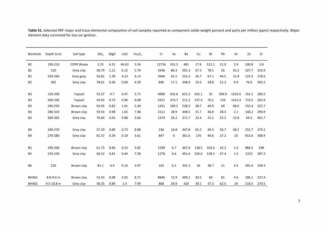
Table S3. RDP classification with 95% confidence threshold and OTU assignment for sequences obtained from iron reducing
consortium isolated from B2 310 sample.
ID* Accession number
Sequence length
Classification using the RDP classifier[17] (95% Confidence threshold)
BHL3_Fe_Consort_1 FR820910 521 Firmicutes, Clostridia, Clostridiales, Incertae Sedis XI, Tissierella
BHL3_Fe_Consort_2 FR820911 511 BHL3_Fe_Consort_3 FR820912 516 Firmicutes, Clostridia, Clostridiales, Clostridiaceae
BHL3_Fe_Consort_4 FR820913 506 Firmicutes, Clostridia, Clostridiales, Clostridiaceae, Clostridiaceae 2, Natronincola
BHL3_Fe_Consort_5 FR820914 516 Firmicutes, Clostridia, Clostridiales, Clostridiaceae
BHL3_Fe_Consort_6 FR820915 512 Firmicutes, Clostridia, Clostridiales, Incertae Sedis XIV, Anaerobranca
BHL3_Fe_Consort_7 FR820916 517 Firmicutes, Clostridia, Clostridiales, Clostridiaceae
BHL3_Fe_Consort_8 FR820917 516 Firmicutes, Clostridia, Clostridiales, Clostridiaceae
BHL3_Fe_Consort_9 FR820918 514 Firmicutes, Clostridia
BHL3_Fe_Consort_10 FR820919 515 Firmicutes, Clostridia, Clostridiales, Clostridiaceae
BHL3_Fe_Consort_11 FR820920 521 Firmicutes, Clostridia, Clostridiales, Incertae Sedis XI, Tissierella
BHL3_Fe_Consort_12 FR820921 516 Firmicutes, Clostridia, Clostridiales, Clostridiaceae
BHL3_Fe_Consort_13 FR820922 496 Firmicutes
BHL3_Fe_Consort_14 FR820923 516 Firmicutes, Clostridia, Clostridiales, Clostridiaceae
BHL3_Fe_Consort_15 FR820924 516 Firmicutes, Clostridia, Clostridiales, Clostridiaceae
BHL3_Fe_Consort_16 FR820925 586 Firmicutes, Clostridia, Clostridiales
BHL3_Fe_Consort_17 FR820926 516 Firmicutes, Clostridia, Clostridiales, Clostridiaceae
BHL3_Fe_Consort_18 FR820927 557 Firmicutes, Clostridia, Clostridiales, Peptococcaceae, Peptococcaceae 1, Desulfitibacter
BHL3_Fe_Consort_19 FR820928 521 Firmicutes, Clostridia, Clostridiales
BHL3_Fe_Consort_20 FR820929 521 Firmicutes, Clostridia, Clostridiales, Incertae Sedis XI
BHL3_Fe_Consort_21 FR820930 518 Firmicutes, Clostridia, Clostridiales, Incertae Sedis XIV, Anaerovirgula
BHL3_Fe_Consort_22 FR820931 589 Firmicutes, Clostridia, Clostridiales
BHL3_Fe_Consort_23 FR820932 512 Firmicutes, Clostridia, Clostridiales, Incertae Sedis XIV, Anaerobranca
BHL3_Fe_Consort_24 FR820933 521 Firmicutes, Clostridia, Clostridiales
BHL3_Fe_Consort_25 FR820934 518 Firmicutes, Clostridia, Clostridiales, Incertae Sedis XIV, Anaerovirgula
BHL3_Fe_Consort_26 FR820935 511 BHL3_Fe_Consort_27 FR820936 584 Firmicutes, Clostridia, Clostridiales
BHL3_Fe_Consort_28 FR820937 512 Firmicutes, Clostridia, Clostridiales, Incertae Sedis XIV, Anaerobranca
BHL3_Fe_Consort_30 FR820938 521 Proteobacteria, Betaproteobacteria, Burkholderiales, Comamonadaceae, Comamonas
BHL3_Fe_Consort_31 FR820939 512 Firmicutes, Clostridia, Clostridiales, Incertae Sedis XIV, Anaerobranca
BHL3_Fe_Consort_32 FR820940 583 Firmicutes, Clostridia, Clostridiales
BHL3_Fe_Consort_33 FR820941 630 Firmicutes, Clostridia, Clostridiales, Incertae Sedis XIV, Anaerovirgula
BHL3_Fe_Consort_34 FR820942 582 Firmicutes, Clostridia, Clostridiales, Incertae Sedis XIV, Anaerobranca
BHL3_Fe_Consort_35 FR820943 511 Firmicutes, Clostridia, Clostridiales, Incertae Sedis XIV, Anaerobranca
BHL3_Fe_Consort_36 FR820944 516 Firmicutes, Clostridia, Clostridiales, Clostridiaceae
BHL3_Fe_Consort_37 FR820945 516 Firmicutes, Clostridia, Clostridiales, Clostridiaceae
BHL3_Fe_Consort_38 FR820946 512 Firmicutes, Clostridia, Clostridiales, Incertae Sedis XIV, Anaerobranca
BHL3_Fe_Consort_39 FR820947 521 Firmicutes, Clostridia, Clostridiales, Peptococcaceae
BHL3_Fe_Consort_40 FR820948 512 Firmicutes, Clostridia, Clostridiales, Incertae Sedis XIV, Anaerobranca
BHL3_Fe_Consort_41 FR820949 584 Firmicutes, Clostridia, Clostridiales
BHL3_Fe_Consort_42 FR820950 521 Firmicutes, Clostridia, Clostridiales, Incertae Sedis XI, Tissierella
BHL3_Fe_Consort_43 FR820951 516 Firmicutes, Clostridia, Clostridiales, Clostridiaceae, Clostridiaceae 2, Alkaliphilus
BHL3_Fe_Consort_44 FR820952 516 Firmicutes, Clostridia, Clostridiales, Clostridiaceae
BHL3_Fe_Consort_45 FR820953 516 Firmicutes, Clostridia, Clostridiales, Clostridiaceae
BHL3_Fe_Consort_46 FR820954 511 Firmicutes
BHL3_Fe_Consort_47 FR820955 504 BHL3_Fe_Consort_48 FR820956 516 Firmicutes, Clostridia, Clostridiales, Clostridiaceae
* BHL3 was renumbered B2 after sequences had been submitted to GenBank

11
Table S4. Idealised Cr molecular co-ordination environment in the chromite structure where n is the number of atoms in each shell and R is the bond distance. Calculated from structural information presented in Derbyshire et al. [18].
Structure Shell n R (Å)
Chromite
O 6 1.99
Cr-Cr 6 2.97
Cr-Fe 6 3.47
12
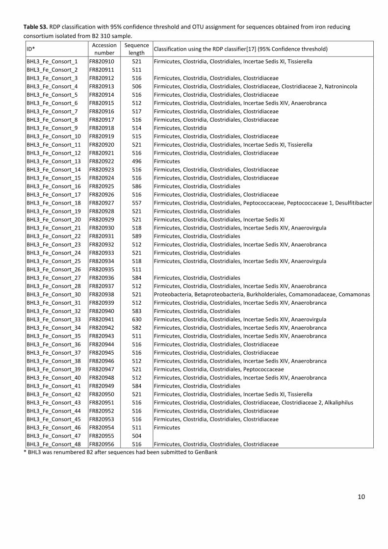
A B
Figure S1. Backscatter SEM images of resin embedded thin section from B2-310 soil sample, showing (A) low magnification view of angular quartz grains in a fine grained matrix (scale bar 1000 µM); (B) higher magnification view (from white arrow, frame A) of fine grained clay-like matrix material (scale bar 10 µM).
0 1 2 3 4 5 6 7 8 9 10Energy (keV)
0
500
1000
1500
Co
un
ts
EDX 1
EDX 2
EDX 3
Fe
FeCr Cr
Cu
Cu
KCa
Si
Al
Mg
C
O
Fe
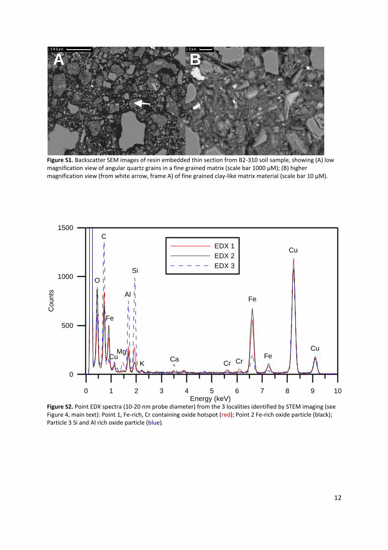
Cu
Figure S2. Point EDX spectra (10-20 nm probe diameter) from the 3 localities identified by STEM imaging (see Figure 4, main text): Point 1, Fe-rich, Cr containing oxide hotspot (red); Point 2 Fe-rich oxide particle (black); Particle 3 Si and Al rich oxide particle (blue).

13
4 6 8 10
k(Å )-1
Norm
alis
ed
A
bso
rba
nce
Figure S3. Chromite ore Cr K-edge EXAFS spectra. Arrows highlight the characteristic sub peak features at k=5.1 and 8 Å
-1, redrawn from Doelsch et al. [19].
Recommended