Ch 27: Prokaryotes - Bacteria and Archaea
Great Salt Lake – pink color from living prokaryotes; survive in 32% salt
• Prokaryotes are divided into two domains– bacteria and archaea
• thrive in diverse habitats– including places too acidic,
salty, cold, or hot for most other organisms
• Most are microscopic– but what they lack in size
they make up for in numbers
– For example: more in a handful of fertile soil than the number of people who have ever lived
Prokaryotes• Single cell
– Some form colonies• Very small
– 0.5–5 µm (10-20 times smaller than Eukaryotes)
• Lacks nucleus and most other membrane bound organelles
• Reproduce very quickly– Asexual binary fission– Genetic recombination
• variety of shapes– spheres (cocci)– rods (bacilli)– spirals
• Cell wall
More structural & functional characteristics in (Ch.27)
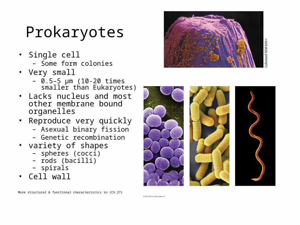
Bacilli
• Rod shaped– Example: E. coli
• Usually solitary• Sometimes chains– streptobacilli
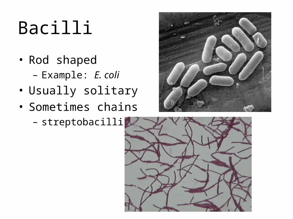
Cocci
• Spherical– Clumps or clusters (like
grapes)• E.g. Staphylococcus aureus
– Streptococci – chains of spheres
– Diplococci – pairs of spheres• E.g. Neisseria gonnorheae
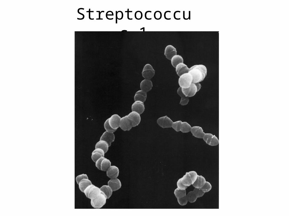
Streptococcus 1
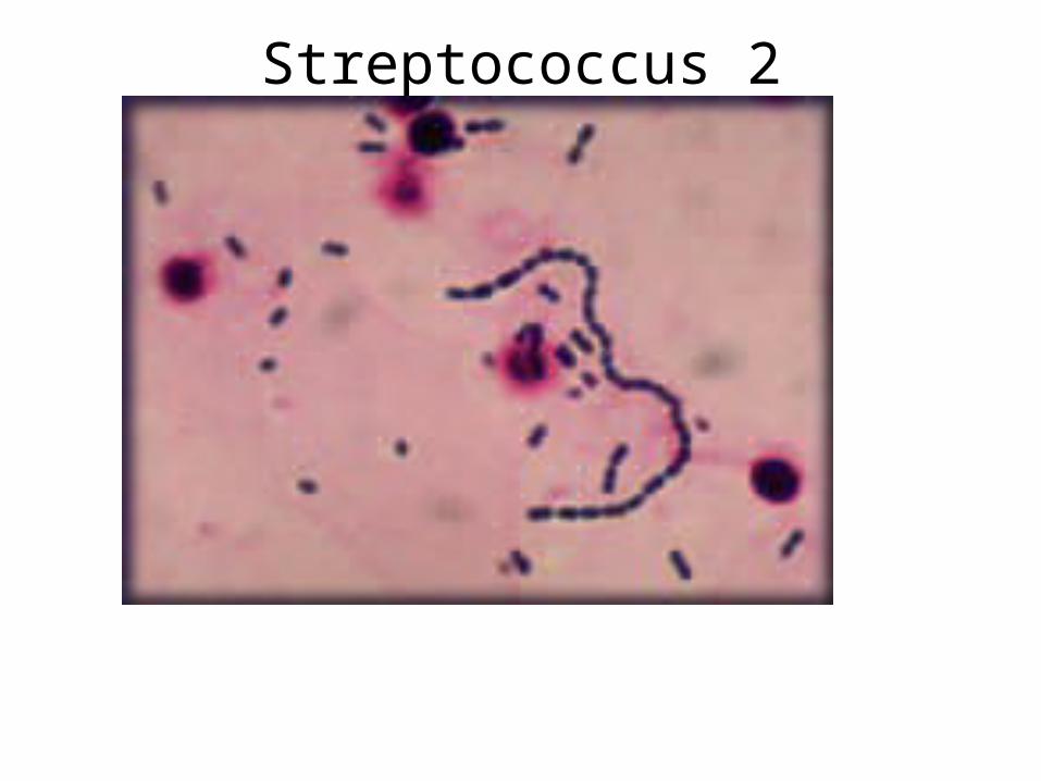
Streptococcus 2
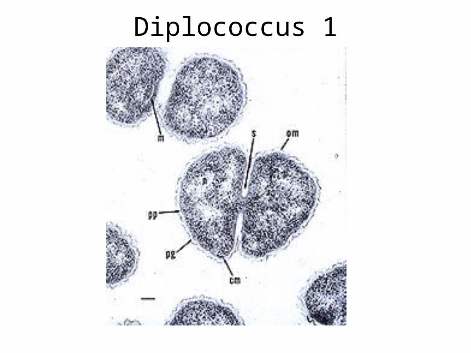
Diplococcus 1
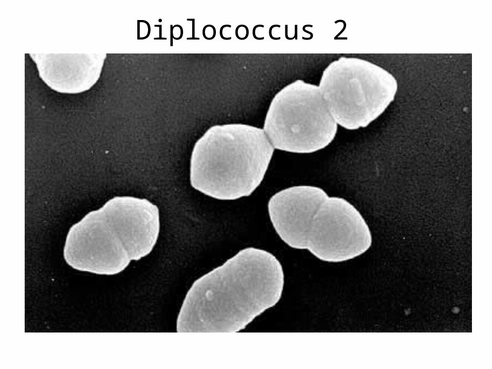
Diplococcus 2
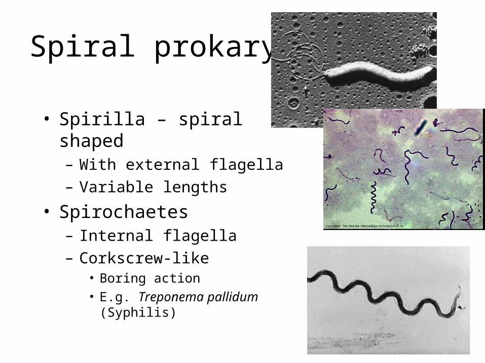
Spiral prokaryotes
• Spirilla – spiral shaped– With external flagella– Variable lengths
• Spirochaetes– Internal flagella– Corkscrew-like
• Boring action• E.g. Treponema pallidum (Syphilis)
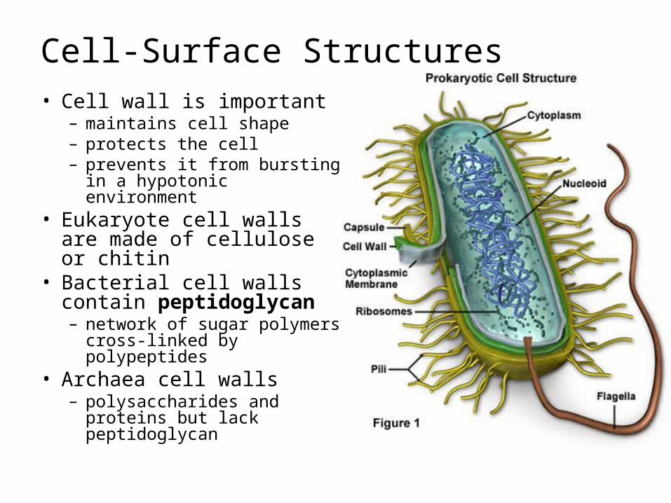
Cell-Surface Structures• Cell wall is important
– maintains cell shape– protects the cell– prevents it from bursting in a
hypotonic environment• Eukaryote cell walls are
made of cellulose or chitin• Bacterial cell walls contain
peptidoglycan– network of sugar polymers
cross-linked by polypeptides• Archaea cell walls
– polysaccharides and proteins but lack peptidoglycan
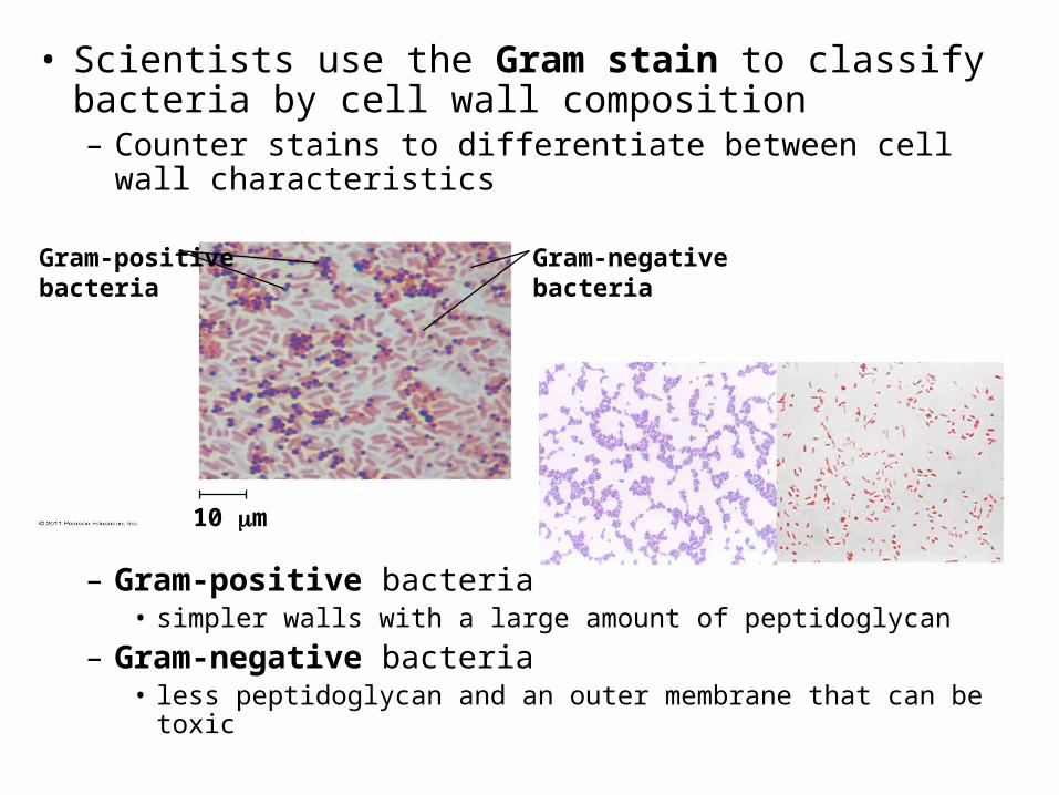
• Scientists use the Gram stain to classify bacteria by cell wall composition– Counter stains to differentiate between cell wall
characteristics
– Gram-positive bacteria• simpler walls with a large amount of peptidoglycan
– Gram-negative bacteria• less peptidoglycan and an outer membrane that can be toxic
Gram-positivebacteria
10 m
Gram-negativebacteria
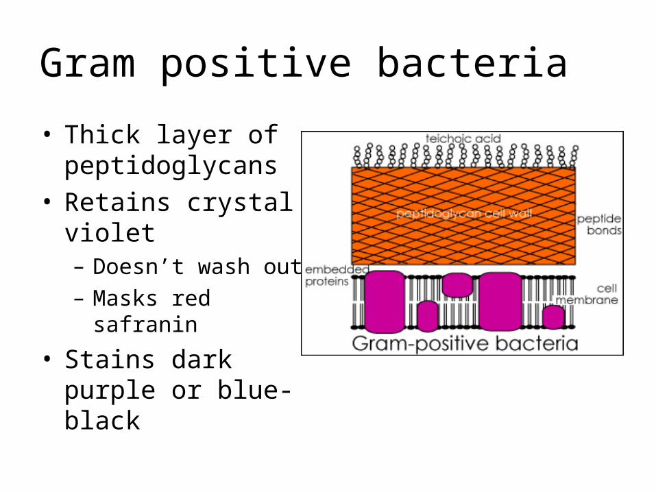
Gram positive bacteria
• Thick layer of peptidoglycans
• Retains crystal violet– Doesn’t wash out– Masks red safranin
• Stains dark purple or blue-black
Gram negative bacteria
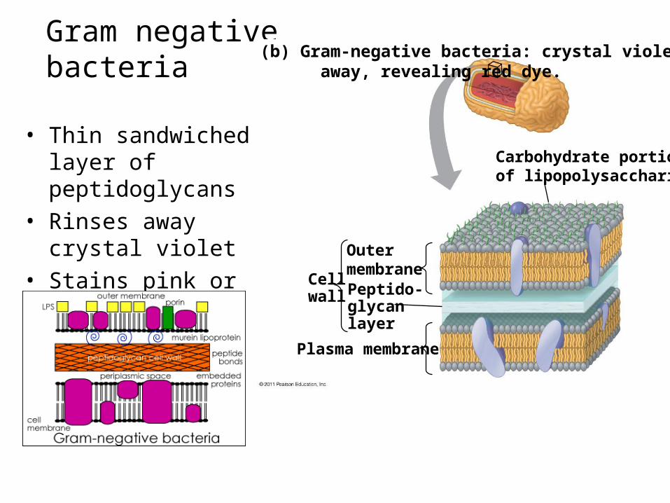
• Thin sandwiched layer of peptidoglycans
• Rinses away crystal violet
• Stains pink or red OutermembranePeptido-glycanlayer
Plasma membrane
Cellwall
Carbohydrate portionof lipopolysaccharide
(b) Gram-negative bacteria: crystal violet is easily rinsed away, revealing red dye.
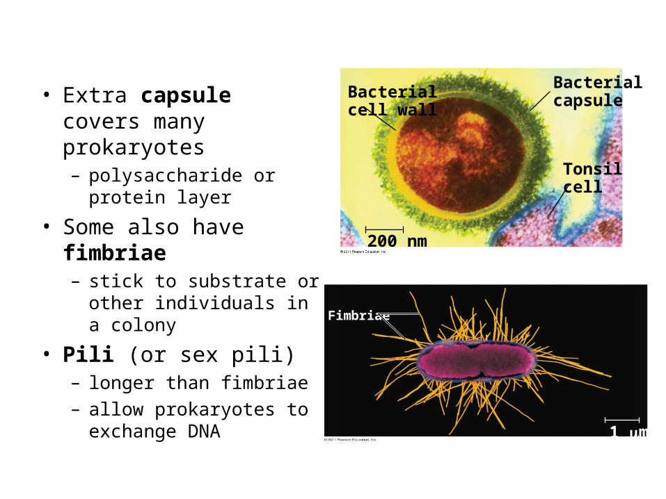
• Extra capsule covers many prokaryotes– polysaccharide or protein
layer
• Some also have fimbriae– stick to substrate or other
individuals in a colony
• Pili (or sex pili)– longer than fimbriae– allow prokaryotes to
exchange DNA
Bacterialcell wall
Bacterialcapsule
Tonsilcell
200 nm
Fimbriae
1 m
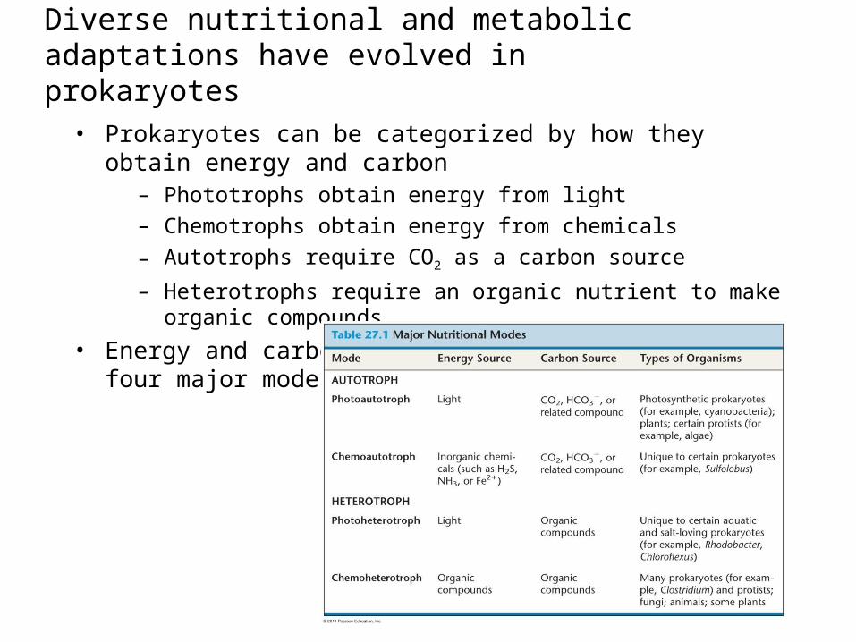
Diverse nutritional and metabolic adaptations have evolved in prokaryotes
• Prokaryotes can be categorized by how they obtain energy and carbon
– Phototrophs obtain energy from light– Chemotrophs obtain energy from chemicals– Autotrophs require CO2 as a carbon source
– Heterotrophs require an organic nutrient to make organic compounds
• Energy and carbon sources are combined to give four major modes of nutrition
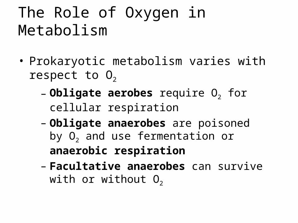
The Role of Oxygen in Metabolism
• Prokaryotic metabolism varies with respect to O2
– Obligate aerobes require O2 for cellular respiration
– Obligate anaerobes are poisoned by O2 and use fermentation or anaerobic respiration
– Facultative anaerobes can survive with or without O2

Nitrogen Metabolism
• Nitrogen is essential for the production of amino acids and nucleic acids – nitrogen fixation– some prokaryotes convert atmospheric
nitrogen (N2) to ammonia (NH3)– Some cooperate between cells of a colony
• allows them to use environmental resources they could not use as individual cells– E.g. cyanobacterium Anabaena, photosynthetic
cells and nitrogen-fixing cells called heterocysts (or heterocytes) exchange metabolic products
Photosyntheticcells
Heterocyst
20 m
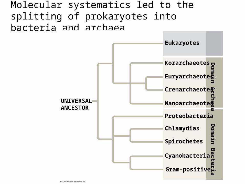
Molecular systematics led to the splitting of prokaryotes into bacteria and archaea
Eukaryotes
Korarchaeotes
Euryarchaeotes
Crenarchaeotes
Nanoarchaeotes
Proteobacteria
Chlamydias
Spirochetes
Cyanobacteria
Dom
ain BacteriaD
omain ArchaeaUNIVERSAL
ANCESTOR
Gram-positive

Clades of Domain Bacteria
• Fig 27.18 (27.13 in 7th ed.)• Proteobacteria– diverse & includes gram-negatives– Subgroups: α, β, γ, δ, ε
• Chlamydias• Spirochaetes• Cyanobacteria• Gram positive bacteria
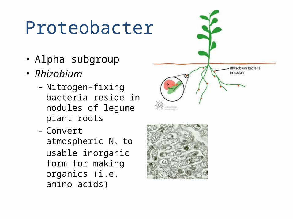
• Alpha subgroup• Rhizobium– Nitrogen-fixing
bacteria reside in nodules of legume plant roots
– Convert atmospheric N2 to usable inorganic form for making organics (i.e. amino acids)
Proteobacteria
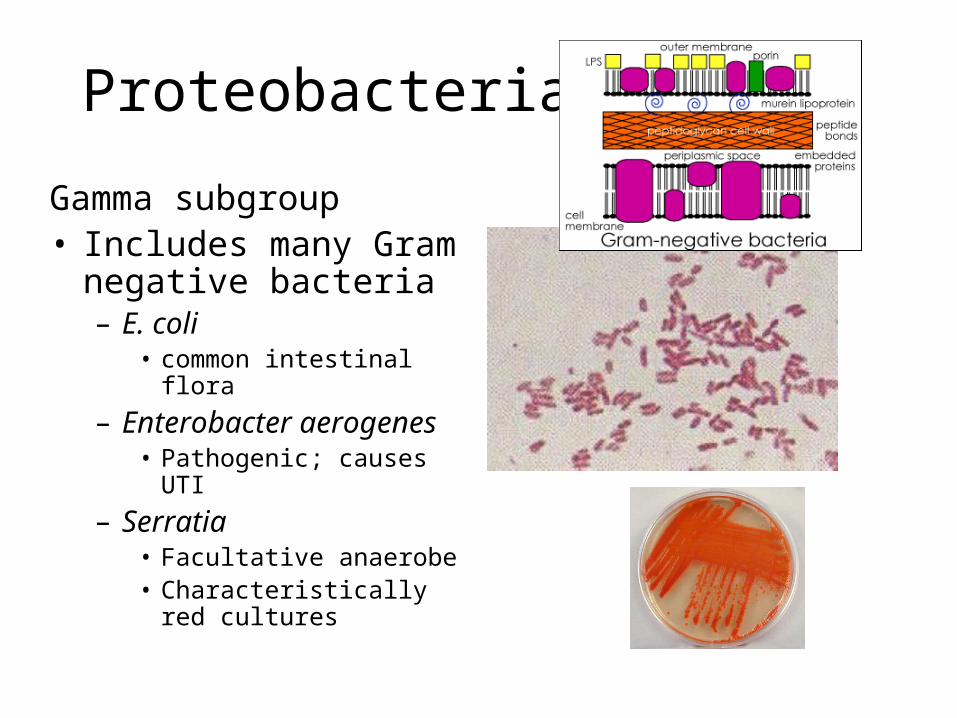
Proteobacteria
Gamma subgroup• Includes many Gram
negative bacteria– E. coli
• common intestinal flora– Enterobacter aerogenes
• Pathogenic; causes UTI– Serratia
• Facultative anaerobe• Characteristically red
cultures
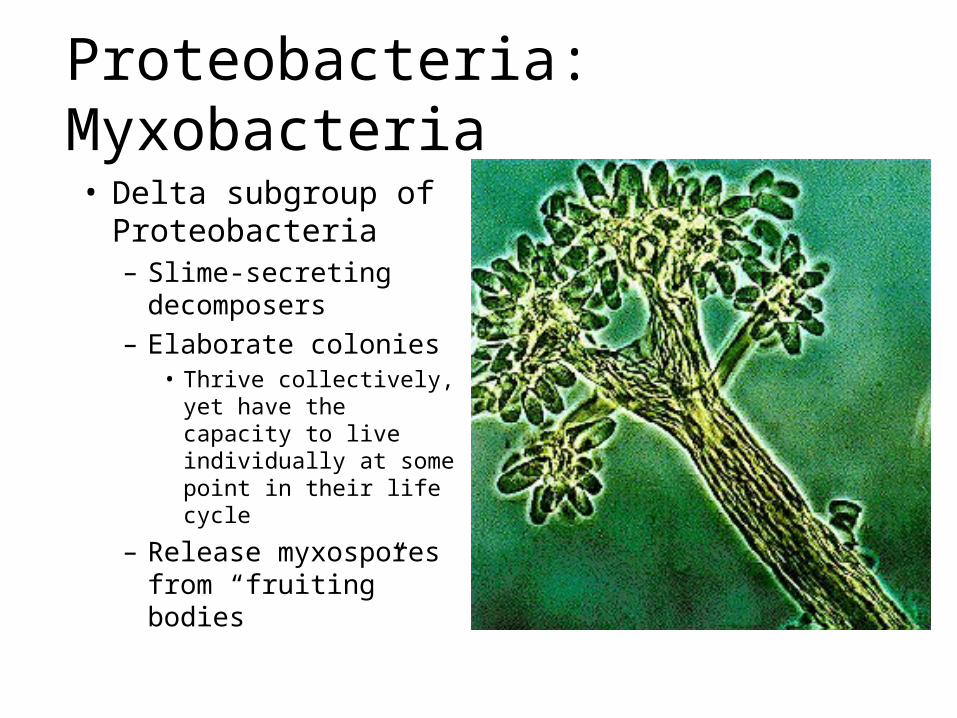
Proteobacteria: Myxobacteria
• Delta subgroup of Proteobacteria– Slime-secreting
decomposers– Elaborate colonies
• Thrive collectively, yet have the capacity to live individually at some point in their life cycle
– Release myxospores from “fruiting” bodies
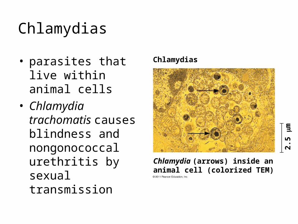
Chlamydias
• parasites that live within animal cells
• Chlamydia trachomatis causes blindness and nongonococcal urethritis by sexual transmission
Chlamydias
2.5
m
Chlamydia (arrows) inside ananimal cell (colorized TEM)
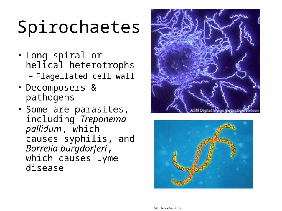
Spirochaetes
• Long spiral or helical heterotrophs– Flagellated cell wall
• Decomposers & pathogens
• Some are parasites, including Treponema pallidum, which causes syphilis, and Borrelia burgdorferi, which causes Lyme disease
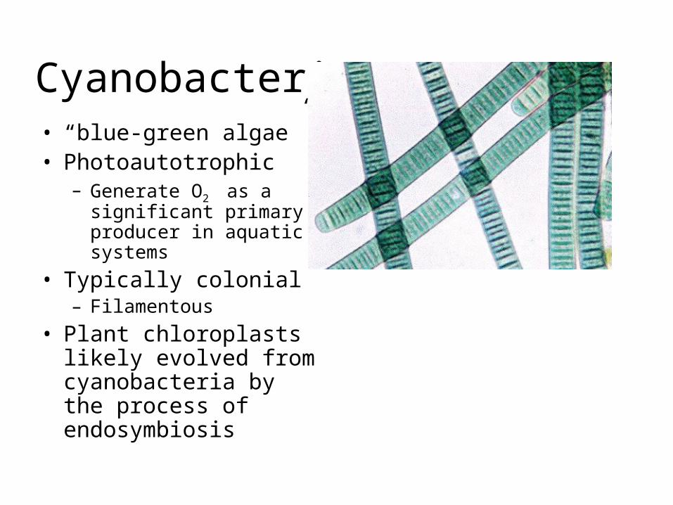
Cyanobacteria• “blue-green algae”• Photoautotrophic
– Generate O2 as a significant primary producer in aquatic systems
• Typically colonial– Filamentous
• Plant chloroplasts likely evolved from cyanobacteria by the process of endosymbiosis
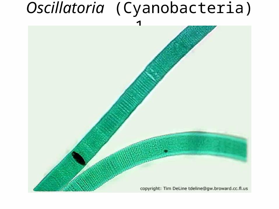
Oscillatoria (Cyanobacteria) 1
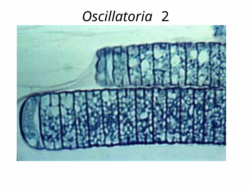
Oscillatoria 2
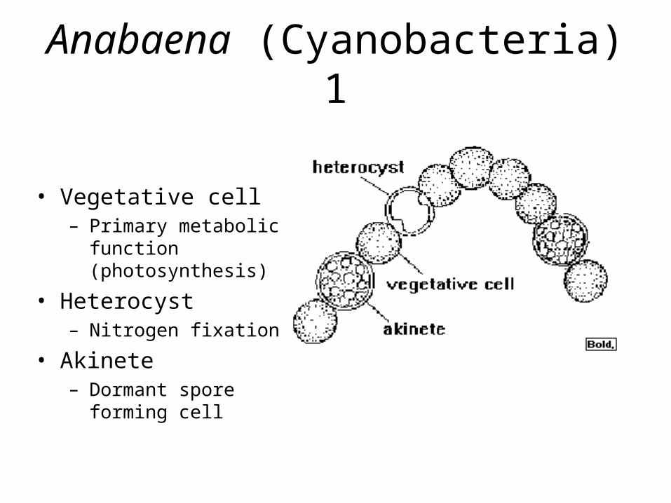
Anabaena (Cyanobacteria) 1
• Vegetative cell– Primary metabolic function
(photosynthesis)
• Heterocyst– Nitrogen fixation
• Akinete– Dormant spore forming cell
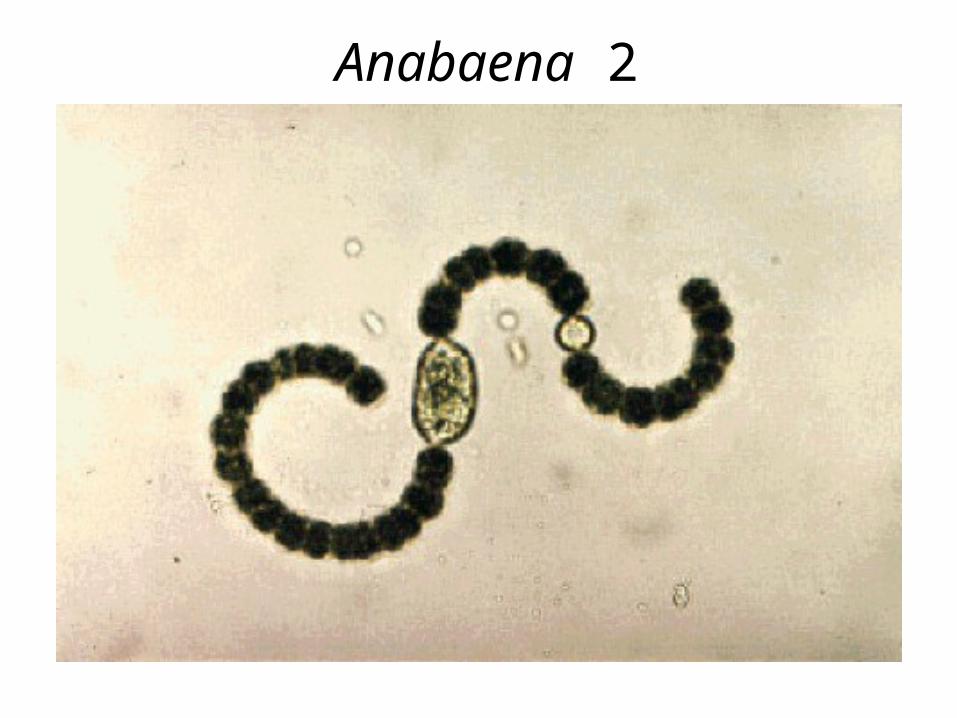
Anabaena 2
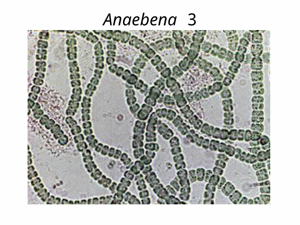
Anaebena 3
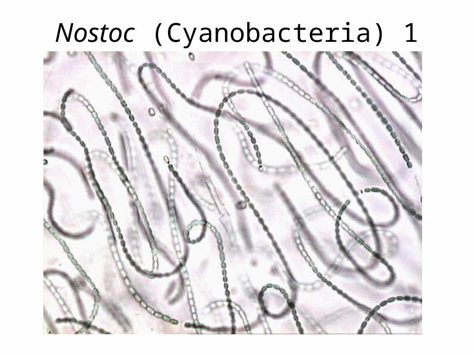
Nostoc (Cyanobacteria) 1
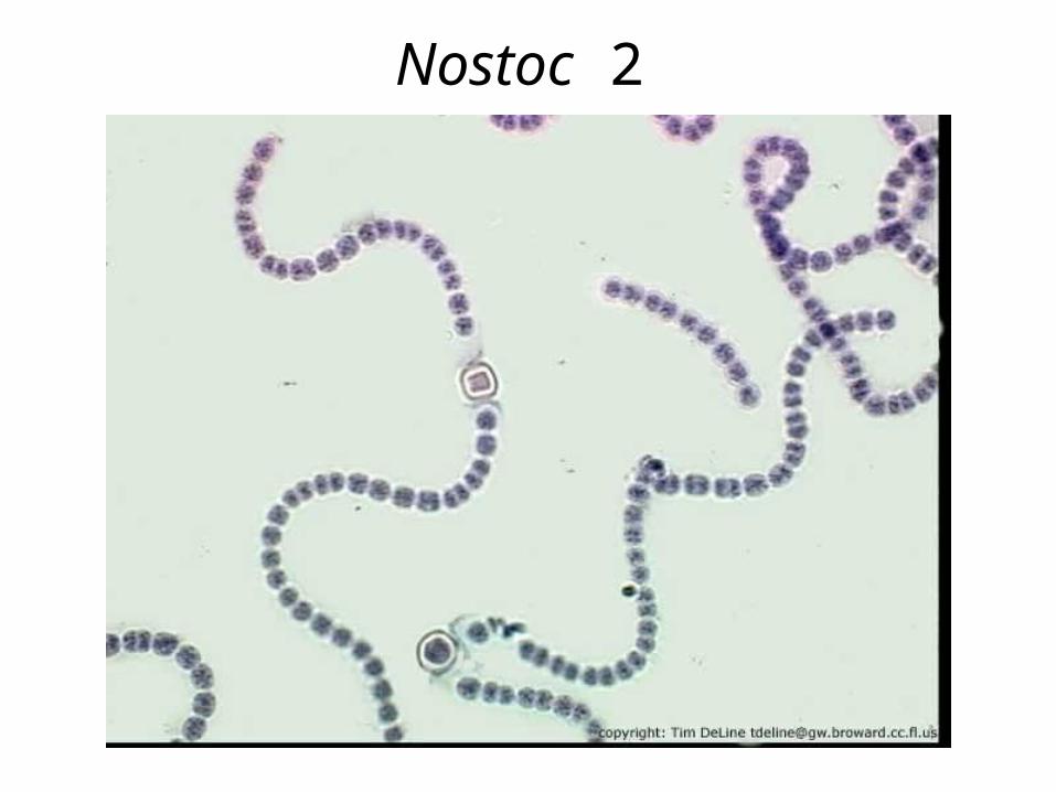
Nostoc 2
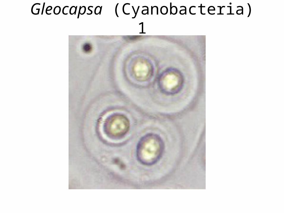
Gleocapsa (Cyanobacteria) 1
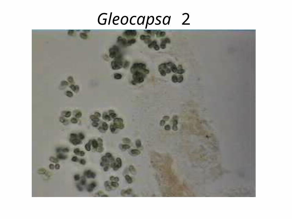
Gleocapsa 2
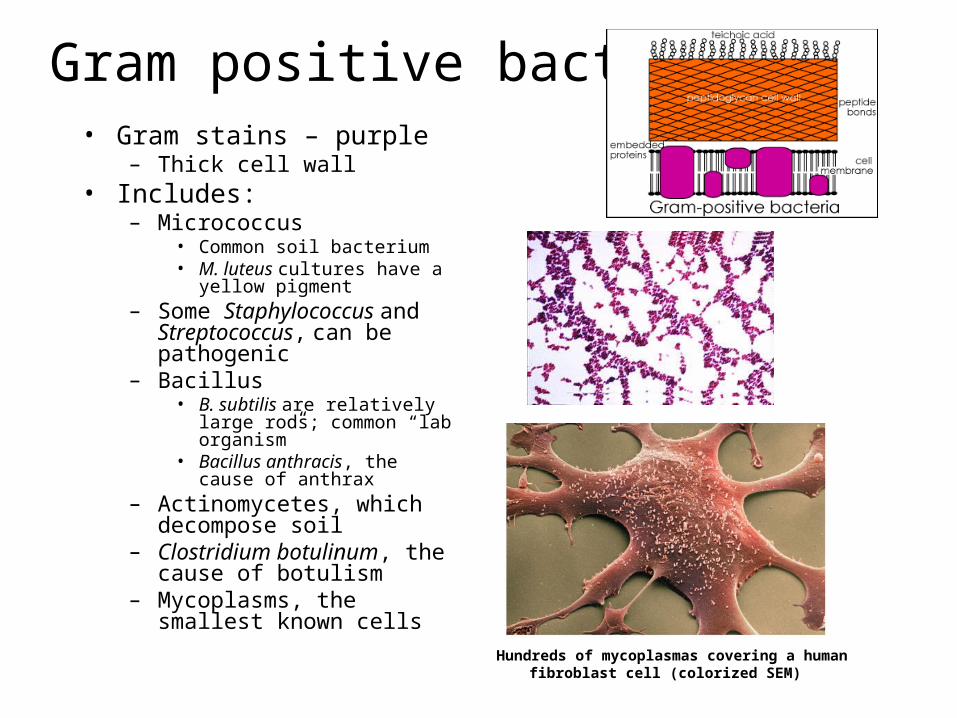
Gram positive bacteria• Gram stains – purple
– Thick cell wall• Includes:
– Micrococcus• Common soil bacterium• M. luteus cultures have a yellow
pigment– Some Staphylococcus and
Streptococcus, can be pathogenic
– Bacillus• B. subtilis are relatively large rods;
common “lab organism”• Bacillus anthracis, the cause of
anthrax– Actinomycetes, which
decompose soil– Clostridium botulinum, the
cause of botulism– Mycoplasms, the smallest
known cells
Hundreds of mycoplasmas covering a human fibroblast cell (colorized SEM)
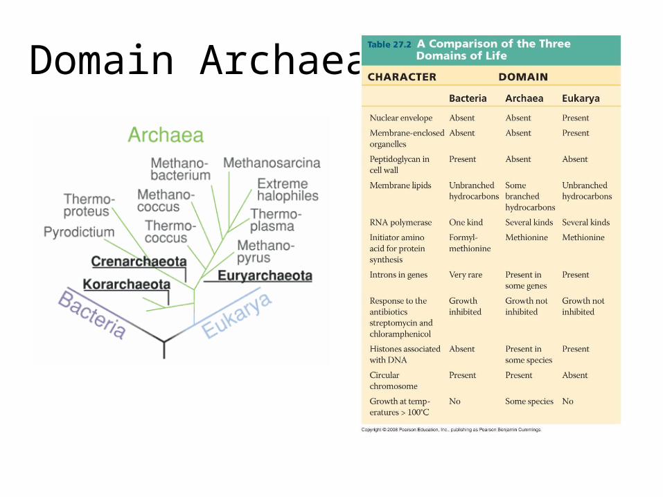
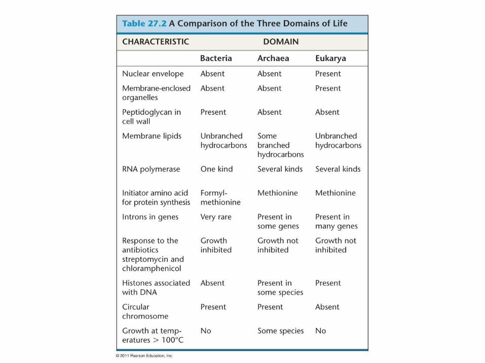
Domain Archaea
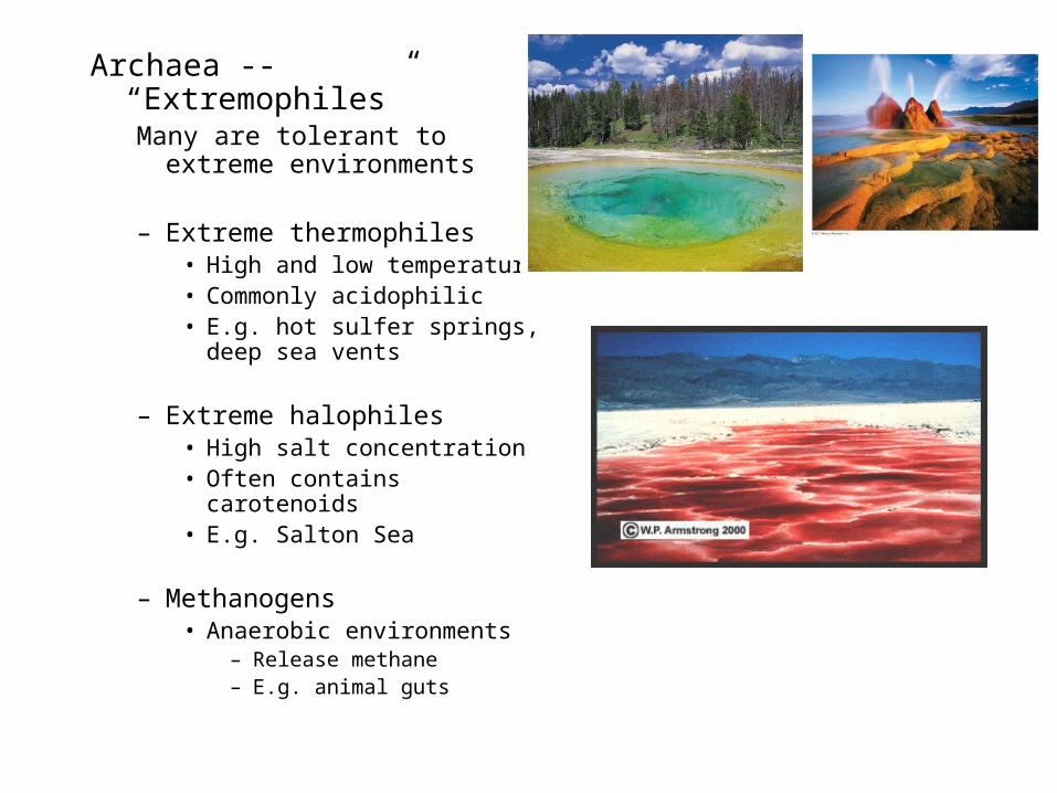
Archaea -- “Extremophiles”Many are tolerant to extreme
environments
– Extreme thermophiles • High and low temperature• Commonly acidophilic• E.g. hot sulfer springs, deep
sea vents
– Extreme halophiles• High salt concentration• Often contains carotenoids• E.g. Salton Sea
– Methanogens• Anaerobic environments
– Release methane– E.g. animal guts
Recommended