Development and
Characterization of Novel 3D
Airway Cell Models
Carolin Boecking, M.D.
University of California, San Francisco

Human Airway Cell Models for CF Research
Focus of our laboratory
is the development and
characterization of in
vitro (cell culture)
models of the
conducting airway
surface epithelium and
submucosal glands for
use in CF research
Central conducting airway
M
S

Human Airway Cell Models for CF Research
Important roles for airway cell models in CF research
include:
Understanding the functions of CFTR and how alterations in
its function lead to disease
Defining the pathophysiology of secondary lung problems in
CF, e.g., infection and mucus hypersecretion
CF drug discovery
In vitro testing of potential CF gene therapies
Understanding the significance of airway gland hyperplasia
and the mechanism of mucus hypersecretion in CF
Improved human airway cell models are needed for:
Facilitating CF drug discovery
Identifying therapeutics for CF treatment of secondary lung
problems
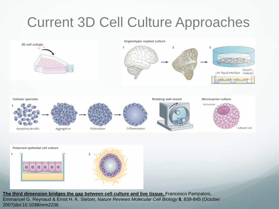
Current 3D Cell Culture Approaches
The third dimension bridges the gap between cell culture and live tissue, Francesco Pampaloni,
Emmanuel G. Reynaud & Ernst H. K. Stelzer, Nature Reviews Molecular Cell Biology 8, 839-845 (October
2007)doi:10.1038/nrm2236
Organotypic explant culture2D cell culture
Cellular speroids Microcarrier cultureRotating wall vessel
Polarized epithelial cell culture
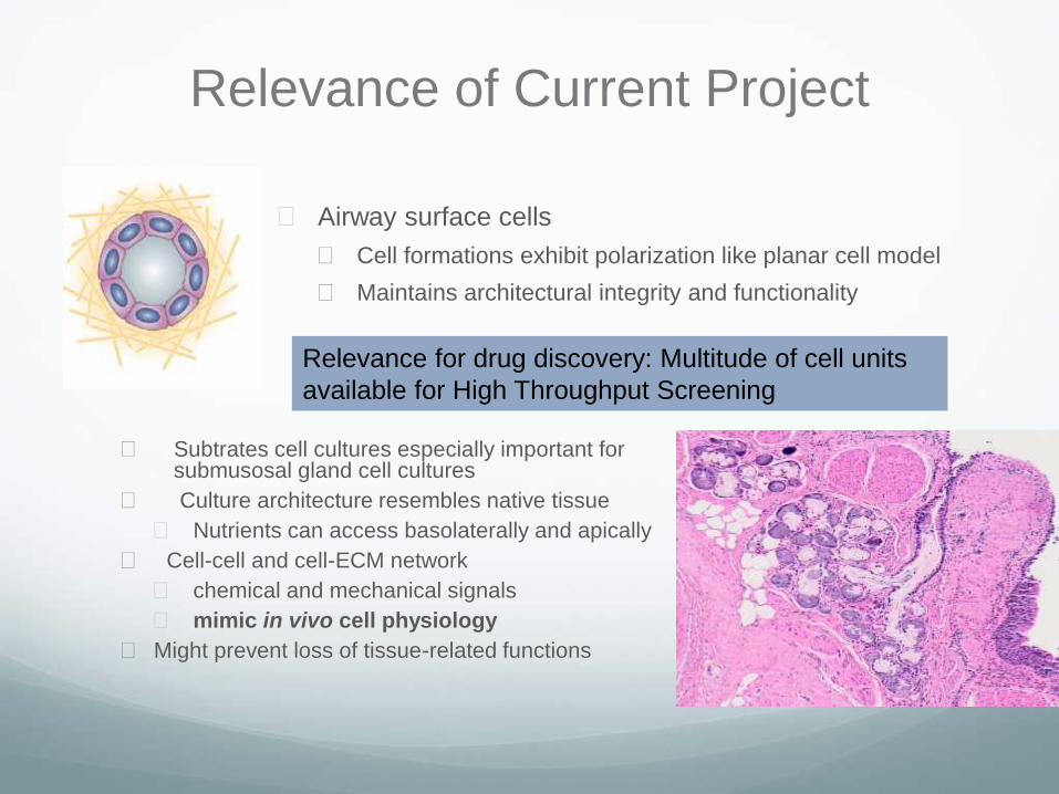
Relevance of Current Project
Subtrates cell cultures especially important for submusosal gland cell cultures
Culture architecture resembles native tissue
Nutrients can access basolaterally and apically
Cell-cell and cell-ECM network
chemical and mechanical signals
mimic in vivo cell physiology
Might prevent loss of tissue-related functions
Airway surface cells
Cell formations exhibit polarization like planar cell model
Maintains architectural integrity and functionality
Relevance for drug discovery: Multitude of cell units
available for High Throughput Screening
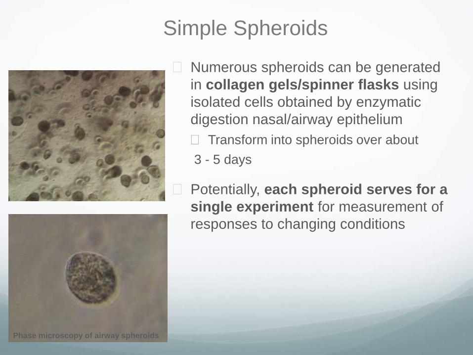
Simple Spheroids
Numerous spheroids can be generated
in collagen gels/spinner flasks using
isolated cells obtained by enzymatic
digestion nasal/airway epithelium
Transform into spheroids over about
3 - 5 days
Potentially, each spheroid serves for a
single experiment for measurement of
responses to changing conditions
Phase microscopy of airway spheroids
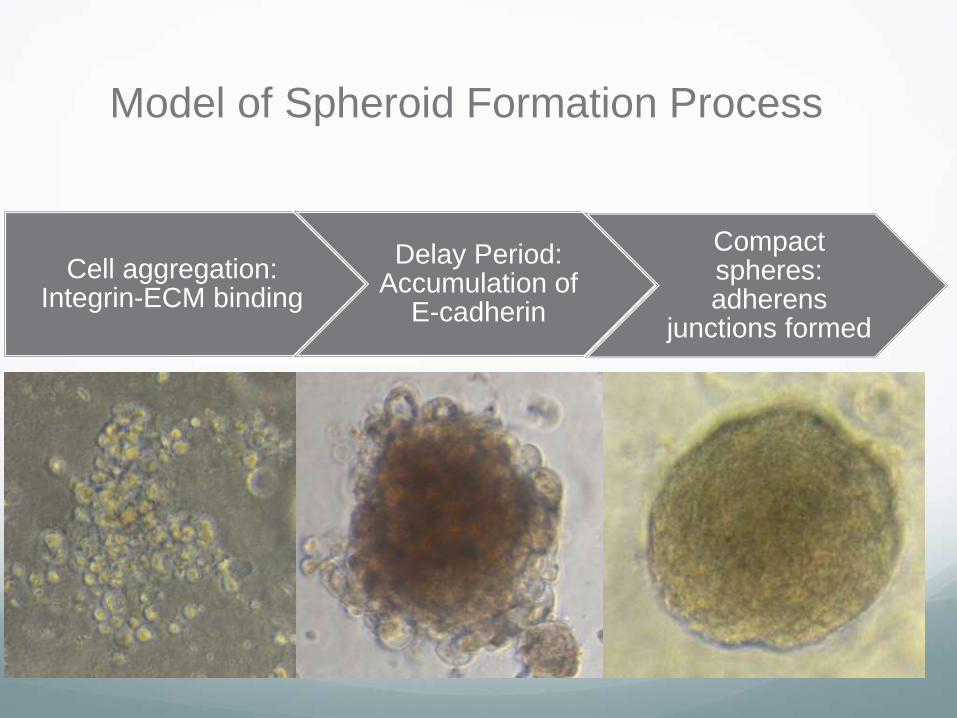
Model of Spheroid Formation Process
Cell aggregation: Integrin-ECM binding
Delay Period: Accumulation of
E-cadherin
Compact spheres: adherens
junctions formed

Morphology of Airway Epithelial
Spheroid
Spheroid formed from isolated cells--
arrow shows cilia
Developing spheroid from epithelial cell
clusters
ABC
Drawbacks of Simple Spheroids
Differentiated features decline with time in
culture
CFTR expressed but ion transport is not
detectable

Promoting Spheroid Differentiation
Differentiation of airway epithelial spheroids
may be maintained/improved by adding stem
cell growth factors R-spondin 2 and Noggin
Liu et al. (Functional Cftr in crypt epithelium of organotypic
enteroid cultures from murine small intestine) grew
“enteroid” crypts with functional CFTR using R-spondin 1
and Noggin
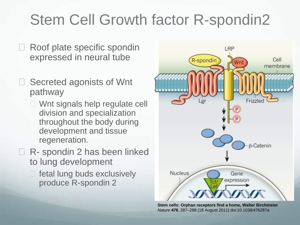
Stem Cell Growth factor R-spondin2
Roof plate specific spondinexpressed in neural tube
Secreted agonists of Wntpathway
Wnt signals help regulate cell division and specialization throughout the body during development and tissue regeneration.
R- spondin 2 has been linked to lung development
fetal lung buds exclusively produce R-spondin 2
Stem cells: Orphan receptors find a home, Walter Birchmeier
Nature 476, 287–288 (18 August 2011) doi:10.1038/476287a

Stem Cell Growth Factor Noggin
Noggin, a BMP antagonist
BMP4
transcribed in lung mesenchyme
involved in branching of lungs and surface epithelium differentiation
Noggin causes
proximalization of
distal airway
epithelium
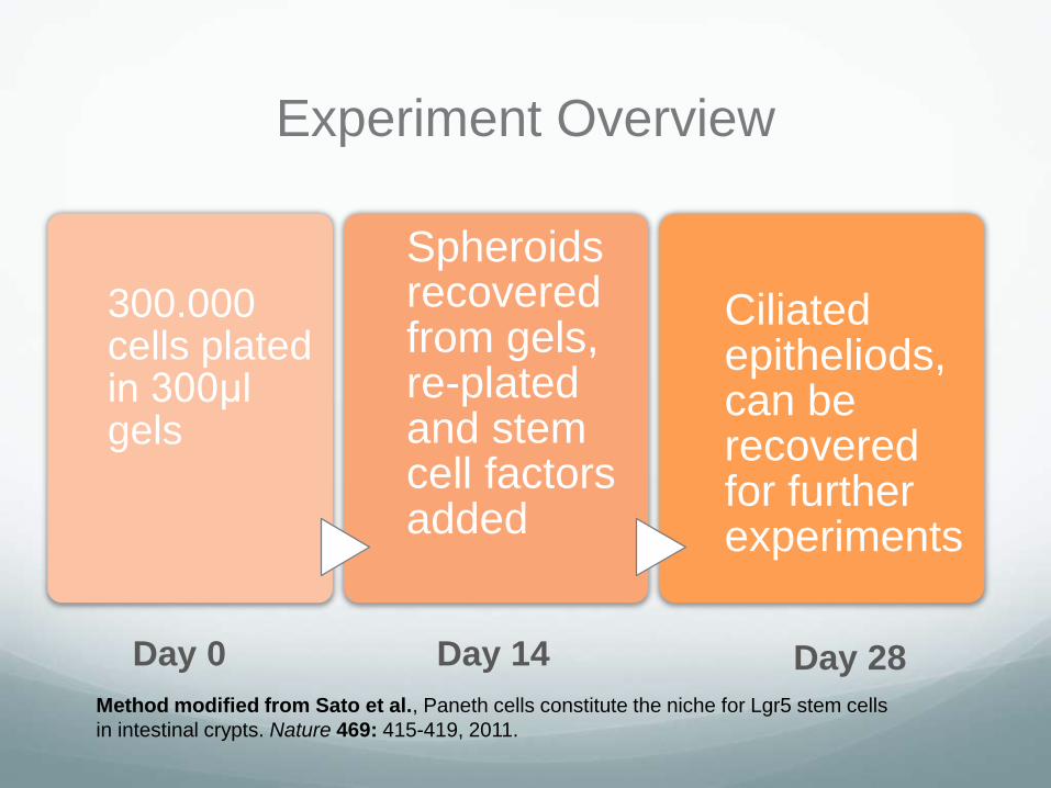
Experiment Overview
300.000 cells plated in 300μl gels
Spheroids recovered from gels, re-plated and stem cell factors added
Ciliated epitheliods, can be recovered for further experiments
Method modified from Sato et al., Paneth cells constitute the niche for Lgr5 stem cells
in intestinal crypts. Nature 469: 415-419, 2011.
Day 0 Day 28Day 14
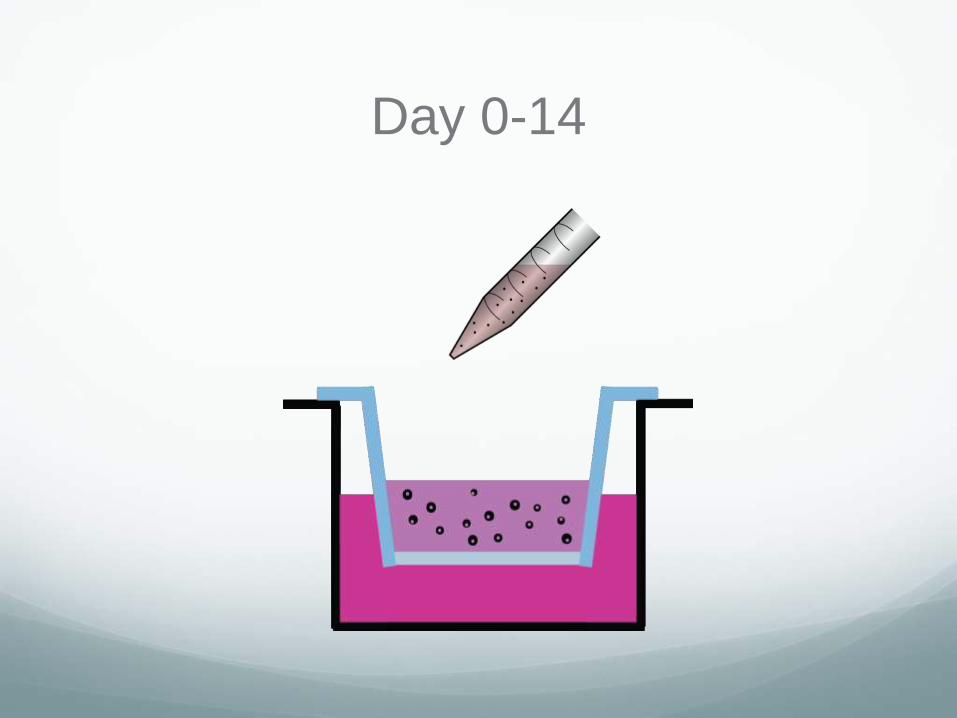
Day 0-14

Day 14-28
+200ng/ml R-spondin2
+25ng/ml Noggin

Ready For Further Studies

Step 1- Airway Epithelial Cells
Embedded in Collagen Gels
Matrigel only Vitrogen only
Mixed matrix: Matrigel/Vitrogen

Step 2- Recovery of Spheroids without
Disrupting Their 3D Orientation
• Spheroids were
recovered from
collagen gels on day
14 using:
• Dispase for
Matrigel
• 0.2%
Collagenase for
Vitrogen
• Spheroids were re-
plated in same gel
Airway epithelial spheroids recovered from
and re-plated in Matrigel
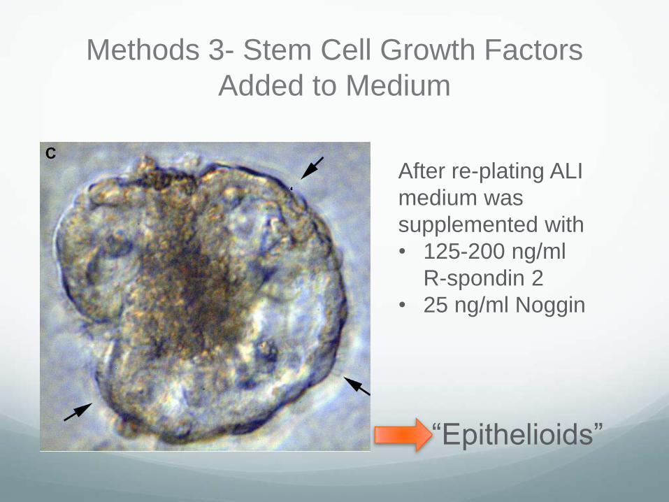
Methods 3- Stem Cell Growth Factors
Added to Medium
After re-plating ALI
medium was
supplemented with
• 125-200 ng/ml
R-spondin 2
• 25 ng/ml Noggin
“Epithelioids”

“Epithelioids”
Cilia
Spheroid Cavity

Epitheliod Architecture Maintained by Growth factors
Control Rspondin-2 and
Noggin added


Immunofluorescence
Protein Quantification

DNA Quantification
Future Studies
EM/ SEM
Histochemical and immunocytochemical stains for
relevant elements of airway surface epithelium,
including CFTR, e-cadherin ciliated cells, basal cells
and mucin genes (MUC5AC, MUC5B)
CFTR function assessed by fluorescence assays,
intracellular microelectrodes and microfluidics
Ciliary beat pattern and beating frequency will be
studied using high-speed video imaging

Established Cell Models of Airway GlandsBrief summary of methods
After epithelium stripped from airway, submucosal tissue sharply dissected
Tissue minced and then dissociated in enzymes--collagenase,hyaluronidase, DNase
Isolated gland aciniplated in collagen coated flask
Near confluent cultures trypsinized onto cell culture inserts

Gland Hyperplasia in Cystic Fibrosis

+IL-13 +IL-17
Control
IL-13 and IL-17 Induce Mucin Production in
Planar Submucosal Gland Cell Cultures

IL-13 and IL-17 Induce Mucin Production in
Planar Submucosal Gland Cell Cultures
• Pink mucicarmine stain
identifies variable
amounts of mucin in
luminal cells
• A. Control cells
• B. Cells exposed to Il-
13 (10 ng/ml)
• C. Cells exposed to IL-17
(10 ng/ml)
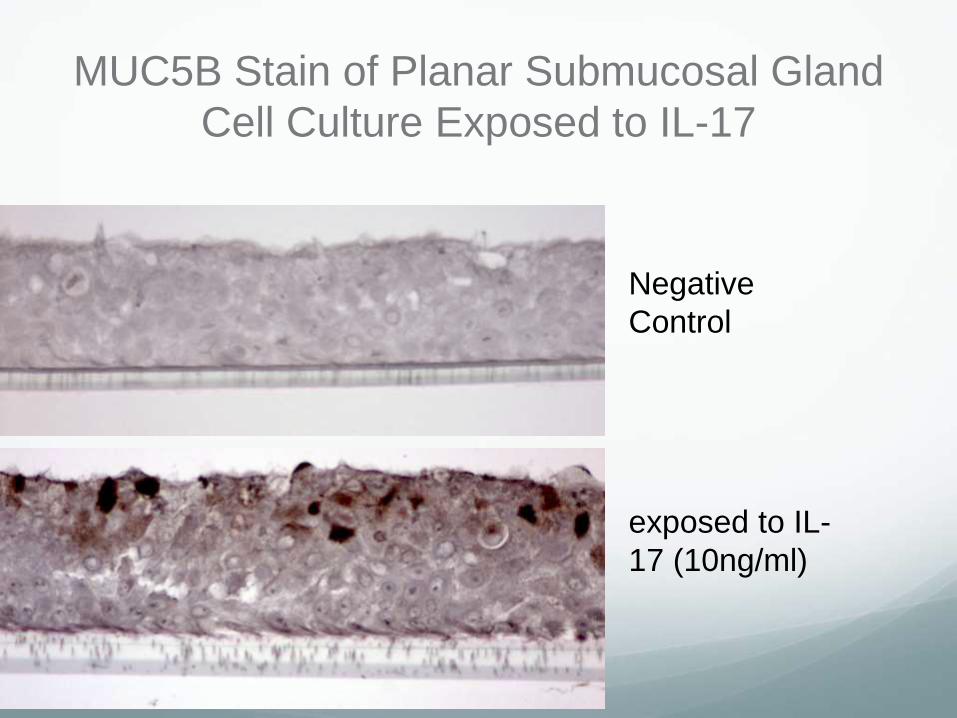
MUC5B Stain of Planar Submucosal Gland
Cell Culture Exposed to IL-17
Negative
Control
exposed to IL-
17 (10ng/ml)
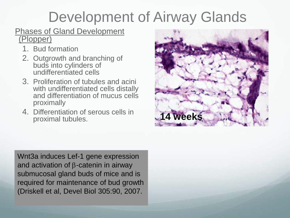
Development of Airway Glands Phases of Gland Development (Plopper)
1. Bud formation
2. Outgrowth and branching of buds into cylinders of undifferentiated cells
3. Proliferation of tubules and aciniwith undifferentiated cells distally and differentiation of mucus cells proximally
4. Differentiation of serous cells in proximal tubules. 14 weeks
Wnt3a induces Lef-1 gene expression
and activation of -catenin in airway
submucosal gland buds of mice and is
required for maintenance of bud growth
(Driskell et al, Devel Biol 305:90, 2007.

Adding Wnt/beta catenin enhancers to Submucosal
Gland Cells 3D cultures induced Cilia growth
Contamination from
epithelial cells
Ciliated duct cells
present
Pluripotent “stem cell”
within gland tissue
reprogrammed by
growth factors to exhibit
characteristics of surface
cells

Submucosal Gland Cultures in Gels Day 5
Control HGF HGF & IL-1399% round or oval
1% cylindrical or stellate
60% round or oval
40% cylindrical or stellate
53% round or oval
47% cylindrical or stellate

Submucosal Gland Cells cultured in Gels
initial gland formation in
gels, day 7
Glands cultured with
MammoCult®
Glands grown on mixed
matrices, day 14
Glands grown in Matrigel alone, day 14
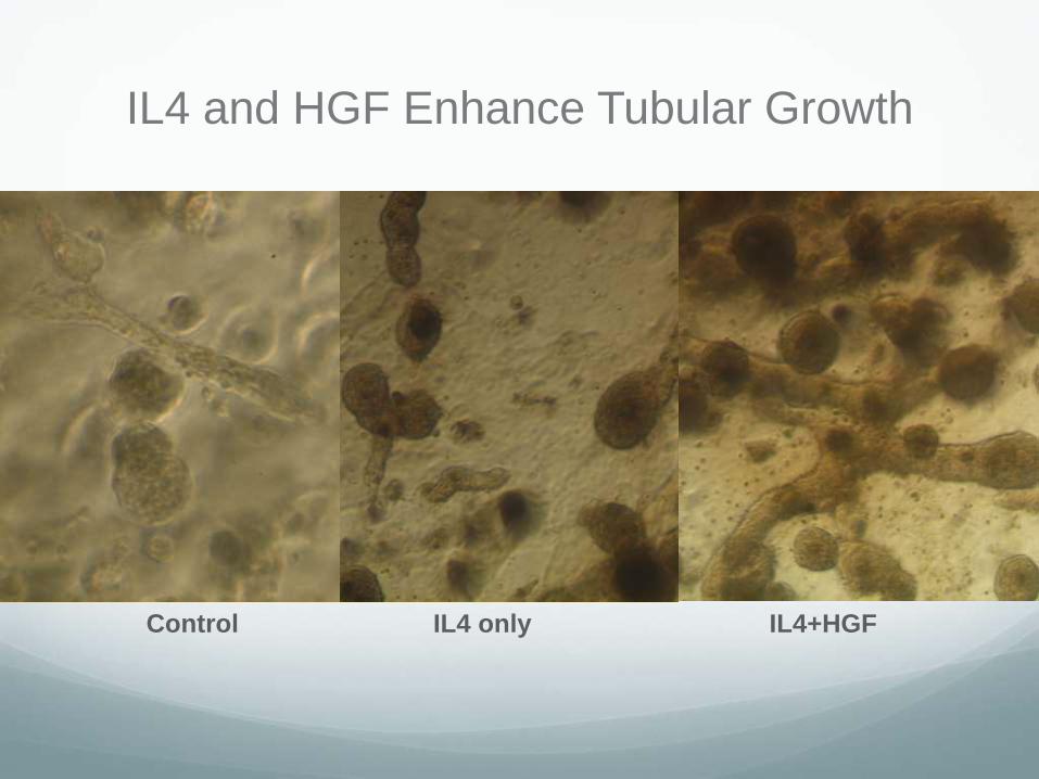
IL4 and HGF Enhance Tubular Growth
Control IL4 only IL4+HGF

IL4 and HGF Enhance Tubular Growth
Control IL13 only IL13+HGF

Future studies
Gene expression analysis
Thank You!
Finkbeiner Lab
Walter E. Finkbeiner, M.D., PhD
Lorna Zlock, Staff Research Associate
Cystic Fibrosis Research, Inc.
For all your support
Recommended