Breast Cancer Screening
Gail Ward PSM – State Manager, Population Screening and Cancer Prevention

Breast Cancer in Tasmania
• 394 breast cancers were diagnosed in Tasmanian
women in 2016*
• 5 breast cancers diagnosed in Tasmanian men in 2016*
• * Tasmanian Cancer Registry 2016 report
• Excludes DCIS
• BreastScreen Tasmania diagnoses just over half of
these cancers
Breast Cancer in Tasmania
• 394 breast cancers were diagnosed in Tasmanian
women in 2016*
• 5 breast cancers diagnosed in Tasmanian men in 2016*
• * Tasmanian Cancer Registry 2016 report
• Excludes DCIS
• BreastScreen Tasmania diagnoses over half of these
cancers (BST participation rate of women 50-74 is
59.8%)
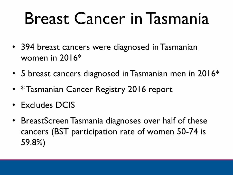
Breast Screening - AustraliaAims
1. To ensure significant reductions can be achieved in morbidity and mortality attributable to breast cancer.
2. To maximise the early detection of breast cancer in the target population.
3. Screening must be provided in dedicated and accredited Screening and Assessment Services as part of the BreastScreen Australia Program.
4. To ensure equitable access for women aged 50−69 years to the Program.
5. To ensure that services are acceptable and appropriate to the needs of the eligible population.
6. To achieve high standards of program management, service delivery, monitoring and evaluation, and accountability

BreastScreen Tasmania
• Targets asymptomatic women aged 50 – 74 (although all Tasmanian women aged over 40 are eligible)
• Delivers services in accordance with the Principles of BreastScreen Australia (Aims and Objectives)
• Meet the criteria of the Australian Population Based Screening Framework
• Based on the World Health Organization (WHO) principles of screening

WHO Principles

WHO Principles

WHO Principles

Forms of Health Test
• Opportunistic Testing
• Targeted Risk Screening
• Routine Exam or Planned surveillance

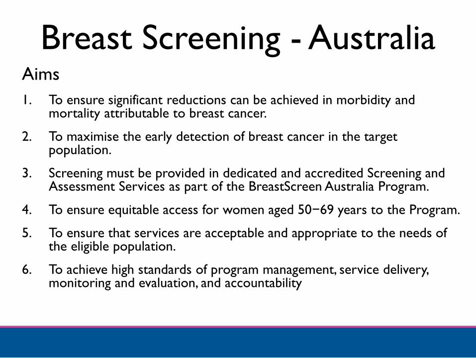
Screening vs. Diagnostic
Screening vs. Diagnostic

Screening vs. DiagnosticWhat happens when women go in the wrong direction?
Assuming the asymptomatic woman has no clinical findings, the
GP has a choice of two paths.

Screening vs. DiagnosticSending the asymptomatic woman in the eligible age group
(40-74) to BreastScreen is the right patient for the right pathway

Screening vs. DiagnosticWhat happens if the asymptomatic woman is sent down
the diagnostic pathway?
Screening vs. Diagnostic
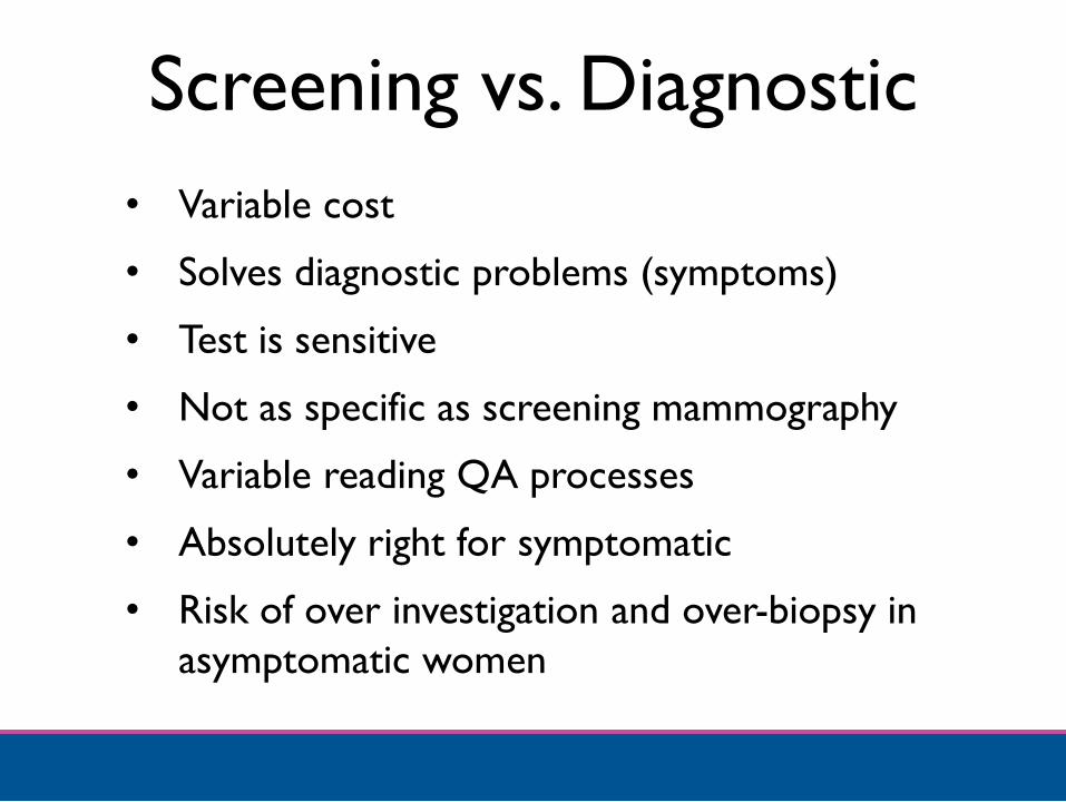
• Variable cost
• Solves diagnostic problems (symptoms)
• Test is sensitive
• Not as specific as screening mammography
• Variable reading QA processes
• Absolutely right for symptomatic
• Risk of over investigation and over-biopsy in
asymptomatic women

Screening vs. DiagnosticWhat happens when symptomatic women are sent to
BreastScreen?
This is usually because of cost or accessibility
Women with symptoms will not be turned away, but…..

Screening vs. DiagnosticOnly a minority will have a screen detected abnormality and
go on to assessment
Women who have no abnormality detected (and their GP) receive
a letter advising they should consult their GP for a referral to a
diagnostic mammography service for their breast symptom, which
is the pathway they should have been on initially

Screening vs. Diagnostic
It’s about safety and costs to women and the community, and
using the right tool for the job!
BreastScreen is specifically designed and quality controlled for
screening. Diagnostic radiology is not.
Reality is: many women
go down the wrong
pathway but eventually
find their way to the
appropriate assessment
Breast Density


BSA Breast Density Research
• http://www.cancerscreening.gov.au/internet/screening/publishing.nsf/Content/breast-density-literature-review

Early Detection of Breast
Cancer – The Role of the GPMr David Finkelde

Outline• Facts about Breast Cancer
• GP Breast Assessment
• Pathways for your patient
– Asymptomatic
– Symptomatic
• What I do in Practice
• Newly diagnosed cancer
• Decisions, decisions.
• Breast density
• New Breast Imaging techniques
• Psychosocial support
• Cases for discussion

Facts About Breast Cancer
• Heterogeneous group of malignancies arising from
breast tissue
• Now estimated to be the most commonly diagnosed
cancer across all sexes and in females
• Likelihood of being diagnosed with breast cancer
before age 85 is 1 in 8 for females
• Second most common cause of cancer death in
females (behind lung) and fourth most common
cause of cancer death in the community

Facts About Breast Cancer
• 2009 – 2013 5yr relative survival 90% c.f general
population
• 1984 – 1988 5yr relative survival 72%
• Figures are worse for younger women and specific
subtypes of breast cancer

Facts About Breast Cancer
• Age <40; 5% of all breast cancers
• Survival worse than for >40
• Leading cause of cancer death in women aged 20 – 39
• Most common cancer in women 20 – 39
• Higher grade / hormone receptor negative / HER2
positive
• Worse outcome for hormone receptor positive
tumours

Facts About Breast Cancer
• Increasing life expectancy in the community
• Elderly also get hormone receptor negative tumours
• No reason to compromise standard treatment in the
era of breast conservation surgery and sentinel node
biopsy

GP Breast Assessment
• History (including BSE)
• Inspection
– Symmetry
– Contour
• Examine both sides
• Physical examination is the starting point
– Symptomatic assessment
– Surveillance

Early Detection – Advice to Women
• Perspective on risks (especially family history)
• Symptoms
• Breast Awareness (Self Examination)
• Surveillance
– Screening
– Higher risk groups
• Lifestyle factors

Pathway for Asymptomatic Women
• No increased risk
– BreastScreen Tasmania (if aged ≥40)
– Breast Awareness
– Annual clinical check
• Increased/High Risk
– Seek Advice

Pathway for Symptomatic Women
1. Suspect Everyone
2. Imaging points the way but is not diagnostic
3. Pathology rules
4. The Triple Test
5. What I do in Practice
Imaging (Symptomatic Women)
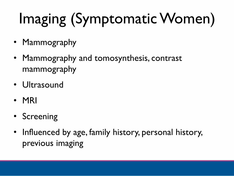
• Mammography
• Mammography and tomosynthesis, contrast
mammography
• Ultrasound
• MRI
• Screening
• Influenced by age, family history, personal history,
previous imaging

Pathology Rules (Symptomatic Women)
• Pathology can explain imaging, but not vice versa
• We treat pathology, not images
• Surgery in the absence of a pathological diagnosis
cannot be considered to automatically be therapeutic

The “Triple Test”
• Strictly applies only to assessment of a lump
• Original study was only 234 patients
– Clinical Examination, mammography, FNA
• Combined triad had negative predictive value of 100%
– These patients could be safely observed
What I do in Practice
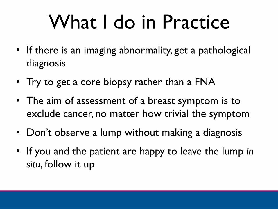
• If there is an imaging abnormality, get a pathological
diagnosis
• Try to get a core biopsy rather than a FNA
• The aim of assessment of a breast symptom is to
exclude cancer, no matter how trivial the symptom
• Don’t observe a lump without making a diagnosis
• If you and the patient are happy to leave the lump in
situ, follow it up

Rules to Live By• The only breast lump diagnosable by imaging alone is a
simple cyst
• Breast tissue is living and changes – any assessment is
only valid at the time it was made.
• A benign diagnosis does NOT mean that it cannot
become malignant
• The patient has the last word in deciding whether to
excise a non-malignant lump
• Be Paranoid – it’s what they came to you for
A New Diagnosis of a Cancer
• Assume that the starting point will be surgery
• <1% will have evidence of secondary spread at
diagnosis
– Beware recent onset of site specific pain

Decisions, Decisions
• What we know at diagnosis
• Major changes in recent practice
• What we do now and why
What We Know at Diagnosis
• Size
• Nodal status (clinical)
• Histology

Major Changes in Recent Practice
• Management of the Axilla
• Guidelines on margins for breast conservation surgery
• Mastectomy and reconstruction

Management of the Axilla
• Sentinel Node Biopsy
– First node or nodes in the regional basin that receive lymph
from the primary site
– Accurately predicts status of regional node basin
• ACOSOG Z0011 Trial
– T1-2 node negative tumours with positive SN (H&E)
randomised to ALND or no further surgery
– Equivalent results (1 or 2+ SN) for LR relapse and survival

Margins for Breast Conserving Surgery
• Negative margins reduce odds of LR
• Increasing distance for defining negative margins is not
associated with reduced odds for LR
• Positive margin is defined as ink on invasive cancer or
DCIS
• A negative margin is no ink on tumour
Mastectomy and Reconstruction
• Increasing rates in US and Australia
• Increasing numbers of reconstructions
• BCS offers equivalent long term survival
• Definite role in the management of high risk genetic
mutations
Molecular Diagnosis
• Receptors
• Gene mutations
• New treatments

What We Do Now and Why• Work-up is everything – the planned operation should be
what is performed
• The surgical options and proposed treatment, as well as
the potential for further surgery and other treatments,
should all have been discussed with the patient before the
operation
• Mastectomy is equivalent to lumpectomy and radiotherapy
and both operations should be explained
• In general, the default operation for a palpable lump with a
clinically negative axilla is wide local excision of the lump
and sentinel node biopsy

What We Do Now and Why• Is it operable? Is it suitable for breast conservation?
• If a mastectomy is needed, does the lady want a
reconstruction?
• If the plan is eventually a mastectomy and
reconstruction, then what type of mastectomy?
• How will surgery fit in with the timeline and priorities
of the other treatments? Is there a role for neo-
adjuvant therapy?

What We Do Now and Why• Lumpectomy and SNB
– 2x small incisions, 2-3 cm long, no drains
– Think about cosmesis and potential for mastectomy
• Axillary dissection
– Level 2-3 clearance, single drain
– Lymphoedema rate 12%
• Simple mastectomy
– Removal of breast, NAC and redundant skin, single drain

What We Do Now and Why• Absorbable sutures, lots of local anaesthetic,
waterproof dressings, padded support
• Aim for early discharge; outpatient support
• Not usually painful compared to muscle cutting
procedures
• 4-5 working days for useful pathology results

Breast Density
• Definition
• Risk
• Management
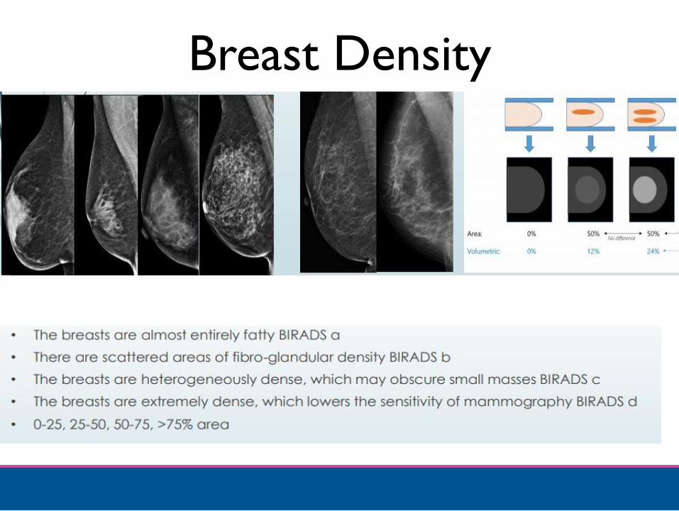
Breast Density – Definition• What we are talking about is mammographic breast
density
• It is an assessment of the mammographic appearance
of the breast
• MBD is defined in the relative amount of radiopaque
elements to radiolucent fat in the breast image
• There is no consistently agreed method for describing
breast density and determining the value at which the
breast is ‘dense’
Breast Density – Risk• Multiple issues with defining ‘dense’ breasts
• Multiple methods of assessing breast density
• Variability with results both between differing measuring
systems and within the same system
• Generally accepted that increased breast density
correlates with an increased relative risk of 1.2 - 2.1 c.f.
average population
• Level of risk is equivalent to that of having a post-
menopausal first degree relative diagnosed with breast
cancer
Breast Density – Risk• This is not new!
• Two potential avenues of increased risk
– Masking
– More rapid growth in dense tissue
• Limitations of cancer detection based on
mammographic images with large amounts of
radiopaque tissue have always been recognised
• This underpins selection of screening age ranges

Breast Density – Management
• Supplemental imaging
– Whole breast ultrasound
– Molecular breast imaging (ultrasound, isotopes)
– Digital Breast Tomography
– MRI
– Contrast Enhanced Mammography
• Public awareness
– Breast density reporting mandatory in >30 US States
Breast Density – Management
• Is it necessary?
– Small increase in cancer detection
– False positives, cost
– No evidence for improved survival
• Individual risk management

New Breast Imaging• Tomosynthesis
• Ultrasound
• MRI
• Contrast Enhanced Mammography
• Selective OSR
• Screening vs. Diagnosis
• Pathology still rules
Psychosocial Support
• GP
• Breast Care Nurse
• Oncologist
• Cancer Council/Support Groups
• Social Worker
• Psychologist
Psychosocial Support
• Age-related needs
• Diagnosis
• Impact of treatment
– Surgical
– Adjuvant

Cases for Discussion - 1• 2014
• 29 y.o. 6/52 lump in breast
• No children, no FHx, only med OCP
• Palpable mass LUOQ, palpable L axillary LN
• Mammogram and U/S – 35 mm mass with microcalc and multiple
abnormal LN
• Core and FNA – Grade 2 IDC, positive LN on cytology
• Elected mastectomy (with AD)
• D/w Med Onc post-op chemotherapy
• Fertility referral – embryo preservation
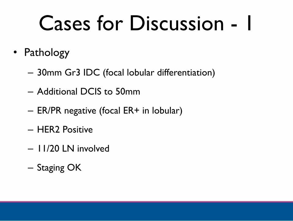
Cases for Discussion - 1
• Pathology
– 30mm Gr3 IDC (focal lobular differentiation)
– Additional DCIS to 50mm
– ER/PR negative (focal ER+ in lobular)
– HER2 Positive
– 11/20 LN involved
– Staging OK

Cases for Discussion - 1
• Post-operative treatment
– TC and Herceptin, Zoladex for ovarian protection
– DXRT to chest wall and SCF
– Tamoxifen (LN ER+)
• Outcome
– 2018 left reconstruction with flap and implant
– Alive and well with no evidence of recurrence, no immediate
plans for a family.

Screening Images

Tomosynthesis Image
• The breast lesion is
clearly visible on
the screening
images and using
tomosynthesis

Cases for Discussion - 2• 52 y.o. 6/12 intermittent swelling (L) arm and ‘change’ in (L)
breast
• No family history
• TBSS 28/11/18
• Recalled for assessment 14/12/18
– 35mm stellate lesion
– Abnormal mass left axilla
• Biopsy results 18/12/18
– Grade 3 ductal carcinoma (+) cytology (L) axillary LN
– ER/PR (+) HER29 (-) on core

Cases for Discussion - 2
• Consideration of neo-adjuvant therapy
• Staging
• Elected Breast Conservation Surgery
– ChG and AD

Screening Images

Tomosynthesis Image
• The breast lesion
(close to chest
wall) and masses
in axilla become
clearly visible
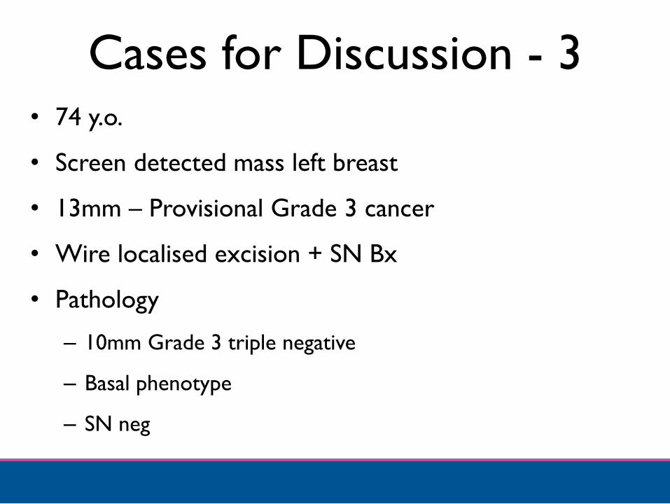
Cases for Discussion - 3• 74 y.o.
• Screen detected mass left breast
• 13mm – Provisional Grade 3 cancer
• Wire localised excision + SN Bx
• Pathology
– 10mm Grade 3 triple negative
– Basal phenotype
– SN neg

Cases for Discussion - 3
• Referred for adjuvant treatment
– Chemotherapy
– Radiotherapy
Recommended