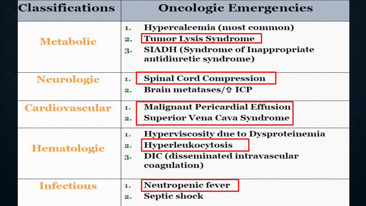
APPROACH TO HEME/ONC
EMERGENCIESBy Kashif Khan (PGY-3)

LEARNING OBJECTIVE
• Identify commonly encountered Hematological and
Oncological emergencies
•Order appropriate labs/radiological tests
• Initiate timely treatment
• Early sub-specialty consultations

WHAT IS AN ONCOLOGICAL EMERGENCY
•A clinical condition resulting from a metabolic,
neurologic, cardiovascular, hematologic, and/or
infectious change caused by cancer or its
treatment that requires immediate intervention
to prevent loss of life or quality of life.

CASE - 1

A 60 yo M with PMH of HTN, GERD, depression, who presents with 1 month history
of progressively worsening generalized fatigue, weakness, and feeling unwell.
ROS: Positive for dry cough, night sweats, 20 lb unintentional wt loss in last 3
months
Vitals: HR 110/m, otherwise stable
Being an excellent new intern, you perform a full physical and find bilateral
cervical and supraclavicular lymphadenopathy and palpable hepatosplenomegaly.
Remainder of the exam was unremarkable.
Labs:
CBC/diff: WBC 15k H/h 7.9/25 Plt 75k Diff: 85%L 10%N 5%M
RFP: Na138, K 5.6, Cl 105, Bicarb 22, BUN 35, Cr 1.9(baseline Cr 0.8)
LDH 1205, uric acid 12.6, calcium 7.0, phosphorus 6.5, albumin 3.2

TUMOR LYSIS SYNDROME

TUMOR LYSIS SYNDROME (TLS)
K+
K+
PO4
Ca2+
Ca2+
CaPO4
TLS is the result of a massive and abrupt release of
cellular contents into the bloodstream after rapid lysis of
malignant cells
Uric Acid

TUMOR LYSIS SYNDROME (TLS)
• Seen in high grade liquid tumors like leukemia with leukocytosis, high grade
lymphomas, and some solid tumors like small cell lung ca
• Clinical Features: weakness, arrhythmias, paralysis, acute renal failure, tetany,
altered mental status, seizures
Diagnosis:
• Laboratory
o ≥ 2 laboratory abnormalities OR
o ≥ 25% change in 2 values from baseline value
• Clinical
o Laboratory diagnosis + end organ damage
HyperKalemia
HyperUricemia
HyperPhosphatemia
HypoCalcemia

TUMOR LYSIS SYNDROME (TLS)Treatment:
• Aggressive IV Fluids &/or diuresis (Most important)
• Manage electrolyte abnormalities
• Rasburicase (Check G-6PD before!)
• Allopurinol (Does not decrease uric acid)
• HD
Prevention:
Fluids, Allopurinol, Rasburicase

CASE - 2

A middle aged F with HTN, DM, metastatic breast ca, presents with worsening
back pain x 2 weeks. Developed after lifting boxes while moving. More recently
has been feeling some RLE numbness and worsening pain.
ROS: Denies fevers/chills, weakness, loss of sensation, bowel/bladder
incontinence
Oncologic history: diagnosed with metastatic breast ca 3 yrs ago, last PET-CT 2
months ago showed stable/shrinking osseous mets in L scapula, multiple ribs,
thoracic spine (T8), and R femur.
O/E: severe pain on palpation of mid-thoracic spine, strength and sensation
intact throughout, normal reflexes, no saddle anesthesia, normal rectal tone
Labs: Unremarkable

MALIGNANT SPINAL CORD COMPRESSION

SPINAL CORD COMPRESSION

SPINAL CORD COMPRESSION
• Majority from:
Breast
Lung
Prostate
Lymphoma
Myeloma
• About 6-10% of patients with cancer
• Thoracic spine (up to 70%)
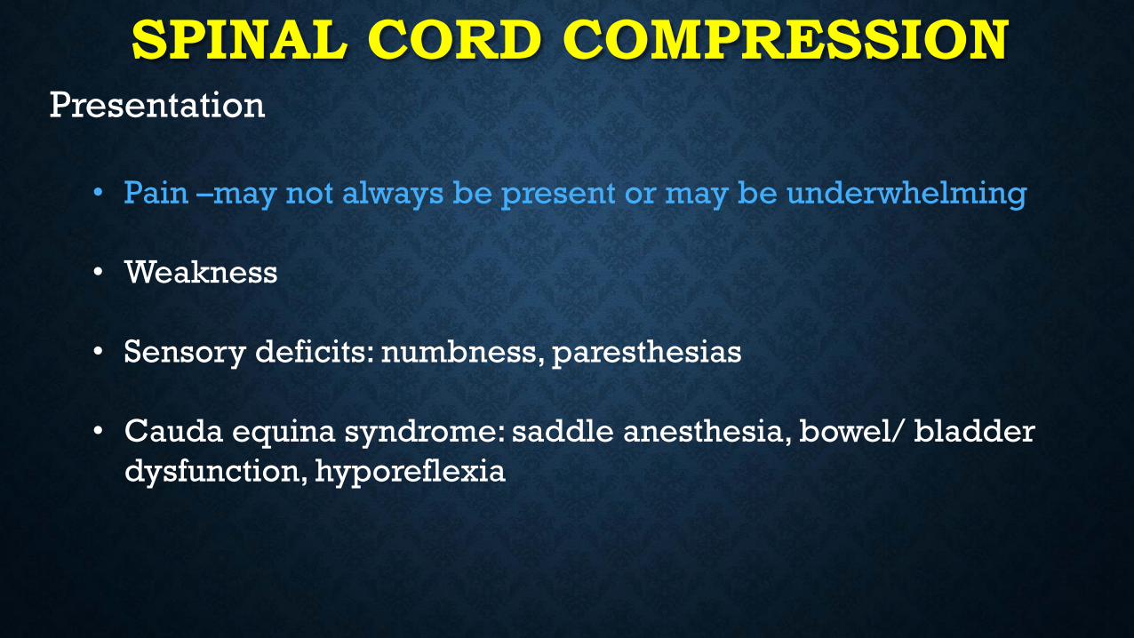
SPINAL CORD COMPRESSIONPresentation
• Pain –may not always be present or may be underwhelming
• Weakness
• Sensory deficits: numbness, paresthesias
• Cauda equina syndrome: saddle anesthesia, bowel/ bladder
dysfunction, hyporeflexia

SPINAL CORD COMPRESSION• Obtain a good history and neurologic exam
• MRI (CT Myelography)
• Steroids: dexamethasone 10mg IV STAT then 4mg q6
• Time is money! ortho/ neurosurgery, radiation oncology
• Pain control
• Primary determinant of the efficacy of therapy is the
patient's neurologic status at time treatment

CASE - 3
75 yo M, heavy smoker, presents with 4 weeks of SOB, worsening
non-productive cough, and 20 lb weight loss. Developed neck
swelling last week, which worsens on bending forwards. ROS -
negative
Vitals: Stable
O/E: R mid-lung crackles, no wheezing/stridor, swelling of neck and
right upper extremity, distension of superficial anterior chest and
neck veins, no focal neuro deficits
Labs: unremarkable

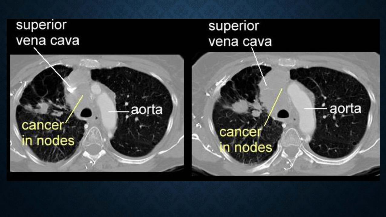
SUPERIOR VENA CAVA SYNDROME
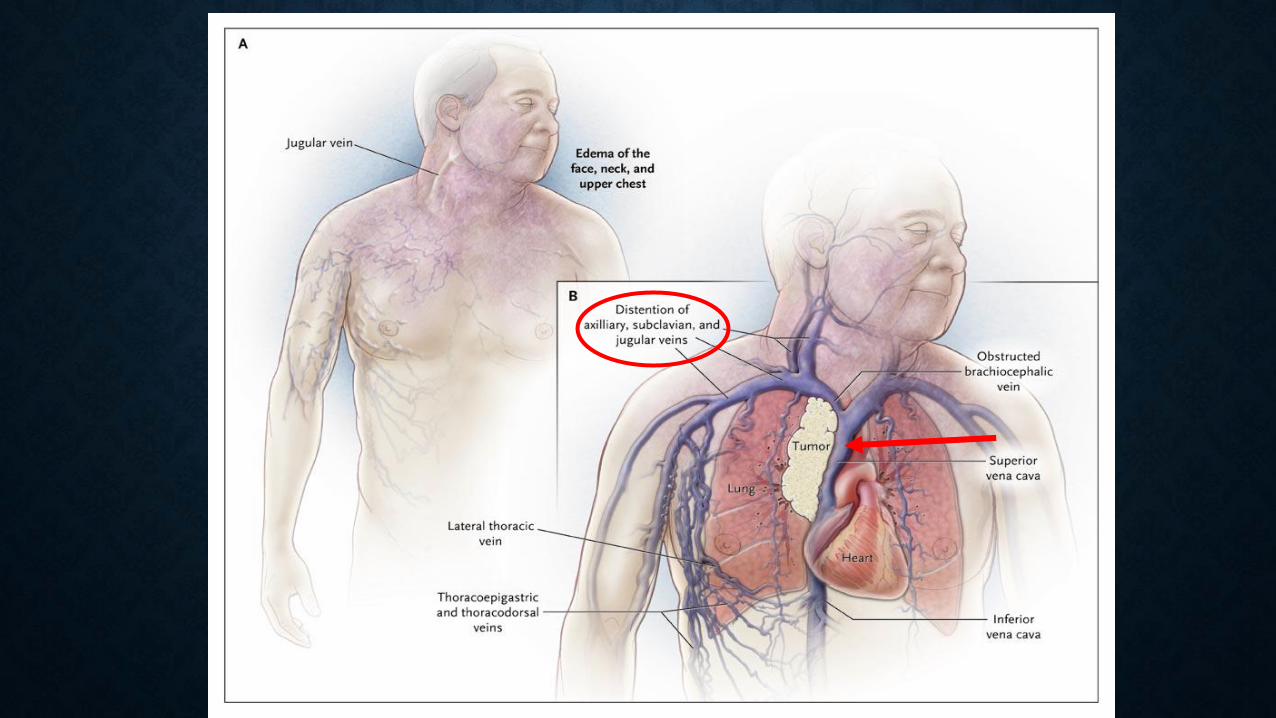
• Most cases are not a true emergency
• Majority of cases are due to Lung Ca or NHL (intrathoracic malignancies)
• Dyspnea (most common). Facial edema, arm edema, distended veins, facial
plethora, cough, airway etc.
• Diagnosis:
o CT/ MRI
o Histological Dx
SUPERIOR VENA CAVA SYNDROME (SVC)

Treat underlying cancer!
Endovascular stents/ Radiotherapy
Supportive care:
• Head elevation, Diuretics
• Avoid high volume fluid infusion through upper extremities
• Anticoagulation
• Steroids
o Severe Airway Obstruction
o Lymphoma
SUPERIOR VENA CAVA SYNDROME (SVC)

CASE - 4

55Y F with AML presents with profound fatigue and 1 week of SOB.
Accompanying family members report report she initially
complained of a headache and dizziness, and since then has started
acting confused and been more somnolent.
Vitals: T:100.6, PR: 115/m, BP: 110/76, RR: 26/m, 80%RA
Pertinent exam: A&Ox1 (wrong date and location), confused,
strength and sensation intact, ataxic gait, bilateral lung crackles
Labs:
CBC: WBC 88K with 78% blasts, H/h 10/30 Plt 3k
RFP: 140 5.0 108 24 20 1.2 100


• Increased blood viscosity impedes blood flow, and local
hypoxemia is worsened by high metabolic activity of cells and
cytokine release
• Symptoms: (CNS/Eyes/Lungs)Pulmonary: hypoxia, interstitial/alveolar infiltrates
Neurological: headache, dizziness, ataxia, confusion, somnolence,
blurry vision
• Management:Rapid cytoreduction with chemotherapy
Consider hydroxyurea or leukapheresis if unable to give
chemo
LEUKOSTASIS
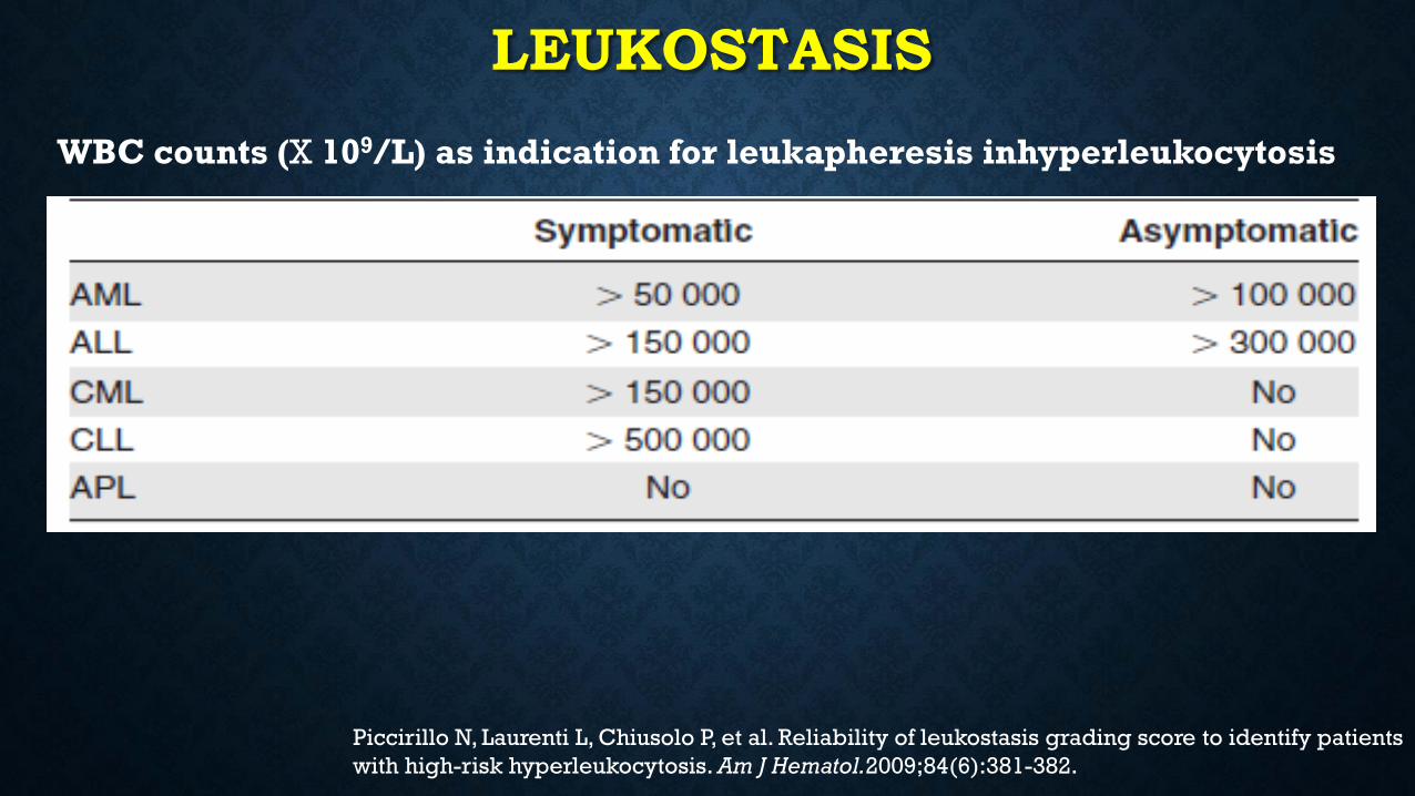
LEUKOSTASIS
WBC counts (X 109/L) as indication for leukapheresis inhyperleukocytosis
Piccirillo N, Laurenti L, Chiusolo P, et al. Reliability of leukostasis grading score to identify patients
with high-risk hyperleukocytosis. Am J Hematol.2009;84(6):381-382.

CASE - 5
A 70 yo M with metastatic colon ca (finished cycle #3 FOLFOX ten days ago),
who presents with fevers for 2 days at home. He states he checked his
temperature at home and it has ranged from 99 to 102 °F.
ROS: Unremarkable
Vitals: T: 38.7oC, PR: 105/m, BP: 115/75, RR: 16/m,SP02 97%RA
Exam: Mild oral mucositis, Mediport c/d/i, lungs clear, abdominal exam
benign, otherwise no focal findings
Labs:
CBC/diff: WBC 2.0k H/h 8.5/28 Plt 85k Diff: 68%L 20%N 9%M 3%E
RFP: WNL

FEBRILE NEUTROPENIA

FEBRILE NEUTROPENIA
Infection in a neutropenic patient is an emergency
Pathogenesis:
• Breeches in host defenses (breakdown of mucosal barriers)
• Immune system suppression
• Majority of cases of neutropenic fever thought to be caused by
bloodstream seeding from GI tract flora
Infectious source: 30%.

FEBRILE NEUTROPENIADiagnosis:
ANC<500 or ANC<1000 with expected nadir <500 over next 48 hours
+T: 38oC for >1 hour or T>38.3oC once
Next Steps:• Is patient HDS? Stable for floor?
• Examine patient: any localizing symptoms? Any role for imaging?
• Cultures STAT (2 sets bld cx peripheral, culture from lines or ports, sputum or
stool cx/C Diff or wound cx as indicated), UA, UCx, CXR
• Antibiotics (30-60min)

FEBRILE NEUTROPENIA
Clinical Scenario Medication

FEBRILE NEUTROPENIA – LOW RISK PATIENTS
Certain low-risk patients can be treated at home with PO antibiotics (typically Ciprofloxacin +
Augmentin) after initial IV dose and brief observation
IDSA: anticipated neutropenia ≤ 7d, clinically stable, ANC > 100, and no medical comorbidities
ASCO: MASCC score ≥ 21

MALIGNANT PERICARDIAL
EFFUSION

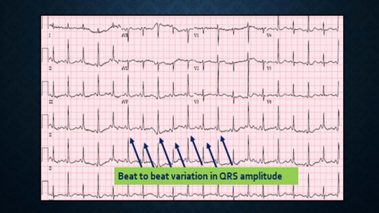
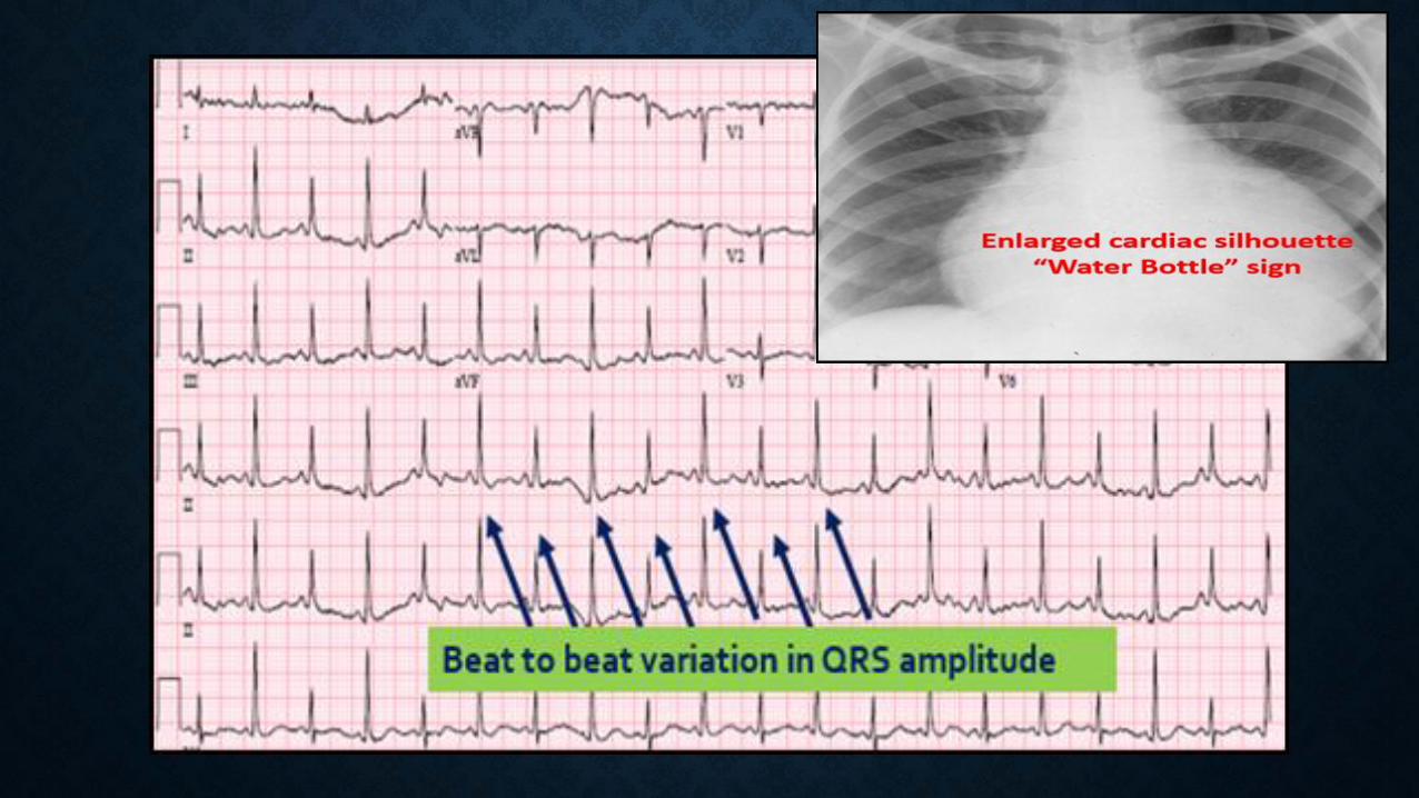
MALIGNANT PERICARDIAL EFFUSION
• Can be related to cancer OR chemo/RT/infection/autoimmune
• Clinical manifestations
Dyspnea, cough, Chest pain, orthopnea, palpitations.
Exam findings: Beck’s triad (JVD, hypotension, decreased heart sounds),
narrow pulse pressure, pulsus paradoxus
Treatment:
• Small/moderate effusions are usually asymptomatic and do not require urgent
treatment
• Acute management: drainage with pericardiocentesis
• Prevention of re-accumulation: drainage catheter, pericardial window
• Treat underlying cancer

ACUTE CHEST SYNDROME
ACUTE CHEST SYNDROME
• Vaso-occlusive crises of pulmonary vasculature in patients with
sickle cell anemia.
• Leading cause of death in SCD.
• New radio-density on CXR AND any one (T >38.5 OC, >2% drop in
SpO2, CP, cough, wheezing, rales, tachypnea)
• Maintain high suspicion, as some may develop ACS within 48-
72hours after initial pain episode!
ACUTE CHEST SYNDROME
Don’t Forget other D/D
Treatment:
• T&S, adequate pain control, IV access, fluids, oxygen, incentive
spirometry, antibiotics, VTE prophylaxis, hematology consult.
• Consider simple Vs exchange transfusion & MICU transfer
• Can use simple transfusion to bridge to exchange transfusion
while waiting for MICU bed (does not remove HgbS)

OTHER TIPS

• Primary Oncologist?
• Date of last chemo? (Check on EMR IV chemo)
• What was their last chemo? (know your acronyms)
• Did they get any medications with chemo? (G-CSF)
• What is their previous oncologic course?
• Access for Chemo? (mediport, PICC?)
• Sickle cell crises: check care path in portal and OARRS
• Inform primary oncologist of patient’s admission

REFERENCES• Wilson FP et al. Onco-nephrology: tumor lysis syndrome. Clin J Am Soc Nephrol. 2012 Oct;7(10):1730–9.
[PMID: 22879434]
• McCurdy MT et al. Oncologic emergencies. Crit Care Med. 2012 Jul;40(7):2212–22. [PMID: 22584756]
• Freifeld AG et al. Clinical practice guideline for the use of antimicrobial agents in neutropenic patients
with cancer: 2010 Update by the Infectious Diseases Society of America. Clin Infect Dis. 2011 Feb
15;52(4):427–31. [PMID: 21205990]
• Chen KN, Xu SF, Gu ZD, et al. Surgical treatment of complex malignant anterior mediastinal tumors
invading the superior vena cava. World J Surg 2006; 30:162
• Wilson LD, Detterbeck FC, Yahalom J. Clinical practice. Superior vena cava syndrome with malignant
causes. N Engl J Med 2007; 356:1862
• Symposium on neoplastic hematology and medical oncology - Emergencies in Hematology and
Oncology. Mayo Clinic 2017
• Savage P, Sharkey R, Kua T, et al. Clinical characteristics and outcomes for patients with an initial
emergency presentation of malignancy: a 15 month audit of patient level data. Cancer Epidemiol.
2015;39(1):86-90

THANK YOU
Recommended