University of South FloridaScholar Commons
Graduate Theses and Dissertations Graduate School
1-20-2016
Antigen Specific Induced T Regulatory CellularTherapy for Graft-Versus-Host Disease FollowingAllogeneic Bone Marrow TransplantationJessica Lauren Heinrichs
Follow this and additional works at: http://scholarcommons.usf.edu/etd
Part of the Immunology and Infectious Disease Commons, and the Oncology Commons
This Dissertation is brought to you for free and open access by the Graduate School at Scholar Commons. It has been accepted for inclusion inGraduate Theses and Dissertations by an authorized administrator of Scholar Commons. For more information, please [email protected].
Scholar Commons CitationHeinrichs, Jessica Lauren, "Antigen Specific Induced T Regulatory Cellular Therapy for Graft-Versus-Host Disease FollowingAllogeneic Bone Marrow Transplantation" (2016). Graduate Theses and Dissertations.http://scholarcommons.usf.edu/etd/6093

Antigen Specific Induced T Regulatory Cellular Therapy for Graft-Versus-Host Disease Following
Allogeneic Bone Marrow Transplantation
by
Jessica Lauren Heinrichs
A dissertation submitted in partial fulfillment of the requirements for the degree of
Doctor of Philosophy Department of Pathology and Cell Biology
College of Medicine University of South Florida
Co-Major Professor: Xue-Zhong Yu, M.D. & M.S. Co-Major Professor: Patricia Kruk, Ph.D.
Claudio Anasetti, M.D. Amer Beg, Ph.D.
Srinivas Bharadwaj, Ph.D. Burt Anderson, Ph.D.
Date of Approval: January 15, 2016
Keywords: Alloreactive, Graft-versus-Leukemia, CD8 Induced Tregs, Minor-mismatched antigens, and Combination therapy
Copyright © 2016, Jessica Lauren Heinrichs

Dedication
This dissertation is dedicated to my loving and supportive father and mother, Dennis and
Shirley Heinrichs. Their constant encouragement and devotion got me through the most challenging
parts of this process. A special dedication to my sister Erica Heinrichs; she motivated me to pursue
my dreams even if the road to success was long and treacherous. Also I dedicate this work to my
grandparents, Francis and Dolly Heinrichs and Lou Franklin who left this world too soon, but whose
love lives on in me.
Finally, a special dedication to my future husband Dr. Kellen Voss; he stood by my side
through the worst and best of times. He continues to encourage my dreams and turn them into our
dreams. Life without him would truly not be worth living. I am so happy to end this chapter of my
life and begin a new one with him by my side.

Acknowledgements
I am very thankful to my mentor Dr. Xue-Zhong Yu for his scientific guidance and support for
me throughout my PhD career. His knowledge and expertise in transplantation immunology is
unsurpassed. I appreciate that he encouraged me to move to the Medical University of South Carolina
with him during my second year, which really enhanced my PhD experience and helped me grow as
an individual. I also greatly appreciate my co-mentor Dr. Patricia Kruk for being a strong supporter of
my scientific and personal goals throughout my PhD. She always reminded me to keep my eyes on the
prize and pushed me to my highest potential. I would like to extend my great gratitude to my
committee members, Dr. Claudio Anasetti, Dr. Sri Nagaraj, Dr. Amer Beg and Dr. Burt Anderson, for
their kindness and helpful guidance these last four years.
I would like to especially thank all the past and current members of the Yu lab: Dr. Kelley
Haarberg, Dr. Jianing Fu, Dr. Yongxia Wu, Dr. Hung Nguyen, Dr. Hanief Sofi, David Bastian, Kane
Koassard, Steven Schutt, Dr. Anusara Daenthanasanmak, and Yuejun Liu. They have provided me
with experimental assistance, editing manuscripts, and emotional support and encouragement these
last four years.
A special thank you to my co-PhD students and best friends: Stephanie Blankenship,
Adonis Mcqueen, Krishna Reddy, and Geofery Ciarolone for our study sessions and for being the
coolest nerds around.
Finally, I would like to thank the technical support from the animal facilities and flow
cytometry cores at both Moffitt Cancer Center and the Medical University of South Carolina for
providing top of the line core that made my research a seamless enjoyable process.

i
Table of Contents
List of Tables ........................................................................................................................................... iv
List of Figures........................................................................................................................................... v
Abstract .................................................................................................................................................. vii
Chapter 1: Background ............................................................................................................................. 1
1.1 Allogeneic HCT ...................................................................................................................... 1
1.2 GVHD Pathophysiology ......................................................................................................... 1
1.3 GVL Effect ............................................................................................................................. 2
1.4 Regulatory T Cells .................................................................................................................. 3
1.4.1 Initial Discovery ............................................................................................... 3
1.4.2 CD25 and Foxp3 ............................................................................................... 4
1.5 Natural and Induced T Regulatory Cells ................................................................................. 4
1.5.1 Development and Generation ............................................................................ 5
1.6 Suppressive Mechanisms ........................................................................................................ 6
1.6.1 Contact-Dependent Mechanisms ...................................................................... 7
1.6.2 Contact-Independent Mechanisms .................................................................... 7
1.6.3 Mechanisms Specific to GVH and GVL ........................................................... 8
1.7 Stability .................................................................................................................................. 9
1.7.1 Epigenetic Control of Foxp3 ........................................................................... 10
1.7.2 Foxp3 Protein Stability ................................................................................... 10
1.8 nTregs in GVHD................................................................................................................... 11
1.8.1 Pre-Clinical Findings ...................................................................................... 12
1.8.2 Expansion of Human nTregs .......................................................................... 12
1.8.3 Antigen-Specific nTreg ................................................................................... 13
1.8.4 Clinical Trails ................................................................................................. 13
1.9 iTregs in GVHD ................................................................................................................... 14
1.9.1 Pre-Clinical Findings ...................................................................................... 15
1.9.2 Polyclonal versus Antigen Specific iTregs ..................................................... 15
1.9.3 HY Antigen ..................................................................................................... 16
1.9.4 Polarizing Cytokines ....................................................................................... 17

ii
1.9.5 Infusion Schedule ........................................................................................... 17
1.10 CD8 iTregs in GVHD ......................................................................................................... 18
1.11 Tregs Effect on GVL Preservation ...................................................................................... 19
1.12 Modifying Tregs ................................................................................................................. 20
1.13 Combinational Therapy ...................................................................................................... 21
Chapter 2: HY-Specific Induced Regulatory T Cells Display High Specificity and Efficacy in the Prevention of Acute Graft-versus-Host Disease ................................................................................ 22
2.1 Abstract ................................................................................................................................ 22
2.2 Introduction .......................................................................................................................... 23
2.3 Materials and Methods .......................................................................................................... 24
2.3.1 Mice ................................................................................................................ 24
2.3.2 T-cell purification and iTreg generation ......................................................... 25
2.3.3 Immuno-fluorescence analysis. ....................................................................... 25
2.3.4 BMT and bioluminescent imaging (BLI). ....................................................... 25
2.4 Results .................................................................................................................................. 26
2.4.1 HY-specific iTregs suppress polyclonal T-cell response to alloantigens in vitro………………………………………………………………….. ....... 26
2.4.2 HY-specific iTregs prevent GVHD in activation-dependent manner .............. 28
2.4.3 HY-specific iTregs suppress the expansion and activation of donor T cells………………………………………………………………..…… ....... 31
2.4.4 Expression of target antigen on epithelial tissues is not required for iTregs to prevent GVHD……………………………………………… ......... 36
2.4.5 HY-specific iTregs essentially preserve the GVL effect ................................. 37
2.6 Discussion ............................................................................................................................ 39
Chapter 3: CD8 Tregs Promote GVHD Attenuation and Overcome the Impaired GVL Effect Mediated by CD4 Tregs in Mice…………………………………………………………………. ....... 43
3.1 Abstract ................................................................................................................................ 43
3.2 Introduction .......................................................................................................................... 44
3.3 Materials and Methods .......................................................................................................... 46
3.3.1 Mice ................................................................................................................ 46
3.3.2 iTreg Generation ............................................................................................. 46
3.3.3 GVH/GVL Models ......................................................................................... 46
3.3.4 Flow Cytometry and Intracellular Cytokine Staining ..................................... 47
3.3.5 Microarray ...................................................................................................... 47
3.3.6 Statistics ......................................................................................................... 47
3.4 Results .................................................................................................................................. 48

iii
3.4.1 Alloreactive CD4 iTregs suppression of GVHD is antigen specific ............... 48
3.4.2 Alloreactive CD4 iTregs impair GVL activity against aggressive leukemia ......................................................................................................... 50
3.4.3 CD8 iTregs moderately attenuate GVHD and fully preserve GVL function .......................................................................................................... 52
3.4.4 Combination of CD4 and CD8 iTregs is superior to singular iTregs therapy in GVHD attenuation………………………………………….. ....... 53
3.4.5 Combinational therapy rescues inhibited GVL response of CD4 singular therapy .............................................................................................. 56
3.4.6 CD8 iTregs contribute to the preserved GVL effect by maintaining cytolytic functions .......................................................................................... 58
3.4.7 Alloreactive CD4 and CD8 iTregs display differential gene expression profiles .......................................................................................... 59
3.5 Discussion ............................................................................................................................. 61
Chapter 4: Conclusions and Future Directions ....................................................................................... 65
Chapter 5: List of References ................................................................................................................. 67
Chapter 6: Appendices ............................................................................................................................ 83
Appendix A: JIRT Copyright Transfer ....................................................................................... 83
Appendix B: JI Copyright Transfer ............................................................................................. 84
Appendix C: Blood Copyright Transfer ...................................................................................... 85
Appendix D: IACUC Approval .................................................................................................. 86
About the Author .................................................................................................................... END PAGE

iv
List of Tables
Table 1.1 Summation of Differences in iTreg Pre-Clinical Results ...................................................... 15

v
List of Figures
Figure 1.1 Delicate balance between GVH and GVL responses ............................................................. 3
Figure 1.2 nTregs and iTregs: Generation, Suppressive Mechanisms, and Stability ............................... 6
Figure 2.1 Generation and isolation of HY-specific iTregs ................................................................... 27
Figure 2.2 HY-specific iTreg attenuation of GVHD is antigen dependent ............................................ 29
Figure 2.3 Effects of HY-specific iTregs on long-term donor immune reconstitution .......................... 29
Figure 2.4 Effect of HY-specific iTregs in the prevention of GVHD in miHAg- or
haplo-mismatched BMT model ............................................................................................ 30
Figure 2.5 Pathogenicity of T cells from B6 Luc-Tg mice in GVHD.................................................... 31
Figure 2.6 Effects of HY-specific iTregs on Teff expansion in vivo ..................................................... 32
Figure 2.7 Stability and Efficiency of HY-specific iTregs .................................................................... 33
Figure 2.8 HY-specific iTregs suppress activation and expansion of Teffs .......................................... 34
Figure 2.9 HY-specific iTregs are highly stable under inflammatory conditions .................................. 35
Figure 2.10 Effect of HY-antigen distribution on HY-specific iTreg-medicated protection ................. 36
Figure 2.11 HY-Specific iTregs spare the GVL effect .......................................................................... 37
Figure 2.12 HY-specific iTregs largely preserve GVL effect in pre-established tumor
model……………………………………………………………………………………….38
Figure 2.13 Education with HY antigen enhances suppressive function of iTregs ............................... 41
Figure 3.1 Alloreactive CD4 iTregs display superior suppressive function than
polyclonal iTregs. ................................................................................................................. 48
Figure 3.2 Alloreactive CD4 iTregs attenuation of GVHD is antigen-specific ..................................... 49
Figure 3.3 Alloantigen specificity is essential for iTreg function and stability ..................................... 50

vi
Figure 3.4 Non-enriched alloreactive iTregs are not pathogenic ........................................................... 51
Figure 3.5 Alloreactive CD4 iTregs impair GVL activity against aggressive leukemia........................ 51
Figure 3.6 Generation and Function of Alloreactive CD8 iTregs .......................................................... 52
Figure 3.7 CD8 iTregs moderately attenuate GVHD and maintain GVL responses ............................. 53
Figure 3.8 Combination of CD4 and CD8 iTregs superior to singular iTregs therapy in GVHD
attenuation ............................................................................................................................ 55
Figure 3.9 CD4 iTregs display greater stability than CD8 iTregs ......................................................... 55
Figure 3.10 Combinational therapy reduce GVHD pathology and tumor burden ................................. 57
Figure 3.11 Combinational therapy preserves the GVL response ......................................................... 57
Figure 3.12 CD8 iTregs contribute to the preserved GVL effect mediated by
combinational therapy .......................................................................................................... 58
Figure 3.13 Alloreactive CD4 and CD8 iTregs display differential gene expression profiles............... 60

vii
Abstract
Allogeneic hematopoietic stem cell transplantation (allo-HCT) has been a successful cellular
therapy for patients suffering from hematological malignancies for many decades; however, the
beneficial effects of graft-versus-leukemia (GVL) are classically offset by graft-versus-host disease
(GVHD). GVHD occurs when major and/or minor human leukocyte antigen (HLA) mismatches
between donor and recipient cause rapid expansion and activation of donor effector T cells (Teffs)
resulting in end organ damage to the recipient’s epithelial tissues. Given the lymphoproliferative
nature of this disease, the standard treatment option is broad immunosuppression, which can result in
primary disease relapse, steroid refractory GVHD, and/or opportunistic infection. A more targeted
therapy that can selectively suppress GVH responses with maintained GVL responses would achieve
the optimal goal of allo-HCT. Regulatory T cells (Tregs) both natural (nTregs) or induced (iTregs)
could be potential cellular therapies for the treatment of GVHD, given their innate suppressive
function. Initial clinical trials using nTregs have yielded positive results; however, nTreg cellular
therapy has been cumbersome due to the necessity for large scale ex vivo expansion given their low
yield within an apheresis product and non-specific suppression. Conversely, iTregs can be generated
from naïve T cells thus decreasing ex vivo culture times and can be educated with specific antigen
thus providing targeted suppression, but a consensus on their efficacy for GVHD therapy has not been
reached. Therefore, we investigated the efficacy of antigen specific iTreg therapy for the prevention
of GVHD while maintaining GVL responses.
In Chapter 2, we evaluated the effectiveness of monoclonal HY-specific iTregs in GVHD
attenuation. We chose HY as a target antigen because it is a naturally processed, ubiquitously
expressed minor mismatch antigen carried by only male donors/recipients cited to increase GVHD
prevalence when donor and recipient are sex-mismatched. Utilizing HY-transgenic mice in which all T

viii
cells recognize HY antigen exclusively, we generated HY specific iTregs which effectively attenuating
GVHD in male, but not female recipients in three murine bone marrow transplantation (BMT) models
(major mismatch, parent to F1, and miHAg mismatch). We found HY specific iTregs lost stability in
female recipients but remained stable and suppressive in male recipients suggesting expression of HY
antigen was required for their suppressive function and stability. GVL responses were not
compromised with the addition of HY specific iTregs in recipient mice using a pre-established tumor
model. Thus, HY-specific iTregs can be generated and suppress GVHD in an antigen-dependent
manner while sparing the GVL effect.
In Chapter 3, we extend our findings in Chapter 2, which provided proof of principle that
antigen specific iTregs effectively control GVHD; however, this therapy has a limited translational
potential. Therefore, we generated alloreactive CD4 and CD8 iTregs and evaluated GVHD attenuation
and GVL preservation in either full or haplo-MHC mismatched BMT models. We found alloreactive
CD4 iTregs significantly suppress lethal GVHD, but completely abrogated the GVL effect against
aggressive tumors. Conversely, alloreactive CD8 iTregs moderately attenuated GVHD and possessed
direct cytotoxicity against tumor cells. Therefore, to rescue the impaired GVL effect mediated by CD4
iTregs, we established a combinational therapy with CD8 iTregs. Indeed we found combination CD4
and CD8 iTreg therapy significantly suppressed GVHD while sparing GVL responses compared to
either CD4 or CD8 singular therapy. Mechanistically, this was achieved by potent suppression of both
CD4 and CD8 Teffs coupled with preserved cytolytic molecule expression by both CD8 iTregs and
Teffs.
Taken together, we propose antigen specific iTreg therapy can effectively attenuate GVHD
while preserving GVL responses. We further uncovered unique characteristics of CD4 and CD8
iTregs that can be exploited to achieve the optimal cellular therapy following allo-HCT.

1
Chapter 1: Background
Note to Reader
Portions of this chapter have been previously published (Heinrichs et al 2015, Journal of
Immunology Research and Therapy) and are utilized with permissions from the publisher (see
Appendix A)
1.1 Allogeneic HCT
Allogeneic hematopoietic cell transplantation (allo-HCT) provides a reconstituted, healthy
immune system for patients suffering from bone marrow failure syndromes and hematological
malignancies such as leukemias, lymphomas, and myelomas. Donors are identified by high-resolution
typing of class I and II human leukocyte antigen (HLA), and typically selected by recipient matching at
HLA-A, -B, -C, -DRB1, DQB1, and –DPB1 (1). Disparity within the major HLA, or even minor
histocompatibility antigens (2), may stimulate donor T cells to induce GVHD. However, this is offset by
the anti-cancer graft-versus-leukemia (GVL) effect of the allograft.
1.2 GVHD Pathophysiology
The pathophysiology of GVHD is complex, involving many different T-helper cell types, which
contribute to disease manifestation; we refer the readers to our extensive review discussing the
characteristics of these cells (3). In brief, following conditioning, damage to host tissues causes the
release of pro-inflammatory cytokines and danger-associated molecular pattern molecules (DAMPs),

2
which in turn activate recipient antigen-presenting cells (APCs). These host APCs then present host
antigens to the donor T cells, which rapidly expand and differentiate into effector T cells (Teffs).
Following differentiation, Teffs migrate to the GVHD target organs (skin, liver, lung, and gut) and cause
end organ damage (3). Despite extensive advancements in HLA matching, immunosuppressive drugs,
and conditioning therapies, many patients receiving allo-HCT still succumb to primary disease (37%),
GVHD (20%), or infection (17%), respectively (4). Clearly, there is room for improving the success of
allo-HCT.
1.3 GVL Effect
Early evidence of the beneficial GVL responses was apparent when increased disparity
between donor and recipient resulted in less primary disease relapse in allo-HCT patients, displayed by
increased disease free survival compared to syngeneic recipients (5, 6). When recipients received T-
cell depleted grafts in order to negate deleterious GVHD responses; they also displayed increased rates
of leukemic relapse, pointing to the T-cell compartment as the driving force for GVL responses (7-9).
Although NK cells can play a very important role in GVL function (10), the cytotoxic CD8 and CD4 T
cell mediate the most potent killing of residual tumor cells. This is classically demonstrated with the
use of donor lymphocyte infusion (DLI) in order to treat patients with relapse after allogeneic HCT,
however DLI also often offsets by the induction of GVHD (11, 12). Recently, it was discovered that
CD4 and CD8 cytotoxic lymphocytes (CTL) used distinct mechanisms to mediate tumor clearance
with CD4 CTLs preferentially using Fas/Fas-L mediated apoptosis and CD8 CTLs using direct release
of cytotoxic perforin (13, 14). To adequately harness the GVL effect without deleterious GVH
responses deeper understanding of tumor specific antigens like HA-1 or HA-2 (15) and how to isolate
and enrich T cells specific for those antigens will greatly enhance the outcome of allo-HCT.

3
1.4 Regulatory T Cells
Many clinicians and scientists have begun to embrace the concept of harnessing our own suppressive
immune cells, T regulatory cells (Tregs), to improve recipient survival and quality of life (16-18). A
delicate balance exists between GVL and GVHD responses, with too much suppression leading to
tumor relapse and too little suppression leading to alloreactivity and end organ damage (Figure 1.1).
Alas, balancing these fine cellular mechanisms has yet to be realized. Nonetheless, Tregs, with their
ability to acquire antigen specificity, may be the answer clinicians and scientists have been looking for.
Figure 1.1 Delicate balance between GVH and GVL responses.Following allogeneic HSCT, effector T cells within the graft inoculum recognize non-hematopoietic and hematopoietic allo-antigens presented by host and/or donor APCs resulting in both graft-versus-host (GVH) and graft-versus-leukemia (GVL) responses. Treg therapy could improve outcomes in allo-HSCT by greatly inhibiting Teffs cells causing GVHD with little or partial inhibition of the GVL effect.
1.4.1 Initial Discovery
Tregs are relatively young, first being described as “suppressor T cells” in the 1970’s by
4
Gershon and Kondo, who conducted elegant experiments illustrating that induction of tolerance, was
dependent on thymus-derived lymphocytes, and not B cells (19, 20). However, due to the inability to
clearly characterize this suppressive lymphocyte population, controversial findings within the I-J region
(21), and limitations in scientific techniques, the “suppressor T cells” fell off the scientific map for 12
years. In 1982, Sakaguchi and colleagues, while studying the effects of neonatal thymectomy on normal
immune homeostasis, stumbled upon a very important discovery: within the CD4 T lymphocyte
compartment were cells capable of causing autoimmune disease and those capable of preventing it (22).
1.4.2 CD25 and Foxp3
Thirteen years later, Sakaguchi was able to distinguish a reliable cell surface marker (CD25),
which could differentiate between the protective CD4 T cells (CD25hi) fraction from the pathologic
CD4 cells (CD25low)(23). However, activated T cells can also express CD25, therefore negating the
exclusivity of CD25 as marker for Tregs (24). Luckily, advances in intracellular staining techniques
allowed for the discovery of Foxp3 (a member of the forkhead winged helix family), the master
transcription factor for determining Treg fate and suppressive function (25). The finding that patients
suffering from the autoimmune disease immunedysregulation polyendocrinopathy enteropathy X-
linked syndrome (IPEX) had inherited germline mutations within the FOXP3 gene, which resulted in
non-functional Tregs, solidified the specificity of Foxp3 to the Treg lineage (26). Scrufy mice,
harboring a deletion of the Foxp3 gene, also display a lymphoproliferative disease characterized by
multiorgan damage (27). The ability to definitively isolate and study Tregs (CD4+CD25+Foxp3+) in
autoimmune diseases clearly shows the major function of these cells is to maintain immune
homeostasis (28, 29).
1.5 Natural and Induced T Regulatory Cells
With the identification of Foxp3, studies on Tregs increased exponentially and soon after we found
5
regulatory cells of the immune system were not just confined to expression of Foxp3 or even the T cell
compartment. Over the years, multiple different flavors of regulatory cells have been discovered: Tr1
cells (30), CD8+-Tregs (31, 32), myeloid derived suppressor cells (MDSC) (33), and B cells (B10 cells)
(34). In this chapter, we will focus on CD4+CD25+Foxp3+ regulatory T cells.
1.5.1 Development and Generation
As stated in the previous section, early neonatal thymectomy on day 3 versus day 7 of life
pointed to the thymus as a major tissue associated with generation of Tregs (35). Experiments
transferring the CD25+CD4+ Tregs from the periphery and the resulting abolition of autoimmune
disease in mice harboring genetic mutation within the Foxp3 gene (Scurfy) (25) hinted the Treg pool
was actually comprised of two distinct subsets. Indeed, it is now widely accepted that Tregs can be
either naturally derived from the thymus (nTregs) or converted from naïve CD4+CD25- T cells in the
periphery termed as inducible Tregs (iTregs).
Both nTregs and iTregs have differential requirements for their generation, which helps
characterize these two distinct subsets. nTregs are derived exclusively from the thymus. Upon
recognition of self-antigen/self-MHC (major histocompatibility complex) with high affinity (36, 37),
co-stimulation from CD28/B7 interactions (38), and IL-2 (although not required) (39), nTregs begin to
increase expression of Foxp3 and acquire suppressive function (40) (41). iTregs, on the other hand,
arise in the periphery from a population of naïve T cells, and therefore do not recognize self-antigens
with high affinity (42). Instead, during chronic antigen exposure, including microbes in the gut and
with suboptimal co-stimulation through CD28/B7, iTregs initiate the expression of Foxp3. In contrast
to nTregs, iTregs require the presence of exogenous cytokines, IL-2 (39) and TGFβ (42), to fully
differentiate into the commonly known suppressor T cells. Retinoic acid (RA) produced by CD103+
dendritic cells (DC) in the gut, has also been shown to further drive conventional T cells to express
Foxp3 (43, 44) (Figure 1.2).

6
Figure 1.2 nTregs and iTregs: Generation, Suppressive Mechanisms, and Stability For generation nTregs and iTregs are distinct, with nTregs requiring recognition of self-antigen, costimulation, and IL-2; whereas iTregs recognize foreign antigen and require IL-2, TGFβ, and RA. nTregs and iTregs share suppressive mechanisms, broadly defined as direct cytolysis, suppressive cytokines, metabolic disruption, IL-2 deprivation, and contact dependent suppression. nTregs are more stable than iTregs with a fully demethylated CNS2 region with the foxp3 gene whereas iTregs sometimes display a partially methylated CNS2.
1.6 Suppressive Mechanisms
While nTregs and iTregs may differ in their requirements for generation, they utilize a
multitude of similar mechanisms in order to maintain immune homeostasis (45, 46) (Figure 1.2). Tregs
are activated via TCR engagement, which is absolutely necessary to mediate their suppressive function
in vivo. In an elegant study using inducible genetic ablation of cell surface TCR complexes, Levine and
colleagues found TCR stimulation was not required for Foxp3 expression, stability, or the ability of

7
Tregs to consume IL-2 (47). Instead, TCR activation is necessary for the expression of a limited
number of genes, like IRF4, is required for activated Tregs to maintain suppressive function (47). The
suppressive mechanisms of Tregs can be broadly classified into contact-dependent or contact-
independent suppression.
1.6.1 Contact-Dependent Mechanisms
Contact-dependent suppression involves the expression of inhibitory molecules: CTLA-4
(cytotoxic T-lymphocyte-associated protein 4), LAG-3 (lymphocyte activation gene 3), and Neuropilin-
1. CTLA-4 inhibits expression of the costimulatory markers CD80/CD86 on the surface of APCs
through trans-endocytosis (48), and thus results in decreased proliferation of T cells. Specific deletion of
CTLA4 in Tregs resulted in decreased suppressive function (49). LAG-3 binds to MHC-Class II with a
high affinity (31) on immature DCs and inhibits their maturation and co-stimulatory capacity (50).
Neuropilin- 1, a recently discovered component of the Treg suppressive arsenal, was found to potentiate
long-lasting interactions between Tregs and DCs. Neuropilin-1 ablation resulted in attenuated Treg
suppressive function (51, 52).
1.6.2 Contact-Independent Mechanisms
In conjunction with contact-dependent mechanisms, Tregs utilize contact-independent
mechanisms that create an immunosuppressive milieu to counteract the inflammatory milieu. A brief
list of such mechanism involves the secretion on anti-inflammatory cytokines (IL-10, TGFβ, and IL-
35), IL-2 consumption, release of granzymes, and generation of adenosine through ectoenzymes
CD39/CD73 on Treg cell surface. IL-10, the immunomodulatory cytokine, seems to be a tissue specific
suppressive mechanism utilized by Treg cells at intestinal interfaces, as IL-10 deficient Tregs could not
protect mice during transfer of CD45RBhighCD4+ T cells induced colitis (53). Supporting this tissue
specificity, Rubtsov generated specific IL-10 ablation within Foxp3 expressing cells and found 40% of

8
IL-10 deficient mice developed spontaneous colitis by 6 months of age, these same mice did not
develop systemic autoimmunity (54). The major function of TGFβ mediated suppression by Tregs is
surprisingly through contact-dependent, but APC-independent, induction of infectious tolerance, a
process of conversion of naïve or effector T cells into suppressive CD4+Foxp3+ suppressor T cells (55).
IL-35, much like TGFβ, has been implicated in conferring infectious tolerance by inducing iTr35
regulatory cell mediated suppression via IL-35 (56).
Interestingly, high expression of CD25 (IL-2 receptor alpha chain) not only aids in the
identification of Treg cells but Tregs can also non-specifically sequester IL-2 from the inflammatory
microenvironment. This was seen in experiments where addition of common-γ chain cytokines reversed
Treg-mediated T cell apoptosis in vitro and in vivo (57). Since Tregs require activation through TCR
signaling, it is no surprise they also express the ectoenzymes CD39/CD73, which convert extracellular
adenosine triphosphate (ATP) into adenosine (58, 59). Tregs utilize adenosine by increasing its
concentration within the inflammatory microenvironment which increases adenosine binding to A2A
adenosine receptors expressed on DCs and T cells, subsequent increase of cyclic AMP and resulting
inhibition of DC/T cells (60). Finally, Tregs can cause direct apoptosis of effector T cells (Teffs)
through the release of granzymes (61).
1.6.3 Mechanisms Specific to GVH and GVL
With regards to GVL/GVHD responses the role of Treg generated granzymes is complex, Ley
and colleague found granzyme B-expressing Tregs specifically accumulated in the tumor
microenvironment and directly used granzyme-mediated apoptosis of NK and CD8 Teffs to inhibit
tumor clearance (62). However, some years later Ley also noted Tregs do not use this granzyme-B
mediated apoptosis to control Teffs during GVHD (63). More recently, Granzyme A has been shown to
be critical for Tregs to control intestinal GVHD, where mice treated with Tregs deficient for granzyme
A failed to rescue hosts from gastrointestinal GVHD (64). IL-10 was also found to be a key suppressive

9
molecule nTregs use to suppress GVHD, shown by the inability of CD4+CD25+ Tregs from IL-10-/-
mice to alleviate acute GVHD (65). Homing to the proper sites, lymph nodes and target organs, by
expressing of CCR5 is also indispensable for Tregs to suppress GVHD as genetic ablation of CCR5
expression negates Tregs ability to attenuate GVHD (66). Therefore, it seems Tregs use their vast
arsenal of suppressive mechanism to suppressive immune reactions in a context and tissue specific
manner and further research is needed to exploit this aspect of Tregs for maximal therapeutic efficacy.
1.7 Stability
In order to realize the use of nTregs or iTregs for cellular therapy, whether for GVHD or
autoimmune disorders, the safety of the therapy must be unequivocally harmless to the patients. To the
advantage of cellular therapy is the fact that these cells arise naturally during immune homeostasis and
off target side effects, like those seen with pharmacological therapy, should be reduced significantly.
However, two different lineage-tracing studies revealed Foxp3 expression could be lost in a subset of
Treg cells, name ex-Tregs. Whether these “ex-Tregs” displayed an activated effector phenotype or
promiscuous transient Foxp3 expression differed based whether Foxp3 was tagged using the NOD BAC
transgenic mice expressing GFP-Cre within the Foxp3 promoter crossed with ROSA-LSL-YFP mice
(67) or using a tamoxifen-inducible GFP-Cre fusion with the estrogen receptor mutant (GFP-creERt2)
crossed with ROSA-LSL-YFP (68), respectively. Hesitation among clinicians and scientists began with
these initial lineage-tracing studies and was strengthened by demonstrating nTregs can lose expression
of Foxp3 after repeated rounds of ex vivo stimulation (69, 70). Therefore, how can we ensure Treg
cellular therapy remains suppressive and safe if the mater transcription factor and regulator of
suppressive function, Foxp3, is lost following expansion? The environmental factors, external stimuli,
and intrinsic mechanisms maintaining or negating the expression and stability of Foxp3 have exploded
in the field of Tregs and still remain a hot topic of debate. Recently, multiple extensive reviews have

10
explored the notion of Treg stability versus Treg plasticity; with the general consensus Tregs poses the
ability to display both of these characteristics depending on the microenvironmental signals (71, 72).
Treg stability can be broadly separated into two subsets: the epigenetic control of Foxp3 (gene
regulation) and the stability of Foxp3 (transcription factor).
1.7.1 Epigenetic Control of Foxp3
Classically, a stable Treg genetic signature consisted of highly demethylated CpG islands within
the conserved non-coding sequence 2 (CNS2) of the Treg specific demethylation region (TSDR), with
nTregs displaying fully demethylated CNS2 and iTregs displaying partially demethylated CNS2 regions
(73). However, the field of Treg genetic stability has moved from a Foxp3 centric view to a multiple
Treg signature gene view, termed “nTreg-Me” by Ohkura et al. (74). In these experiments, it was
elegantly found CpG hypomethylation of four Treg signature genes Foxp3, Tnfrs18 (GITR), Ctla4, and
Ikzf4 (Eos) was independent of Foxp3 expression and occurred following strong and/or chronic TCR
signaling. Importantly, it was found cells can express Foxp3 but without a full nTreg-Me signature can
lose stability and become plastic, secreting proinflammatory cytokines (74) (Figure 2). In line with this
study was the establishment of the Treg-quintet, a complex of five redundant transcription factors that
act in conjunction with Foxp3 to fully establish the Treg-signature (75). Any one of these factors, Eos,
IRF4, GATA-1, Lef- 1, and Stab1 helps stabilize Foxp3 to its bind target site, either repressing (IL-2) or
activating (CTLA-4) expression, and thus fully committing the cell to the Treg phenotype.
1.7.2 Foxp3 Protein Stability
Given expression of Foxp3 protein itself ensures inheritable maintenance of the Treg
phenotype by directly binding to the CNS2 in a Cbfb-Runx1 demethylation dependent manner (73),
many investigators have shifted focus as to what factors contribute to the stability of Foxp3 expression.
Recently, some key negative (CDK2 and Stub1) and positive (PTEN and Ezh2) regulators have

11
emerged. Cyclin-dependent kinase 2 (CDK2) was found to phosphorylate Foxp3, which then recruited
the E3 ubiquitin ligase Scf/Fwb7, interesting when CDK2 was genetically deleted, the half-life of
Foxp3 was dramatically increased resulting in a more potently suppressive Treg (76). Likewise, the E3
ubiquitin ligase, Stub1, was found to polyubiquinate Foxp3 in a heat shock protein 70 dependent
fashions during inflammatory responses (77). Silencing of Stub1 decreased the degradation of Foxp3
and enhanced protection from T cell mediated colitis in mice (77). On the converse side, phosphatase
and tensin homolog (PTEN) deficiency was found to lead to loss of CD25 expression and then eventual
loss of Foxp3 expression and suppressive function, probably through overt signaling through PI3(K), a
direct target of PTEN (78, 79). Finally the chromatin-modifying enzyme (Ezh2) was found to aid
Foxp3 in binding to repression targets (IL-2 and IFNγ) genes to mediate their silencing, genetic
ablation of Ezh2 lead to a decrease in Foxp3+ cells in non-lymphoid tissues and expression of genes
resembling Teff cells at those sites (80) and failed to protect mice from autoimmune colitis (81). More
specifically, Ezh2 may impact Tregs in tissue specific manner as Ezh2 deficient Tregs displayed
reduced expansion on the spleen and lymph nodes, but not in the thymus and lamina propria (81).
Furthermore, He et al. demonstrated Ezh2 plays an important role in Treg survival and expansion post
BMT (82). Much more research is needed to understand exactly what can make and more importantly
maintain a stable Treg cell if their use as a cellular therapy will be fully realized.
1.8 nTregs in GVHD
Given their natural presence, high stability, and important function to maintain
homeostasis nTregs were the first subset of Tregs to be explored as a cellular therapy. The
uncontrolled immune activation, high likelihood of disease (GVHD), limited therapeutic options,
and steroid refraction surrounding allo-HCT made it the ideal candidate to test nTreg cellular
therapy

12
1.8.1 Pre-Clinical Findings
Initial experiments in pre-clinical models found donor-type CD25+CD4+ regulatory T cells
could suppress lethal acute GVHD in BALB/c recipients but only if a high ratio of 1:1 Treg to Teff
cell was maintained (65). The knowledge nTregs only account for 5-10% of the total CD4 T cell
population and high number needed to achieve GVHD attenuation; it became apparent nTregs would
need to be expanded ex vivo in order to achieve a more efficient translatable therapy. A seminal study
from Blazar’s group in 2002, tested ex vivo polyclonal activated and expanded nTregs in three
different models of lethal acute GVHD (83). Importantly, this study established nTregs could be
expanded (67-fold) to sufficient numbers to attenuate GVHD thus solving the problem of low
circulating nTregs. To further move to clinical reality, investigators strove to see if nTregs would
suppress the beneficial GVL effect. Using two different tumor models, A20 and BCL1, freshly isolated
CD4+CD25+ Tregs did not impair the ability of Teff cells to clear tumor at a 1:1 ratio, however, if the
effector T cell dose was below a certain threshold the tumor relapsed (84).
1.8.2 Expansion of Human nTregs
With the strong preclinical findings indicating nTregs could functionally attenuate GVHD while
maintaining GVL, the field moved quickly to translate murine findings to human nTregs. Levings
isolated CD4+CD25+ human nTregs from peripheral blood and expanded them with IL-2 and allogeneic
feeder cells. These expanded nTregs remained unresponsive to allogeneic DCs and anti-CD3 activation,
while maintaining the ability to suppress autologous CD25- T cells in vitro (85). nTreg expansion of
100-fold was reached by Godfrey in 2004, using cell-sized dynabeads with anti-CD3 and anti-CD28
attached, CD4 feeder cells, and IL-2 (86). It was found these activated and expanded nTregs could
potently suppress DC-driven allogeneic mixed lymphocyte reactions by 90%, and completely prevent
the secretion of pro- inflammatory cytokines (86). Since cord blood transplants are often used in the
clinic, researchers also tested whether nTreg isolation and expansion from this source could also be

13
effective. Cord blood was found to contain a larger CD25bright population compared to adult peripheral
blood, in which the population was CD25dim indicating a non-suppressive function. These nTregs
displayed a comparable growth rate to peripheral nTregs, and were also potently suppressive against
allogeneic CD4+CD25- Teffs (87). Lastly, based on the finding human nTregs could be expanded more
robustly using anti-CD3 loaded artificial APCs and could potently suppress xenogeneic GVHD (88), the
first clinical trials were initiated for nTreg therapy for the treatment of GVHD.\
1.8.3 Antigen-Specific nTreg
Recently, a new concept has emerged regarding the expansion of nTreg cells for cellular
therapy: selective expansion of the alloreactive nTregs within an apheresis product. This more
personalized approach, using nTregs specific for both HLA-mismatched (18) and HLA-matched but
minor antigen mismatched (miHAg) (89), yielded a high number of potently suppressive nTregs.
Generation of these alloantigen specific nTregs differs from institutions with some using monocyte
derived DCs (89, 90) or B cells (Leventhal and Wood personal communication); however each
generates sufficient numbers for infusion into patients. These results have initiated the first clinical trial
using personalized nTregs to prevent acute GVHD (17).
1.8.4 Clinical Trails
In 2009, the first patients were treated with ex vivo expanded CD4+CD25+CD127- nTregs from
donor peripheral blood (91). In this initial trial only two patients were enrolled, nTreg therapy could
only be initiated once standard immunosuppression failed. One patient developed acute GVHD and
displayed transient alleviation of disease, however the Treg source was exhausted and the patient later
succumb to multiorgan failure (91). The other patient developed chronic GVHD and once nTreg therapy
was initiated significant reduction in symptoms was seen (91). Even though the sample size was very
small, this study lead to the first dose escalation study for ex vivo expanded nTregs isolated from

14
umbilical cord blood (92). A dosing of 1, 3, 10, or 30 x 106 Tregs/kg was tested; of the 23 patients
enrolled 17 patients received their target dose and no dose-limited toxicities were observed. A modest
reduction in acute GVHD was observed in the 23 patients compared with historical controls (43% versus
61%) respectively (92). In a very bold clinical trial, freshly isolated nTregs from donor peripheral blood
were administered four days prior to transplant, followed by no post-transplant immunosuppression. Of
the 26 patients enrolled, only 2 developed GVHD, given no immunosuppression was administered; this
trial proved nTregs could be used as a prophylactic for GVHD (93). However, 13 of the 26 patients died
within 3 months post-transplant from other co-morbidities. These three clinical trials really opened the
door for Treg therapy; however, there are still improvements to be made. For instance, the expansion
potential of nTregs was still a major obstacle, with 5 patients not receiving sufficient cell doses (92) and
despite the high success of freshly isolated nTregs from the Di Ianni group; a high number 2:1 (Treg:
Teff) was still needed to prevent GVHD (93).
1.9 iTregs in GVHD
The study of iTregs in pre-clinical models of GVHD has been restricted to the in vitro
generated iTreg due to the fact an adequate marker to fully distinguish nTregs from iTregs has not been
established. Given conventional T cells constitute a larger percentage of peripheral blood or cord blood
products and have an increased activation capacity compared to nTreg cells, protocols to polarize these
cells into iTregs are currently being investigated. It is now well established that conventional CD4 T
cells isolated from peripheral lymphoid organs can begin to express Foxp3 upon polyclonal stimulation
with anti-CD3/anti- CD28 in the presence of TGFβ and IL-2 (42, 94, 95) and retinoic acid (RA) can
further enhance the expression of Foxp3 (43).

15
Table 1.1 Summation of Differences in iTreg Pre-Clinical Results. Differences in activation cue, polarizing
cytokines, and infusion schedule have results in dramatically different results for iTreg therapy. Many more tests
on GVL function are highly warranted.
1.9.1 Pre-Clinical Findings
Unlike, nTreg preclinical findings, which displayed similar results even across different
expansion and GVHD models, there is still considerable controversy in the literature regarding iTreg
therapy for the prevention or treatment of GVHD. This controversy seems to encompass differences
in activation reagents, polarizing cytokines, and infusion schedules (Table 1.1).
1.9.2 Polyclonal versus Antigen Specific iTregs
iTregs generated using polyclonal activation (anti-CD3/anti-CD28) (96-98) are inferior to
antigen-specific (99, 100) /alloantigen specific iTregs (101-103). Beres et al found a high percentage of
iTreg conversion using polyclonal activation however even at a 1:1 (Treg: Teff) ratio these iTregs could
not effectively attenuate acute GVHD (96); they claim the ineffectiveness of iTreg therapy directly
stems from the loss of Foxp3 expression. This finding is directly in line with the subsequent study by
Zhang et al. showing polyclonal activated iTregs failed to protect recipient mice and could even be

16
pathogenic if systemic rapamycin and IL-2 complexes were not co-administrated (97). Despite these
two pre-clinical findings, Hippen et al was able to induce naïve T cells from human peripheral blood
products to 240 x 109 iTregs when stimulated with KT64/86 cells (a K562 cell-based artificial APC
expression CD86 and high affinity Fc receptor loaded with anti-CD3) and these iTregs potently
suppressed xenogeneic GVHD (98). In contrast to polyclonal activation, we have shown, using OT-II
and HY-transgenic naïve T cells stimulated with either OVA or HY peptide, OVA (99) and HY-
specific (100) CD4 iTregs potently suppress acute GVHD, even at low Treg: Teff ratios. This higher
potency correlates with the ability of antigen-specific iTregs to recognize antigen, as antigen-specific
iTregs failed to protect recipient mice when the cognate antigen was not expressed. Emphasizing
continuous activation of Tregs through TCR engagement is essential for their suppressive function.
Naïve B6 T cells generated with either BALB/c BM-derived mature DCs (101) or CD11c+ splenic
DCs(102) to induce alloreactive iTregs yield conflicting results, with the former being ineffective at
protecting mice from GVHD due to loss of Foxp3 expression, whereas the latter significantly attenuates
GVHD in a non-irradiation BMT model and iTregs persist for 6 months in recipient mice. We have
generated alloreactive CD4 iTregs using the same generation conditions as Sela et al (CD11c DCs) and
have found these iTregs to be potently suppressive and effective attenuate GVHD in a fully MHC-
mismatched irradiated BMT (103). It is no surprise antigen-specific iTregs are more potent and
suppressive than polyclonal iTregs as a recently study found the two different activation signals impart
different phenotypic profiles to each iTreg (104). Physiologically activated iTregs displayed better
control of Th1 responses and a broader range of chemokine and chemokine receptor expression than anti-
CD3/CD28 activated iTregs (104); this could explain the differences seen between investigators with
regards to iTregs ability to attenuate GVHD.
1.9.3 HY Antigen
The diversity of minor histocompatibility antigens (miHAg) among the population is quite

17
extensive and represents a hindrance following allo-BMT in HLA-matched donor and recipient pairs
(105). miHAg, like HY, are fragments or peptides of endogenous host proteins that are bound to the
clefts of HLA molecules (106). Whereas, in HLA- mismatched transplants the T cells recognize the
HLA molecule as foreign, in a HLA-matched transplant, the T cells recognize the miHAg present
within the HLA binding groove and mediate GVHD. HY is the most well studied miHAg due to the
high incidence of sex-disparity allo-BMTs (107) (108-110). Given its high polymorphism HY antigen
has been associated acute GVHD risk between female donors and male recipients, which makes it an
ideal target antigen for iTreg therapy (107).
1.9.4 Polarizing Cytokines
Differences in the polarizing conditions also accounts for the discrepancy seen in iTreg therapy
for GVHD. IL-2 and TGFβ are present throughout all experiments however some investigators use
rapamycin (97, 98) and others using RA (96, 99-103). Since Rapamycin has been shown to
preferentially suppress Teff cells while allowing for the growth/conversion of iTregs, the addition of
this compound to generation conditions should yield a more pure population of iTreg cells (111).
However, we and others have proven RA greatly increases conversion of naïve T cells into iTregs
displaying potent suppressive function. RA was shown to increase the histone acetylation and
methylation within the CNS elements of the Foxp3 promoter region thus increasing accessibility of
binding partners to the Foxp3 promoter (112).
1.9.5 Infusion Schedule
Finally the infusion schedule seems to play a major part in the outcome of GVHD attenuation
with iTreg therapy. Almost all studies use iTregs as a prophylactic therapy; iTregs have yet to be shown
to be beneficial as a treatment modality. Most investigators infused iTregs with T-cell depleted bone
marrow and CD25-depleted Teff cells all within 24 hours of irradiation (96, 97, 101, 102). With the

18
finding that initial infusion of nTregs two days prior to Teff infusion resulted in robust expansion of
nTregs with 10-fold less needed to attenuate GVHD (113), we strove to apply this infusion schedule to
iTreg cell therapy. Indeed we found infusion of iTregs prior to Teff cells greatly increased the potency
of iTregs to attenuate GVHD (100, 103). Despite all these conflicting results the first dose escalation
clinical trial using iTregs induced as described in (98) will be tested in adults receiving non-
myeloablative HLA- identical sibling donor transplantation (114). The outcome of this trial will greatly
increase of understanding of iTreg cellular therapy and results are eagerly awaited.
1.10 CD8 iTregs in GVHD
A less understood population of suppressor T cells is derived from the CD8 T cell lineage (31,
32). Surprisingly after allogeneic BMT in murine models, a significant population of
CD8+CD25+Foxp3+ iTregs emerges early after transplant (115, 116) and not after syngeneic transplant.
These CD8 iTregs were found to express similar suppressive molecules as compared with CD4 iTregs
(GITR, CD44, CTLA-4, and CD25) and could compensate for the CD4 iTregs to attenuate GVHD
(115). However, CD8 iTregs did express increased levels of α4β7 as compared to CD4 iTregs (116).
Importantly, when these CD8 iTregs were isolated from recipient mice and used as a prophylactic in
secondary recipients they could significantly attenuate GVHD (116). To correlate these findings to
human cells, patients’ peripheral blood was tested 6 months post-transplant and surprisingly no
CD8+CD25+Foxp3+ iTregs were found. Authors later found all patients had received cyclosporine as a
prophylaxis and thus concluded CD8 iTregs were acutely sensitive to cyclosporine inhibition (116).
Future experiments are needed to see if this population arises in patients receiving different
prophylactic therapy like rapamycin.
Currently only two groups have published pre-clinical experiments on in vitro generated CD8
iTregs testing the ability of these cells to attenuate GVHD, each with opposite findings. While testing
polyclonal CD4 iTregs, Zhang et al simultaneously, generated polyclonal CD8 iTregs and found them to

19
be equally pathogenic due to loss of Foxp3 expression 3 weeks post-transplant (97). They also found
CD8 iTregs to be less responsive to Foxp3 stabilization, using rapamycin and IL-2 complex treatments,
as compared to CD4 iTregs (97). Due to the inability to attenuate GVHD, the GVL function was not
assessed in the study. Further work on CD8 iTreg therapy by Zheng and colleagues was to isolate naïve
human CD8+CD25-CD45RA+CD45RO- and generate alloreactive CD8 iTregs (termed CD8hi) by
stimulating solely with hCD40-B cells (117). These CD8hi iTregs potently suppressed GVHD induced by
hPBMC injected into Rag2-/-γc-/- mice (humanized mouse model of GVHD). Authors then used
lymphoblastoid cell line (LCL) to assess CD8hi ability to maintain the GVL effect; infusion of CD8hi
iTregs did not impair the GVL effect as LCL tumor was cleared within the blood of recipient mice as
compared to PBS treated controls (117). Interestingly, it was found CD8hi iTregs had direct cytotoxicity
against LCL tumors through Fas- FasL, perforin, and granzyme B pathways as inhibition of either
negated the lysis of LCL tumors in vitro. The direct cytotoxic effect of CD8 iTregs is in direct
correlation with our own findings using murine alloreactive CD8 iTregs in GVHD (103). We find CD8
iTregs possess some direct toxicity against P815 mastocytoma, however not enough to fully eradicate
tumor without Teff cell infusion (103). Interesting, these cytolytic CD8 iTregs were moderately
effective at GVHD attenuation (103).
1.11 Tregs Effect on GVL Preservation
Although attenuation of GVHD is the main focus of investigators in assessing the potential of
Treg therapy, suppression of Teffs can only reach a certain threshold before these cells are unable to
clear recipients of residual tumor cells (the GVL effect). In fact, the increase in Treg numbers in the
peripheral blood and/ or tumor microenvironment positively correlates with tumor relapse or growth in
mice and humans (118, 119). With regards to nTreg therapy, pre-clinical models show contrasting
results depending on the type of tumor tested. In models using A20 (84, 120) and BCL1 (84), Tregs did
not inhibit the GVL effect. However, the GVL effect was inhibited in a model using P815 mastocytoma

20
(120). Be that as it may, initial nTreg clinical trials indicated no increased incidence of tumor relapse
compared to historical controls (92). iTregs, on the other hand, seem to be more complex. Zhang et al.
found polyclonal activated CD4 iTregs, despite being unable to attenuate GVHD without the addition of
rapamycin, also impaired the capacity of Teffs to clear primary myeloid blast crisis CML. This
impairment was not due to rapamycin administration, as mice treated only with rapamycin did not
succumb to tumor mortality (97). In our lab, we found HY-specific iTregs could attenuate GVHD and
still maintain the GVL effect, even against pre-established P815 mastocytoma tumors (100). However,
our recent data shows alloreactive CD4 iTregs, when infused three days prior to Teffs, significantly
impairs the GVL effect (103).
1.12 Modifying Tregs
Tregs are the master regulators of balance in our immune systems. Given their natural function,
we have tried to exploit them to control immune disorders characterized by unbridled inflammation
(namely autoimmunity/GVHD). However, isolation, expansion, and reinfusion of Tregs did not result
in an adequate therapy. Thus, investigators are eagerly testing new strategies to increase the specificity,
stability, and activity of Tregs. Chimeric antigen receptor (CAR) modified T cells have shown great
promise for increasing the antitumor effects in acute and chronic B cell malignancies (121, 122) as well
as some solid tumors (123). Since Tregs are themselves derived from the same lymphocyte progenitors,
it is tempting to envision the use of CARs to increase Treg specificity and stability. In this regard,
specificity was easily achieved, illustrated by two studies using hapten-specific CAR Tregs were more
potent in alleviating experimental colitis than unmodified Tregs (124, 125). Given the increased
potency of antigen-specific iTregs compared to polyclonal iTregs, it would be ideal to find a way to
engineer iTregs that could specifically suppress responses against tissue damage (GVHD), while
ignoring responses to tumor antigens (GVL). To increase stability, silencing of Stub1, a molecule that
ubquinates Foxp3, was tested by infecting Tregs with lentivirus containing sh-Stub1 (silencing RNA).

21
It was found sh-Stub1 Tregs were more stable during experimental colitis induction (77). Likewise,
Restifo and colleagues established BACH2 as a key partner for Foxp3 stability, as genetic deletion
resulted in Tregs inability to suppress lethal inflammation in RAG KO mice (126). Therefore, retroviral
induced expression of BACH2 in iTregs could potentially increase their stability and should be further
investigated.
1.13 Combinational Therapy
The dichotomy we have seen between CD4 and CD8 iTregs, especially with regards to GVL
and GVHD responses, raises the question as to whether these cells can work together to optimize the
outcome of allo-HCT? As our knowledge about cellular and biological processes continues to expand,
clinicians and scientists have moved from a singular approach in order to incorporate a combinational
therapeutic approach in a vast majority of disease models. Alloreactive CD4 iTregs are able to potently
attenuate GVHD, yet severely compromise GVL function. Alloreactive CD8 iTregs only modestly
attenuate GVHD, but possess GVL capability (103). We hypothesized combining these two cellular
therapies would result in attenuation of GVHD while preserving the GVL effect. Indeed, we found in
allogeneic BMTs, a combination of CD4 iTregs and CD8 iTregs was effectively able to decrease
GVHD while maintaining GVL (103). With regards to combinational therapy, two investigators have
found the addition of Rapamycin and IL-2 complexes in conjunction with iTreg infusion creates
optimal attenuation of GVHD (97, 116). We believe even more beneficial combinational therapies will
emerge for GVHD in years to come.

22
Chapter 2: HY-Specific Induced Regulatory T Cells Display High Specificity and Efficacy in the
Prevention of Acute Graft-versus-Host Disease
Note to Reader
This chapter has been previously published (Li & Heinrichs et al. J Immunol. 2015, Jul 15;
195(2): 717-25) and are utilized with permissions from the publisher (see Appendix B)
2.1 Abstract
Naturally derived regulatory T cells (nTregs) may prevent graft-versus-host disease (GVHD)
while preserving graft-versus-leukemia (GVL) activity. However, clinical application of nTregs has
been severely hampered by their scarce availability and non-selectivity. To overcome these limitations,
we took alternative approaches to generate Ag-specific induced Tregs (iTregs) and tested their efficacy
and selectivity in the prevention of GVHD in pre-clinical models of bone marrow transplantation
(BMT). We selected HY as a target antigen because it is a naturally processed, ubiquitously expressed
minor histocompatibility antigen (miHAg) with a proven role in GVHD and GVL effect. We generated
HY- specific iTregs (HY-iTregs) from resting CD4 T cells derived from TCR transgenic mice, in which
CD4 cells specifically recognize HY peptide. We found HY-iTregs were highly effective in preventing
GVHD in male (HY+) but not female (HY-) recipients using MHC II-mismatched, parent → F1 and
miHAg- mismatched murine BMT models. Interestingly, the expression of target Ag (HY) on the
hematopoietic or non- hematopoietic compartment alone was sufficient for iTregs to prevent GVHD.
Furthermore, treatment with HY-iTregs still preserved the GVL effect even against pre-established
leukemia. We found HY-iTregs were more stable in male than in female recipients. Furthermore, HY-

23
iTregs expanded extensively in male but not female recipients, which in turn significantly reduced donor
effector T-cell (Teff) expansion, activation, and migration into GVHD target organs resulting in
effective prevention of GVHD. This study demonstrates iTregs specific for HY miHAg are highly
effective in controlling GVHD in an Ag-dependent manner while sparing the GVL effect.
2.2 Introduction
Allogeneic bone marrow transplantation (BMT), as a treatment for leukemias, lymphomas, and
myelomas, has historically been hampered by the detrimental effects of graft-versus-host disease
(GVHD). Allogeneic T cells within the graft inoculum recognize both major and minor mismatch
antigens on leukemic and host tissues, resulting in either beneficial graft versus leukemic (GVL) or
deleterious graft-versus host (GVH) effect. Clinicians and scientists still struggle to separate the GVL
and GVH responses; among other strategies, the use of naturally derived regulatory T cells (nTregs) has
been shown to be a promising approach to effectively control GVHD in animal studies and initial
clinical trials. However, isolation and expansion of nTregs still remains a significant obstacle to
establishing nTreg therapy as a standard for GVHD treatment. This is due to the low frequency and high
number of nTregs needed to effectively control GVHD. Another concern regarding nTreg therapy
centers on the loss of the GVL effect. Given nTregs are non-selective suppressors; this therapy could
result in suppression of allogeneic T cells responding to leukemic cells and therefore increased relapse in
patients. Establishing Ag-specific inducible T regulatory (iTreg) cell therapy for the treatment of GVHD
may solve the previously stated disadvantages of nTreg therapy. First, iTregs can be generated from
naïve T cells, under specific polarizing conditions, offering a greater number of primary cells for initial
expansion. Secondly, we propose, by conferring antigen specificity or antigen education during iTreg
generation, we can overcome the high number needed for efficiency as compared to non-specific nTreg
cell therapy. Finally, we propose drawing the fine line between GVL and GVH responses can be
obtained by conferring Ag-specificity.

24
In experimental autoimmune disease models, Ag-specific Tregs are highly effective in
controlling autoimmune diabetes, gastritis, and encephalomyelitis (127-129). Others and we have
initiated studies to evaluate the effects of Ag-specific iTregs in the prevention of GVHD and in the
maintenance of GVL activity. We previously generated OVA-specific iTregs by foxp3 transduction or
TGFβ-induction, and demonstrated they persist long-term in vivo and suppress GVHD in non-
myeloablative and myeloablative BMT models when activated by the cognate Ag; either constitutively
expressed or introduced via immunization (130, 131). However, we used a nominal Ag to activate Ag-
specific iTregs in our preliminary studies, which may not represent clinical settings. Therefore, it is
crucial to extend these studies by testing iTregs specific for naturally processed alloantigens, in this
case, HY Ag. HY is a minor histocompatibility Ag (miHAg) expressed solely by male recipients.
Clinical data shows MHC-matched BMT between female donors and male recipients increased the risk
for acute GVHD development (132) and HY-specific alloresponses (109, 133-135). Therefore, due to its
clinical relevance, we generated HY specific iTregs and tested their efficiency, stability, and selectivity
in suppressing acute murine GVHD.
2.3 Materials and Methods
2.3.1 Mice
C57BL/6 (B6, H-2b, CD45.2+, BALB/c (H-2d) and (B6 x DBA2) F1 (BDF1, H-2b/d) mice were
purchased from the National Cancer Institute. B6 Ly5.1 (H-2b, CD45.1+), B6 bm12 (H-2b), BALB.b (H-
2b) mice were purchased from the Jackson Laboratory (Bar Harbor, ME). Foxp3gfp knock-in (KI) strain
was obtained from A. Rudensky’s laboratory (136, 137). Luciferase-transgenic (Luc-Tg) strain on B6
background was kindly provided by R. Negrin (Stanford Univ., CA) (138). Anti-HY TCR Tg Marilyn
mice (CD4+Tg, H-2b, I-Ab restricted) was kindly provided by C.R. Mainhart (NIAID, Bethesda, MD).

25
Marilynn Foxp3gfp knock-in (KI) and (B6 x bm12) F1 strains were produced by crossbreeding. All the
mice were housed in a pathogen-free condition at H. Lee Moffitt Cancer Center and Drug Discovery
Building at MUSC. All experimental procedures were approved by the IACUC.
2.3.2 T-cell purification and iTreg generation
Total T cells or CD4+CD25- T cells were purified through negative selection using magnetic
beads as described in our previous work (131, 139). The purity of CD4+CD25- cells ranged from 85 to
95%, but CD4+CD25+ cells was always less than 1% among total CD4+ cells. To generate HY-specific
iTregs, CD4+CD25- T cells from TCR Tg (Marilynn) Foxp3gfp KI mice were seeded at 2.5 x 10⁵/ml
and stimulated with 0.5 µg/ml HY peptide in the presence of 1.25 x 10⁶/ml irradiated syngeneic T-cell
depleted (TCD)-splenocytes as APCs with 5 ng/ml TGF-β1, 5 ng/ml IL-2 and 10 nM retinoic acid for
6 days.
2.3.3 Immuno-fluorescence analysis.
Multiple-color flow cytometry was performed to measure the expression of surface molecules
according to standard techniques. Intracellular Foxp3 expression was measured with a Foxp3 detection kit
from eBioscience (San Diego, CA), according to manufacturer’s instruction. Intracellular cytokines were
measured according to standard techniques, as described in our previous work (140).
2.3.4 BMT and bioluminescent imaging (BLI).
The procedures for induction of acute GVHD were described in previous publications (131,
140). BALB.b mice were exposed to total body irradiation (TBI) at 850-900 cGy (2 split doses) at day -
1. (B6 x bm12) F1 or BDF1 mice were exposed to 1200 - 1300 cGy TBI (2 split doses). TCD-BM cells
alone or in combination with purified T cells from B6 donors were injected via the tail vein into
recipients within 24 hrs after irradiation. Recipient mice were monitored every other day for clinical

26
signs of GVHD, such as ruffled fur, hunched back, lethargy or diarrhea, and mortality. Animals judged
to be moribund were euthanized and counted as GVHD lethality. To generate BM chimeras, female or
male recipients were lethally irradiated and transplanted with TCD-BM from male or female syngeneic
donors, and thus HY antigens were expressed only on epithelial tissues (F → M) or hematopoietic cells
(M → F). In vivo BLI of the recipients transplanted with allogeneic T cells from Luc-Tg B6 donors and
BM from non-Tg B6 donors was performed using an IVIS200 charge-coupled device imaging system
(Xenogen). For GVL induction, p815-luc mastocytoma cells were injected either on day of transplant
with iTregs or three days prior to irradiation (pre-established tumor model).
2.4 Results
2.4.1 HY-specific iTregs suppress polyclonal T-cell response to alloantigens in vitro
iTregs can be generated from conventional CD4 T cells upon TCR stimulation in the presence
of TGFβ, and addition of retinoic acid (RA) further increases the generation of iTregs (42, 139, 141). In
this study we selected HY as target antigen, because it is a naturally processed and ubiquitously
expressed miHAg with a proven role in GVHD and GVL responses (6-10). HY-specific iTregs were
generated from CD4+CD25- T cells from Marilyn Foxp3gfp KI mice by stimulating with HYAbDby, in
the presence of IL-2, TGFβ and RA (Figure 2.1A, upper panels) and purified (purity 94%± 3%) by
FACS sorting (Figure 2.1A, lower panels). Foxp3gfp reporter gene allows us to obtain purified Foxp3+
iTregs, but this strategy cannot be applied in humans and GFP KI may affect the function of Tregs
(142, 143). Therefore, to exclude any confounding effect, we generated HY-specific iTregs from
CD4+CD25- T cells of Marilyn mice (Figure 2. 1B, upper panels). CD4+CD25hi cells (purity 92% ± 3%)
were purified through positive selection for CD25 using magnetic beads (Figure 2. 1B, lower panels).
Thus, iTregs were routinely generated from non-Foxp3gfp CD4+CD25- T cells and isolated for
CD4+CD25hi using magnetic beads. We then tested the suppressive function of HY-specific iTregs in

27
vitro, and found these iTregs suppressed ~50% proliferation of polyclonal T cells in response to
allogeneic APCs at 1:16 ratio of Treg: Teff in the presence of HY-peptide, but the same iTregs had
little suppressive activity in the presence of nominal OVA peptide (Figure 2.1C), confirming the
activation of iTregs is required for their suppressive function in vitro.
Figure 2.1 Generation and isolation of HY-specific iTregs. (A) CD4+CD25- cells were purified from spleen and
lymph nodes of TCR Marilynn Foxp3gfp KI mice, and stimulated with HY-peptide (0.5 µM) in the presence of irradiated TCD-splenocytes plus IL-2 (5 ng/ml). To generate iTregs, media was supplied with either TGFβ (5 ng/ml) alone or TGFβ and RA (10 nM). Five to six days after culture, cells were harvested and tested for expression of CD4, CD25, and GFP by flow cytometry. The phenotype of cultured cells under the different condition is shown
on gated live CD4+ cells (upper panels). CD4+ CD25+GFP+ (iTregs) and CD25+GFP- cells (controls) were purified
by FACS sorting (lower panels). (B) CD4+CD25- cells were purified from spleen and lymph node of TCR Marilyn mice, and iTregs were generated in the presence of TGFβ and RA as described in A. Six days after culture,
CD4+CD25hi cells (iTregs) were isolated by enriching CD4+ cells through negative selection and then purifying
CD25hi cells through positive selection using magnetic beads. The phenotype of cultured cells is shown on gated
live CD4+ cells before (upper panels) and after (lower panels) iTreg isolation. These results represent accumulative
data obtained from more than 10 experiments. (C) CD4+CD25- purified T cells from B6 Ly5.1+ mice were labeled
with CFSE and stimulated at 2 x 10⁵/well with irradiated TCD-splenocytes from female (B6 x bm12) F1 mice at 6 x
105/well in 96-well plates. Various numbers of HY-specific iTregs were added into culture to achieve indicated

28
Treg: Teff ratios in the presence of HY (upper panels) or control OVA (lower panels) peptide at 0.5 µg/mL. Six days after cell stimulation, cultured cells were harvested and stained for the expression of Ly5.1 and CD4. CSFE
profiles were shown on gated Ly5.1+CD4+ Teffs. These data represents 1 of 3 replicate experiments. ND: not done
2.4.2 HY-specific iTregs prevent GVHD in activation-dependent manner
Next, we examined whether HY-specific iTregs were able to prevent GVHD induced by
polyclonal T cells in a B6 → (B6 x bm12) F1 BMT model, in which donor effector CD4+ T cells (Teff)
recognize mismatched recipient MHC II alloantigen (H2bm12). In this model, Teffs at indicated dose
induced ~60% GVHD lethality while addition of iTregs at the same time of BMT significantly reduced
GVHD lethality in male (p < 0.01) but not in female recipients (p = 0.7) (Figure 2.2 A ,B), indicating
recognition of HY antigen by HY-specific iTregs was indispensable for their suppressive function of
allogeneic responses in vivo. To assess whether donor reconstitution was impaired by iTreg therapy, 80
days post BMT, we observed male recipients who received Teffs plus HY-specific iTregs had
comparable numbers of total spleen, B and T cells to those of BM alone (controls without GVHD),
whereas the recipients of BM + Teff (GVHD controls) had significantly reduced numbers of spleen, B
and T cells (vs. BM alone, p<0.05, Figure 2.3). These results indicate HY-specific iTregs promoted
long-term immune reconstitution and did not cause chronic GVHD in male recipients. We next
determined whether infusion of HY-specific iTregs prior to Teffs promotes Treg expansion and
increases therapeutic potential of Tregs (113). To this end, we utilized the same model and infused HY-
specific iTregs 3 days prior to Teffs and found these iTregs completely prevented GVHD lethality in
male recipients (p<0.01) (Figure 2.2 C, D).
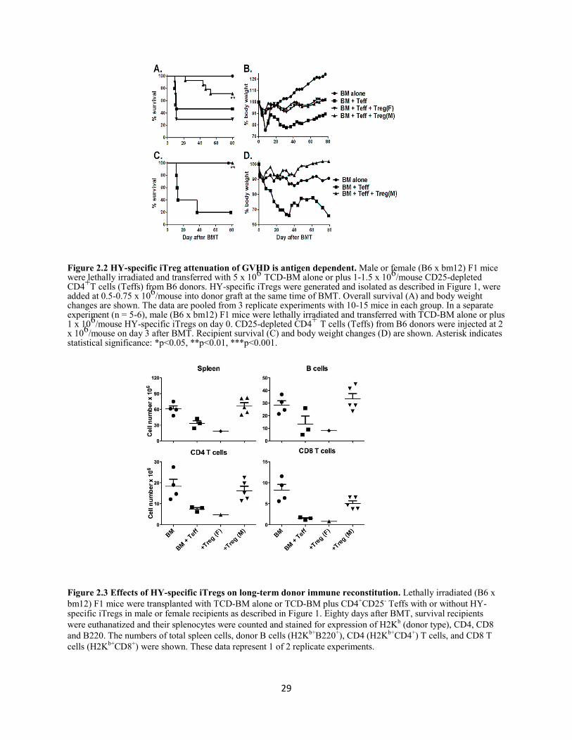
29
Figure 2.2 HY-specific iTreg attenuation of GVHD is antigen dependent. Male or female (B6 x bm12) F1 mice were lethally irradiated and transferred with 5 x 10⁶ TCD-BM alone or plus 1-1.5 x 10⁶/mouse CD25-depleted CD4+T cells (Teffs) from B6 donors. HY-specific iTregs were generated and isolated as described in Figure 1, were added at 0.5-0.75 x 10⁶/mouse into donor graft at the same time of BMT. Overall survival (A) and body weight changes are shown. The data are pooled from 3 replicate experiments with 10-15 mice in each group. In a separate experiment (n = 5-6), male (B6 x bm12) F1 mice were lethally irradiated and transferred with TCD-BM alone or plus 1 x 10⁶/mouse HY-specific iTregs on day 0. CD25-depleted CD4+ T cells (Teffs) from B6 donors were injected at 2 x 10⁶/mouse on day 3 after BMT. Recipient survival (C) and body weight changes (D) are shown. Asterisk indicates statistical significance: *p<0.05, **p<0.01, ***p<0.001.
Figure 2.3 Effects of HY-specific iTregs on long-term donor immune reconstitution. Lethally irradiated (B6 x
bm12) F1 mice were transplanted with TCD-BM alone or TCD-BM plus CD4+CD25- Teffs with or without HY- specific iTregs in male or female recipients as described in Figure 1. Eighty days after BMT, survival recipients
were euthanatized and their splenocytes were counted and stained for expression of H2Kb (donor type), CD4, CD8
and B220. The numbers of total spleen cells, donor B cells (H2Kb+B220+), CD4 (H2Kb+CD4+) T cells, and CD8 T
cells (H2Kb+CD8+) were shown. These data represent 1 of 2 replicate experiments.

30
In clinical BMT, most patients receive grafts from MHC-matched and multiple miHAg-
mismatched donors. In an effort to mimic a clinical scenario, we used the B6 → BALB.b (both H2b)
model, in which donor and recipient mice differ by at least 29 different miHAg loci (144). HY-specific
iTregs were highly effective in preventing GVHD in male (p<0.05) but not female BALB.B recipients
(Figure 2.4 A, B). Likewise, haploidentical transplantation is extensively used in clinic. Utilizing B6
→DF1 model, we further confirmed HY-specific iTregs were highly effective in preventing GVHD in
male (p<0.05) but not female BDF1 recipients (Figure 2.4 C, D). To further support our long-term data
in the B6 → BDF1 model, we analyzed pathology scores and found male recipients who received
iTregs had significantly reduced pathologic damage within the liver, small intestine, large intestine, and
lung compared to all other groups (Figure 2.4E; p < 0.01). In agreement with survival data, female
recipients receiving iTregs had comparable pathologic injury in target organs to Teff alone groups,
further supporting necessity for iTregs to recognize specific Ag to exert their suppressive function.
Taken together, these findings support the use of HY-specific iTregs in clinically relevant BMT models.
Figure 2.4 Effect of HY-specific iTregs in the prevention of GVHD in miHAg- or haplo-mismatched BMT model. Male or female BALB.b mice were lethally irradiated and transferred with TCD-BM alone or plus 4 x
106/mouse HY-specific iTregs. On day 3, 25 x 106/mouse total splenocytes from normal B6 donors were injected into the recipients previously transferred with BM alone or BM plus iTregs. Recipient survival (A) and body weight changes (B) are shown. The data are pooled from 2 replicate experiments with 9-10 mice in each group.

31
Male or female BDF1 mice were lethally irradiated and transferred with TCD-BM alone or plus at 4 x 10⁶/mouse
CD25- depleted total T cells from normal B6 donors. HY-specific iTregs were also included at 2 x 10⁶/mouse into donor graft 3 days after BMT for some recipients. Recipient survival (C) and body weight changes (D) are shown. The experiments were done 2-3 times for each BMT model, but the data presented are from one experiment with 5-8 mice in each group using cell doses indicated. Seven days post Teff injection, BDF1 recipients as described in the legend were sacrificed and pathology samples of the skin, lung, liver, small and large intestine were collected (E) represents the pathology score of the various organs with 4 mice per group. Asterisk indicates statistical significance: *p<0.05, **p<0.01, ***p<0.001
2.4.3 HY-specific iTregs suppress the expansion and activation of donor T cells
We next assessed the cellular mechanism by which HY-specific iTregs suppress alloreactive
Teffs in vivo. Taking advantage of Luc-Tg mice, the expansion and infiltration of Luc-Tg Teffs can be
measured in vivo over time using BLI assay (145). To use this method, we titrated the dose of T cells
required for mediating GVHD and found at least 4-fold lower numbers of Luc-Tg T cells were required
to cause GVHD lethality comparable to normal B6 T cells (Figure 2.5).
Figure 2.5 Pathogenicity of T cells from B6 Luc-Tg mice in GVHD. CD25-depleted total T cells (Teffs) were purified from spleens and lymph nodes of normal B6 or B6 Luc-Tg mice through negative selection. Lethally irradiated BALB/c mice were transplanted with TCD-BM or plus Teffs from normal or Luc-Tg B6 donor at the doses indicated. Recipient survival (A) and body weight changes (B) are shown. The data are pooled from 2 replicate experiments with 8-10 mice per group.
This data suggested Luc-Tg T cells might be significantly more pathogenic in the induction of
acute GVHD. Using 0.25 x 10⁶ Luc-Tg CD4+, although there were relatively low signal intensities and
no significant difference among groups on day 6 following Teff injection (Figure 2.6 A), throughout
later observation periods, the BLI intensity was significantly reduced in male recipients receiving HY-
specific iTregs compared to recipients with Teff alone (p< 0.001) or in female recipients transplanted

32
HY-specific iTregs (p = 0.02) (Figure 2.6 A, B). Furthermore, male recipients receiving HY-specific
iTreg showed less dispersed BLI signal, mainly confined to the spleen, compared to other recipients
(Figure 2.6 B). Similar results were observed in miHAg- or haplo-mismatched BMT models (Figure 2.6
C-F). These data suggest HY specific iTregs regulate allogeneic Teff expansion and infiltration into
GVHD target organs, such as the gut and liver.
Figure 2.6 Effects of HY-specific iTregs on Teff expansion in vivo. The results from experiments using
B6→Bm12 as described in Figure 2. The BLI signal strength was shown on one representative mouse from each
group (A). Average signal intensity from each group of recipients throughout the experimental time points (B).
The results were from the experiments using B6 → BALB.b BMT model as described in Figure 3 A and B. The
BLI signal strength was shown on one representative mouse from each group at day 14 after BMT (C). Average
signal intensity from each group of recipients was shown at the time points indicated (D). The results were from
the experiments using B6 → BDF1 BMT model as described in Figure 3 C and D. The BLI signal strength was
shown on one representative mouse from each group (E), and average signal intensity from each group of
recipients was shown (F) on day 13 after BMT. The image is shown in one representative mouse in each group.
The data represent one of 2 replicate experiments with 5-6 mice per group in each experiment.

33
To further evaluate the effect of iTregs on the expansion and migration of Teffs, we transferred
Teffs isolated from B6 Ly5.1+ mice and HY-specific iTregs along with TCD-BM isolated from normal
B6 donors into (B6 x bm12) F1 recipients. Seven days after BMT, we measured Teffs (CD4+Ly5.1+)
and iTregs (CD4+TCRVb6+Ly5.1-) in recipient spleen (Figure 2.7 A, C) and liver (Figure 2.7 B, D). We
found iTregs expanded extensively in the spleen and migrated substantially to the liver of male but not
female recipients, (p < 0.01, in spleen and liver), and the number of Teffs in the liver of male recipients
was dramatically reduced (Figure 2.7 D).
Figure 2.7 Stability and Efficiency of HY-specific iTregs. Lethally irradiated male or female (B6 x bm12) F1 mice were transplanted with B6 TCD-BM plus 1.5 x 10⁶/mouse Teffs (CD4+CD25-) isolated from B6 Ly5.1+
mice. At the same time, 0.75 x 10⁶/mouse HY-specific iTregs were also included into the donor graft to some recipients. Seven days after BMT, recipient spleen and liver were harvested and measured for expansion and infiltration of iTregs and Teffs. Mononuclear cells were isolated from recipient spleen (A) and liver (B), and expression of TCRVb6 and Ly5.1 was shown in gated CD4+ live cells. The average numbers of total cells, Teffs (CD4+Ly5.1+) and iTregs (CD4+Vβ6+Ly5.1-) per mouse were shown in recipient spleen (C) and liver (D). Each group includes 3-4 mice, and the data represents 1 of 3 replicate experiments. Asterisk indicates statistical significance: *p<0.05, **p<0.01, ***p<0.001

34
To extend these findings to the haploidentical BMT model, we transferred BM and Teffs from
B6 Ly5.1+ mice and HY-specific iTregs (Ly5.1-) into irradiated B6D2F1 recipients. Fourteen days after
BMT, analysis of IFNγ and TNFα secretion within the spleen of effector CD4 and CD8 T cells, showed
male recipients had significantly decreased secretion of pro-inflammatory cytokines ( p<0.01) whereas
there was no significant difference between Teff alone and female recipients (Figure 2.8 A, B). In
correlation with our flow data, analysis of the serum cytokine levels, 14 days post BMT, revealed male
recipients had significantly reduced levels of IFNγ and TNFα (p<0.01) whereas there was no difference
between female recipients and Teff alone groups (Figure 2.8 C). These cellular findings are consistent
with our long-term survival data, proving the recognition of cognate antigen is necessary for iTregs to
control Teffs in order to attenuate GVHD.
Figure 2.8 HY-specific iTregs suppress activation and expansion of Teffs. Male or Female BDF1 mice were
lethally irradiated and transferred with TCD-BM alone or plus 4 x 10⁶/mouse CD25-depleted total T cells from
normal B6 Ly5.1+ donors. HY-specific iTregs were also transplanted at 2 x 10⁶/mouse on day 0 of BMT. Fourteen days after Teff injection, recipient spleen and liver were harvested and total T cells isolated 4 mice per group. (A)

35
Representative flow analysis of IFNγ and TNFα with effector CD4 and CD8 T cells. B) Represents the absolute number of CD4 and CD8 Teffs secreting IFNγ and TNFα within the recipient spleen. (C) Serum was collected on day 14 and cytokine levels were assessed by cytometric bead analysis, one representative analysis of two independent experiments. Asterisk indicates statistical significance: *p<0.05, **p<0.01, ***p<0.001.
To address the stability of Ag-specific iTregs after injection, we performed a time course analysis of
HY- specific iTregs within male and female recipient’s spleen and liver 7, 14 and 21 days after our
previously described B6→BDF1 model. The male recipients retained the high number of
CD4+Vβ6+Foxp3+ iTregs in their spleens on day 7, 14, and 21 post BMT (p< 0.001), whereas the
number of HY-specific iTregs declined over time in the female recipients (Figure 2.9 A, B). The male
recipients also had higher numbers of CD4+Vβ6+ iTregs than female recipients on day 7 in their livers,
although Foxp3 expression was retained similarly at this time point (Figure 2.9 C, D). In the liver, the
numbers of iTregs were more strikingly different on days 14 or 21 between male and female recipients.
Furthermore, iTregs were highly stable in the male recipients reflected by their Foxp3 expression,
whereas iTregs rapidly lost their Foxp3 expression in female recipients (p<0.001) (Figure 2.9 D). Taken
together, iTregs were highly stable and expanded extensively after Ag-stimulation, and in turn
effectively suppress Teff expansion and activation and migration into GVHD target organs regardless of
BMT models
Figure 2.9 HY-specific iTregs are highly stable under inflammatory conditions. Male or Female BDF1 mice
were lethally irradiated and transferred with TCD-BM alone or plus 4 x 10⁶/mouse CD25-depleted total T cells
from normal B6 Ly5.1+ donors. HY-specific iTregs were also transplanted at 2 x 10⁶/mouse on day 0 of BMT. On days 7, 14, and 21 post BMT spleen (A, C) and liver (B, D) were collected from recipient mice for analysis. Phenotypes of cells isolated from recipient spleen (A) or liver (B) are displayed. Absolute numbers of originally

36
infused iTregs (H2Kb+CD4+Vβ6+) in spleen (C) or liver (D) are depicted on the left panels. Percentages of Foxp3 expression on gated iTregs in spleen (C) or liver (D) were shown on the right panels. Asterisk indicates statistical significance:
*p<0.05, **p<0.01, ***p<0.001.
2.4.4 Expression of target antigen on epithelial tissues is not required for iTregs to
prevent GVHD
It has been widely accepted donor T cells have to recognize alloantigens expressed on
epithelial tissues in order to cause GVHD in myeloablative BMT models (146). However, it is not
clear whether Tregs require expression of target antigen on epithelial tissues in order to suppress
GVHD. To address this question, we created 2 types of BM chimeras by transplanting donor (male
or female) BM into lethally irradiated syngeneic recipients (female or male), so the HY antigen was
only expressed either on hematopoietic cells (M → F chimeras) or on epithelial tissues (F → M
chimeras). We then transplanted TCD-BM plus Teffs from B6 donors with or without additional
HY-specific iTregs into these lethally irradiated chimeric recipients. In B6 → BALB.b (miHAg-
mismatched) and B6 → BDF1 (haplo- mismatched) BMT models, we found HY-specific iTregs
were highly capable in preventing GVHD, and the efficacy was comparable in either type of
chimeric recipients (Figure 2.10 A, B), indicating target antigen expressed on either compartment is
sufficient for iTregs to exert their suppression in GVHD.
Figure 2.10 Effect of HY-antigen distribution on HY-specific iTreg-medicated protection. (A) Male → female or female → male BM chimeras were generated using BALB.b mice as described in “Material and Method”. These BM chimeras were lethally irradiated again and divided into 2 cohorts, each of which were

37
transferred with TCD- BM alone or plus 25 x 106/mouse total splenocytes from normal B6 donors. HY-specific
iTregs were also included at 4 x 10⁶/mouse into donor graft at the day of BMT for some recipients. Recipient survival is shown, and the data represent 5-8 mice in each group. (B) Male → female or female → male BM chimeras were generated using BDF1 mice as described in “Material and Method”. These BM chimeras were lethally irradiated again and divided into 2 cohorts, each of which were transferred with TCD-BM alone or plus 4
x 106/mouse CD25-depleted total T cells from normal B6 donors. HY-specific iTregs were also included at 2 x
106/mouse into donor graft at the day of BMT for some recipients. Recipient survival is shown, and the data represent 7-8 mice in each group. Asterisk indicates statistical significance: *p<0.05, **p<0.01, ***p<0.001
2.4.5 HY-specific iTregs essentially preserve the GVL effect
To determine the effect of HY- specific iTregs on the GVL activity, we utilized the clinically
relevant B6→BDF1 (haplo-mismatched) BMT model with the injection of p815-luc+ mastocytoma cell
line. One day after lethal irradiation, we injected TCD-BM from B6 donors and HY-specific iTregs
into male recipients, three days later we then injected B6 Teff cells and p815-luc+ cells. We observed
mice receiving BM + p815 alone all succumbed to tumor mortality within 20 days post BMT, as seen
by high BLI signal with little weight loss, however mice received an addition of Teff cells died from
GVHD indicated by decreased weight loss with little to no BLI signal (Figure 2.11 A-D). The addition
of HY- specific iTregs significantly increased survival (p<0.001) and significantly delayed tumor
mortality (p<0.001) as seen by maintained body weight and low BLI signal (Figure 2.11 A-D).
Figure 2.11 HY-Specific iTregs spare the GVL effect. B6D2F1 male recipient mice were lethally irradiated and injected with B6 BM with or without HY-specific iTregs, three days later CD25 depleted Teffs plus 5000 p815-luc mastocytoma cells were injected. Mice were monitored for body weight loss (A), survival (B), and tumor morality using the IVIS 200 imager throughout the course of study. The data depicted in A and B is

38
pooled from 2 replicate experiments, but the imaging shown (D) is from one of these 2 replicate experiments. Asterisk indicates statistical significance: *p<0.05, **p<0.01, ***p<0.001
In order to better mimic clinical circumstance where patients have already established tumor,
we generated a pre-established tumor model by injecting p815-luc+ cells to the recipients 3 days prior to
irradiation and 7 days prior to Teff infusion. One day after irradiation, male recipients were transplanted
with BM plus HY-specific iTregs and three days later Teffs were infused. As shown in Figure 2.12,
50% of the recipients of BM plus p815 tumor died within 50 days of BMT without body weight loss
and strong BLI signal indicating tumor relapse (Figure 2.12, A-C). The recipients of BM plus Teffs also
died within 62 days with body weight loss and no detectable BLI signal, indicating GVHD mortality.
HY- specific iTreg infusion significantly attenuated GVHD (p<0.05), reflected by higher percentage of
survival and no tumor relapse reflected by no BLI signal (Figure 2.12 A-C, p < 0.05). Taken together,
these data indicate the HY- specific iTregs largely preserved the GVL activity mediated by Teffs.
Figure 2.12 HY-specific iTregs largely preserve GVL effect in pre-established tumor model. Male BDF1 mice injected with 5000 P815-luc cells three days prior to irradiation. Tumor bearing mice were lethally
irradiated and transplanted with 5 x 10⁶ BM and HY-specific iTregs 2 x 10⁶/mouse from B6 donors. Three days
later, the recipient mice received 4 x 10⁶ CD25-depleted total T cells. Recipient body weight (A) and survival (B) are shown. Data are from one experiment of two replicates with similar but not the same settings. Mice were imaged once weekly and then once every two-three weeks to monitor tumor growth (C and D) using an IVIS 200 imager. Asterisk indicates statistical significance: *p<0.05, **p<0.01, ***p<0.001.\

39
2.6 Discussion
Aiming to increase the potency and selectivity of Treg therapy, by using TGFβ-induced Ag-
specific iTregs, our previous studies have demonstrated Ag-specific iTregs, once activated in the
recipient, are significantly more effective than expanded polyclonal nTregs in the prevention of GVHD
(130, 131). The current study substantially extended our previous work by generating and testing
iTregs specific for naturally processed alloantigens. Given the knowledge female to male transplants
occur frequently in the clinic and these patients are at a greater risk of developing GVHD due to
miHAg mismatched antigens, like HY, we strove to provide clinical relevance by generating HY-
specific iTregs. We found monoclonal iTregs specific to HY miHAg were highly effective in
preventing GVHD in activation-dependent manner. Furthermore, we observed HY-specific iTregs
largely preserve the GVL effect (Figure 2.11 and 2.12). Given p815 used in our study is a mastocytoma
cell line originally derived from male DBA2 mice (147), therefore is susceptible to antigen-specific T
cell- rather than NK cell- mediated killing. Our results indicate miHAg-specific iTregs still permit the
GVL activity against the tumor that expresses such a miHAg. This observation is important and
clinically relevant, because many miHAg, such as HY, are ubiquitously expressed.
Unlike freshly isolated nTregs, iTregs are generated from naïve CD4 T cells and thus the
number of iTregs is essentially unlimited (Figure 2.1). In current nTreg cell expansion protocols for
clinical application, long culture period and multiple rounds of expansion are required to reach an
optimal number of cells (88, 148) still with potential loss of Foxp3 expression (69). Given iTregs rapid
expansion potential (98), this will decrease culture times and in turn resolve the fear of Foxp3 loss in
vitro, however there is still concern regarding iTregs stability in vivo. Given our results showing iTregs
remain highly stable even under extreme inflammatory conditions (Figure 2.9), this current work gives
strong rationale to move iTreg therapy into the clinic. A potential concern was raised by some studies
showing in vitro generated iTregs were less suppressive than nTregs (149, 150) and failed to prevent
GVHD (96, 151). On the other hand, there is also substantial evidence in the literature supporting

40
iTregs as or more effective than nTregs in suppressing immune responses in vivo (42, 127, 152-158).
Consistent with our previous studies using OVA-specific iTregs4,5, the current work demonstrated HY-
specific iTregs were highly effective in preventing GVHD in clinically relevant murine models of
allogeneic BMT in an Ag- dependent manner (Figure 2.2 and 2.4).
The stability and efficacy of iTregs still appears to be controversial with regards to controlling
GVHD. However, our results are also supported by the reports from Steinman’s group (102), who
demonstrated iTregs generated with allogeneic DCs in the presence of TGF-β and RA maintained
Foxp3 expression and exhibited higher levels of CNS2 demethylation in the Foxp3 gene, a marker for
stability. Stability of iTregs generated by others and us may be partially attributed to the presence of
RA, which was shown to promote iTregs through increasing histone methylation and acetylation within
the promoter and conserved non-coding DNA sequence elements at the Foxp3 gene (112)(38). More
strikingly, we interpret the efficacy of iTregs in the attenuation of GVHD is directly related to TCR-
driven activation and expansion in vivo (Figure 2.7 and 2.9). The iTregs used in our studies were ~
100% Ag-specific and able to expand when recognizing cognate Ag, whereas the iTregs used in studies
by others were polyclonal and only a small fraction (e.g. < 1%) of them were able to expand when
recognizing alloAgs. Our data clearly shows that iTregs not activated by cognate Ag were unable to
expand and were ineffective in the prevention of GVHD. Along this line, Sela et al. showed DC-
induced, alloAg-specific iTregs are capable of preventing GVHD (36). However, the therapeutic
efficacy of their iTregs was lower than HY-specific iTregs used in our current study but higher than
polyclonal iTregs used in other studies (27-29), indicating the efficacy directly correlates with the
frequency of alloreactive cells among different types of iTregs. Taken together, these data provide
direct evidence TCR-driven activation and expansion of iTregs after infusion is essential for their
therapeutic efficacy in the control of GVHD.
Since it is commonly accepted GVHD development requires donor T cells that recognize
alloantigens expressed on epithelial tissues, we hypothesized Tregs must also recognize antigens

41
expressed on epithelia in order to prevent GVHD. A recent study by Tawara et al. proposed the hosts
APCs are necessary and sufficient for GVHD protection by donor Tregs (159). By creating BM
chimeras as recipients in which the alloantigens to be recognized by Tregs are expressed on either
hematopoietic cells or parenchymal tissues, we observed HY-specific iTregs were highly capable in
preventing GVHD in either type of chimeric recipients (Figure 2.10). These results indicate target
antigen expressed on either compartment is sufficient for iTregs to exert their suppression in GVHD.
Figure 2.13 Education with HY antigen enhances suppressive function of iTregs. HY- TCR transgenic iTregs were generated as described in Figure 1. Polyclonal HY iTregs were generated by isolating WT B6 resting CD4+CD25- T cells and stimulated with syngeneic DCs with HY peptide, IL-2 (5ng/mL), TGFβ (5ng/mL), and RA (10nM) for 5 days. After 5 days, cells were harvested and restimulated with syngeneic DC’s with HY peptide for an additional 5 days. Anti-CD3 polyclonal iTregs were generated by stimulating resting CD4+CD25- T cells with syngeneic APCs and α-CD3 with IL-2 (5ng/mL), TGFβ (5ng/mL), and RA (10nM) for 3 days. Different iTregs were harvested and purified through CD25-magnetic bead positive selection (A). iTreg
suppressive function was tested by CFSE labeling CD4+CD25- B6 T cells and stimulating them with BDF1 male APCs. HY transgenic, HY polyclonal, or αCD3 iTregs were then titrated at the indicated ratios. After 5 days culture, cells were harvested and CFSE dilution was assessed through flow cytometry. Percentage of suppression was calculated by using the control (without iTregs) as the baseline for uninhibited T cell proliferation (B).
The current work using TCR transgenic T cells clearly provides the evidence miHAg-specific
iTregs were effective in the prevention of acute GVHD. To translate the finding into clinical
application, one could generate Ag-specific human iTregs by transducing TCR-gene into CD4 T cells
and then induce them into iTregs in vitro. Alternatively and also more practically, miHAg-reactive

42
iTregs could be generated from polyclonal CD4 T cells. In fact, we were able to generate HY-reactive
polyclonal iTregs by 2-rounds of stimulation of CD4 T cells with dendritic cells from normal female
B6 mice in the presence of HY-peptide (Figure 2.13A). These iTregs enriched for HY-specificity
exhibited significantly higher efficiency in suppressing B6 CD4 T cells in response to APCs from
BDF1 male mice as compared to polyclonal iTregs generated after anti-CD3 stimulation (Figure
2.13B). Furthermore, we recently have shown human nTregs specific for HY miHAg (89) can be
extensively expanded ex vivo, which demonstrates the feasibility to acquire sufficient human HY-
specific iTregs for clinic trials. In conclusion, the current pre-clinical study provides strong rationale
to apply human miHAg-specific iTregs in the clinic for the prevention of GVHD in patients after
allogeneic HCT.

43
Chapter 3: CD8 Tregs Promote GVHD Attenuation and Overcome the Impaired GVL Effect
Mediated by CD4 Tregs in Mice
Note to reader
This chapter has been previously published (Heinrichs et al, Annual ASH meeting, Orland FL
2015) and are utilized with permissions from the publisher (see Appendix C)
3.1 Abstract
Adoptive natural regulatory T cell (nTreg) therapy has improved the outcome for patients
suffering from graft-versus-host disease (GVHD) following allogeneic hematopoietic cell
transplantation (allo-HCT). However, fear of broad immune suppression and subsequent dampening of
beneficial graft- versus-leukemic (GVL) responses remains a challenge. To address this concern, we
generated alloreactive induced Tregs (iTregs) from resting CD4 or CD8 T cells and tested their ability
to suppress GVH and maintain GVL responses. We utilized major mismatched and haploidentical
murine models of HCT with host-derived lymphoma or leukemia cell lines to evaluate GVH and GVL
responses simultaneously. Alloreactive CD4 iTregs were effective in preventing GVHD, but abrogated
the GVL effect against aggressive leukemia. Alloreactive CD8 iTregs moderately attenuated GVHD
while sparing the GVL effect. Hence, we hypothesized a combination of CD4 and CD8 iTregs could
achieve the optimal goal of allo-HCT. Indeed, the combinational therapy was superior to CD4 or CD8
iTreg singular therapy in GVHD control; importantly, the combinational therapy maintained GVL
responses. Cellular analysis uncovered potent suppression of both CD4 and CD8 effector T cells by the
combinational therapy resulting in effective prevention of GVHD, which could not be achieved by

44
either singular therapy. Gene expression profiles revealed alloreactive CD8 iTregs possess elevated
expression of multiple cytolytic molecules compared to CD4 iTregs, which likely contributes to GVL
preservation. Our study uncovers unique differences between alloreactive CD4 and CD8 iTregs can be
harnessed to create an optimal iTreg therapy for GVHD prevention with maintained GVL responses.
3.2 Introduction
Allogeneic hematopoietic cell transplantation (allo-HCT) was established to treat patients
suffering from hematological malignancies encompassing leukemias and lymphomas. Allo-HCT
replaces a patient’s diseased hematopoietic system with a healthy donor stem cell source. The degree of
human leukocyte antigen (HLA) disparity dictates the beneficial graft-versus-leukemia (GVL) response,
where donor T cells recognize and kill residual leukemic cells, and the deleterious graft-versus-host
disease (GVHD), where donor T cells recognize and damage host epithelial tissues (3). Unfortunately, a
therapy separating these two responses within allo-HCT recipients has yet to be established. Presently,
the primary therapy for GVHD is broad immune suppression, which can lead to relapse of the
underlying malignancy or life-threatening infections (160).
Natural T regulatory cells (nTregs) and induced T regulatory cells (iTregs) play key roles in
suppressing autoimmunity (26) and in maintaining immune homeostasis (41). nTregs arise from the
thymus, recognize self-antigen with high affinity, and represent a stable cell lineage(36). iTregs arise
in the periphery (42), recognize non-self-antigens, and are less stable due to their propensity to lose
Foxp3 expression, the master transcription factor (40) associated with Treg suppressive function.
Harnessing Treg’s inherent suppressive nature to control GVHD has been an ongoing area of
investigation. Initial clinical trials using nTregs have yielded positive results (91-93). However, nTregs
constitute a small percentage of circulating T cells and require expansion due to the high number
needed for GVHD attenuation (93); thus, reaching optimal cell doses for patients (92) remains an

45
obstacle. Likewise, nTregs are non-selective suppressors, which could have significant impairment of
the beneficial GVL response. Conversely, iTregs can be generated in large numbers (98) and enriched
for antigen specificity (99, 161), which could result in targeted separation of GVH and GVL responses.
However, iTreg therapy has been surrounded by controversy, with some investigators citing beneficial
effects (88, 102, 115, 117) and others a complete lack of efficacy (96, 97, 101). The lack of a
consensus on iTreg therapy seems to stem from differences in activation agents (polyclonal vs. antigen-
specific), polarizing cytokines, and infusion schedules employed across laboratories. Yet, it is clear that
antigen specificity enhances iTregs potency in GVHD attenuation, but these findings used monoclonal
antigen specific iTregs that cannot be readily translated to the clinic (99, 161). Therefore, to extend
antigen-specific iTreg therapy into clinical settings, we tested the efficacy of alloreactive iTregs in
attenuating GVHD and preserving GVL responses.
Utilizing well-established murine models of allo-HCT, we demonstrated alloreactive CD4
iTregs potently suppress GVHD; yet GVL is completely lost against aggressive leukemia. Alloreactive
CD8 iTregs are less stable than CD4 iTregs, but still moderately attenuated GVHD while preserving the
GVL effect. To overcome the obstacles of each singular therapy, we established a combination therapy
using both CD4 and CD8 iTregs, which was superior in GVHD attenuation and preservation of GVL
responses. Mechanistic studies demonstrated, even under the same polarization conditions, CD4 and
CD8 iTregs represent two distinct regulatory populations that suppress GVH or maintain GVL
responses. Finally, we uncovered unique gene expression patterns between alloreactive CD4 and CD8
iTregs, which provide further understanding of these cells types and rationale to apply combinational
iTregs therapy after allo- HCT.

46
3.3 Materials and Methods
3.3.1 Mice
C57BL/6 (B6; H-2b), BALB/c (H-2d), B6-Ly5.2 (H-2b), B6DF1 (B6 x DBA2) F1 (H-2b/d) were
purchased from NCI. B6-Foxp3-GFP (H-2b) was kindly provided by A. Rudensky. All animals were
housed in specific pathogen-free conditions in the America Association for Laboratory Animal Care-
accredited Animal Resource Center at the Medical University of South Carolina. The Institutional Animal
Care and Use Committee of the Medical University of South Carolina approved all work.
3.3.2 iTreg Generation
CD4+CD25- and CD8+CD25- resting T cells were purified from C57BL6 spleen and lymph
nodes as previously described (161). CD11c+ dendritic cells were purified from BALB/c mice using
CD11c microbeads (Miltenyi) positive selection. CD4 or CD8 T cells were stimulated with CD11c DCs
at a 1:10 (T cell: DC) ratio in the presence of IL-2 (5ng/mL), TGFβ (5μg/mL), and retinoic acid (RA)
(40nM) for 5 or 4 days respectively.
3.3.3 GVH/GVL Models
Major histocompatibility complex (MHC)-mismatched (B6→BALB/c) (162) and haploidentical
(B6→BDF1) (163) BMT models were used as previously established. Recipients were monitored for
survival and body weight loss twice/week. In GVL models, tumor doses ranged from 5,000-10,000 per
mouse. Tumor mortality and GVHD mortality were distinguished as previously described (162).
Representative samples of GVHD target organs were excised from recipient’s 14 days post-BMT and
subjected to pathology scoring as previously described (164).

47
3.3.4 Flow Cytometry and Intracellular Cytokine Staining
Mononuclear cells were isolated from recipient spleen and liver as previously described (165)
and stained for surface molecules using standard flow cytometry protocols. Foxp3 was detected using
established manufacturer’s instructions Foxp3 fixation/permeablization kit (eBioscience).
Intracellular cytokines and granzyme expression were detected from spleen and liver lymphocytes
following in vitro phorbol-myristate-acetate/ionomycin stimulation as previously described (163). Cells
were analyzed using the LSRII Diva software (BD Biosciences) and FlowJo (Treestar).
3.3.5 Microarray
CD8+Foxp3+ and CD4+Foxp3+ cells were generated three independent times from Foxp3-GFP
reporter mice and sorted purified (BD FACSAria II cell sorter), CD8+Foxp3- and CD4+Foxp3- were
used as controls. RNA was extracted using Trizol and isolated using Qaigen RNAeasy Mini Kit and
quality assessed using 2100 Bioanalyzer (Agilent). Total RNA (250ng) was converted using the WT
Plus Kit (Affymetrix) and hybridized to Mouse 430 2.0T Genechips (Affymetrix) according to
manufacturer’s instructions. Raw data (CEL files) were normalized by Robust Multi-array average
(RMA) 70 using the gene level summarization method in Expression Console software (Affymetrix).
Comparisons were performed using dChip software 71 using strict criteria of absolute fold change> 2
and p<0.05. Calculations based on 50 iterations estimated a false discovery rate (FDR) of < 1 % for all
comparisons.
3.3.6 Statistics
For comparison of recipient survival among groups in GVHD experiments and tumor
mortality, the log-rank test was used to determine statistical significance. To compare pathology
scores, cytokine levels, and effector T cells, a Student t test was performed.

48
3.4 Results
3.4.1 Alloreactive CD4 iTregs suppression of GVHD is antigen specific
Our previous findings illustrate antigen specificity increases iTregs suppressive potential (161).
However, HY-specific monoclonal iTregs have limited potential to be translated to the clinic. Therefore,
we generated alloreactive CD4 iTregs and tested their ability to alleviate GVHD while maintaining GVL
activity. We utilized generation conditions previously described (102); as a control, polyclonal CD4
iTregs were generated (99). iTregs generated with H2d+ DCs (Figure 3.1 A) were highly suppressive
toward CD4 and CD8 T-cell responses to H2d+ alloantigens, and more effective than polyclonal iTregs in
vitro (Figure 3.1B). These alloreactive iTregs were also capable of suppressing allogeneic responses in
vivo, reflected by a significant reduction of T-cell proliferation and IFNγ secretion (Figure 3.1C, D).
Using an MHC-mismatched B6 to BALB/c HCT model, we injected enriched H2d-alloreactive iTregs
three days prior to effector T cells (Teffs), and found these iTregs significantly attenuated GVHD,
displayed by improved survival and body weight maintenance (Figure 3.2A, B) and decreased
pathological damage in all tissues (Figure 3.2C).
Figure 3.1 Alloreactive CD4 iTregs display superior suppressive function than polyclonal iTregs. Naïve
CD4+
CD25-
T cells were isolated from B6 mice and plated with either BALB/c DCs (alloreactive) or with α-CD3 + B6 APCs (polyclonal) with IL-2, TGFβ, and retinoic acid. Foxp3 expression was assess through Flow cytometry, Activity of iTregs was assessed by suppressing allo-response of T effector cells in vitro (B) and in vivo (C,D).* p < 0.05, ** p < 0.01, *** p < 0.001.

49
Figure 3.2 Alloreactive CD4 iTregs attenuation of GVHD is antigen-specific. BALB/c mice were lethally
irradiated (700cGy) and transplanted with 5 x 10⁶ TCD-BM from B6 donors and 1 x 10⁶ CD25hi H2d specific iTregs. Three days later 1.0 x 10⁶ CD25 depleted B6 Teff cells were injected alone or with a second dose of H2d iTregs (1- 0.5 x 10⁶). Recipients were monitored for survival (A) and body weight loss (B) for 80 days. During on replicate experiment GVHD target organs were excised, subjected to H&E staining, and sent to a collaborator for pathological
scoring (C). Recipient BALB/c mice were lethally irradiated (700cGy) and transplanted with 5 x 10⁶ TCD-BM from B6 donors and 1 x 10⁶ CD25hi H2d or H2k specific iTregs. Three days later 1.0 x 10⁶ CD25-depleted B6 Teff cells were injected alone or with a second dose of H2d or H2k specific iTregs (1-0.5 x 10⁶). Recipients were monitored for survival (D) and body weight loss (E) for 80 days. * P<.05; ** P<.01; *** P<.001. All error bars indicate standard error of the mean (SEM)
To evaluate the impact of iTreg allo-reactivity in the prevention of GVHD, we generated third-
party (H2k)-reactive iTregs and compared the ability of H2d- vs. H2k-alloreactive iTregs to suppress
GVHD. As expected, host (H2d)-reactive iTregs were superior to third-party (H2k)-reactive iTregs in
suppressing GVHD (Figure3.2D, E). To understand the mechanism of how iTreg allo-reactivity affects
iTreg function, we followed the fate of CD4 iTregs to observe their effect on Teffs. Host (H2d)-reactive
iTregs displayed greater persistence as compared to H2k-iTregs (Figure 3.3A, B). Furthermore, host
(H2d)-reactive iTregs potently suppressed the expansion of CD4 Teffs and reduced the expansion of
CD8 Teffs (Figure 3.3A, B). Conversely, the third-party (H2k)-reactive iTregs reduced the expansion of
CD4 Teffs but had no effect of CD8 Teffs, which may account for their ability to moderately attenuate
GVHD lethality. Taken together, alloreactive iTregs effectively attenuate GVHD and host- reactive
iTregs have greater stability and potency in GVHD prevention

50
Figure 3.3 Alloantigen specificity is essential for iTreg function and stability. BALB/c mice were lethally
irradiated and transplanted with TCD-BM and either H2d or H2k iTregs as described in Figure 3.1. Three days
later Ly5.1+ CD25-depleted Teff cells were injected. Fourteen days later recipients spleen excised, mononuclear cells isolated, and stained for indicated surface molecules, representative flow gating strategy is shown (A). Based on flow percentages absolute numbers of Teff and iTregs were calculated for the spleen (B) * P<.05; ** P<.01; *** P<.001. All error bars indicate standard error of the mean (SEM)
3.4.2 Alloreactive CD4 iTregs impair GVL activity against aggressive leukemia
Since GVHD is intimately linked to GVL activity, we tested whether alloreactive CD4 iTregs
could suppress GVHD without impairing GVL responses. To further increase iTreg yield and
translational potential, we tested non-enriched iTregs (bulk after culture) pathogenicity following allo-
HCT and found non-enriched alloreactive iTregs (CD4, CD8, or in combination) were not pathogenic
(Figure 3.4A and B). Consequently, we used non-enriched iTregs in all experiment thereafter. Initially,
we found CD4 iTregs significantly attenuated GVHD (Figure 3.5A) while largely preserving GVL
activity against A20 B cell lymphoma (Figure 3.5B, C). To ensure the maintenance of GVL function
was not tumor specific, we used a haploidentical B6 to BDF1 HCT model with host-original
mastocytoma (P815). Surprisingly, the addition of CD4 iTregs completely abrogated the ability of
Teffs to mediate GVL function, reflected by all recipients with CD4 iTregs succumbing to tumor
mortality (Figure 3.5D, E) with heavy tumor load (Figure 3.5F) similar to those with BM plus tumor
alone. To understand the strikingly different outcomes we encountered between A20 and P815 models,
we found A20 cells express significantly higher levels of MHC-II and CD86 compared to P815 (data

51
not shown), suggesting A20 lymphoma is more immunogenic than P815, possibly contributing to the
differential sensitivities we observed. Taken together, while CD4 alloreactive iTregs are effective in
GVHD control, they may impair the GVL activity against aggressive and low immunogenic tumors.
Figure 3.4 Non-enriched alloreactive iTregs are not pathogenic. BALB/c mice were lethally irradiated
and transplanted with 5 x 10⁶ TCD-BM, 1 x 10⁶ CD4 iTregs, 1 x 10⁶ CD8 iTregs, or 0.5 x 10⁶ CD4 + 0.5 x
106 CD8 iTregs. Three days later 0.5 x 10⁶ CD25-depleted Teff cells were injected. Recipients were monitored for survival and body weight loss for 80 days, n=8. ***P<0.005
Figure 3.5 Alloreactive CD4 iTregs impair GVL activity against aggressive leukemia. BALB/c mice were
lethally irradiated and transplanted with 5 x 10⁶ TCD-BM, 1 x 10⁶ CD4 iTregs, and 1 x 10⁴ luc-A20 cells. Three
days later 0.5 x 10⁶ CD25-depleted Teff cells were injected. Recipients were monitored for survival (A) tumor mortality (B) and tumor load (C) for 80 days n=10/group. BDF1 recipient mice were lethally irradiated
(1200cGy) split dose 3 hours apart and transplanted with 5 x 10⁶ TCD-BM, 2 x 10⁶ CD4 iTregs, and 5 x 10³ luc-
P815 cells. Three days later 2 x 10⁶ CD25-depleted Teff cells were injected. Recipients were monitor for survival (D) tumor mortality (E) and tumor load (F) for 60 days, n=15/group. * P<.05; ** P<.01; *** P<.001. All error bars indicate standard error of the mean (SEM).

52
3.4.3 CD8 iTregs moderately attenuate GVHD and fully preserve GVL function
Recent studies showed CD8 iTregs arise after allogeneic HCT, which play a significant role
in GVHD attenuation (116). Interestingly, a recent report also suggested human CD8 iTregs possess
direct cytotoxicity against LCL-tumor cell lines (117). We therefore generated alloreactive CD8
iTregs and evaluated their ability to control GVHD and preserve the GVL response. Using similar
polarizing conditions, we were able to generate 30-40% Foxp3+ CD8 iTregs (Figure 3.6A). These
CD8 iTregs displayed significantly higher levels of CD39+CD73+, CTLA-4, and granzyme B
compared to nTregs and CD4 iTregs (Figure 3.6B), and were equally suppressive of allogeneic
responses as CD4 iTregs (Figure 3.6C). Despite potent suppressive function in vitro, alloreactive CD8
iTregs only moderately attenuated GVHD (Figure 3.7A), but preserved the GVL effect against P815
tumor (Figure 3.7B, C). Interestingly, CD8 iTregs themselves could significantly delay tumor
mortality (Figure 3.7A-C). In vitro cytotoxic assays confirmed CD8 iTregs possess direct cytotoxicity
against tumor cells (Figure 3.7SD). These results provide evidence alloreactive CD4 and CD8 iTregs
are fundamentally different with regards to GVH and GVL responses, with CD4 iTregs potently
suppressing Teffs and CD8 iTregs contributing to GVL maintenance.
Figure 3.6 Generation and Function of Alloreactive CD8 iTregs. Naïve CD8+CD25-B6 T cells were stimulated with BALB/c DCs with IL-2 (5ng/mL), TGFβ (5ng/mL) and retinoic acid (40nM) for 4 days (A). Expression of CD39, CD73, CTLA4, and granzyme B were assessed between nTregs, alloreactive CD4 iTregs, and alloreactive CD8 iTregs post generation (B) and mean fluorescence intensity quantified (B). In vitro suppressive function between alloreactive CD4 and alloreactive CD8 iTregs was assessed through suppressive of CFSE labeled alloreactive B6 T cells (C). Error bars indicate the mean of standard error.

53
Figure 3.7 CD8 iTregs moderately attenuate GVHD and maintain GVL responses. BDF1 recipient mice were lethally irradiated (1200cGY,split doses) and transplanted with 5 x 10⁶ B6 TCD-BM, 4 x 10⁶ CD8 iTregs, and 5 x 10³ luc-P815 cells. Three days later 2 x 10⁶ CD25-depleted Teff cells were injected. Recipients were monitored for survival (A) and tumor mortality (B). Every week to every other week mice were subjected to whole body imaging, region of signal intensity was quantified using IVIS software for each group (C). BDF1 mice were transplanted as described in (A), fourteen days post HCT splenocytes were plated with either CFSEhi P815 or CFSElow EL4 tumor cells for 16 hours. Percentage tumor killing was calculated as ratio EL4:P815 (no splenocytes control) / EL4: P815 (each group), tumor killing was normalized to total donor CD8 T cells in splenocytes (D). n=15. * P<.05; ** P<.01; *** P<.001. Error bars indicate the mean of standard error.
3.4.4 Combination of CD4 and CD8 iTregs is superior to singular iTregs therapy in GVHD
attenuation
We further reasoned these two types of iTregs could compensate for each other’s weaknesses
and provide an optimized cellular therapy. We first assessed the potency of a combination therapy
(CD4 + CD8 iTregs) to suppress GVHD using B6 to BALB/c HCT model. To negate an additive effect
within the combinational therapy, the absolute iTreg product injected was equivalent to CD4 or CD8
iTregs singular therapy. We observed the inclusion of CD8 iTregs did not abrogate the ability of CD4
iTregs to alleviate GVHD, and in fact attenuated GVHD more potently than CD4 iTreg singular therapy
(Figure 3.8A). By using β-actin luciferase transgenic B6 donors to monitor the expansion of Teffs in
vivo, we found the combinational therapy significantly reduced the expansion of Teffs compared to
either CD4 or CD8 iTreg singular therapy reflected by significantly decreased bioluminescent signal
over time (Figure 3.8B).

54
To understand the cellular mechanism of iTreg therapy on GVHD, we performed a time course
study to monitor both Teff and iTregs after allo-HCT. Recipient’s BALB/c spleen, liver, and
mesenteric lymph nodes (mLN) were harvested on day 5, 8, and 11 post HCT. On day 5, Thy1.1+ Teffs
were not readily detectable in any of the recipients. On day 8, the expansion of Teffs was significantly
suppressed by all iTreg therapies (Figure 3.8C). On day 11, all the recipients with Teffs alone
succumbed to GVHD. Combinational therapy with CD4 and CD8 iTregs controlled Teff expansion
significantly better than either singular therapy (Figure 3.8C), which correlated with a less intense
bioluminescence signal observed in the combinational therapy (Figure 3.8B). Interestingly, CD4 iTregs
potently controlled the expansion of CD4, but not CD8 Teffs, whereas CD8 iTregs potently controlled
the expansion of CD8, but not CD4 Teffs (Figure 3.8D). Furthermore, the combination of CD4 and
CD8 iTregs potently controlled the expansion of both CD4 and CD8 Teffs (Figure 3.8D). To evaluate
the fate of infused iTregs, we tabulated absolute numbers of residual Foxp3+ infused individual iTregs
(Thy1.2+), which reflects iTregs expansion and stability. Overall, equivalent or greater
expansion/stability of iTregs was observed when infused in combination as compared to each alone in
recipient spleens and mLNs (Figure 3.8E). While CD8 iTregs accumulated in mLN, there were
significantly fewer in the liver when infused alone versus together with CD4 iTregs (Figure 3.8E). To
track the maintenance and stability of infused iTregs, we generated iTregs from Ly5.1+ B6 mice,
performed HCT as described in Figure 3.8, and evaluated Foxp3 expression 17 days post-transplant.
Consistently, we found CD8 iTregs expanded and/or survived longer than CD4 iTregs (Figure 3.9A,
C), albeit at the loss of Foxp3 expression. Given the injected iTregs were non-enriched (~60% Foxp3
expression) at the day of transplant, we found CD4 iTregs had significant retention of Foxp3 expression
(~40%) and were more stable than CD8 iTregs (Figure 3.9A, B). Taken together, the combinational
therapy was more effective in preventing GVHD than either singular therapy, likely because the former
was able to control both CD4 and CD8 Teffs simultaneous

55
Figure 3.8 Combination of CD4 and CD8 iTregs superior to singular iTregs therapy in GVHD attenuation. BALB/c mice were lethally irradiated and transplanted with 5 x 10⁶ TCD-BM, 1 x 10⁶ CD4 iTregs, 1 x 10⁶ CD8
iTregs, or 0.5 x 10⁶ CD4 + 0.5 x 10⁶ CD8 iTregs. Three days later 0.5 x 10⁶ CD25-depleted Teff cells were injected. Recipients were monitored for survival and body weight loss for 80 days (A), n=20/group. During one
replicate experiment, 0.25 x 10⁶ luc-Teff were injected and expansion monitored over the first 40 days post BMT (B). BALB/c mice were lethally irradiated and transplanted as described in A, however Teff cells were injected
from Thy1.1+ reporter mice. On days 5, 8, and 11 recipient spleens, liver, and mesenteric lymph nodes harvested and Teff expansion analyzed over time (C) as well as characteristics (D). iTreg absolute number was also quantified over time in all three organs (E). * P<.05; ** P<.01; *** P<.001. All error bars indicate standard error of the mean (SEM).
Figure 3.9 CD4 iTregs display greater stability than CD8 iTregs. BALB/c mice were lethally irradiated
and transplanted with 5 x 10⁶ B6 TCD-BM, 1 x 10⁶ CD4, 1 x 10⁶ CD8, or 0.5 CD4 + 0.5 x 10⁶ CD8 iTregs
generated from Ly5.1+ mice. Three days later, CD25-depleted 0.5 x 10⁶ Teff cells were injected. Seventeen days post- transplant, spleens were collected and iTregs analyzed as described in the representative flow

56
diagram (A). Quantification of percentage of Foxp3 (B) and absolute number (C) per mouse n=4. Error bars indicate the mean of standard error
3.4.5 Combinational therapy rescues inhibited GVL response of CD4 singular therapy
To exclude potential model specific phenomena, we further evaluated the efficacy of
combinational therapy in B6 to BDF1 HCT model. We identified an appropriate combined dose of
CD4 and CD8 iTregs potently attenuated GVHD significantly better than either iTreg therapy alone.
This was supported by pathological analysis of GVHD target organs; including the liver, lung, small
intestine and colon (Figure 3.10A, B). Taken together with the data presented in Figure 3.8, we
conclude the combinational therapy is superior to single iTreg therapy across different HCT models.
We next addressed the most pertinent question as to whether the combinational therapy can
spare the GVL effect against P815. Fourteen days post HCT, we observed the recipients given CD4
iTregs alone had significantly increased tumor burden within the liver, lung, spleen, and gut as
compared to those receiving Teffs alone (Figure 3.10C, D). However, the recipients given either CD8
iTregs alone or combinational therapy had significantly reduced tumor burden as compared to those
receiving CD4 iTregs alone within all organs examined (Figure 3.10C, D). These results suggest
CD8 iTregs could overcome the impaired GVL effect mediated by CD4 iTregs. To extend this
observation to long-term survival studies, we simultaneously evaluated the effect of combinational
therapy on GVHD and GVL responses against P815. Indeed, the recipients with the combinational
therapy had significantly improved long-term survival, resulting from reduced GVHD lethality and
tumor mortality (Figure 3.11 A-C). Taken together with data presented in Figure 3.5 and Figure 3.7,
we conclude the combinational therapy has distinct advantages over either CD4 or CD8 iTreg singular
therapy resulting in GVHD prevention and GVL preservation.

57
Figure 3.10 Combinational therapy reduce GVHD pathology and tumor burden. BDF1 recipient mice were
lethally irradiated (1200cGy) and transplanted with 5 x 10⁶ B6 TCD-BM, and either 2 x 10⁶ CD4 iTregs, 4 x 10⁶
CD8 iTregs, or a combination of both CD4 and CD8 iTregs, and 5 x 10³ luc-P815 cells. Three days later 2 x 10⁶
Ly5.1+ CD25-depleted Teffs were injected. Fourteen days post BM injection; GVHD target organs were excised for pathological analysis. A representative photomicrograph of each group depicting the average disease score morphology (A) and quantification of pathological damages to organs (B) are shown. Before mice were euthanized, luciferin was injected, and organs were excised followed by imaging using IVIS 200 imager (C), a representative mice from each group n=4. Organ bioluminescent was quantified using Living Imager software (D). * P<.05; ** P<.01; *** P<.001. Error bars indicate the mean of standard error.
Figure 3.11 Combinational therapy preserves the GVL response. BDF1 recipient mice were lethally
irradiated (1200cGY) and transplanted with 5 x 10⁶ TCD-BM, 2 x 10⁶ CD4 iTregs, 4 x 10⁶ CD8 iTregs, and 5 x
103 luc-P815 cells. Three days later 2 x 10⁶ B6 CD25-depleted Teffs were injected. Recipients were monitored for survival (A), tumor mortality (B), and tumor load (C) for 80 days, n=15. * P<.05; ** P<.01; *** P<.001.

58
3.4.6 CD8 iTregs contribute to the preserved GVL effect by maintaining cytolytic functions
To understand why the GVL effect was preserved upon addition of CD8 iTregs (as opposed to
CD4 iTregs alone) we analyzed the levels of effector molecules on all donor cells injected (Teffs +
iTregs) to accurately reflect the total cytolytic potential within recipient mice. Importantly, this
analysis will not exclude the cytolytic molecules, such as IFNγ, granzyme B, and fas-L, which are
secreted or expressed by CD8 iTregs themselves (Figure 3.6). Fourteen days post HCT; CD4 iTregs
alone significantly reduced the percentage of IFNγ+ cells among total donor T cells (H2d-, Teff +
iTregs) in recipient spleens and livers (Figure 3.12A). The percentage of IFNγ+ cells among donor T
cells was partially restored with addition of CD8 iTregs (Figure 3.12B). To a lesser extent, the absolute
number of H2d-IFNγ+ cells was also significantly increased in the recipients given combinational
therapy compared to those of CD4 iTregs alone (Figure 3.12C). Combinational therapy was also able
to restore the expression of granzyme B and fas-L on H2d-CD8+ cells, which were impaired by CD4
iTregs (Figure 3.12D). These results indicate the combinational therapy achieves GVL preservation by
maintaining sufficient levels of IFNγ secretion and cytolytic molecule expression.
Figure 3.12 CD8 iTregs contribute to the preserved GVL effect mediated by combinational therapy.
BDF1 mice were transplanted as stated in Fig 6. Fourteen days post BMT spleen and liver mononuclear cells

59
were isolated. Representative flow gating strategy (A) displays IFNγ secretion from Teffs and injected iTregs
(H2Kd-). Percentages of indicated cell populations, IFNγ production (B), and absolute number are quantified (C). Cells were also stained for granzyme B and Fas-L, with a corresponding isotype control (D) representative histogram analysis of the isotype control, CD4 singular or combination therapy. Graphical quantification of the mean fluorescence intensity for each molecule is displayed in (D), n=4. * P<.05; ** P<.01; *** P<.001. Error bars indicate the mean of standard error.
3.4.7 Alloreactive CD4 and CD8 iTregs display differential gene expression profiles
Given CD4 and CD8 iTregs had distinct effects on GVH and GVL responses; we strove to
understand molecular differences between these two types of iTregs and if different molecular
signatures could confirm our long-term studies. By using T cells from Foxp3-GFP reporter mice, we
generated alloreactive iTregs and sorted pure CD4+Foxp3+ and CD8+Foxp3+ iTregs. The gene
expression of alloreactive iTregs (Foxp3+) and Teffs (Foxp3-) was compared at the genomic level.
Among a total of 34,356 genes analyzed, there were 235 genes differentially expressed between CD4
and CD8 Foxp3+ iTregs and 688 genes differentially expressed between CD4 and CD8 Foxp3- Teffs.
By excluding genes that differ between CD4 and CD8 T cells independent of Foxp3 expression, we
further identified 85 of the 235 genes uniquely upregulated and 71 of the 235 genes uniquely
downregulated in CD8+Foxp3+ vs. CD4+Foxp3+ iTregs; as the expression of those 156 (85 + 71) genes
were not significantly different in CD8+Foxp3- vs. CD4+Foxp3- Teffs (Figure 3.13A, B). To identify
the genes or molecules that may contribute to the distinct activity of CD4 and CD8 iTregs on
GVH/GVL responses, we focused on those that regulate cytolytic function, Foxp3 stability, Treg
suppressive mechanisms, and plasticity. 32 representative genes among those were displayed in a heat
map (Figure 3.13A). Consistent with increased cytolytic activity of CD8 iTregs and hence GVL
preservation (Figure 3.6 and 3.7), we found granzyme B, granzyme C, fas-L, perforin, and DAPK2
(death associated protein kinase 2) were significantly and uniquely upregulated in CD8+Foxp3+ vs.
CD4+Foxp3+ iTregs (Figure 3.13C). Among five transcription factors associated with Foxp3 stability,
also known as the “Treg quintet”(75), 3 of the 5 genes were significantly downregulated in
CD8+Foxp3+ iTregs; reflecting their instability as compared to CD4 counterparts (Figure 3.13D).

60
Among the effector molecules Tregs utilize to mediate suppression, we found LAG3, CD39, and FGL2
(fibrinogen-like protein 2) were upregulated, whereas CD28 and Neuropilin-1 were downregulated in
CD8+Foxp3+ vs. CD4+Foxp3+ iTregs (Figure 3.13E), suggesting CD8 and CD4 iTregs may utilize
different mechanisms to control the immune response. In addition, CD4+Foxp3+ cells upregulated
genes associated with Treg plasticity, such as RORγt, IL-17R, IL-6R and CCR6, as compared with
CD8+Foxp3+ cells, suggesting CD8 iTregs, although less stable, represent a more terminally
differentiated population (Figure 3.13F). Overall, CD8 iTregs and CD4 iTregs are quite distinct on the
molecular level, with CD8 iTregs expressing more cytolytic molecules and less plasticity than CD4
iTregs.
Figure 3.13 Alloreactive CD4 and CD8 iTregs display differential gene expression profiles. Naïve CD4 or
CD8 B6-Foxp3-GFP T cells were stimulated with BALB/c CD11chi DC with IL-2 only, or with TGFβ and RA
three separate times. GFP/Foxp3- CD4 or CD8 cells were FACS sorted from the culture with IL-2 alone, and
GFP/Foxp3+ CD4 or CD8 cells were sorted from the culture with IL-2 plus TGFβ and RA. Total RNA was
isolated from 2 x 10⁶ cells, and then labeled and hybridized to microarray chips for gene profiling. 32

61
representative genes display differential gene expression were shown (A). dChip software using FC>2 and p<0.05
identified unique genes that were exclusive to either Foxp3 expression or independent of Foxp3 expression (B).
Signal values for genes relevant to cytotoxic function (C) Treg stability (D) suppressive mechanisms (E) or Treg
plasticity were displayed (F). * P<. 05; ** P<. 01; *** P<. 001. Error bars indicate the mean of standard error.
3.5 Discussion
In the current study, we demonstrate host alloreactive iTregs are more potent in GVHD
prevention than third-party alloreactive iTregs following allogeneic HCT. This is in accordance with our
previous findings (161) that antigen recognition increases iTreg therapeutic potential. To date, an
effective therapy distinguishing between GVH and GVL responses is highly desirable for patients with
hematologic malignancies. Here, we established the first combinational alloreactive iTreg therapy, using
both CD4 and CD8 iTregs, to provide distinct suppression of GVHD with maintained GVL responses.
We uncovered unique roles for alloreactive CD4 and CD8 iTregs following allo-HCT. CD4 iTreg
singular therapy potently suppressed GVHD but significantly impaired GVL responses. Conversely,
CD8 iTreg singular therapy had a lesser impact on GVHD but fully preserved GVL activity. Although
the current study utilizes iTregs reactive to major (MHC) antigen, we have validated the generation
protocols can be applied to minor antigen (miHAg) settings (data not shown); suggesting miHAg-
reactive iTregs could be utilized to prevent GVHD induction from miHAg disparity. Together, the
combinational therapy using these two types of iTregs had complementary roles: CD4 iTregs reduced
the expansion of Teffs to attenuate GVHD, and CD8 iTregs maintained optimal activation and cytolytic
potential of Teffs, thus preserving GVL function.
Although controversy surrounds iTreg therapy for GVHD attenuation, a critical look into
the variability among generation conditions clearly demonstrates why differential results exist.
First, the activating agent (polyclonal vs. physiological) results in noticeably different outcomes for
GVHD, as polyclonal activated iTregs are unable to mitigate disease (96, 97) while physiological
activated iTregs significantly attenuate GVHD (99, 102, 161). It has been found physiologic

62
activation imparts iTregs with more homing characteristics, like chemokine receptors, than
polyclonal activated iTregs (104), which may provide advantages for physiologic iTregs on
GVHD attenuation. Second, the difference between the third polarizing cytokine (rapamycin vs.
RA) has a significant effect on the iTreg product. Interestingly, the use of rapamycin resulted in an
unstable iTreg therapy (97), which could only be effective if rapamycin was continuously
administered post-transplant, whereas RA has been shown to yield a stable iTreg product (99, 102,
161). The mechanism of action of these two compounds is quite different, with rapamycin
indirectly suppressing expansion of contaminating Teffs through mTOR inhibition (166); whereas
RA directly enhances the accessibility of the conserved non-coding sequence within the Foxp3
promoter, thus specifically enhancing Foxp3 stability. In this study, we established the optimal
generation conditions for iTreg therapy on GVHD, physiological activation (allogeneic DCs) and
RA polarization.
Of biological importance, we uncovered unique roles for CD4 and CD8 iTregs following allo-
HCT. To our knowledge, this is the first study to show preferential suppression of CD4 Teff by CD4
iTregs and CD8 Teff by CD8 iTregs. Our cellular and molecular analysis uncovered increased
expression of multiple cytolytic molecules on CD8, but not on CD4 iTregs, which likely contributes to
the ability of CD8 iTregs to delay tumor relapse on their own. Of clinical importance, human
alloreactive CD8 iTregs were also found to possess direct cytotoxicity against LCL tumor cell lines
(117), providing a correlation between our findings and human specimens. Given their innate cytotoxic
nature, these findings could be further expanded to use CD8 iTregs as a novel cell source for donor
lymphocyte infusion (DLI) given to patients suffering from primary disease relapse (167-169). The
stability of Foxp3 expression was another unique difference harbored by CD4 and CD8 iTregs, with
CD4 iTregs displaying increased expression of multiple Treg stabilizing molecules, like the “Treg
quintet”, which was lacking in CD8 iTregs. These molecular findings mirror our cellular time course
study of alloreactive iTregs and other’s observations (97) that CD4 iTregs represent a stable cell subset

63
following allo-HCT; whereas CD8 iTregs are less stable, especially in GVHD target organs.
Importantly, we observed that non-enriched CD4, CD8, or combinational therapy, even with loss of
Foxp3 expression, was not pathogenic following allo-HCT, suggesting those iTregs are safe in cellular
therapy. Together, our study proves CD4 and CD8 iTregs, even when generated under similar
conditions, possess distinctly different characteristics. More work is needed to optimize the dose ratio of
CD4 and CD8 iTregs and to manipulate either CD4 or CD8 iTreg characteristics to create a superior
singular iTreg for GVH prevention and GVL maintenance. Similarly, multiple infusions of either CD4
or CD8 iTregs may be necessary to achieve sufficient efficacy and superior outcome in clinical settings.
Primary disease relapse is the leading cause of mortality, occurring in 37% of recipients (4),
following allo-HCT. Therefore, when establishing a new therapeutic option for GVHD attenuation,
maintaining GVL function is essential. A consensus on whether Tregs may preserve the GVL effect has
not been reached, due to the limited tumor models tested. Notably, our studies show CD4 iTregs
abrogate GVL activity against evasive P815 mastocytoma, which correlates with impaired CML
clearance found by others (97). Mirroring our observations, alloreactive nTreg (120) therapy resulted in
maintained GVL activity against A20 but not against P815. The authors hypothesize clearance of P815
is strongly dependent on alloreactive T cells, which are sharply reduced by infused Tregs; which we also
observed in our CD4 iTreg therapy. In the current study, we demonstrate a striking reduction in cytolytic
molecules, such as granzyme B and fas-L on Teffs by CD4 iTregs, which likely contributes to the
compromised GVL effect. This finding also provides a rationale to combine CD4 iTregs with cytolytic
CD8 iTregs for GVL preservation.
We consider the most exciting finding in this work to be the combinational therapy’s ability to
rescue the impaired GVL effect mediated by CD4 singular therapy. Two main factors contribute to the
unhindered GVL effect observed within the combinational therapy: 1) CD8 iTregs expression of
cytolytic molecules that can directly lyse tumor cells and, 2) although the expansion of Teffs is
dramatically inhibited, the residual cells are poly-functional, with sustained expression of IFNγ,

64
granzyme B, and fas-L. While underlying mechanisms of GVL maintenance may still contribute, we
identify these two factors, which provide a significant therapeutic effect. An effective therapy following
allo-HCT requires conflicting immune responses, both suppression and cytolytic potential, and rarely
can one immune cell type simultaneously possess each of these functions without complicated cellular
manipulation. Uniquely, our combinational therapy utilizes the innate strengths of two immune cell
types to achieve both suppression (CD4) and cytolytic potential (CD8) following allo-HCT. In
summary, these findings provide the rationale for a targeted dual cellular therapy to reduce GVHD
propensity and primary disease relapse, the two major hindrances obstructing allo-HCT success.

65
Chapter 4: Conclusions and Future Directions
The field of regulatory T cell therapy has come a long way since their discovery in 1970.
However, there is still a long way to go. Although nTregs are an effective source for therapy, their low
proliferative potential remains a major issue. Additionally, the expansion of GMP-grade nTreg for use in
allo-HCT recipients requires significant clinical infrastructure and coordination. This work has shown
both biologically (100) and translationally (103) iTregs can be generated rapidly and potently suppress
GVHD. However GVL can be impaired using CD4 singular iTreg therapy. With the field trending
towards investigating the differential abilities between CD4 versus CD8 iTregs with regards to GVHD
and GVL responses, the convergence of these two therapies seems inevitable. The upcoming clinical
trials involving both alloreactive nTregs and polyclonal iTregs will give us detailed insight into the next
steps for improving iTreg cellular therapy for the treatment of GVHD.
The most compelling finding during this work was the therapeutic potential of alloreactive CD8
iTregs, which has the greatest potential for future experimental investigations. The molecular and
cellular findings in Chapter 3 show alloreactive CD8 iTregs to be significantly less stable compared to
alloreactive CD4 iTregs, a future project could focus on stabilizing Foxp3 expression within CD8 iTregs
to provide a superior singular iTreg therapy. For example, Foxp3 stabilization can be achieved via
inhibition of DNA methyltransferase by utilizing azacytidine thus resulting in a more demethylated
Foxp3 promoter and stable Foxp3 expression. Azacytidine has been shown to generate induced Tregs in
vivo following allogeneic transplantation giving this compound strong promise as an agent to stabilize/
increase iTreg conversion ex vivo (170). With new discoveries of molecules or proteins affecting Foxp3
stability, the list of agents is endless, for example, Stub1 (77), complement receptor inhibition (171), and
Sirtuin1 (172) all represent potential targets to stabilize Foxp3 expression with alloreactive CD8 iTregs.

66
Stub1 is an E3-ubiquitin ligase that was shown to ubiquitate Foxp3 that lead to it being targeted for
degradation, inhibiting Stub1 activity could greatly increase the half-life of the Foxp3 protein which is
essential for lineage stability. Recently, binding of pro-inflammatory C3a and C5a (complement) to their
cognate receptors C3aR and C5aR on T cells was shown to increase differentiation of T cells into Th1 or
Th17 phenotype, whereas blocking these receptors lead to increase in Treg generation. Finally, Sirtuin1,
a histone deacetylase which facilitates condensation of chromatin, was recently found to deacetylate
Foxp3, by inhibiting Sirtuin1 activity acetylation of Foxp3 was increase and subsequently increasing
their suppressive function.
On the other hand given alloreactive CD8 iTregs possess direct cytotoxicity against tumor cell
lines however do not induce GVHD on their own characterizes these cells as a potential new cell
source for DLI infusion. The anti-tumor effects of DLI infusion have substantially helped patients
suffering from primary disease relapse however 50% of those patients end up with GVHD (167, 168).
It would be an interesting approach to utilize alloreactive CD8 iTregs for DLI infusion given their
inability to cause acute GVHD but can mediate beneficial GVL clearance.
Finally, all the data presented in the current studies utilized either monoclonal or MHC-major
mismatch settings in order to generate iTregs. This firmly establishes that antigen-specific Tregs
potent efficiency to control GVHD, however, MHC-major mismatch transplant are never performed
clinically due to high resolution typing of donor and recipient prior to transplant. Therefore, extension
of these findings into minor-antigen mismatch setting would be even more clinically relevant and
directly translational. There are multiple minor antigen preclinical models that could be utilized to
generate alloreactive minor antigen specific iTregs (B10D2 to DBA2) (173). Consequently, these pre-
clinical studies provide the rationale to support the first clinical trial in testing host-alloantigen
reactive Tregs in GVHD (NCT01795573 led by Dr. Anasetti). Overall, our studies have significantly
enhanced the knowledge and the translational potential of antigen-specific iTreg therapy for allo-HCT

67
Chapter 5: List of References
1. Pidala, J., S. J. Lee, K. W. Ahn, S. Spellman, H. L. Wang, M. Aljurf, M. Askar, J. Dehn, M.
Fernandez Vina, A. Gratwohl, V. Gupta, R. Hanna, M. M. Horowitz, C. K. Hurley, Y. Inamoto, A.
A. Kassim, T. Nishihori, C. Mueller, M. Oudshoorn, E. W. Petersdorf, V. Prasad, J. Robinson, W.
Saber, K. R. Schultz, B. Shaw, J. Storek, W. A. Wood, A. E. Woolfrey, and C. Anasetti. 2014.
Nonpermissive HLA-DPB1 mismatch increases mortality after myeloablative unrelated allogeneic
hematopoietic cell transplantation. Blood 124: 2596-2606.
2. Goulmy, E., R. Schipper, J. Pool, E. Blokland, J. H. Falkenburg, J. Vossen, A. Gratwohl, G.
B. Vogelsang, H. C. van Houwelingen, and J. J. van Rood. 1996. Mismatches of minor
histocompatibility antigens between HLA-identical donors and recipients and the development of
graft-versus-host disease after bone marrow transplantation. The New England journal of medicine
334: 281-285.
3. Fu, J., J. Heinrichs, and X. Z. Yu. 2014. Helper T-cell differentiation in graft-versus-host
disease after allogeneic hematopoietic stem cell transplantation. Archivum immunologiae et therapiae
experimentalis 62: 277-301.
4. Pasquini MC, Z. X. 2014. Current uses and outcomes of hematopoietic stem cell
transplantation: 2014 CIBMTR Summary Slides.
5. Horowitz, M. M., R. P. Gale, P. M. Sondel, J. M. Goldman, J. Kersey, H. J. Kolb, A. A. Rimm,
O. Ringden, C. Rozman, B. Speck, and et al. 1990. Graft-versus-leukemia reactions after bone marrow
transplantation. Blood 75: 555-562.
6. Marmont, A. M., M. M. Horowitz, R. P. Gale, K. Sobocinski, R. C. Ash, D. W. van Bekkum, R.
E. Champlin, K. A. Dicke, J. M. Goldman, R. A. Good, and et al. 1991. T-cell depletion of HLA-
identical transplants in leukemia. Blood 78: 2120-2130.
7. Apperley, J. F., L. Jones, G. Hale, H. Waldmann, J. Hows, Y. Rombos, C. Tsatalas, R. E. Marcus,
A. W. Goolden, E. C. Gordon-Smith, and et al. 1986. Bone marrow transplantation for patients with
chronic myeloid leukaemia: T-cell depletion with Campath-1 reduces the incidence of graft-versus-
host disease but may increase the risk of leukaemic relapse. Bone marrow transplantation 1: 53-66.
8. Goldman, J. M., R. P. Gale, M. M. Horowitz, J. C. Biggs, R. E. Champlin, E. Gluckman, R. G.
Hoffmann, S. J. Jacobsen, A. M. Marmont, P. B. McGlave, and et al. 1988. Bone marrow
transplantation for chronic myelogenous leukemia in chronic phase. Increased risk for relapse
associated with T-cell depletion. Annals of internal medicine 108: 806-814.

68
9. Mitsuyasu, R. T., R. E. Champlin, R. P. Gale, W. G. Ho, C. Lenarsky, D. Winston, M. Selch,
R. Elashoff, J. V. Giorgi, J. Wells, and et al. 1986. Treatment of donor bone marrow with monoclonal
anti- T-cell antibody and complement for the prevention of graft-versus-host disease. A prospective,
randomized, double-blind trial. Annals of internal medicine 105: 20-26.
10. Uharek, L., B. Glass, T. Gaska, M. Zeiss, W. Gassmann, H. Loffler, and W. Muller-
Ruchholtz. 1993. Natural killer cells as effector cells of graft-versus-leukemia activity in a murine
transplantation model. Bone marrow transplantation 12 Suppl 3: S57-60.
11. Champlin, R., I. Khouri, A. Shimoni, J. Gajewski, S. Kornblau, J. Molldrem, N. Ueno, S.
Giralt, and P. Anderlini. 2000. Harnessing graft-versus-malignancy: non-myeloablative preparative
regimens for allogeneic haematopoietic transplantation, an evolving strategy for adoptive
immunotherapy. British journal of haematology 111: 18-29.
12. Mielcarek, M., P. J. Martin, W. Leisenring, M. E. Flowers, D. G. Maloney, B. M. Sandmaier, M.
B. Maris, and R. Storb. 2003. Graft-versus-host disease after nonmyeloablative versus
conventional hematopoietic stem cell transplantation. Blood 102: 756-762.
13. Hsieh, M. H., and R. Korngold. 2000. Differential use of FasL- and perforin-mediated
cytolytic mechanisms by T-cell subsets involved in graft-versus-myeloid leukemia responses. Blood
96: 1047- 1055.
14. Hsieh, M. H., A. E. Patterson, and R. Korngold. 2000. T-cell subsets mediate graft-versus-
myeloid leukemia responses via different cytotoxic mechanisms. Biology of blood and marrow
transplantation : journal of the American Society for Blood and Marrow Transplantation 6: 231-
240.
15. Marijt, W. A., M. H. Heemskerk, F. M. Kloosterboer, E. Goulmy, M. G. Kester, M. A. van der
Hoorn, S. A. van Luxemburg-Heys, M. Hoogeboom, T. Mutis, J. W. Drijfhout, J. J. van Rood, R.
Willemze, and J. H. Falkenburg. 2003. Hematopoiesis-restricted minor histocompatibility antigens HA-
1- or HA-2-specific T cells can induce complete remissions of relapsed leukemia. Proceedings of the
National Academy of Sciences of the United States of America 100: 2742-2747.
16. Brunstein, C. G., B. R. Blazar, J. S. Miller, Q. Cao, K. L. Hippen, D. H. McKenna, J. Curtsinger,
P. B. McGlave, and J. E. Wagner. 2013. Adoptive transfer of umbilical cord blood-derived regulatory T
cells and early viral reactivation. Biology of blood and marrow transplantation : journal of the
American Society for Blood and Marrow Transplantation 19: 1271-1273.
17. Anasetti, C. 2015. Ex-vivo Expanded Donor Regulatory T Cells for Prevention of Acute
Graft- Versus-Host Disease. clinicaltrails.gov.
18. Veerapathran, A., J. Pidala, F. Beato, X. Z. Yu, and C. Anasetti. 2011. Ex vivo expansion
of human Tregs specific for alloantigens presented directly or indirectly. Blood 118: 5671-5680.
19. Gershon, R. K., and K. Kondo. 1970. Cell interactions in the induction of tolerance: the role
of thymic lymphocytes. Immunology 18: 723-737.

69
20. Gershon, R. K., P. Cohen, R. Hencin, and S. A. Liebhaber. 1972. Suppressor T cells. Journal
of immunology 108: 586-590.
21. Kronenberg, M., M. Steinmetz, J. Kobori, E. Kraig, J. A. Kapp, C. W. Pierce, C. M. Sorensen,
G. Suzuki, T. Tada, and L. Hood. 1983. RNA transcripts for I-J polypeptides are apparently not encoded
between the I-A and I-E subregions of the murine major histocompatibility complex. Proceedings of the
National Academy of Sciences of the United States of America 80: 5704-5708.
22. Sakaguchi, S., T. Takahashi, and Y. Nishizuka. 1982. Study on cellular events in post-
thymectomy autoimmune oophoritis in mice. II. Requirement of Lyt-1 cells in normal female mice for
the prevention of oophoritis. The Journal of experimental medicine 156: 1577-1586.
23. Sakaguchi, S., N. Sakaguchi, M. Asano, M. Itoh, and M. Toda. 1995. Immunologic self-
tolerance maintained by activated T cells expressing IL-2 receptor alpha-chains (CD25). Breakdown of
a single mechanism of self-tolerance causes various autoimmune diseases. Journal of immunology 155:
1151- 1164.
24. Sakaguchi, S., M. Miyara, C. M. Costantino, and D. A. Hafler. 2010. FOXP3+ regulatory T
cells in the human immune system. Nature reviews. Immunology 10: 490-500.
25. Fontenot, J. D., M. A. Gavin, and A. Y. Rudensky. 2003. Foxp3 programs the development
and function of CD4+CD25+ regulatory T cells. Nature immunology 4: 330-336.
26. Gambineri, E., T. R. Torgerson, and H. D. Ochs. 2003. Immune dysregulation,
polyendocrinopathy, enteropathy, and X-linked inheritance (IPEX), a syndrome of systemic
autoimmunity caused by mutations of FOXP3, a critical regulator of T-cell homeostasis. Current
opinion in rheumatology 15: 430-435.
27. Ramsdell, F., and S. F. Ziegler. 2014. FOXP3 and scurfy: how it all began. Nature
reviews. Immunology 14: 343-349.
28. Chatenoud, L., B. Salomon, and J. A. Bluestone. 2001. Suppressor T cells--they're back
and critical for regulation of autoimmunity! Immunological reviews 182: 149-163.
29. Brusko, T. M., A. L. Putnam, and J. A. Bluestone. 2008. Human regulatory T cells: role
in autoimmune disease and therapeutic opportunities. Immunological reviews 223: 371-390.
30. Roncarolo, M. G., R. Bacchetta, C. Bordignon, S. Narula, and M. K. Levings. 2001. Type 1
T regulatory cells. Immunological reviews 182: 68-79.
31. Guillonneau, C., E. Picarda, and I. Anegon. 2010. CD8+ regulatory T cells in solid
organ transplantation. Current opinion in organ transplantation 15: 751-756.
32. Cortesini, R., J. LeMaoult, R. Ciubotariu, and N. S. Cortesini. 2001. CD8+CD28- T suppressor
cells and the induction of antigen-specific, antigen-presenting cell-mediated suppression of Th
reactivity. Immunological reviews 182: 201-206.

70
33. Talmadge, J. E., and D. I. Gabrilovich. 2013. History of myeloid-derived suppressor cells.
Nature reviews. Cancer 13: 739-752.
34. Candando, K. M., J. M. Lykken, and T. F. Tedder. 2014. B10 cell regulation of health
and disease. Immunological reviews 259: 259-272.
35. Asano, M., M. Toda, N. Sakaguchi, and S. Sakaguchi. 1996. Autoimmune disease as a
consequence of developmental abnormality of a T cell subpopulation. The Journal of
experimental medicine 184: 387-396.
36. Jordan, M. S., A. Boesteanu, A. J. Reed, A. L. Petrone, A. E. Holenbeck, M. A. Lerman, A.
Naji, and A. J. Caton. 2001. Thymic selection of CD4+CD25+ regulatory T cells induced by an agonist
self- peptide. Nature immunology 2: 301-306.
37. Lio, C. W., and C. S. Hsieh. 2011. Becoming self-aware: the thymic education of regulatory
T cells. Current opinion in immunology 23: 213-219.
38. Salomon, B., D. J. Lenschow, L. Rhee, N. Ashourian, B. Singh, A. Sharpe, and J. A.
Bluestone. 2000. B7/CD28 costimulation is essential for the homeostasis of the CD4+CD25+
immunoregulatory T cells that control autoimmune diabetes. Immunity 12: 431-440.
39. D'Cruz, L. M., and L. Klein. 2005. Development and function of agonist-induced
CD25+Foxp3+ regulatory T cells in the absence of interleukin 2 signaling. Nature immunology 6:
1152-1159.
40. Hori, S., T. Nomura, and S. Sakaguchi. 2003. Control of regulatory T cell development by
the transcription factor Foxp3. Science 299: 1057-1061.
41. Setoguchi, R., S. Hori, T. Takahashi, and S. Sakaguchi. 2005. Homeostatic maintenance of
natural Foxp3(+) CD25(+) CD4(+) regulatory T cells by interleukin (IL)-2 and induction of
autoimmune disease by IL-2 neutralization. The Journal of experimental medicine 201: 723-735.
42. Chen, W., W. Jin, N. Hardegen, K. J. Lei, L. Li, N. Marinos, G. McGrady, and S. M. Wahl.
2003. Conversion of peripheral CD4+CD25- naive T cells to CD4+CD25+ regulatory T cells by TGF-
beta induction of transcription factor Foxp3. The Journal of experimental medicine 198: 1875-1886.
43. Benson, M. J., K. Pino-Lagos, M. Rosemblatt, and R. J. Noelle. 2007. All-trans retinoic
acid mediates enhanced T reg cell growth, differentiation, and gut homing in the face of high levels
of co- stimulation. The Journal of experimental medicine 204: 1765-1774.
44. Coombes, J. L., K. R. Siddiqui, C. V. Arancibia-Carcamo, J. Hall, C. M. Sun, Y. Belkaid, and
F. Powrie. 2007. A functionally specialized population of mucosal CD103+ DCs induces Foxp3+
regulatory T cells via a TGF-beta and retinoic acid-dependent mechanism. The Journal of experimental
medicine 204: 1757-1764.
45. Schmidt, A., N. Oberle, and P. H. Krammer. 2012. Molecular mechanisms of treg-mediated
T cell suppression. Frontiers in immunology 3: 51.

71
46. Shevach, E. M., R. S. McHugh, C. A. Piccirillo, and A. M. Thornton. 2001. Control of T-
cell activation by CD4+ CD25+ suppressor T cells. Immunological reviews 182: 58-67.
47. Levine, A. G., A. Arvey, W. Jin, and A. Y. Rudensky. 2014. Continuous requirement for the
TCR in regulatory T cell function. Nature immunology 15: 1070-1078.
48. Qureshi, O. S., Y. Zheng, K. Nakamura, K. Attridge, C. Manzotti, E. M. Schmidt, J. Baker, L.
E. Jeffery, S. Kaur, Z. Briggs, T. Z. Hou, C. E. Futter, G. Anderson, L. S. Walker, and D. M. Sansom.
2011. Trans-endocytosis of CD80 and CD86: a molecular basis for the cell-extrinsic function of CTLA-
4. Science 332: 600-603.
49. Wing, K., Y. Onishi, P. Prieto-Martin, T. Yamaguchi, M. Miyara, Z. Fehervari, T. Nomura, and
S. Sakaguchi. 2008. CTLA-4 control over Foxp3+ regulatory T cell function. Science 322: 271-275.
50. Liang, B., C. Workman, J. Lee, C. Chew, B. M. Dale, L. Colonna, M. Flores, N. Li, E.
Schweighoffer, S. Greenberg, V. Tybulewicz, D. Vignali, and R. Clynes. 2008. Regulatory T cells
inhibit dendritic cells by lymphocyte activation gene-3 engagement of MHC class II. Journal of
immunology 180: 5916-5926.
51. Sarris, M., K. G. Andersen, F. Randow, L. Mayr, and A. G. Betz. 2008. Neuropilin-1
expression on regulatory T cells enhances their interactions with dendritic cells during antigen
recognition. Immunity 28: 402-413.
52. Solomon, B. D., C. Mueller, W. J. Chae, L. M. Alabanza, and M. S. Bynoe. 2011.
Neuropilin-1 attenuates autoreactivity in experimental autoimmune encephalomyelitis. Proceedings
of the National Academy of Sciences of the United States of America 108: 2040-2045.
53. Asseman, C., S. Mauze, M. W. Leach, R. L. Coffman, and F. Powrie. 1999. An essential role
for interleukin 10 in the function of regulatory T cells that inhibit intestinal inflammation. The Journal
of experimental medicine 190: 995-1004.
54. Rubtsov, Y. P., J. P. Rasmussen, E. Y. Chi, J. Fontenot, L. Castelli, X. Ye, P. Treuting, L. Siewe,
A. Roers, W. R. Henderson, Jr., W. Muller, and A. Y. Rudensky. 2008. Regulatory T cell-derived
interleukin-10 limits inflammation at environmental interfaces. Immunity 28: 546-558.
55. Andersson, J., D. Q. Tran, M. Pesu, T. S. Davidson, H. Ramsey, J. J. O'Shea, and E. M.
Shevach. 2008. CD4+ FoxP3+ regulatory T cells confer infectious tolerance in a TGF-beta-dependent
manner. The Journal of experimental medicine 205: 1975-1981.
56. Chaturvedi, V., L. W. Collison, C. S. Guy, C. J. Workman, and D. A. Vignali. 2011. Cutting
edge: Human regulatory T cells require IL-35 to mediate suppression and infectious tolerance. Journal
of immunology 186: 6661-6666.
57. Pandiyan, P., L. Zheng, S. Ishihara, J. Reed, and M. J. Lenardo. 2007.
CD4+CD25+Foxp3+ regulatory T cells induce cytokine deprivation-mediated apoptosis of
effector CD4+ T cells. Nature immunology 8: 1353-1362.

72
58. Borsellino, G., M. Kleinewietfeld, D. Di Mitri, A. Sternjak, A. Diamantini, R. Giometto, S.
Hopner, D. Centonze, G. Bernardi, M. L. Dell'Acqua, P. M. Rossini, L. Battistini, O. Rotzschke, and
K. Falk. 2007. Expression of ectonucleotidase CD39 by Foxp3+ Treg cells: hydrolysis of extracellular
ATP and immune suppression. Blood 110: 1225-1232.
59. Kobie, J. J., P. R. Shah, L. Yang, J. A. Rebhahn, D. J. Fowell, and T. R. Mosmann. 2006. T
regulatory and primed uncommitted CD4 T cells express CD73, which suppresses effector CD4 T cells
by converting 5'-adenosine monophosphate to adenosine. Journal of immunology 177: 6780-6786.
60. Ernst, P. B., J. C. Garrison, and L. F. Thompson. 2010. Much ado about adenosine:
adenosine synthesis and function in regulatory T cell biology. Journal of immunology 185: 1993-
1998.
61. Gondek, D. C., L. F. Lu, S. A. Quezada, S. Sakaguchi, and R. J. Noelle. 2005. Cutting
edge: contact-mediated suppression by CD4+CD25+ regulatory cells involves a granzyme B-
dependent, perforin-independent mechanism. Journal of immunology 174: 1783-1786.
62. Cao, X., S. F. Cai, T. A. Fehniger, J. Song, L. I. Collins, D. R. Piwnica-Worms, and T. J.
Ley. 2007. Granzyme B and perforin are important for regulatory T cell-mediated suppression of
tumor clearance. Immunity 27: 635-646.
63. Cai, S. F., X. Cao, A. Hassan, T. A. Fehniger, and T. J. Ley. 2010. Granzyme B is not
required for regulatory T cell-mediated suppression of graft-versus-host disease. Blood 115: 1669-
1677.
64. Velaga, S., S. N. Ukena, U. Dringenberg, C. Alter, J. Pardo, O. Kershaw, and A. Franzke.
2015. Granzyme A Is Required for Regulatory T-Cell Mediated Prevention of Gastrointestinal Graft-
versus- Host Disease. PloS one 10: e0124927.
65. Hoffmann, P., J. Ermann, M. Edinger, C. G. Fathman, and S. Strober. 2002. Donor-type
CD4(+)CD25(+) regulatory T cells suppress lethal acute graft-versus-host disease after allogeneic
bone marrow transplantation. The Journal of experimental medicine 196: 389-399.
66. Wysocki, C. A., Q. Jiang, A. Panoskaltsis-Mortari, P. A. Taylor, K. P. McKinnon, L. Su, B. R.
Blazar, and J. S. Serody. 2005. Critical role for CCR5 in the function of donor CD4+CD25+ regulatory
T cells during acute graft-versus-host disease. Blood 106: 3300-3307.
67. Zhou, X., S. L. Bailey-Bucktrout, L. T. Jeker, C. Penaranda, M. Martinez-Llordella, M. Ashby,
M. Nakayama, W. Rosenthal, and J. A. Bluestone. 2009. Instability of the transcription factor Foxp3
leads to the generation of pathogenic memory T cells in vivo. Nature immunology 10: 1000-1007.
68. Rubtsov, Y. P., R. E. Niec, S. Josefowicz, L. Li, J. Darce, D. Mathis, C. Benoist, and A.
Y. Rudensky. 2010. Stability of the regulatory T cell lineage in vivo. Science 329: 1667-1671.
69. Hoffmann, P., T. J. Boeld, R. Eder, J. Huehn, S. Floess, G. Wieczorek, S. Olek, W. Dietmaier,
R. Andreesen, and M. Edinger. 2009. Loss of FOXP3 expression in natural human CD4+CD25+
regulatory T cells upon repetitive in vitro stimulation. European journal of immunology 39: 1088-
1097.

73
70. Duarte, J. H., S. Zelenay, M. L. Bergman, A. C. Martins, and J. Demengeot. 2009. Natural
Treg cells spontaneously differentiate into pathogenic helper cells in lymphopenic conditions.
European journal of immunology 39: 948-955.
71. Sawant, D. V., and D. A. Vignali. 2014. Once a Treg, always a Treg? Immunological reviews
259: 173-191.
72. Hori, S. 2014. Lineage stability and phenotypic plasticity of Foxp3(+) regulatory T cells.
Immunological reviews 259: 159-172.
73. Zheng, Y., S. Josefowicz, A. Chaudhry, X. P. Peng, K. Forbush, and A. Y. Rudensky. 2010.
Role of conserved non-coding DNA elements in the Foxp3 gene in regulatory T-cell fate. Nature 463:
808-812.
74. Ohkura, N., M. Hamaguchi, H. Morikawa, K. Sugimura, A. Tanaka, Y. Ito, M. Osaki, Y. Tanaka,
R. Yamashita, N. Nakano, J. Huehn, H. J. Fehling, T. Sparwasser, K. Nakai, and S. Sakaguchi. 2012.
T cell receptor stimulation-induced epigenetic changes and Foxp3 expression are independent and
complementary events required for Treg cell development. Immunity 37: 785-799.
75. Fu, W., A. Ergun, T. Lu, J. A. Hill, S. Haxhinasto, M. S. Fassett, R. Gazit, S. Adoro, L. Glimcher,
S. Chan, P. Kastner, D. Rossi, J. J. Collins, D. Mathis, and C. Benoist. 2012. A multiply redundant
genetic switch 'locks in' the transcriptional signature of regulatory T cells. Nature immunology 13:
972- 980.
76. Morawski, P. A., P. Mehra, C. Chen, T. Bhatti, and A. D. Wells. 2013. Foxp3 protein stability
is regulated by cyclin-dependent kinase 2. The Journal of biological chemistry 288: 24494-24502.
77. Chen, Z., J. Barbi, S. Bu, H. Y. Yang, Z. Li, Y. Gao, D. Jinasena, J. Fu, F. Lin, C. Chen, J.
Zhang, N. Yu, X. Li, Z. Shan, J. Nie, Z. Gao, H. Tian, Y. Li, Z. Yao, Y. Zheng, B. V. Park, Z. Pan, J.
Zhang, E. Dang, Z. Li, H. Wang, W. Luo, L. Li, G. L. Semenza, S. G. Zheng, K. Loser, A. Tsun, M. I.
Greene, D. M. Pardoll, F. Pan, and B. Li. 2013. The ubiquitin ligase Stub1 negatively modulates
regulatory T cell suppressive activity by promoting degradation of the transcription factor Foxp3.
Immunity 39: 272-285.
78. Huynh, A., M. DuPage, B. Priyadharshini, P. T. Sage, J. Quiros, C. M. Borges, N. Townamchai,
V. A. Gerriets, J. C. Rathmell, A. H. Sharpe, J. A. Bluestone, and L. A. Turka. 2015. Control of PI(3)
kinase in Treg cells maintains homeostasis and lineage stability. Nature immunology 16: 188-196.
79. Shrestha, S., K. Yang, C. Guy, P. Vogel, G. Neale, and H. Chi. 2015. Treg cells require
the phosphatase PTEN to restrain TH1 and TFH cell responses. Nature immunology 16: 178-187.
80. DuPage, M., G. Chopra, J. Quiros, W. L. Rosenthal, M. M. Morar, D. Holohan, R. Zhang, L.
Turka, A. Marson, and J. A. Bluestone. 2015. The chromatin-modifying enzyme Ezh2 is critical for
the maintenance of regulatory T cell identity after activation. Immunity 42: 227-238.

74
81. Yang, X. P., K. Jiang, K. Hirahara, G. Vahedi, B. Afzali, G. Sciume, M. Bonelli, H. W. Sun,
D. Jankovic, Y. Kanno, V. Sartorelli, J. J. O'Shea, and A. Laurence. 2015. EZH2 is crucial for both
differentiation of regulatory T cells and T effector cell expansion. Scientific reports 5: 10643.
82. He, S., F. Xie, Y. Liu, Q. Tong, K. Mochizuki, P. E. Lapinski, R. S. Mani, P. Reddy, I.
Mochizuki, A. M. Chinnaiyan, S. Mineishi, P. D. King, and Y. Zhang. 2013. The histone
methyltransferase Ezh2 is a crucial epigenetic regulator of allogeneic T-cell responses mediating
graft- versus-host disease. Blood 122: 4119-4128.
83. Taylor, P. A., C. J. Lees, and B. R. Blazar. 2002. The infusion of ex vivo activated and
expanded CD4(+)CD25(+) immune regulatory cells inhibits graft-versus-host disease lethality. Blood
99: 3493- 3499.
84. Edinger, M., P. Hoffmann, J. Ermann, K. Drago, C. G. Fathman, S. Strober, and R. S. Negrin.
2003. CD4+CD25+ regulatory T cells preserve graft-versus-tumor activity while inhibiting graft-
versus- host disease after bone marrow transplantation. Nature medicine 9: 1144-1150.
85. Levings, M. K., R. Sangregorio, and M. G. Roncarolo. 2001. Human cd25(+)cd4(+) t
regulatory cells suppress naive and memory T cell proliferation and can be expanded in vitro without
loss of function. The Journal of experimental medicine 193: 1295-1302.
86. Godfrey, W. R., Y. G. Ge, D. J. Spoden, B. L. Levine, C. H. June, B. R. Blazar, and S. B.
Porter. 2004. In vitro-expanded human CD4(+)CD25(+) T-regulatory cells can markedly inhibit
allogeneic dendritic cell-stimulated MLR cultures. Blood 104: 453-461.
87. Godfrey, W. R., D. J. Spoden, Y. G. Ge, S. R. Baker, B. Liu, B. L. Levine, C. H. June, B. R.
Blazar, and S. B. Porter. 2005. Cord blood CD4(+)CD25(+)-derived T regulatory cell lines express
FoxP3 protein and manifest potent suppressor function. Blood 105: 750-758.
88. Hippen, K. L., S. C. Merkel, D. K. Schirm, C. M. Sieben, D. Sumstad, D. M. Kadidlo, D. H.
McKenna, J. S. Bromberg, B. L. Levine, J. L. Riley, C. H. June, P. Scheinberg, D. C. Douek, J. S.
Miller,
J. E. Wagner, and B. R. Blazar. 2011. Massive ex vivo expansion of human natural regulatory T cells
(T(regs)) with minimal loss of in vivo functional activity. Science translational medicine 3: 83ra41.
89. Veerapathran, A., J. Pidala, F. Beato, B. Betts, J. Kim, J. G. Turner, M. K. Hellerstein, X. Z. Yu,
W. Janssen, and C. Anasetti. 2013. Human regulatory T cells against minor histocompatibility
antigens: ex vivo expansion for prevention of graft-versus-host disease. Blood 122: 2251-2261.
90. Cherai, M., Y. Hamel, C. Baillou, S. Touil, M. Guillot-Delost, F. Charlotte, L. Kossir, G.
Simonin, S. Maury, J. L. Cohen, and F. M. Lemoine. 2014. Generation of human alloantigen-
specific regulatory T-cells under good manufacturing practice-compliant conditions for cell
therapy. Cell transplantation.

75
91. Trzonkowski, P., M. Bieniaszewska, J. Juscinska, A. Dobyszuk, A. Krzystyniak, N. Marek,
J. Mysliwska, and A. Hellmann. 2009. First-in-man clinical results of the treatment of patients with
graft versus host disease with human ex vivo expanded CD4+CD25+CD127- T regulatory cells.
Clinical immunology 133: 22-26.
92. Brunstein, C. G., J. S. Miller, Q. Cao, D. H. McKenna, K. L. Hippen, J. Curtsinger, T. Defor, B.
L. Levine, C. H. June, P. Rubinstein, P. B. McGlave, B. R. Blazar, and J. E. Wagner. 2011. Infusion of ex
vivo expanded T regulatory cells in adults transplanted with umbilical cord blood: safety profile and
detection kinetics. Blood 117: 1061-1070.
93. Di Ianni, M., F. Falzetti, A. Carotti, A. Terenzi, F. Castellino, E. Bonifacio, B. Del Papa, T. Zei,
R. I. Ostini, D. Cecchini, T. Aloisi, K. Perruccio, L. Ruggeri, C. Balucani, A. Pierini, P. Sportoletti, C.
Aristei, B. Falini, Y. Reisner, A. Velardi, F. Aversa, and M. F. Martelli. 2011. Tregs prevent GVHD
and promote immune reconstitution in HLA-haploidentical transplantation. Blood 117: 3921-3928.
94. Davidson, T. S., R. J. DiPaolo, J. Andersson, and E. M. Shevach. 2007. Cutting Edge: IL-2
is essential for TGF-beta-mediated induction of Foxp3+ T regulatory cells. Journal of immunology
178: 4022-4026.
95. Fantini, M. C., C. Becker, G. Monteleone, F. Pallone, P. R. Galle, and M. F. Neurath. 2004.
Cutting edge: TGF-beta induces a regulatory phenotype in CD4+CD25- T cells through Foxp3
induction and down-regulation of Smad7. Journal of immunology 172: 5149-5153.
96. Beres, A., R. Komorowski, M. Mihara, and W. R. Drobyski. 2011. Instability of Foxp3
expression limits the ability of induced regulatory T cells to mitigate graft versus host disease.
Clinical cancer research : an official journal of the American Association for Cancer Research 17:
3969-3983.
97. Zhang, P., S. K. Tey, M. Koyama, R. D. Kuns, S. D. Olver, K. E. Lineburg, M. Lor, B. E. Teal,
N. C. Raffelt, J. Raju, L. Leveque, K. A. Markey, A. Varelias, A. D. Clouston, S. W. Lane, K. P.
MacDonald, and G. R. Hill. 2013. Induced regulatory T cells promote tolerance when stabilized
by rapamycin and IL-2 in vivo. Journal of immunology 191: 5291-5303.
98. Hippen, K. L., S. C. Merkel, D. K. Schirm, C. Nelson, N. C. Tennis, J. L. Riley, C. H. June, J.
S. Miller, J. E. Wagner, and B. R. Blazar. 2011. Generation and large-scale expansion of human
inducible regulatory T cells that suppress graft-versus-host disease. American journal of transplantation
: official journal of the American Society of Transplantation and the American Society of Transplant
Surgeons 11: 1148-1157.
99. Semple, K., Y. Yu, D. Wang, C. Anasetti, and X. Z. Yu. 2011. Efficient and selective
prevention of GVHD by antigen-specific induced Tregs via linked-suppression in mice. Biology of
blood and marrow transplantation : journal of the American Society for Blood and Marrow
Transplantation 17: 309-318.
100. Li, J., J. Heinrichs, K. Haarberg, K. Semple, A. Veerapathran, C. Liu, C. Anasetti, and X. Z.
Yu. 2015. HY-Specific Induced Regulatory T Cells Display High Specificity and Efficacy in the
Prevention of Acute Graft-Versus-Host Disease. Journal of immunology.

76
101. Koenecke, C., N. Czeloth, A. Bubke, S. Schmitz, A. Kissenpfennig, B. Malissen, J. Huehn, A.
Ganser, R. Forster, and I. Prinz. 2009. Alloantigen-specific de novo-induced Foxp3+ Treg revert in
vivo and do not protect from experimental GVHD. European journal of immunology 39: 3091-3096.
102. Sela, U., P. Olds, A. Park, S. J. Schlesinger, and R. M. Steinman. 2011. Dendritic cells induce
antigen-specific regulatory T cells that prevent graft versus host disease and persist in mice. The
Journal of experimental medicine 208: 2489-2496.
103. Heinrichs, J. L., J,Nguyen,H.,Wu,Y.Lui,C. Annasetti, C. Yu, X.Z. 2015. CD8 Tregs Promote
GVHD Attenuation and Restore the Impaired GVL Effect Mediated by CD4 Tregs in Mice. In 57th
ASH Annual Meeting. A. S. o. Hematology, ed, Orlando FL.
104. Zhao, C., G. Shi, B. P. Vistica, S. J. Hinshaw, W. S. Wandu, C. Tan, M. Zhang, and I. Gery.
2014. Induced regulatory T-cells (iTregs) generated by activation with anti-CD3/CD28 antibodies
differ from those generated by the physiological-like activation with antigen/APC. Cellular
immunology 290: 179-184.
105. Chao, N. J. 2004. Minors come of age: Minor histocompatibility antigens and graft-versus-
host disease. Biology of blood and marrow transplantation : journal of the American Society for
Blood and Marrow Transplantation 10: 215-223.
106. Claverie, J. M., P. Kourilsky, P. Langlade-Demoyen, A. Chalufour-Prochnicka, G. Dadaglio,
F. Tekaia, F. Plata, and L. Bougueleret. 1988. T-immunogenic peptides are constituted of rare
sequence patterns. Use in the identification of T epitopes in the human immunodeficiency virus gag
protein. European journal of immunology 18: 1547-1553.
107. Vogt, M. H., E. Goulmy, F. M. Kloosterboer, E. Blokland, R. A. de Paus, R. Willemze, and J.
H. Falkenburg. 2000. UTY gene codes for an HLA-B60-restricted human male-specific minor
histocompatibility antigen involved in stem cell graft rejection: characterization of the critical
polymorphic amino acid residues for T-cell recognition. Blood 96: 3126-3132.
108. Warren, E. H., M. A. Gavin, E. Simpson, P. Chandler, D. C. Page, C. Disteche, K. A. Stankey, P.
D. Greenberg, and S. R. Riddell. 2000. The human UTY gene encodes a novel HLA-B8-restricted H-
Y antigen. Journal of immunology 164: 2807-2814.
109. Wang, W., L. R. Meadows, J. M. den Haan, N. E. Sherman, Y. Chen, E. Blokland, J.
Shabanowitz, A. I. Agulnik, R. C. Hendrickson, C. E. Bishop, and et al. 1995. Human H-Y: a
male- specific histocompatibility antigen derived from the SMCY protein. Science 269: 1588-
1590.
110. Goulmy, E., A. Termijtelen, B. A. Bradley, and J. J. van Rood. 1977. Y-antigen killing by T
cells of women is restricted by HLA. Nature 266: 544-545.
111. Gao, W., Y. Lu, B. El Essawy, M. Oukka, V. K. Kuchroo, and T. B. Strom. 2007. Contrasting
effects of cyclosporine and rapamycin in de novo generation of alloantigen-specific regulatory T cells.
American journal of transplantation : official journal of the American Society of Transplantation and
the American Society of Transplant Surgeons 7: 1722-1732.

77
112. Lu, L., J. Ma, Z. Li, Q. Lan, M. Chen, Y. Liu, Z. Xia, J. Wang, Y. Han, W. Shi, V. Quesniaux,
B. Ryffel, D. Brand, B. Li, Z. Liu, and S. G. Zheng. 2011. All-trans retinoic acid promotes TGF-beta-
induced Tregs via histone modification but not DNA demethylation on Foxp3 gene locus. PloS one 6:
e24590.
113. Nguyen, V. H., R. Zeiser, D. L. Dasilva, D. S. Chang, A. Beilhack, C. H. Contag, and R. S.
Negrin. 2007. In vivo dynamics of regulatory T-cell trafficking and survival predict effective strategies
to control graft-versus-host disease following allogeneic transplantation. Blood 109: 2649-2656.
114. MacMillan, M. 2015. Inducible Regulatory T Cells (iTregs) in Non-Myeloablative Sibling
Donor Peripheral Blood Stem Cell Transplantation. clinical. This is a phase I single center dose
escalation study with an extension at the best available dose to determine the tolerability of inducible
regulatory T cells (iTregs) when given to adult patients undergoing non-myeloablative HLA-identical
sibling donor peripheral blood stem cell (PBSC) transplantation for the treatment of a high risk
malignancy. Up to 5 dose cohorts will be tested. Once the tolerable dose is determined for iTregs,
enrollment will continue with an additional 10 patients using sirolimus/Mycophenolate mofetil (MMF)
graft-versus-host disease (GVHD) prophylaxis to gain further safety information and to provide pilot
data in this treatment setting.
115. Beres, A. J., D. Haribhai, A. C. Chadwick, P. J. Gonyo, C. B. Williams, and W. R. Drobyski.
2012. CD8+ Foxp3+ regulatory T cells are induced during graft-versus-host disease and mitigate
disease severity. Journal of immunology 189: 464-474.
116. Robb, R. J., K. E. Lineburg, R. D. Kuns, Y. A. Wilson, N. C. Raffelt, S. D. Olver, A. Varelias, K.
A. Alexander, B. E. Teal, T. Sparwasser, G. J. Hammerling, K. A. Markey, M. Koyama, A. D. Clouston,
C. R. Engwerda, G. R. Hill, and K. P. MacDonald. 2012. Identification and expansion of highly
suppressive CD8(+)FoxP3(+) regulatory T cells after experimental allogeneic bone marrow
transplantation. Blood 119: 5898-5908.
117. Zheng, J., Y. Liu, Y. Liu, M. Liu, Z. Xiang, K. T. Lam, D. B. Lewis, Y. L. Lau, and W. Tu.
2013. Human CD8+ regulatory T cells inhibit GVHD and preserve general immunity in humanized
mice. Science translational medicine 5: 168ra169.
118. Nishikawa, H., E. Jager, G. Ritter, L. J. Old, and S. Gnjatic. 2005. CD4+ CD25+ regulatory T
cells control the induction of antigen-specific CD4+ helper T cell responses in cancer patients. Blood
106: 1008-1011.
119. Shimizu, J., S. Yamazaki, and S. Sakaguchi. 1999. Induction of tumor immunity by
removing CD25+CD4+ T cells: a common basis between tumor immunity and autoimmunity.
Journal of immunology 163: 5211-5218.
120. Trenado, A., F. Charlotte, S. Fisson, M. Yagello, D. Klatzmann, B. L. Salomon, and J. L.
Cohen. 2003. Recipient-type specific CD4+CD25+ regulatory T cells favor immune reconstitution and
control graft-versus-host disease while maintaining graft-versus-leukemia. The Journal of clinical
investigation 112: 1688-1696.
78
121. Porter, D. L., M. Kalos, Z. Zheng, B. Levine, and C. June. 2011. Chimeric Antigen
Receptor Therapy for B-cell Malignancies. Journal of Cancer 2: 331-332.
122. Kalos, M., B. L. Levine, D. L. Porter, S. Katz, S. A. Grupp, A. Bagg, and C. H. June. 2011. T
cells with chimeric antigen receptors have potent antitumor effects and can establish memory in
patients with advanced leukemia. Science translational medicine 3: 95ra73.
123. Pule, M. A., B. Savoldo, G. D. Myers, C. Rossig, H. V. Russell, G. Dotti, M. H. Huls, E. Liu, A.
P. Gee, Z. Mei, E. Yvon, H. L. Weiss, H. Liu, C. M. Rooney, H. E. Heslop, and M. K. Brenner. 2008.
Virus-specific T cells engineered to coexpress tumor-specific receptors: persistence and antitumor
activity in individuals with neuroblastoma. Nature medicine 14: 1264-1270.
124. Elinav, E., N. Adam, T. Waks, and Z. Eshhar. 2009. Amelioration of colitis by genetically
engineered murine regulatory T cells redirected by antigen-specific chimeric receptor.
Gastroenterology 136: 1721-1731.
125. Elinav, E., T. Waks, and Z. Eshhar. 2008. Redirection of regulatory T cells with
predetermined specificity for the treatment of experimental colitis in mice. Gastroenterology 134:
2014-2024.
126. Roychoudhuri, R., K. Hirahara, K. Mousavi, D. Clever, C. A. Klebanoff, M. Bonelli, G. Sciume,
H. Zare, G. Vahedi, B. Dema, Z. Yu, H. Liu, H. Takahashi, M. Rao, P. Muranski, J. G. Crompton, G.
Punkosdy, D. Bedognetti, E. Wang, V. Hoffmann, J. Rivera, F. M. Marincola, A. Nakamura, V.
Sartorelli, Y. Kanno, L. Gattinoni, A. Muto, K. Igarashi, J. J. O'Shea, and N. P. Restifo. 2013. BACH2
represses effector programs to stabilize T(reg)-mediated immune homeostasis. Nature 498: 506-510.
127. DiPaolo, R. J., C. Brinster, T. S. Davidson, J. Andersson, D. Glass, and E. M. Shevach.
2007. Autoantigen-specific TGFbeta-induced Foxp3+ regulatory T cells prevent autoimmunity by
inhibiting dendritic cells from activating autoreactive T cells. Journal of immunology 179: 4685-
4693.
128. Tang, Q., K. J. Henriksen, M. Bi, E. B. Finger, G. Szot, J. Ye, E. L. Masteller, H. McDevitt,
M. Bonyhadi, and J. A. Bluestone. 2004. In vitro-expanded antigen-specific regulatory T cells
suppress autoimmune diabetes. The Journal of experimental medicine 199: 1455-1465.
129. Zhang, H., J. R. Podojil, J. Chang, X. Luo, and S. D. Miller. 2010. TGF-beta-induced
myelin peptide-specific regulatory T cells mediate antigen-specific suppression of induction of
experimental autoimmune encephalomyelitis. Journal of immunology 184: 6629-6636.
130. Albert, M. H., Y. Liu, C. Anasetti, and X. Z. Yu. 2005. Antigen-dependent suppression of
alloresponses by Foxp3-induced regulatory T cells in transplantation. European journal of
immunology 35: 2598-2607.

79
131. Semple, K., Y. Yu, D. Wang, C. Anasetti, and X. Z. Yu. 2011. Efficient and selective
prevention of GVHD by antigen-specific induced Tregs via linked-suppression in mice. Biology of
blood and marrow transplantation : journal of the American Society for Blood and Marrow
Transplantation 17: 309-318.
132. Randolph, S. S., T. A. Gooley, E. H. Warren, F. R. Appelbaum, and S. R. Riddell. 2004.
Female donors contribute to a selective graft-versus-leukemia effect in male recipients of HLA-
matched, related hematopoietic stem cell transplants. Blood 103: 347-352.
133. Zorn, E., D. B. Miklos, B. H. Floyd, A. Mattes-Ritz, L. Guo, R. J. Soiffer, J. H. Antin, and J.
Ritz. 2004. Minor histocompatibility antigen DBY elicits a coordinated B and T cell response after
allogeneic stem cell transplantation. J Exp Med 199: 1133-1142.
134. Vogt, M. H., J. W. van den Muijsenberg, E. Goulmy, E. Spierings, P. Kluck, M. G. Kester, R.
A. van Soest, J. W. Drijfhout, R. Willemze, and J. H. Falkenburg. 2002. The DBY gene codes for an
HLA- DQ5-restricted human male-specific minor histocompatibility antigen involved in graft-versus-
host disease. Blood 99: 3027-3032.
135. Miklos, D. B., H. T. Kim, K. H. Miller, L. Guo, E. Zorn, S. J. Lee, E. P. Hochberg, C. J. Wu, E.
P. Alyea, C. Cutler, V. Ho, R. J. Soiffer, J. H. Antin, and J. Ritz. 2005. Antibody responses to H-Y
minor histocompatibility antigens correlate with chronic graft-versus-host disease and disease
remission. Blood 105: 2973-2978.
136. Fontenot, J. D., J. P. Rasmussen, L. M. Williams, J. L. Dooley, A. G. Farr, and A. Y.
Rudensky. 2005. Regulatory T cell lineage specification by the forkhead transcription factor foxp3.
Immunity 22: 329-341.
137. Fontenot, J. D., J. P. Rasmussen, M. A. Gavin, and A. Y. Rudensky. 2005. A function
for interleukin 2 in Foxp3-expressing regulatory T cells. Nature immunology 6: 1142-1151.
138. Negrin, R. S., and C. H. Contag. 2006. In vivo imaging using bioluminescence: a tool for
probing graft-versus-host disease. Nature reviews. Immunology 6: 484-490.
139. Guo, F., C. Iclozan, W. K. Suh, C. Anasetti, and X. Z. Yu. 2008. CD28 controls differentiation
of regulatory T cells from naive CD4 T cells. Journal of immunology 181: 2285-2291.
140. Yu, Y., D. Wang, C. Liu, K. Kaosaard, K. Semple, C. Anasetti, and X. Z. Yu. 2011. Prevention
of GVHD while sparing GVL by targeting Th1 and Th17 transcription factor T-bet and ROR{gamma}t
in mice. Blood 118: 10.
141. Zheng, S. G., J. Wang, P. Wang, J. D. Gray, and D. A. Horwitz. 2007. IL-2 is essential for
TGF- beta to convert naive CD4+CD25- cells to CD25+Foxp3+ regulatory T cells and for expansion
of these cells. J Immunol 178: 2018-2027.
142. Bettini, M. L., F. Pan, M. Bettini, D. Finkelstein, J. E. Rehg, S. Floess, B. D. Bell, S. F. Ziegler,
J. Huehn, D. M. Pardoll, and D. A. Vignali. 2012. Loss of epigenetic modification driven by the Foxp3
transcription factor leads to regulatory T cell insufficiency. Immunity 36: 717-730.

80
143. Darce, J., D. Rudra, L. Li, J. Nishio, D. Cipolletta, A. Y. Rudensky, D. Mathis, and C.
Benoist. 2012. An N-terminal mutation of the Foxp3 transcription factor alleviates arthritis but
exacerbates diabetes. Immunity 36: 731-741.
144. Bailey, D. W., and L. E. Mobraaten. 1969. Estimates of the number of loci contributing to
the histoincompatibility between C57BL-6 and BALB-c strains of mice. Transplantation 7: 394-
400.
145. Zeiser, R., V. H. Nguyen, A. Beilhack, M. Buess, S. Schulz, J. Baker, C. H. Contag, and R. S.
Negrin. 2006. Inhibition of CD4+CD25+ regulatory T-cell function by calcineurin-dependent
interleukin- 2 production. Blood 108: 390-399.
146. Asakura, S., D. Hashimoto, S. Takashima, H. Sugiyama, Y. Maeda, K. Akashi, M. Tanimoto, and
T. Teshima. 2010. Alloantigen expression on non-hematopoietic cells reduces graft-versus-
leukemia effects in mice. The Journal of clinical investigation 120: 2370-2378.
147. Colmenero, P., P. Liljestrom, and M. Jondal. 1999. Induction of P815 tumor immunity
by recombinant Semliki Forest virus expressing the P1A gene. Gene therapy 6: 1728-1733.
148. Chakraborty, R., A. Mahendravada, S. K. Perna, C. M. Rooney, H. E. Heslop, J. F. Vera,
B. Savoldo, and G. Dotti. 2013. Robust and cost effective expansion of human regulatory T cells
highly functional in a xenograft model of graft-versus-host disease. Haematologica 98: 533-537.
149. Floess, S., J. Freyer, C. Siewert, U. Baron, S. Olek, J. Polansky, K. Schlawe, H. D. Chang, T.
Bopp, E. Schmitt, S. Klein-Hessling, E. Serfling, A. Hamann, and J. Huehn. 2007. Epigenetic control
of the foxp3 locus in regulatory T cells. PLoS biology 5: e38.
150. Tran, D. Q., H. Ramsey, and E. M. Shevach. 2007. Induction of FOXP3 expression in naive
human CD4+FOXP3 T cells by T-cell receptor stimulation is transforming growth factor-beta
dependent but does not confer a regulatory phenotype. Blood 110: 2983-2990.
151. Koenecke, C., N. Czeloth, A. Bubke, S. Schmitz, A. Kissenpfennig, B. Malissen, J. Huehn, A.
Ganser, R. Forster, and I. Prinz. 2009. Alloantigen-specific de novo-induced Foxp3(+) Treg revert in
vivo and do not protect from experimental GVHD. European journal of immunology 39: 3091-3096.
152. Hill, J. A., J. A. Hall, C. M. Sun, Q. Cai, N. Ghyselinck, P. Chambon, Y. Belkaid, D. Mathis, and
C. Benoist. 2008. Retinoic acid enhances Foxp3 induction indirectly by relieving inhibition from
CD4+CD44hi Cells. Immunity 29: 758-770.
153. Luo, X., K. V. Tarbell, H. Yang, K. Pothoven, S. L. Bailey, R. Ding, R. M. Steinman, and
M. Suthanthiran. 2007. Dendritic cells with TGF-beta1 differentiate naive CD4+CD25- T cells into
islet- protective Foxp3+ regulatory T cells. Proceedings of the National Academy of Sciences of the
United States of America 104: 2821-2826.
154. Selvaraj, R. K., and T. L. Geiger. 2007. A kinetic and dynamic analysis of Foxp3 induced in
T cells by TGF-beta. J Immunol 179: 11 p following 1390.
81
155. Zang, W., M. Lin, S. Kalache, N. Zhang, B. Kruger, A. M. Waaga-Gasser, M. Grimm, W.
Hancock, P. Heeger, B. Schroppel, and B. Murphy. 2008. Inhibition of the alloimmune response
through the generation of regulatory T cells by a MHC class II-derived peptide. J Immunol 181: 7499-
7506.
156. Zheng, S. G., J. Wang, and D. A. Horwitz. 2008. Cutting edge: Foxp3+CD4+CD25+ regulatory
T cells induced by IL-2 and TGF-beta are resistant to Th17 conversion by IL-6. J Immunol 180: 7112-
7116.
157. Pilat, N., U. Baranyi, C. Klaus, E. Jaeckel, N. Mpofu, F. Wrba, D. Golshayan, F.
Muehlbacher, and T. Wekerle. Treg-therapy allows mixed chimerism and transplantation tolerance
without cytoreductive conditioning. Am J Transplant 10: 751-762.
158. Kong, N., Q. Lan, M. Chen, T. Zheng, W. Su, J. Wang, Z. Yang, R. Park, G. Dagliyan, P. S.
Conti, D. Brand, Z. Liu, H. Zou, W. Stohl, and S. G. Zheng. 2012. Induced T regulatory cells
suppress osteoclastogenesis and bone erosion in collagen-induced arthritis better than natural T
regulatory cells. Annals of the rheumatic diseases 71: 1567-1572.
159. Tawara, I., W. D. Shlomchik, A. Jones, W. Zou, E. Nieves, C. Liu, T. Toubai, R. Duran-Struuck,
Y. Sun, S. G. Clouthier, R. Evers, K. P. Lowler, R. B. Levy, and P. Reddy. 2010. A crucial role for host
APCs in the induction of donor CD4+CD25+ regulatory T cell-mediated suppression of experimental
graft-versus-host disease. Journal of immunology 185: 3866-3872.
160. ZX, P. M. 2014. Current uses and outcomes of hematopoietic stem cell
transplantation:2014 CIBMTR summary Slides.
161. Li, J., J. Heinrichs, K. Haarberg, K. Semple, A. Veerapathran, C. Liu, C. Anasetti, and X. Z.
Yu. 2015. HY-Specific Induced Regulatory T Cells Display High Specificity and Efficacy in the
Prevention of Acute Graft-versus-Host Disease. Journal of immunology 195: 717-725.
162. Haarberg, K. M., J. Li, J. Heinrichs, D. Wang, C. Liu, C. C. Bronk, K. Kaosaard, A. M. Owyang,
S. Holland, E. Masuda, K. Tso, B. R. Blazar, C. Anasetti, A. A. Beg, and X. Z. Yu. 2013.
Pharmacologic inhibition of PKCalpha and PKCtheta prevents GVHD while preserving GVL activity
in mice. Blood 122: 2500-2511.
163. Wu, Y., J. Heinrichs, D. Bastian, J. Fu, H. Nguyen, S. Schutt, Y. Liu, J. Jin, C. Liu, Q. J. Li,
C. Xia, and X. Z. Yu. 2015. MicroRNA-17-92 controls T-cell responses in graft-versus-host disease
and leukemia relapse in mice. Blood.
164. Liang, Y., C. Liu, J. Y. Djeu, B. Zhong, T. Peters, K. Scharffetter-Kochanek, C. Anasetti, and X.
Z. Yu. 2008. Beta2 integrins separate graft-versus-host disease and graft-versus-leukemia effects. Blood
111: 954-962.
165. Yu, Y., D. Wang, C. Liu, K. Kaosaard, K. Semple, C. Anasetti, and X. Z. Yu. 2011. Prevention
of GVHD while sparing GVL effect by targeting Th1 and Th17 transcription factor T-bet and
RORgammat in mice. Blood 118: 5011-5020.

82
166. Thomson, A. W., H. R. Turnquist, and G. Raimondi. 2009. Immunoregulatory functions
of mTOR inhibition. Nature reviews. Immunology 9: 324-337.
167. Luznik, L., and E. J. Fuchs. 2002. Donor lymphocyte infusions to treat hematologic
malignancies in relapse after allogeneic blood or marrow transplantation. Cancer control : journal of the
Moffitt Cancer Center 9: 123-137.
168. Loren, A. W., and D. L. Porter. 2006. Donor leukocyte infusions after unrelated
donor hematopoietic stem cell transplantation. Current opinion in oncology 18: 107-114.
169. Porter, D., and J. E. Levine. 2006. Graft-versus-host disease and graft-versus-leukemia
after donor leukocyte infusion. Seminars in hematology 43: 53-61.
170. Goodyear, O. C., M. Dennis, N. Y. Jilani, J. Loke, S. Siddique, G. Ryan, J. Nunnick, R. Khanum,
M. Raghavan, M. Cook, J. A. Snowden, M. Griffiths, N. Russell, J. Yin, C. Crawley, G. Cook, P. Vyas,
P. Moss, R. Malladi, and C. F. Craddock. 2012. Azacitidine augments expansion of regulatory T cells
after allogeneic stem cell transplantation in patients with acute myeloid leukemia (AML). Blood 119:
3361- 3369.
171. van der Touw, W., P. Cravedi, W. H. Kwan, E. Paz-Artal, M. Merad, and P. S. Heeger. 2013.
Cutting edge: Receptors for C3a and C5a modulate stability of alloantigen-reactive induced regulatory
T cells. Journal of immunology 190: 5921-5925.
172. Beier, U. H., L. Wang, T. R. Bhatti, Y. Liu, R. Han, G. Ge, and W. W. Hancock. 2011. Sirtuin-
1 targeting promotes Foxp3+ T-regulatory cell function and prolongs allograft survival. Molecular and
cellular biology 31: 1022-1029.
173. Schroeder, M. A., and J. F. DiPersio. 2011. Mouse models of graft-versus-host disease:
advances and limitations. Disease models & mechanisms 4: 318-333.

83
Chapter 6: Appendices
Appendix A: JIRT Copyright Transfer
Copyright
• All works published by SciTech Central are under the terms of the Creative Commons
Attribution License. This permits anyone to copy, distribute, transmit and adapt the work
provided the original work and source is appropriately cited.
APC
The Open Access format of publication allows all the readers (researchers, scholars, scientists etc.) a
wide and open source of scientific literature and for the same reason, this format allows free access to all
the published work available on World Wide Web.
Unlike subscription based model of publishing, the Open Access model provide wide visibility and
creditability with the citations for each view of the published article which indicates the quality of the
article published.
The Article Processing Charges(APC) or Manuscript Handling Fee(MHF) is always associated with the
Open Access Model of publishing. The minimum Fee which is required for the article consists of
various components such as manuscript handling including processing of article, quality check, double
blind peer review, pdf conversion, xml conversion, html conversion, editing at the international level,
increased number of pages, colour effects, video effects(in video-articles), professional graphical
editing, unlimited author’s corrections, etc.
The APC/MHF can be covered by the Author/Sponsor/University Grant/any financial aid from author’s
side. This APC/MHF is always associated with each article to be published in SciTech Central
International Journals. The Article Processing Changes or Manuscript Handling Fee for SciTech
Central International Journals will be 1527$(USD)/1042£ (GBP)/1439€

84
Appendix B: JI Copyright Transfer
Copyright Transfer
All manuscripts are considered to be the property of AAI from the time of submission. Should AAI not publish the paper, AAI releases its rights therein at the time the manuscript is returned to the corresponding author.
Manuscripts published in The Journal of Immunology become the sole property of, with all rights in copyright reserved to, The American Association of Immunologists, Inc. (AAI).
The corresponding author, on behalf of all authors, signs a copyright transfer form. When all authors of an article have written it as part of their official duties as employees of the U.S. government, the article is exempt from this requirement for transfer of copyright.
Authors retain the following permissions as long as proper attribution (Originally published in The Journal of Immunology. Author(s). Year. Title. J. Immunol. Vol: pp-pp. Copyright © [year] The American Association of Immunologists, Inc.) is included:
1. If mandated by the funder of the research, authors may deposit the accepted, uncopyedited version of
the manuscript into PubMed Central, with an embargo period (time at which the manuscript is
available to the public) of 12 months after publication. This right applies solely to deposition into
PubMed Central; the authors agree not to deposit the manuscript to any other repository (except a
thesis repository if required - see item 3), agency, or entity. Authors whose funder mandates a
PubMed Central embargo of 6 months have permission to make the manuscript available at 6 months
after publication. This right applies only to articles under a funder mandate to submit to PubMed
Central (for research funded by NIH, this means articles accepted for publication on or after April 7,
2008).
2. To reuse original figures or tables in the author's own work. This permission extends to the author only,
not to the institution. The author may not permit others to reproduce their figures or tables in works
not written by the author.
3. To include the final, published version of the article in a thesis and/or dissertation in print. If required by
the degree-conferring institution, an electronic version of the final, published version may be deposited
into a thesis repository as long as a link to the article on The Journal of Immunology website is
included.
4. To reprint the article in print collections composed solely of the author's own writings. Permission
must be obtained from AAI to reprint the article in any publication that includes the work of others.
5. To post a copy of the accepted, uncopyedited version on the author's personal website with a notice
that "This is the accepted, uncopyedited version of the manuscript. The definitive version was
published
in The Journal of Immunology Vol: pp-pp. DOI." A hyperlink to the published article on The Journal
of Immunology website must be included; the published PDF may not be displayed. For posting on
any other website, including any social media, corporate or government (other than PubMed Central
according to the terms in item 1 above) website, permission must be requested from AAI.
POSTING TO INSTITUTIONAL REPOSITORIES IS NOT PERMITTED.
6. To present the work orally in part or in its entirety.
7. If the work was prepared as a work-for-hire, the author's employer may print out PDFs or make
photocopies for internal use only. Distribution of the article by the author's employer in other formats
or for any other purpose requires permission from AAI.
Authors reusing their own material as described in items 1-7 do not need to contact AAI for permission, except where noted. For all other reuse, the authors should contact AAI.

85
Appendix C: Blood Copyright Transfer
Republication Requests
Authors reusing their own material
Authors have permission to do the following after their article has been published in Blood, either in
print or online as a First Edition Paper.
• Reprint the article in print collections of the author's own writing.
• Present the work orally in its entirety.
• Use the article in theses and/or dissertation.
• Reproduce the article for use in courses the author is teaching. If the author is employed by an
academic institution, that institution may also reproduce the article for course teaching.
• Distribute photocopies of the article to colleagues, but only for noncommercial purposes.
• Reuse figures and tables created by the author in future works.
• Post a copy of the article on the author's personal website, departmental website, and/or the
university intranet. A hyperlink to the article on the Blood website must be included.
The author must include the following citation when citing material that appeared in the print edition of Blood:
"This research was originally published in Blood. Author(s). Title. Blood. Year;Vol:pp-pp. ©
the American Society of Hematology."
Material from a First Edition Paper can only be reused if the article has not yet been published in
its final form in the print version of Blood. When using material from a First Edition Paper, it
should be cited as follows (shown here with sample date and DOI):
"This research was originally published in Blood Online. Author(s). Title. Blood. Prepublished June
15, 2002; DOI 10.1182/blood-2001-12-1234."
Authors reusing their own material in the above ways do not need to contact Blood for
permission. For all other uses, the author must request permission from ASH by visiting the
Copyright Clearance Center.

86
Appendix D: IACUC Approval

About the Author
Jessica Heinrichs was born in Tampa, Florida in 1986 to Shirley and Dennis Heinrichs. She
received her Bachelors of Science degree in 2008 from Florida State University. During her
undergraduate works she published one coauthor manuscript on the morphology of the spade foot
tadpole in Dr. Bryant Chase’s lab. After graduating she worked as a lab manager for 2 years in Dr.
Kimberly Hughes lab, where her work was focused on increasing the longevity of Drosophila
melanogaster by controlling age of first reproduction. She then entered the Ph.D. program in
biomedical sciences at the University of South Florida’s College of Medicine in the fall of 2011.
During her first two years she worked at Moffitt Cancer Center in the laboratory of Dr. Xue-Zhong
Yu studying the use of iTreg cellular therapy to attenuate GVHD in pre-clinical animal models. In
2013, she moved with Dr. Yu to the Medical University of South Carolina where he had accepted a
new position to complete her dissertation. At the Medical University of South Carolina she published
two first author manuscripts and one review paper on the use of regulatory T cells following
allogeneic bone marrow transplantation. Her work received two abstract achievement awards from the
American Society of Hematology for the annual 2014 and 2015 meetings in San Francisco and
Orlando, respectively. Following completion of her degree, she has accepted a post- doctoral position
at Northwestern University in Chicago, Illinois in Dr. Joseph Leventhal’s lab working on antigen-
specific nTregs therapy to establish tolerance in kidney recipients.
Recommended